Article

The Antioxidant and Antiproliferative Activities of 1,2,3-Triazolyl-L-Ascorbic Acid Derivatives

by
Anja Harej
1,
Andrijana Meščić Macan
2,*,
Višnja Stepanić
3,
Marko Klobučar
1,
Krešimir Pavelić
4,
Sandra Kraljević Pavelić
1,* and
Silvana Raić-Malić
2
1
Centre for High-throughput Technologies, Department of Biotechnology, University of Rijeka, Radmile Matejčić 2, 51000 Rijeka, Croatia
2
Department of Organic Chemistry, Faculty of Chemical Engineering and Technology, University of Zagreb, Marulićev trg 20, 10000 Zagreb, Croatia
3
Division of Electronics, Ruđer Bošković Institute, Bijenička cesta 54, 10000 Zagreb, Croatia
4
Faculty of medicine, Juraj Dobrila University of Pula, Zagrebačka 30, 52100 Pula, Croatia
*
Authors to whom correspondence should be addressed.
Int. J. Mol. Sci. 2019, 20(19), 4735; https://doi.org/10.3390/ijms20194735
Submission received: 30 August 2019 / Revised: 17 September 2019 / Accepted: 19 September 2019 / Published: 24 September 2019
(This article belongs to the Special Issue Modulation of Oxidative Stress: Molecular and Pharmacological Aspects)

Abstract

:
The novel 4-substituted 1,2,3-triazole L-ascorbic acid (L-ASA) conjugates with hydroxyethylene spacer as well as their conformationally restricted 4,5-unsaturated analogues were synthesized as potential antioxidant and antiproliferative agents. An evaluation of the antioxidant activity of novel compounds showed that the majority of the 4,5-unsaturated L-ASA derivatives showed a better antioxidant activity compared to their saturated counterparts. m-Hydroxyphenyl (7j), p-pentylphenyl (7k) and 2-hydroxyethyl (7q) substituted 4,5-unsaturated 1,2,3-triazole L-ASA derivatives exhibited very efficient and rapid (within 5 min) 2,2-diphenyl-1-picrylhydrazyl (DPPH) radical scavenging activity (7j, 7k: IC50 = 0.06 mM; 7q: IC50 = 0.07 mM). In vitro scavenging activity data were supported by in silico quantum-chemical modelling. Thermodynamic parameters for hydrogen-atom transfer and electron-transfer radical scavenging pathways of anions deprotonated at C2-OH or C3-OH groups of L-ASA fragments were calculated. The structure activity analysis (SAR) through principal component analysis indicated radical scavenging activity by the participation of OH group with favorable reaction parameters: the C3-OH group of saturated C4-C5(OH) derivatives and the C2-OH group of their unsaturated C4=C5 analogues. The antiproliferative evaluation showed that p-bromophenyl (4e: IC50 = 6.72 μM) and p-pentylphenyl-substituted 1,2,3-triazole L-ASA conjugate (4k: IC50 = 26.91 μM) had a selective cytotoxic effect on breast adenocarcinoma MCF-7 cells. Moreover, compound 4e did not inhibit the growth of foreskin fibroblasts (IC50 > 100 μM). In MCF-7 cells treated with 4e, a significant increase of hydroxylated hypoxia-inducible transcription factor 1 alpha (HIF-1α) expression and decreased expression of nitric oxide synthase 2 (NOS2) were observed, suggesting the involvement of 4e in the HIF-1α signaling pathway for its strong growth-inhibition effect on MCF-7 cells.

Graphical Abstract

1. Introduction

Biochemical processes in our body, including aerobic metabolism and inflammatory responses, as well as exposure to the environment, result in the generation of unstable and highly reactive free radicals, such as reactive oxygen (ROS) and nitrogen (RNS) species [1,2,3,4]. While ROS have an important biological role in cell signaling, homeostasis and defense against micro-organisms, an imbalance between ROS production and ROS removal through the cell antioxidant protection system, comprised of endogenous antioxidants, enzymes, dietary antioxidants, and metal-binding proteins, can induce oxidative stress and various pathological conditions [2,5]. Free radicals may damage cellular components as they react with membrane lipids, nucleic acids, proteins, carbohydrates, and other small molecules [6,7]. Increasing evidence indeed, suggests that many chronic diseases, such as cancer, cardiovascular diseases, inflammation, neurodegenerative diseases, aging, diabetes, and atherosclerosis, are associated with high levels of ROS [8,9].
Vitamin C (L-ascorbic acid, L-ASA) or more accurately the ascorbate anion (A, Figure 2), is an excellent reducing agent and a powerful antioxidant co-factor required for key enzyme reactions [10,11,12,13,14]. Due to the low one electron reduction potential of the ascorbate radical (RA)/ascorbate (A) couple (Figure 2), almost every oxidizing radical formed in the biological system, including the hydroxyl (HO), alkoxyl, peroxyl, thiol, and tocopheroxyl radicals cause one-electron oxidation of the ascorbate anion, resulting in the formation of the resonance stabilized ascorbate radical which can be recycled back to ascorbate [15,16]. Furthermore, structural modifications of hydroxyl groups of L-ASA by the introduction of lipophilic moieties have led to L-ASA derivatives with improved radical scavenging ability compared to L-ASA, especially in lipophilic parts of biological system [17,18,19,20,21,22]. While L-ASA has an antioxidant property and protects the cell from damage caused by free radicals, when used in pharmacological (millimolar) concentrations achieved through intravenous administration L-ASA shows pro-oxidant activity [13,23,24,25]. The pro-oxidant activity can be explained by the ability of ascorbate to reduce catalytic metals, for instance, Fe3+ to Fe2+, which than leads to the generation of the superoxide anion, hydrogen peroxide and HO by Fenton type reactions [26]. The higher concentration of ROS in this case, may lead to a cancer cell’s death.
Recent findings showed that high levels of vitamin C selectively kill colorectal cancer cells with KRAS and BRAF mutations by increasing ROS production, which blocks glucose metabolism and subsequently causes the energy crisis and cell death [27,28]. Additionally, high-doses of vitamin C showed a cytotoxic effect on breast adenocarcinoma (MCF7) and colon cancer (HT29) cells by inducing metabolic changes, which caused insufficient ATP formation and cell death [29]. Additionally, high concentrations of vitamin C showed cellular toxicity on proliferating neural stem/progenitor cells [30]. Furthermore, some lipophilic ASA derivatives with modified hydroxyl groups, as well as conjugates of ASA with pyrimidine, purine, triazole and imidazole bases, exhibited antitumor and antiviral activity [14].
Additionally, it was found that ascorbate decreases the activity of the hypoxia-inducible transcription factor 1 (HIF-1) [31,32,33,34]. As a response to hypoxic stress, HIF-1 and HIF-2 are overexpressed in human cancers, and upregulate the expression of genes involved in angiogenesis, glucose uptake, anaerobic metabolism, and cell proliferation, making HIF an important target for cancer therapy [13,35,36,37]. Under normoxic conditions, the HIF-1α subunit is continually synthesized and then degraded in the proteasome which requires HIF-hydroxylase enzymes to hydroxylate its proline and asparagine residues. It is presumed that ascorbate decreases the HIF-1α protein levels by acting as a co-factor for iron-dependent HIF-hydroxylase enzymes [38,39].
Encouraged by the observed antitumor effect of vitamin C and its derivatives, in our previous work, we designed and synthesized 2,3-dibenzylated L-ASA derivatives and their C4=C5 unsaturated analogues with a 1,4-disubstituted 1,2,3-triazole core on the C-6 atom of the lactone ring that exhibited a pronounced and selective antitumoral and antiviral activity [40]. In continuation of our work and with the aim to enhance the antioxidant activity of L-ASA, we have prepared a library of 1,2,3-triazole L-ASA conjugates (Scheme 1) with free C-2 and C-3 hydroxyl groups, hypothesizing that diverse substituted triazole moiety and hydroxyethylene and ethylidene linkers will have an impact on free-radical-scavenging and antitumor properties.

2. Results and Discussion
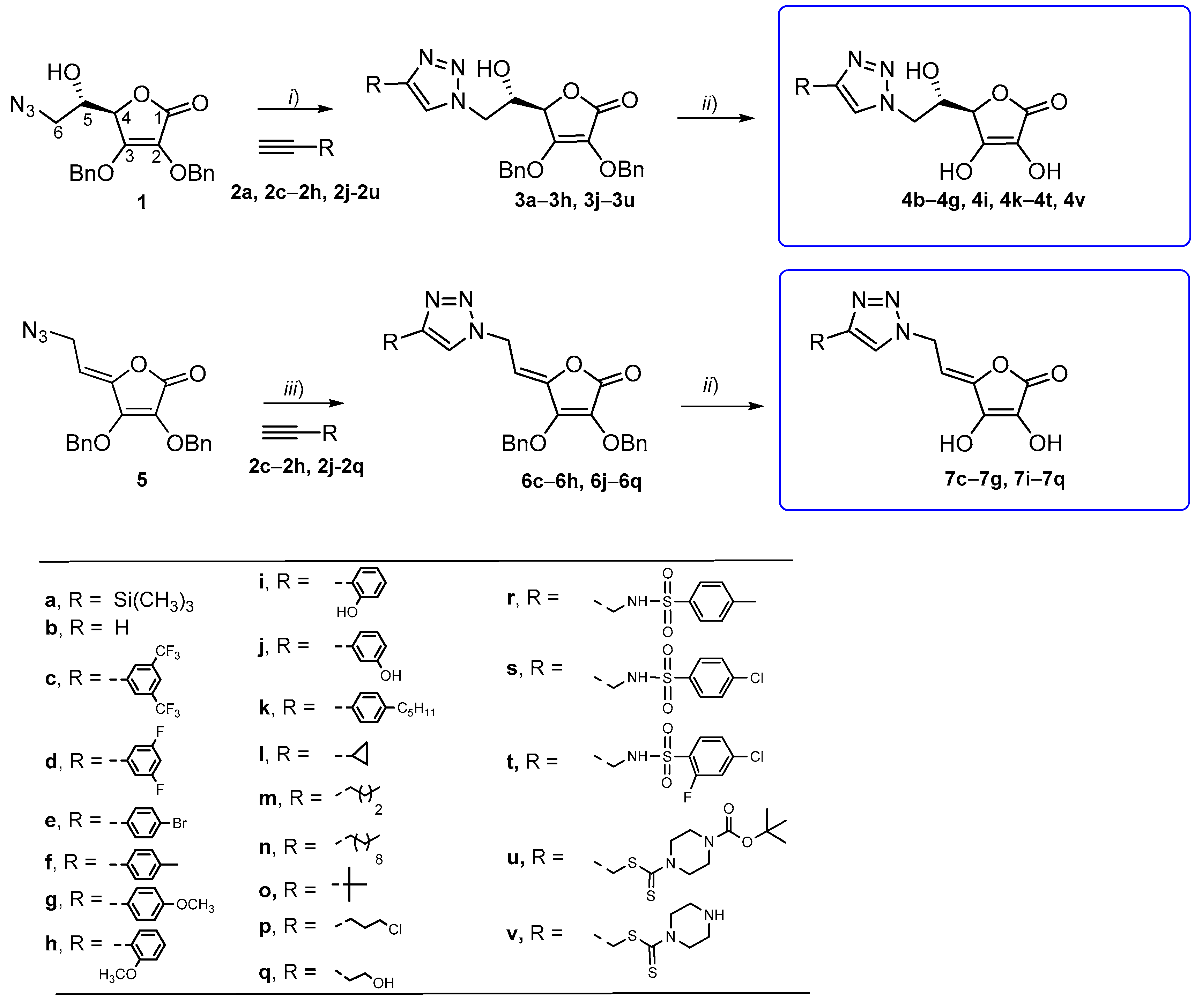
2.1. Synthesis

The target 1,2,3-triazole appended L-ASA derivatives were synthesized following Scheme 1. The 1,2,3-triazole ring was introduced at the C-6 position of L-ascorbic acid via copper(I)-catalyzed azide-alkyne (3 + 2) dipolar cycloaddition (CuAAC) [41] of the corresponding C-6 azido L-ascorbic acid derivatives 1 and 5 with versatile terminal alkynes (2a, 2c–2h, 2j–2u) and 6-(1,2,3-triazolyl)-6-deoxy (3a–3h, 3j–3u). Then, 6-(1,2,3-triazolyl)-4,5-didehydro-5,6-dideoxy-L-ASA (6c–6h, 6j–6g) derivatives were obtained [40].
The removal of the benzyl group was achieved via reaction of the 1,2,3-triazole L-ascorbic acid derivatives 3a–3h, 3j–3u, 6c–6h, and 6j–6q with boron trichloride (BCl3) in dry dichloromethane, affording compounds 4b–4q, 4i, 4k–4t, 4v, 7c–7g, and 7i–7q with yields from 42% to 86%.

2.2. DPPH Radical Scavenging Activity in Vitro

Likewise, L-ASA, the 1,2,3-triazole L-ASA derivatives with free C2-OH and C3-OH groups were expected to be potent antioxidants. Therefore, they were tested as radical scavengers by reaction with the stable free radical 2,2-diphenyl-1-picrylhydrazyl (DPPH). The scavenging activity data are expressed as IC50 values and are summarized in Table 1.
L-ASA was used as control and its experimentally assessed IC50 was 0.18 mM for the 5 min treatment. The scavenging activity data showed that conjugates having a hydroxyethylene spacer with a 4-substituted 1,2,3-triazole moiety at the C-6 position of L-ASA (4b4v), in general, exhibited activity similar to that of L-ASA. Compounds 4b, 4f, 4k, 4l, 4n, 4p, and 4t from the 6-(1,2,3-triazolyl)-6-deoxy-L-ASA and 7f, 7i7k, 7n, and 7q from the 6-(1,2,3-triazolyl)-4,5-didehydro-5,6-dideoxy-L-ASA series had higher antioxidant potencies in comparison with L-ASA. When a halogen (4c, 4d, or 4e) and methoxy-substituted (4g) aryl moiety was introduced in the C-4 of 1,2,3-triazole, the antioxidant activity was slightly lower (IC50 = 0.19–0.27 mM) in comparison with L-ASA and the unsubstituted 1,2,3-triazole L-ASA derivative 4b (IC50 = 0.13 mM at 5 and 10 min treatments). The introduction of an alkyl in the para position of phenyl in compounds 4f and 4k, improved the antioxidant activity after the 10 min treatment. Among 4-alkyl-substituted triazoles, cyclopropyl in 4l, decyl in 4n, and 3-chloropropyl substituents in 4p also improved the antioxidant activity compared to L-ASA. L-ASA derivatives containing the benzenesulphonamide and dithiocarbamate moiety showed lower radical scavenging activity after the 5 min treatment then L-ASA and the unsubstituted triazole 4b, but benzenesulphonamide-substituted 1,2,3-triazole L-ASA derivative 4t had a better antioxidant activity after the 10 min treatment then L-ASA and 4b, with an IC50 value of 0.12 mM.
It was observed that the majority of the 4,5-unsaturated L-ASA derivatives showed a better antioxidant activity compared to their saturated counterparts (Figure 1). Interestingly, the introduction of hydroxyl groups in 4,5-unsaturated L-ASA derivatives 7i, 7j, and 7q improved the radical scavenging activities. Besides, the electron-donating p-methyl groups in 4f/7f, the p-pentyl-substituted aryl groups in 4k/7k, and the long aliphatic chains in 4n/7n improved the antioxidant activity compared to L-ASA as well.

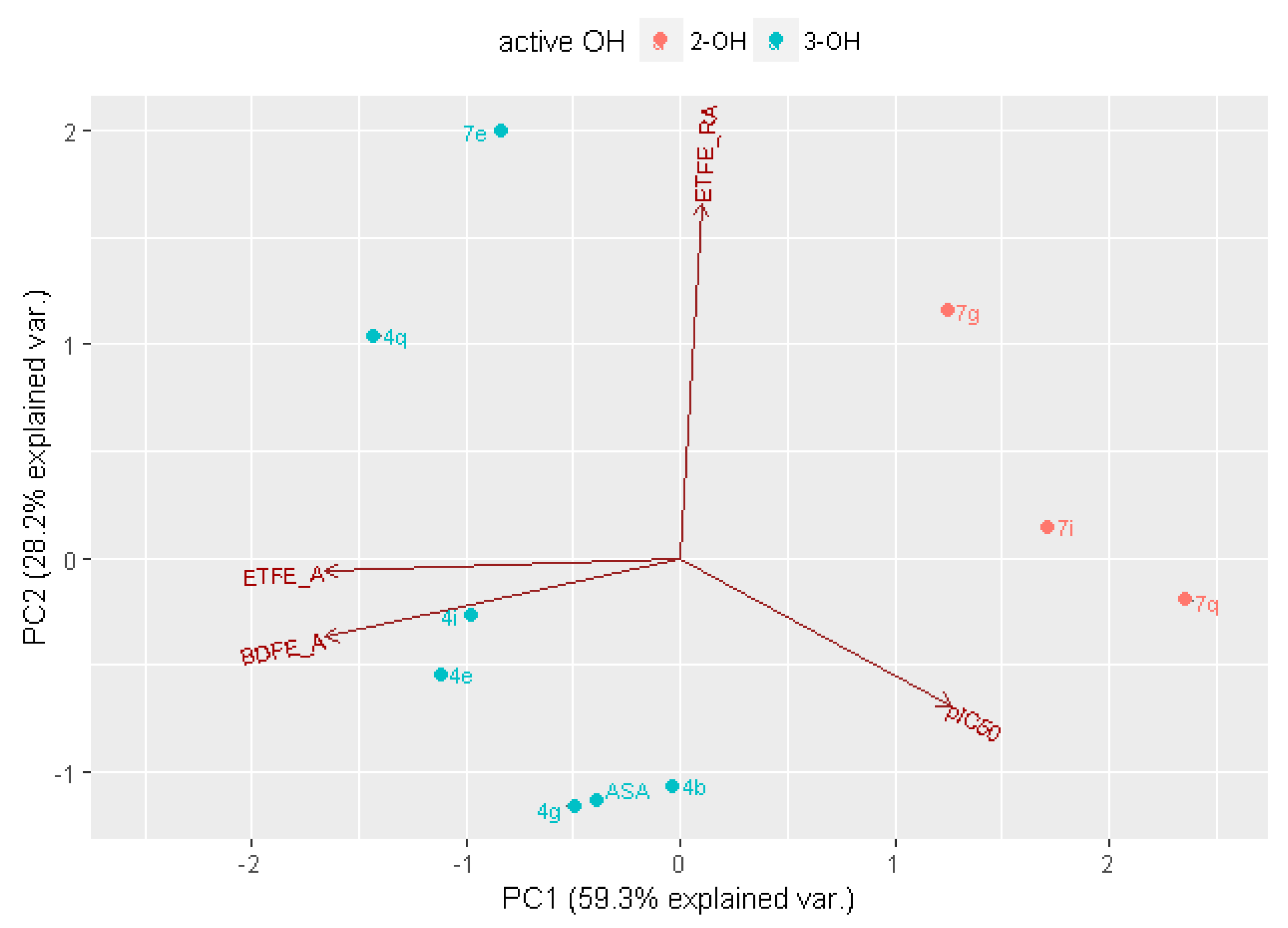
2.3. In Silico Analysis of Radical Scavenging Capacities

According to the pIC50 values, calculated as the negative logarithm of IC50 values (Table 1)—which are within the range of 3.55−4.22 and have the pIC50 medians of 3.7, regardless the incubation time of 5 min or 10 min—the majority of L-ASA derivatives have similar DPPH radical scavenging capacities. The pIC50 value for the well-known antioxidant L-ASA was 3.75, demonstrating that most of compounds have a similar or somewhat higher radical scavenging capacity compared to L-ASA.
For some pairs of L-ASA derivatives differing in the presence of C4-C5(OH) or C4=C5 fragments, somewhat greater difference in their IC50 values has been observed (Table 1). For these pairs of compounds—4b/7b, 4e/7e, 4g/7g, 4i/7i and 4q/7q—the extensive quantum-chemical calculations of radical scavenging capacities were performed by using an approach widely exploited for the estimation of the radical scavenging potential of polyphenols [42]. L-ASA is highly dissociated acidic molecule at physiological pH of 7.4 (the negative logarithms of experimental acid dissociation constants (pKa) are pKa,1 of 4.1 and pKa,2 of 11.4) [43].
The novel 1,2,3-triazole L-ASA conjugates were shown to be acidic molecules with similar pKa values (Table 2). Accordingly, L-ASA reacts as a radical scavenger by donating an electron or a hydrogen H-atom from its anion to a free radical (Figure 2). The first radical scavenging mechanism of L-ASA is described as a two-step sequential proton loss electron transfer (SPLET) process: after the first step of deprotonation of the most acidic OH group (described by its acidity constant), an anion (A) formed donates an electron with a capacity described by the electron transfer (free) energy (ETFE) and changes into neutral radical (NR) (cyan path in Figure 2). However, an anion may also donate a H-atom from the adjacent OH group through the hydrogen-atom transfer (HAT) mechanism which is described by the O-H bond dissociation free energy (BDFE) parameter. The product of this radical scavenging pathway is a radical anion (RA) which may release an additional electron and oxidize to a diketo form of L-ASA known as dehydroascorbic acid (DHA, green path in Figure 2). However, the neutral radical formed by SPLET, immediately releases a proton (pKaNR in Table 2) and converts to a RA. Thus, both pathways have the same final dehydroascorbic acid product. The thermodynamic preference of one of these two pathways has been estimated by comparison of reaction free energies of underlying step processes (Table 2). The multivariate principal component analysis (PCA) analysis in terms of the experimental pIC50 values and the calculated aqueous parameters ETFEA, BDFEA, and ETFERA describing radical scavenging capacities of reducing anions and the intermediate radical anion, explain 87.5% variance in the DPPH activity of modelled compounds (Figure 3).
The most significant difference between saturated C4-C5(OH) and unsaturated C4=C5 derivatives is in acidity of the C2-OH group. While in saturated C4-C5(OH) L-ASA derivatives, the C3-OH group is the only one deprotonated at pH 7.4, in derivatives with the C4=C5 bond, both C2-OH group and C3-OH groups are acidic and may participate in the described DPPH radical scavenging mechanisms (Table 2). However, in the C4=C5 derivatives, intermediate C2-O anion is generally characterized by more favorable, lower ETFEA and BDFEA values than the C3-O anion and it may be expected that it is a primary reactive locus for scavenging of the DPPH radical (Table 2). Thus, the best variance explanation in the PCA analysis of DPPH radical scavenging activity has been obtained by using the more favorable parameter values for C3-OH groups of saturated C4-C5(OH) derivatives and for the C2-OH group of the C4=C5 unsaturated derivatives, except for the compound 7e, for which C3-OH parameters were used (Figure 3).
By comparing ETFEA, BDFEA, and ETFERA values for saturated and unsaturated analogues, all three parameters are clearly more favorable for both groups C2-OH and C3-OH of 7q than for the C3-OH group of its saturated analogue 4q, and it has been observed as significantly more active in DPPH radical scavenging, having a higher pIC50 value (Table 1 and Table 2). ETFEA and BDFEA are also more favorable for the stronger scavenger 7i in the 4i/7i pair. While BDFEA values for the 4e/7e pair are comparable, the radical scavenging capacity of intermediate radical anion is somewhat better for 4e than 7e, which may explain the somewhat higher pIC50 value of the saturated analogue 4e. However, according to the calculation, unsaturated 7g should be a stronger scavenger than the saturated 4g, which was not in agreement with the measured pIC50 values during 5 min and 10 min time periods of incubation.

2.4. Biological Evaluations

2.4.1. Antiproliferative Activities in Vitro

The C-6 substituted 1,2,3-triazole derivatives of L-ASA within the C5-OH group (4b4q, 4i, 4k4t, and 4v) and with the C4=C5 double bond (7c7g and 7i7q) were evaluated against seven malignant tumor cell lines: lung adenocarcinoma (A549), ductal pancreatic adenocarcinoma (CFPAC-1), colorectal carcinoma (HCT-116), cervical carcinoma (HeLa), hepatocellular carcinoma (HepG2), breast adenocarcinoma (MCF-7), colorectal adenocarcinoma, and metastatic (SW620); as well as normal cell lines: lung fibroblasts (WI-38) and foreskin fibroblasts (HFF-1) (Table 3, Table S1, Supplementary Information). L-ASA was used as a positive control, and carboxyamidotriazole, a known antiproliferative agent that inhibits tumor cell growth, invasion, and metastasis, and 5-fluorouracil (5-FU), were used as reference compounds.
It can be observed that the majority of C-4 substituted 1,2,3-triazolyl-6-deoxy-L-ASA derivatives were not cytotoxic. While compound 4e showed a selective antiproliferative effect on MCF-7 and A549 cells and 4k on MCF-7 cells, compounds 4d, 4o4q, and 4v showed an only minor antiproliferative effect at the highest tested concentrations (Table 1). However, except for 4e, 4q and 4v, other compounds exhibited cytotoxicity on foreskin fibroblasts HFF-1 as well. The compounds showed themselves to be more active than L-ASA, which showed no effects on the cell growth of the cells tested. Carboxyamidotriazole, that contains a 1,2,3-triazole ring like the other tested compounds, showed a non-selective, moderate antiproliferative effect on all evaluated cell lines.
The unsubstituted 1,2,3-triazolyl derivative 4b was not cytotoxic toward any of the tested cell lines, indicating that C-4 substitution at 1,2,3-triazole moiety has an impact on growth-inhibition. Among the aryl-substituted triazole derivatives, the fluoro and bromo-substituted aromatic moieties in 4d and 4e, respectively, had influences on the antiproliferative effect. While 4d exhibited a nonspecific cytostatic effect, 4e displayed a strong and selective antiproliferative effect on breast adenocarcinoma MCF-7 (IC50 = 6.72 µM) and moderate activity on lung adenocarcinoma A549 cells (IC50 = 25.44 µM). Additionally, compound 4e did not exhibit cytotoxicity against foreskin fibroblasts HFF-1 (IC50 > 100 µM) and had some cytotoxicity on fibroblasts WI-38 (IC50 = 73.93 µM; selectivity index, SI = 11). When comparing the activities of compounds 4e, 4f, and 4g it can be concluded that the nature of the substituent in the p-position of the phenyl has an influence on the antiproliferative effect. While electron-withdrawing bromine in the structure of 4e increased the activity, compounds 4f and 4g with electron-donating groups on the 4-aryl-1,2,3-triazole units, did not show any cytotoxic effects. The only exception to the observed impact of electron-donating groups on the inhibition effect was p-pentylphenyl-1,2,3-triazolyl-6-deoxy-L-ASA derivative 4k that exerted a selective cytotoxic effect on MCF-7 cells (IC50 = 26.91 µM), albeit, it showed cytotoxicity on foreskin fibroblasts (IC50 = 0.21 µM). From the 4-alkyl substituted triazole derivatives, 4-tert-butyl in 4o, 3-chloropropyl in 4p, and 2-hydroxyethyl chains in 4g caused moderate to marginal cytostatic effects on cervical carcinoma HeLa, colorectal carcinoma HCT-116, and breast adenocarcinoma MCF-7 cells. Compounds 4r4t with benzenesulphonamide substituents, did not show any cytotoxicity toward evaluated tumor cell lines. The dithiocarbamate derivative 4v showed a selective but moderate antiproliferative effect toward MCF-7 cells (IC50 = 37.33 µM). Referring to the lipophilicity of the newly synthesized compounds, their clogP values are within the range of −3.00–2.67 (Table 3 and Table S1, Supplementary Information). Compounds 4e and 4k with the best inhibitory activity and clogP values of −0.42 and 0.98, respectively, showed themselves to be more lipophilic than L-ASA (clogP = −2.46).
The C-6 substituted 1,2,3-triazole 4,5-unsaturated L-ASA derivatives (7c7g and 7i7q) showed no antiproliferative effect on the tested cell panel at concentrations lower than 100 µM (Table S1, Supplementary Information), which implies that the linker connecting the lactone ring and the 1,2,3-triazole moiety has an influence on the antitumor activity of tested compounds, which is in accordance with the previously shown antitumor effects of 2,3-O,O-dibenzylated L-ASA derivatives [40].

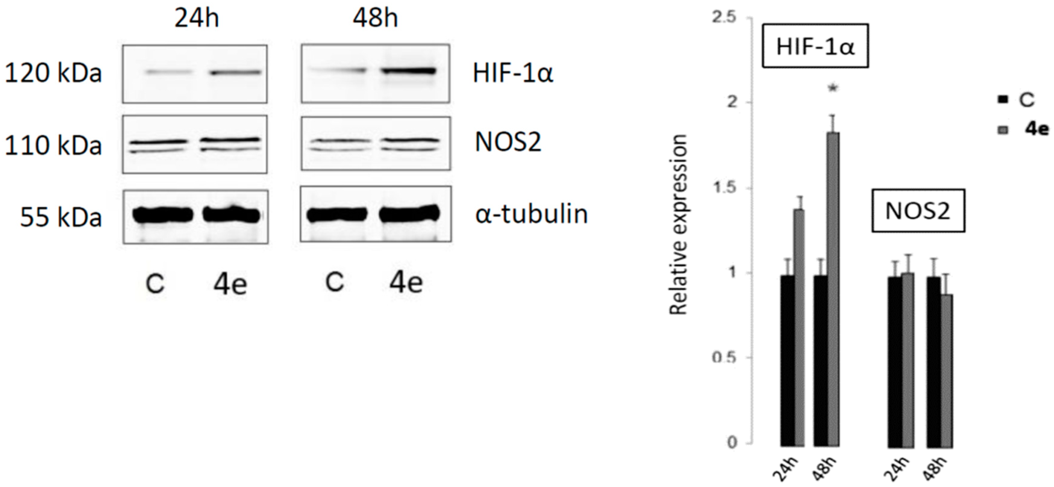
2.4.2. The Detection of Apoptosis and the Validation of HIF-1α and NOS2 Protein Targets for 4e

In order to examine whether the antitumor effect of p-bromophenyl-substituted L-ASA derivative 4e, which showed a selective antiproliferative effect on MCF-7 tumor cell line, can be connected to the induction of apoptosis or any other kind of cell death, an Annexin V assay was performed following a procedure described previously [45].
The results obtained indicate that compound 4e induced apoptosis in MCF-7, and showed a moderate increase in early apoptotic cell populations of 12.4% and late apoptotic cell populations, 2.0%, after 24 h-treatment at concentration 1 × IC50 (Table 4). After 48 h the cell death rate in the treated cells was similar to the untreated MCF-7 control cells for the higher tested concentration, while the percentage of cells in early apoptosis was higher by 35.2%, in treated cells at concentration 1 × IC50, compared with untreated cells.
To further understand the observed antiproliferative and cell death effects on MCF-7 cells by p-bromophenyl 1,2,3-triazole L-ASA conjugate 4e, the analysis of the relative expressions of Hypoxia-inducible factor I alpha (HIF-1α) and Nitric oxide synthase 2 (NOS2, iNOS) protein, as potential targets of 4e, was performed by western blot (Figure 4). The results showed that in MCF-7 cells treated with 4e for 48 h, a statistically significant increase of hydroxylated HIF-1α (Pro564) expression was observed. Increased levels of hydroxylated HIF-1α (Pro564) would imply an increased form of HIF-1α for proteasomal degradation. Furthermore, 48 h after incubation of MCF-7 cells with 4e, a decreased expression of NOS2 was observed.
NOS2s are homodimer enzymes that catalyze the formation of nitric oxide radicals (NO) from L-arginine and oxygen. NO is an important signaling molecule and mediates various physiological and pathological processes, including systemic blood pressure, the upregulation of hypoxic genes, the regulation of stress response pathways, host–microbe interactions, immune signaling, and apoptosis [46,47,48]. The majority of reports indicate that NO induces HIF-1α accumulation but the molecular mechanisms of the regulation of HIF-1α activity by NO are not yet defined and are dependent on conditions being hypoxic or normoxic, and on the concentration of NO [47,48,49,50,51,52,53]. Additionally, it was reported that vitamin C reduces the NO-induced stabilization of HIF-1α in endothelial cells from umbilical cords (HUVECs) under normoxic conditions [54].
Based on the literature overview, we can conclude that the decreased expression of NOS2 is consistent with the enhanced expression of hydroxylated HIF-1α and the antiproliferative effects of 4e on MCF-7 observed.

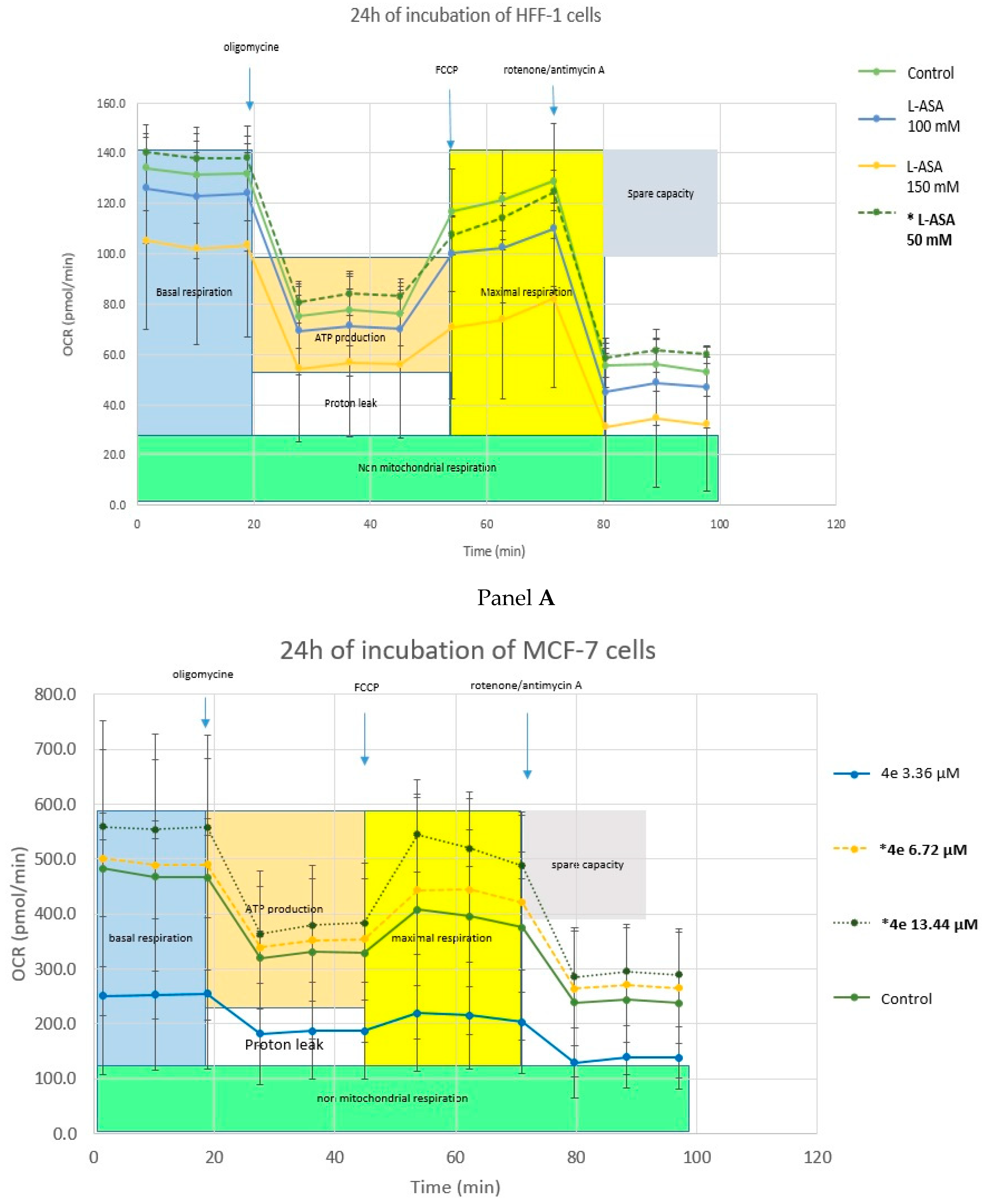
2.4.3. Cell Metabolism Analysis—Mitochondrial Toxicity

The effects on the mitochondrial respiration after 24 h exposure of MCF-7 and HFF-1 cells to L-ASA and 4e tested at concentrations comparable to the DPPH test (IC50 values and correspondingly higher or lower concentrations) were assessed by use of the Seahorse XF Cell Mito Stress Test kit (Figure 5). Even though no statistically relevant changes were observed, interesting trends were consistent. The basal respiration and ATP production of MCF-7 and HFF-1 cells after sequential injection of the standard assay compounds, as described in the Material and methods section, was affected by higher concentrations of L-ASA and compound 4e that both induced a decrease in the cellular-metabolism rate. A lower L-ASA concentration (50 mM) did not act on the mitochondrial activity (dashed lines in Figure 5, panel A). In MCF-7 tumor cells, it was clearly visible that higher L-ASA concentrations decreased the basal cell respiration and ATP production in the way that the whole cell metabolism switched towards non-mitochondrial respiration while this effect was not observed in the normal HFF-1 fibroblasts. Compound 4e increased the basal respiration and ATP production of MCF-7 cells at its IC50 concentration (6.72 µM) and 2 × IC50 concentration (13.44 µM), while it lowered cellular mitochondrial metabolism, switching it towards protein leaking at the lower tested concentration (3.36 µM). Compound 4e did not affect the basal cell respiration or ATP production of HFF-1 cells which correlates with MTT-results where 4e showed no cytotoxicity on this cell line at micromolar concentrations (Table 3; IC50 > 100 µM). Interestingly, 4e showed a slightly positive effect on the cellular metabolism and mitochondrial activity of HFF-1 at a concentration of 100 µM (Figure 5, panel B). The observed differences in compound 4e activity on MCF-7 tumor cells in comparison with normal fibroblasts HFF-1 may be due to the specific metabolic status of tumor cells, in which case an antioxidant compound with ROS scavenging properties, such as L-ASA, may have a cytotoxic effect; i.e., through a pro-oxidant effect on cancer cells associated with increase of ROS in tumor cells [55]. Particularly in MCF-7 cells, a small amount of an important cellular antioxidant enzyme manganese-dependent superoxide dismutase (MnSOD) was found in comparison with other cell lines [56]. It is therefore, possible that the compound 4e exerts a pro-oxidant activity in MCF-7 cells, which elevates the levels of ROS and induces cell death. This hypothesis should be further studied, however, in more experimental detail.

3. Materials and Methods

3.1. Chemistry

3.1.1. General

The melting points of novel compounds were determined using a Kofler micro hot-stage (Reichert, Wien, Austria) apparatus. The progress of all reactions was monitored by thin-layer chromatography (TLC) on silica gel 60F-254 plates (Merck, Darmstadt, Germany) and the spots were observed under UV light (254 nm). Purification of compounds using column chromatography was carried out with silica gel (0.063–0.2 mm) (Fluka, Buchs, Switzerland). All 1H and 13C NMR spectra were recorded in DMSO-d6 at 298 K on a Bruker 300 or 600 MHz NMR spectrometer (Bruker Biospin, Rheinstetten, Germany). Chemical shifts were referenced to the signal of DMSO at δ: 2.50 ppm (1H NMR) and δ: 39.50 ppm (13C NMR). High-resolution mass spectra (HRMS) of the final compounds were recorded on a 4800 Plus MALDI TOF/TOF mass spectrometar (Applied Biosystems, Foster City, CA, USA). Acquisitions were performed in positive ion reflectron mode.

3.1.2. Preparation of Compounds

General procedure for the removal of the benzyl groups: 1,2,3-triazole-L-ascorbic acid derivatives 3a3h, 3j3u, 6c6h, and 6j6q were dried using a vacuum pump. Dry dichloromethane (around 20 mL) was added to the compound under an argon atmosphere. The reaction mixture was cooled to −65 °C and 1 M boron trichloride solution (BCl3, 6 equivalents) was added dropwise to the reaction mixture. The reaction mixture was stirred for 3 h at −65 °C and was left in the freezer overnight. A mixture of dichloromethane and methanol (10 mL) was then added and the reaction mixture was stirred for 30 min at room temperature. The solvent was evaporated and Amberlite IRA 743 was added to neutralize the reaction mixture. The reaction mixture was filtered and the solvent was evaporated. The compounds were purified using a very short column filled with silica (CH2CH2:MeOH = 100:1; 50:1; 10:1). After trituration of the obtained product with n-hexane, diethyl ether, dichloromethane, acetone, and methanol, compounds 4b–4q, 4i, 4k–4t, 4v, 7c−7g, and 7i−7q were isolated as white powders.
The preparation of 6-deoxy-6-(1,2,3-triazol-1-yl)-L-ascorbic acid (4b): Compound 4b (61.8 mg; 57%; m.p. = 219–221 °C) was synthesized according to the general procedure using compound 3a (194.9 mg; 0.478 mmol); 1 M BCl3 (2.9 mL) and CH2Cl2 (20 mL). 1H NMR (600 MHz, DMSO) δ: 11.12 (bs, 1H, OH), 8.43 (bs, 1H, OH), 8.10 (s, 1H, H-8), 7.72 (s, 1H, H-7), 5.45 (bs, 1H, OH), 4.71 (d, J = 1.4 Hz, 1H, H-4), 4.65 (dd, J = 13.8 Hz, 3.9 Hz, 1H, H-6), 4.41 (dd, J = 13.8 Hz, 9.3 Hz, 1H, H-6), 4.19 (d, J = 6.4 Hz, 1H, H-5) ppm. 13C NMR (151 MHz, DMSO) δ: 170.3 (C-1), 152.0 (C-3), 133.0 (C-8), 125.8 (C-7), 118.3 (C-2), 75.6 (C-4), 67.2 (C-5), 52.4 (C-6) ppm. HRMS (ESI Q-TOF): calculated for C8H9N3O5 [M + H]+ = 228.0620; found = 228.0626.
Preparation of 6-deoxy-6-[4-(3,5-bis(trifluoromethyl)phenyl)-1,2,3-triazol-1-yl]-L-ascorbic acid (4c): Compound 4c (119.0 mg; 86%; m.p. ≥ 250 °C) was synthesized according to the general procedure using compound 3c (194.5 mg; 0.314 mmol); 1 M BCl3 (1.9 mL) and CH2Cl2 (20 mL). 1H NMR (600 MHz, DMSO) δ: 11.18 (s, 1H, OH), 8.99 (s, 1H, H-7), 8.53 (s, 2H, Ph-2, Ph-6), 8.46 (s, 1H, OH), 8.07 (s, 1H, Ph-4), 5.60 (d, J = 6.9 Hz, 1H, OH), 4.79 (d, J = 1.6 Hz, 1 H, H-4), 4.74 (dd, J = 13.8 Hz, 4.0 Hz, 1H, H-6), 4.46 (dd, J = 13.9 Hz, 9.3 Hz, 1H, H-6), 4.29–4.23 (m, 1H, H-5) ppm. 13C NMR (151 MHz, DMSO) δ: 170.2 (C-1), 151.9 (C-3), 143.4 (C-8), 133.4 (Ph-1), 131.4; 131.1; 130.9; 130.7 (q, 2JCF = 32.9 Hz, Ph-3, Ph-5), 126.0; 124.1; 122.3; 120.5 (q, 1JCF = 272.8 Hz), 125.3 (Ph), 124.5 (Ph), 121.0 (C-7), 118.3 (C-2), 75.4 (C-4), 67.0 (C-5), 53.1 (C-6) ppm. HRMS (ESI Q-TOF): calculated for C16H11F6N3O5 [M + H]+ = 440.0681; found = 440.0671.
Preparation of 6-deoxy-6-[4-(3,5-difluorophenyl)-1,2,3-triazol-1-yl]-L-ascorbic acid (4d): Compound 4d (44.4 mg; 46%; m.p. = 189–191 °C) was synthesized according to the general procedure using compound 3d (148.6 mg; 0.286 mmol); 1 M BCl3 (1.7 mL) and CH2Cl2 (20 mL). 1H NMR (600 MHz, DMSO) δ: 11.16 (s, 1H, OH), 8.72 (s, 1H, H-7), 8.45 (s, 1H, OH), 7.59–7.56 (m, 2H, Ph-2, Ph-6), 7.35–7.10 (m, 1H, Ph-4), 5.57 (s, 1H, OH), 4.78 (d, J = 1.5 Hz, 1H, H-4), 4.71 (dd, J = 13.8 Hz, 3.8 Hz, 1H, H-6), 4.42 (dd, J = 13.8 Hz, 9.4 Hz, 1H, H-6), 4.26–4.22 (m, 1H, H-5) ppm. 13C NMR (151 MHz, DMSO) δ: 170.2 (C-1), 163.7; 163.6; 162.1; 162.0 (dd, 1JCF = 245.6 Hz, 3JCF = 13.7 Hz, Ph-3, Ph-5), 151.9 (C-3), 144.1 (C-8), 134.4; 134.3; 134.3 (t, 3JCF = 10.7 Hz, Ph-1), 123.9 (C-7), 118.3 (C-2), 108.3–107.7 (m, Ph-2, Ph-6), 103.1; 103.0; 102.8 (t, 2JCF = 25.9 Hz, Ph-4), 75.5 (C-4), 67.0 (C-5), 53.0 (C-6) ppm. HRMS (ESI Q-TOF): calculated for C14H11F2N3O5 [M + H]+ = 340.0745; found = 340.0750.
Preparation of 6-[4-(4-bromophenyl)-1,2,3-triazol-1-yl]-6-deoxy-L-ascorbic acid (4e): Compound 4e (111.7 mg; 61%; m.p. = 159–160 °C) was synthesized according to the general procedure using compound 3e (268.5 mg; 0.477 mmol); 1 M BCl3 (2.86 mL) and CH2Cl2 (20 mL). 1H NMR (300 MHz, DMSO) δ: 11.10 (s, 1H, OH), 8.62 (s, 1H, H-7), 8.48 (bs, 1H, OH), 7.81 (d, J = 8.5 Hz, 2H, Ph), 7.65 (d, J = 8.5 Hz, 2H, Ph), 5.55 (s, 1H, OH), 4.77 (d, J = 1.2 Hz, 1H, H-4), 4.69 (dd, J = 13.7 Hz, 3.7 Hz, 1H H-6), 4.42 (dd, J = 13.7 Hz, 9.3 Hz, 1H, H-6), 4.30–4.18 (m, 1H, H-5). 13C NMR (151 MHz, DMSO) δ: 170.2 (C-1), 157.3 (C-3), 145.0 (C-8), 131.8 (Ph), 130.1 (Ph-q), 127.0 (Ph), 122.8 (C-7), 120.7 (Ph-q), 118.2 (C-2), 75.5 (C-4), 67.1 (C-5), 52.9 (C-6) ppm. HRMS (ESI Q-TOF): calculated for C14H12BrN3O5 [M + H]+ = 382.0039; found = 382.0023.
Preparation of 6-deoxy-6-[4-tolyl-1,2,3-triazol-1-yl]-L-ascorbic acid (4f): Compound 4f (42.0 mg; 42%; m.p. = 152–154 °C) was synthesized according to the general procedure using compound 3f (156.6 mg; 0.315 mmol); 1 M BCl3 (1.9 mL); CH2Cl2 (20 mL). 1H NMR (300 MHz, DMSO) δ: 11.17 (bs, 1H, OH), 8.50 (s, 1H, H-7), 8.46 (bs, 1H, OH), 7.73 (d, J = 8.0 Hz, 2H, Ph), 7.25 (d, J = 8.0 Hz, 2H, Ph), 5.54 (d, J = 6.7 Hz, 1H, OH), 4.77 (d, J = 1.0 Hz, 1H, H-4), 4.67 (dd, J = 13.6 Hz, 3.7 Hz, 1H, H-6), 4.41 (dd, J = 13.7 Hz, 9.3 Hz, 1H, H-6), 4.31–4.17 (m, 1H, H-5), 2.33 (s, 3H, CH3) ppm. 13C NMR (75 MHz, DMSO) δ: 170.3 (C-1), 152.1 (C-3), 146.1 (C-8), 137.0 (Ph-q), 129.4 (Ph), 128.1 (Ph-q), 125.0 (Ph), 122.1 (C-7), 118.3 (C-2), 75.6 (C-4), 67.1 (C-5), 52.8 (C-6), 20.7 (CH3) ppm. HRMS (ESI Q-TOF): calculated for C15H15N3O5 [M + H]+ = 318.1090; found = 318.1090.
Preparation of 6-deoxy-6-[4-methoxyphenyl-1,2,3-triazol-1-yl]-L-ascorbic acid (4g): Compound 4g (81.8 mg; 54%; 185–187 °C) was synthesized according to the general procedure using compound 3g (234 mg; 0.456 mmol); 1 M BCl3 (2.74 mL) and CH2Cl2 (20 mL). 1H NMR (300 MHz, DMSO) δ: 11.18 (bs, 1H, OH), 8.45 (s, 2H, H-7, OH), 7.77 (d, J = 8.8 Hz, 2H, Ph), 7.01 (d, J = 8.8 Hz, 2H, Ph), 5.53 (bs, 1H, OH), 4.76 (d, J = 1.5 Hz, 1H, H-4), 4.66 (dd, J = 13.7 Hz, 3.9 Hz, 1H, H-6), 4.40 (dd, J = 13.7 Hz, 9.3 Hz, 1H, H-6), 4.28–4.19 (m, 1H, H-5), 3.79 (s, 3H, OCH3) ppm. 13C NMR (75 MHz, DMSO) δ: 170.3 (C-1), 158.9 (Ph-q), 152.0 (C-3), 145.9 (C-8), 126.4 (Ph), 123.4 (Ph-q), 121.5 (C-7), 118.3 (C-2), 114.3 (Ph), 75.5 (C-4), 67.1 (C-5), 55.1 (OCH3), 52.7 (C-6) ppm. HRMS (ESI Q-TOF): calculated for C34H45NO16 [M + H]+ = 334.1039; found = 334.1025.
Preparation of 6-deoxy-6-[2-hydroxyphenyl-1,2,3-triazol-1-yl]-L-ascorbic acid (4i): Compound 4i (56.5 mg; 63%; m.p. = 129–131 °C) was synthesized according to the general procedure using compound 3h (145 mg; 0.282 mmol); 1 M BCl3 (1.69 mL) and CH2Cl2 (20 mL). 1H NMR (300 MHz, DMSO) δ: 11.13 (bs, 1H, OH), 10.20 (s, 1H, OH), 8.56–8.28 (m, 2H, H-7, OH), 8.01 (dd, J = 7.7 Hz, 1.5 Hz, 1H, Ph), 7.29–7.10 (m, 1H, Ph), 7.01–6.86 (m, 2H, Ph), 5.53 (bs, 1H, OH), 4.77 (d, J = 1.4 Hz, 1H, H-4), 4.70 (dd, J = 13.7 Hz, 3.7 Hz, 1H, H-6), 4.46 (dd, J = 13.7 Hz, 9.4 Hz, 1H, H-6), 4.25 (bs, 1H, H-5) ppm. 13C NMR (151 MHz, DMSO) δ: 170.4 (C-1), 153.8; 152.1 (C-3, Ph-q), 142.7 (C-8), 128.6 (Ph), 126.4 (Ph), 124.4 (C-7), 119.3 (Ph), 118.3 (C-2), 117.1 (Ph-q), 116.0 (Ph), 75.7 (C-4), 67.3 (C-5), 52.7 (C-6) ppm. HRMS (ESI Q-TOF): calculated for C14H13N3O6 [M + H]+ = 320.0883; found = 320.0869.
Preparation of 6-deoxy-6-[4-(4-pentylphenyl)-1,2,3-triazol-1-yl]-L-ascorbic acid (4k): Compound 4k (62 mg; 57%; m.p. = 237–239 °C) was synthesized according to the general procedure using compound 3k (162.0 mg; 0.293 mmol); 1 M BCl3 (1.8 mL) and CH2Cl2 (20 mL). 1H NMR (600 MHz, DMSO) δ: 11.14 (bs, 1H, OH), 8.49 (s, 1H, H-7), 8.43 (bs, 1H, OH), 7.74 (d, J = 8.2 Hz, 2H, Ph), 7.26 (d, J = 8.1 Hz, 2H, Ph), 5.53 (bs, 1H, OH), 4.76 (d, J = 1.6 Hz, 1H, H-4), 4.67 (dd, J = 13.8 Hz, 4.0 Hz, 1H, H-6), 4.41 (dd, J = 13.8 Hz, 9.3 Hz, 1H, H-6), 4.34–4.04 (m, 1H, H-5), 2.59 (t, J = 7.6 Hz, 2H, H-1′), 1.68–1.47 (m, 2H, CH2′), 1.44–1.18 (m, 4H, CH2’), 0.87 (t, J = 7.0 Hz, 3H, CH3) ppm. 13C NMR (151 MHz, DMSO) δ: 170.2 (C-1), 152.0 (C-3), 146.1 (C-8), 142.0 (Ph-q), 128.7 (Ph), 128.3 (Ph-q), 125.0 (Ph), 122.1 (C-7), 118.3 (C-2), 75.5 (C-4), 67.1 (C-5), 52.7 (C-6), 34.8 (C-1′), 30.8 (CH2’), 30.5 (CH2’), 21.9 (CH2’), 13.9 (CH3) ppm. HRMS (ESI Q-TOF): calculated for C19H23N3O5 [M + H]+ = 374.1716; found = 374.1700.
Preparation of 6-[4-cyclopropyl-1,2,3-triazol-1-yl]-6-deoxy-L-ascorbic acid (4l): Compound 4l (80.4 mg; 78%; m.p. = 119–121 °C) was synthesized according to the general procedure using compound 3l (172.2 mg; 0.385 mmol); 1 M BCl3 (2.3 mL) and CH2Cl2 (20 mL). 1H NMR (600 MHz, DMSO) δ: 11.13 (bs, 1H, OH), 8.43 (bs, 1H, OH), 7.80 (s, 1H, H-7), 5.44 (bs, 1H, OH), 4.68 (d, J = 1.2 Hz, 1H, H-4), 4.54 (dd, J = 13.8 Hz, 4.0 Hz, 1H, H-6), 4.29 (dd, J = 13.7 Hz, 9.2 Hz, 1H, H-6), 4.17–4.12 (m, 1 H, H-5), 2.11–1.44 (m, 1H, H-1′), 0.96–0.82 (m, 2H, CH2’), 0.78–0.59 (m, 2H, CH2’) ppm. 13C NMR (75 MHz, DMSO) δ: 170.3 (C-1), 152.1 (C-3), 148.5 (C-8), 121.8 (C-7), 118.3 (C-2), 75.5 (C-4), 67.1 (C-5), 52.5 (C-6), 7.6 (CH2’), 7.5 (CH2’), 6.5 (C-1′) ppm. HRMS (ESI Q-TOF): calculated for C11H13N3O5 [M + H]+ = 268.0933; found = 268.0938.
Preparation of 6-(4-butyl-1,2,3-triazol-1-yl)-6-deoxy-L-ascorbic acid (4m): Compound 4m (36.5 mg; 47%; m.p. = 98–99 °C) was synthesized according to the general procedure using compound 3m (127.9 mg; 0.276 mmol); 1 M BCl3 (1.7 mL) and CH2Cl2 (20 mL). 1H NMR (300 MHz, DMSO) δ: 11.14 (bs, 1H, OH), 8.45 (bs, 1H, OH), 7.82 (s, 1H, H-7), 5.44 (d, J = 7.0 Hz, 1H, OH), 4.68 (d, J = 1.5 Hz, 1H, H-4), 4.56 (dd, J = 13.7 Hz, 4.1 Hz, 1H, H-6), 4.32 (dd, J = 13.7 Hz, 9.0 Hz, 1H, H-6), 4.22–4.12 (m, 1H, H-5), 2.63–2.57 (m, 2H, H-1′), 1.65–1.49 (m, 2H, CH2’), 1.45–1.19 (m, 2H, CH2’), 0.89 (t, J = 7.3 Hz, 3H, CH3) ppm. 13C NMR (75 MHz, DMSO) δ: 170.3 (C-1), 152.1 (C-3), 146.5 (C-8), 122.8 (C-7), 118.3 (C-2), 75.6 (C-4), 67.2 (C-5), 52.4 (C-6), 31.1 (C-1′), 24.6 (CH2’), 21.7 (CH2’), 13.7 (CH3) ppm. HRMS (ESI Q-TOF): calculated for C12H17N3O5 [M + H]+ = 284.1246; found = 284.1250.
Preparation of 6-(4-decyl-1,2,3-triazol-1-yl)-6-deoxy-L-ascorbic acid (4n): Compound 4n (59.1 mg; 42%; m.p. = 140–142 °C) was synthesized according to the general procedure using compound 3n (209.1 mg; 0.382 mmol); 1 M BCl3 (1.6 mL) and CH2Cl2 (20 mL). 1H NMR (300 MHz, DMSO) δ: 11.14 (bs, 1H, OH), 8.43 (bs, 1H, OH), 7.81 (s, 1H, H-7), 5.44 (d, J = 5.9 Hz, OH), 4.68 (d, J = 1.5 Hz, 1H, H-4), 4.56 (dd, J = 13.7 Hz, 4.0 Hz, 1H, H-6), 4.32 (dd, J = 13.6 Hz, 9.1 Hz, 1H, H-6), 4.22–4.11 (m, 1H, H-5), 2.59 (t, J = 7.5 Hz, 2H, H-1′), 1.67–1.43 (m, 2H, H-2′), 1.36–1.17 (m, 14H, CH2’), 0.85 (t, J = 6.6 Hz, 3H, CH3) ppm. 13C NMR (75 MHz, DMSO) δ: 170.3 (C-1), 152.0 (C-3), 146.6 (C-8), 122.8 (C-7), 118.3 (C-2), 75.6 (C-4), 67.2 (C-5), 52.4 (C-6), 31.3 (C-1′), 29.0 (CH2’), 28.8 (CH2’), 28.7 (CH2’), 28.6 (CH2’), 25.0 (CH2’), 22.1 (CH2’), 143.0 (CH3) ppm. HRMS (ESI Q-TOF): calculated for C18H29N3O5 [M + H]+ = 368.2185; found = 368.2175.
Preparation of 6-[4-(tert-butyl)-1,2,3-triazol-1-yl]-6-deoxy-L-ascorbic acid (4o): Compound 4o (64.5 mg; 48%; m.p. ≥ 250 °C) was synthesized according to the general procedure using compound 3o (220.0 mg; 0.475 mmol); 1 M BCl3 (2.9 mL) and CH2Cl2 (20 mL). 1H NMR (300 MHz, DMSO) δ: 11.17 (bs, 1H, OH), 8.45 (bs, 1H, OH), 7.83 (s, 1H, H-7), 5.45 (bs, 1H, OH), 4.70 (d, J = 1.2 Hz, 1H, H-4), 4.56 (dd, J = 13.6 Hz, 3.8 Hz, 1H, H-6), 4.32 (dd, J = 13.5 Hz, 9.1 Hz, 1H, H-6), 4.25–4.12 (m, 1H, H-5), 1.27 (s, 9H, 3 × CH3) ppm. 13C NMR (75 MHz, DMSO) δ: 170.3 (C-1), 155.9 (C-8), 152.1 (C-3), 120.8 (C-7), 118.3 (C-2), 75.6 (C-4), 67.2 (C-5), 52.4 (C-6), 30.3 (3 × CH3), 28.9 (C-1′) ppm. HRMS (ESI Q-TOF): calculated for C12H17N3O5 [M + H]+ = 284.1246; found = 284.1247.
Preparation of 6-[4-(3-chloropropyl)-1,2,3-triazol-1-yl]-6-deoxy-L-ascorbic acid (4p): Compound 4p (104.0 mg; 83%; m.p. = 86–88 °C) was synthesized according to the general procedure using compound 3p (200.0 mg; 0.413 mmol); 1 M BCl3 (2.5 mL) and CH2Cl2 (20 mL). 1H NMR (300 MHz, DMSO) δ: 11.17 (s, 1H, OH), 8.47 (s, 1H, OH), 7.88 (s, 1H, H-7), 5.47 (d, J = 7.2 Hz, 1H, OH), 4.71 (d, J = 1.4 Hz, 1H, H-4), 4.58 (dd, J = 13.7 Hz, 3.9 Hz, 1H, H-6), 4.33 (dd, J = 13.7 Hz, 9.2 Hz, 1H, H-6), 4.26–4.11 (m, 1H, H-5), 3.68 (t, J = 6.5 Hz, 2H, H-3′), 2.75 (t, J = 7.4 Hz, 2H, H-1′), 2.08–2.01 (m, 2H, H-2′) ppm. 13C NMR (151 MHz, DMSO) δ: 170.3 (C-1), 152.1 (C-3), 145.2 (C-8), 123.2 (C-7), 118.3 (C-2), 75.6 (C-4), 67.2 (C-5), 52.5 (C-6), 44.7 (C-3′), 31.8 (C.-1′), 22.3 (C-2′) ppm. HRMS (ESI Q-TOF): calculated for C11H14ClN3O5 [M + H]+ = 304.0700; found = 304.0697.
Preparation of 6-deoxy-6-(4-(2-hydroxyethyl)-1,2,3-triazol-1-yl)-L-ascorbic acid (4q): Compound 4q (59.5 mg; 70%; m.p. = 184–186 °C) was synthesized according to the general procedure using compound 3q (140.3 mg; 0.311 mmol); 1 M BCl3 (1.9 mL) and CH2Cl2 (20 mL). 1H NMR (600 MHz, DMSO) δ: 11.17 (bs, 1H, OH), 8.46 (bs, 1H, OH), 7.85 (s, 1H, H-7), 5.46 (bs, 1H, OH), 4.73–4.66 (m, 2H, H-4, OH), 4.57 (dd, J = 13.8 Hz, 4.0 Hz, 1H, H-6), 4.33 (dd, J = 13.8 Hz, 9.2 Hz, 2H, H-6), 4.25–4.10 (m, 2H, H-5), 3.64–3.60 (m, 2H, H-2′), 2.76 (t, J = 7.0 Hz, 2H, H-1′) ppm. 13C NMR (151 MHz, DMSO) δ: 170.4 (C-1), 152.1 (C-3), 144.1 (C-8), 123.5 (C-7), 118.3 (C-2), 75.6 (C-4), 67.2 (C-5), 60.4 (C-2′), 52.4 (C-6), 29.2 (C-1′) ppm. HRMS (ESI Q-TOF): calculated For C10H13N3O6 [M + H]+ = 272.0883; found = 272.0895.
Preparation of 6-deoxy-6-[(4-(4-methylbenzenesulphonamide)methyl-1,2,3-triazol-1-yl]-L-ascorbic acid (4r): Compound 4r (115.9 mg; 85%; m.p. = 125–127 °C) was synthesized according to the general procedure using compound 3r (196.0 mg; 0.330 mmol); 1 M BCl3 (2.0 mL) and CH2Cl2 (20 mL). 1H NMR (600 MHz, DMSO) δ: 11.15 (bs, 1H, OH), 8.44 (bs, 1H, OH), 8.03 (t, J = 6.0 Hz, 1H, NH), 7.83 (s, 1H, H-7), 7.68 (d, J = 8.2 Hz, 2H, Ph), 7.38 (d, J = 8.0 Hz, 2H, Ph), 5.44 (d, J = 7.4 Hz, 1H, OH), 4.64 (d, J = 1.5 Hz, 1H, H-4), 4.55 (dd, J = 13.8 Hz, 4.2 Hz, 1H, H-6), 4.32 (dd, J = 13.8 Hz, 9.1 Hz, 1H, H-6), 4.16–4.11 (m, 1H, H-5), 4.01 (d, J = 6.0 Hz, 2H, CH2NH), 2.38 (s, 3H, CH3) ppm. 13C NMR (75 MHz, DMSO) δ: 170.3 (C-1), 152.0 (C-8), 143.13 (C-3), 142.65 (Ph-q), 137.5 (Ph-q), 129.6 (Ph), 126.6 (Ph), 124.2 (C-7), 118.3 (C-2), 75.5 (C-4), 67.2 (C-5), 52.4 (C-6), 38.1 (CH2NH), 21.0 (CH3) ppm. HRMS (ESI Q-TOF): calculated for C16H18N4O7S [M + H]+ = 411.0974; found = 411.0961.
Preparation of 6-[(4-(4-chlorobenzenesulphonamide)methyl-1,2,3-triazol-1-yl]-6-deoxy-L-ascorbic acid (4s): Compound 4s (87.0 mg; 80%; m.p. = 132–134 °C) was synthesized according to the general procedure using compound 3s (154.0 mg; 0.252 mmol); 1 M BCl3 (1.5 mL) and CH2Cl2 (20 mL). 1H NMR (300 MHz, DMSO) δ: 11.17 (bs, 1H, OH), 8.45 (bs, 1H, OH), 8.26 (t, J = 5.9 Hz, 1H, NH), 7.88 (s, 1H, H-7), 7.81–7.71 (m, 2H, Ph), 7.67–7.61 (m, 2H, Ph), 5.46 (d, J = 7.4 Hz, 1H, OH), 4.67 (d, J = 1.5 Hz, 1H, H-4), 4.56 (dd, J = 13.7 Hz, 4.0 Hz, 1H, H-6), 4.32 (dd, J = 13.6 Hz, 9.1 Hz, 1H, H-6), 4.18–4.09 (m, 1H, H-5), 4.06 (d, J = 6.0 Hz, 2H, CH2NH) ppm. 13C NMR (75 MHz, DMSO) δ: 170.1 (C-1), 152.0 (C-3), 142.9 (C-8), 139.3 (Ph-q), 137.2 (Ph-q), 129.2 (Ph), 128.5 (Ph), 124.3 (C-7), 118.4 (C-2), 75.5 (C-4), 67.2 (C-5), 52.5 (C-6), 38.0 (CH2NH) ppm. HRMS (ESI Q-TOF): calculated for C15H5ClN4O7S [M + H]+ = 431.0428; found = 431.0412.
Preparation of 6-[(4-(2-chloro-4-fluorobenzenesulphonamide)methyl-1,2,3-triazol-1-yl]-6-deoxy-L-ascorbic acid (4t): Compound 4t (88.7 mg; 59%; m.p. = 198–200 °C) was synthesized according to the general procedure using compound 3t (210.0 mg; 0.334 mmol); 1 M BCl3 (2.0 mL) and CH2Cl2 (20 mL). 1H NMR (300 MHz, DMSO) δ: 11.18 (bs, 1H), 8.45 (t, J = 5.9 Hz, 1H, NH), 7.96 (dd, J = 8.9, 6.0 Hz, 1H, Ph), 7.83 (s, 1H, H-7), 7.62 (dd, J = 8.7, 2.5 Hz, 1H, Ph), 7.36 (td, J = 8.6, 2.6 Hz, 1H, Ph), 4.65 (d, J = 1.3 Hz, 1H, H-4), 4.54 (dd, J = 13.8 Hz, 4.0 Hz, 1H, H-6), 4.30 (dd, J = 13.7 Hz, 9.1 Hz, 1H, H-6), 4.17 (d, J = 5.9 Hz, 2H, CH2NH), 4.14–4.06 (m, 1H, H-5) ppm. 13C NMR (75 MHz, DMSO) δ: 170.2 (C-1), 165.3; 161.9 (d, 1JCF = 254.3 Hz, Ph-4), 151.9 (C-3), 142.9 (C-8), 134.8; 137.7 (d, 4JCF = 3.6 Hz, Ph-1), 132.8; 132.6 (d, 3JCF = 10.1 Hz, Ph-6), 132.6; 132.4 (d, 3JCF = 10.1 Hz, Ph-2), 124.2 (C-7), 119.2; 118.9 (d, 2JCF = 25.9 Hz, Ph), 118.3 (C-2), 114.8; 114.5 (d, 2JCF = 21.8 Hz, Ph), 75.5 (C-4), 67.2 (C-5), 52.4 (C-6), 37.8 (CH2NH) ppm. HRMS (ESI Q-TOF): calculated for C15H14ClFN4O7S [M + H]+ = 449.0334; found = 449.0326.
Preparation of 6-[4-((tert-butyl 1-carboxylate)-4-(methylthiocarbothionyl)piperazine))-1,2,3-triazol-1-yl]-6-deoxy-L-ascorbic acid (4v): Compound 4v (31.2 mg; 35%; m.p. ≥ 250 °C) was synthesized according to the general procedure using compound 3u (147.7 mg; 0.221 mmol); 1 M BCl3 (1.3 mL) and CH2Cl2 (15 mL). 1H NMR (600 MHz, DMSO) δ: 9.74 (bs, 1H, NH), 8.52 (bs, 1H, OH), 8.08 (s, 1H, H-7), 5.46 (bs, 1H, OH), 4.71 (d, J = 1.5 Hz, 1H, H-4), 4.67–4.54 (m, 3H, H-6, CH2S), 4.39–4.11 (m, 4H, H-6, H-5, 2 × CH2), 3.2–3.1 (m, 4H, 2 × CH2) ppm. 13C NMR (151 MHz, DMSO) δ: 195.8 (CS), 170.1 (C-1), 151.7 (C-3), 141.2 (C-8), 124.8 (C-7), 118.3 (C-2), 75.6 (C-4), 67.2 (C-5), 52.7 (C-6), 42.3 (CH2’), 31.9 (CH2S) ppm. HRMS (ESI Q-TOF): calculated for C14H19N5O5S2 [M + H]+ = 402.0906; found = 402.0891.
Preparation of 4,5-didehydro-5,6-dideoxy-(4-(3,5-di-(trifluoromethyl)phenyl)-1,2,3-triazol-1-yl)-L-ascorbic acid (7c): Compound 7c (72.1 mg; 52%; m.p. ≥ 200 °C) was synthesized according to the general procedure using compound 6c (200 mg; 0.33 mmol); 1 M BCl3 (1.98 mL) and CH2Cl2 (20 mL). 1H-NMR (300 MHz, DMSO) δ: 11.21 (s, 1H, OH), 9.95 (s, 1H, OH), 9.01 (s, 1H, H-7), 8.54 (s, 2H, Ph-2, Ph-6), 8.07 (s, 1H, Ph-4), 5.61 (t, J = 7.6 Hz, 1H, H-5), 5.30 (d, J = 7.7 Hz, 2H, H-6) ppm. 13C-NMR (151 MHz, DMSO) δ: 164.6 (C-1), 145.8 (C-3), 143.8 (C-4), 142.9 (C-8), 133.2 (Ph-1), 131.3; 131.1; 130.9; 130.7 (q, 2JCF = 33.0 Hz, Ph-3, Ph-5), 125.9; 124.1; 122.3 (m, 1JCF = 272.8 Hz, CF3), 125.3 (Ph-2, Ph-6), 123.3 (C-7), 122.1 (C-2), 121.1 (Ph-4), 98.2 (C-5), 44.5 (C-6) ppm. HRMS (ESI Q-TOF): calculated for C16H9F6N3O4 [M + H]+ = 422.0576; found = 422.0580.
Preparation of 4,5-didehydro-5,6-dideoxy-(4-(3,5-difluoromethyl)phenyl)-1,2,3-triazol-1-yl)-L-ascorbic acid (7d): Compound 7d (61.1 mg; 48%; m.p. ≥ 200 °C) was synthesized according to the general procedure using compound 6d (200 mg; 0,39 mmol); 1 M BCl3 (2.27 mL) and CH2Cl2 (20 mL): Compound 7d was isolated as white powder. 1H-NMR (600 MHz, DMSO) δ: 11.31 (s, 1H, OH), 9.83 (s, 1H, OH), 8.75 (s, 1H, H-7), 7.59 (d, J = 6.6 Hz, 2H, Ph-2, Ph-6), 7.31–7.03 (m, 1H, Ph-4), 5.58 (t, J = 7.7 Hz, 1H, H-5), 5.26 (d, J = 7.7 Hz, 2H, H-6) ppm. 13C-NMR (151 MHz, DMSO) δ: 164.6 (C-1), 163.7; 163.6; 162.1; 162.0 (dd, 1JCF = 245.6 Hz, 3JCF = 13.7 Hz, Ph-3, Ph-5), 145.7 (C-3), 144.6 (C-8), 142.9 (C-4), 134.2; 134.2; 134.1 (t, J = 10.5 Hz, Ph-1), 122.7 (C-7), 122.1 (C-2), 108.2; 108.1; 108.0; 108.0 (dd, 2JCF = 21.1 Hz, 4JCF = 5.5 Hz, Ph-2, Ph-6), 103.2; 103.1; 102.9 (t, 2J = 26.0 Hz, Ph-4), 98.4 (C-5), 44.5 (C-6) ppm. HRMS (ESI Q-TOF): calculated for C14H9F2N3O4 [M + H]+ = 322.0639; found = 322.0642.
Preparation of 6-[4-(4-bromophenyl)-1,2,3-triazole-1-yl]-4,5-didehydro-5,6-dideoxy-L-ascorbic acid (7e): Compound 7e (24 mg; 21%; m.p. = 219–222 °C) was synthesized according to the general procedure using compound 6e (177 mg; 0.324 mmol); 1 M BCl3 (1.94 mL) and CH2Cl2 (20 mL). 1H NMR (300 MHz, DMSO) δ: 10.48 (s, OH), 8.67 (s, 1H, H-7), 7.82 (d, J = 8.5 Hz, 2H, Ph), 7.64 (d, J = 8.6 Hz, 2H, Ph), 5.58 (t, J = 7.5 Hz, 1H, H-5), 5.25 (d, J = 7.5 Hz, 2H, H-6). 13C NMR (75 MHz, DMSO-d6) δ: 164.7 (C-1), 145.5 (C-3), 143.0 (C-8, C-4), 131.8 (Ph), 129.9 (Ph-q), 127.1 (Ph), 122.0 (C-7), 121.8 (C-2), 120.8 (Ph-q), 98.7 (C-5), 44.4 (C-6) ppm. HRMS (ESI Q-TOF): calculated for C14H10BrN3O4 [M + H]+ = 363.9933; found = 363.9915.
Preparation of 6-[4-tolyl-1,2,3-triazole-1-yl]-4,5-didehydro-5,6-dideoxy-L-ascorbic acid (7f): Compound 7f (20 mg; 15%; m.p. = 188–192 °C) was synthesized according to the general procedure using compound 6f (203 mg; 0.407 mmol); 1 M BCl3 (2.45 mL) and CH2Cl2 (20 mL). 1H NMR (600 MHz, DMSO) δ: 11.40 (s, 1H, OH), 9.78 (s, 1H, OH), 8.56 (s, 1H, H-7), 7.74 (d, J = 8.0 Hz, 2H, Ph), 7.25 (d, J = 7.9 Hz, 2H, Ph), 5.59 (t, J = 7.6 Hz, 1H, H-5), 5.24 (d, J = 7.6 Hz, 2H, H-6), 2.33 (s, 3H, CH3) ppm. 13C NMR (151 MHz, DMSO) δ: 164.7 (C-1), 146.5 (C-3), 145.3 (C-4), 143.0 (C-8), 137.2 (Ph-q), 129.4 (Ph), 127.9 (Ph-q), 125.0 (Ph), 121.9 (C-7), 120.9 (C-2), 98.8 (C-5), 44.3 (C-6), 20.7 (CH3) ppm. HRMS (ESI Q-TOF): calculated for C15H13N3O4 [M + H]+ = 300.0984; found 300.0974.
Preparation of 4,5-didehydro-5,6-dideoxy-6-[4-(4-methoxyphenyl)-1,2,3-triazole-1-yl]-L-ascorbic acid (7g): Compound 7g (58.1 mg; 28%; m.p. = 198–199 °C) was synthesized according to the general procedure using compound 6g (328 mg; 0.662 mmol); 1 M BCl3 (3.96 mL) and CH2Cl2 (25 mL). 1H NMR (600 MHz, DMSO) δ: 11.05 (bs, 1H, OH), 9.95 (bs, 1H, OH), 8.49 (s, 1H, H-7), 7.78 (d, J = 8.7 Hz, 2H, Ph), 7.01 (d, J = 8.7 Hz, 2H, Ph), 5.58 (t, J = 7.6 Hz, 1H, H-5), 5.23 (d, J = 7.6 Hz, 2H, H-6) ppm. 13C NMR (151 MHz, DMSO) δ: 164.7 (C-1), 159.0 (Ph-q), 146.5 (C-3), 145.3 (C-8), 126.5 (Ph), 123.3 (Ph-q), 121.9 (C-2), 120.4 (C-7), 114.3 (Ph), 99.0 (C-5), 55.1 (OCH3), 44.3 (C-6) ppm. HRMS (ESI Q-TOF): calculated for C15H13N3O5 [M + H]+ = 316.0933; found = 316.0930.
Preparation of 4,5-didehydro-5,6-dideoxy-6-[4-(2-hydroxyphenyl)-1,2,3-triazole-1-yl]-L-ascorbic acid (7i): Compound 7i (64.2 mg; 56%; m.p. = 167–169 °C) was synthesized according to the general procedure using compound 6h (188 mg; 0.379 mmol); 1 M BCl3 (2.28 mL) and CH2Cl2 (20 mL). 1H NMR (300 MHz, DMSO) δ: 11.13 (bs, 1H, OH), 10.15 (s, 1H, OH), 9.74 (bs, 1H, OH), 8.41 (s, 1H, H-7), 7.97 (dd, J = 7.7 Hz, 1.7 Hz, 1H, Ph), 7.43–7.05 (m, 1H, Ph), 6.99–6.78 (m, 2H, Ph), 5.58 (t, J = 7.5 Hz, 1H, H-5), 5.25 (d, J = 7.5 Hz, 2H, H-6) ppm. 13C NMR (75 MHz, DMSO) δ: 164.6 (C-1), 153.9 (Ph-q), 145.1 (C-3), 143.1 (C-8), 143.0 (C-4), 128.7 (Ph), 126.5 (Ph), 123.1 (C-7), 121.9 (C-2), 119.2 (Ph), 116.9 (Ph-q), 115.9 (Ph), 99.3 (C-5), 44.2 (C-6) ppm. HRMS (ESI Q-TOF): calculated for C14H11N3O5 [M + H]+ = 302.0777; found = 302.0782.
Preparation of 4,5-didehydro-5,6-dideoxy-6-[4-(3-hydroxyphenyl)-1,2,3-triazole-1-yl]-L-ascorbic acid (7j): Compound 7j (45.3 mg; 41%; m.p. = 155–157 °C) was synthesized according to the general procedure using compound 6j (175 mg; 0.363 mmol); 1 M BCl3 (2.18 mL) and CH2Cl2 (20 mL). 1H NMR (600 MHz, DMSO) δ: 11.33 (s, 1H, OH), 9.90 (s, 1H, OH), 9.55 (s, 1H, OH), 8.55 (s, 1H, H-7), 7.54–7.02 (m, 3H, Ph), 6.72 (d, J = 7.0 Hz, 1H, Ph), 5.59 (t, J = 7.6 Hz, 1H, H-5), 5.23 (d, J = 7.6 Hz, 2H, H-6) ppm. 13C NMR (151 MHz, DMSO) δ: 164.7 (C-1), 157.8 (Ph-q), 146.7 (C-3), 145.3 (C-4), 143.0 (C-8), 131.9 (Ph-q), 130.0 (Ph), 122.0 (C-7), 121.4 (C-2), 116.1 (Ph), 114.9 (Ph), 111.9 (Ph), 99.0 (C-5), 44.4 (C-6) ppm. HRMS (ESI Q-TOF): calculated for C14H11N3O5 [M + H]+ = 302.0777; found = 302.0765.
Preparation of 4,5-didehydro-5,6-dideoxy-6-[4-(4-pentylphenyl)-1,2,3-triazole-1-yl]-L-ascorbic acid (7k): Compound 7k (31 mg; 24%; m.p. = 230–232 °C) was synthesized according to the general procedure using compound 6k (195 mg; 0.364 mmol); 1 M BCl3 (2.18 mL) and CH2Cl2 (20 mL). 1H NMR (300 MHz, DMSO) δ: 11.10 (bs, 1 H, OH), 9.98 (bs, 1H, OH), 8.55 (s, 1H, H-7), 7.75 (d, J = 8.1 Hz, 2H, Ph), 7.26 (d, J = 8.1 Hz, 2H, Ph), 5.59 (t, J = 7.6 Hz, 1H, H-5), 5.24 (d, J = 7.6 Hz, 1H, H-6), 2.59 (t, J = 7.6 Hz, 2H, H-1′), 1.65–1.51 (m, 2H, CH2), 1.38–1.17 (m, 4H, CH2), 0.86 (t, J = 6.8 Hz, 3H, CH3) ppm. 13C NMR (75 MHz, DMSO) δ: 164.7 (C-1), 146.6 (C-3), 145.3 (C-4), 143.0 (C-8), 142.1 (Ph-q), 128.8 (Ph), 128.1 (Ph-q), 125.1 (Ph), 122.0 (C-2), 121.0 (C-7), 99.0 (C-5), 44.3 (C-6), 34.8 (C-1′), 30.9 (C-2′), 30.5 (CH2), 21.9 (CH2), 13.9 (CH3) ppm. HRMS (ESI Q-TOF): calculated for C19H21N3O4 [M + H]+ = 356.1610; found = 356.1598.
Preparation of 6-[4-cyclopropyl-1,2,3-triazole-1-yl]-4,5-didehydro-5,6-dideoxy-L-ascorbic acid (7l): Compound 7l (52.5 mg; 53%; m.p. = 176–178 °C) was synthesized according to the general procedure using compound 6l (170 mg; 0.396 mmol); 1 M BCl3 (2.38 mL) and CH2Cl2 (20 mL). 1H NMR (300 MHz, DMSO) δ: 11.10 (bs, 1H, OH), 9.88 (bs, 1H, OH), 7.83 (s, 1H, H-7), 5.50 (t, J = 7.5 Hz, 1H, H-5), 5.11 (d, J = 7.6 Hz, 2H, H-6), 1.98–1.86 (m, 1H, CH), 0.95–0.84 (m, 2H, CH2), 0.73–0.65 (m, 2H, CH2) ppm. 13C NMR (75 MHz, DMSO) δ: 164.6 (C-1), 149.2 (C-8), 145.0 (C-3), 143.0 (C-4), 121.9 (C-2), 120.6 (C-7), 99.2 (c-5), 44.0 (C-6), 7.5 (CH2), 6.4 (CH) ppm. HRMS (ESI Q-TOF): calculated for C11H11N3O4 [M + H] + = 250.0828; found = 250.0826.
Preparation of 6-[4-butyl-1,2,3-triazole-1-yl]-4,5-didehydro-5,6-dideoxy-L-ascorbic acid (7m): Compound 7m (66.8 mg; 47%; m.p. = 180–183 °C) was synthesized according to the general procedure using compound 6m (237 mg; 0.532 mmol); 1 M BCl3 (3.19 mL) and CH2Cl2 (20 mL). 1H NMR (600 MHz, DMSO) δ: 11.32 (s, 1H, OH), 9.76 (s, 1H, OH), 7.88 (s, 1H, H-7), 5.52 (t, J = 7.6 Hz, 1H, H-5), 5.14 (d, J = 7.6 Hz, 2H, H-6), 2.60 (t, J = 7.6 Hz, 2H, H-1′), 1.68–1.42 (m, 2H, CH2), 1.41–1.26 (m, 2H, CH2), 0.89 (t, J = 7.4 Hz, 3H, CH3) ppm. 13C NMR (151 MHz, DMSO) δ: 164.7 (C-1), 147.2; 145.0; 143.1 (C-3, C-4, C-8), 121.9 (C-2), 121.8 (C-7), 99.4 (C-5), 44.0 (C-6), 31.1 (C-1′), 24.7 (CH2), 21.7 (CH2), 13.7 (CH3) ppm.
Preparation of 6-(4-decyl-1,2,3-triazol-1-il)-4,5-didehydro-5,6-dideoxy-L-ascorbic acid (7n): Compound 7n (55 mg; 42%; m.p. = 169–171 °C) was synthesized according to the general procedure using compound 6n (200 mg; 0.38 mmol); 1 M BCl3 (2.28 mL) and CH2Cl2 (20 mL). 1H-NMR (600 MHz, DMSO) δ: 11.28 (s, 1H, OH), 9.73 (s, 1H, OH), 7.86 (s, 1H, H-7), 5.52 (t, 1H, J = 7.5 Hz, H-5), 5.14 (s, 2H, H-6), 2.60–2.57 (m, 2H, H-1′), 1.58–1.56 (m, 2H, H-2′), 1.33–1.20 (m, 14H, CH2), 0.84 (t, J = 7.0 Hz, 3H, CH3) ppm. 13C-NMR (151 MHz, DMSO) δ: 164.6 (C-1), 147.2 (C-3), 144.9 (C-4), 143.0 (C-8), 121.9 (C-2), 121.7 (C-7), 99.3 (C-5), 43.9 (C-6), 31.2 (C-1′), 28.9 (CH2’), 28.7 (CH2’), 28.6 (CH2’), 28.5 (CH2’), 24.9 (CH2’), 22.1 (CH2’), 13.9 (CH3) ppm. HRMS (ESI Q-TOF): calculated for C18H27N3O4 [M + H] + = 350.2080; found = 350.2086.
Preparation of 6-[4-tert-butyl-1,2,3-triazole-1-yl]-4,5-didehydro-5,6-dideoxy-L-ascorbic acid (7o): Compound 7o (30 mg; 24%; m.p. = 182–184 °C) was synthesized according to the general procedure using compound 6o (203 mg; 0.455 mmol); 1 M BCl3 (2.72 mL) and CH2Cl2 (20 mL). 1H NMR (300 MHz, DMSO) δ: 11.31–9.54 (bs, 2H, OH × 2), 7.88 (s, 1H, H-7), 5.53 (t, J = 7.5 Hz, 1H, H-5), 5.14 (d, J = 7.5 Hz, 2H, H-6), 1.27 (s, 9H, CH3 × 3). 13C NMR (75 MHz, DMSO) δ: 164.6 (C-1), 144.9 (C-3), 143.0 (C-4), 121.9 (C-2), 119.7 (C-7), 99.3 (C-5), 43.9 (C-6), 30.2 (C-1′), 30.2 (CH3) ppm. HRMS (ESI Q-TOF): calculated for C12H15N3O4 [M + H] + = 266.1141; found = 266.1129.
Preparation of 6-[4-chloropropyl-1,2,3-triazole-1-yl]-4,5-didehydro-5,6-dideoxy-L-ascorbic acid (7p): Compound 7p (53.2 mg; 38%; m.p. = 160–162 °C) was synthesized according to the general procedure using compound 6p (230 mg; 0.494 mmol); 1 M BCl3 (2.96 mL); CH2Cl2 (20 mL). 1H NMR (300 MHz, DMSO) δ: 11.18–9.67 (bs, 2H, OH × 2), 7.93 (s, 1H, H-7), 5.53 (t, J = 7.5 Hz, 1H, H-5), 5.15 (d, J = 7.5 Hz, 2H, H-6), 3.67 (t, J = 6.5 Hz, 2H, H-3′), 2.82–2.66 (m, 2H, H-1′), 2.10–1.98 (m, 2H, H-2′) ppm. 13C NMR (75 MHz, DMSO) δ: 164.6 (C-1), 145.7; 145.0; 143.0 (C-3, C-4, C-8), 122.1 (C-7), 121.9 (C-2), 99.2 (C-5), 44.6 (C-1′), 44.0 (C-6), 31.7 (C-1′), 22.3 (C-3′) ppm. HRMS (ESI Q-TOF): calculated for C11H12ClN3O4 [M + H] + = 286.0595; found = 286.0599.
Preparation of 6-[4-(2-hydroxyethyl)-1,2,3-triazole-1-yl]-4,5-didehydro-5,6-dideoxy-L-ascorbic cid (7q): Compound 7q (81.2 mg; 70%; m.p. = 172–173 °C) was synthesized according to the general procedure using compound 6q (200 mg; 0.461 mmol); 1 M BCl3 (2.77 mL); CH2Cl2 (20 mL). 1H NMR (300 MHz, DMSO) δ: 11.21 (bs, 1H, OH), 9.87 (bs, 1H, OH), 7.87 (s, 1H, H-7), 5.52 (t, J = 7.6 Hz, 1H, H-5), 5.15 (d, J = 7.6 Hz, 2H, H-6), 4.65 (bs, 1H, OH), 3.62 (t, J = 6.9 Hz, 2H, H-2′), 2.76 (t, J = 6.9 Hz, H-1′) ppm. 13C NMR (75 MHz, DMSO) δ: 164.6 (C-1), 145.0 (C-3), 144.7 (C-8), 143.0 (C-4), 122.3 (C-7), 121.9 (C-2), 99.3 (C-5), 60.3 (C-2′), 43.9 (C-6), 29.1 (C-1′) ppm. HRMS (ESI Q-TOF): calculated for C10H11N3O5 [M + H] + = 254.0777; found = 254.0770.

3.2. Biological Evaluation

3.2.1. DPPH Assay

Antioxidant capacity of selected compounds was assessed by 2,2-diphenyl-1-picrylhydrazyl assay (DPPH, Sigma Aldrich, Darmstadt, Germany), in which the antioxidant activity of compounds is measured by the reaction between a stable radical of 1,1-diphenyl-2-picylhydrazil (DPPH) and a selected compound according to the manufacturer recommendations. Briefly, compounds 4b–4e, 4f–4h, 4k, 4l, 4o, 4p–4s, 4t, 4v, 7c–7f, 7i–7n, 7q, 7p, and 7o were tested at 0.06, 0.12, 0.18, 0.23, 0.25, and 0.28 mM. L-ascorbic acid was used as the reference compound. Absorbance was measured 5 and 10 min after incubation of the compounds with the DPPH reagent. The reduction of DPPH with selected compounds was monitored by measuring the absorbance at a wavelength of 517 nm on the monochromator (Infinite M200 PRO, TECAN, Männedorf, Switzerland).

3.2.2. In Silico Approaches—DFT Calculations and PCA Analysis

The gas-phase (g) reaction parameters for radical scavenging models: bond dissociation enthalpy (BDE), basicity, and electron transfer enthalpy (ETE), and their aqueous free energy (FE) counterparts BDFE, pKa, and ETFE, respectively, were calculated by using thermochemical cycles [42] in the density functional theory (DFT) model (U)M052X/6-311++G** implemented in Gaussian 09 [57,58]. The gas-phase equilibrium geometries were optimized for species in neutral and anion closed-shell singlet and neutral monoradical and radical anion open-shell doublet ground electronic states. The minima were confirmed by absence of imaginary vibrational frequencies. Calculations were done for the compounds with a free C5OH group in the 4R,5S-configuration as a starting L-ASA (series 4). The compounds with a C4=C5 double bond were determined in Z-configuration (series 7) (Scheme 1) [59]. The free energies of hydratation ΔG*hyd were determined at the gas phase geometries by using the SMD method [60]. The gas-phase thermodynamic reaction parameters were calculated in terms of the sums of electronic and thermal enthalpies or free energies estimated at temperature of 298.15 K and pressure of 1 atm. For the electron and the proton, values reported in the reference [61] were used. Regarding free energies of hydratation ΔG°hyd(H), ΔG°hyd (H+), and ΔG°hyd (e), the experimental values of 27.8 kJ/mol [62], −1104.62 kJ/mol [63] and −148.5 kJ/mol [64] were used, respectively.
The PCA analyses were done by R package princomp [65]. The values of partition n-octanol/water coefficients—clogP were calculated by DataWarrior by using SMILES as inputs [44].

3.2.3. Cell Culturing

Human tumor cell lines HeLa (cervical carcinoma), SW620 (colorectal adenocarcinoma, metastatic), A549 (lung adenocarcinoma), MCF−7 (breast adenocarcinoma), CFPAC-1 (pancreatic adenocarcinoma, metastatic), HCT-116 (colorectal carcinoma), and HepG2 (hepatocellular carcinoma), and normal cell lines HFF-1 (foreskin fibroblasts) and WI-38 (immortalized human lung fibroblast), were from the US American Type Culture Collection (ATCC, Manassas, VA, USA). Cells were cultured in a liquid nutrient medium (Dulbecco’s Modified Eagle Medium, LONZA, Basel, Switzerland) supplemented with 10% fetal bovine serum (FBS, Gibco, Grand Island, NY, USA), 2 mM L-glutamine (GIBCO, Grand Island, NY, USA), 100 μg/mL penicillin, and 100 μg/mL streptomycin (LONZA, Basel, Switzerland), in an incubator (NÜVE, Ankara, Turkey), at a humid atmosphere with 5% CO2 at 37 °C. Trypsin/EDTA Solution (Lonza, Basel, Switzerland), was used for cell passaging and cell detachment.

3.2.4. Proliferation Assay- MTT Assay

Adherent cells were seeded on 96 well microtiter plates (Falcon, New York, NY, USA) at a seeding density of 5000 cells per well, added in 150 μL of cell suspension in a nutrient medium. 24 h after seeding, cells were treated with compound 4b–4g, 4i, 4k–4t, 4v, 7c–7g, and 7i–7q dilutions in the range of 0.01 μM to 100 μM. L-ascoric acid (Sigma Aldrich, Darmstadt, Germany) and Carboxyamidotriazole (Sigma Aldrich, Darmstadt, Germany) were used as control compounds. Incubation of the cells with the compounds were performed for 72 h in a 5% CO2, in humid atmosphere at 37 °C. After incubation, nutrient medium was removed and 40 μL of the MTT reagent (3-(4-dimethylthiazol-2-yl)-2,5-diphenyltetrazole bromide (Invitrogen, Carlsbad, CA, USA) was added to each well. 3 h after incubation, 160 μL of DMSO (VWR CHEMICALS, Radnor, PA, USA) were added to each well to dissolve the crystals of purple formazane. The absorbance was measured at 570 nm (TECAN SUNRISE Microtiter plate reader (Männedorf, Switzerland). The experimentally measured optical densities (OD) were converted to the PG (percentage of growth), using the formulas according to the protocol assumed by NIH (National Institute of Health). The IC50 values (concentration causing 50% cell growth inhibition) for each compound were calculated by regression analysis of the survival count curve of the concentrations of the tested compounds. The results were processed in Excel 2010 (Microsoft, Redmond, WA, USA).

3.2.5. Apoptosis Detection—Annexin V Assay

A total of 2 × 10⁴ MCF-7 cells were seeded into 8-well Nunc Lab-Tek II Chamber Slide system (Thermo Fischer Scientific, Waltham, MA, USA), and treated with compound 4e at 1 × IC50 and 2 × IC50 for 24 and 48 h. The assay (Annexin-V-FLUOS Staining kit, Roche Applied Science, Penzberg, Germany) was preformed according to the manufacturer protocol. Slides were analyzed by fluorescence microscopy (Zeiss Axio Observer Z1 Inverted Phase Contrast Fluorescent Microscope, Jena, Germany)).

3.2.6. Western Blot Analysis

MCF-7 cells were seeded in Petri dishes, at 1 × 10⁶ cells, and treated with compound 4e at IC50 6.72 µM for 24 and 48 h. Protein lysates were prepared using RIPA buffer containing cocktail of protease and phosphatase inhibitors (Roche, Basel, Switzerland). Amounts of 50 μg of proteins were resolved on 12% SDS polyacrylamide gels using the Mini-protean cell (Bio-Rad, Hercules, CA, USA). The membranes were incubated with primary antibodies raised against hydroxy-HIF-1α (Pro564) (HIF-1α, 1:1000, rabbit mAb, Cell Signaling Technology, NL) and NOS2 (1:500, mouse mAb, SantaCruzBiotechnology, Dallas, TX, USA) at 4 °C overnight. A secondary antibody linked to anti-mouse (1:1000, Dako, Santa Clara, CA, USA) was used. The signal was visualized by the Western Lightening Chemiluminescence Reagent Plus Kit (Perkin Elmer, Massachusetts, MA, USA) on the ImageQuant LAS500 (GE Healthcare, Chicago, IL, USA), and as a loading control, α-tubulin (1:1000, mouse mAb, Sigma, Sigma Aldrich, Darmstadt, Germany) was used. The signal intensities of bands were normalized and compared in Quantity One software (Bio-Rad, Hercules, CA, USA). Differences in protein relative expression status obtained by western blot analysis were analyzed by two-tailed, paired t-tests (p < 0.05) in the Statistica software package (v.12.0).

3.2.7. Seahorse XF Cell Myto Stress Test

The Seahorse XF Cell Mito Stress Test was used for measurements of mitochondrial respiration by direct assessment of the oxygen consumption rate (OCR), of treated and untreated cells on the Seahorse XFe24 Analyzer, (Agilent, Santa Clara, CA, USA). Key parameters of the mitochondrial function were measured by direct assessment of the OCR, by use of the Mito Stress Test (Agilent, Agilent, Santa Clara, CA, USA). The Mito Stress Test uses modulators of respiration that target components of the electron transport chain in the mitochondria. The compounds added to the reaction were: oligomycin, mitochondrial oxidative phosphorylation uncoupler carbonyl cyanide-4-(trifluoromethoxy)phenylhydrazone (FCCP), and the mix of rotenone and antymicin A. These were directly injected to the cell growth medium containing cells treated with selected L-ASA derivatives to determine the production of ATP, respiration at maximal level, and the non-mitochondrial respiration. Measured parameters were used for calculation of the proton leakage, spare respiratory capacity, and basal respiration. The assay was performed according to the manufacturer’s instructions (SeaHorse—Mito Stress Test: Agilent, Santa Clara, CA, USA). Briefly, selected compound 4e was tested due to its selective antiproliferative effect on MCF-7 cells. MCF-7 cells were treated for 24 h with compound 4e at 3 different concentrations (at 12.72 µM, 25.44 µM and 50.88 µM) while the cells HFF-1 were treated for 24 h with compound 4e at concentrations 50 µM, 100 µM, and 200 µM. L-ascorbic acid, was used as control compound and tested at concentrations 50, 100, and 150 mM (same as those used for DPPH assay) on MCF-7 and HFF-1 cells. The experiment was performed with biological duplicates.

4. Conclusions

With the aim to find compounds with superior antioxidant and antiproliferative activity compared to L-ASA, two novel classes of L-ASA derivatives, 6-(1,2,3-triazolyl)-6-deoxy-L-ASA (series 4) with the C4-C5(OH) group, and their analogues (series 7) with the C4=C5 double bond were synthesized. The assessment of the antioxidant activity by DPPH radical scavenging assay showed that the majority of the 4,5-unsaturated L-ASA derivatives (series 7) showed better antioxidant activity compared to their saturated analogues (series 4). Moreover, the introduction of an additional hydroxyl group at 1,2,3-triazole of the 4,5-unsaturated L-ASA derivatives (in 7i, 7j, and 7q) resulted in improvement of the radical scavenging activity. An explanation of the observed differences in the DPPH radical scavenging activities of the L-ASA−triazole conjugates has been obtained using in silico analysis, which showed that the triazole conjugates have an analogous radical scavenging mechanism to L-ASA and that the most significant difference between saturated C4-C5(OH) and unsaturated C4=C5 derivatives is in acidity of C2-OH group. The active radical scavenging center in L-ASA derivatives with a C4-C5(OH) fragment is the C3-OH group, while for majority unsaturated C4=C5 derivatives, the radical scavenging center can be ascribed to C2-OH group. Furthermore, the antiproliferative evaluation on seven malignant tumor cell lines proved that the linker connecting the lactone ring and the 1,2,3-triazole moiety has an influence on the antitumor activity. Hence, 1,2,3-triazole L-ASA derivatives with a hydroxyethylene C4-C5(OH) unit showed better growth-inhibition effects on tumor cells compared to their 4,5-unsaturated C4=C5 analogues. Additionally, unsubstituted 1,2,3-triazolyl derivative 4b was not cytotoxic toward any of the tested cell lines, which shows that C-4 substitution at the 1,2,3-triazole moiety has an impact on growth-inhibition related effects. Compound 4e with a p-bromophenyl and 4k with a p-pentylphenyl-substituted 1,2,3-triazole L-ASA both exerted a selective cytostatic effect on MCF-7 cells (IC50 = 6.72 μM; IC50 = 26.91 μM, respectively). Western blot analysis of the relative expression of HIF-1α and NOS2 proteins showed that compound 4e strongly increased the expression of hydroxylated HIF-1α and somewhat decreased the expression of NOS2, indicating its role in the HIF-1-triggered response of MCF-7 cells to hypoxia. Cellular-metabolism analysis showed that compound 4e increased the basal respiration and ATP production of MCF-7 cells, while it did not affect the basal cell respiration and ATP production of HFF-1 cells, which is in agreement with MTT-results, in which 4e showed no cytotoxicity on HFF-1 cells. Overall, 4-substituted 1,2,3-triazole L-ASA conjugates were identified as a promising chemical entity that affected the HIF-1α signaling pathway, and further structure optimization of the saturated C4-C5(OH) derivatives could lead to a more potent and selective growth-inhibition effect on breast adenocarcinoma (MCF-7) cells.

Supplementary Materials

Supplementary materials can be found at https://www.mdpi.com/1422-0067/20/19/4735/s1.

Author Contributions

conceptualization, S.R.-M., K.P., and S.K.P.; methodology, S.R.-M. and S.K.P.; writing—original draft preparation, writing—review and editing, A.H., A.M.M, S.R.-M., V.S., and S.K.P.; investigation, A.H., A.M.M., V.S., and M.K.; supervision, A.M.M., K.P., S.K.P., and S.R.-M.; project administration, S.R.-M. and S.K.P.; funding acquisition, S.R.-M. and S.K.P.

Funding

Financial support from the Croatian Science Foundation under the project HRZZ-IP-2018-01-4682 is gratefully acknowledged. We greatly appreciate the access granted to equipment owned by the University of Rijeka within the project “Research Infrastructure for Campus-based Laboratories at University of Rijeka,” financed by the European Regional Development Fund (ERDF). We also acknowledge the University of Rijeka’s research support, uniri-biomed-18-133. We would like to thank Croatian Government and the European Union (European Regional Development Fund—the Competitiveness and Cohesion Operational Programme—KK.01.1.1.01) for funding this research through project Bioprospecting of the Adriatic Sea (KK.01.1.1.01.0002) granted to The Scientific Centre of Excellence for Marine Bioprospecting—BioProCro.

Conflicts of Interest

The authors declare no conflict of interest.

References

  1. Hussain, S.P.; Hofseth, L.J.; Harris, C.C. Radical causes of cancer. Nat. Rev. Cancer 2003, 3, 276–285. [Google Scholar] [CrossRef] [PubMed]
  2. Di Meo, S.; Reed, T.T.; Venditti, P.; Victor, V.M. Role of ROS and RNS Sources in Physiological and Pathological Conditions. Oxid. Med. Cell. Longev. 2016, 2016, 1245049. [Google Scholar] [CrossRef] [PubMed]
  3. Nathan, C.; Cunningham-Bussel, A. Beyond oxidative stress: An immunologist’s guide to reactive oxygen species. Nat. Rev. Immunol. 2013, 13, 349–361. [Google Scholar] [CrossRef] [PubMed]
  4. Salman, K.A.; Ashraf, S. Reactive oxygen species: A link between chronic inflammation and cancer. Asia-Pacific J. Mol. Biol. Biotechnol. 2013, 21, 42–49. [Google Scholar]
  5. Krishnamurthy, P.; Wadhwani, A. Antioxidant Enzymes and Human Health. In Antioxidant Enzyme; Mohammed Amr El-Missiry, Ed.; IntechOpen: London, UK, 2012. [Google Scholar] [Green Version]
  6. Adly, A.A.M. Oxidative Stress and Disease: An Updated Review. Res. J. Immunol. 2010, 3, 129–145. [Google Scholar]
  7. Marengo, B.; Nitti, M.; Furfaro, A.L.; Colla, R.; De Ciucis, C.; Marinari, U.M.; Pronzato, M.A.; Traverso, N.; Domenicotti, C. Redox Homeostasis and Cellular Antioxidant Systems: Crucial Players in Cancer Growth and Therapy. Oxid. Med. Cell. Longev. 2016, 2016, 1–16. [Google Scholar] [CrossRef] [PubMed] [Green Version]
  8. Fang, Y.-Z.; Yang, S.; Wu, G. Free radicals, antioxidants, and nutrition. Nutrition 2002, 18, 872–879. [Google Scholar] [CrossRef]
  9. Liguori, I.; Russo, G.; Curcio, F.; Bulli, G.; Aran, L.; Della-Morte, D.; Gargiulo, G.; Testa, G.; Cacciatore, F.; Bonaduce, D.; et al. Oxidative stress, aging, and diseases. Clin. Interv. Aging 2018, 13–757. [Google Scholar] [CrossRef]
  10. Ryan, M.J.; Dudash, H.J.; Docherty, M.; Geronilla, K.B.; Baker, B.A.; Haff, G.G.; Cutlip, R.G.; Alway, S.E. Vitamin E and C supplementation reduces oxidative stress, improves antioxidant enzymes and positive muscle work in chronically loaded muscles of aged rats. Exp. Gerontol. 2010, 45, 882–895. [Google Scholar] [CrossRef] [Green Version]
  11. Monacelli, F.; Acquarone, E.; Giannotti, C.; Borghi, R.; Nencioni, A. Vitamin C, Aging and Alzheimer’s Disease. Nutrients 2017, 9, 670. [Google Scholar] [CrossRef]
  12. Padayatty, S.J.; Katz, A.; Wang, Y.; Eck, P.; Kwon, O.; Lee, J.H.; Chen, S.; Corpe, C.; Levine, M.; Dutta, A.; et al. Vitamin C as an Antioxidant: Evaluation of Its Role in Disease Prevention. J. Am. Coll. Nutr. 2003, 22, 18–35. [Google Scholar] [CrossRef] [PubMed]
  13. Du, J.; Cullen, J.J.; Buettner, G.R. Ascorbic acid: Chemistry, biology and the treatment of cancer. Biochim. Biophys. Acta. 2012, 1826, 443–457. [Google Scholar] [CrossRef] [PubMed] [Green Version]
  14. Meščić Macan, A.; Gazivoda Kraljević, T.; Raić-Malić, S. Therapeutic perspectives of vitamin C and its derivatives. Antioxidants 2019, 8, 247. [Google Scholar] [CrossRef] [PubMed]
  15. Buettner, G.R.; Jurkiewicz, B.A. Ascorbate free radical as a marker of oxidative stress: An EPR study. Free Radic. Biol. Med. 1993, 14, 49–55. [Google Scholar] [CrossRef]
  16. Buettner, G.R. The Pecking Order of Free Radicals and Antioxidants: Lipid Peroxidation, α-Tocopherol, and Ascorbate. Arch. Biochem. Biophys. 1993, 300, 535–543. [Google Scholar] [CrossRef] [PubMed]
  17. Weber, V.; Coudert, P.; Rubat, C.; Duroux, E.; Leal, F.; Couqulet, J.; Pharmacologie, L. De Antioxidant Properties of Novel Lipophilic Ascorbic Acid Analogues. J. Pharm. Pharmacol. 2000, 2, 523–530. [Google Scholar] [CrossRef] [PubMed]
  18. Nihro, Y.; Miyataka, H.; Sudo, T.; Matsumoto, H.; Satoh, T. 3-O-Alkylascorbic acids as free-radical quenchers: Synthesis and inhibitory effect on lipid peroxidation. J. Med. Chem. 1991, 34, 2152–2157. [Google Scholar] [CrossRef] [PubMed]
  19. Terao, S.; Shimamoto, N.; Hirata, M. Studies on Scavengers of Active Oxygen Species. 1. Synthesis and Biological Activity of 2 - 0 -Alkylascorbic Acids. J. Med. Chem. 1988, 31, 793–798. [Google Scholar]
  20. Tao, Z.; Ren, Y.; Tong, W.; Wei, D. Synthesis of 6-O-acyl-L-ascorbic acid-2-O-phosphates and study of their antioxidant effects in 95-D cells. Pharmacol. Rep. 2005, 57, 77–83. [Google Scholar]
  21. Du, C.B.; Liu, J.W.; Su, W.; Ren, Y.H.; Wei, D.Z. The protective effect of ascorbic acid derivative on PC12 cells: Involvement of its ROS scavenging ability. Life Sci. 2003, 74, 771–780. [Google Scholar] [CrossRef]
  22. Lopez, E.; del Carmen Ortega-Liébana, M.; Salido, S.; Salido, G.M.; Altarejos, J.; Rosado, J.A.; Redondo, P.C. Evaluation of the antiaggregant activity of ascorbyl phenolic esters with antioxidant properties. J. Physiol. Biochem. 2015, 71, 415–434. [Google Scholar] [CrossRef] [PubMed]
  23. Fritz, H.; Flower, G.; Weeks, L.; Cooley, K.; Callachan, M.; McGowan, J.; Skidmore, B.; Kirchner, L.; Seely, D. Intravenous vitamin C and cancer: A systematic review. Integr. Cancer Ther. 2014, 13, 280–300. [Google Scholar] [CrossRef] [PubMed]
  24. Padayatty, S.J.; Sun, H.; Wang, Y.; Riordan, H.D.; Hewitt, S.M.; Katz, A.; Wesley, R.A.; Levine, M. Vitamin C Pharmacokinetics: Implications for Oral and Intravenous Use. Ann. Intern. Med. 2004, 140, 533–537. [Google Scholar] [CrossRef] [PubMed]
  25. Padayatty, S.J.; Sun, A.Y.; Chen, Q.; Espey, M.G.; Drisko, J.; Levine, M. Vitamin C: Intravenous Use by Complementary and Alternative Medicine Practitioners and Adverse Effects. PLoS ONE 2010, 5, e11414. [Google Scholar] [CrossRef] [PubMed]
  26. Buettner, G.R.; Jurkiewicz, B.A. Catalytic metals, ascorbate and free radicals: Combinations to avoid. Radiat. Res. 1996, 145, 532–541. [Google Scholar] [CrossRef] [PubMed]
  27. Yun, J.; Mullarky, E.; Lu, C.; Bosch, K.N.; Kavalier, A.; Rivera, K.; Roper, J.; Chio, I.I.C.; Giannopoulou, E.G.; Rago, C.; et al. Vitamin C selectively kills KRAS and BRAF mutant colorectal cancer cells by targeting GAPDH. Science 2015, 350, 1391–1396. [Google Scholar] [CrossRef] [PubMed]
  28. Reczek, C.R.; Chandel, N.S. Revisiting vitamin C and cancer. Science 2015, 350, 1317–1318. [Google Scholar] [CrossRef] [PubMed]
  29. Uetaki, M.; Tabata, S.; Nakasuka, F.; Soga, T.; Tomita, M. Metabolomic alterations in human cancer cells by Vitamin C-induced oxidative stress. Sci. Rep. 2015, 5, 1–9. [Google Scholar] [CrossRef] [PubMed]
  30. Kim, T.J.; Byun, J.S.; Kwon, H.S.; Kim, D.Y. Cellular toxicity driven by high-dose vitamin C on normal and cancer stem cells. Biochem. Biophys. Res. Commun. 2018, 497, 347–353. [Google Scholar] [CrossRef]
  31. Kawada, H.; Kaneko, M.; Sawanobori, M.; Uno, T.; Matsuzawa, H.; Nakamura, Y.; Matsushita, H.; Ando, K. High Concentrations of L-Ascorbic Acid Specifically Inhibit the Growth of Human Leukemic Cells via Downregulation of HIF-1α Transcription. PLoS ONE 2013, 8, e62717. [Google Scholar] [CrossRef]
  32. Miles, S.L.; Fischer, A.P.; Joshi, S.J.; Niles, R.M. Ascorbic acid and ascorbate-2-phosphate decrease HIF activity and malignant properties of human melanoma cells. BMC Cancer 2015, 15, 867. [Google Scholar] [CrossRef] [PubMed]
  33. Knowles, H.J.; Raval, R.R.; Harris, A.L.; Ratcliffe, P.J. Effect of ascorbate on the activity of hypoxia-inducible factor in cancer cells. Cancer Res. 2003, 63, 1764–1768. [Google Scholar] [PubMed]
  34. Fischer, A.P.; Miles, S.L.; Venturelli, S.; Sinnberg, T.W.; Niessner, H.; Busch, C.; Osipyants, A.I.; Poloznikov, A.A.; Smirnova, N.A.; Hushpulian, D.M.; et al. Pharmacologic ascorbate (P-AscH − ) suppresses hypoxia-inducible Factor-1α (HIF-1α) in pancreatic adenocarcinoma. Free Radic. Biol. Med. 2018, 8, 1–10. [Google Scholar]
  35. Blaszczak, W.; Barczak, W.; Masternak, J.; Kopczyński, P.; Zhitkovich, A.; Rubiś, B. Vitamin C as a Modulator of the Response to Cancer Therapy. Molecules 2019, 24, 453. [Google Scholar] [CrossRef]
  36. Vissers, M.C.M.; Das, A.B. Potential Mechanisms of Action for Vitamin C in Cancer: Reviewing the Evidence. Front. Physiol. 2018, 9, 809. [Google Scholar] [CrossRef] [PubMed] [Green Version]
  37. Semenza, G.L. Targeting HIF-1 for cancer therapy. Nat. Rev. Cancer 2003, 3, 721–732. [Google Scholar] [CrossRef]
  38. Kuiper, C.; Vissers, M.C.M. Ascorbate as a co-factor for fe- and 2-oxoglutarate dependent dioxygenases: Physiological activity in tumor growth and progression. Front. Oncol. 2014, 4, 359. [Google Scholar] [CrossRef]
  39. Kuiper, C.; Dachs, G.U.; Currie, M.J.; Vissers, M.C.M. Intracellular ascorbate enhances hypoxia-inducible factor ()-hydroxylase activity and preferentially suppresses the HIF-1 transcriptional response. Free Radic. Biol. Med. 2014, 69, 308–317. [Google Scholar] [CrossRef]
  40. Meščić Macan, A.; Harej, A.; Cazin, I.; Klobučar, M.; Stepanić, V.; Pavelić, K.; Kraljević Pavelić, S.; Schols, D.; Snoeck, R.; Andrei, G.; et al. Antitumor and antiviral activities of 4-substituted 1,2,3-triazolyl-2,3-dibenzyl-L-ascorbic acid derivatives. Eur. J. Med. Chem. (submitted for review).
  41. Wang, X.; Huang, B.; Liu, X.; Zhan, P. Discovery of bioactive molecules from CuAAC click-chemistry-based combinatorial libraries. Drug Discov. Today 2016, 21, 118–132. [Google Scholar] [CrossRef]
  42. Stepanić, V.; Gall Trošelj, K.; Lučić, B.; Marković, Z.; Amić, D. Bond dissociation free energy as a general parameter for flavonoid radical scavenging activity. Food Chem. 2013, 141, 1562–1570. [Google Scholar] [CrossRef]
  43. Warren, J.J.; Tronic, T.A.; Mayer, J.M. Thermochemistry of Proton-Coupled Electron Transfer Reagents and its Implications. Chem. Rev. 2010, 110, 6961–7001. [Google Scholar] [CrossRef] [PubMed] [Green Version]
  44. Sander, T.; Freyss, J.; von Korff, M.; Rufener, C. DataWarrior: An Open-Source Program For Chemistry Aware Data Visualization And Analysis. J. Chem. Inf. Model. 2015, 55, 460–473. [Google Scholar] [CrossRef] [PubMed]
  45. Gregorić, T.; Sedić, M.; Grbčić, P.; Tomljenović Paravić, A.; Kraljević Pavelić, S.; Cetina, M.; Vianello, R.; Raić-Malić, S. Novel pyrimidine-2,4-dione–1,2,3-triazole and furo [2,3-d]pyrimidine-2-one–1,2,3-triazole hybrids as potential anti-cancer agents: Synthesis, computational and X-ray analysis and biological evaluation. Eur. J. Med. Chem. 2017, 125, 1247–1267. [Google Scholar] [CrossRef] [PubMed]
  46. Choi, M.S. Pathophysiological Role of S-Nitrosylation and Transnitrosylation Depending on S-Nitrosoglutathione Levels Regulated by S-Nitrosoglutathione Reductase. Biomol. Ther. (Seoul). 2018, 26, 533–538. [Google Scholar] [CrossRef] [PubMed]
  47. Poyton, R.; Hendrickson, M. Crosstalk between nitric oxide and hypoxia-inducible factor signaling pathways: An update. Res. Rep. Biochem. 2015, 5, 147–161. [Google Scholar] [CrossRef]
  48. Olson, N.; van der Vliet, A. Interactions between nitric oxide and hypoxia-inducible factor signaling pathways in inflammatory disease. Nitric Oxide 2011, 25, 125–137. [Google Scholar] [CrossRef] [PubMed] [Green Version]
  49. Ball, K.A.; Nelson, A.W.; Foster, D.G.; Poyton, R.O. Nitric oxide produced by cytochrome c oxidase helps stabilize HIF-1α in hypoxic mammalian cells. Biochem. Biophys. Res. Commun. 2012, 420, 727–732. [Google Scholar] [CrossRef] [PubMed]
  50. Li, F.; Sonveaux, P.; Rabbani, Z.N.; Liu, S.; Yan, B.; Huang, Q.; Vujaskovic, Z.; Dewhirst, M.W.; Li, C.-Y. Regulation of HIF-1α Stability through S-Nitrosylation. Mol. Cell 2007, 26, 63–74. [Google Scholar] [CrossRef]
  51. Berchner-Pfannschmidt, U.; Tug, S.; Kirsch, M.; Fandrey, J. Oxygen-sensing under the influence of nitric oxide. Cell. Signal. 2010, 22, 349–356. [Google Scholar] [CrossRef]
  52. Metzen, E.; Zhou, J.; Jelkmann, W.; Fandrey, J.; Brüne, B. Nitric Oxide Impairs Normoxic Degradation of HIF-1α by Inhibition of Prolyl Hydroxylases. Mol. Biol. Cell 2003, 14, 3470–3481. [Google Scholar] [CrossRef]
  53. Mateo, J.; García-Lecea, M.; Cadenas, S.; Hernández, C.; Moncada, S. Regulation of hypoxia-inducible factor-1alpha by nitric oxide through mitochondria-dependent and -independent pathways. Biochem. J. 2003, 376, 537–544. [Google Scholar] [CrossRef] [PubMed]
  54. Muellner, M.K.; Schreier, S.M.; Schmidbauer, B.; Moser, M.; Quehenberger, P.; Kapiotis, S.; Goldenberg, H.; Laggner, H. Vitamin C inhibits NO-induced stabilization of HIF-1α in HUVECs. Free Radic. Res. 2010, 44, 783–791. [Google Scholar] [CrossRef] [PubMed]
  55. Pathi, S.S.; Lei, P.; Sreevalsan, S.; Chadalapaka, G.; Jutooru, I.; Safe, S. Pharmacologic doses of ascorbic acid repress specificity protein (Sp) transcription factors and Sp-regulated genes in colon cancer cells. Nutr. Cancer 2011, 63, 1133–1142. [Google Scholar] [CrossRef] [PubMed]
  56. Isnaini, I.; Permatasari, N.; Mintaroem, K.; Prihardina, B.; Widodo, M.A. Oxidants-Antioxidants Profile in the Breast Cancer Cell Line MCF-7. Asian Pac. J. Cancer Prev. 2018, 19, 3175–3178. [Google Scholar] [CrossRef] [PubMed] [Green Version]
  57. Zhao, Y.; Schultz, N.E.; Truhlar, D.G. Design of Density Functionals by Combining the Method of Constraint Satisfaction with Parametrization for Thermochemistry, Thermochemical Kinetics, and Noncovalent Interactions. J. Chem. Theory Comput. 2006, 2, 364–382. [Google Scholar] [CrossRef] [PubMed]
  58. Frisch, M.J.; Trucks, G.W.; Schlegel, H.B.; Scuseria, G.E.; Robb, M.A.; Cheeseman, J.R.; Scalmani, G.; Barone, V.; Mennucci, B.; Petersson, G.A.; et al. Gaussian 09 Revision D.01; Gaussian, Inc.: Wallingford, CT, USA, 2013. [Google Scholar]
  59. Meščić, A.; Šalić, A.; Gregorić, T.; Zelić, B.; Raić-Malić, S. Continuous flow-ultrasonic synergy in click reactions for the synthesis of novel 1,2,3-triazolyl appended 4,5-unsaturated l -ascorbic acid derivatives. RSC Adv. 2017, 7, 791–800. [Google Scholar] [CrossRef]
  60. Marenich, A.V.; Cramer, C.J.; Truhlar, D.G. Universal Solvation Model Based on Solute Electron Density and on a Continuum Model of the Solvent Defined by the Bulk Dielectric Constant and Atomic Surface Tensions. J. Phys. Chem. B 2009, 113, 6378–6396. [Google Scholar] [CrossRef] [PubMed]
  61. Bartmess, J.E. Thermodynamics of the Electron and the Proton. J. Phys. Chem. 1994, 98, 6420–6424. [Google Scholar] [CrossRef]
  62. Roduner, E. Hydrophobic solvation, quantum nature, and diffusion of atomic hydrogen in liquid water. Radiat. Phys. Chem. 2005, 72, 201–206. [Google Scholar] [CrossRef]
  63. Alongi, K.S.; Shields, G.C. Theoretical Calculations of Acid Dissociation Constants: A Review Article. Annu. Rep. Comput. Chem. 2010, 6, 113–138. [Google Scholar]
  64. Zhan, C.-G.; Dixon, D.A. The Nature and Absolute Hydration Free Energy of the Solvated Electron in Water. J. Phys. Chem. 2003, 107, 4403–4417. [Google Scholar] [CrossRef]
  65. The R Project for Statistical Computing. Available online: https://www.r-project.org/ (accessed on 28 January 2019).
Scheme 1. Reagents and conditions: (i) Cu, 1 M copper(II) sulphate (CuSO4), tert-butanol, DMF, water, and a microwave reactor; (ii) 1 M boron trichloride (BCl3) and dry dichloromethane; (iii) copper(II) acetate (Cu(OAc)2) and methanol.
Ijms 20 04735 sch001
Figure 1. Summarized representation of the importance of some structural features in antioxidant/radical-scavenging activity. The arrows point to structure-activity observations.
Ijms 20 04735 g001
Figure 2. Radical scavenging mechanisms and associated calculated aqueous free energies (bond dissociation free energy (BDFE) and electron transfer free energy (ETFE) in kcal/mol) and negative logarithm of acid dissociation constant for the most acidic OH group, pKa,1, Table 2), of L-ascorbic acid (L-ASA) at physiological pH. The ascorbate anion deprotonated at the O-3 position may neutralize a free radical by the donation of an H-atom (green pathway) or an electron (cyan pathway). The intermediate radical anion transforms to the final product, dehydroascorbic acid (DHA), by further giving an electron. The synthesized L-ASA derivatives may follow the same radical scavenging mechanisms.
Ijms 20 04735 g002
Figure 3. Principal component analysis (PCA) biplot for derivatives containing C4C5(OH) (series 4) or C4=C5 (series 7) fragments obtained by the PCA analysis performed in terms of the in vitro pIC50 values and three significant in silico radical scavenging parameters (Table 2). For saturated/unsaturated derivatives, the more favorable parameter values for C3-O/C2-O anions have been used. The first two principal components explain 87.5% variance in the data set. Coloring is according to the active OH group in the 1st radical scavenging step. More active compounds have the higher pIC50 values and are more on the right side of the plot.
Ijms 20 04735 g003
Figure 4. Western blot analysis of predicted protein targets of compounds 4e. Representative western blots are shown on the left panel, where cellular levels of selected proteins before and after treatment of MCF-7 cells with compound 4e, at IC50 = 6.72 µM for 24 and 48 h, can be observed. C denotes control (untreated) cells. Approximate molecular weights (kDa) of target proteins are presented. Relative signal intensities of target proteins were normalized to the alpha-tubulin loading control and are shown in the right panel. Data are presented as mean values ± SDs. Statistically significant (p < 0.5) differences in the expression levels are marked by an asterisk (*).
Ijms 20 04735 g004
Figure 5. Results of the cell metabolism analysis for cells MCF-7 and HFF1 treated for 6 and 24 h with compound 4e having a p-bromophenyl substituent at C-4 of triazole. *The zero (0) on the X axis denotes the starting point of measurment upon addition of oligomycine, mitochondrial oxidative phosphorylation uncoupler carbonyl cyanide-4-(trifluoromethoxy)phenylhydrazone (FCCP), and rotenone/antimycin A, respectively (marked with arrows), on cells incubated with L-ASA or 4e for 24 h. Panel A—cells treated with L-ASA; Panel B—cells treated with compound 4e.
Ijms 20 04735 g005aIjms 20 04735 g005bIjms 20 04735 g005c
Table 1. The scavenging activity of L-ascorbic acid derivatives assessed by DPPH test. Results are presented as IC50 values (mM) of DPPH inhibition after 5 and 10 min treatments.
Ijms 20 04735 i001 Ijms 20 04735 i002
Comp.RIC50 (mM)aComp.RIC50 (mM)a
Treatm. 5 minTreatm. 10 min Treatm. 5 minTreatm. 10 min
L-ASA0.18 ± 0.060.17 ± 0.054t Ijms 20 04735 i0030.22 ± 0.080.12 ± 0.02
4bH0.13 ± 0.050.13 ± 0.054v Ijms 20 04735 i0040.27 ± 0.010.27
4c Ijms 20 04735 i0050.26 ± 0.020.257c Ijms 20 04735 i0060.280.28
4d Ijms 20 04735 i0070.27 ± 0.010.277d Ijms 20 04735 i0080.230.23
4e Ijms 20 04735 i0090.20 ± 0.010.19 ± 0.017e Ijms 20 04735 i0100.280.28
4f Ijms 20 04735 i0110.20 ± 0.020.14 ± 0.017f Ijms 20 04735 i0120.10 ± 0.020.10 ± 0.02
4g Ijms 20 04735 i0130.19 ± 0.010.19 ± 0.017g Ijms 20 04735 i014> 0.280.26
4i Ijms 20 04735 i015>0.28>0.287i Ijms 20 04735 i0160.10 ± 0.050.10 ± 0.04
4k Ijms 20 04735 i0170.21 ± 0.070.13 ± 0.047j Ijms 20 04735 i0180.060.06
4l Ijms 20 04735 i0190.17 ± 0.160.16 ± 0.147k Ijms 20 04735 i0200.060.06
4m Ijms 20 04735 i021>0.28>0.287l Ijms 20 04735 i0220.20 ± 0.010.20
4n Ijms 20 04735 i0230.15 ± 0.060.10 ± 0.037m Ijms 20 04735 i0240.200.19
4o Ijms 20 04735 i0250.24 ± 0.0280.24 ± 0.047n Ijms 20 04735 i0260.140.10
4p Ijms 20 04735 i0270.15 ± 0.060.15 ± 0.067o Ijms 20 04735 i0280.200.20
4q Ijms 20 04735 i0290.24 ± 0.020.237p Ijms 20 04735 i0300.200.20
4r Ijms 20 04735 i0310.250.27 ± 0.027q Ijms 20 04735 i0320.070.07
4s Ijms 20 04735 i0330.27 ± 0.020.25
a IC50 —concentration of compound required to scavenge 50% of DPPH free radicals.
Table 2. Calculated aqueous reaction free energy parameters (kcal/mol): acid dissociation constant of neutral specie (pKa) and its neutral radical (pKaNR), electron transfer free energies, and bond dissociation free energy for an anion (ETFEA and BDFEA), and the ETFE of an intermediate radical anion (ETFERA) (Figure 2) for C2-OH and C3-OH groups of selected compound pairs (saturated C4-C5(OH) analogues from series 4 and unsaturated C4=C5 analogues from series 7).
Comp.Active OHpKaETFEApKaNRBDFEAETFERA
L-ASA2-OH10.372.6−6.357.3
3-OH3.679.6−4.466.976.6
L-ASA_C4=C5a2-OH7.873.9−4.461.3
3-OH3.681.3−5.666.977.9
4b2-OH10.472.7−6.756.9
3-OH3.779.3−5.066.676.9
7b2-OH7.475.1−5.061.6
3-OH3.582.0−5.867.079.0
4e2-OH10.471.0−5.360.4
3-OH3.582.0−4.167.977.3
7e2-OH9.572.7−4.260.4
3-OH5.180.6−5.466.579.7
4g2-OH10.279.8−11.058.2
3-OH3.479.2−3.867.376.5
7g2-OH7.674.7−4.661.8
3-OH4.380.3−5.466.378.3
4i2-OH10.472.5−6.157.6
3-OH3.379.8−4.267.377.1
7i2-OH7.476.0−5.162.3
3-OH4.080.5−5.066.978.1
4q2-OH10.772.8−6.560.4
3-OH2.482.3−5.168.679.0
7q2-OH7.374.5−4.262.0
3-OH3.580.8−5.167.178.0
aNot synthetized.
Table 3. The IC50 values (µM) for compounds 4b–4g, 4i, 4k–4t, and 4v for tested cell lines.
Ijms 20 04735 i034
Comp.RIC50 a (µM)clogPb
A549CFPAC-1HCT-116HeLaHepG2MCF-7SW620WI-38HFF-1
4bH>100>100>100>100>100>100>100>100NA−2.89
4c Ijms 20 04735 i035>100>100>100>100>100>100>100>100>1000.56
4d Ijms 20 04735 i03681.6575.8264.7956.7695.3439.8192.04>1000.12−0.94
4e Ijms 20 04735 i03725.44>100>100>100>1006.72>10073.93>100−0.42
4f Ijms 20 04735 i038>100>100>100>100>100>100>100>10069.32−0.80
4g Ijms 20 04735 i039>100>100>100>100>100>100>100>100>100−1.21
4i Ijms 20 04735 i040>100>100>100>100>100>100>100>1000.92−1.49
4k Ijms 20 04735 i041>100>100>100>100>10026.91>100>1000.210.98
4l Ijms 20 04735 i042>100>100>100>100>100>100>100>1004.61−1.73
4m Ijms 20 04735 i043>100>100>100>100>100>100>100>1000.12−1.17
4n Ijms 20 04735 i044>100>100>100>100>100>100>100>1000.061.56
4o Ijms 20 04735 i045>100>100>10093.04>100>100>10094.95<0.01−1.26
4p Ijms 20 04735 i046>100>100>10048.01>10099.59>10052.656.05−1.39
4q Ijms 20 04735 i047>100>10063.34>100>100>100>100>100>100−3.00
4r Ijms 20 04735 i048>100>100>100>100>100>100>100>100>100−1.95
4s Ijms 20 04735 i049>100>100>100>100>100>100>100>1000.40−1.69
4t Ijms 20 04735 i050>100>100>100>100>100>100>100>100>100−1.59
4v Ijms 20 04735 i05197.98>100>100>100>10037.33>100>100NA−2.40
L-ASA>100>100>100>100>100>100>100NA>100−2.46
Carboxyamidotriazole51.6141.5519.8831.8440.0914.6941.33/32.61
5-FU2.800.14/8.819.040.0960.080.94/
a Concentration of compounds required to inhibit human tumor cell lines and normal cell line growth by 50%; b values of n-octanol/water partition coefficients; clogP, were calculated by DataWarrior [44].
Table 4. Results of the Annexin V assay a.
Cell LineDuration of TreatmentComp.ConcentrationEarly Apoptosis (%)Late Apoptosis (%)Necrosis (%)
MCF-724 hControl18.24.40
4e (6.72 µM)30.66.40.8
4e (13.44 µM)5.338.115.9
48 hControl11.410.60
4e (6.72 µM)46.64.20.8
4e (13.44 µM)9.12.30
a Results are presented as percentages of early apoptotic cells, late apoptotic/primary necrotic cells, and necrotic cells after 24 h and 48 h treatment. Cells were treated with 4e at 6.72 µM (1 × IC50) and at 13.44 µM (2 × IC50). Control—untreated MCF-7 cells.

