Review

Genetic Landscape of Rett Syndrome Spectrum: Improvements and Challenges

1
Sant Joan de Déu Research Foundation, 08950 Barcelona, Spain
2
Institut de Recerca Pediàtrica Hospital Sant Joan de Déu, 08950 Barcelona, Spain
3
Neurology Service, Hospital Sant Joan de Déu, 08950 Barcelona, Spain
4
CIBER-ER (Biomedical Network Research Center for Rare Diseases), Institute of Health Carlos III (ISCIII), 28029 Madrid, Spain
5
Molecular and Genetics Medicine Section, Hospital Sant Joan de Déu, 08950 Barcelona, Spain
*
Author to whom correspondence should be addressed.
Int. J. Mol. Sci. 2019, 20(16), 3925; https://doi.org/10.3390/ijms20163925
Submission received: 3 July 2019 / Revised: 8 August 2019 / Accepted: 10 August 2019 / Published: 12 August 2019

Abstract

:
Rett syndrome (RTT) is an early-onset neurodevelopmental disorder that primarily affects females, resulting in severe cognitive and physical disabilities, and is one of the most prevalent causes of intellectual disability in females. More than fifty years after the first publication on Rett syndrome, and almost two decades since the first report linking RTT to the MECP2 gene, the research community’s effort is focused on obtaining a better understanding of the genetics and the complex biology of RTT and Rett-like phenotypes without MECP2 mutations. Herein, we review the current molecular genetic studies, which investigate the genetic causes of RTT or Rett-like phenotypes which overlap with other genetic disorders and document the swift evolution of the techniques and methodologies employed. This review also underlines the clinical and genetic heterogeneity of the Rett syndrome spectrum and provides an overview of the RTT-related genes described to date, many of which are involved in epigenetic gene regulation, neurotransmitter action or RNA transcription/translation. Finally, it discusses the importance of including both phenotypic and genetic diagnosis to provide proper genetic counselling from a patient’s perspective and the appropriate treatment.

1. Introduction

Rett syndrome (OMIM#312750) is an early-onset neurodevelopmental disorder, which was first described by Doctor Andreas Rett in 1966 [1]. However, it was not until 1999 when Zoghbi’s laboratory identified mutations in the X-linked methyl-CpG-binding protein 2 gene (MECP2; OMIM*300005) in RTT patients. This gene encodes a chromatin-associated protein that contains a methyl-CpG binding domain and can activate and repress transcription; it is essential for the maturation of neurons and normal function of nerves cells [2]. The molecular pathogenesis of MECP2 mutations is complex, involving multiple functions and tissues. MeCP2 has two differentially spliced isoforms of exons 1 and 2 (MeCP2-e1 and MeCP2-e2) which contribute to the diverse functions of MeCP2, but only mutations in exon 1, not exon 2, are observed in RTT. However, the majority of RTT mutations occur in MECP2 exons 3 and 4 [3]. MeCP2-e1 contains three exons and the start codon is located in exon 1, while MeCP2-e2 contains exons 2, 3 and 4 and the start codon is located in exon 2 [4]. Moreover, MeCP2-e1 is mainly expressed in the central nervous system [3], suggesting that it is the dominant isoform in the brain [5]. Studies using mice models have shown that Mecp2-e1 deficiency alone contributes to neurologic symptoms, while Mecp2-e2 has no essential function in the nervous system [5]. More than 800 different mutations in MECP2 have been identified in more than 95% of patients with classic RTT and 75% of patients with atypical RTT (RettBASE: MECP2 Variation Database: http://mecp2.chw.edu.au/) [6,7]. There are also some atypical RTT variants, such as the early onset seizure variant and the congenital variant, which have been associated with mutations in cyclin-dependent kinase-like 5 (CDKL5; Xp22; OMIM*300203) and forkhead box protein G1 (FOXG1; 14q12; OMIM*164874), respectively [8,9]. However, the etiology of a subset of patients with a clinical diagnosis of RTT still remains unknown.
Nowadays, with the increasing use of Next-Generation Sequencing (NGS) techniques, and the improvement of the techniques themselves and bioinformatics analysis tools, more patients can obtain a genetic diagnosis, which is important for proper genetic counselling, the patient’s future perspective, and treatment options. Consequently, the number of known genes which are disease-causing for RTT-like phenotypes increased remarkably in the last years. This development can actually be observed in the hugely heterogeneous group of neurodevelopmental disorders [10]. This study underlines the current molecular genetic studies performed in RTT patients, highlights the phenotype overlap with other monogenic disorders, and reviews the new treatments that are being performed.

2. RTT and RTT-Like Syndrome

The diagnosis criteria used to establish the clinical diagnosis of RTT was described in 2002 and revised in 2010 [11]. The patients diagnosed with classical RTT should present four main criteria: Partial or complete loss of spoken language, partial or complete loss of purposeful hand movements, gait abnormality, and stereotypic hand movements. In contrast to classical RTT, a diagnosis of atypical RTT is described when the patient present two or more of the main criteria in addition to five or more of the supportive criteria. Nearly all classical RTT patients are characterized by a period of apparently normal development followed by a regression phase, but some atypical forms are congenital and early seizure shows developmental impairment/delay from the first months of life. In addition, there are some clinical features that can exclude a diagnosis of classical RTT, such as brain injury, a neurometabolic disease, or neurological infection [12]. Nowadays, in RTT, as in other diseases, the term “like” is used in patients that do not fulfill established clinical criteria, but present an overlapping phenotype with the disease. Formal consensus criteria for a Rett-like syndrome (RTT-like) are not published yet and a combination of distinct features of RTT can be described as RTT-like phenotype.
The report of pathogenic or likely pathogenic variants in different genes in patients with overlapping phenotypes creates a huge challenge in the clinical diagnosis. NGS, such as gene panels or whole exome/genome sequencing, allows us to solve difficulties and improves results, complementing the clinical diagnosis with a genetics diagnosis. However, making an accurate phenotypic description of the patients is crucial to enable the selection of the most relevant genes to be analyzed and for assessing the clinical significance of genomic variants identified in them.

3. New Technologies for a Rare Genetic Diagnosis

In recent years, NGS technology—a method of simultaneously sequencing millions of fragments of DNA—has emerged as a powerful tool for the study of this type of genetic disease. Now, with the possibility of multiplexing genes and patients, sequencing them at the same time, the cost-efficiency of the technique is comparable to the Sanger sequencing analysis of a single gene [13]. Therefore, the global implementation of these technologies in research laboratories has led to an important increase in the identification of diseases or genes related to RTT/RTT-like phenotype that in some cases had previously been associated with other well-described diseases [14,15,16]. While the added value of NGS diagnostics in all of these patients is clear, an optimal implementation strategy for diagnostic laboratories is yet to be established [17].
Basically, there are three NGS approaches for DNA sequencing which can be used to improve the diagnostic rate in this hugely heterogeneous group of diseases: (1) Targeted enrichment of a set of genes (gene panels); (2) whole-exome sequencing (WES); (3) whole-genome sequencing (WGS). Targeted panels focus on individual genes, specific regions of interest, or a subset of genes associated with a wide variety of inherited disorders. This approach is usually the first line of testing, while WES is reserved for cases in which targeted testing has been uninformative [18]. Moreover, panels can be customized and optimized for different regions and sample types, allowing determination of single nucleotide variants (SNVs) from NGS in a more cost-effective manner. The targeted panels are constantly improving because with basic research and WES and WGS of patients without a genetic diagnosis, new genes are discovered or their functions are more clearly understood and, subsequently, are associated with human diseases. For this reason, targeted panels are the best approach in terms of genetics diagnosis.
WES testing often involves testing the child and both parents (trio testing) to assist in the interpretation of variants [19,20]. The current challenge of WES is to determine benign variants from pathological variants, since the exome of a healthy person reveals about 30,000–100,000 variations if compared with the standard reference genome. Using variation databases and software tools, such as the ones that are described in Table 1, the potential disease-causing variation can be detected, although the exact method is uncertain and functional studies continue to be necessary to fully demonstrate the pathogenicity of the variations found [21]. However, mutations in intronic and promoter regions are not covered and nor are greater structural variants like inversions and translocations.
Nowadays, WGS is considered to be the most comprehensive genetic test available, but it is not applied to patient diagnostics because of the complex and challenging data analysis, the high cost compared to targeted panels and WES, and the unknown diagnosis potential of the test. With the sequencing of the whole genome we can detect, besides from SNV in coding regions such as WES, variations in non-coding regions, but in the majority of cases functional studies are required to determine their pathogenicity. To date, there are no publications about WGS in a cohort of RTT nor RTT-like, but studies in intellectual disability (ID) or other diseases are published [22,23]. However, targeted panels have the best cost-efficiency value.
Moreover, NGS not only allows for the detection of SNV variants but with this technology we are also able to detect copy number variations (CNVs); the size and complexity of the genomic regions of interest will determine which NGS method is the best to use. WGS offers the potential to capture all genetic variations, including CNVs and structural changes such as inversions and translocations. WES can detect indels (CNVs ~1–100 bp), but approaches are steadily improving to provide data suitable for larger CNVs because WES covers many different regions and it can be particularly tricky to optimize for uniformity of coverage. Finally, targeted NGS panels offer high uniformity of coverage of targeted regions, and the targeted nature results in lower costs with increasing depth, opening up the possibility for reliable CNV calling.
Recent studies have shown that germline and somatic mosaicism is present in genes related with severe encephalopathies such as Dravet syndrome [24,25], focal cortical dysplasia [26] and intellectual disability [27]. Mosaicism is being postulated as the cause to explain differential phenotype expression of the disease among patients (somatic mosaicism due to a postzygotic mutation) and to explain recurrent mutations in the same family assumed de novo (due to low-grade parental mosaicism). To study mosaicism, other techniques besides NGS must be considered, such as single-molecule molecular inversion probes (smMIP), a technique with high sensitivity for detecting low-grade mosaic variants. Using this technique, it has been shown that parental mosaicism occurs in a substantial proportion of families with mutations in SCN1A (7%–13%), which has important implications for recurrence risks in subsequent pregnancies. On the other hand, to study disease severity ranges between patients with mutations in the same gene, a better prediction technique is needed because Dravet syndrome patients with mosaicism have milder phenotypes than those with heterozygous mutations [28].

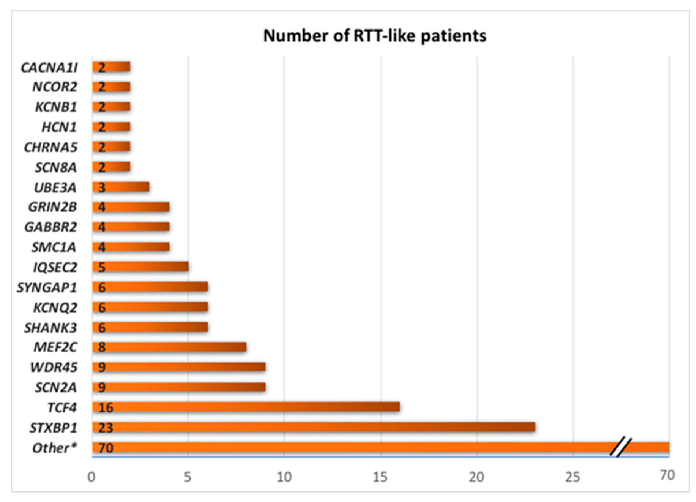
4. NGS Results: Many Genes, Many Disorders

Apart from RTT and RTT-like patients, which have mutations in the MEPC2, CDKL5 and FOXG1 genes, there are a percentage of patients without a genetic diagnosis. Now, with WES and panels that incorporate more and more genes related to the central nervous system, the number of patients with a pathogenic variant detected has increased in genes that were previously not related to RTT nor RTT-like. All studies published about these genes are summarized in Table 2.
Using NGS, in only five years more than eighty genes were related to the RTT-like phenotype and some of these genes were identified as causative for aRTT or RTT-like phenotype in these patients, although some of them were associated with well-known syndromes such as Pitt–Hopkins syndrome (TCF4, CNTNAP2 and NRXN1 genes), Phelan–McDermid syndrome (SHANK3 gene), Angelman syndrome (UBE3A gene), Kleefstra syndrome (EHMT1) and Cornelia de Lange syndrome (SMC1A). In addition, a substantial number of genes are epileptic encephalopathy genes (STXBP1, SCN1A, SCN2A, SCN8A, GRIN2A, GRIN2B, HCN1, SLC6A1, KCNA2, EEF1A2, KCNB1 and SYNGAP1) and are related with mental retardation and epilepsy (IQSEC2 and MEF2C).
Note that, regarding the number of patients described in these publications, there are some genes that are more represented (Figure 1). STXBP1, TCF4, SCN2A, WDR45, and MEF2C are the most common genes with a pathogenic (or likely pathogenic) variant detected in patients with RTT/RTT-like phenotype.
The STXBP1 gene (syntaxin-binding protein 1; OMIM#602926) encodes a transmembrane attachment protein receptor that plays an important role in the release of neurotransmitters via regulation of syntaxin, a transmembrane attachment protein receptor [47]. Pathogenic variants in this gene, reducing its expression, depress the functions of GABAergic and glutamatergic synapses, particularly in GABAergic interneurons [48], a process that has been shown to be altered in RTT patients [49]. Mutations in STXBP1 have been associated with epileptic encephalopathy, early infantile 4 (EEIE4) and a series of neurodevelopmental disorders, including RTT-like syndrome.
The TCF4 gene (transcription factor 4; OMIM*602272) is a broadly expressed basic helix–loop–helix (bHLH) protein that functions as a homodimer or as a heterodimer with other bHLH proteins. These dimers bind DNA at Ephrussi (E) box sequences motif (‘CANNTG’). Alternative splicing creates numerous N-terminally distinct TCF4 isoforms that differ in their subcellular localization and transactivational capacity [50]. Mutations in TCF4 have been associated with Pitt–Hopkins syndrome (PTHS), which is characterized by severe ID, delayed motor development, seizures, wide mouth and distinctive facial features, hypotonia, microcephaly, limited walking abilities, and intermittent hyperventilation followed by apnea [31]. The microcephaly, intermittent hyperventilation, and stereotype hand movements may steer clinicians towards a diagnosis of RTT-like rather than PTHS. The distinct facial features presented in patients with a clear PTHS phenotype, such as thin eyebrows, sunken eyes, a pronounced double curve of the upper lip, and a wide mouth with full lips, is more consistent with PTHS and helps to distinguish PTHS from RTT, but some patients do not always present distinctly these facial dismorphisms, or they are often not well-defined during the first year of life [51].
SCN2A (neuronal voltage-gated sodium channel NaV1.2; OMIM*182390) encodes one member of the sodium channel alpha subunit gene family, responsible for generation and propagation of action potentials, chiefly in nerve and muscle. Pathogenic variants in the SCN2A gene that produce loss-of-function of the protein lead to ASD/ID and increased channel activity that lead to epileptic encephalopathy early infantile 11 (EEIE11) and benign familial neonatal-infantile seizures (BFIS) [52].
WD40 repeat proteins are an important key component that regulates the assembly of multiprotein complexes by presenting a beta-propeller platform for simultaneous and reversible protein–protein interactions [53]. Variants in WDR45 (WD repeat-containing protein 45; OMIM*300526) are associated with developmental delay in early childhood and progressive neurodegeneration in adolescence or adulthood related to iron accumulation in the globus pallidus and substantia nigra [30,54]. Affected patients may have features overlapping those of RTT, including developmental regression, hand-wringing, and seizures. Some may even have a diagnosis of typical or atypical RTT [55].
MEF2C (OMIM*600662) belongs to the myocyte enhancer factor-2 (MEF2) family of transcription factors. MEF2C plays an important role in myogenesis, development of the anterior heart field, neural crest and craniofacial development, and neurogenesis, among others [56]. It is well-described the MEF2C haploinsufficiency syndrome that has been recognized as a neurodevelopmental disorder. Until now, fourteen patients with point mutation pathogenic, or likely pathogenic, variants in MEF2C have been identified in RTT-like patients, including three nonsense, three missense and three frameshift variants [37,57].
In Table 3, the most common genes for RTT-like phenotypes are summarized, and all of them present an autosomal dominant inheritance pattern, either autosomal or linked to the X chromosome and are caused by de novo heterozygous mutations in the germline. Note that all the diseases are severe IDs and most of them meet the four main criteria for RTT; all of them present a loss or speech severe deficit, gait abnormalities and lost or absent purposeful hand movements linked to stereotypical hand movements, such as hand wringing. Only developmental regression, one of the four main criteria, is absent in PTHS. Moreover, other common symptoms in RTT are present in these diseases too, such as epilepsy, breathing disturbances and microcephaly, although not all the RTT patients present them. In contrast, other symptoms that are not present in RTT are present in atypical forms related to CDKL5 and FOXG1, such as dysmorphic facial features in PTHS or CNS abnormalities in neurodegeneration with brain iron accumulation.
Focusing on the pathophysiology of RTT at the brain level, female mice heterozygous for the null MECP2 present microcephaly without gross neuropathological changes. Specifically, mouse and human neurons without MeCP2 have smaller somas and decreased dendritic complexity [58,59]. A decrease in synaptic plasticity and abnormalities in neurotransmitter concentrations is also observed in many neuronal types [60,61,62]. Looking at the established knockout mice models of those of the most common genes detected in RTT patients, morphological and/or physiological features resemble RTT mouse models. For instance, young adult Mef2c and Scn2a cKO mice present a normal gross brain morphology and cortical layer organization, as murine RTT models. In contrast, Mef2c cKO mice cause an increase in dendritic spine density on dentate granule neurons of the hippocampal dentate gyrus [63] and Scn2a cOK in pre-oligodendrocyte alters their morphology, impairs myelination and reduces axon-oligodendrocyte interactions [64,65]. Moreover, depletion of specific presynaptic proteins involved in exocytosis, such as STXBP1, causes abnormalities in neurotransmitter concentrations and produces neuronal cell death [66]. TCF4 haploinsufficiency mice exhibit a delay in neuronal migration, and a significant increase in the number of upper-layer cortical neurons, as well as abnormal dendrite and synapse formation [67]. WDR45 cOK mice show a loss of neurons in prefrontal cortex and basal ganglion in aged mice and increased apoptosis in prefrontal cortex, recapitulating a hallmark of neurodegeneration [68].
At the moment, more than 80 genes have been associated with RTT/RTT-like, but their link with RTT should be critically evaluated. In order to relate a gene with RTT several points have to be taken into consideration: (1) Proper clinical characterizations of the patients; (2) deep comprehension of the functions of the candidate gene; (3) validated evidence that mutation found it is pathogenic. The more patients with a similar phenotype have pathogenic mutations in the same gene, the more consistent evidence we will have of that gene being part of the list of genes related to RTT.

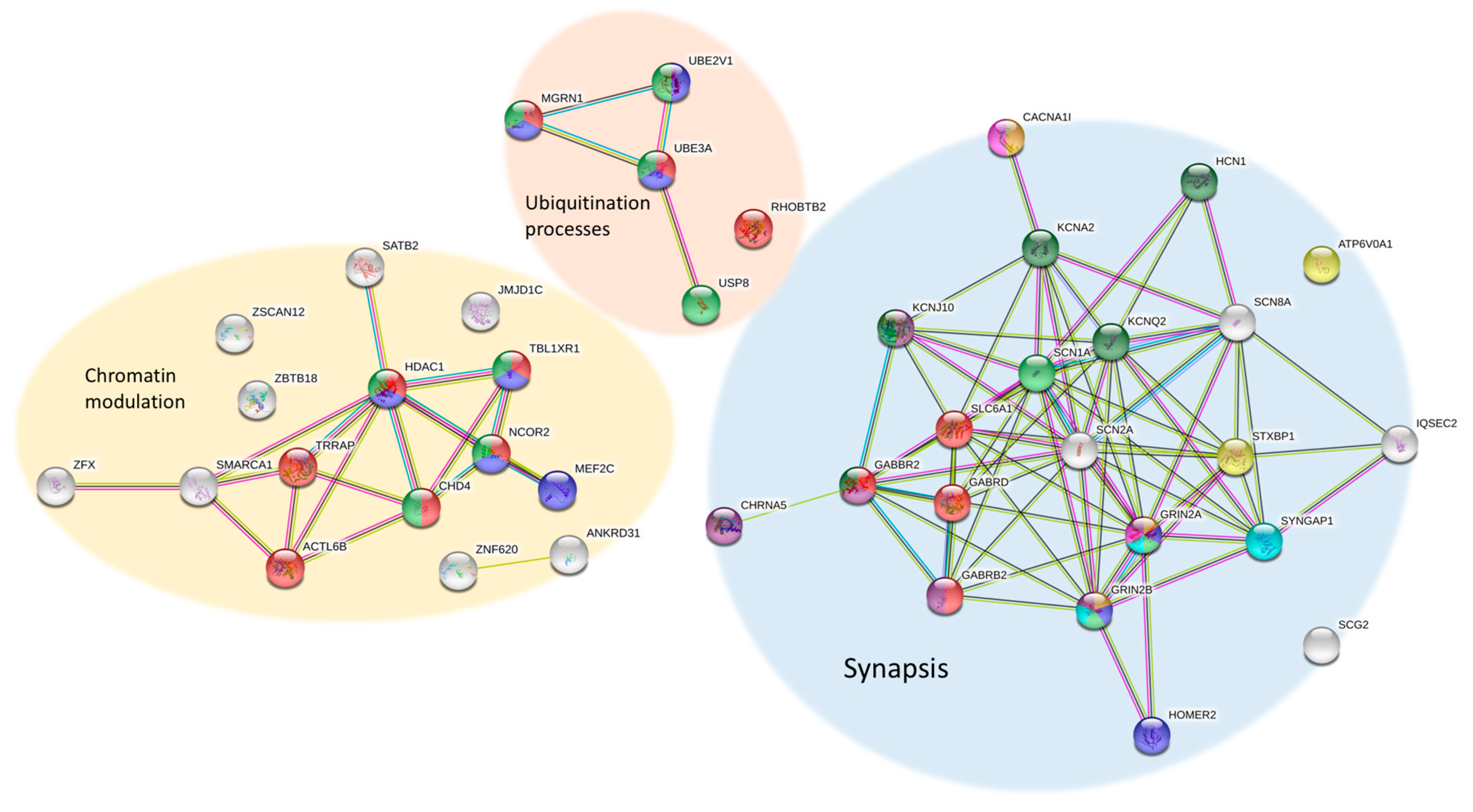
5. Functions and Pathways around RTT

The list of genes related to RTT/RTT-like phenotypes is complex and diverse. However, using REACTOME (an open-source, open access, manually curated and peer-reviewed pathway database; https://reactome.org) and STRING (a database of known and predicted protein–protein interactions; https://string-db.org/), we can identify some groups of genes with functions involved in common mechanisms. Several pathways can be studied in RTT/RTT-like patients, such as chromatin modulation, synaptic function and ubiquitin conjugation (Figure 2).
This list currently includes 15 genes (ACTL6B, ANKRD31, CHD4, HDAC1, JMJD1C, MEF2C, NCOR2, SATB2, SMARCA1, TBL1XR1, TRRAP, ZFX. ZNF238, ZNF620 and ZSCAN12) involved in chromatin modulation pathways, such as chromatin-modifying enzymes and histone deacetylases (HDACs) and 21 genes (ATP6V0A1, CACNA1I, CHRNA5, GABBR2, GABRB2, GABRD, GRIN2A, GRIN2B, HCN1, IQSEC2, KCNA2, KCNJ10, KCNQ2, SCG2, SCN1A, SCN2A, SCN8A, SHANK3, SLC6A1, STXBP1 and SYNGAP1) involved in synaptic function, necessary for GABAergic, glutamatergic and dopaminergic synapses, synaptic vesicles trafficking, ion homeostasis in neurons and circadian entrainment. The link of all these genes to the same pathways could explain why these patients’ phenotypes overlap, causing impaired synaptic function, sleep disturbances and major dysregulation of gene expression. Notably, there are also a few genes (MGRN1, RHOBTB2 and USP8) involved in ubiquitination processes, which UBE3A (the gene responsible for Angelman syndrome) is also linked to. The considerable overlapping of RTT and Angelman features could be explained due to this relationship.
MeCP2 performs many tasks during the neurodevelopment, such as regulating the gene expression of other genes, modulating epigenetic imprinting and neurotransmitter actions. Hence it is challenging to create a well-defined pathways involving MeCP2 and predict the downstream effects that disruption of the MeCP2 function can generate. Ehrhart et al. 2016 [69] created a comprehensive visualization of the biologic pathways showing how MECP2 upstream and downstream regulation developed. Moreover, it had been and published on WikiPathways which will serve as template for future omics data driven research (http://www.wikipathways.org/instance/WP3584) [69].
Discovering the pathways related to MEPC2, we could better link genes that are mutated in patients without MECP2 defects. Nowadays, the combination of the omics data analysis and prior knowledge databases are a powerful approach to identify connections between mutation and phenotype. Ehrhart et al. 2019 [70] identified a subset of genes, which are significantly different in several transcriptomics datasets and were not described yet in the context of RTT. They described that these genes are involved in molecular pathways and several processes known to be affected in RTT patients [70]. For example, altered calcium homeostasis seems to be responsible for an abnormal neuronal development and generates epilepsy; and tubulin, ERM and MEF2C are some of the altered proteins related to cytoskeletal abnormalities that are present not only in RTT, but also in Angelman syndrome [69]. In the same way, cholesterol biosynthesis is altered in RTT and in Smith–Lemli–Opitz syndrome, in which it has been pointed out to be the cause of the autism and malformations [71]. The NF-kB pathway, which is involved in nervous system development, synaptic transmission and cognition, is altered in RTT and RTT-like patients and seems to be the cause of mental retardation [72]. Another pathway observed in patients with RTT, autism, and Parkinson’s disease (PD) is the neurotransmitter imbalance of GABA, Glutamine and Dopamine. This imbalance seems to be responsible for the autism features RTT patients present and the motor difficulties that patients with PD and RTT have [73].
An important feature of MeCP2 reduction in RTT mouse models and individuals with RTT is a propensity for seizure, a prominent signature in many brain diseases, including RTT [74,75]. The deletion of MeCP2 from all forebrain GABAergic interneurons recapitulates major phenotype of RTT [75], demonstrating that altered inhibitory function is critical for normal function of GABA-releasing neurons and an important aspect of RTT pathophysiology. It has been demonstrated recently that human neurons derived from patients with RTT and RTT mouse models show a significant reduced the SLC12A5 gene expression, resulting in a delayed GABA functional switch [76]. This gene encodes a neuron-specific K+/Cl− cotransporter 2, the major extruder of intracellular chloride in mature neurons. Moreover, it has been established that MeCP2 regulates KCC2 expression through inhibiting RE1-silencing transcriptional factor [77], and it is suggested that KCC2 should play a role in the pathophysiology of RTT
Recently, Cosentino et al. (2019) have described several alterations in RTT patients and animal models during the pre-symptomatic stage [78]. During this stage some compensatory mechanisms keep the phenotypic outcome to a minimum until MECP2 deficiency cannot be supplied and the known phenotype of RTT becomes apparent [79]. Thus, since the alterations found in RTT spectrum disorders are due to both direct and indirect effects of MECP2 and related genes’ deficiencies/malfunctions, the best approach for characterization and consequent treatment would be the comprehensive study of all the altered pathways that have been discovered in these patients. If we were able to find a way to compensate those altered pathways, a treatment could be implemented in very early stages of development, even before the onset of the most remarkable features of the syndrome [78,79].

6. Future Perspectives and Treatment Options

In recent years, impressive advances have been achieved not only in the genetics diagnosis of neurodevelopmental disorders, but also in elucidating the physiological pathway and molecular mechanisms involved in the clinical manifestations of the diseases. An increasing amount of evidence is emerging that understanding these mechanisms is relevant for the selection of the most appropriate treatment in the affected individual.
More than 50 years have passed since the description and clinical characterization of RTT and the current standard of care for patients remains limited to supportive and symptomatic therapies that can palliate the symptomatology of the patients, but not cure the disease per se. Drug treatment consists mainly of off-label prescriptions due to the lack of approved medications for the disorder. Until now, all RTT trials have been based on the essential role of MECP2 in the development and maintenance of neurons in the central nervous system, and its specific role in the distinct cellular subtypes focused on the following specific neurotransmitters: dextromethorphan (an NMDA receptor antagonist, mainly used for cognition and seizures [80]); desipramine (a noradrenaline reuptake inhibitor, for breathing abnormalities [81]); and IGF-1 [82]. To date, emulating the MeCP2 function, using a pharmacology strategy, as a treatment for RTT is not the best and most successful strategy.
However, it is not only patients with RTT or RTT-like that are caused by defects in the MECP2 gene. An improved understanding of the different genes mutated in the RTT-like phenotype generates important therapeutic clues and opportunities to develop novel and better treatments. A treatment which targets neuronal maturational defects seen in MECP2 mutations may not be effective for an ion channelopathy due to KCNB1 alterations. In this way, therapies must focus on personalized treatments for each individual, depending on which gene and which type of mutation carries. For example, for the SCN2A gene that encodes the voltage-gated sodium channel Nav1.2, gain-of-function versus loss-of-function variants in SCN2A determine whether sodium channel blockers improve or worsen seizure control [52]. Another example is that L-serine supplementation might ameliorate GRIN2B-related severe encephalopathy [83]. Additionally, for STXBP1, which plays an important role in presynaptic vesicle docking and fusion [84], current treatments are largely limited to seizure control and future therapies will also need to target the developmental aspects of the disease [83]. It has been hypothesized that enhancing KCC2 expression could increase the efficacy of GABAergic inhibition and also improve the dendritic spine and excitatory synapse development, both of which are abnormal in RTT [85,86] and may be caused by an aberrant interaction between KCC2 and the dendritic cytoskeleton [87,88]. Recently, two works have considered the pathogenic role of diminished KCC2 expression in the Mecp2 null model: The first found that the use of bumetanide can attenuate the unbalance glutamatergic/GABAergic ratio if treated in the early stages of the disorder [89]; and Tang et al. (2019) have shown that the injection of KEEC KW-2449 or piperine (small-compounds) in Mecp2 mutant mice ameliorated disease-associated respiratory and locomotion phenotypes [90].

7. Conclusions

All of the individuals summarized in this review met the diagnostic criteria for RTT or RTT-like; however, lacking a defect in MECP2 underscores the importance of carrying out additional genetic testing, whether it is by specific gene panels, WES or WGS, to identify the specific etiology and to direct appropriate diagnostic and therapeutic strategies. Many disorders can be caused by multiple genes, such as West syndrome or Charcot-Marie-Tooth, and these are considered as the same disorder as long as they share a common phenotype. Therefore, we consider that all classical, atypical and all RTT-like phenotypes could be grouped into an RTT spectrum disorder with many causative genes.
From a research point of view, the new genes recently associated with RTT-like phenotypes without a clear description of their biological functions network involved need further study. Furthermore, variants with an unknown clinical significance in genes without clear defined functions have been found through screening RTT spectrum cohorts without genetic diagnosis. These new candidate genes need functional studies to establish their potential role in the disease pathogenesis. From a clinical perspective, a better definition of the pathways that connect of all these genes involved in all RTT and RTT-like phenotypes would enable a better understanding of the genetic landscape of the RTT spectrum. The elucidation of the functional pathways involved in all these patients could support the development of future targeted therapies.

Author Contributions

S.V., C.X., M.P. and J.A. conceived and supervised the review; S.V. and C.X. wrote the original draft. S.V., C.X., A.P.-A., M.O., M.P. and J.A., wrote—reviewed & edited. J.A. and M.P. supervised. All authors reviewed the article critically for intellectual content.

Funding

The work was supported by grants from the Spanish Ministry of Health (Instituto de Salud Carlos III/FEDER, PI15/01159), the crowd-funding program PRECIPITA, the Spanish Ministry of Health (Fundación Española para la Ciencia y la Tecnología), the Catalan Association for Rett Syndrome, FondoBiorett and Mi Princesa Rett.

Conflicts of Interest

The authors declare no competing interests. The funders had no role in the design of the study; in the collection, analyses, or interpretation of data; in the writing of the manuscript, or in the decision to publish the results.

References

  1. Rett, A. On a unusual brain atrophy syndrome in hyperammonemia in childhood. Wien. Med. Wochenschr. 1966, 116, 723–726. [Google Scholar] [PubMed]
  2. Amir, R.E.; Van den Veyver, I.B.; Wan, M.; Tran, C.Q.; Francke, U.; Zoghbi, H.Y. Rett syndrome is caused by mutations in X-linked MECP2, encoding methyl-CpG-binding protein 2. Nat. Genet. 1999, 23, 185–188. [Google Scholar] [CrossRef] [PubMed]
  3. Kriaucionis, S.; Bird, A. The major form of MeCP2 has a novel N-terminus generated by alternative splicing. Nucleic Acids Res. 2004, 32, 1818–1823. [Google Scholar] [CrossRef] [PubMed]
  4. Mnatzakanian, G.N.; Lohi, H.; Munteanu, I.; Alfred, S.E.; Yamada, T.; MacLeod, P.J.; Jones, J.R.; Scherer, S.W.; Schanen, N.C.; Friez, M.J.; et al. A previously unidentified MECP2 open reading frame defines a new protein isoform relevant to Rett syndrome. Nat. Genet. 2004, 36, 339–341. [Google Scholar] [CrossRef] [PubMed] [Green Version]
  5. Yasui, D.H.; Gonzales, M.L.; Aflatooni, J.O.; Crary, F.K.; Hu, D.J.; Gavino, B.J.; Golub, M.S.; Vincent, J.B.; Carolyn Schanen, N.; Olson, C.O.; et al. Mice with an isoform-ablating Mecp2 exon 1 mutation recapitulate the neurologic deficits of Rett syndrome. Hum. Mol. Genet. 2014, 23, 2447–2458. [Google Scholar] [CrossRef] [PubMed]
  6. Christodoulou, J.; Grimm, A.; Maher, T.; Bennetts, B. RettBASE: The IRSA MECP2 variation database-a new mutation database in evolution. Hum. Mutat. 2003, 21, 466–472. [Google Scholar] [CrossRef] [PubMed]
  7. Neul, J.L.; Fang, P.; Barrish, J.; Lane, J.; Caeg, E.B.; Smith, E.O.; Zoghbi, H.; Percy, A.; Glaze, D.G. Specific mutations in methyl-CpG-binding protein 2 confer different severity in Rett syndrome. Neurology 2008, 70, 1313–1321. [Google Scholar] [CrossRef] [PubMed]
  8. Kalscheuer, V.M.; Freude, K.; Musante, L.; Jensen, L.R.; Yntema, H.G.; Gecz, J.; Sefiani, A.; Hoffmann, K.; Moser, B.; Haas, S.; et al. Mutations in the polyglutamine binding protein 1 gene cause X-linked mental retardation. Nat. Genet. 2003, 35, 313–315. [Google Scholar] [CrossRef] [Green Version]
  9. Ariani, F.; Hayek, G.; Rondinella, D.; Artuso, R.; Mencarelli, M.A.; Spanhol-Rosseto, A.; Pollazzon, M.; Buoni, S.; Spiga, O.; Ricciardi, S.; et al. FOXG1 is responsible for the congenital variant of Rett syndrome. Am. J. Hum. Genet. 2008, 83, 89–93. [Google Scholar] [CrossRef]
  10. Srivastava, S.; Love-Nichols, J.A.; Dies, K.A.; Ledbetter, D.H.; Martin, C.L.; Chung, W.K.; Firth, H.V.; Frazier, T.; Hansen, R.L.; Prock, L.; et al. Meta-analysis and multidisciplinary consensus statement: Exome sequencing is a first-tier clinical diagnostic test for individuals with neurodevelopmental disorders. Genet. Med. 2019. [Google Scholar] [CrossRef]
  11. Hagberg, B.; Hanefeld, F.; Percy, A.; Skjeldal, O. An update on clinically applicable diagnostic criteria in Rett syndrome. Comments to Rett Syndrome Clinical Criteria Consensus Panel Satellite to European Paediatric Neurology Society Meeting, Baden Baden, Germany, 11 September 2001. Eur. J. Paediatr. Neurol. 2002, 6, 293–297. [Google Scholar] [CrossRef]
  12. Neul, J.L.; Kaufmann, W.E.; Glaze, D.G.; Christodoulou, J.; Clarke, A.J.; Bahi-Buisson, N.; Leonard, H.; Bailey, M.E.; Schanen, N.C.; Zappella, M.; et al. Rett syndrome: Revised diagnostic criteria and nomenclature. Ann. Neurol. 2010, 68, 944–950. [Google Scholar] [CrossRef] [Green Version]
  13. Koboldt, D.C.; Steinberg, K.M.; Larson, D.E.; Wilson, R.K.; Mardis, E.R. The next-generation sequencing revolution and its impact on genomics. Cell 2013, 155, 27–38. [Google Scholar] [CrossRef]
  14. Wang, J.; Zhang, Q.; Chen, Y.; Yu, S.; Wu, X.; Bao, X.; Wen, Y. Novel MEF2C point mutations in Chinese patients with Rett (-like) syndrome or non-syndromic intellectual disability: Insights into genotype-phenotype correlation. BMC Med. Genet. 2018, 19, 191. [Google Scholar] [CrossRef]
  15. Vidal, S.; Brandi, N.; Pacheco, P.; Gerotina, E.; Blasco, L.; Trotta, J.R.; Derdak, S.; Del Mar O’Callaghan, M.; Garcia-Cazorla, A.; Pineda, M.; et al. The utility of Next Generation Sequencing for molecular diagnostics in Rett syndrome. Sci. Rep. 2017, 7, 12288. [Google Scholar] [CrossRef]
  16. Schonewolf-Greulich, B.; Bisgaard, A.M.; Moller, R.S.; Duno, M.; Brondum-Nielsen, K.; Kaur, S.; Van Bergen, N.J.; Lunke, S.; Eggers, S.; Jespersgaard, C.; et al. Clinician’s guide to genes associated with Rett-like phenotypes-Investigation of a Danish cohort and review of the literature. Clin. Genet. 2019, 95, 221–230. [Google Scholar] [CrossRef]
  17. Di Resta, C.; Galbiati, S.; Carrera, P.; Ferrari, M. Next-generation sequencing approach for the diagnosis of human diseases: Open challenges and new opportunities. EJIFCC 2018, 29, 4–14. [Google Scholar]
  18. Srivastava, S.; Desai, S.; Cohen, J.; Smith-Hicks, C.; Baranano, K.; Fatemi, A.; Naidu, S. Monogenic disorders that mimic the phenotype of Rett syndrome. Neurogenetics 2018, 19, 41–47. [Google Scholar] [CrossRef]
  19. Lucariello, M.; Vidal, E.; Vidal, S.; Saez, M.; Roa, L.; Huertas, D.; Pineda, M.; Dalfo, E.; Dopazo, J.; Jurado, P.; et al. Whole exome sequencing of Rett syndrome-like patients reveals the mutational diversity of the clinical phenotype. Hum. Genet. 2016, 135, 1343–1354. [Google Scholar] [CrossRef] [Green Version]
  20. Mak, C.C.; Leung, G.K.; Mok, G.T.; Yeung, K.S.; Yang, W.; Fung, C.W.; Chan, S.H.; Lee, S.L.; Lee, N.C.; Pfundt, R.; et al. Exome sequencing for paediatric-onset diseases: Impact of the extensive involvement of medical geneticists in the diagnostic odyssey. NPJ Genom. Med. 2018, 3, 19. [Google Scholar] [CrossRef]
  21. Lelieveld, S.H.; Veltman, J.A.; Gilissen, C. Novel bioinformatic developments for exome sequencing. Hum. Genet. 2016, 135, 603–614. [Google Scholar] [CrossRef] [Green Version]
  22. Lupski, J.R.; Reid, J.G.; Gonzaga-Jauregui, C.; Rio Deiros, D.; Chen, D.C.; Nazareth, L.; Bainbridge, M.; Dinh, H.; Jing, C.; Wheeler, D.A.; et al. Whole-genome sequencing in a patient with Charcot-Marie-Tooth neuropathy. N. Engl. J. Med. 2010, 362, 1181–1191. [Google Scholar] [CrossRef]
  23. Gilissen, C.; Hehir-Kwa, J.Y.; Thung, D.T.; van de Vorst, M.; van Bon, B.W.; Willemsen, M.H.; Kwint, M.; Janssen, I.M.; Hoischen, A.; Schenck, A.; et al. Genome sequencing identifies major causes of severe intellectual disability. Nature 2014, 511, 344–347. [Google Scholar] [CrossRef]
  24. Halvorsen, M.; Petrovski, S.; Shellhaas, R.; Tang, Y.; Crandall, L.; Goldstein, D.; Devinsky, O. Mosaic mutations in early-onset genetic diseases. Genet. Med. 2016, 18, 746–749. [Google Scholar] [CrossRef]
  25. de Lange, I.M.; Koudijs, M.J.; van ’t Slot, R.; Sonsma, A.C.M.; Mulder, F.; Carbo, E.C.; van Kempen, M.J.A.; Nijman, I.J.; Ernst, R.F.; Savelberg, S.M.C.; et al. Assessment of parental mosaicism in SCN1A-related epilepsy by single-molecule molecular inversion probes and next-generation sequencing. J. Med. Genet. 2019, 56, 75–80. [Google Scholar] [CrossRef]
  26. Uddin, M.; Woodbury-Smith, M.; Chan, A.; Brunga, L.; Lamoureux, S.; Pellecchia, G.; Yuen, R.K.C.; Faheem, M.; Stavropoulos, D.J.; Drake, J.; et al. Germline and somatic mutations in STXBP1 with diverse neurodevelopmental phenotypes. Neurol. Genet. 2017, 3, e199. [Google Scholar] [CrossRef] [Green Version]
  27. D’Gama, A.M.; Walsh, C.A. Somatic mosaicism and neurodevelopmental disease. Nat. Neurosci. 2018, 21, 1504–1514. [Google Scholar] [CrossRef]
  28. de Lange, I.M.; Koudijs, M.J.; van ’t Slot, R.; Gunning, B.; Sonsma, A.C.M.; van Gemert, L.; Mulder, F.; Carbo, E.C.; van Kempen, M.J.A.; Verbeek, N.E.; et al. Mosaicism of de novo pathogenic SCN1A variants in epilepsy is a frequent phenomenon that correlates with variable phenotypes. Epilepsia 2018, 59, 690–703. [Google Scholar] [CrossRef]
  29. Baasch, A.L.; Huning, I.; Gilissen, C.; Klepper, J.; Veltman, J.A.; Gillessen-Kaesbach, G.; Hoischen, A.; Lohmann, K. Exome sequencing identifies a de novo SCN2A mutation in a patient with intractable seizures, severe intellectual disability, optic atrophy, muscular hypotonia, and brain abnormalities. Epilepsia 2014, 55, e25–e29. [Google Scholar] [CrossRef]
  30. Saitsu, H.; Tohyama, J.; Walsh, T.; Kato, M.; Kobayashi, Y.; Lee, M.; Tsurusaki, Y.; Miyake, N.; Goto, Y.; Nishino, I.; et al. A girl with West syndrome and autistic features harboring a de novo TBL1XR1 mutation. J. Hum. Genet. 2014, 59, 581–583. [Google Scholar] [CrossRef]
  31. Okamoto, N.; Miya, F.; Tsunoda, T.; Kato, M.; Saitoh, S.; Yamasaki, M.; Shimizu, A.; Torii, C.; Kanemura, Y.; Kosaki, K. Targeted next-generation sequencing in the diagnosis of neurodevelopmental disorders. Clin. Genet. 2015, 88, 288–292. [Google Scholar] [CrossRef]
  32. Hara, M.; Ohba, C.; Yamashita, Y.; Saitsu, H.; Matsumoto, N.; Matsuishi, T. De novo SHANK3 mutation causes Rett syndrome-like phenotype in a female patient. Am. J. Med. Genet. A 2015, 167, 1593–1596. [Google Scholar] [CrossRef]
  33. Olson, H.E.; Tambunan, D.; LaCoursiere, C.; Goldenberg, M.; Pinsky, R.; Martin, E.; Ho, E.; Khwaja, O.; Kaufmann, W.E.; Poduri, A. Mutations in epilepsy and intellectual disability genes in patients with features of Rett syndrome. Am. J. Med. Genet. A 2015, 167A, 2017–2025. [Google Scholar] [CrossRef]
  34. Hoffjan, S.; Ibisler, A.; Tschentscher, A.; Dekomien, G.; Bidinost, C.; Rosa, A.L. WDR45 mutations in Rett (-like) syndrome and developmental delay: Case report and an appraisal of the literature. Mol. Cell Probes 2016, 30, 44–49. [Google Scholar] [CrossRef]
  35. Lee, J.S.; Yoo, Y.; Lim, B.C.; Kim, K.J.; Choi, M.; Chae, J.H. SATB2-associated syndrome presenting with Rett-like phenotypes. Clin. Genet. 2016, 89, 728–732. [Google Scholar] [CrossRef]
  36. Saez, M.A.; Fernandez-Rodriguez, J.; Moutinho, C.; Sanchez-Mut, J.V.; Gomez, A.; Vidal, E.; Petazzi, P.; Szczesna, K.; Lopez-Serra, P.; Lucariello, M.; et al. Mutations in JMJD1C are involved in Rett syndrome and intellectual disability. Genet. Med. 2016, 18, 378–385. [Google Scholar] [CrossRef] [Green Version]
  37. Rocha, H.; Sampaio, M.; Rocha, R.; Fernandes, S.; Leao, M. MEF2C haploinsufficiency syndrome: Report of a new MEF2C mutation and review. Eur. J. Med. Genet. 2016, 59, 478–482. [Google Scholar] [CrossRef]
  38. Lopes, F.; Barbosa, M.; Ameur, A.; Soares, G.; de Sa, J.; Dias, A.I.; Oliveira, G.; Cabral, P.; Temudo, T.; Calado, E.; et al. Identification of novel genetic causes of Rett syndrome-like phenotypes. J. Med. Genet. 2016, 53, 190–199. [Google Scholar] [CrossRef]
  39. Sajan, S.A.; Jhangiani, S.N.; Muzny, D.M.; Gibbs, R.A.; Lupski, J.R.; Glaze, D.G.; Kaufmann, W.E.; Skinner, S.A.; Annese, F.; Friez, M.J.; et al. Enrichment of mutations in chromatin regulators in people with Rett syndrome lacking mutations in MECP2. Genet. Med. 2017, 19, 13–19. [Google Scholar] [CrossRef] [Green Version]
  40. Allou, L.; Julia, S.; Amsallem, D.; El Chehadeh, S.; Lambert, L.; Thevenon, J.; Duffourd, Y.; Saunier, A.; Bouquet, P.; Pere, S.; et al. Rett-like phenotypes: Expanding the genetic heterogeneity to the KCNA2 gene and first familial case of CDKL5-related disease. Clin. Genet. 2017, 91, 431–440. [Google Scholar] [CrossRef]
  41. Yoo, Y.; Jung, J.; Lee, Y.N.; Lee, Y.; Cho, H.; Na, E.; Hong, J.; Kim, E.; Lee, J.S.; Lee, J.S.; et al. GABBR2 mutations determine phenotype in rett syndrome and epileptic encephalopathy. Ann. Neurol. 2017, 82, 466–478. [Google Scholar] [CrossRef]
  42. Vuillaume, M.L.; Jeanne, M.; Xue, L.; Blesson, S.; Denomme-Pichon, A.S.; Alirol, S.; Brulard, C.; Colin, E.; Isidor, B.; Gilbert-Dussardier, B.; et al. A novel mutation in the transmembrane 6 domain of GABBR2 leads to a Rett-like phenotype. Ann. Neurol. 2018, 83, 437–439. [Google Scholar] [CrossRef]
  43. Huisman, S.; Mulder, P.A.; Redeker, E.; Bader, I.; Bisgaard, A.M.; Brooks, A.; Cereda, A.; Cinca, C.; Clark, D.; Cormier-Daire, V.; et al. Phenotypes and genotypes in individuals with SMC1A variants. Am. J. Med. Genet. A 2017, 173, 2108–2125. [Google Scholar] [CrossRef]
  44. Percy, A.K.; Lane, J.; Annese, F.; Warren, H.; Skinner, S.A.; Neul, J.L. When Rett syndrome is due to genes other than MECP2. Transl. Sci. Rare Dis. 2018, 3, 49–53. [Google Scholar] [CrossRef] [Green Version]
  45. Iwama, K.; Mizuguchi, T.; Takeshita, E.; Nakagawa, E.; Okazaki, T.; Nomura, Y.; Iijima, Y.; Kajiura, I.; Sugai, K.; Saito, T.; et al. Genetic landscape of Rett syndrome-like phenotypes revealed by whole exome sequencing. J. Med. Genet. 2019. [Google Scholar] [CrossRef]
  46. Vidal, S.; Brandi, N.; Pacheco, P.; Maynou, J.; Fernandez, G.; Xiol, C.; Pascual-Alonso, A.; Pineda, M.; Rett Working, G.; Armstrong, J. The most recurrent monogenic disorders that overlap with the phenotype of Rett syndrome. Eur. J. Paediatr. Neurol. 2019. [Google Scholar] [CrossRef]
  47. Swanson, D.A.; Steel, J.M.; Valle, D. Identification and characterization of the human ortholog of rat STXBP1, a protein implicated in vesicle trafficking and neurotransmitter release. Genomics 1998, 48, 373–376. [Google Scholar] [CrossRef]
  48. Toonen, R.F.; Wierda, K.; Sons, M.S.; de Wit, H.; Cornelisse, L.N.; Brussaard, A.; Plomp, J.J.; Verhage, M. Munc18-1 expression levels control synapse recovery by regulating readily releasable pool size. Proc. Natl. Acad. Sci. USA 2006, 103, 18332–18337. [Google Scholar] [CrossRef] [Green Version]
  49. Medrihan, L.; Tantalaki, E.; Aramuni, G.; Sargsyan, V.; Dudanova, I.; Missler, M.; Zhang, W. Early defects of GABAergic synapses in the brain stem of a MeCP2 mouse model of Rett syndrome. J. Neurophysiol. 2008, 99, 112–121. [Google Scholar] [CrossRef]
  50. Sepp, M.; Pruunsild, P.; Timmusk, T. Pitt-Hopkins syndrome-associated mutations in TCF4 lead to variable impairment of the transcription factor function ranging from hypomorphic to dominant-negative effects. Hum. Mol. Genet. 2012, 21, 2873–2888. [Google Scholar] [CrossRef]
  51. de Winter, C.F.; Baas, M.; Bijlsma, E.K.; van Heukelingen, J.; Routledge, S.; Hennekam, R.C. Phenotype and natural history in 101 individuals with Pitt-Hopkins syndrome through an internet questionnaire system. Orphanet J. Rare Dis. 2016, 11, 37. [Google Scholar] [CrossRef]
  52. Sanders, S.J.; Campbell, A.J.; Cottrell, J.R.; Moller, R.S.; Wagner, F.F.; Auldridge, A.L.; Bernier, R.A.; Catterall, W.A.; Chung, W.K.; Empfield, J.R.; et al. Progress in Understanding and Treating SCN2A-Mediated Disorders. Trends Neurosci. 2018, 41, 442–456. [Google Scholar] [CrossRef]
  53. Proikas-Cezanne, T.; Waddell, S.; Gaugel, A.; Frickey, T.; Lupas, A.; Nordheim, A. WIPI-1alpha (WIPI49), a member of the novel 7-bladed WIPI protein family, is aberrantly expressed in human cancer and is linked to starvation-induced autophagy. Oncogene 2004, 23, 9314–9325. [Google Scholar] [CrossRef]
  54. Haack, T.B.; Hogarth, P.; Kruer, M.C.; Gregory, A.; Wieland, T.; Schwarzmayr, T.; Graf, E.; Sanford, L.; Meyer, E.; Kara, E.; et al. Exome sequencing reveals de novo WDR45 mutations causing a phenotypically distinct, X-linked dominant form of NBIA. Am. J. Hum. Genet. 2012, 91, 1144–1149. [Google Scholar] [CrossRef]
  55. Ohba, C.; Nabatame, S.; Iijima, Y.; Nishiyama, K.; Tsurusaki, Y.; Nakashima, M.; Miyake, N.; Tanaka, F.; Ozono, K.; Saitsu, H.; et al. De novo WDR45 mutation in a patient showing clinically Rett syndrome with childhood iron deposition in brain. J. Hum. Genet. 2014, 59, 292–295. [Google Scholar] [CrossRef]
  56. Zweier, M.; Gregor, A.; Zweier, C.; Engels, H.; Sticht, H.; Wohlleber, E.; Bijlsma, E.K.; Holder, S.E.; Zenker, M.; Rossier, E.; et al. Mutations in MEF2C from the 5q14.3q15 microdeletion syndrome region are a frequent cause of severe mental retardation and diminish MECP2 and CDKL5 expression. Hum. Mutat. 2010, 31, 722–733. [Google Scholar] [CrossRef]
  57. Bienvenu, T.; Diebold, B.; Chelly, J.; Isidor, B. Refining the phenotype associated with MEF2C point mutations. Neurogenetics 2013, 14, 71–75. [Google Scholar] [CrossRef]
  58. Guy, J.; Hendrich, B.; Holmes, M.; Martin, J.E.; Bird, A. A mouse Mecp2-null mutation causes neurological symptoms that mimic Rett syndrome. Nat. Genet. 2001, 27, 322–326. [Google Scholar] [CrossRef]
  59. Chapleau, C.A.; Calfa, G.D.; Lane, M.C.; Albertson, A.J.; Larimore, J.L.; Kudo, S.; Armstrong, D.L.; Percy, A.K.; Pozzo-Miller, L. Dendritic spine pathologies in hippocampal pyramidal neurons from Rett syndrome brain and after expression of Rett-associated MECP2 mutations. Neurobiol. Dis. 2009, 35, 219–233. [Google Scholar] [CrossRef] [Green Version]
  60. Guy, J.; Gan, J.; Selfridge, J.; Cobb, S.; Bird, A. Reversal of neurological defects in a mouse model of Rett syndrome. Science 2007, 315, 1143–1147. [Google Scholar] [CrossRef]
  61. Moretti, P.; Levenson, J.M.; Battaglia, F.; Atkinson, R.; Teague, R.; Antalffy, B.; Armstrong, D.; Arancio, O.; Sweatt, J.D.; Zoghbi, H.Y. Learning and memory and synaptic plasticity are impaired in a mouse model of Rett syndrome. J. Neurosci. 2006, 26, 319–327. [Google Scholar] [CrossRef]
  62. Samaco, R.C.; Mandel-Brehm, C.; Chao, H.T.; Ward, C.S.; Fyffe-Maricich, S.L.; Ren, J.; Hyland, K.; Thaller, C.; Maricich, S.M.; Humphreys, P.; et al. Loss of MeCP2 in aminergic neurons causes cell-autonomous defects in neurotransmitter synthesis and specific behavioral abnormalities. Proc. Natl. Acad. Sci. USA 2009, 106, 21966–21971. [Google Scholar] [CrossRef] [Green Version]
  63. Harrington, A.J.; Raissi, A.; Rajkovich, K.; Berto, S.; Kumar, J.; Molinaro, G.; Raduazzo, J.; Guo, Y.; Loerwald, K.; Konopka, G.; et al. MEF2C regulates cortical inhibitory and excitatory synapses and behaviors relevant to neurodevelopmental disorders. Elife 2016, 5. [Google Scholar] [CrossRef]
  64. Berret, E.; Barron, T.; Xu, J.; Debner, E.; Kim, E.J.; Kim, J.H. Oligodendroglial excitability mediated by glutamatergic inputs and Nav1.2 activation. Nat. Commun. 2017, 8, 557. [Google Scholar] [CrossRef] [Green Version]
  65. Shin, W.; Kweon, H.; Kang, R.; Kim, D.; Kim, K.; Kang, M.; Kim, S.Y.; Hwang, S.N.; Kim, J.Y.; Yang, E.; et al. Scn2a Haploinsufficiency in Mice Suppresses Hippocampal Neuronal Excitability, Excitatory Synaptic Drive, and Long-Term Potentiation, and Spatial Learning and Memory. Front. Mol. Neurosci. 2019, 12, 145. [Google Scholar] [CrossRef]
  66. Santos, T.C.; Wierda, K.; Broeke, J.H.; Toonen, R.F.; Verhage, M. Early Golgi Abnormalities and Neurodegeneration upon Loss of Presynaptic Proteins Munc18-1, Syntaxin-1, or SNAP-25. J. Neurosci. 2017, 37, 4525–4539. [Google Scholar] [CrossRef]
  67. Li, H.; Zhu, Y.; Morozov, Y.M.; Chen, X.; Page, S.C.; Rannals, M.D.; Maher, B.J.; Rakic, P. Disruption of TCF4 regulatory networks leads to abnormal cortical development and mental disabilities. Mol. Psychiatry 2019, 24, 1235–1246. [Google Scholar] [CrossRef]
  68. Wan, H.; Wang, Q.; Chen, X.; Zeng, Q.; Shao, Y.; Fang, H.; Liao, X.; Li, H.S.; Liu, M.G.; Xu, T.L.; et al. WDR45 contributes to neurodegeneration through regulation of ER homeostasis and neuronal death. Autophagy 2019. [Google Scholar] [CrossRef]
  69. Ehrhart, F.; Coort, S.L.; Cirillo, E.; Smeets, E.; Evelo, C.T.; Curfs, L.M. Rett syndrome - biological pathways leading from MECP2 to disorder phenotypes. Orphanet J. Rare Dis. 2016, 11, 158. [Google Scholar] [CrossRef]
  70. Ehrhart, F.; Coort, S.L.; Eijssen, L.; Cirillo, E.; Smeets, E.E.; Bahram Sangani, N.; Evelo, C.T.; Curfs, L.M.G. Integrated analysis of human transcriptome data for Rett syndrome finds a network of involved genes. World J. Biol. Psychiatry 2019. [Google Scholar] [CrossRef]
  71. Lekman, A.Y.; Hagberg, B.A.; Svennerholm, L.T. Membrane cerebral lipids in Rett syndrome. Pediatr. Neurol. 1991, 7, 186–190. [Google Scholar] [CrossRef]
  72. Kishi, N.; MacDonald, J.L.; Ye, J.; Molyneaux, B.J.; Azim, E.; Macklis, J.D. Reduction of aberrant NF-kappaB signalling ameliorates Rett syndrome phenotypes in Mecp2-null mice. Nat. Commun. 2016, 7, 10520. [Google Scholar] [CrossRef]
  73. di Michele, F.; Luchetti, S.; Bernardi, G.; Romeo, E.; Longone, P. Neurosteroid and neurotransmitter alterations in Parkinson’s disease. Front. Neuroendocrinol. 2013, 34, 132–142. [Google Scholar] [CrossRef]
  74. Goffin, D.; Brodkin, E.S.; Blendy, J.A.; Siegel, S.J.; Zhou, Z. Cellular origins of auditory event-related potential deficits in Rett syndrome. Nat. Neurosci. 2014, 17, 804–806. [Google Scholar] [CrossRef] [Green Version]
  75. Chao, H.T.; Chen, H.; Samaco, R.C.; Xue, M.; Chahrour, M.; Yoo, J.; Neul, J.L.; Gong, S.; Lu, H.C.; Heintz, N.; et al. Dysfunction in GABA signalling mediates autism-like stereotypies and Rett syndrome phenotypes. Nature 2010, 468, 263–269. [Google Scholar] [CrossRef]
  76. Banerjee, A.; Rikhye, R.V.; Breton-Provencher, V.; Tang, X.; Li, C.; Li, K.; Runyan, C.A.; Fu, Z.; Jaenisch, R.; Sur, M. Jointly reduced inhibition and excitation underlies circuit-wide changes in cortical processing in Rett syndrome. Proc. Natl. Acad. Sci. USA 2016, 113, E7287–E7296. [Google Scholar] [CrossRef] [Green Version]
  77. Tang, X.; Kim, J.; Zhou, L.; Wengert, E.; Zhang, L.; Wu, Z.; Carromeu, C.; Muotri, A.R.; Marchetto, M.C.; Gage, F.H.; et al. KCC2 rescues functional deficits in human neurons derived from patients with Rett syndrome. Proc. Natl. Acad. Sci. USA 2016, 113, 751–756. [Google Scholar] [CrossRef] [Green Version]
  78. Cosentino, L.; Vigli, D.; Franchi, F.; Laviola, G.; De Filippis, B. Rett syndrome before regression: A time window of overlooked opportunities for diagnosis and intervention. Neurosci. Biobehav. Rev. 2019. [Google Scholar] [CrossRef]
  79. Bedogni, F.; Cobolli Gigli, C.; Pozzi, D.; Rossi, R.L.; Scaramuzza, L.; Rossetti, G.; Pagani, M.; Kilstrup-Nielsen, C.; Matteoli, M.; Landsberger, N. Defects During Mecp2 Null Embryonic Cortex Development Precede the Onset of Overt Neurological Symptoms. Cereb Cortex 2016, 26, 2517–2529. [Google Scholar] [CrossRef]
  80. Smith-Hicks, C.L.; Gupta, S.; Ewen, J.B.; Hong, M.; Kratz, L.; Kelley, R.; Tierney, E.; Vaurio, R.; Bibat, G.; Sanyal, A.; et al. Randomized open-label trial of dextromethorphan in Rett syndrome. Neurology 2017, 89, 1684–1690. [Google Scholar] [CrossRef]
  81. Mancini, J.; Dubus, J.C.; Jouve, E.; Roux, J.C.; Franco, P.; Lagrue, E.; Castelnau, P.; Cances, C.; Chaix, Y.; Rougeot-Jung, C.; et al. Effect of desipramine on patients with breathing disorders in RETT syndrome. Ann. Clin. Transl. Neurol. 2018, 5, 118–127. [Google Scholar] [CrossRef]
  82. O’Leary, H.M.; Kaufmann, W.E.; Barnes, K.V.; Rakesh, K.; Kapur, K.; Tarquinio, D.C.; Cantwell, N.G.; Roche, K.J.; Rose, S.A.; Walco, A.C.; et al. Placebo-controlled crossover assessment of mecasermin for the treatment of Rett syndrome. Ann. Clin. Transl. Neurol. 2018, 5, 323–332. [Google Scholar] [CrossRef]
  83. Stamberger, H.; Weckhuysen, S.; De Jonghe, P. STXBP1 as a therapeutic target for epileptic encephalopathy. Expert. Opin. Ther. Targets 2017, 21, 1027–1036. [Google Scholar] [CrossRef]
  84. Gerber, S.H.; Rah, J.C.; Min, S.W.; Liu, X.; de Wit, H.; Dulubova, I.; Meyer, A.C.; Rizo, J.; Arancillo, M.; Hammer, R.E.; et al. Conformational switch of syntaxin-1 controls synaptic vesicle fusion. Science 2008, 321, 1507–1510. [Google Scholar] [CrossRef]
  85. Monteggia, L.M.; Kavalali, E.T. Rett syndrome and the impact of MeCP2 associated transcriptional mechanisms on neurotransmission. Biol. Psychiatry 2009, 65, 204–210. [Google Scholar] [CrossRef]
  86. Dani, V.S.; Nelson, S.B. Intact long-term potentiation but reduced connectivity between neocortical layer 5 pyramidal neurons in a mouse model of Rett syndrome. J. Neurosci. 2009, 29, 11263–11270. [Google Scholar] [CrossRef]
  87. Chamma, I.; Chevy, Q.; Poncer, J.C.; Levi, S. Role of the neuronal K-Cl co-transporter KCC2 in inhibitory and excitatory neurotransmission. Front. Cell Neurosci. 2012, 6, 5. [Google Scholar] [CrossRef] [Green Version]
  88. Duarte, S.T.; Armstrong, J.; Roche, A.; Ortez, C.; Perez, A.; O’Callaghan Mdel, M.; Pereira, A.; Sanmarti, F.; Ormazabal, A.; Artuch, R.; et al. Abnormal expression of cerebrospinal fluid cation chloride cotransporters in patients with Rett syndrome. PLoS ONE 2013, 8, e68851. [Google Scholar] [CrossRef]
  89. Lozovaya, N.; Nardou, R.; Tyzio, R.; Chiesa, M.; Pons-Bennaceur, A.; Eftekhari, S.; Bui, T.T.; Billon-Grand, M.; Raserom, J.; Bonifazi, P.; et al. Early alterations in a mouse model of Rett syndrome: The GABA developmental shift is abolished at birth. Sci. Rep. 2019, 9, 9276. [Google Scholar] [CrossRef]
  90. Tang, X.; Drotar, J.; Li, K.; Clairmont, C.D.; Brumm, A.S.; Sullins, A.J.; Wu, H.; Liu, X.S.; Wang, J.; Gray, N.S.; et al. Pharmacological enhancement of KCC2 gene expression exerts therapeutic effects on human Rett syndrome neurons and Mecp2 mutant mice. Sci. Transl. Med. 2019, 11, eaau0164. [Google Scholar] [CrossRef]
Figure 1. Overall results of all the summarized genetic studies. Patients grouped by gene defect.
Ijms 20 03925 g001
Figure 2. RTT-related protein known functional interaction networks. (A) Chromatin modulation: Red for chromatin modifying enzymes, blue for proteins involved in MECP2-mediated transcriptional regulation and green for histone deacetylases (HDACs). (B) Ubiquitination processes: Red for proteins linked to ubiquitin-mediated proteolysis, green for proteins involved in ubiquitin-like modifier conjugation pathway and blue for ubiquitination and proteasome degradation proteins. (C) Synapsis: Red for proteins involved in GABAergic synapses, dark blue for proteins involved in glutamatergic synapses, light green for proteins involved in dopaminergic synapses, yellow for proteins involved in the synaptic vesicle cycle, pink for proteins in the calcium signalling pathway, dark green for potassium channels, light blue for proteins in the Ras signalling pathway, orange for proteins related to circadian entrainment and purple for neurotransmitter receptors and postsynaptic transmission proteins.
Ijms 20 03925 g002
Table 1. List of databases and software tools used in variant analysis.
Data BasesDescriptionWebsite
Human mutation database (HGMD)Database that represents an attempt to collate all known (published) gene lesions responsible for human inherited disease.www.hgmd.cf.ac.uk/
VarsomeThe human genomic variant search engine.https://varsome.com/
GnomADData from exome and genome sequencing from a variety of large-scale sequencing projects.https://gnomad.broadinstitute.org/
dbSNPPublic-domain archive for a broad collection of simple genetic polymorphisms. www.ncbi.nlm.nih.gov/snp/
ClinVarPublic archive of reports of the relationships among human variations and phenotypes, with supporting evidence.www.ncbi.nlm.nih.gov/clinvar/
Specific disease databasesDatabases such as RettBASE that are freely-available resources for mutation and polymorphism data pertaining to Rett syndrome and other related clinical disorders. mecp2.chw.edu.au
Software ToolsDescriptionWebsite
Mutation TasterAn in silico prediction tool for the pathogenicity of a variant based on evolutionary conservation, splice-site, mRNA, protein and regulatory features.www.mutationtaster.org/
SIFTAn in silico prediction tool for nonsynonymous variants based on sequence homology derived from closely related sequences collected through PSI-BLAST.https://sift.bii.a-star.edu.sg/
Polyphen-2Tool which predicts possible impact of an amino acid substitution on the structure and function of a human protein using straightforward physical and comparative considerations. genetics.bwh.harvard.edu/pph2/
ProveanAn in silico tool that predicts how nonsynonymous or in-frame indel variant will affect a protein’s biological function.provean.jcvi.org/
Humans Splicing FinderThis tool is aimed to help studying the pre-mRNA splicing.http://www.umd.be/HSF/
Table 2. List of recent publications about genetic studies in Rett syndrome (RTT) and the genes reported.
PublicationsGenes
Gilissen et al. 2014 [23]SMC1A
Baasch et al. 2014 [29]CN2A
Saitsu et al. 2014 [30]TBL1XR1
Okamoto et al. 2015 [31]GABRD
Hara et al. 2015 [32]SHANK3
Olson et al. 2015 [33]STXBP1, SCN8A, IQSEC2
Hoffjan et al. 2016 [34]WDR45
Lee et al. 2016 [35]SATB2
Saez et al. 2016 [36]JMJD1C
Rocha et al. 2016 [37]MEF2C
Lucariello et al. 2016 [19]ANKRD31, CHRNA5, HCN1, SCN1A, TCF4, GRIN2B, SLC6A1, MGRN1, BTBD9, SEMA6B, AGAP6, MGRN1,VASH2, ZNF620, GRAMD1A, GABBR2, ATP8B1, HAP1, PDLIM7, SRRM3, CACNA1I
Lopes et al. 2016 [38]TCF4, EEF1A2, STXBP1, ZNF238, SLC35A2, ZFX, SHROOM4, EIF2B2, RHOBTB2, SMARCA1, GABBR2, EIF4G1, HTT
Vidal et al. 2017 [15]GRIN2B, GABBR2, MEF2C, STXBP1, KCNQ2, SLC2A1, TCF4, SCN2A, SYNGAP1, CACNA1I, CHRNA5, HCN1
Sajan et al. 2017 [39]PWP2, SCG2, IZUMO4, XAB2, ZSCAN12, IQSEC2, FAM151A, SYNE2, SMC1A, ARHGEF10L, HDAC1, TAF1B, KCNJ10, CHD4, LRRC40, LAMB2, GRIN2B, IMPDH2, SAFB2, ACTL6B, STXBP1, TRRAP, WDR45, SLC39A13, FAT3, IQGAP3, NCOR2, GABRB2, TCF4, GRIN2A
Allou et al. 2017 [40]IQSEC2, KCNA2
Yoo et al. 2017 [41]GABBR2
Vuillaume et al. 2018 [42]GABBR2
Huisman et al. 2017 [43]SMC1A
Wang et al. 2018 [14]MEF2C
Percy et al. 2018 [44]CTNNB1, WDR45
Srivastava et al. 2018 [18]KCNB1, IQSEC2, MEIS2, TCF4, WDR45
Iwama et al. 2019 [45]ATP6V0A1, USP8, MAST3, NCOR2, WDR45, STXBP1, SHANK3, UBE3A, GABRA1, SCN2A, SCN8A, GRIN2B, IQSEC2, CAMK2B, CUX2, CACNA1D, CACNA1G, ITPR1, KIF1A, SYNGAP1, NALCN, NR2F1, IRF2BPL, MAST1, COL4A1, HDAC8, TCF4, PDHA1, PPT1, DNMT3A, MEF2C
Schönewolf-Greulich et al. 2019 [16] STXBP1, SCN2A, KCNB1, TCF4, SHANK3, SMC1A
Vidal et al. 2019 [46]STXBP1, TCF4, SCN2A, KCNQ2, MEF2C, SYNGAP1
Table 3. List of causative genes in RTT-like diagnoses or differential diagnoses and the phenotypical overlap with Rett syndrome (modified from Schönewolf-Greulich et al., 2019 [16]).
RTT Genes
MECP2CDKL5FOXG1STXBP1TCF4SCN2AWDR45MEF2C
DisorderRett syndromeEEP 2RTT, congenital variantEEP 4Pitt- Hopkins syndromeEEP 11Neurodegeneration with brain iron accumulation 5MEF2C haploinsufficiency syndrome
OMIM#312750300672613454612164610954613721300894613443
InheritanceXLDXLDADADADADXLDAD
Present in RTTRequiredDevelopmental regression++++-++-
Four main criteriaPurposeful hand movements lost/absent++++++++
Speech severe deficit/loss++++++++
Gait abnormality++++++++
Stereotypic hand movements++++++++
Other common symptomsBreathing abnormality++--+---
ID++++++++
Epilepsy++++++++
Microcephaly+++++++-
Not present in RTTExclusion criteriaCNS abnormality--+-+-++
Other symptomsDysmorphic facial features-+--+-++
Abbreviations: EEP, epileptic encephalopathy; XLD, X-linked dominant; AD, autosomal dominant. Plus (+) is noted if the symptom has been described in one or more patients with a pathogenic variant in the gene. The symptoms emphasized are the main clinical features according to the 2010 classification of clinical Rett and other specific features of RTT. The gray colour indicates clinical symptoms in common with RTT.

