Article

Protein Expression Analysis in Reversible Photobleached Cells of Scenedesmus vacuolatus after High Temperature Stress

1
Anxi College of Tea Sciences, Fujian Agriculture and Forestry University, Fuzhou 350002, China
2
Department of Cosmetic Science, Chia Nan University of Pharmacy and Science, No. 60, Section 1, Erren Rd. Rende Dist., Tainan City 71710, Taiwan
3
Department of Life Science, National Tsing Hua University, No.101, Section 2, Kuang-Fu Road, Hsinchu City 30013, Taiwan
*
Author to whom correspondence should be addressed.
Int. J. Mol. Sci. 2019, 20(12), 3082; https://doi.org/10.3390/ijms20123082
Submission received: 24 May 2019 / Revised: 19 June 2019 / Accepted: 21 June 2019 / Published: 24 June 2019
(This article belongs to the Section Biochemistry)

Abstract

:
We have analyzed protein expression in the bleached small vegetative cells of synchronous Scenedesmus vacuolatus to investigate how unicellular algae lived through stress. These cells were subjected to heat treatment (46.5 °C for 1h in dark condition) and then cultured under continuous illumination for 24 h. Flow cytometry analysis of the chlorophyll autofluorescence intensity of S. vacuolatus cells indicated that heat-treated cells were completely bleached within 24 h of light cultivation. Transmission electron microscopy (TEM) images showed that bleached cells maintained thylakoid membrane structure, but with lower contrast. The bleached cells regained green color after 72 h, along with a recovery in contrast, which indicated a return of photosynthetic ability. Two-dimensional gel electrophoresis (2DE) showed that the protein expression patterns were very difference between control and bleached cells. ATP synthase subunits and glutamine synthetase were down-regulated among the many differences, while some of phototransduction, stress response proteins were up-regulated in bleached cells, elucidating bleached cells can undergo changes in their biochemical activity, and activate some stress response proteins to survive the heat stress and then revive. In addition, small heat shock proteins (HSPs), but not HSP40 and HSP70 family proteins, protected the bleaching cells.

1. Introduction

Green algae are growing in a variety of habitats and are widely distributed around the earth. They can survive under many stress conditions and reestablish new populations when conditions are favorable. For instance, desiccated desert green algae can survive four weeks in darkness, and then recover photosynthetic activity within 1 h of rehydration [1]. Similarly, terrestrial algae, Prasiola calophylla, lost the quantum yield in desiccation stress and it can completely recover within 100 min. after rehydration [2]. Soil alga Rhizoclonium crassipellitum was found to be absent from summer to winter, and it appeared in spring of next year [3]. Aquatic algae Chlorella can degenerate chloroplasts to accommodate the changes in environment and regenerate the organelle under favorable condition [4].
Heat stress is a general stress in natural environment and cultivation systems (e.g., photobioreactors, open ponds). Torzillo et al. [5] reported that the temperature of an outdoor photobioreactor could be higher than 40 °C for several hours per day in the summer in Firenze (Italy). Tredici and Materassi [6] also observed a temperature as high as 56 °C in the vertical alveolar panels.
In chloroplasts, heat stress affects the metabolism of reactive oxygen species (ROS) and inactivates photosystem II (PSII) and Rubisco activase [7,8,9]. ROS are signal molecules. At low concentration, they can activate stress protection mechanisms in cells [10,11]. However, large amounts of ROS can attack cell membrane systems, proteins and nucleic acids induce cell damage. Plants and algae can activate many stress response genes (heat stress transcription factors) and synthesize protective proteins (e.g., heat shock proteins, superoxide dismutase) to stabilize protein structure and remove harmful substrate in order to cope with heat stress [12,13].
In a previous report, the synchronous small cells of S. vacuolatus were subjected to a heat treatment (46.5 °C for 1 h in the dark) and then cultured right away under continuous illumination [14]. It was found that the heat treatment inhibited the photosynthetic activity and the illumination during cultivation at an intensity that was normally not harmful, induced chlorophyll degradation [14]. Nevertheless, the S. vacuolatus cells, after going through the heat stress and continuous light cultivation, were able to maintain their cell membrane integrity and become green again after 72 h [14]. In this study, the changes of chlorophyll fluorescence intensity of the cell population, the structure of thylakoid membranes, and protein compositions before stress (control) and after stress (bleached) were investigated. The results suggest that these bleached S. vacuolatus cells down-regulated their biochemical activity, and they displayed a different protein expression pattern. This transformation may help the bleached cells to revive under suitable conditions.

2. Results

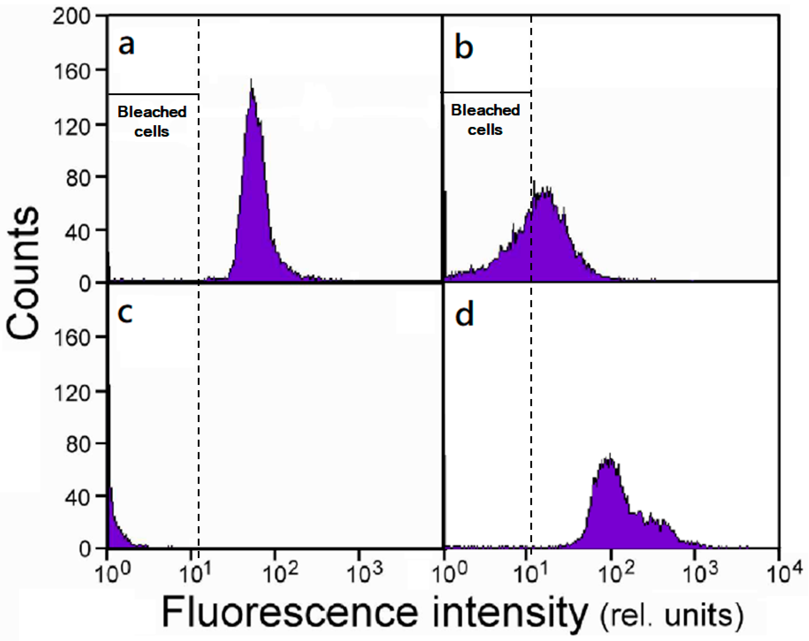
2.1. Population Responses.

S. vacuolatus cells that were subjected to a heat treatment were immediately cultured under continuous illumination, and the flow cytometry analyzed for chlorophyll a fluorescence was used to continuously monitor the changes in the chlorophyll content of the cell population. Here, we took the fluorescence intensity of 20 (relative units) as the dividing line to distinguish between the green and bleached cells. Figure 1 depicts the frequency distribution of the fluorescence intensity of control cells and the heat treated cells that had been cultured for various times. After light culture for 6 h, the shift of distribution toward the left indicated cells were losing chlorophylls (Figure 1b). Until 24 h, all of the heat treated cells were bleached (Figure 1c). However, starting from 24 h of cultivation, an increase in the high-fluorescent cell population becomes progressively significant (Figure 1d). Note that a fraction of these newly emerged cells could emit fluorescence at an intensity (~300 in relative unit) that was about five times higher than the control (~60). This might have something to do with the new cells population was not synchronous, and continued growth could generate cells that contain multiple daughter cells/chloroplasts.

2.2. Morphological Studies of Thylakoid Membranes.

The TEM study found that the S. vacuolatus cell has typical triplet parallel thylakoid membranes in chloroplast (Figure 2a). After light cultivating for 6 h, the light and dark segments showed up along the length of thylakoid membranes (Figure 2b,c, arrows). In bleached cell (light cultivated for 24 h), thylakoid membranes appeared to maintain intact, but with lower contrast when compared with the control image (Figure 2c). At a longer time of cultivation, the thylakoid membranes in the recovered cells showed a partial recovery, in contrast (Figure 2d).

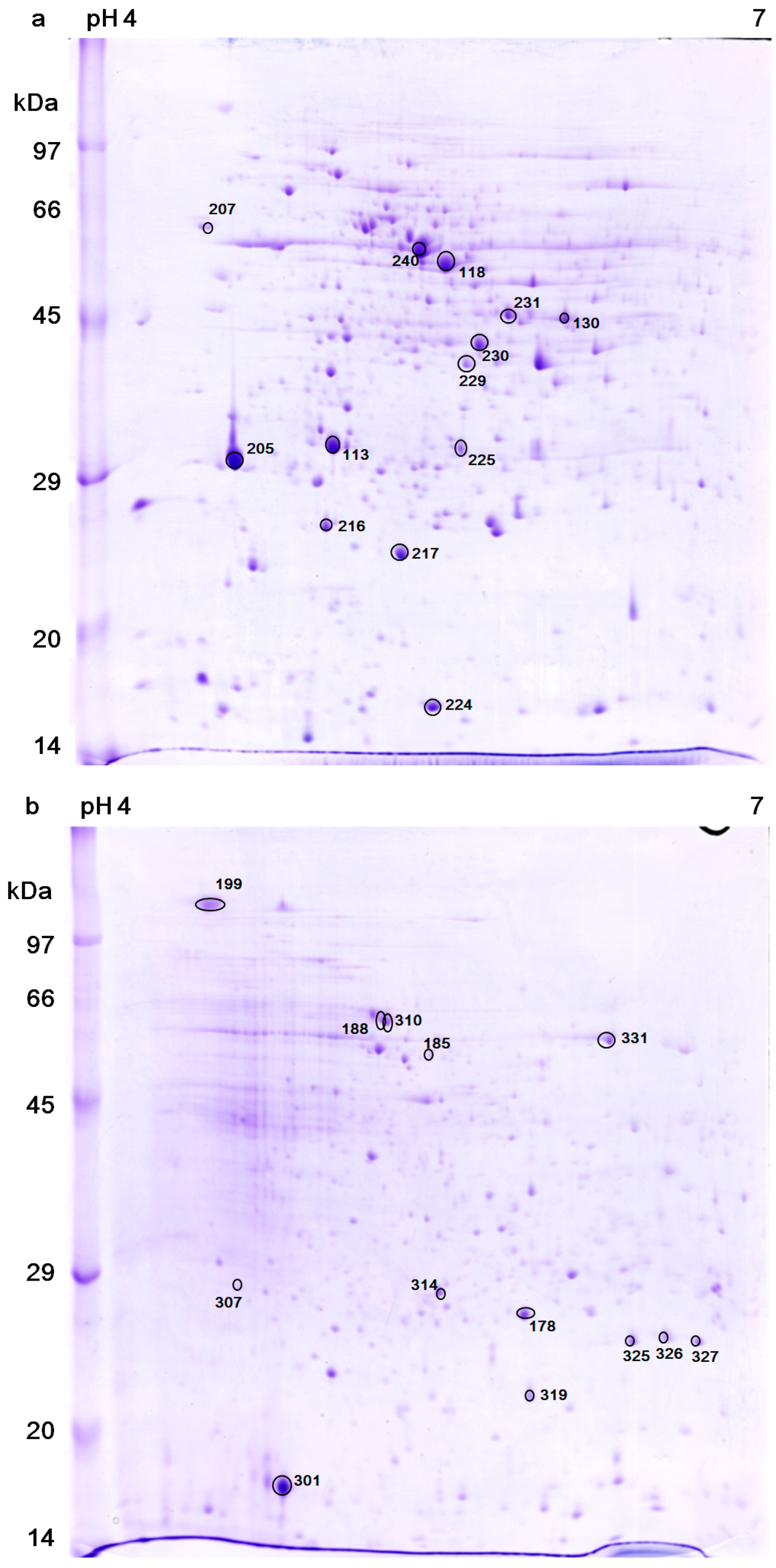
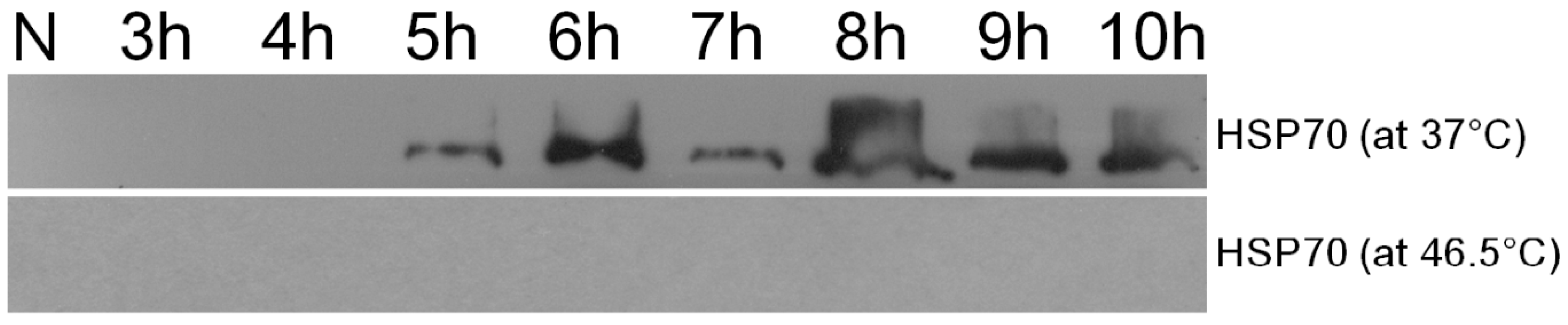
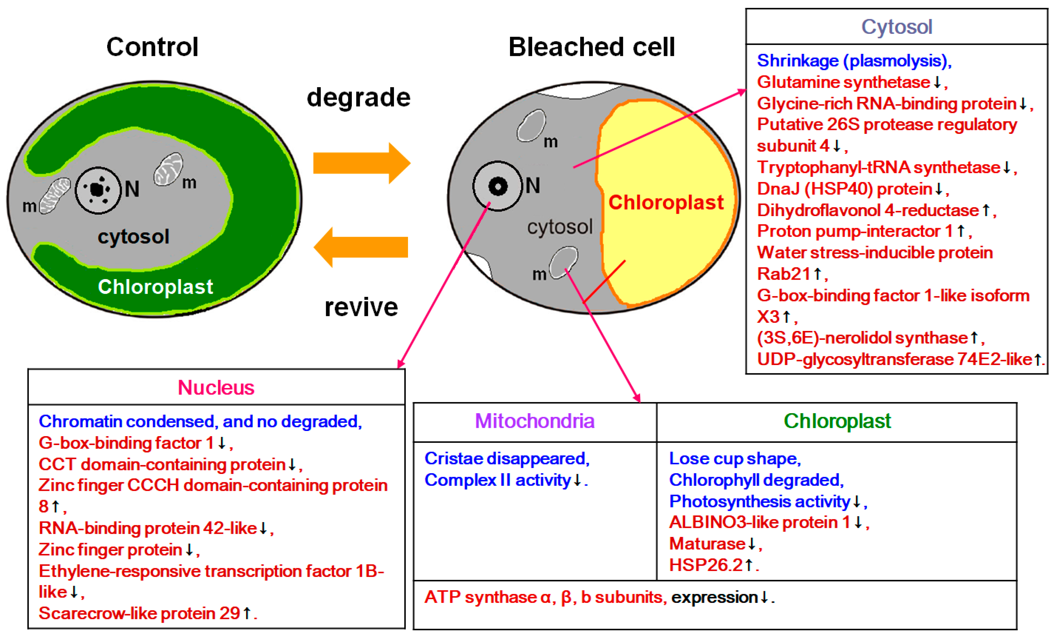
2.3. Proteins Expression Analysis.

Figure 3a,b showed the two dimensional gel electrophoresis (2DE) of the control cells and the bleached cells, respectively. The patterns of spots are quite different between the two samples; this might be a result of protein modification, new expression and/or down regulation. 150 highly expressed proteins were selected for protein identification, but only 26 of them were identified. The protein spots expression levels that were identified by using LC-MS/MS and MALDI-TOF MS are listed and annotated in Table 1 and Table 2, respectively. Protein analysis reveals that bleached cells down-regulated several important metabolism-related proteins, such as ATP synthase β, α, b subunits (protein No. 118, 240, 216, respectively), glutamine synthetase (No. 130), tryptophanyl- tRNA synthetase (No. 230), and dnaJ (heat shock protein 40 family) homolog protein (No. 229), which suggested that these cells actually entered a low activity state upon bleaching. On the other hand, the bleached cells mainly up-regulated the transcription factors or stress response proteins, including a most abundant protein “water stress-inducible protein Rab21” (No. 301), stress response proteins (No. 310, 326), light response transcription factors (e.g., No. 307, 314), and small HSP (e.g., No. 326), which indicated that these cells had entered a protective state against a harsh environment. In addition, western blot analysis found that HSP70 can be induced when the cells were cultured at a slightly higher temperature (37 °C instead of 32 °C) for 5 h. However, HSP70 could not be detected in the cells that had been heat treated at 46.5 °C for 1 h, followed by continuous light culture at 32 °C (Figure 4).

3. Discussion

The heat treatment completely inhibits the light reactions of photosynthesis and temporarily represses cell multiplication [14]. Previous reports suggest that the targets of thermal damage are at PSII with its oxygen-evolving complex, ATP generating system, and carbon fixation by Rubisco [7,15].
The subsequent light cultivation induced the complete chlorophyll degradation in all cells within 24 h (Figure 1a–c). The process was only found in light cultivation, but not in dark cultivation [16], which indicated that chlorophyll degradation proceeded through photooxidation [17]. However, flow cytometry analysis found some of newly emerged cells in 85 h culture that could emit chlorophyll fluorescence at an intensity that was much higher than the control (Figure 1d). This can be explained by the observation in one of our previous TEM study, which found that, among the cell population from 72 h culture, there were many cells that contained several chloroplasts in the regreening cells [14].
Chlorophylls can turn into harmful pigments if algae cells are under irradiation when photosyhthetic ability is completely inhibited. This is because chlorophyll can be photooxidized in the absence of electron transport. The electrons would be transferred to oxygen, which leads to the formation of ROS [18,19]. These ROS are primarily generated in chloroplasts and can cause oxidative damage. Therefore, thylakoid membranes and the proteins within them are the targets of damage that is caused by exceed light [20]. The loss of chlorophylls was accompanied by structural changes in chloroplasts. In this study, the thylakoid membranes of the bleached cells still maintained their integrity, lost contrast with the background, and showed black-white bands along their length (Figure 2b,c). Notably, these black-white bands only presented on thylakoid membrane, but not in lumen. Similar phenomena were observed when the plants are under stress [21,22,23]. After regreening, the appearance of the thylakoid membranes of heat-treated cells was largely restored to that of control cells (Figure 2d). It is worth mentioning that photooxidation can also regulate the gene expression of some pigments’ biosynthetic pathway [24]. In this study, we found dihydroflavonol 4-reductase (protein No. 178), which is a protein that regulates flavonoid metabolism were up-regulated in bleached cells.
The proteins profile was another significant change brought about by heat plus light stress (Figure 3a,b). In 2DE, many of the proteins decreased in amount or disappeared completely, and many new spots were synthesized in the bleached cells (Table 2), which indicated that these bleached cells were functionally active and had entered a different physiological state.
FOF1 ATP synthase is located in mitochondria inner membranes and chloroplast thylakoids. The F1 subunit α (No. 240), β (No. 118) and FO subunit b (No. 216) of ATP synthase were down-regulated in the bleached cells (Table 1 and Table 2). This is in line with our previous finding that mitochondria were only partially inhibited, but photosynthesis had been completely inhibited, so the source of ATP could only come from the mitochondria in bleached cells [14]. The presence of partially operating mitochondria implied that these bleached cells were still functioning to a certain extent, such as generating the energy that is necessary for maintaining integrity of cell membranes.
Tryptophanyl-tRNA synthetase (No. 230) can catalyze the aminoacylation of tRNA, which is an enzyme that is associated with protein synthesis [25]. The enzyme was not detected in the bleached cells (Table 2). It might be that the protein expression level became too low to be detected by Coomassie Brilliant Blue, or a protein modification changed its location in the 2DE gel. In addition, the expression of glutamine synthetase (No. 130) was also down-regulated. These results indicated that these bleached cells have down-regulated their bioactivity and protein expression. A similar result can be observed in another unicellular algae Chlamydomonas reinhardtii after heat stress [15].
Maximal photosynthetic activity (Fv/Fm) and chlorophyll contents were gradually recovered after light culture for 24 h [14]. It is likely that PSII and light-harvesting complex (LHC) were synthesized in bleached cells in this time period, which required newly synthesized chlorophylls to bind with the chlorophyll a–b binding proteins of LHC. ALBINO3-like protein 1 (No. 188) is necessitated for the insertion of LHC proteins into the thylakoid membrane and assembly of LHC I and II [26]. The increasing of this protein expression level might indicate that the assembly of LHCs is enhanced in the bleached cells.
HSPs can stabilize protein structure and their expression levels are changed under abiotic stresses. In this study, a member of HSP40 protein family, dnaJ protein P58IPK homolog protein (No. 229), was down-regulated, while chloroplast HSP26.2 (No. 326) was up-regulated in the bleached cells (Table 2). Moreover, HSP70 could not be detected in the cells that had been heat treated at 46.5 °C for 1 h, followed by continuous light culture at 32 °C (Figure 4). Similar results were observed in unicellular algae, C. reinhardtii, in which many HSP60, HSP70, and HSP100 family proteins were down-regulated under irradiance stress [27]. The results indicated that the dnaJ protein (No. 229) and HSP70 might not function as protective proteins in the bleached cells. However, chloroplast HSP26 was inducible by heat stress and it has been found to interact with ATP synthase α, β subunits, chlorophyll a–b binding proteins of LHC, oxygen-evolving enhancer protein 1 of PSII, and PSI subunit VI protein [28,29]. As mentioned above, ATP could only come from mitochondria in the bleached cells. This information might imply that the ATP synthase in chloroplast maintains a “unwork state” in bleached cells. The overexpression of HSP26 in transgenic tobacco could alleviate photoinhibition under extreme temperatures [30], and protected PSII activity (Fv/Fm) during heat and oxidative stress in tall fescue [31]. In the bleached cells, chloroplast HSP26.2 (No. 326) might play a role in protecting chloroplast and cell regreening.
A reversible shrinkage of the cytoplasm was observed between 2 and 24 h of light cultivation in the heat treated cells [14]. This phenomenon is generally observed in osmotic stressed or water stressed cells. In this study, the most abundant protein in bleached cells is water stress-inducible protein Rab21 (Responsive to abscisic acid 21; No. 301; Table 2). The protein was only located in cytosol and can be induced by water stress [32]. Many reports showed that abscisic acid (ABA) could improve the tolerance of unicellular algae to dehydration, osmotic stress, and oxidative stress [33,34,35,36,37], and also increase biomass production in Chlamydomonas and Scenedesmus cells [38,39]. In another thread, proton pump-interactor 1(PPI1), which is the second abundant protein in bleached cells, was also up-regulated (No. 199; Table 1). In Solanum tuberosum L., drought stress slightly increases the StPPI1 mRNA expression level [40]. PPI1 can stimulate H+ATPase activity and generate a membrane potential that plays a role in the adaptation of plants to stress conditions [40,41]. The two proteins Rab21 and PPI1 may not be directly correlated with each other. However, their common interacting moleculars, ABA have two different mechanisms that regulate H+ATPase activity in guard cell and root cell under salt stress [42,43,44]. It is still unknown how ABA regulates H+ATPase activity in unicellular cells. We suggest that these proteins’ up-regulation might be associated with the shrinkage of the cytoplasm we observed, indicating that osmotic stress or water stress was present in bleached cells [14]. They all could increase stress tolerance in the bleached cells.
Our findings can help to explain how algae survive and reproduce in the transition of environmental stress. The bleached cells in this study were found to down-regulate many biochemical activities and, in the meantime, activated some stress response proteins to survive the damage that is incurred by stress treatment. These changes in proteins’ expression pattern might elucidate many phenomena observed in the previous study [14].

4. Materials and Methods

4.1. Cell Culture and Heat Treatment.

Unicellular S. vacuolatus (SAG 211-8b cells, obtained from the Algal Collection Center, University of Göttingen, Germany) were photoautotrophically cultured. Axenic cultures were conducted, as described in Lee and Hsu [14]. In briefly, the algae cells were grown at 32 °C with slow bubbling at 3.5% CO2 in air. Illumination was provided by daylight fluorescence tubes at an intensity of 150 μmol photon m−2s−1. Synchronous cultures were obtained by the programmed 14 h light/10 h dark regime. The culture started with a cell density of 2 × 10⁶ cells mL−1 and dilution (~15-16×) was made after each 24 h cycle.
Heat treatment was applied by incubating the synchronous culture at the stage of small vegetative cell (5 μm in diameter) at 46.5 °C for 1 h in the dark. The heat-treated cells were cultured immediately thereafter at 32 °C with slow bubbling at 3.5% CO2 in air, and continuously illuminated at 100 μmol photon m−2s−1 [14].

4.2. Flow Cytometry Analysis.

Flow cytometry analysis was carried out with a FACSCaliburTM flow cytometer (Becton Dickinson, California), which was equipped with a 15 mW argon-ion laser. For chlorophyll content measurement, cell samples (2 mL) were collected at different time points of cultivation and the fluorescence of intrinsic chlorophyll a was detected with an excitation at 488 nm and emission longer than 650 nm. Each measurement was analyzed for 104 particles.

4.3. Transmission Electron Microscopy (TEM).

The same procedure, as delineated in Lee and Hsu [14], was adopted. S. vacuolatus cells were collected by centrifugation at 1800× g for 5 min. at 4 °C and then fixed for 4 h in 2% glutaraldehyde in 66 mM potassium phosphate buffer (pH = 7.1) at 4 °C. The samples were washed three times at 15 min. intervals in 66 mM potassium phosphate buffer (pH = 7.1) at 4 °C and then post-fixed in 2% aqueous OsO4 in 66 mM potassium phosphate buffer (pH = 7.1) for 4 h at 4 °C. This was followed by three washes at 15 min. intervals with the same buffer. The tissues were dehydrated in a grade acetone series, infiltrated through acetone: Spurr resin mixtures, then embedded in pure Spurr resin, and polymerized at 70 °C for 15 h. Ultrathin gold sections were cut on a Reichert-Jung ultramicrotome, which was collected on formvar-coated grids, stained with saturated solution of uranyl acetate in 100% methanol, and post-stained with lead citrate. Observations were made while using a Hitach-7500 transmission electron microscope (Hitachi, Japan).

4.4. Two Dimensional Gel Electrophoresis (2DE).

Lee and Hsu has described the same procedure [14]. S. vacuolatus cells (2 × 10⁹, control cells and heat-treated cells that had been cultured for 24 h under light) were collected by centrifugation at 1800× g for 10 min. and pulverized in liquid nitrogen. The samples were then homogenized by MagNA Lyser (Roche, Mannheim, Germany) at 6500 rpm for 30 s in 500 μL lysis buffer (7 M urea, 2 M thiourea, 4% (w/v) CHAPS ([(3-cholamidopropyl) dimetylammonio]-1-propanesulphonate), 0.5% (w/v) NP-40 (Nonidet P-40) and 10 mM Tris-HCl (pH = 8.3)), followed by centrifugation at 13,000× g for 10 min. to remove the debris. The protein concentrations were determined using two-dimensional (2D) Quant Kit, according to the manufacturer’s protocol (GE healthcare, San Francisco, CA, USA). Before analysis, the samples were stored at −80°C. 2DE was as described by Zhang and Koay (2008) [45], except that the first-dimensional gel separation was carried out with 18 cm pH 4-7 IPG strips. 900 μg protein was loaded per gel. The gels were visualized while using Coomassie Brilliant Blue R250 staining.

4.5. Protein Expression Level Analysis.

2DE image analysis was performed while using PowerLook 1120 scanner (UMAX, CA) and ImageMasterTM 2-D Platinum version 5.0 (Amersham Bioscience, NJ).

4.6. In-gel Digestion.

Referred to the procedure of Shevchenko et al. [46], but skipped the steps of reduction and alkylation. An Ettan automated spot picker from 2DE was used to pick the interesting spots, which were then put into 0.5 mL microtubes (Axygen; extrapure). The gel pieces were destained with 50 μL of 10 mM ammonium bicarbonate/50% acetonitrile and incubated with vortexing for 15 min. After removing the supernatant, 100 μL of 100% acetonitrile was added for gel pieces drying. Subsequently, the liquid was eliminated and 25 μL of 25 mM ammonium bicarbonate was added for 5 min., followed by a wash with 25 μL of 100% acetonitrile for 15 min. After removing all remaining liquid, 100 μL neat acetonitrile was added with vortexing for 3 min., until the gel pieces become white and shrinking. The gel pieces then were dried in a SpeedVac, and reswelled in 20 ng/μL sequencing grade modified procine trypsin (Promega sequencing grade modified trypsin (V511A)) in 10 mM ammonium bicarbonate for 1 h. Subsequently, the gel pieces were covered with 10mM ammonium bicarbonate and incubated at 37 °C overnight. Peptides were extracted by 15 μL of 50% acetonitrile with 1% trifluoroacetic acid for 15 min. twice, and then stored at −20 °C prior to LC-MS/MS or MALDI-TOF MS analysis.

4.7. Liquid Chromatography–Tandem Mass Spectrometry (LC-MS/MS).

The procedure was performed based on the method that was described by Chan et al. [47]. The 5 μL tryptic digested protein samples were injected onto a reversed phase capillary column (PepMap 75 μm × 150 mm, LC Packings) while using a nanoflow high pressure liquid chromatography system (Ultimate, Dionex) that was connected on-line to an electrospray ionization Q-TOF I mass spectrometer (Waters). The flow rate was 300 nL min−1 and separation was performed by gradient elution from 5 to 50% solution B (80% (v/v) acetonitrile, 0.1% formic acid) for 60 min., followed by an isocratic step at 100% solution B for 10 min. Balance solution A was 0.1% formic acid. Data-dependent acquisition was used with a mass spectrometry scans set every second (m/z 350–1500), and MS/MS was performed on automatically selected peptide ions, also for 1 s (m/z 50–2000, continuum mode), while using the function switching in MassLynx version 4.0 software. Raw MS/MS data were smoothed (Savitzky Golay, two channels twice) and centroided (at 80%) and peaks lists were generated while using MassLynx software.

4.8. Matrix Assisted Laser Desorption Ionization–Time of Flight Mass Spectrometry (MALDI-TOF MS).

The tryptic digested protein samples were resuspended in 5 μL of 0.1% formic acid. 0.65 μL of the samples were spotted onto an anchorchip target plate (600 μM/384 well, Bruker Daltonics) and further covered with 0.5 μL of matrix solution (1 mg/mL α-cyano-4-hydro xycinammic acid (HCCA) in 50% acetonitrile and 0.1% trifluoroacetic acid). The peptide calibration standard (Bruker Daltonics. m/z 757.39916, 1046.54180, 1296.68480, 1347.73540, 1619.82230, 1758.93261, 2096.08620, 2465.19630, 3147.47100) was also spotted onto the target plate for peptide calibration. The peptide mass fingerprints were obtained while using MALDI-TOF MS (autoflex III, Bruker Daltonics).

4.9. Protein Identification.

The LC-MS/MS spectra were submitted to the MASCOT server (Matrix Science, v. 2.6.2) and compared against the protein database from NCBI (NCBInr 20180429). The taxonomy was set to Viridiplantae (green plants). The following parameters were used for database searches: peptide tolerance at ±1.2 Da and MS/MS tolerance at ±1.5 Da; peptide charge of 2+ or 3+; and, trypsin as enzyme allowing for up to two missed cleavages. The fixed modification is carbamidomethyl (C). Variable modification, like oxidation (M) and phosphorylation (ST), were also used for consideration through queries. MALDI-TOF MS analysis followed that outlined by Lai et al. [48]. The peaks in the mass range m/z 600–4000 were used to generate a peptide mass fingerprint that was matched to the theoretical trypsin digests of proteins from the NCBI database. The accepted protein identifications required that the Mascot scores be higher than the threshold value in MS/MS analysis or in MALDI-TOF MS analysis. Protein location and functional description were referred to UniProt Knowledgebase (https://www.uniprot.org/)

4.10. Western Blotting Analysis

The total proteins of 1 × l07 Scenedesmus cells were subjected to SDS-PAGE and the resolved proteins were transferred to nitrocellulose membranes. The transfer buffer included 192 mM glycine, 25 mM Tris-HCl buffer (pH = 8.3), and 1% methanol. The mernbranes were incubated for 2 h at room temperature in blocking buffer [5% non-fat milk in TTBS buffer (150 mM NaCl, 20 mM Tris-HCl (pH = 7.4), and 0.1% Tween 20)], and then immunoblotted with 50 ng/mL anti-hsp70/hsc70 primary monoclonal antibody (Stressgen) in TTBS buffer containing 3% non-fat milk at room temperature for 2 h. The membranes were washed with 150 mM NaCl, 20 mM Tris-HCl (pH = 7.4), 0.1% Tween 20, and probed with secondary antibodies (1:3000), coupled to horseradish peroxidase (HRP) at room temperature for 1 h. The ECL system was used for detection.

5. Conclusions

In this study, heat treatment followed by cultivation under continuous illumination resulted in bleaching in the S. vacuolatus cells and the loss of photosynthetic activity. However, these bleached cell down-regulated many biochemical related proteins, such as ATP synthase α, β subunits, and glutamine synthetase, while up-regulated some phototransduction transcription factors, stress response proteins, and protective proteins, like small HSPs (but not HSP40 and HSP70 family proteins). The molecular evidence indicates that bleached cells can regulate their biochemical activity and activate certain protective mechanisms to remedy the damage that is incurred by the heat stress and successfully resurrect (Figure 5).

Author Contributions

T.-C.L. and B.-D.H. designed the experiments. T.-C.L. and T.-H.K. performed the experiments measurement and protein database search. K.-L.L. analyzed western blot. B.-D.H. and T.-C.L. wrote the paper.

Conflicts of Interest

The authors declare that they have no conflict of interest.

Abbreviations

2DETwo-dimensional gel electrophoresis
ABAAbscisic aci
HSPHeat shock protein
LC-MS/MSLiquid chromatography–tandem mass spectrometry
LHCLight harvesting complex
MALDI-TOF MSMatrix assisted laser desorption ionization–time of flight mass spectrometry
ROSReactive oxygen species
PSIIPhotosystem II
TEMTransmission electron microscopy

References

  1. Gray, D.W.; Lewis, L.A.; Cardon, Z.G. Photosynthetic recovery following desiccation of desert green algae (Chlorophyta) and their aquatic relatives. Plant. Cell Environ. 2007, 30, 1240–1255. [Google Scholar] [CrossRef] [PubMed]
  2. Holzinger, A.; Herburger, K.; Blaas, K.; Lewis, L.A.; Karsten, U. The terrestrial green macroalga Prasiola calophylla (Trebouxiophyceae, Chlorophyta): ecophysiological performance under water-limiting conditions. Protoplasma 2017, 254, 1755–1767. [Google Scholar] [CrossRef] [PubMed]
  3. Gupta, S.; Agrawal, S.C. Survival of blue-green and green algae under stress conditions. Folia Microbiol. 2006, 51, 121–128. [Google Scholar] [CrossRef]
  4. Aoki, S.; Hase, E. De- and re-generation of chloroplasts in the cells of Chlorella protothecoides. I. Syntheses of nucleic acids and protein in relation to the process of regeneration of chloroplast. Plant. Cell Physiol. 1964, 5, 473–484. [Google Scholar]
  5. Torzillo, G.; Pushparaj, B.; Bocci, F.; Balloni, W.; Materassi, R.; Florenzano, G. Production of Spirulina biomass in closed photobioreactors. Biomass 1986, 11, 61–74. [Google Scholar] [CrossRef]
  6. Tredici, M.R.; Materassi, R. From open ponds to vertical alveolar panels: the Italian experience in the development of reactors for the mass cultivation of phototrophic microorganisms. J. Appl. Phycol. 1992, 4, 221–231. [Google Scholar] [CrossRef]
  7. Allakhverdiev, S.I.; Kreslavski, V.D.; Klimov, V.V.; Los, D.A.; Carpentier, R.; Mohanty, P. Heat stress: an overview of molecular responses in photosynthesis. Photosynth. Res. 2008, 98, 541–550. [Google Scholar] [CrossRef] [PubMed]
  8. Feller, U.; Crafts-Brandner, S.J.; Salvucci, M.E. Moderately high temperatures inhibit ribulose-1,5-bisphosphate carboxylase/oxygenase (Rubisco) activase-mediated activation of Rubisco. Plant. Physiol. 1998, 116, 539–546. [Google Scholar] [CrossRef] [PubMed]
  9. Temple, M.D.; Perrone, G.G.; Dawes, I.W. Complex cellular responses to reactive oxygen species. Trends Plant. Sci. 2005, 15, 319–326. [Google Scholar] [CrossRef]
  10. Suzuki, N.; Mittler, R. Reactive oxygen species and temperature stresses: A delicate balance between signaling and destruction. Physiol. Plant. 2006, 126, 45–51. [Google Scholar] [CrossRef]
  11. Møller, I.M.; Sweetlove, L.J. ROS signaling-specificity is required. Trends Plant. Sci. 2010, 15, 370–374. [Google Scholar] [CrossRef] [PubMed]
  12. Kotak, S.; Larkindale, J.; Lee, U.; von Koskull-Döring, P.; Vierling, E.; Scharf, K.D. Complexity of the heat stress response in plants. Curr. Opin. Plant. Biol. 2007, 10, 310–316. [Google Scholar] [CrossRef] [PubMed]
  13. Wang, W.; Vinocur, B.; Shoseyov, O.; Altman, A. Role of plant heat-shock proteins and molecular chaperones in the abiotic stress response. Trends Plant. Sci. 2004, 9, 244–252. [Google Scholar] [CrossRef] [PubMed]
  14. Lee, T.C.; Hsu, B.D. The reversible disintegration of heat-treated Scenedesmus vacuolatus under continuous light cultivation conditions. Protoplasma 2014, 251, 1201–1211. [Google Scholar] [CrossRef] [PubMed]
  15. Hemme, D.; Veyel, D.; Mühlhaus, T.; Sommer, F.; Jüppner, J.; Unger, A.-K.; Sandmann, M.; Fehrle, I.; Schönfelder, S.; Steup, M.; et al. Systems-wide analysis of acclimation responses to long-term heat stress and recovery in the photosynthetic model organism Chlamydomonas reinhardtii. Plant. Cell 2014, 26, 4270–4297. [Google Scholar] [CrossRef] [PubMed]
  16. Lee, T.C.; Hsu, B.D. Characterization of the decline and recovery of heat-treated Scenedesmus vacuolatus. Bot. Stud. 2013, 54, 3. [Google Scholar] [CrossRef] [PubMed]
  17. Matile, P.; Hörtensteiner, S.; Thomas, H. Chlorophyll degradation. Annu. Rev. Plant. Physiol. Plant. Mol. Biol. 1999, 50, 67–95. [Google Scholar] [CrossRef] [PubMed]
  18. Pospíšil, P. Production of reactive oxygen species by photosystem II as a response to light and temperature stress. Front. Plant. Sci. 2016, 7, 1950. [Google Scholar] [CrossRef] [PubMed]
  19. Takagi, D.; Takumi, S.; Hashiguchi, M.; Sejima, T.; Miyake, C. Superoxide and singlet oxygen produced within the thylakoid membranes both cause photosystem I photoinhibition. Plant. Physiol. 2016, 171, 1626–1634. [Google Scholar] [CrossRef]
  20. Vincent, D.; Zivy, M. Plant proteome responses to abiotic stress. In Plant Proteomics; Samaj, J., Thelen, J.J., Eds.; Springer-Verlag: Berlin/Heidelberg, Germany, 2007; pp. 346–364. [Google Scholar]
  21. Taylor, A.O.; Craig, A.S. Plants under climatic stress. II. Low temperature, high light effects on chloroplast ultrastructure. Plant. Physiol. 1971, 47, 719–725. [Google Scholar] [CrossRef]
  22. Munné-Bosch, S.; Jubany-Marí, T.; Alegre, L. Drought-induced senescence is characterized by loss of antioxidant defences in chloroplasts. Plant. Cell Environ. 2001, 24, 1319–1327. [Google Scholar] [CrossRef]
  23. Yamane, K.; Rahman, M.S.; Kawasaki, M.; Taniguchi, M.; Miyake, H. Pretreatment with antioxidants decreases the effects of salt stress on chloroplast ultrastructure in rice leaf segments (Oryza sativa L.). Plant. Prod. Sci. 2004, 7, 292–300. [Google Scholar] [CrossRef]
  24. Park, J.H.; Tran, L.H.; Jung, S. Perturbations in the photosynthetic pigment status result in photooxidation-induced crosstalk between carotenoid and porphyrin biosynthetic pathways. Front. Plant. Sci. 2017, 8, 1992. [Google Scholar] [CrossRef]
  25. Ibba, M.; Soll, D. Aminoacyl-tRNA synthesis. Annu. Rev. Biochem. 2000, 69, 617–650. [Google Scholar] [CrossRef]
  26. Bellafiore, S.; Ferris, P.; Naver, H.; Goehre, V.; Rochaix, J.D. Loss of Albino3 leads to the specific depletion of the light-harvesting system. Plant. Cell 2002, 14, 2303–2314. [Google Scholar] [CrossRef]
  27. Mahong, B.; Roytrakul, S.; Phaonaklop, N.; Wongratana, J.; Yokthongwattana, K. Proteomic analysis of a model unicellular green alga, Chlamydomonas reinhardtii, during short-term exposure to irradiance stress reveals significant down regulation of several heat-shock proteins. Planta 2012, 235, 499–511. [Google Scholar] [CrossRef]
  28. Chauhan, H.; Khurana, N.; Nijhavan, A.; Khurana, J.P.; Khurana, P. The wheat chloroplastic small heat shock protein (sHSP26) is involved in seed maturation and germination and imparts tolerance to heat stress. Plant. Cell Environ. 2012, 35, 1912–1931. [Google Scholar] [CrossRef]
  29. Hu, X.; Yang, Y.; Gong, F.; Zhang, D.; Zhang, L.; Wu, L.; Li, C.; Wang, W. Protein sHSP26 improves chloroplast performance under heat stress by interacting with specific chloroplast proteins in maize (Zea mays). J. Proteomics 2015, 115, 81–92. [Google Scholar] [CrossRef]
  30. Guo, S.J.; Zhou, H.Y.; Zhang, X.S.; Li, X.G.; Meng, Q.W. Overexpression of CaHSP26 in transgenic tobacco alleviates photoinhibition of PSII and PSI during chilling stress under low irradiance. J. Plant. Physiol. 2007, 164, 126–136. [Google Scholar] [CrossRef]
  31. Kim, K.H.; Alam, I.; Kim, Y.G.; Sharmin, S.A.; Lee, K.W.; Lee, S.H.; Lee, B.H. Overexpression of a chloroplast-localized small heat shock protein OsHSP26 confers enhanced tolerance against oxidative and heat stresses in tall fescue. Biotechnol. Lett. 2012, 34, 371–377. [Google Scholar] [CrossRef]
  32. Mundy, J.; Chua, N.H. Abscisic acid and water-stress induce the expression of a novel rice gene. EMBO J. 1988, 7, 2279–2286. [Google Scholar] [CrossRef] [PubMed]
  33. Kobayashi, M.; Hirai, N.; Kurimura, Y.; Ohigashi, H.; Tsuji, Y. Abscisic acid-dependent algal morphogenesis in the unicellular green alga Haematococcus pluvialis. Plant. Growth Regul. 1997, 22, 79–85. [Google Scholar] [CrossRef]
  34. Yoshida, K.; Igarashi, E.; Mukai, M.; Hirata, K.; Miyamoto, K. Induction of tolerance to oxidative stress in the green alga, Chlamydomonas reinhardtii, by abscisic acid. Plant. Cell Environ. 2003, 26, 451–457. [Google Scholar] [CrossRef]
  35. Yoshida, K.; Igarashi, E.; Wakatsuki, E.; Miyamoto, K.; Hirata, K. Mitigation of osmotic and salt stresses by abscisic acid through reduction of stress-derived oxidative damage in Chlamydomonas reinhardtii. Plant. Sci. 2004, 167, 1335–1341. [Google Scholar] [CrossRef]
  36. Lu, Y.; Xu, J. Phytohormones in microalgae: a new opportunity for microalgal biotechnology? Trends Plant. Sci. 2015, 20, 273–282. [Google Scholar] [CrossRef]
  37. Holzinger, A.; Pichrtová, M. Abiotic stress tolerance of charophyte green algae: new challenges for omics techniques. Front. Plant. Sci. 2016, 7, 678. [Google Scholar] [CrossRef]
  38. Park, W.K.; Yoo, G.; Moon, M.; Kim, C.W.; Choi, Y.E.; Yang, J.W. Phytohormone supplementation significantly increases growth of Chlamydomonas reinhardtii cultivated for biodiesel production. Appl. Biochem. Biotechnol. 2013, 171, 1128–1142. [Google Scholar] [CrossRef]
  39. Tatiana, A.K.; Bruce, P.H.; Priti, K.; Levin, B.D. Effect of phytohormones on growth and accumulation of pigments and fatty acids in the microalgae Scenedesmus quadricauda. Algal Res. 2017, 27, 325–334. [Google Scholar]
  40. García, M.N.M.; País, S.M.; Téllez-Iñón, M.T.; Capiati, D.A. Characterization of StPPI1, a proton pump interactor from Solanum tuberosum L. that is up-regulated during tuber development and by abiotic stress. Planta 2011, 233, 661–674. [Google Scholar] [CrossRef]
  41. Morandini, P.; Valera, M.; Albumi, C.; Bonza, M.; Giacometti, S.; Ravera, G.; Murgia, I.; Soave, C.; DeMichelis, M. A novel interaction partner for C-terminus of Arabidopsis thaliana plasma membrane H+-ATPase (AHA1 isoform): site and mechanism of action on H+-ATPase activity differ from those of 14-3-3 proteins. Plant. J. 2002, 31, 487–497. [Google Scholar] [CrossRef]
  42. Falhof, J.; Pedersen, J.T.; Fuglsang, A.T. Plasma membrane H+-ATPase regulation in the center of plant physiology. Mol. Plant. 2016, 9, 323–337. [Google Scholar] [CrossRef]
  43. Hayashi, M.; Inoue, S.; Takahashi, K.; Kinoshita, T. Immunohistochemical detection of blue light-induced phosphorylation of the plasma membrane H+-ATPase in stomatal guard cells. Plant. Cell Physiol. 2011, 52, 1238–1248. [Google Scholar] [CrossRef]
  44. Janicka-Russak, M.; Kłobus, G. Modification of plasma membrane and vacuolar H+-ATPase in response to NaCl and ABA. J. Plant. Physiol. 2007, 164, 295–302. [Google Scholar] [CrossRef]
  45. Zhang, D.; Koay, E.S.C. Analysis of laser capture microdissected cells by 2-dimensional gel electrophoresis. In Clinical Proteomics, Methods and Protocols; Vlahou, A., Ed.; Humana Press: Totowa, NJ, USA, 2008; pp. 77–91. [Google Scholar]
  46. Shevchenko, A.; Tomas, H.; Havliš, J.; Olsen, J.V.; Mann, M. In-gel digestion for mass spectrometric characterization of proteins and proteomes. Nat. Protoc. 2006, 1, 2856–2860. [Google Scholar] [CrossRef]
  47. Chan, H.L.; Chou, H.C.; Duran, M.C.; Gruenewald, J.; Waterfield, M.D.; Ridley, A.; Timms, J.F. Major role of EGFR and SRC kinases in promoting oxidative stress-dependent loss of adhesion and apoptosis in epithelial cells. J. Biol. Chem. 2010, 285, 4307–4318. [Google Scholar] [CrossRef]
  48. Lai, T.C.; Chou, H.C.; Chen, Y.W.; Lee, T.R.; Chan, H.T.; Shen, H.H.; Lee, W.T.; Lin, S.T.; Lu, Y.C.; Wu, C.L.; et al. Secretomic and proteomic analysis of potential breast cancer markers by two-dimensional differential gel electrophoresis. J. Proteome. Res. 2010, 9, 1302–1322. [Google Scholar] [CrossRef]
Figure 1. The frequency distribution histograms of Scenedesmus cells analyzed for chlorophyll a fluorescence. (a) Untreated synchronous small cells, and the cells that had been heat treated (46.5 °C, 1 h) and then cultured under illumination for (b) 6 h, (c) 24 h, and (d) 96 h. A fluorescence intensity lower than 20 are identified as bleached cells. Three independent measurements were conducted at each time point.
Ijms 20 03082 g001
Figure 2. Transmission electron micrographs of the thylakoids of (a) freshly harvested S. vacuolatus (control) cell and the cells that had been heat-treated and then cultured under illumination for (b) 6 h, (c) 24 h, and (d) 72 h. Bars = 100 nm. The light and dark segments are indicated by white and black arrows, respectively.
Ijms 20 03082 g002
Figure 3. Two dimensional gel electrophoresis (2DE) analysis of the total protein extracts. (a) Freshly harvested (control) cells and (b) the bleached cells that had been heat-treated (46.5 °C for 1 h) and then cultured under illumination for 24 h. Identified protein spots were marked with numbers.
Ijms 20 03082 g003
Figure 4. Western blotting analysis of the HSP70 in heat-treated Scenedesmus cells. Cells (1 × 10⁷) were either subjected to 37 °C treatment for various times (upper panel) or 46.5 °C for one hour then cultivated at 32 °C for various time (lower panel). N, normal cell.
Ijms 20 03082 g004
Figure 5. A summary of the results that combine the morphological and physiological studies from a previous report [14], and the proteins expression analysis in this study. The changes observed in the previous study are marked in blue, whereas those in the present study in red. m: mitochondria; N: nucleus.
Ijms 20 03082 g005
Table 1. List of identified proteins from S. vacuolatus using liquid chromatography–tandem mass spectrometry (LC-MS/MS).
No.Homologous Protein (LC/MS/MS) Obs/Theor M. W. (kD)Obs/Theor PIAccession No.Mascot Score Identified Peptide Fragment LocationFunctional DescriptionExpression Fold Change (Bleached/Control)
118ATP synthase β subunit [Spathelia wrightii]55/50.1185.52/5.14CBY80088.167K.IGLFGGAGTGK.TChloroplast and mitochondriaProduces ATP from ADP.
The catalytic sites are primarily by the subunits.
0.02
130Glutamine synthetase [Chlorella sorokiniana]42/41.6716.06/6.17PRW34037.179K.HETCDINTFR.FCytosolCatalyzes the ATP-dependent biosynthesis of glutamine from glutamate and ammonia. 0.10
178Dihydroflavonol 4-reductase [Vitis davidii]28/38.1395.86/5.81AHK10250.166R.ATVRDPTNVK.KExtrinsic component of endoplasmic reticulum This protein is involved in flavonoid metabolism.1.13
199Proton pump-interactor 1 [Zea mays]170/170.1784.48/4.59ONM22629.160K.AENMVEVKSAAR.EEndoplasmic reticulum and plasma membraneRegulation of proton transport.
Enhances H+ATPase activity.
4.82
205Maturase K (chloroplast) [Trochodendron aralioides]28/28.0354.57/9.98AAB58649.177R.SQMIENAFLIDSTSKKFDTIVPISPLIGSLAK.AChloroplastProbably assists in splicing its own and other chloroplast group II introns. mRNA and tRNA processing. 0.04
224Glycine-rich RNA-binding protein [Dorcoceras hygrometricum]16/16.9235.46/5.54KZV53374.166R.GGGGGGYGGDR.GShuttling between nucleus and cytoplasmPlays a role in RNA transcription.0.05
240ATP synthase α subunit [Eriachne mucronata]56/55.7045.42/5.94YP_009410967.169K.TAIATNTILNQK.SChloroplast and mitochondria Produces ATP from ADP.
The α subunit is a regulator of ATP synthase.
0.01
Table 2. List of identified proteins from S. vacuolatus using matrix assisted laser desorption ionization–time of flight mass spectrometry (MALDI-TOF MS).
No. Homologous Protein (MALDI-TOF) Obs/Theor M. W. (kD) Obs/Theor PI Accession No. Mascot Score No. Match Peptides (Seq. Coverage)LocationFunctional DescriptionExpression Fold Change (Bleached/Control)
113RNA-binding protein 42-like [Erythranthe guttata]32/27.2335.0/9.73XP_012840025.18329(49%)NucleusEssential polysome-associated RNA-binding protein.0.03
185 Zinc finger protein [Macleaya cordata]55/55.2935.5/5.58OUZ99355.113022(15%)NucleusRNA methyltransferase activity.0.18
188ALBINO3-like protein 1, chloroplastic isoform X1 [Cucumis melo]58/56.0395.28/7.71XP_008444397.111023(20%)ChloroplastMembrane insertase activity0.65
207Putative 26S protease regulatory subunit 4 [Zostera marina]66/68.6784.47/9.12KMZ68817.19154(50%)CytoplasmPolyubiquitin modification- dependent protein binding.Only detected in control cell
216ATP synthase FO subunit b [Chlorokybus atmophyticus]25/21.0215.0/9.72YP_001315093.110520(62%)Chloroplast and mitochondriaConnect FO, F1 of ATP synthesis.Only detected in control cell
217Ethylene-responsive transcription factor 1B-like [Cajanus cajan]24/28.2945.49/5.53XP_020237863.110217(33%)NucleusDNA-binding transcription factor activity. Only detected in control cell
225G-box-binding factor 1 [Arabidopsis lyrata subsp. lyrata]33/33.7165.6/6.03XP_002866995.18624(22%)NucleusBinds to the G-box motif (5’-CCACGTGG-3’) of the rbcS-1A gene promoter, and 5’-CACGTG-3’ of LHCB2.4 (At3g27690) promoter.Only detected in control cell
229DnaJ protein P58IPK homolog [Cucurbita pepo subsp. pepo]40/53.8955.65/6.85XP_023517230.18527(34%)Endoplasmic reticulumChaperone (Hsp40 family).
May play a role in protein folding in the endoplasmic reticulum.
Only detected in control cell
230Tryptophanyl-tRNA synthetase [Chlorella sorokiniana]39/34.2875.69/6.97PRW57418.18423(62%)CytoplasmAminoacyl-tRNA ligase activityOnly detected in control cell
231CCT domain-containing protein [Cephalotus follicularis]48/49.8435.77/6.17GAV71350.18329(24%)NucleusRegulation of gene expression.0.07
301Water stress-inducible protein Rab21 [Setaria italica]16/15.1654.7/10.11XP_004980462.114817(54%)CytoplasmResponsive to abscisic acid.Only detected in bleached cell
307Scarecrow-like protein 29 [Camelina sativa]28/28.3174.63/5.41XP_010489817.18220(27%)NucleusDNA-binding transcription factor activity. Red, far-red light phototransductionOnly detected in bleached cell
310(3S,6E)-nerolidol synthase 1 [Herrania umbratica]59/59.0635.3/6.44XP_021294536.119828(35%)CytosolStress response protein.Only detected in bleached cell
314 G-box-binding factor 1-like isoform X3 [Tarenaya hassleriana]29/33.6375.53/6.99XP_010548353.18524(43%)NucleusRegulates blue light-mediated photomorphogenic growth.Only detected in bleached cell
319 Unnamed protein product [Coffea canephora]23/27.2306.95/5.78CDP02738.19223(34%)N/AN/AOnly detected in bleached cell
325 UDP-glycosyltransferase 74E2-like [Hevea brasiliensis]26/25.7516.33/4.87 XP_021651079.18218(33%)Intracellular membrane-bounded organelleGlycosyltransferase, transferase activity, transferring hexosyl groupsOnly detected in bleached cell
326 Chloroplast low molecular weight heat shock protein HSP26.2 [Agrostis stolonifera var. palustris]26/26.1966.5/7.85AAN74534.111814(47%)ChloroplastStress response protein.Only detected in bleached cell
327 Uncharacterized protein LOC104232495 [Nicotiana sylvestris]27/28.8216.7/6.15XP_009784020.19514(24%)N/AN/AOnly detected in bleached cell
331 Glutathione S-transferase T2-like [Chenopodium quinoa]56/51.0876.3/8.01XP_021721044.112056(54%)N/ATransferase activity2.21

