Theoretical Analysis for Wireless Magnetothermal Deep Brain Stimulation Using Commercial Nanoparticles


Abstract

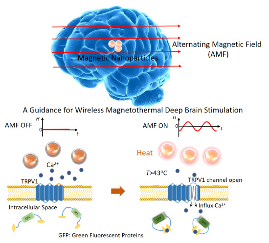
1. Introduction
2. Results and Discussion
2.1. Preliminary Determination of Minimum Limits for WMS without Blood Flow and Cerebrospinal Fluid (CSF)
2.2. Temperature Distribution inside the Brain before Stimulation
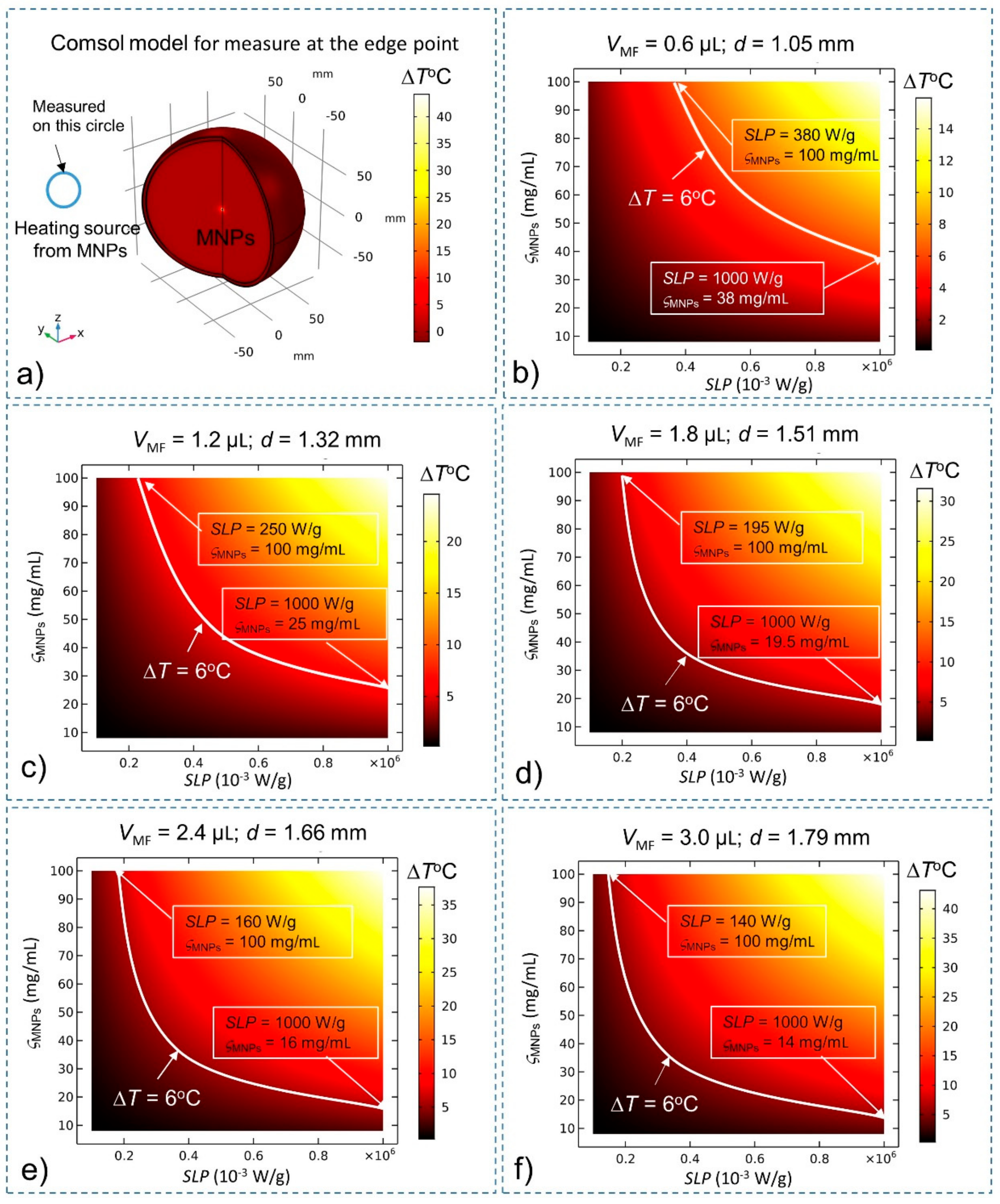
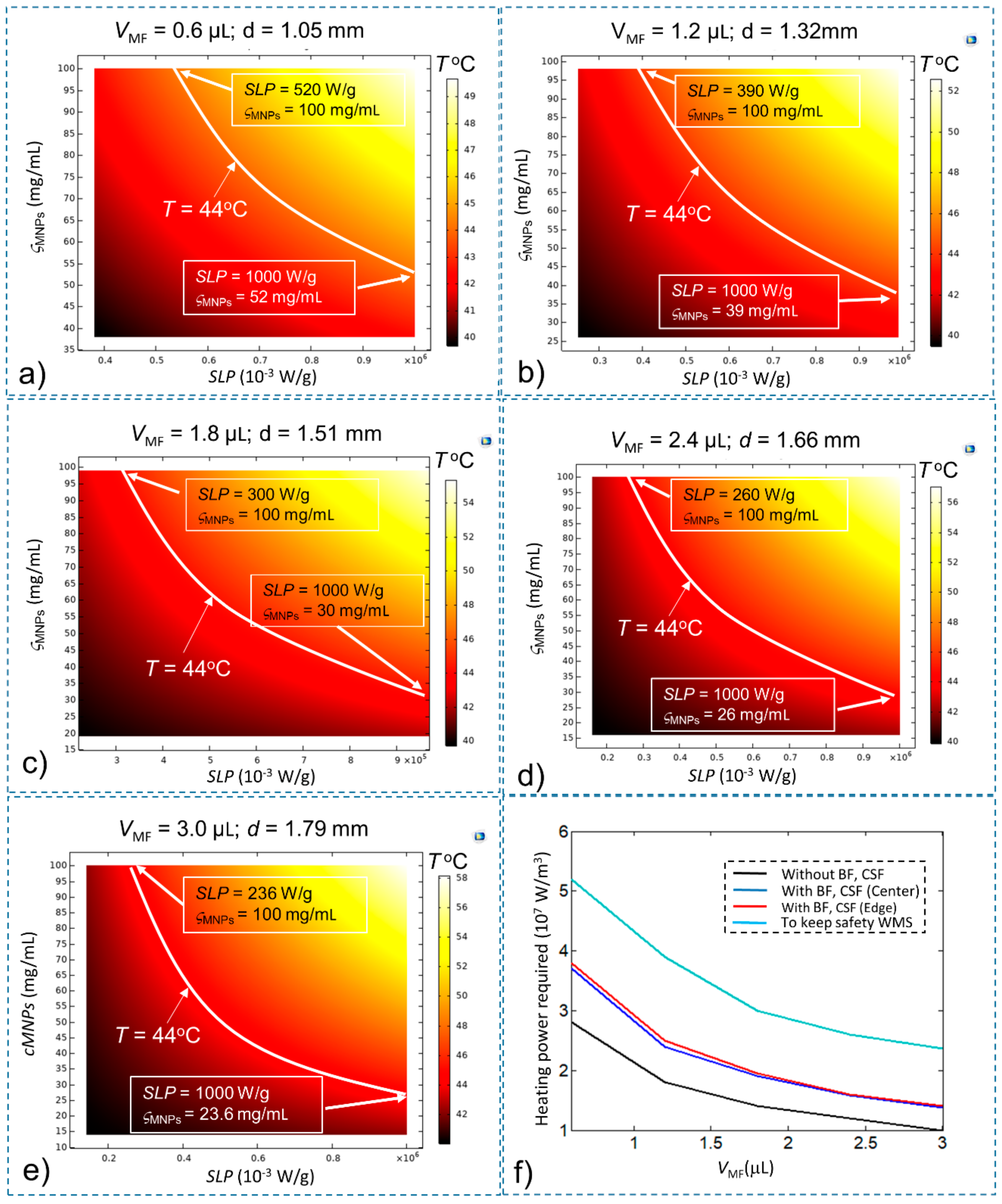
2.3. Determination of Minimum Limits for WMS in the Presences of Blood Flow and CSF
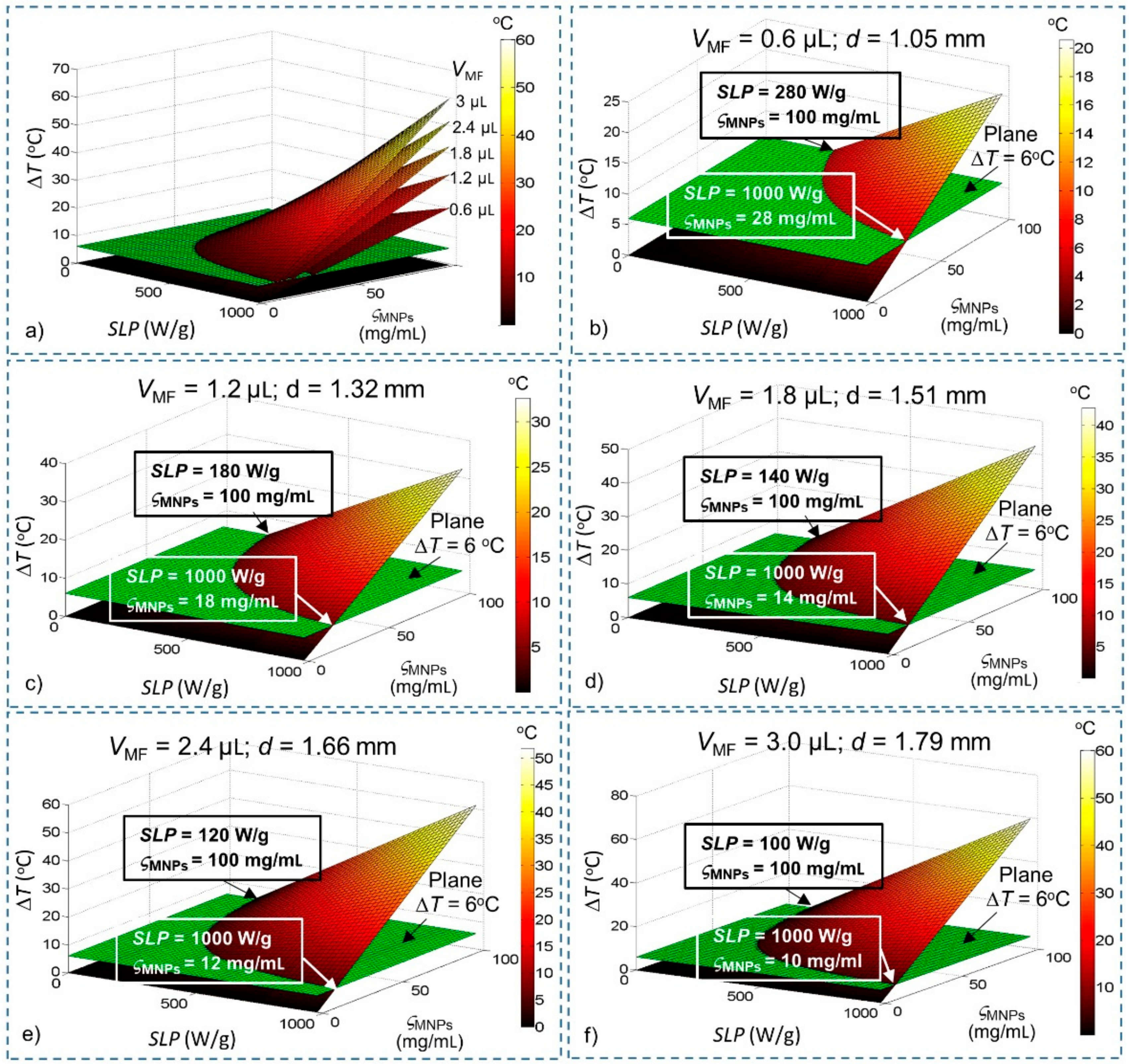
2.4. Prediction of a Feasbile Experiment Condition for WMS
3. Materials and Methods
3.1. Fourier’s Law for Steady-State Temperature Rise
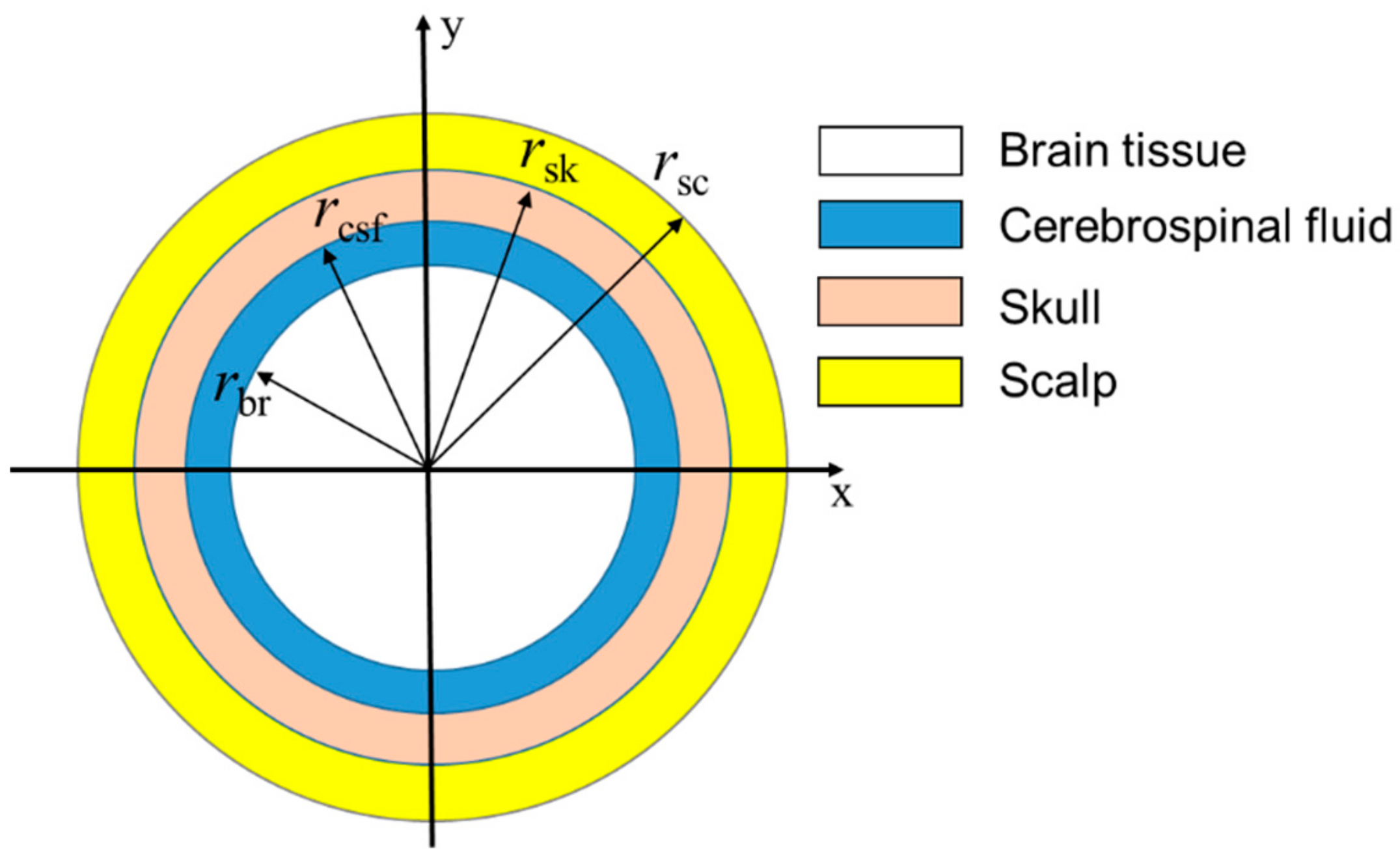
3.2. Bio-Heat Transfer Model for Heat Distribution
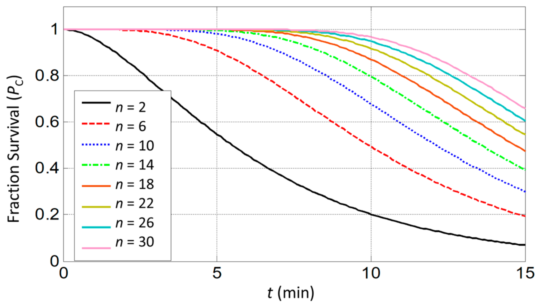
3.3. Cytotoxic Effects
4. Conclusions
Author Contributions
Funding
Acknowledgments
Conflicts of Interest
Abbreviations
| SLP | Specific loss power |
| SNPs | Super paramagnetic nanoparticles |
| MNPs | Magnetic nanoparticles |
| WMS | Wireless magnetothermal stimulation |
| CSF | Cerebrospinal fluid |
| BF | Blood flow |
| MF | Magnetic fluid |
| AMF | Alternating magnetic fields |
References
- Wyszkowska, J.; Jankowska, M.; Gas, P. Electromagnetic fields and neurodegenerative diseases. Przegląd Elektrotechniczny 2019, 95, 229–257. [Google Scholar] [CrossRef]
- Perlmutter, J.S.; Mink, J.W. Deep brain stimulation. Annu. Rev. Neurosci. 2006, 29, 229–257. [Google Scholar] [CrossRef]
- Banghart, M.; Borges, K.; Isacoff, E.; Trauner, D.; Kramer, R.H. Light-activated ion channels for remote control of neuronal firing. Nat. Neurosci. 2004, 7, 1381–1386. [Google Scholar] [CrossRef]
- Tufail, Y.; Matyushov, A.; Baldwin, N.; Tauchmann, M.L.; Georges, J.; Yoshihiro, A.; Tillery, S.I.H.; Tyler, W.J. Transcranial pulsed ultrasound stimulates intact brain circuits. Neuron 2010, 66, 681–694. [Google Scholar] [CrossRef]
- Walsh, V.; Cowey, A. Transcranial magnetic stimulation and cognitive neuroscience. Nat. Rev. Neurosci. 2000, 1, 73. [Google Scholar] [CrossRef]
- Boyden, E.S.; Zhang, F.; Bamberg, E.; Nagel, G.; Deisseroth, K. Millisecond-timescale, genetically targeted optical control of neural activity. Nat. Neurosci. 2005, 8, 1263. [Google Scholar] [CrossRef]
- Wu, W.; Wu, Z.; Yu, T.; Jiang, C.; Kim, W.-S. Recent progress on magnetic iron oxide nanoparticles: Synthesis, surface functional strategies and biomedical applications. Sci. Technol. Adv. Mater. 2015, 16, 23501. [Google Scholar] [CrossRef]
- Kurgan, E.; Gas, P. Magnetophoretic placement of ferromagnetic nanoparticles in rf hyperthermia. In Proceedings of the 2017 Progress in Applied Electrical Engineering (PAEE), Koscielisko, Poland, 25–30 June 2017; pp. 1–4. [Google Scholar]
- Périgo, E.A.; Hemery, G.; Sandre, O.; Ortega, D.; Garaio, E.; Plazaola, F.; Teran, F.J. Fundamentals and advances in magnetic hyperthermia. Appl. Phys. Rev. 2015, 2, 041302. [Google Scholar] [CrossRef]
- Wiley, D.T.; Webster, P.; Gale, A.; Davis, M.E. Transcytosis and brain uptake of transferrin-containing nanoparticles by tuning avidity to transferrin receptor. Proc. Natl. Acad. Sci. USA 2013, 110, 8662–8667. [Google Scholar] [CrossRef]
- Pankhurst, Q.A.; Connolly, J.; Jones, S.K.; Dobson, J. Applications of magnetic nanoparticles in biomedicine. J. Phys. D: Appl. Phys. 2003, 36, 167–181. [Google Scholar] [CrossRef]
- Maysinger, D.; Ji, J.; Hutter, E.; Cooper, E. Nanoparticle-based and bioengineered probes and sensors to detect physiological and pathological biomarkers in neural cells. Front. Neurosci. 2015, 9, 480. [Google Scholar] [CrossRef] [PubMed]
- Chen, R.; Romero, G.; Christiansen, M.G.; Mohr, A.; Anikeeva, P. Wireless magnetothermal deep brain stimulation. Science 2015, 347, 1477–1480. [Google Scholar] [CrossRef] [PubMed]
- Syrek, P.; Skowron, M.; Ciesla, A. Passive shielding of magnetic field in transcranial magnetic stimulation–outline of the problem. In Proceedings of the 2019 11th International Symposium on Advanced Topics in Electrical Engineering (ATEE), Bucharest, Romania, Romania, 28–30 March 2019; pp. 1–4. [Google Scholar]
- Caterina, M.J.; Schumacher, M.A.; Tominaga, M.; Rosen, T.A.; Levine, J.D.; Julius, D. The capsaicin receptor: A heat-activated ion channel in the pain pathway. Nature 1997, 389, 816. [Google Scholar] [CrossRef] [PubMed]
- Tominaga, M.; Caterina, M.J.; Malmberg, A.B.; Rosen, T.A.; Gilbert, H.; Skinner, K.; Raumann, B.E.; Basbaum, A.I.; Julius, D. The cloned capsaicin receptor integrates multiple pain-producing stimuli. Neuron 1998, 21, 531–543. [Google Scholar] [CrossRef]
- Huang, H.; Delikanli, S.; Zeng, H.; Ferkey, D.M.; Pralle, A. Remote control of ion channels and neurons through magnetic-field heating of nanoparticles. Nat. Nanotechnol. 2010, 5, 602. [Google Scholar] [CrossRef] [PubMed]
- Stanley, S.A.; Gagner, J.E.; Damanpour, S.; Yoshida, M.; Dordick, J.S.; Friedman, J.M. Radio-wave heating of iron oxide nanoparticles can regulate plasma glucose in mice. Science 2012, 336, 604–608. [Google Scholar] [CrossRef] [PubMed]
- Munshi, R.; Qadri, S.M.; Zhang, Q.; Rubio, I.C.; del Pino, P.; Pralle, A. Magnetothermal genetic deep brain stimulation of motor behaviors in awake, freely moving mice. eLife 2017, 6, e27069. [Google Scholar] [CrossRef] [PubMed]
- Sukstanskii, A.L.; Yablonskiy, D.A. Theoretical model of temperature regulation in the brain during changes in functional activity. Proc. Natl. Acad. Sci. USA 2006, 103, 12144–12149. [Google Scholar] [CrossRef]
- Nybo, L.; Møller, K.; Volianitis, S.; Nielsen, B.; Secher, N.H. Effects of hyperthermia on cerebral blood flow and metabolism during prolonged exercise in humans. J. Appl. Physiol. 2002, 93, 58–64. [Google Scholar] [CrossRef]
- Patterson, J.; Strang, R. The role of blood flow in hyperthermia. Int. J. Radiat. Oncol. Biol. Phys. 1979, 5, 235–241. [Google Scholar] [CrossRef]
- Hergt, R.; Hiergeist, R.; Hilger, I.; Kaiser, W.A.; Lapatnikov, Y.; Margel, S.; Richter, U. Maghemite nanoparticles with very high ac-losses for application in rf-magnetic hyperthermia. J. Magn. Magn. Mater. 2004, 270, 345–357. [Google Scholar] [CrossRef]
- Zhong, J.; Schilling, M.; Ludwig, F. Spatial and temperature resolutions of magnetic nanoparticle temperature imaging with a scanning magnetic particle spectrometer. Nanomaterials 2018, 8, 866. [Google Scholar] [CrossRef]
- Mason, E.E.; Cooley, C.Z.; Cauley, S.F.; Griswold, M.A.; Conolly, S.M.; Wald, L.L. Design analysis of an mpi human functional brain scanner. Int. J. Magn. Part. Imaging 2017, 3, 1703008. [Google Scholar]
- Le, T.-A.; Zhang, X.; Hoshiar, A.K.; Yoon, J. Real-time two-dimensional magnetic particle imaging for electromagnetic navigation in targeted drug delivery. Sensors 2017, 17, 2050. [Google Scholar] [CrossRef]
- Zhang, X.; Le, T.-A.; Hoshiar, A.K.; Yoon, J. A soft magnetic core can enhance navigation performance of magnetic nanoparticles in targeted drug delivery. IEEE/ASME Trans. Mechatron. 2018, 23, 1573–1584. [Google Scholar] [CrossRef]
- Hoshiar, A.; Le, T.-A.; Amin, F.; Kim, M.; Yoon, J. A novel magnetic actuation scheme to disaggregate nanoparticles and enhance passage across the blood–brain barrier. Nanomaterials 2017, 8, 3. [Google Scholar] [CrossRef]
- Amin, F.U.; Hoshiar, A.K.; Do, T.D.; Noh, Y.; Shah, S.A.; Khan, M.S.; Yoon, J.; Kim, M.O. Osmotin-loaded magnetic nanoparticles with electromagnetic guidance for the treatment of alzheimer’s disease. Nanoscale 2017, 9, 10619–10632. [Google Scholar] [CrossRef]
- Rabin, Y. Is intracellular hyperthermia superior to extracellular hyperthermia in the thermal sense? Int. J. Hyperth. 2002, 18, 194–202. [Google Scholar] [CrossRef]
- Jordan, A.; Scholz, R.; Wust, P.; Schirra, H.; Schiestel, T.; Schmidt, H.; Felix, R. Endocytosis of dextran and silan-coated magnetite nanoparticles and the effect of intracellular hyperthermia on human mammary carcinoma cells in vitro. J. Magn. Magn. Mater. 1999, 194, 185–196. [Google Scholar] [CrossRef]
- Ng, E.Y.K.; Kumar, S.D. Physical mechanism and modeling of heat generation and transfer in magnetic fluid hyperthermia through néelian and brownian relaxation: A review. Biomed. Eng. Online 2017, 16, 36. [Google Scholar]
- Kozissnik, B.; Bohorquez, A.C.; Dobson, J.; Rinaldi, C. Magnetic fluid hyperthermia: Advances, challenges, and opportunity. Int. J. Hyperth. 2013, 29, 706–714. [Google Scholar] [CrossRef] [PubMed]
- Noh, S.-H.; Moon, S.H.; Shin, T.-H.; Lim, Y.; Cheon, J. Recent advances of magneto-thermal capabilities of nanoparticles: From design principles to biomedical applications. Nano Today 2017, 13, 61–76. [Google Scholar] [CrossRef]
- Gas, P.; Miaskowski, A. Specifying the ferrofluid parameters important from the viewpoint of magnetic fluid hyperthermia. In Proceedings of the 2015 Selected Problems of Electrical Engineering and Electronics (WZEE), Kielce, Poland, 17–19 September 2015; pp. 1–6. [Google Scholar]
- Soetaert, F.; Kandala, S.K.; Bakuzis, A.; Ivkov, R. Experimental estimation and analysis of variance of the measured loss power of magnetic nanoparticles. Sci. Rep. 2017, 7, 6661. [Google Scholar] [CrossRef] [PubMed]
- Hervault, A.; Thanh, N.T.K. Magnetic nanoparticle-based therapeutic agents for thermo-chemotherapy treatment of cancer. Nanoscale 2014, 6, 11553–11573. [Google Scholar] [CrossRef] [PubMed]
- Schuerle, S.; Dudani, J.S.; Christiansen, M.G.; Anikeeva, P.; Bhatia, S.N. Magnetically actuated protease sensors for in vivo tumor profiling. Nano Lett. 2016, 16, 6303–6310. [Google Scholar] [CrossRef]
- Christiansen, M.G. Magnetothermal multiplexing for biomedical applications. Massachusetts Institute of Technology. Ph.D. Thesis, Massachusetts Institute of Technology, Cambridge, MA, USA, 2017. [Google Scholar]
- Gas, P.; Kurgan, E. Cooling effects inside water-cooled inductors for magnetic fluid hyperthermia. In Proceedings of the 2017 Progress in Applied Electrical Engineering (PAEE), Koscielisko, Poland, 25–30 June 2017; pp. 1–4. [Google Scholar]
- Okawa, K.; Sekine, M.; Maeda, M.; Tada, M.; Abe, M.; Matsushita, N.; Nishio, K.; Handa, H. Heating ability of magnetite nanobeads with various sizes for magnetic hyperthermia at 120 khz, a noninvasive frequency. J. Appl. Phys. 2006, 99, 08H102. [Google Scholar] [CrossRef]
- Dutz, S.; Hergt, R. Magnetic nanoparticle heating and heat transfer on a microscale: Basic principles, realities and physical limitations of hyperthermia for tumour therapy. Int. J. Hyperth. 2013, 29, 790–800. [Google Scholar] [CrossRef]
- Lepock, J.R. Cellular effects of hyperthermia: Relevance to the minimum dose for thermal damage. Int. J. Hyperth. 2003, 19, 252–266. [Google Scholar] [CrossRef]
- Diller, K.R. Fundamentals of bioheat transfer. Phys. Therm. Ther. Fundam. Clin. Appl. 2012, 3–22. [Google Scholar]
- Olsen, R.; Hayes, L.; Wissler, E.; Nikaidoh, H.; Eberhart, R. Influence of hypothermia and circulatory arrest on cerebral temperature distributions. J. Biomech. Eng. 1985, 107, 354–360. [Google Scholar] [CrossRef]
- Diao, C.; Zhu, L.; Wang, H. Cooling and rewarming for brain ischemia or injury: Theoretical analysis. Ann. Biomed. Eng. 2003, 31, 346–353. [Google Scholar] [CrossRef] [PubMed]
- Sukstanskii, A.; Yablonskiy, D. Theoretical limits on brain cooling by external head cooling devices. Eur. J. Appl. Physiol. 2007, 101, 41–49. [Google Scholar] [CrossRef] [PubMed]
- Leith, J.T.; Miller, R.C.; Gerner, E.W.; Boone, M.L. Hyperthermic potentiation. Biological aspects and applications to radiation therapy. Cancer 1977, 39, 766–779. [Google Scholar] [CrossRef]


| VMF (µL) | Diameter of Stimulation Region d (mm) | Minimum SLP (W/g) at Maximum ςMNPs (100 mg/mL) | Minimum ςMNPs (mg/mL) at Maximum SLP(1000 W/g) | Maximum ΔT with Maximum SLP and ςMNPs (°C) | Minimum Heating Power Required QMNPs (W/m3) |
|---|---|---|---|---|---|
| 0.6 | 1.05 | 280 | 28 | 20 | 2.8 × 10⁷ |
| 1.2 | 1.32 | 180 | 18 | 30 | 1.8 × 10⁷ |
| 1.8 | 1.51 | 140 | 14 | 40 | 1.4 × 10⁷ |
| 2.4 | 1.66 | 120 | 12 | 50 | 1.2 × 10⁷ |
| 3.0 | 1.79 | 100 | 10 | 60 | 1.0 × 10⁷ |
| VMF (µL) | Minimum SLP (W/g) at Maximum ςMNPs (100 mg/mL) | Minimum ςMNPs (mg/mL) at Maximum SLP (1000 W/g) | Maximum ΔT with Maximum SLP and ςMNPs ( °C) | Minimum Heating Power Required QMNPs (W/m3) | ||||
|---|---|---|---|---|---|---|---|---|
| Center | Edge | Center | Edge | Center | Edge | Center | Edge | |
| 0.6 | 370 | 380 | 37 | 38 | 16.31 | 15.94 | 3.7 × 10⁷ | 3.8 × 10⁷ |
| 1.2 | 240 | 250 | 24 | 25 | 25.18 | 24.58 | 2.4 × 10⁷ | 2.5 × 10⁷ |
| 1.8 | 190 | 195 | 19 | 19.5 | 32.36 | 31.57 | 1.9 × 10⁷ | 1.95 × 10⁷ |
| 2.4 | 158 | 160 | 15.8 | 16 | 38.60 | 37.65 | 1.58 × 10⁷ | 1.6 × 10⁷ |
| 3.0 | 138 | 140 | 13.8 | 14 | 44.23 | 43.13 | 1.38 × 10⁷ | 1.4 × 10⁷ |
| VMF (µL) | Minimum SLP (W/g) at Maximum ςMNPs (100 mg/mL) | Minimum ςMNPs (mg/mL) at Maximum SLP (1000 W/g) | Minimum Heating Power Required QMNPs (W/m3) |
|---|---|---|---|
| 0.6 | 520 | 52 | 5.2 × 10⁷ |
| 1.2 | 390 | 39 | 3.9 × 10⁷ |
| 1.8 | 300 | 30 | 3.0 × 10⁷ |
| 2.4 | 260 | 26 | 2.6 × 10⁷ |
| 3.0 | 236 | 23.6 | 2.36 × 10⁷ |
| Name | Minimal Value | Maximal Value |
|---|---|---|
| SLP (W/g) | 0 | 1000 |
| ςMNPs (mg/mL) | 0 | 100 |
| VMF (µL) | 0 | 3 |
| Parameters | Specific Heat C (J/(kg °C)) | Density ρ (kg/m3) | Thermal Conductivity k (W/(m °C)) | Blood Flow Rate ω (1/s) | Metabolic Rate Qm (W/m3) | Radius (Adult) r (mm) | Temperature Tb (°C) | Initial Temperature T0 (°C) |
|---|---|---|---|---|---|---|---|---|
| Blood (b) | 3800 | 1050 | 0.5 | – | – | – | 36.64 | 36.64 |
| Scalp (sc) | 4000 | 1000 | 0.34 | 0.00143 | 363 | 88 | 37.00 | 37.00 |
| Skull (sk) | 2300 | 1500 | 1.16 | 0.000143 | 70 | 87 | 37.00 | 37.00 |
| CSF | 3800 | 1007 | 0.61 | 0 | 0 | 84 | 37.00 | 37.00 |
| Brain tissue (br) | 3700 | 1050 | 0.51 | 0.008 | 10437 | 83 | 37.00 | 37.00 |
| Brain surroundings | – | – | – | – | – | – | 25.00 | 25.00 |
© 2019 by the authors. Licensee MDPI, Basel, Switzerland. This article is an open access article distributed under the terms and conditions of the Creative Commons Attribution (CC BY) license (http://creativecommons.org/licenses/by/4.0/).
Share and Cite
Le, T.-A.; Bui, M.P.; Yoon, J. Theoretical Analysis for Wireless Magnetothermal Deep Brain Stimulation Using Commercial Nanoparticles. Int. J. Mol. Sci. 2019, 20, 2873. https://doi.org/10.3390/ijms20122873
Le T-A, Bui MP, Yoon J. Theoretical Analysis for Wireless Magnetothermal Deep Brain Stimulation Using Commercial Nanoparticles. International Journal of Molecular Sciences. 2019; 20(12):2873. https://doi.org/10.3390/ijms20122873
Chicago/Turabian StyleLe, Tuan-Anh, Minh Phu Bui, and Jungwon Yoon. 2019. "Theoretical Analysis for Wireless Magnetothermal Deep Brain Stimulation Using Commercial Nanoparticles" International Journal of Molecular Sciences 20, no. 12: 2873. https://doi.org/10.3390/ijms20122873
APA StyleLe, T.-A., Bui, M. P., & Yoon, J. (2019). Theoretical Analysis for Wireless Magnetothermal Deep Brain Stimulation Using Commercial Nanoparticles. International Journal of Molecular Sciences, 20(12), 2873. https://doi.org/10.3390/ijms20122873