Review

Na/K-ATPase Signaling and Cardiac Pre/Postconditioning with Cardiotonic Steroids

by
Pauline V. Marck
and
Sandrine V. Pierre
*
Marshall Institute for Interdisciplinary Research, Marshall University, Huntington, West Virginia, WV 25701, USA
*
Author to whom correspondence should be addressed.
Int. J. Mol. Sci. 2018, 19(8), 2336; https://doi.org/10.3390/ijms19082336
Submission received: 5 July 2018 / Revised: 5 August 2018 / Accepted: 6 August 2018 / Published: 9 August 2018

Abstract

:
The first reports of cardiac Na/K-ATPase signaling, published 20 years ago, have opened several major fields of investigations into the cardioprotective action of low/subinotropic concentrations of cardiotonic steroids (CTS). This review focuses on the protective cardiac Na/K-ATPase-mediated signaling triggered by low concentrations of ouabain and other CTS, in the context of the enduring debate over the use of CTS in the ischemic heart. Indeed, as basic and clinical research continues to support effectiveness and feasibility of conditioning interventions against ischemia/reperfusion injury in acute myocardial infarction (AMI), the mechanistic information available to date suggests that unique features of CTS-based conditioning could be highly suitable, alone /or as a combinatory approach.

1. Introduction

Without a doubt, the prompt restoration of blood flow to reinstate the perfusion of the ischemic myocardium has substantially improved the outcomes for patients hospitalized with acute myocardial infarction (AMI) [1]. It is also clear that life-saving reperfusion therapy is double-edged, as it ineluctably brings about the structural and functional damage of reperfusion injury to the myocardium [2]. With an estimated potential to reduce the size of the infarct by up to 40%, the development of clinically effective strategies to reduce reperfusion injury in AMI is one of the most-anticipated advances in cardiovascular therapies for the current decade [3]. To date, there is no stronger protection against reperfusion injury than the one afforded by adjunct conditioning treatment at reperfusion. As basic and clinical research continues to support the effectiveness and feasibility of conditioning interventions, this review covers insights into the protective cardiac Na/K-ATPase-mediated signaling triggered by ouabain and other cardiotonic steroids (CTS), as well as its potential application in the context of the enduring debate over the use of CTS in the ischemic heart.

2. Cardiac Pre- and Post-Conditioning against Ischemia/Reperfusion Injury

In 1986, Murry et al. first described the ability to precondition (PC) the heart to protect against infarction following ischemia/reperfusion injury (I/R). In dogs, the seminal study showed that a sequence of few very brief ischemic episodes induced by coronary occlusion, interspersed among brief periods of reperfusion, limited the cardiac damage induced by a subsequent prolonged ischemic insult. Strikingly, the infarct size was substantially smaller in the dogs that were exposed to the short periods of ischemia, than in the controls. The authors coined the term ischemic preconditioning (IPC) to describe this phenomenon [4]. Given its invasive nature and the need for intervention prior to the ischemic event, IPC’s limited clinical application was recognized early on. However, pre-clinical and clinical research has since shown that IPC-like protection can be obtained through ischemic post-conditioning (IPost) (applied at the time of reperfusion) and/or remote ischemic conditioning (RIC) (applied non-invasively to a limb during or after myocardial ischemia) [5].

3. Pharmacologically-Induced Cardiac Protection

With the characterization of the cellular mechanisms involved in IPC, the concept of pharmacological PC took form [6]. Indeed, although PC was initially described as a response of the myocardium to ischemia, it soon became apparent that a similar phenotype could be elicited by other stimuli. Pharmacological strategies to activate IPC-like protective signal transduction pathways, while avoiding the vascular and myocardial injury that could result from coronary artery occlusion, were widely seen as less harmful, and thus more clinically suitable than the IPC-based strategies. A number of pharmacological agents, including agonists of G protein-coupled receptor (GPCR)s (adenosine A1 or A3, bradykinin B2, α1-adrenergic, muscarinic M2, angiotensin AT1, and endothelin, δ1-opioid), nitric oxide (NO) donors, phosphodiesterase inhibitors, and various noxious stimuli (such as endotoxin derivatives, various cytokines, and reactive oxygen species), have been found to elicit an IPC-like protection (reviewed in the literature [7]). Figure 1 summarizes the main signaling cascades that have been involved to date, including the eNOS/PKG, reperfusion injury salvage kinase (RISK) [8], and the survivor factor enhancement (SAFE) pathways, which ultimately result in the opening of the mitoK-ATP channel (mKATP) and inhibition of the mitochondrial permeability transition pore (mPT) [9].

4. Enduring Challenges and Current Goals for Clinical Application of Conditioning

Although a variety of ischemia- and drug-based conditioning strategies have been tested, the results of the clinical studies for improving the patient outcomes have been largely disappointing. Beyond the need to refine the design of experimental and clinical studies [11], a now well-recognized core issue faced by basic scientists and clinicians alike is the influence of confounding risk factors. Indeed, frequent comorbidities such as diabetes or hypercholesterolemia, as well as co-medications [12] differentially alter the key elements of the cardioprotective signaling pathways (Figure 1), and consequently, the efficacy of a given tested PC intervention. As an approach to overcome this limitation, combination therapies targeting multiple non-redundant pathways are increasingly being explored [5]. In this context, the unique properties of the CTS signaling described in the following section represent a potential safe approach to consider in the protection against cardiac I/R injury.

5. Pre/Postconditioning with Cardiotonic Steroid through Cardiac Na/K-ATPase Signaling

CTS (digitalis in particular) have been used to treat heart failure for hundreds of years [13,14], long before the late Nobel Laureate Jens C. Skou uncovered their molecular target as the Na/K-ATPase [15,16,17]. Na/K-ATPase is the membrane-spanning enzyme complex that uses the energy of ATP hydrolysis for the coupled active transport of Na+ and K+ across the plasmalemma of mammalian cells [18,19,20]. CTS induce moderate inotropy by inhibiting sarcolemmal Na/K-ATPase, which raises the intracellular Na+ and Ca2+ through the Na+/Ca2+-exchanger, and subsequently increases myocardial contractility [21,22]. Alteration of cardiac Na/K-ATPase has long been recognized as a key aspect of I/R pathophysiology [23,24], which contributes to cardiomyocyte demise through mechanisms that go far beyond a simple disruption of Na+ and Ca2+ homeostasis secondary to ATP depletion, and remains incompletely understood [25,26,27,28,29,30,31]. Clinically, interest in CTS for the management of acute myocardial infarction sparked early in modern cardiology, before molecular knowledge of Na/K-ATPase function in health and I/R became available [32]. Somewhat surprisingly, in the context of the molecular mechanism described above, experimental and clinical reports still suggested beneficial effects of an “appropriate and judicious” use of the CTS digitalis in patients with failing myocardium associated with acute myocardial infarction. There were also concerns over increased sensitivity to inotropic and toxic effects, and under the principle of “primum non nocere”, the prevalent message in the clinical arena has persisted as “there is no role for the prophylactic use of digitalis in the uncomplicated myocardial infarction” [33].
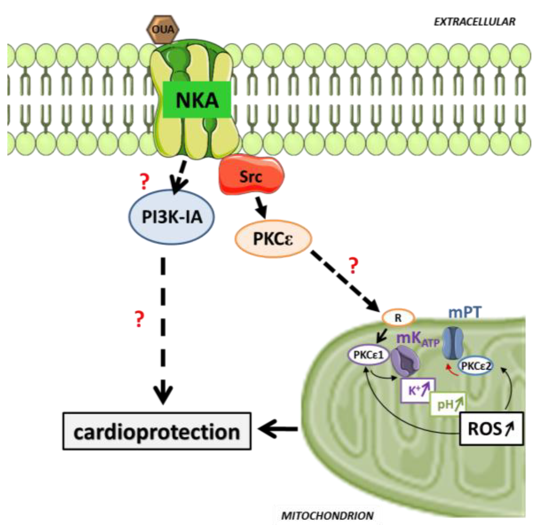
Unsurprisingly, given such inauspicious pharmacological and clinical circumstances, the CTS pre/postconditioning hypothesis was not formulated until the 20 year-old discovery of Na/K-ATPase signaling, recognized in this special issue, came about. Indeed, it is the discovery of elements of the molecular signature of the CTS-induced signaling through the cardiac Na/K-ATPase that revealed striking similarities with those of the ischemic and pharmacological PC, and prompted further investigation. Specifically, by early 2000, it had become clear that exposure to the CTS ouabain triggers Src, protein kinase C epsilon (PKCε), ERK, mKATP, and mitochondrial reactive oxygen species (ROS) production in the cardiac tissue [34,35,36]. Collectively, these represented a hallmark of the RISK pathway, which is common to most pharmacological preconditioners known at the time (Figure 1). Two studies specifically tested the hypothesis that exposure to the CTS ouabain could trigger preconditioning, and uncovered the first key characteristics in Langendorff-perfused rat heart preparations. In Pierre et al. [37], transient exposure to a subinotropic concentration of ouabain and wash-out prior to ischemia/reperfusion induced a structural and functional protection comparable to that observed with IPC in this model. By analogy to IPC, this phenomenon was termed Ouabain PreConditioning (OPC). Pharmacological inhibition further revealed that OPC requires both Src and PKCε activities, and that Src is required for PKCε activation. In Pasdois et al. [38], an alternate protocol consisting of a continuous exposure to increasing concentrations of ouabain also triggered OPC. Mechanistically, the study demonstrated the requirement for mKATP-opening and ROS production, and it also revealed that OPC is independent of protein kinase G (PKG) and guanylyl cyclase (GC) activation, contrary to bradykinin-induced PC. This independence from PKG/GC was of particular interest, not only because it was the first indication that a unique mechanism of protection could set CTS apart from other pharmacological triggers of PC, but also because it indicated that CTS-induced PC signaling and inotropy relied on distinct mechanisms. Hence, although mKATP-opening and ROS production are required for both OPC and positive inotropy, GC and PKG are required only for the latter. Collectively, these two studies also revealed that ouabain inotropy and OPC have distinct dose-dependence curves, and illustrated that although inotropy stops when ouabain is removed, OPC protection occurs even after ouabain is withdrawn, consistent with the general model of persistence of cardioprotective signaling cascades observed after washout of their triggers. Subsequently, D’Urso et al. successfully triggered CTS PC using the FDA-approved digoxin [39], indicating potentials in the clinical setting. Additionally, Morgan et al. [40] reported that CTS-induced PC protection could be achieved in the rabbit heart, a model that recapitulates human myocardial physiology, vulnerability to ischemic injury, and CTS pharmacology better than the rat or mouse heart [35,40,41,42,43,44]. The lower ouabain concentration (nM range) [40] correlated with the higher ouabain affinity of the Na/K-ATPase α1 isoform in the rabbit species compared to the rat, suggesting a key role of α1 in CTS PC signaling. As summarized in Figure 2, OPC signaling includes an intramitochondrial signaling pathway, common to most if not all forms of PC [45,46,47]. It involves at least two mitochondrial PKCε in the sequence that leads to a mKATP-opening, production of ROS, and inhibition of mPT. Remarkably, activation of the Na/K-ATPase cardioprotective signaling pathway by OPC protects the myocardial Na/K-ATPase enzyme function itself against I/R [29,48], a feature that was also noted in IPC [49]. Several aspects of OPC signaling are unique, and contrast with IPC and GPCR-based forms of pharmacological preconditioning (Figure 1). Firstly, as mentioned earlier, it is a cGMP-independent pathway, in contrast to numerous major forms of cardioprotection [50]. Secondly, it is mediated by PI3K-IA (rather than PI3K-IB), in parallel rather than upstream from the PKCε activation [51]. The reliance on PI3K-IA rather than IB in the pre-ischemic phase (as observed for IPC or adenosine) is a rare occurrence in known pharmacological PC, and suggests that OPC could trigger “insulin-like” protective effects related to the substrate utilization or cell survival. Studies have also shown that the PI3K-IA activation is highly protective at reperfusion [52]. Finally, and perhaps even more surprisingly (yet consistent with independence from the cGMP pathway) is the lack of requirement for Akt activation in OPC [51]. Potentially, these unique mechanistic features could make a CTS-based approach very suitable, alone or in combination, for PC in patients whose disease and/or treatment may have altered cGMP, PI3K-IB, and/or Akt pathways. Therefore, we recently tested a more clinically applicable CTS-based protocol by comparing very low doses of ouabain and digoxin’s protective effects when given as a bolus at reperfusion, following 40 min of zero flow ischemia in Langendorff-perfused mouse heart preparation. The results showed that Na/K-ATPase cardioprotective signaling activation, increased cell survival, and improved functional recovery as effective as those obtained with IPostC can be obtained using digoxin [53].

6. Prospect and Future Directions

The first reports of cardiac Na/K-ATPase signaling [54,55,56], 20 years ago, have opened several major fields of investigation into the cardioprotective action of CTS drugs given at low doses, particularly in hypertrophy [57,58], as well as PC, as reviewed here. The mechanistic information available on PC suggests distinct features of CTS-based PC that could make this modality clinically relevant, alone or as a combinatory approach. There are also a number of remaining gaps and questions in our current knowledge of the pathway and its connection to other modalities of PC. For instance, the role of the key players of cell metabolism/survival Bcl2/Bax, ERK1/2, and GSK3-β, which are fixtures of PC signaling (Figure 1) and have been shown to be modulated by CTS/Na/K-ATPase signaling [54,59,60,61], remain to be tested on CTS-based PC.
As studies continue to explore the mechanism and efficacy of CTS conditioning, in vivo investigations in pre-clinical rodent and non-rodent models of AMI will be critical. Indeed, important aspects of the complex pathophysiology of I/R injury, such as sterile inflammation and cardiac/vascular remodeling, cannot be adequately evaluated ex vivo. Fundamentally, and given the proposed role of the Na/K-ATPase non ion-pumping function in cardiac myocyte survival after I/R injury [29], those models could also redefine the role of Na/K-ATPase signaling and endogenous CTS in the pathophysiology of I/R injury. Indeed, a role for endogenous CTS as hormones with distinct but related modulatory effects on cardiovascular homeostasis has been suggested in AMI and other physiological and pathophysiological conditions, such as pregnancy, exercise, salt-loading, or heart failure) [62,63,64,65,66], and could include a PC-based effect. In the context of I/R injury, the CTS release from the rat myocardium has been observed ex vivo within a short (15 min) ischemia [67], suggesting that the ischemia-induced release of CTS may occur during IPC. Ouabain, and potentially other CTS, could therefore be added to the list of paracrine/autocrine factors that are released during preconditioning ischemia and trigger protection by binding to their respective receptors. Some of the most promising cardioprotective candidate drugs to date, opioid and cannabinoid receptor agonists [68,69] or adenosine receptor agonists [70], belong to this category.

Author Contributions

P.V.M. and S.V.P. wrote the paper.

Acknowledgments

This research was funded by NIH P01HL036573 and MIIR Funds.

Conflicts of Interest

The authors declare no conflict of interest.

References

  1. Krumholz, H.M.; Wang, Y.; Chen, J.; Drye, E.E.; Spertus, J.A.; Ross, J.S.; Curtis, J.P.; Nallamothu, B.K.; Lichtman, J.H.; Havranek, E.P.; et al. Reduction in acute myocardial infarction mortality in the United States: Risk-standardized mortality rates from 1995−2006. JAMA 2009, 302, 767–773. [Google Scholar] [CrossRef] [PubMed]
  2. Hausenloy, D.J.; Yellon, D.M. Myocardial ischemia-reperfusion injury: A neglected therapeutic target. J. Clin. Investig. 2013, 123, 92–100. [Google Scholar] [CrossRef] [PubMed]
  3. Fuster, V. Top 10 cardiovascular therapies and interventions for the next decade. Nat. Rev. Cardiol 2014, 11, 671–683. [Google Scholar] [CrossRef] [PubMed]
  4. Murry, C.E.; Jennings, R.B.; Reimer, K.A. Preconditioning with ischemia: A delay of lethal cell injury in ischemic myocardium. Circulation 1986, 74, 1124–1136. [Google Scholar] [CrossRef] [PubMed]
  5. Hausenloy, D.J.; Garcia-Dorado, D.; Botker, H.E.; Davidson, S.M.; Downey, J.; Engel, F.B.; Jennings, R.; Lecour, S.; Leor, J.; Madonna, R.; et al. Novel targets and future strategies for acute cardioprotection: Position Paper of the European Society of Cardiology Working Group on Cellular Biology of the Heart. Cardiovasc. Res. 2017, 113, 564–585. [Google Scholar] [CrossRef] [PubMed] [Green Version]
  6. Parratt, J.R. Possibilities for the pharmacological exploitation of ischaemic preconditioning. J. Mol. Cell Cardiol. 1995, 27, 991–1000. [Google Scholar] [CrossRef]
  7. Bulluck, H.; Yellon, D.M.; Hausenloy, D.J. Reducing myocardial infarct size: Challenges and future opportunities. Heart 2016, 102, 341–348. [Google Scholar] [CrossRef] [PubMed]
  8. Do Carmo, H.; Arjun, S.; Petrucci, O.; Yellon, D.M.; Davidson, S.M. The Caspase 1 Inhibitor VX-765 Protects the Isolated Rat Heart via the RISK Pathway. Cardiovasc. Drugs Ther. 2018, 32, 165–168. [Google Scholar] [CrossRef] [PubMed] [Green Version]
  9. Rosenberg, J.H.; Werner, J.H.; Moulton, M.J.; Agrawal, D.K. Current Modalities and Mechanisms Underlying Cardioprotection by Ischemic Conditioning. J. Cardiovasc. Transl. Res. 2018. [Google Scholar] [CrossRef] [PubMed]
  10. Kleinbongard, P.; Heusch, G. Extracellular signalling molecules in the ischaemic/reperfused heart-druggable and translatable for cardioprotection? Br. J. Pharmacol. 2015, 172, 2010–2025. [Google Scholar] [CrossRef] [PubMed]
  11. Heusch, G. Critical Issues for the Translation of Cardioprotection. Circ. Res. 2017, 120, 1477–1486. [Google Scholar] [CrossRef] [PubMed]
  12. Hausenloy, D.J.; Yellon, D.M. Ischaemic conditioning and reperfusion injury. Nat. Rev. Cardiol. 2016, 13, 193–209. [Google Scholar] [CrossRef] [PubMed]
  13. Bessen, H.A. Therapeutic and toxic effects of digitalis: William Withering, 1785. J. Emerg. Med. 1986, 4, 243–248. [Google Scholar] [CrossRef]
  14. Braunwald, E. Effects of digitalis on the normal and the failing heart. J. Am. Coll. Cardiol. 1985, 5, 51A–59A. [Google Scholar] [CrossRef]
  15. Skou, J.C. Nobel Lecture. The identification of the sodium pump. Biosci. Rep. 1998, 18, 155–169. [Google Scholar] [CrossRef] [PubMed]
  16. Skou, J.C. The Identification of the Sodium-Potassium Pump (Nobel Lecture). Angew. Chem. Int. Ed. Engl. 1998, 37, 2320–2328. [Google Scholar] [CrossRef]
  17. Skou, J.C. The influence of some cations on an adenosine triphosphatase from peripheral nerves. Biochim. Biophys. Acta. 1957, 23, 394–401. [Google Scholar] [CrossRef]
  18. Lingrel, J.B.; Kuntzweiler, T. Na+,K(+)-ATPase. J. Biol. Chem. 1994, 269, 19659–19662. [Google Scholar] [PubMed]
  19. Skou, J.C.; Esmann, M. The Na,K-ATPase. J. Bioenerg. Biomembr. 1992, 24, 249–261. [Google Scholar] [PubMed]
  20. Blanco, G. Na,K-ATPase subunit heterogeneity as a mechanism for tissue-specific ion regulation. Semin. Nephrol. 2005, 25, 292–303. [Google Scholar] [CrossRef] [PubMed]
  21. Shattock, M.J.; Ottolia, M.; Bers, D.M.; Blaustein, M.P.; Boguslavskyi, A.; Bossuyt, J.; Bridge, J.H.; Chen-Izu, Y.; Clancy, C.E.; Edwards, A.; et al. Na+/Ca2+ exchange and Na+/K+-ATPase in the heart. J. Physiol. 2015, 593, 1361–1382. [Google Scholar] [CrossRef] [PubMed]
  22. Bai, Y.; Morgan, E.E.; Giovannucci, D.R.; Pierre, S.V.; Philipson, K.D.; Askari, A.; Liu, L. Different roles of the cardiac Na+/Ca2+-exchanger in ouabain-induced inotropy, cell signaling, and hypertrophy. Am. J. Physiol. Heart Circ. Physiol. 2013, 304, H427–H435. [Google Scholar] [CrossRef] [PubMed]
  23. Beller, G.A.; Conroy, J.; Smith, T.W. Ischemia-induced alterations in myocardial (Na+ + K+)-ATPase and cardiac glycoside binding. J. Clin. Investig. 1976, 57, 341–350. [Google Scholar] [CrossRef] [PubMed]
  24. Bersohn, M.M. Sodium pump inhibition in sarcolemma from ischemic hearts. J. Mol. Cell Cardiol. 1995, 27, 1483–1489. [Google Scholar] [CrossRef]
  25. Murphy, E.; Steenbergen, C. Ion transport and energetics during cell death and protection. Physiology (Bethesda) 2008, 23, 115–123. [Google Scholar] [CrossRef] [PubMed]
  26. Hilgemann, D.W.; Yaradanakul, A.; Wang, Y.; Fuster, D. Molecular control of cardiac sodium homeostasis in health and disease. J. Cardiovasc. Electrophysiol. 2006, 17, S47–S56. [Google Scholar] [CrossRef] [PubMed]
  27. Lin, M.J.; Fine, M.; Lu, J.Y.; Hofmann, S.L.; Frazier, G.; Hilgemann, D.W. Massive palmitoylation-dependent endocytosis during reoxygenation of anoxic cardiac muscle. eLife 2013, 2, e01295. [Google Scholar] [CrossRef] [PubMed]
  28. Fuller, W.; Parmar, V.; Eaton, P.; Bell, J.R.; Shattock, M.J. Cardiac ischemia causes inhibition of the Na/K ATPase by a labile cytosolic compound whose production is linked to oxidant stress. Cardiovasc. Res. 2003, 57, 1044–1051. [Google Scholar] [CrossRef] [Green Version]
  29. Belliard, A.; Sottejeau, Y.; Duan, Q.; Karabin, J.L.; Pierre, S.V. Modulation of cardiac Na+,K+-ATPase cell surface abundance by simulated ischemia-reperfusion and ouabain preconditioning. Am. J. Physiol. Heart Circ. Physiol. 2013, 304, H94–H103. [Google Scholar] [CrossRef] [PubMed]
  30. Inserte, J.; Garcia-Dorado, D.; Hernando, V.; Soler-Soler, J. Calpain-mediated impairment of Na+/K+-ATPase activity during early reperfusion contributes to cell death after myocardial ischemia. Circ. Res. 2005, 97, 465–473. [Google Scholar] [CrossRef] [PubMed]
  31. Zhang, X.Q.; Moorman, J.R.; Ahlers, B.A.; Carl, L.L.; Lake, D.E.; Song, J.; Mounsey, J.P.; Tucker, A.L.; Chan, Y.M.; Rothblum, L.I.; et al. Phospholemman overexpression inhibits Na+-K+-ATPase in adult rat cardiac myocytes: Relevance to decreased Na+ pump activity in postinfarction myocytes. J. Appl. Physiol. 2006, 100, 212–220. [Google Scholar] [CrossRef] [PubMed]
  32. Herrick, J.B. Landmark article (JAMA 1912). Clinical features of sudden obstruction of the coronary arteries. JAMA 1983, 250, 1757–1765. [Google Scholar] [CrossRef] [PubMed]
  33. Dodek, A. Digitalis use in acute myocardial infarction: Current concepts. Can. Med. Assoc. J. 1974, 111, 561, 563–564. [Google Scholar] [PubMed]
  34. Mohammadi, K.; Kometiani, P.; Xie, Z.; Askari, A. Role of protein kinase C in the signal pathways that link Na+/K+-ATPase to ERK1/2. J. Biol. Chem. 2001, 276, 42050–42056. [Google Scholar] [CrossRef] [PubMed]
  35. Mohammadi, K.; Liu, L.; Tian, J.; Kometiani, P.; Xie, Z.; Askari, A. Positive inotropic effect of ouabain on isolated heart is accompanied by activation of signal pathways that link Na+/K+-ATPase to ERK1/2. J. Cardiovasc. Pharmacol. 2003, 41, 609–614. [Google Scholar] [CrossRef] [PubMed]
  36. Tian, J.; Liu, J.; Garlid, K.D.; Shapiro, J.I.; Xie, Z. Involvement of mitogen-activated protein kinases and reactive oxygen species in the inotropic action of ouabain on cardiac myocytes. A potential role for mitochondrial K(ATP) channels. Mol. Cell Biochem. 2003, 242, 181–187. [Google Scholar] [CrossRef] [PubMed]
  37. Pierre, S.V.; Yang, C.; Yuan, Z.; Seminerio, J.; Mouas, C.; Garlid, K.D.; Dos-Santos, P.; Xie, Z. Ouabain triggers preconditioning through activation of the Na+,K+-ATPase signaling cascade in rat hearts. Cardiovasc. Res. 2007, 73, 488–496. [Google Scholar] [CrossRef] [PubMed]
  38. Pasdois, P.; Quinlan, C.L.; Rissa, A.; Tariosse, L.; Vinassa, B.; Costa, A.D.; Pierre, S.V.; Dos Santos, P.; Garlid, K.D. Ouabain protects rat hearts against ischemia-reperfusion injury via pathway involving src kinase, mitoKATP, and ROS. Am. J. Physiol. Heart Circ. Physiol. 2007, 292, H1470–H1478. [Google Scholar] [CrossRef] [PubMed]
  39. D’Urso, G.; Frascarelli, S.; Zucchi, R.; Biver, T.; Montali, U. Cardioprotection by ouabain and digoxin in perfused rat hearts. J. Cardiovasc. Pharmacol. 2008, 52, 333–337. [Google Scholar] [CrossRef] [PubMed]
  40. Morgan, E.E.; Li, Z.; Stebal, C.; Belliard, A.; Tennyson, G.; Salari, B.; Garlid, K.D.; Pierre, S.V. Preconditioning by subinotropic doses of ouabain in the Langendorff perfused rabbit heart. J. Cardiovasc. Pharmacol. 2010, 55, 234–239. [Google Scholar] [CrossRef] [PubMed]
  41. Hearse, D.J.; Humphrey, S.M.; Garlick, P.B. Species variation in myocardial anoxic enzyme release, glucose protection and reoxygenation damage. J. Mol. Cell Cardiol. 1976, 8, 329–339. [Google Scholar] [CrossRef]
  42. Kim, H.D.; Kim, C.H.; Rah, B.J.; Chung, H.I.; Shim, T.S. Quantitative study on the relation between structural and functional properties of the hearts from three different mammals. Anat. Rec. 1994, 238, 199–206. [Google Scholar] [CrossRef] [PubMed]
  43. Tanaka, H.; Namekata, I.; Nouchi, H.; Shigenobu, K.; Kawanishi, T.; Takahara, A. New aspects for the treatment of cardiac diseases based on the diversity of functional controls on cardiac muscles: Diversity in the excitation-contraction mechanisms of the heart. J. Pharmacol. Sci. 2009, 109, 327–333. [Google Scholar] [CrossRef] [PubMed]
  44. Ytrehus, K. The ischemic heart-experimental models. Pharmacol. Res. 2000, 42, 193–203. [Google Scholar] [CrossRef] [PubMed]
  45. Garlid, K.D.; Costa, A.D.; Quinlan, C.L.; Pierre, S.V.; Dos Santos, P. Cardioprotective signaling to mitochondria. J. Mol. Cell Cardiol. 2009, 46, 858–866. [Google Scholar] [CrossRef] [PubMed] [Green Version]
  46. Quinlan, C.L.; Costa, A.D.; Costa, C.L.; Pierre, S.V.; Dos Santos, P.; Garlid, K.D. Conditioning the heart induces formation of signalosomes that interact with mitochondria to open mitoKATP channels. Am. J. Physiol. Heart Circ. Physiol. 2008, 295, H953–H961. [Google Scholar] [CrossRef] [PubMed] [Green Version]
  47. Costa, A.D.; Pierre, S.V.; Cohen, M.V.; Downey, J.M.; Garlid, K.D. cGMP signalling in pre- and post-conditioning: The role of mitochondria. Cardiovasc. Res. 2008, 77, 344–352. [Google Scholar] [CrossRef] [PubMed]
  48. Belliard, A.; Gulati, G.K.; Duan, Q.; Alves, R.; Brewer, S.; Madan, N.; Sottejeau, Y.; Wang, X.; Kalisz, J.; Pierre, S.V. Ischemia/reperfusion-induced alterations of enzymatic and signaling functions of the rat cardiac Na+/K+-ATPase: Protection by ouabain preconditioning. Physiol. Rep. 2016, 4, e12991. [Google Scholar] [CrossRef] [PubMed]
  49. Inserte, J.; Garcia-Dorado, D.; Hernando, V.; Barba, I.; Soler-Soler, J. Ischemic preconditioning prevents calpain-mediated impairment of Na+/K+-ATPase activity during early reperfusion. Cardiovasc. Res. 2006, 70, 364–373. [Google Scholar] [CrossRef] [PubMed]
  50. Inserte, J.; Garcia-Dorado, D. The cGMP/PKG pathway as a common mediator of cardioprotection: Translatability and mechanism. Br. J. Pharmacol. 2015, 172, 1996–2009. [Google Scholar] [CrossRef] [PubMed]
  51. Duan, Q.; Madan, N.D.; Wu, J.; Kalisz, J.; Doshi, K.Y.; Haldar, S.M.; Liu, L.; Pierre, S.V. Role of phosphoinositide 3-kinase IA (PI3K-IA) activation in cardioprotection induced by ouabain preconditioning. J. Mol. Cell Cardiol. 2015, 80, 114–125. [Google Scholar] [CrossRef] [PubMed] [Green Version]
  52. Rossello, X.; Riquelme, J.A.; He, Z.; Taferner, S.; Vanhaesebroeck, B.; Davidson, S.M.; Yellon, D.M. The role of PI3Kalpha isoform in cardioprotection. Basic. Res. Cardiol. 2017, 112, 66. [Google Scholar] [CrossRef] [PubMed]
  53. Duan, Q.; Xu, Y.; Marck, P.V.; Kalisz, J.; Morgan, E.E.; Pierre, S.V. Preconditioning and Postconditioning by Cardiac Glycosides in the Mouse Heart. J. Cardiovasc. Pharmacol. 2018, 71, 95–103. [Google Scholar] [PubMed]
  54. Kometiani, P.; Li, J.; Gnudi, L.; Kahn, B.B.; Askari, A.; Xie, Z. Multiple signal transduction pathways link Na+/K+-ATPase to growth-related genes in cardiac myocytes. The roles of Ras and mitogen-activated protein kinases. J. Biol. Chem. 1998, 273, 15249–15256. [Google Scholar] [CrossRef] [PubMed]
  55. Xie, Z.; Kometiani, P.; Liu, J.; Li, J.; Shapiro, J.I.; Askari, A. Intracellular reactive oxygen species mediate the linkage of Na+/K+-ATPase to hypertrophy and its marker genes in cardiac myocytes. J. Biol. Chem. 1999, 274, 19323–19328. [Google Scholar] [CrossRef] [PubMed]
  56. Kometiani, P.; Tian, J.; Li, J.; Nabih, Z.; Gick, G.; Xie, Z. Regulation of Na/K-ATPase beta1-subunit gene expression by ouabain and other hypertrophic stimuli in neonatal rat cardiac myocytes. Mol. Cell Biochem. 2000, 215, 65–72. [Google Scholar] [CrossRef] [PubMed]
  57. Wu, J.; Li, D.; Du, L.; Baldawi, M.; Gable, M.E.; Askari, A.; Liu, L. Ouabain prevents pathological cardiac hypertrophy and heart failure through activation of phosphoinositide 3-kinase alpha in mouse. Cell Biosci. 2015, 5, 64. [Google Scholar] [CrossRef] [PubMed]
  58. Sjogren, B.; Parra, S.; Atkins, K.B.; Karaj, B.; Neubig, R.R. Digoxin-Mediated Upregulation of RGS2 Protein Protects against Cardiac Injury. J. Pharmacol. Exp. Ther. 2016, 357, 311–319. [Google Scholar] [CrossRef] [PubMed]
  59. Lauf, P.K.; Alqahtani, T.; Flues, K.; Meller, J.; Adragna, N.C. Interaction between Na-K-ATPase and Bcl-2 proteins BclXL and Bak. Am. J. Physiol. Cell Physiol. 2015, 308, C51–C60. [Google Scholar] [CrossRef] [PubMed] [Green Version]
  60. Burlaka, I.; Nilsson, L.M.; Scott, L.; Holtback, U.; Eklof, A.C.; Fogo, A.B.; Brismar, H.; Aperia, A. Prevention of apoptosis averts glomerular tubular disconnection and podocyte loss in proteinuric kidney disease. Kidney Int. 2016, 90, 135–148. [Google Scholar] [CrossRef] [PubMed]
  61. Kotova, O.; Al-Khalili, L.; Talia, S.; Hooke, C.; Fedorova, O.V.; Bagrov, A.Y.; Chibalin, A.V. Cardiotonic steroids stimulate glycogen synthesis in human skeletal muscle cells via a Src- and ERK1/2-dependent mechanism. J. Biol. Chem. 2006, 281, 20085–20094. [Google Scholar] [CrossRef] [PubMed]
  62. Hamlyn, J.M.; Manunta, P. Endogenous cardiotonic steroids in kidney failure: A review and an hypothesis. Adv. Chronic. Kidney Dis. 2015, 22, 232–244. [Google Scholar] [CrossRef] [PubMed]
  63. Bagrov, A.Y.; Fedorova, O.V.; Dmitrieva, R.I.; Howald, W.N.; Hunter, A.P.; Kuznetsova, E.A.; Shpen, V.M. Characterization of a urinary bufodienolide Na+,K+-ATPase inhibitor in patients after acute myocardial infarction. Hypertension 1998, 31, 1097–1103. [Google Scholar] [CrossRef] [PubMed]
  64. Schoner, W.; Scheiner-Bobis, G. Endogenous and exogenous cardiac glycosides and their mechanisms of action. Am. J. Cardiovasc. Drugs 2007, 7, 173–189. [Google Scholar] [CrossRef] [PubMed]
  65. Kennedy, D.J.; Vetteth, S.; Periyasamy, S.M.; Kanj, M.; Fedorova, L.; Khouri, S.; Kahaleh, M.B.; Xie, Z.; Malhotra, D.; Kolodkin, N.I.; et al. Central role for the cardiotonic steroid marinobufagenin in the pathogenesis of experimental uremic cardiomyopathy. Hypertension 2006, 47, 488–495. [Google Scholar] [CrossRef] [PubMed]
  66. Dvela-Levitt, M.; Cohen-Ben Ami, H.; Rosen, H.; Ornoy, A.; Hochner-Celnikier, D.; Granat, M.; Lichtstein, D. Reduction in maternal circulating ouabain impairs offspring growth and kidney development. J. Am. Soc. Nephrol. 2015, 26, 1103–1114. [Google Scholar] [CrossRef] [PubMed]
  67. D’Urso, G.; Frascarelli, S.; Balzan, S.; Zucchi, R.; Montali, U. Production of ouabain-like factor in normal and ischemic rat heart. J. Cardiovasc. Pharmacol. 2004, 43, 657–662. [Google Scholar] [CrossRef] [PubMed]
  68. Maslov, L.N.; Khaliulin, I.; Oeltgen, P.R.; Naryzhnaya, N.V.; Pei, J.M.; Brown, S.A.; Lishmanov, Y.B.; Downey, J.M. Prospects for Creation of Cardioprotective and Antiarrhythmic Drugs Based on Opioid Receptor Agonists. Med. Res. Rev. 2016, 36, 871–923. [Google Scholar] [CrossRef] [PubMed] [Green Version]
  69. Maslov, L.N.; Khaliulin, I.; Zhang, Y.; Krylatov, A.V.; Naryzhnaya, N.V.; Mechoulam, R.; De Petrocellis, L.; Downey, J.M. Prospects for Creation of Cardioprotective Drugs Based on Cannabinoid Receptor Agonists. J. Cardiovasc. Pharmacol. Ther. 2016, 21, 262–272. [Google Scholar] [CrossRef] [PubMed]
  70. Lasley, R.D. Adenosine Receptor-Mediated Cardioprotection-Current Limitations and Future Directions. Front. Pharmacol. 2018, 9, 310. [Google Scholar] [CrossRef] [PubMed]
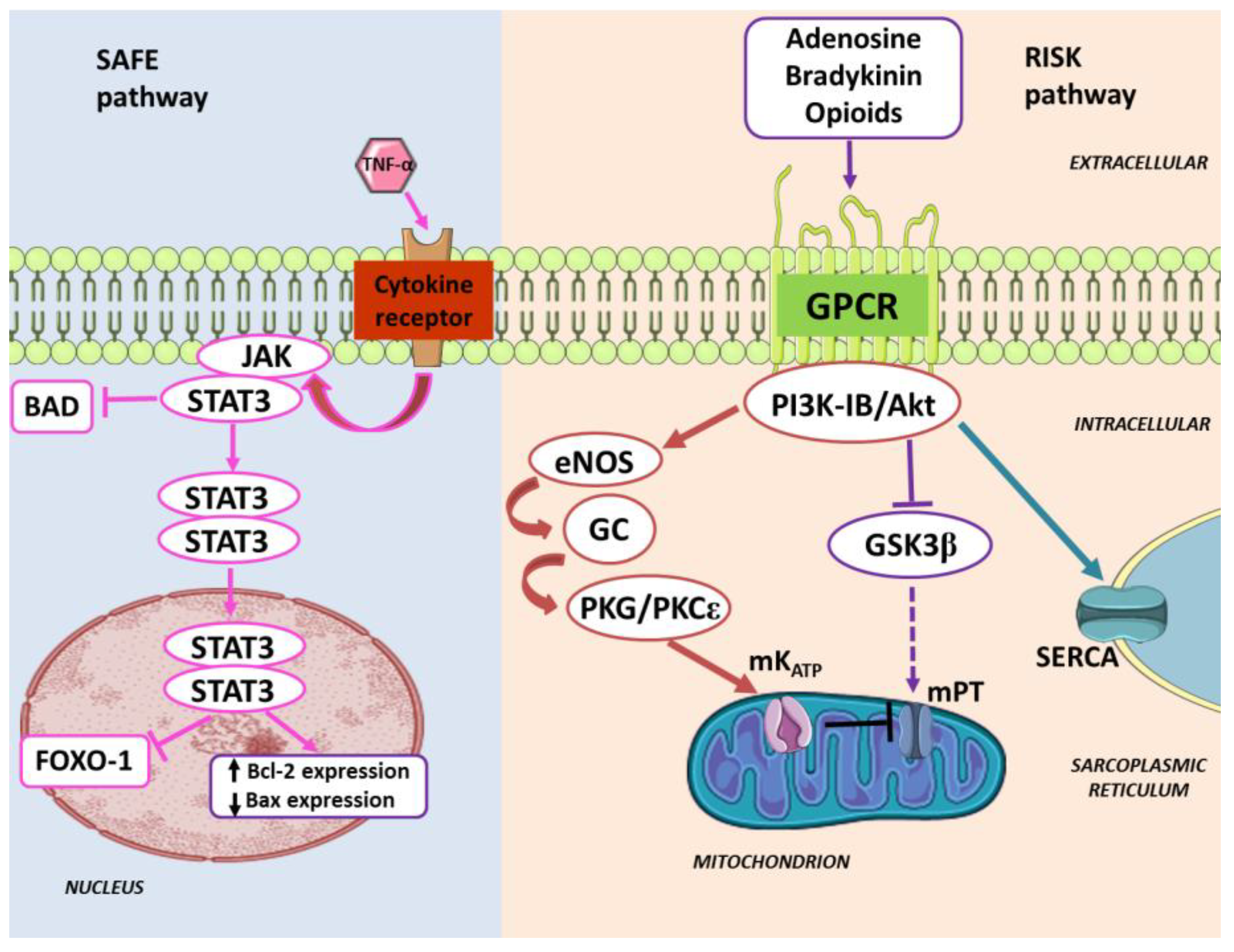
Figure 1. Major cardiac conditioning signaling pathways. The activation of cardioprotective signaling RISK, eNOS/PKG, and SAFE pathways by conditioning triggers is represented (modified from the literature [10]). BAD—Bcl-2-associated death promoter; Bax—Bcl-2-associated X protein; Bcl2—B-cell lymphoma 2; eNOS—endothelial nitric oxide synthase; FOXO-1—forkhead box protein O1; G—guanylyl cyclase; GPCR—G protein-coupled receptors; GSK3β—glycogen synthase kinase 3 beta; JAK—Janus kinase; mKATP—mitochondrial potassium ATP channel; mPT—mitochondrial permeability transition pore; PI3K-IB—phosphoinositide 3-kinase class IB; PKG—protein kinade G; PKCε—protein kinase C epsilon; RISK—reperfusion injury salvage kinase; SAFE—survivor activating factor enhancement; SERCA—sarco/endoplasmic reticulum Ca2+-ATPase; STAT3—signal transducer and activator of transcription 3; TNF-α—tumor necrosis factor alpha.
Ijms 19 02336 g001
Figure 2. The ouabain preconditioning signaling pathway. Na/K-ATPase/Src activation precedes PKCε activation and translocation. An intramitochondrial pathway involves PKCε activation (PKCε1) and mKATP channel opening as a functional complex to trigger an increase in K+-uptake in the mitochondrial matrix. The mKATP-dependent matrix alkalinization is crucial in intramitochondrial signaling, leading to ROS production, which activates the second PKCε, PKCε2. PKCε2 inhibits the mitochondrial permeability transition pore(mPT) in a phosphorylation-dependent reaction. Furthermore, PKCε1 sustains the open state of mKATP channel through mKATP-dependent ROS activation. In addition to this mitochondrial cardioprotective signaling, ouabain-induced PI3K-IA activation is required for protection by OPC. The inhibition of either PKCε or PI3K-IA blunts the OPC-induced protection. mKATP—mitochondrial potassium ATP channel; mPT—mitochondrial permeability transition pore; NKA—Na/K-ATPase; OUA—ouabain; PKCε—protein kinase C epsilon type; PLC-γ—phospholipase C gamma; PI3K-IA—phosphoinositide 3-kinase class IA; ROS—reactive oxygen species; Src—proto-oncogene tyrosine-protein kinase.
Ijms 19 02336 g002