Article

Dietary Supplementation with Lactobacillus casei Alleviates Lipopolysaccharide-Induced Liver Injury in a Porcine Model

1
Hubei Collaborative Innovation Center for Animal Nutrition and Feed Safety, Hubei Key Laboratory of Animal Nutrition and Feed Science, Wuhan Polytechnic University, Wuhan 430023, China
2
Department of Animal Science, Texas A & M University, College Station, TX 77843, USA
*
Author to whom correspondence should be addressed.
These authors contributed equally to this work and should be considered co-first authors.
Int. J. Mol. Sci. 2017, 18(12), 2535; https://doi.org/10.3390/ijms18122535
Submission received: 30 October 2017 / Revised: 20 November 2017 / Accepted: 21 November 2017 / Published: 26 November 2017
(This article belongs to the Section Molecular Pathology, Diagnostics, and Therapeutics)

Abstract

:
This study aims to determine whether Lactobacillus casei (L. casei) could relieve liver injury in piglets challenged with lipopolysaccharide (LPS). Piglets were randomly allocated into one of the three groups: control, LPS, and L. casei. The control and LPS groups were fed a corn- and soybean meal-based diet, whereas the L. casei group was fed the basal diet supplemented with 6 × 10⁶ cfu/g L. casei. On Day 31 of the trial, piglets in the LPS and L. casei groups received intraperitoneal administration of LPS (100 µg/kg body weight), while the control group received the same volume of saline. Blood and liver samples were collected for analysis. Results showed that L. casei supplementation decreased the feed/gain ratio (p = 0.027) and diarrhea incidence (p < 0.001), and attenuated LPS-induced liver histomorphological abnormalities. Compared with the control group, LPS challenge dramatically increased glutamyl transpeptidase activity (p = 0.001) in plasma as well as the concentrations of Interleukin 6 (IL-6) (p = 0.048), Tumor necrosis factor-alpha (TNF-α) (p = 0.041), and Malondialdehyde (MDA) (p = 0.001) in the liver, while decreasing the hepatic SOD activity. LPS also increased (p < 0.05) the mRNA levels for IL-6, IL-8, TNF-α, Toll-like receptors 4 (TLR4), Nuclear factor κB (NF-κB) and Heat shock protein 70 (HSP70) in the liver. The adverse effects of LPS challenge were ameliorated by L. casei supplementation. In conclusion, dietary L. casei alleviates LPS-induced liver injury via reducing pro-inflammatory cytokines and increasing anti-oxidative capacity.

1. Introduction

As a major site for nutrient metabolism and detoxification in the body, the liver plays a critical role in preventing exogenous toxic substances from entering the systemic blood stream [1]. Factors such as bacterial and viral infection or inflammation lead to the activation of macrophages (Kupffer cells), which results in increased productions of pro-inflammatory cytokines (e.g., tumor necrosis factor (TNF)-α, interleukine (IL)-1β and IL-6) and reactive oxygen species (ROS) [2], and consequently parenchymal liver damage and dysfunction. Lipopolysaccharide (LPS), a component of the outer membrane of Gram-negative bacteria, is known to stimulate Kupffer cells (macrophages) and result in inflammatory liver injury [3].
Lactobacillus casei (L. casei) is regarded as a probiotic and is widely used in the food industry [4]. This beneficial bacterium has attracted the focus of research due to its potential immunoregulatory effect. It has been demonstrated that specific L. casei could modulate host immunity, which is positively correlated with the enhanced resistance to various viral and bacterial infections [4]. The immunomodulatory effects are dependent on various factors, such as the intrinsic adjuvant properties, dose, viability, route and timing of administration of the specific L. casei, as well as the physiological state and genetic background of the host [5,6,7]. However, literature concerning the effects of L. casei on liver inflammation and injury is scarce. Therefore, the present study was conducted to determine whether L. casei could attenuate liver injury by using a piglet model with LPS challenge [6].

2. Results

2.1. Growth Performance

During Days 1–30 of the trial, dietary supplementation with L. casei decreased the ratio of feed to gain (F/G) and diarrhea incidence of piglets, in comparison with the control group (Table 1).

2.2. Liver Histology

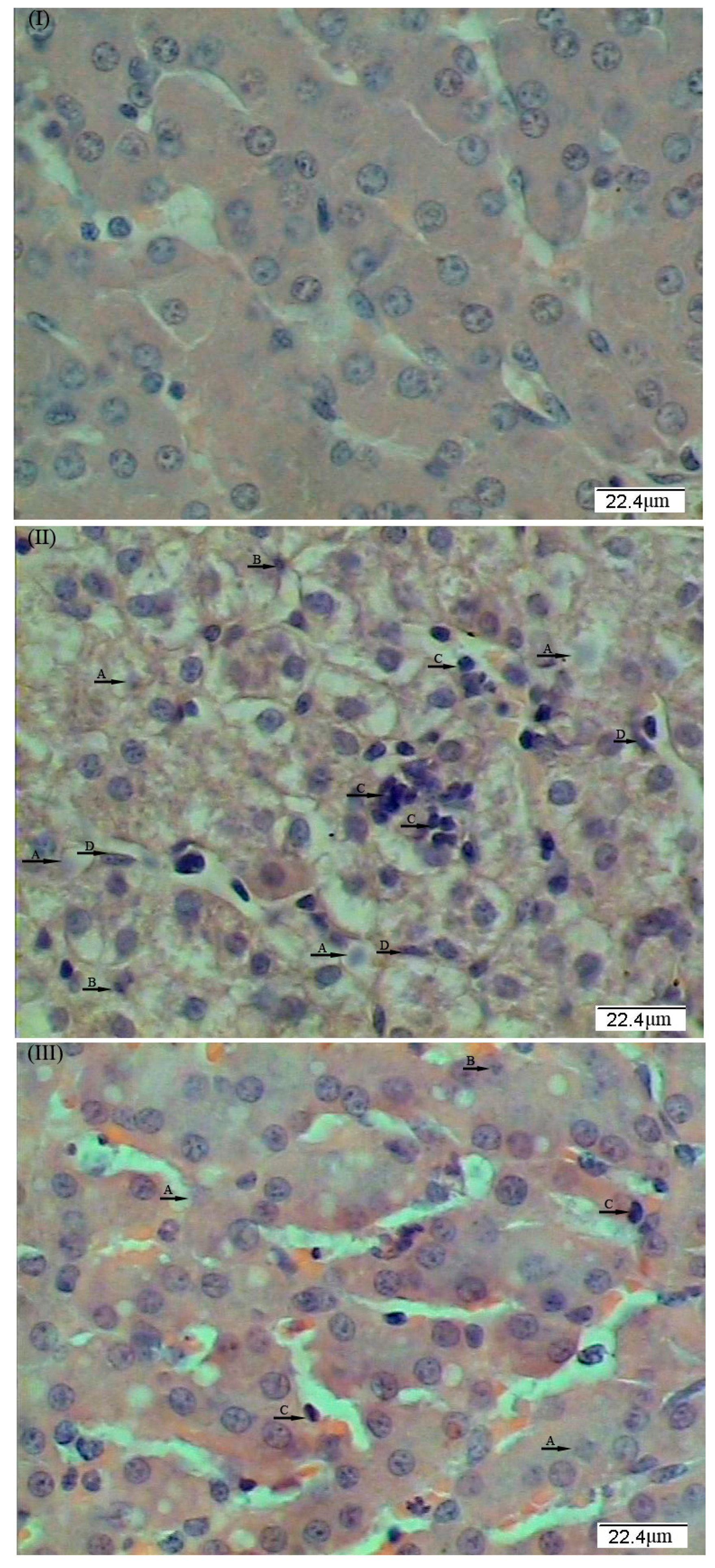
The morphological structure of the liver is shown in Figure 1. The livers of piglets in the control group (Figure 1I) appeared to be normal. The intraperitoneal administration of LPS induced histopathological changes in the liver, including: (A) the infiltration of inflammatory leukocytes into the hepatic lobes; (B) the caryolysis, karyopycnosis, and vacuolation of hepatocytes; (C) the disordered arrangement of hepatic cell cords; and (D) hemorrhage (Figure 1II). The LPS-induced liver injury was attenuated by dietary supplementation with L. casei, as indicated by the normal hepatic structure, the mild vacuolation of hepatic cells, and limited infiltration of inflammatory leukocytes (Figure 1III).

2.3. Alanine Aminotransferase (ALT), Aspartate Aminotransferase (AST), Glutamyl Transpeptidase (GGT) Activity in Plasma

The activities of ALT, AST, GGT in plasma are shown in Table 2. LPS challenge increased (p < 0.05) AST and GGT activities in plasma, compared with the control group. Dietary supplementation with L. casei decreased the blood GGT (p < 0.05) activity in LPS-challenged piglets, despite no effects on ALT and AST activities.

2.4. IL-6 and TNF-α Concentrations in the Liver

As shown in Table 3, LPS challenge increased (p < 0.05) the concentrations of IL-6 and TNF-α in the liver of piglets in comparison with the control group. However, dietary supplementation with L. casei decreased (p < 0.05) the concentrations of those two pro-inflammatory cytokines in the liver of LPS-challenged piglets (Table 3).

2.5. Liver Redox Status

The concentrations of MDA and H2O2, as well as the enzymatic activities of Superoxide dismutase (SOD), Catalase (CAT), and Glutathione peroxidase (GSH-Px), in the liver are summarized in Table 4. LPS challenge reduced (p < 0.05) SOD activity but increased MDA levels in the liver, as compared with the control group. However, dietary supplementation with L. casei attenuated the reduction in hepatic SOD activity (p < 0.05) and the increase in hepatic MDA concentrations in LPS-challenged piglets. The activities of CAT and GSH-Px, and H2O2 levels in the liver were not affected by L. casei supplementation (p > 0.05).

2.6. IL-6, IL-8, TNF-α, TLR4, NF-κB and HSP70 mRNA Expression in the Liver

Data on mRNA levels for hepatic IL-6, IL-8, TNF-α, TLR4, NF-κB and HSP70 are summarized in Table 5. Compared with the control group, LPS-challenged piglets had higher (p < 0.05) mRNA levels for IL-6, IL-8, TNF-α, TLR4, NF-κB, and HSP70 in the liver. However, the increases in hepatic IL-6, IL-8, TNF-α, NF-κB, and HSP70 were attenuated (p < 0.05) in piglets receiving the L. casei-supplemented diet. The mRNA expression of hepatic TLR4 was not altered by L. casei supplementation (p > 0.05).

3. Discussion

In this study, to investigate whether dietary supplementation of L. casei could alleviate liver injury, we utilized a well-established porcine model with LPS-induced hepatic damage. In this animal model, liver injury was induced by intraperitoneal administration of E. coli LPS. LPS, which commonly exists in the outer membrane of all Gram-negative bacteria, can bind to and activate the Kupffer cells (specialized macrophages located in the liver), resulting in the enhanced release of pro-inflammatory cytokines [4,5]. This LPS-induced liver injury model has been commonly used to elucidate the mechanism of inflammatory liver injury and the protective effects of nutritional ingredients, such as probiotics and amino acids [6,8,9,10].
The dose of L. casei used for the present study was based on the results of our previous work regarding the effects of its dietary supplementation on LPS-induced liver injury on improving the growth performance of piglets [11]. Interestingly, piglets fed the L. casei diet exhibited a lower feed/gain ratio and a lower rate of diarrhea in comparison with those fed the basal diet. Diarrhea commonly occurs in early-weaned piglets because of their intestinal dysfunction in response to various challenges, such as social, environmental and dietary stresses [6]. In L. casei-supplemented piglets, reduced diarrhea incidence indicates an improvement in intestinal health. As a probiotic, L. casei has been reported to enhance intestinal-mucosal barrier function and immunity [12] and modulate the intestinal ecology [11,12,13].
Plasma ALT, AST, and GGT activities are sensitive markers for hepatic damage [2]. Results of the present study showed that dietary supplementation with L. casei attenuated LPS-induced increases in plasma GGT activities, indicating a positive beneficial effect of L. casei in ameliorating liver injury. These results are consistent with the histopathological changes in liver morphology, which demonstrated that dietary L. casei supplementation mitigated the LPS-induced damage to the hepatic architecture. However, results on plasma ALT and AST activities indicated an incomplete recovery of the liver from LPS challenge, which may be due to the dosage of L. casei (1 × 10⁸ cfu/day per kg body weight) used in the present study. Of note, a higher dosage of L. casei (6.8 × 10¹⁰ cfu/day per kg body weight) could restore the plasma ALT activity to the normal level and protect the liver from fructose-induced steatosis in mice [14]. Further studies are warranted to determine dose-dependent effects of L. casei on hepatic structure and function.
LPS stimulated the release of inflammatory cytokines, which consequently induced the production of ROS and related peroxides, and ultimately resulted in the aggravation of the liver injury [10]. However, the liver possesses defensive mechanisms against ROS through the actions of radical scavengers, such as SOD, CAT, and GSH-Px [15]. In the present study, the activity of SOD in the liver was much lower in the LPS group than in the control group. Of note, dietary supplementation with L. casei effectively mitigated the oxidative damage caused by LPS. Lactobacillus bacteria act through various mechanisms to defend animals against ROS toxicity, such as synthesizing SODs, producing hydroperoxidases, and accumulating high intracellular levels of metal ions. SOD defends against oxidative stress by scavenging O2 into O2 and H2O2, whereas catalase decomposes H2O2 into H2O and O2. It is likely that dietary supplementation with L. casei improved the liver health in piglets through increasing the activity of SOD to protect against oxidative damage. Similarly, Wang et al. [13] found that pretreatment with L. casei significantly increased SOD activity in the homogenates of the liver challenged with LPS. Taken together, these findings support the notion that L. casei is an effective agent for enhancing the anti-oxidative capacity in animals.
LPS also induces the production of pro-inflammatory cytokines by hepatic Kupffer cells via activating pattern recognition through toll-like receptors (TLRs) [10]. Among the identified TLRs, TLR4 is a well defined receptor for LPS recognition. Upon activation, the TLR4 signaling drives Kupffer cells to produce a variety of inflammation-related cytokines [16]. The nuclear regulatory factor κB (NF-κB) is a central regulator of cellular stress in all cell types in the liver. NF-κB activation is triggered via canonical pathways in response to a wide variety of stimuli, including pro-inflammatory cytokines, as well as the bacterial and viral antigens that act on TLRs [17]. Moreover, NF-κB is a family of dimeric transcription factors that regulate inflammation, innate and adaptive immunity, and wound healing responses, as well as cell fate and function [18]. NF-κB plays these physiological roles by binding to κB sequences found in the regulatory regions of more than 200 target genes. The elevated abundance of NF-κB in the liver plays a mediatory role in the stimulatory effect of LPS on the production of inflammatory cytokines (IL-6 and TNF-α) in piglets. To support this notion, we observed an increase in the concentrations of hepatic IL-6 and TNF-α in LPS-challenged piglets (Table 3). In addition, NF-κB regulates the expression of many downstream genes that control cell proliferation, survival, stress responses, and immunity. Under LPS challenge, TLR4 and NF-κB mRNA levels were markedly enhanced, suggesting that TLR4 and NF-κB signaling pathways may be activated by LPS. However, the diet supplemented with L. casei reduced the hepatic concentrations of IL-6 and TNF-α, as well as the hepatic mRNA levels for pro-inflammatory cytokines (e.g., IL-6 and IL-8) in LPS-challenged piglets (Table 5). Collectively, these results indicate that dietary supplementation with L. casei attenuated the hepatic inflammation possibly via activating the TLR4/NF-κB signaling pathway.
In response to stresses, the mRNA abundance of hepatic heat shock protein HSP70 is usually enhanced to promote the refolding of partially-denatured proteins and prevent their aggregation, thereby protecting cells from injury [8]. This is an adaptive mechanism for allowing organisms to survive heat shock stress. Therefore, a high level of HSP70 is a sensitive indicator of oxidative stress in tissues [19]. In the present study, the expression of the hepatic HSP70 gene was dramatically increased in the liver of LPS-challenged piglets, but was decreased when the diet was supplemented with L. casei. These results further support the notion that L. casei plays an important role in ameliorating liver oxidative stress.
In summary, dietary supplementation with 6 × 10⁶ cfu/g L. casei exerts beneficial effects in alleviating liver injury in lipopolysaccharide-challenged piglets. The hepato-protective effects of L. casei is closely associated with its role in increasing anti-oxidative capacity and reducing pro-inflammatory cytokines in the liver of piglets. These novel findings have important implications for improving the nutritional status of infected animals. As the piglet is a well-established animal model for studying human nutrition and disease [6], findings from the porcine model may be used for the treatment of human liver disease.
4. Material and Methods

4.1. Experimental Design

The animal use protocol for this research (2012-0820, 11 September 2012) was approved by the Institutional Animal Care and Use Committee at Wuhan Polytechnic University. Eighteen healthy crossbred female piglets (Duroc × Landrace × Yorkshire), which were reared by sows, were weaned at 21 ± 2 days of age. After a 4-day period of adaptation, piglets (25 ± 2 days of age, average body weight of 6.40 ± 0.53 kg) were housed individually in stainless steel metabolic cages (1.20 × 1.10 m2) and maintained in an environmentally controlled room (25 °C) by air conditioning. Piglets had free access to food and water. The corn and soybean meal-based diet (Table 6) was formulated to meet National Research Council’s (NRC, 2012) recommended requirements for all nutrients [20]. The dietary content of crude protein (CP), calcium (Ca), and total phosphorus (P) was analyzed according to the Weende method of the feed proximate analysis as described by Henneberg and Stohmann [21]. The dietary content of total lysine, methionine, cystine, threonine, and tryptophan was analyzed by automatic amino acids analyzer (S433D, Sykam GmbH, Eresing, Germany) [22].
All piglets had free access to the basal diet during a 4-day adaptation period to help them adapt to solid food. On Day 1 of the trial (25 days of age), piglets were assigned randomly into three groups (6 piglets/group): (1) control group (piglets fed the basal diet and received intraperitoneal treatment with sterile saline); (2) LPS group (piglets fed the basal diet and received intraperitoneal treatment with Escherichia coli LPS); and (3) L. casei group (LPS + 6 × 10⁶ cfu/g L. casei, piglets fed the basal diet supplemented with 6 × 10⁶ cfu/g L. casei and received intraperitoneal administration of Escherichia coli LPS). LPS was dissolved in sterile saline. L. casei (powder) was well mixed with the basal diet in a one-batch mixing. The dosage of 6 × 10⁶ cfu/g L. casei was chosen because it was found in our previous study to be effective in improving the growth performance and intestinal function of piglets [11]. L. casei was grown overnight at 37 °C in MRS broth (HB0384-1; Oxoid, Haibo, China) and centrifuged at 3000 rpm for 10 min at room temperature, then a L. casei powder with a concentration of 2 × 10¹¹ cfu/g was obtained by vacuum drying. The L. casei powder was mixed with feed to a final concentration of 6 × 10⁶ cfu/g. On Day 31 of the trial, overnight fasted piglets in the LPS and L. casei groups received an intraperitoneal injection of LPS (Escherichia coli serotype O55: B5; Sigma Chemical Inc., St. Louis, MO, USA) at the dose of 100 µg/kg BW, whereas piglets in the control group received an intraperitoneal injection of the same volume of sterile saline [23]. At 6 h post-injection of LPS or saline, all piglets were killed by an intravenous injection of pentobarbital sodium (50 mg/kg BW) and liver samples were collected [8]. During Days 1–30 of the trial, the body weight, feed intake, and diarrhea incidences of piglets were recorded to statistically analyze their growth performance.

4.2. Blood Sample Collection

On Day 31 of the trial, blood samples were collected from the anterior vena cava into heparinized vacuum tubes at 3 h post LPS or saline injection (Becton Dickinson Vacutainer System, Franklin Lake, NJ, USA). Blood samples (7 mL) were centrifuged at 1000× g for 10 min at 4 °C to separate plasma and then stored at −80 °C for further analysis.

4.3. Liver Samples Collection and Histology Analysis

The abdomen of piglets was surgically opened immediately from the sternum to the pubis, and the liver without the cholecyst was collected. A liver sample (~5 g) was collected from the left lobe of the liver and rinsed thoroughly with ice-cold phosphate buffered saline (PBS, pH = 7.4) to remove blood contamination. Liver samples were then rapidly frozen in liquid nitrogen and stored at −80 °C for further analysis. All samples were collected within 15 min after euthanasia.
For histomorphological analysis of liver samples, the 0.5 cm3 segments were cut off from the liver, and flushed with ice-cold PBS. Liver segments were then fixed in fresh 4% paraformaldehyde/phosphate-buffered saline and embedded in paraffin, sectioned at 5 µm and stained with hematoxylin and eosin [8]. Histomorphological examination was performed with a light microscope (American Optical Co., Scientific Instrument Div., Buffalo, NY, USA).

4.4. ALT, AST, GGT Activity in Plasma

Alanine aminotransferase (ALT), aspartate aminotransferase (AST), and glutamyl transpeptidase (GGT) in plasma were assayed by a Hitachi automatic biochemistry analyzer 7100 with WAKO chemical reagents (Wako Pure Chemical Industries, Ltd., Osaka, Japan).

4.5. IL-6 and TNF-α Levels in the Liver

Concentrations of pro-inflammatory cytokines in the liver were determined as previously described [17]. Briefly, frozen liver samples were homogenized in ice-cold PBS-EDTA buffer (0.05 mol/L Na3PO4, 2.0 mol/L NaCl, 2 mmol/L EDTA, pH 7.4), and the homogenates were centrifuged to obtain the supernatant fluid. Tumor necrosis factor-α (TNF-α) and Interleukin-6 (IL-6) in the liver supernatant fluid were analyzed using commercially available 125I kits (Beijing North Institute of Biological Technology, Beijing, China). The detection limit for TNF-α was 0.03 ng/mL, and the intra- and interassay coefficients of variation were 5% and 8%, respectively. The detection limits for IL-6 analyses were 5.0 pg/mL, and the coefficients of variation for intra- and inter-assays of IL-6 were less than 7% and 15%, respectively.

4.6. MDA, H2O2, SOD, CAT and GSH-Px Activity in Liver

The liver tissue (~200 mg) was homogenized in a nine-fold volume of freezing saline, and then centrifuged at 2500 rpm for 10 min at 4 °C to obtain the supernatant fluid used for assays. Malondialdehyde (MDA), H2O2, superoxide dismutase (SOD), catalase (CAT), and glutathione peroxidase (GSH-Px) in the liver were determined using commercially available kits (Nanjing Jiancheng Bioengineering Institute, Nanjing, China).

4.7. IL-6, IL-8, TNF-α, TLR4, NF-κB, and HSP70 mRNA Expression

mRNA levels for Interleukin-6 (IL-6), interleukin-8 (IL-8), tumor necrosis factor-alpha (TNF-α), toll-like receptor 4 (TLR4), nuclear factor kB (NF-κB), and heat shock protein 70 (HSP70) in the liver were quantified using quantitative RT-PCR, as described by Hou et al. [17]. A frozen liver sample of approximately 100 mg was powdered under liquid nitrogen using a mortar and pestle, then homogenized in a buffer. Total RNA was isolated using the TRIzol Reagent protocol (Cat. 15596026, Invitrogen, Carlsbad, CA, USA), and was quantified using the NanoDrop® ND-2000 UV–Vis spectrophotometer (Thermo Scientific, Wilmington, DE, USA) at 260 and 280 nm. The purity of RNA was assessed by determining the OD260/OD280 ratio. All of the samples had an OD260/OD280 ratio above 1.8, corresponding to 90–100% pure nucleic acids. Meanwhile, the integrity of RNA in each sample was assessed using 1% denatured agarose gel electrophoresis. RNA was used for quantitative RT-PCR analysis when the sample had a 28S/18S rRNA ratio ≥ 1.8 [24]. Total RNA was reverse-transcribed using the Prime-Script® RT reagent kit with gDNA Eraser (Cat. RR047A, Takara, Dalian, China) according to the manufacturer’s instructions. cDNA was synthesized and stored at −20 °C for further analysis. The RT-PCR analysis of gene expression was performed using primers for IL-6, IL-8, TNF-α, TLR4, NF-κB, HSP70, ribosomal protein L4 (RPL4) and glyceraldehyde-3-phosphate dehydrogenase (GAPDH) (Table 7), and the SYBR® Premix Ex Taq™ (Cat. 420A, Takara, Dalian, China) on an Applied Biosystems 7500 Real-Time PCR System (Foster City, CA, USA). The total volume of the PCR reaction system was 20 µL. In brief, the reaction mixture contained 10.0 µL SYBR Premix ExTaq, 0.4 µL ROX reference dye II (50×), 2.0 µL cDNA, 6.8 µL RNase free water, 0.4 µL forward primer (10 µmol/L), and 0.4 µL reverse primer (10 µmol/L). All PCRs were done in triplicate on a 96-well RT-PCR plate under the following conditions (two-step amplification): 95 °C for 30 s, followed by 40 cycles of 95 °C for 5 s, and 60 °C for 34 s. A subsequent melting curve (95 °C for 15 s, 60 °C for 1 min and 95 °C for 15 s) with continuous fluorescence measurement and final cooling to 25 °C was processed. Amplification products were verified by melting curves and agarose gel electrophoresis. Each biological sample was run in triplicate. To ensure the sensitivity and accuracy of the results obtained by qPCR, samples were normalized internally using simultaneously RPL4 and GAPDH as references in each sample to avoid any artifact of variation in the target gene. Results were analyzed by 2−ΔΔCt method [25].

4.8. Statistical Analysis

Data expressed as means ± SD were analyzed by one-way analysis of variance. The normality and constant variance for data were tested by the Levene’s test [26]. Differences among treatment means were determined by the Duncan’s post hoc test. All statistical analyses were performed by the SPSS 13.0 software (Chicago, IL, USA). Possibility values <0.05 were taken to indicate statistical significance [27].

5. Conclusions

Diet supplementation with 6 × 10⁶ cfu/g L. casei exerts beneficial effects in alleviating liver injury in lipopolysaccharide-challenged piglets. The hepato-protective effects of L. casei is closely associated with its role in increasing anti-oxidative capacity and reducing pro-inflammatory cytokines in the liver of piglets.

Acknowledgments

This research was jointly supported by National Key R&D Program of China (2016YFD0501210 and 2017YFD0500505); National Natural Science Foundation of China (31372319, 31572416, and 31402084); Hubei Provincial Technology and Innovation Program (2016ABA121 and 2017AHB062); Natural Science Foundation of Hubei Province (2016CFA070); the Hubei Hundred Talent Program, Agriculture and Food Research Initiative Competitive Grants of (2014-67015-21770) of the USDA National Institute of Food and Agriculture; and Texas AgriLife Research (H-82000). All these funding agencies had no role in the design, analysis, or writing of our published studies.

Author Contributions

Yongqing Hou and Di Zhao designed the study and wrote the manuscript. Tao Wu, Dan Yi, Lei Wang, Peng Li, and Junmei Zhang collected and analyzed experimental results. Guoyao Wu participated in the revision of the paper. All authors contributed to the data interpretation and approved the final version of the manuscript.

Conflicts of Interest

The authors declare no conflict of interest.

Abbreviations

L. caseiLactobacillus casei
LPSLipopolysaccharide
ALTAlanine aminotransferase
ASTAspartate aminotransferase
GGTGlutamyl transpeptidase
MDAMalondialdehyde
SODSuperoxide dismutase
CATCatalase
GSH-PxGlutathione peroxidase
IL-6Interleukin 6
IL-8Interleukin 8
TNF-αTumor necrosis factor-alpha
TLR4Toll-like receptors 4
NF-κBNuclear factor κB
HSP70Heat shock protein 70

References

  1. Nakao, A.; Taki, S.; Yasui, M.; Kimura, Y.; Nonami, T.; Harada, A.; Takagi, H. The fate of intravenously injected endotoxin in normal rats and in rats with liver failure. Hepatology 1994, 19, 1251–1256. [Google Scholar] [CrossRef] [PubMed]
  2. He, P.; Noda, Y.; Sugiyama, K. Green tea suppresses lipopolysaccharide-induced liver injury in d-galactosamine sensitized rats. J. Nutr. 2001, 131, 1560–1567. [Google Scholar] [PubMed]
  3. Masaki, T.; Chiba, S.; Tatsukawa, H.; Yasuda, T.; Noguchi, H.; Seike, M.; Yoshimatsu, H. Adiponectin protects LPS-Induced liver injury through modulation of TNF-α in KK-Ay obese mice. Hepatology 2004, 40, 177–184. [Google Scholar] [CrossRef] [PubMed]
  4. Singh, V.P.; Sharma, J.; Babu, S.; Rizwanulla; Singla, A. Role of probiotics in health and disease: A review. J. Pak. Med. Assoc. 2013, 63, 253–257. [Google Scholar] [PubMed]
  5. Ashraf, R.; Shah, N.P. Selective and differential enumerations of Lactobacillus delbrueckii subsp. bulgaricus, Streptococcus thermophilus, Lactobacillus acidophilus, Lactobacillus casei and Bifidobacterium spp. in yoghurt—A review. Int. J. Food Microbiol. 2011, 149, 194–208. [Google Scholar] [PubMed]
  6. Hou, Y.; Wang, L.; Ding, B.; Liu, Y.; Zhu, H.; Liu, J.; Li, Y.; Wu, X.; Yin, Y.; Wu, G. Dietary α-ketoglutarate supplementation ameliorates intestinal injury in lipopolysaccharide-challenged piglets. Amino Acids 2010, 39, 555–564. [Google Scholar] [CrossRef] [PubMed]
  7. Li, X.; Rezaei, R.; Li, P.; Wu, G. Composition of amino acids in feed ingredients for animal diets. Amino Acids 2011, 40, 1159–1168. [Google Scholar] [CrossRef] [PubMed]
  8. Yi, D.; Hou, Y.; Wang, L.; Ding, B.; Yang, Z.; Li, J.; Long, M.; Liu, Y.; Wu, G. Dietary N-acetylcysteine supplementation alleviates liver injury in lipopolysaccharide-challenged piglets. Br. J. Nutr. 2014, 111, 46–54. [Google Scholar] [CrossRef] [PubMed]
  9. Li, Q.; Liu, Y.; Che, Z.; Zhu, H.; Meng, G.; Hou, Y.; Ding, B.; Yin, Y.; Chen, F. Dietary l-arginine supplementation alleviates liver injury caused by Escherichia coli LPS in weaned pigs. Innate Immun. 2012, 18, 804–814. [Google Scholar] [CrossRef] [PubMed]
  10. Chen, F.; Liu, Y.L.; Zhu, H.; Hong, Y.; Wu, Z.; Hou, Y.; Li, Q.; Ding, B.; Yi, D.; Chen, H. Fish oil attenuates liver injury caused by LPS in weaned pigs associated with inhibition of TLR4 and nucleotide-binding oligomerization domain protein signaling pathways. Innate Immun. 2013, 19, 504–515. [Google Scholar] [CrossRef] [PubMed]
  11. Dou, M.; Wu, T. Effect of different types of probiotics on intestinal microflora, pH and volatile base nitrogen of weaned piglets. China Anim. Husb. Vet. Med. 2013, 40, 82–87. [Google Scholar]
  12. Zakostelska, Z.; Kverka, M.; Klimesova, K.; Rossmann, P.; Mrazek, J.; Kopecny, J.; Hornova, M.; Srutkova, D.; Hudcovic, T.; Ridl, J.; et al. Lysate of probiotic Lactobacillus casei DN-114 001 ameliorates colitis by strengthening the gutbarrier function and changing the gut microenvironment. PLoS ONE 2011, 6, e27961. [Google Scholar] [CrossRef] [PubMed]
  13. Wang, Y.; Xie, J.; Li, Y.; Dong, S.; Liu, H.; Chen, J.; Wang, Y.; Zhao, S.; Zhang, Y.; Zhang, H. Probiotic Lactobacillus casei Zhang reduces pro-inflammatory cytokine production and hepatic inflammation in a rat model of acute liver failure. Eur. J. Nutr. 2016, 55, 821–831. [Google Scholar] [CrossRef] [PubMed]
  14. Wagnerberger, S.; Spruss, A.; Kanuri, G.; Stahl, C.; Schröder, M.; Vetter, W.; Bischoff, S.C.; Bergheim, I. Lactobacillus casei Shirota protects from fructose-induced liver steatosis: A mouse model. J. Nutr. Biochem. 2013, 24, 531–538. [Google Scholar] [CrossRef] [PubMed]
  15. Yu, B.P. Cellular defenses against damage from reactive oxygen species. Physiol. Rev. 1994, 74, 139–162. [Google Scholar] [PubMed]
  16. Ben, A.Z.; Avlas, O.; Pappo, O.; Zilbermints, V.; Cheporko, Y.; Bachmetov, L.; Zemel, R.; Shainberg, A.; Sharon, E.; Grief, F.; et al. Reduced hepatic injury in Toll-like receptor 4-deficient mice following d-galactosamine/lipopolysaccharide-induced fulminant hepatic failure. Cell. Physiol. Biochem. 2012, 29, 41–50. [Google Scholar] [CrossRef] [PubMed]
  17. Yao, J.; Pan, D.; Zhao, Y.; Zhao, L.; Sun, J.; Wang, Y.; You, Q.; Xi, T.; Guo, Q.; Lu, N. Wogonin prevents lipopolysaccharide-induced acute lung injury and inflammation in mice via peroxisome proliferator-activated receptor gamma-mediated attenuation of the nuclear factor-κB pathway. Immunology 2014, 143, 241–257. [Google Scholar] [CrossRef] [PubMed]
  18. Muller, J.M.; Ziegler-Heitbrock, H.W.L.; Baeuerle, P.A. Nuclear factor κB, a mediator of lipopolysaccharide effects. Immunobiology 1993, 187, 233–256. [Google Scholar] [CrossRef]
  19. Wang, Y.; Li, Y.; Xie, J.; Zhang, Y.; Wang, J.; Sun, X.; Zhang, H. Protective effects of probiotic Lactobacillus casei Zhang against endotoxin- and d-galactosamine-induced liver injury in rats via anti-oxidative and anti-inflammatory capacities. Int. Immunopharmacol. 2013, 15, 30–37. [Google Scholar] [CrossRef] [PubMed]
  20. Hou, Y.; Wang, L.; Yi, D.; Ding, B.; Yang, Z.; Li, J.; Chen, X.; Qiu, Y.; Wu, G. N-acetylcyseine reduces inflammation in the small intestine by regulating redox, EGF and TLR4 signaling. Amino Acids 2013, 45, 513–522. [Google Scholar] [CrossRef] [PubMed]
  21. Henneberg, W.; Stohmann, F. Beiträge zur Begründungeiner Rationellen Fü Tterung der Wiederkäuer, [Contributions to Establish a Rational Feeding of Ruminants], 2nd ed.; Schwetschtke u. Sohn: Braunschweig, Germany, 1864; 342p. [Google Scholar]
  22. Yi, D.; Hou, Y.; Tan, L.; Liao, M.; Xie, J.; Wang, L.; Ding, B.; Yang, Y.; Gong, J. N-acetylcysteine improves the growth performance andintestinal function in the heat-stressed broilers. Anim. Feed Sci. Technol. 2016, 220, 83–92. [Google Scholar] [CrossRef]
  23. Hou, Y.; Wang, L.; Zhang, W.; Yang, Z.; Ding, B.; Zhu, H.; Liu, Y.; Qiu, Y.; Yin, Y.; Wu, G. Protective effects of N-acetylcysteine on intestinal functions of piglets challenged with lipopolysaccharide. Amino Acids 2012, 43, 1233–1242. [Google Scholar] [CrossRef] [PubMed]
  24. Yi, D.; Hou, Y.; Wang, L.; Zhao, D.; Ding, B.; Wu, T.; Chen, H.; Liu, Y.; Kang, P.; Wu, G. Gene expression profiles in the intestine of lipopolysaccharide-challenged piglets. Front. Biosci. 2016, 21, 487–501. [Google Scholar]
  25. Livak, K.; Schmittgen, T. Analysis of relative gene expression data using real-time quantitative PCR and 2−ΔΔCt method. Methods 2001, 25, 402–408. [Google Scholar] [CrossRef] [PubMed]
  26. Wei, J.W.; Carroll, R.J.; Harden, K.K.; Wu, G. Comparisons of treatment means when factors do not interact in two-factorial studies. Amino Acids 2012, 42, 2031–2035. [Google Scholar] [CrossRef] [PubMed]
  27. Fu, W.J.; Stromberg, A.J.; Viele, K.; Carroll, R.J.; Wu, G. Statistics and bioinformatics in nutritional sciences: Analysis of complex data in the era of systems biology. J. Nutr. Biochem. 2010, 21, 561–572. [Google Scholar] [CrossRef] [PubMed]
Figure 1. Effects of dietary supplementation with L. casei on liver morphology. Representative photomicrographs of liver sections stained with hematoxylin and eosin are shown (×400): (I) piglets fed the basal diet and received intraperitoneal administration of sterile saline; (II) piglets fed the basal diet and received intraperitoneal administration of Escherichia coli lipopolysaccharide (LPS); and (III) piglets fed the basal diet supplemented with 6 × 10⁶ cfu/g L. casei and received intraperitoneal administration of LPS, as described in the Material and Methods Section. Histological analysis revealed abnormalities in hepatic structure, including: caryolysis (A); karyopycnosis (B); infiltration of inflammatory cells (C); injury to hepatic sinusoids (D); and the disordered arrangement of hepatic cell cords.
Ijms 18 02535 g001
Table 1. Effect of dietary supplementation with L. casei on the growth performance of piglets during Days 1–30 of the trial.
ItemsControl GroupL. casei Groupp-Value
BW (kg)6.33 ± 1.076.48 ± 1.280.840
ADG (g/day)410 ± 51412 ± 320.945
F/G ratio1.82 ± 0.03 a1.75 ± 0.06 b0.027
Diarrhea incidence (%)14.6 ± 2.63 a4.17 ± 0.83 b<0.001
Data are means ± SD, n = 12 for the control group and n = 12 for the L. casei group. BW = body weight; ADG = average daily gain; F/G = feed/gain ratio. Control group = piglets fed the basal diet; L. casei group = piglets fed the basal diet supplemented with 6 × 10⁶ cfu/g L. casei; a,b Values within a row with different superscript letters differ (p < 0.05).
Table 2. Effects of dietary supplementation with L. casei on activities of ALT, AST, GGT in the piglet plasma.
ItemsControl GroupLPS GroupL. casei Groupp-Value
ALT (U/L)43.4 ± 7.7045.1 ± 7.5348.8 ± 8.510.354
AST (U/L)34.6 ± 6.78 b56.9 ± 12.8 a50.9 ± 7.06 a0.021
GGT (U/L)27.2 ± 6.12 b46.5 ± 4.15 a27.8 ± 4.60 b0.001
Data are means ± SD, n = 6. ALT = alanine aminotransferase; AST = aspartate aminotransferase; GGT = glutamyl transpeptidase. Control group = piglets fed the basal diet and received administration of saline; LPS group = piglets fed the basal diet and challenged with LPS; L. casei group = piglets fed the basal diet supplemented with 6 × 10⁶ cfu/g L. casei and challenged with LPS; a,b Values within a row with different superscript letters differ significantly (p < 0.05).
Table 3. Effects of dietary supplementation with L. casei on the concentrations of IL-6 and TNF-α in the piglet liver.
ItemsControl GroupLPS GroupL. casei Groupp-Value
IL-6 (pg/mg)5.86 ± 0.33 b7.20 ± 0.31 a5.85 ± 0.79 b0.048
TNF-α (ng/mg)0.15 ± 0.03 b0.20 ± 0.01 a0.15 ± 0.04 b0.041
Data are means ± SD, n = 6. IL-6 = Interleukin 6; TNF-α = tumor necrosis factor-alpha. Control group = piglets fed the basal diet and received administration of saline; LPS group = piglets fed the basal diet and challenged with LPS; L. casei group = piglets fed the basal diet supplemented with 6 × 10⁶ cfu/g L. casei and challenged with LPS; a,b Values within a row with different superscript letters differ significantly (p < 0.05).
Table 4. Effects of dietary supplementation with L. casei on the concentrations of MDA and H2O2, as well as the enzymatic activities of SOD, CAT, and GSH-Px, in the piglet liver.
ItemsControl GroupLPS GroupL. casei Groupp-Value
SOD (U/mg protein)86.7 ± 9.0 a69.8 ± 4.7 b91.4 ± 1.2 a0.002
CAT (U/g protein)30.0 ± 4.528.3 ± 7.126.0 ± 6.10.559
GSH-Px (U/g protein)117 ± 25.595.3 ± 25.3108 ± 25.50.583
MDA (µg/g protein)7.1 ± 1.5 b11.6 ± 1.7 a6.9 ± 0.6 b0.001
H2O2 (µg/g protein)50.1 ± 11.156.7 ± 3.454.7 ± 13.40.818
Data are means ± SD, n = 6. MDA = malondialdehyde; H2O2 = hydrogen peroxide; SOD = superoxide dismutase; CAT = catalase; GSH-Px = glutathione peroxidase. Control group = piglets fed the basal diet and received administration of saline; LPS group = piglets fed the basal diet and challenged with LPS; L. casei group = piglets fed the basal diet supplemented with 6 × 10⁶ cfu/g L. casei and challenged with LPS; a,b Values within a row with different superscript letters differ significantly (p < 0.05).
Table 5. Effects of dietary supplementation with L. casei on mRNA levels for inflammatory genes in the liver of piglets.
GenesControl GroupLPS GroupL. casei Groupp-Value
IL-61 ± 0.23 c9.27 ± 0.44 a5.17 ± 0.15 b0.004
IL-81 ± 0.32 c55.5 ± 10.1 a38.0 ± 3.36 b<0.001
TNF-α1 ± 0.16 c6.43 ± 1.25 a3.02 ± 0.49 b0.001
TLR41 ± 0.25 b1.77 ± 0.08 a1.74 ± 0.01 a0.033
NF-κB1 ± 0.04 b2.10 ± 0.32 a1.36 ± 0.23 b0.006
HSP701 ± 0.18 c20.4 ± 5.81 a10.4 ± 2.31 b0.017
All mRNA levels in the control group were regarded as 1. Data are means ± SD, n = 6. IL-6 = interleukin 6, IL-8 = interleukin 8, TNF-α = tumor necrosis factor-alpha, TLR4 = toll-like receptors 4, NF-κB = nuclear factor κB, HSP70 = heat shock protein 70. Control group = piglets fed the basal diet and received administration of saline; LPS group = piglets fed the basal diet and challenged with LPS; L. casei group = piglets fed the basal diet supplemented with 6 × 10⁶ cfu/g L. casei and challenged with LPS; a,b,c Values within a row with different superscript letters differ significantly (p < 0.05).
Table 6. Composition and nutrient contents of the basal diet (as-fed basis).
IngredientsContent (%)
Corn (DE 14.27 MJ/kg, CP 8.7%)61.88
Soybean meal (DE 13.5 MJ/kg, CP 40%)21.98
Wheat Middling (DE 13.4 MJ/kg, CP 13%)4.00
Fish meal (CP 66%)3.00
Dried whey (CP 12%)3.00
Soy protein concentrate (CP 65%)1.50
CaHPO41.25
Premix 1.00
Limestone (CaCO3 > 35%)0.69
Soy oil0.50
Acidifier (Citric acid > 99%)0.30
NaCl0.30
Mould inhibitor (Calcium propionate > 30%)0.10
Choline chloride0.20
l-Lysine·HCl (78.8% lysine)0.25
dl-Methionine (99% methionine)0.05
Nutrients composition
Digestible energy (MJ/kg)14.22
Crude protein (%) §20.90
Total lysine (%) §1.15
Total methionine (%) §0.30
Total threonine (%) §0.74
Total tryptophan (%) §0.21
Total calcium (%) §0.70
Total phosphorus (%) §0.60
Available phosphorus (%) 0.32
Premix provided the following amounts of vitamins and trace minerals per kilogram of the complete diet: ferrum, 100 mg (FeSO4·H2O); copper, 150 mg (CuSO4·5H2O); manganese, 40 mg (MnSO4·5H2O); zinc, 100 mg (ZnSO4·7H2O); iodine, 0.5 mg (KI); selenium, 0.3 mg (Na2SeO3·5H2O); vitamin A acetate, 3.66 mg; cholecalciferol, 0.10 mg; DL-α-tocopheryl acetate, 36.4 mg; menadione, 4 mg; thiamin, 6 mg; riboflavin, 12 mg; pyridoxine, 6 mg; cyanocobalamin, 0.05 mg; biotin, 0.2 mg; folic acid, 2 mg; niacin, 50 mg; d-calcium pantothenate, 25 mg; Calculated value; § Analyzed value.
Table 7. Sequences of the primers used for quantitative RT-PCR analysis.
GeneForward (5′–3′)Reverse (5′–3′)
IL-6TACTGGCAGAAAACAACCTGGTACTAATCTGCACAGCCTC
IL-8TTCGATGCCAGTGCATAAATACTGTACAACCTTCTGCACCCA
TNF-αTCCAATGGCAGAGTGGGTATGAGCTGGTTGTCTTTCAGCTTCAC
TLR4GCCTTTCTCTCCTGCCTGAGAGCTCCATGCATTGGTAACTAATG
NF-κBCTCGCACAAGGAGACATGAAACTCAGCCGGAAGGCATTAT
HSP70GACGGAAGCACAGGAAGGAGAAGACAGGGTGCGTTTGG
RPL4GAGAAACCGTCGCCGAATGCCCACCAGGAGCAAGTT
GAPDHCGTCCCTGAGACACGATGGTCCCGATGCGGCCAAAT