Article

Frequent Epigenetic Suppression of Tumor Suppressor Gene Glutathione Peroxidase 3 by Promoter Hypermethylation and Its Clinical Implication in Clear Cell Renal Cell Carcinoma

1
Department of Urology, Peking University First Hospital and Institute of Urology, National Research Center for Genitourinary Oncology, Beijing 100034, China
2
Department of Pathology, Cancer Institute and Cancer Hospital, Peking Union Medical College (PUMC), Chinese Academy of Medical Sciences, Beijing 100021, China
*
Author to whom correspondence should be addressed.
Int. J. Mol. Sci. 2015, 16(5), 10636-10649; https://doi.org/10.3390/ijms160510636
Submission received: 28 March 2015 / Revised: 30 April 2015 / Accepted: 5 May 2015 / Published: 11 May 2015
(This article belongs to the Special Issue Molecular Classification of Human Cancer: Diagnosis and Treatment)

Abstract

:
The goal of this study is to identify novel tumor suppressor genes silenced by promoter methylation in clear cell renal cell carcinoma (ccRCC) and discover new epigenetic biomarkers for early cancer detection. Reactive oxygen species (ROS) is a major cause of DNA damage that correlates with cancer initiation and progression. Glutathione peroxidase 3 (GPX3), the only known extracellular glycosylated enzyme of GPXs, is a major scavenger of ROS. GPX3 has been identified as a tumor suppressor in many cancers. However, the role of GPX3 in ccRCC remains unclear. This study aimed to investigate its epigenetic alteration in ccRCC and possible clinicopathological association. In our study, GPX3 methylation and down-regulation were detected in 5 out of 6 ccRCC cell lines and the GPX3 mRNA and protein expression level in ccRCC tumors was significantly lower than in adjacent non-malignant renal tissues (p < 0.0001). Treatment with 5-Aza-2'-deoxycytidine restored GPX3 expression in ccRCC cells. Aberrant methylation was further detected in 77.1% (162/210) of RCC primary tumors, but only 14.6% (7/48) in adjacent non-malignant renal tissues. GPX3 methylation status was significantly associated with higher tumor nuclear grade (p = 0.014). Thus, our results showing frequent GPX3 inactivation by promoter hypermethylation in ccRCC may reveal the failure in the cellular antioxidant system in ccRCC and may be associated with renal tumorigenesis. GPX3 tumor specific methylation may serve as a biomarker for early detection and prognosis prediction of ccRCC.

1. Introduction

Clear cell Renal cell carcinoma (ccRCC) is the most lethal type of urological cancer due to its occult onset and resistance to chemotherapy and radiation. Although radical nephrectomy is effective to cure local and early ccRCCs, 30% of patients develop metastases after surgery [1]. Therefore, novel, specific biomarkers for early tumor detection and anti-tumor agents are urgently required.
It is well recognized that inactivation of tumor suppressor genes (TSGs) may lead to neoplastic changes. TSGs can be inactivated by both genetic and epigenetic mechanisms, such as point mutation, LOH, and promoter hypermethylation. Recently, multiple TSGs associated with promoter hypermethylation have been identified in ccRCC, such as VHL, p16, RASSF1A, SPINT2 and HOXB13 [2,3,4,5]. Our group has also identified some TSGs silenced by promoter methylation in ccRCC, including DLC1, DLEC1 and IRF8 [6,7,8]. These findings provide a new insight to probe the molecular mechanisms of ccRCC and to seek potential diagnostic and therapeutic target for ccRCC. However, most of these known TSGs have a relatively low frequency of methylation in ccRCC. Thus, further studies are needed to identify novel and specific methylation-sensitive tumor suppressor genes in ccRCC.
Increased reactive oxygen species(ROS)levels have been found in a broad range of tumor tissues (e.g., lung, breast, esophagus and liver) [9], indicating a role for ROS as a common cause of human cancers. Under normal circumstances, ROS play a role in signal transduction. However, accumulation of excess cellular ROS has been reported to induce increased DNA mutations that have been associated with increased carcinogenesis [10].
Normal cells have integrated antioxidative systems that protect cells from ROS-induced DNA damage and cell injury. Among these systems, the glutathione peroxidase family (GPXs) is a major antioxidative enzyme family that promotes the reduction of lipid peroxides, hydrogen peroxide and organic hydroperoxide by reduced glutathione [11]. GPX3, located on chromosome 5q32, is the only known extracellular glycosylated enzyme of GPXs that can use thioredoxin, glutaredoxin and glutathione as electron donors, to reduce a wide range of hydroperoxides [12,13]. It is a major scavenger of ROS produced during normal metabolism or after oxidative insult. GPX3 mRNA is expressed in a variety of normal human tissues, including kidney, liver, breast, heart, lung, brain, and gastrointestinal tract, but the majority of plasma GPX3 is kidney-derived [13,14]. Recently, downregulation of GPX3 by promoter hypermethylation has been reported in multiple human cancers, such as prostate, gastric, esophageal, cervical, and bladder cancer [15,16,17], suggesting that GPX3 serves as a tumor suppressor in these cancers. However, the role and the clinical implication of GPX3 in ccRCC remain unclear.
In this study, we examined GPX3 expression and promoter methylation status in ccRCC cell lines and primary tumors, analyzed the relationship between its methylation and clinicopathological features in patients with RCC.

2. Results

2.1. Methylation of the GPX3 Promoter Correlates with Its Downregulation in ccRCC Cell Lines

By Real-Time PCR, we detected for the first time that GPX3 mRNA expression was downregulated in five out of six (83.3%) ccRCC cell lines compared with “normal” human embryonic kidney cell line (Hek293) and “normal” human proximal tubular cell line (HK-2) (Figure 1A). The region spanning the assumed promoter and exon 1 of GPX3 is a typical CpG island and, thus, susceptible to epigenetic silencing. Therefore, we next explored the role of promoter methylation in silencing GPX3 by methylation-specific PCR. Full or partial methylation was detected in five renal cancer cell lines (786-0, Caki-1, Osrc-2, Kert-3 and 769P), which showed downregulated GPX3 expression, whereas only weak or no methylation was detected in the cell lines (Hek293 and HK-2) with GPX3 expression (Figure 1A). To confirm MSP results, BGS was performed to identify methylation status of 14 CpG sites within the GPX3 promoter. The bisulfite genomic sequencing results were consistent with those of methylation specific PCR in which high density methylated alleles were detected in GPX3 downregulated 786-0, Caki-1, Kert-3 and Osrc-2 cell lines, while more unmethylated alleles were detected in Hek293 cell line and HK-2cell line (Figure 1B). These results indicated that promoter methylation was associated with the downregulation of GPX3 in ccRCC cell lines.
Figure 1. GPX3 downregulation by promoter hypermethylation in ccRCC cell lines. (A) Detection of GPX3 mRNA expression and methylation in a panel of ccRCC, Hek293, and HK-2 cell lines by real time RT-PCR and MSP. M, methylated. U, unmethylated; (B) Representative methylation analysis of individual CpG sites in the GPX3 promoter by bisulfite genomic sequencing. Each row represents one bacterial clone with one circle symbolizing one CpG site. Filled ovals indicate methylated. Open ovals indicate unmethylated.
Ijms 16 10636 g001

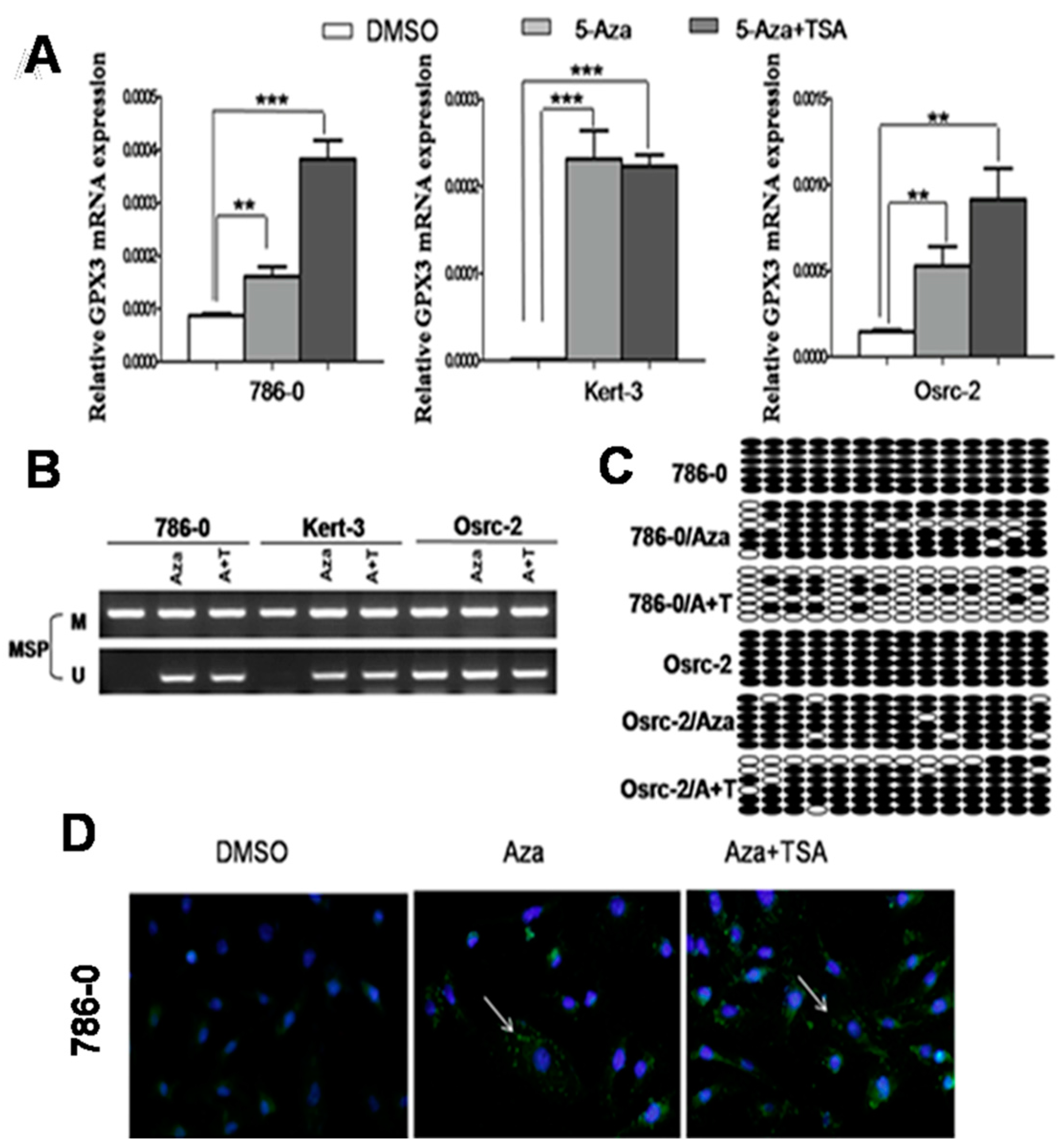
2.2. Pharmacological Demethylation Restored GPX3 Expression in ccRCC Cells

To explore whether promoter methylation directly mediates GPX3 reduction in ccRCC, three methylated cell lines that showed downregulation of GPX3 were treated with the DNA methyltransferase inhibitor 5-Aza-2'-deoxycytidine with or without the histone deacetylase inhibitor trichostatin A. Results showed that 5-Aza treatment could restore GPX3 mRNA expression in ccRCC cells along with a decrease in methylated alleles, and the same results were observed in A + T treated-RCC cells (Figure 2A,B). Further detailed BGS methylation analysis for 786-0 and Osrc-2 before and after Aza treatment confirmed its demethylation (Figure 2C). To check if demethylation treatment also restored GPX3 protein level, we performed an immunofluorescence assay, using antibody against GPX3 in 786-0 cells. As shown in Figure 2D, there was a significant increase in the GPX3 green immunofluorescence signal after 5-Aza and 5-Aza-TSA treatments as compared to DMSO control, suggesting that pharmacological demethylation can restore the GPX3 protein expression in 786-0 cells. These results indicated that CpG methylation of the GPX3 promoter directly led to its suppression in ccRCC cell lines.
Figure 2. (A,B) Pharmalogic demethylation with 5-Aza alone or combined with trichostatin A (A + T) restored GPX3 mRNA expression and induced its demethyation in ccRCC cell lines. ** p < 0.01; and *** p < 0.001; (C) BGS analysis of Aza and A + T treated 786-O and Osrc-2 ccRCC cells. Each row represents one bacterial clone with one circle symbolizing one CpG site. Filled ovals indicate methylated. Open ovals indicate unmethylated; (D) Immunofluorescence staining of GPX3 protein in 786-O cells. Pharmalogic demethylation with 5-Aza alone or combined with trichostatin A (A + T) restored GPX3 protein expression in 786-O cells. Green pellet in the cytoplasm represents positive staining (indicated by arrows).
Ijms 16 10636 g002

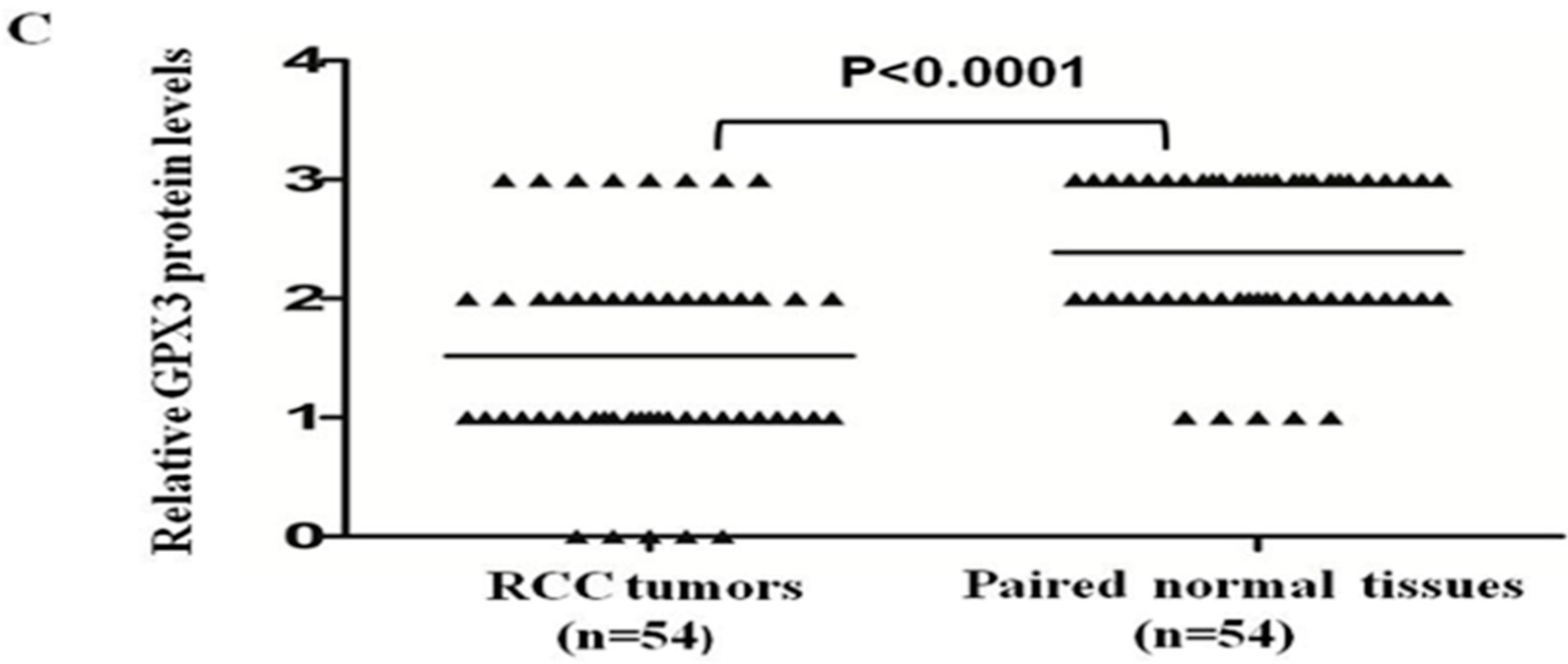
2.3. GPX3 mRNA and Protein Expression Was Frequently Downregulated or Silenced in Primary ccRCC Tumor Tissues

We investigated mRNA expression of GPX3 in 76 paired ccRCC tumor tissues and their adjacent non-tumor tissues using quantitative Real-Time PCR. As shown in Figure 3A, GPX3 was significantly downregulated in renal tumors compared with their adjacent non-tumor tissues (p < 0.0001). We revealed consistently low levels of GPX3 mRNA expression in 94.74% (72 of 76) of ccRCC samples. Moreover, there was a dramatic reduction in levels of GPX3 in 40.79% (31/76) of the cases, with a 20-fold reduction compared with the normal samples (Figure 3A). Inhibition of GPX3 in ccRCC was further confirmed at protein level by using immunohistochemical staining. We examined GPX3 protein expression in 54 ccRCCs and paired adjacent non-tumor tissues. In adjacent non-tumor tissues, intense immunostaining for GPX3 was observed in a cytoplasmic distribution (Figure 3B), whereas absent/weak immunostaining was detected in the cytoplasm of tumor tissues (Figure 3B). Out of 54 ccRCC tumor tissues, GPX3 protein was negative in five (9.3%), weakly positive in 24 (44.4%), moderately positive in 17 (31.5%), and strongly positive in eight (14.8%) (Figure 3C). In contrast, out of 54 adjacent non-tumor tissues, GPX3 protein was negative in 0, weakly positive in five (9.3%), moderately positive in 23 (42.6%), and strongly positive in 26 (48.1%) (Figure 3C). As shown in Figure 3C, statistical analysis of the immunohistochemical results revealed that protein expression of GPX3 in ccRCC tumor tissues was significantly lower than in adjacent non-tumor tissues (p < 0.0001).
Figure 3. Expression pattern of GPX3 in ccRCC. (A) Seventy-six paired ccRCC samples and adjacent non-tumor tissues were analyzed by real time RT-PCR for GPX3 mRNA expression, which was significantly downregulated in ccRCC tumors as compared to adjacent non-tumor samples (p < 0.0001); (B) Representative immunohistochemical staining of a pair of ccRCC specimens and corresponding non-tumor tissue. In adjacent non-tumor tissues, intense immunostaining for GPX3 was detected in a cytoplasmic distribution, whereas absent/weak immunostaining was observed in the cytoplasm of tumor tissues; The place of pictures taken in 400× was indicated by arrows; (C) Evaluation and statistical analysis of GPX3 protein expression in 54 paired ccRCC samples and adjacent non-tumor tissues. GPX3 protein expression was significantly downregulated in ccRCC samples compared to adjacent non-tumor tissues (p < 0.0001).
Ijms 16 10636 g003aIjms 16 10636 g003b

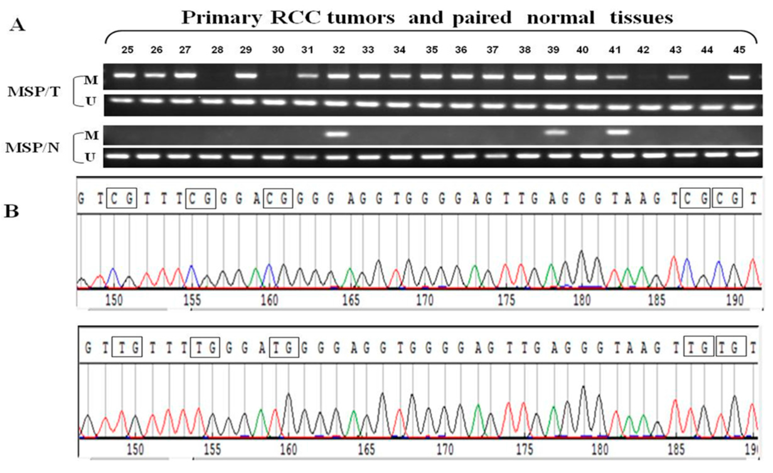
2.4. Frequent GPX3 Promoter Methylation in Primary RCC Tumors Is Associated with Poor Prognosis

We further examined GPX3 methylation status in primary RCC samples and their adjacent non-tumor tissues. Results showed that GPX3 methylation was detected in 77.1% (162/210) of RCC tumors, but only 14.6% (7/48) in adjacent non-malignant renal tissues, suggesting tumor-specific methylation of GPX3 in RCC. Representative methylation status of GPX3 in RCC primary tumors (T) and paired adjacent non-tumor tissues (N) are shown in Figure 4A. MSP results were confirmed by bisulfite genomic sequencing (Figure 4B). We also analyzed the correlation between GPX3 methylation and clinicopathological features of patients with RCC. As shown in Table 1, GPX3 methylation was significantly associated with higher tumor nuclear grade of RCC (p = 0.014), whereas no significant correlation was found between its methylation and gender, age, tumor location, TNM stage and histological type. In addition, our study also showed an increased percentage of GPX3 methylation in Stage 2 or higher tumors compared with Stage 1 patients (although this was not statistically significant). Collectively, these data indicate that GPX3 methylation is a frequent event in pathogenesis of RCC and is associated with patient poor prognosis.
Figure 4. (A) Representative MSP results of GPX3 methylation in RCC primary tumors (T) and paired adjacent non-tumor tissues (N). M, methylated; U, unmethylated; (B) Representative bisulfite genomic sequencing results of cloned BGS-PCR products. Methylated CpG sites will appear as CG during sequencing, while unmethylated CpG sites as TG.
Ijms 16 10636 g004
Table 1. Association between GPX3 methylation and clinicopathological features of patients with RCC.
Clinicopathological FeaturesNumber ( n = 210)GPX3 Methylation Status Methylated Unmethylatedp Value
Overall210162 (77.1) 48 (22.9)
Gender
M146112 (76.7) 34 (23.3)0.860
F6450 (78.1) 14 (21.9)(Fisher’s exact test)
Age
<60 (median)10984 (77.1) 25 (22.9)1.000
≥6010178 (77.2) 23 (22.8)(Fisher’s exact test)
Side
Rt11382 (72.6) 31 (27.4)0.088
Lt9780 (82.5) 17 (17.5)(Fisher’s exact test)
TNM Classification
pT1a7956 (70.9) 23 (29.1)0.206
pT1b7260 (83.3) 12 (16.7)(chi-square test)
pT21715 (83.3) 2 (16.7)
pT34232 (76.2) 10 (23.8)
Nuclear Grade
G14830 (62.5) 18 (37.5)0.014 *
G2135108 (80.0) 27 (20.0)(chi-square test)
G32724 (88.9) 3 (11.1)
Histological Type
Clear cell Rcc193150 (77.7) 43 (23.3)0.734
papillary Rcc96 (66.7) 3 (33.3)(chi-square test)
chromophobe Rcc86 (75.0) 2 (25.0)
* Significant difference.

3. Discussion

Recent studies have shown increased evidences demonstrating that reactive oxygen species (ROS) are a major cause of DNA damage that correlate with a wide range of human diseases including cancer. For instance, carcinoma cells utilize ROS to stimulate their proliferation, angiogenesis, migration, and escape of apoptotic mechanism [18]. In addition, ROS promote the motility and invasion of carcinoma cells by activating protein kinase-C (PKC) and the ERK/MAPK signaling pathways, thus increasing the risk of metastasis [19,20].
The glutathione peroxidases, a family of oxidation-reduction enzymes, play central roles in balancing the signaling, immunomodulatory and detrimental effects of ROS. GPX3, also named plasma glutathione peroxidase, is the only known selenocysteine-containing extracellular form of GPXs and accounts for nearly all of the glutathione peroxidase activity in plasma [21]. This peculiar feature makes GPX3 an extremely important part, not only in GPXs family, but also among cellular antioxidant system, serving as a first line of defense against ROS prior to their entry into the cell.
GPX3 mRNA is expressed in a wide range of normal human tissues, however, downregulation of GPX3 has been found in multiple human cancers, such as prostate, gastric, esophageal, cervical, and bladder cancer [15,16,17], suggesting its importance in human tumorigenesis. Yu, et al. reported that GPX3 suppression in prostate cancer correlates with increased incidence of lymph node metastasis and poor clinical prognosis. Overexpression of GPX3 in prostate cancer cell lines seemed to restrain tumor growth and metastasis through downregulation of c-met, a receptor tyrosine tumor transforming gene involved in a variety of cellular processes [17,22,23]. In addition, GPX3 was found to increase apoptotic cell death by interacting directly with p53-induced gene 3 (PIG3) both in vivo and in vitro [24], suggesting a novel signaling pathway of GPX3-PIG3 in the regulation of cell death in prostate cancer. Recently, a study in colitis-associated carcinoma reported that GPX3-deficient mice exhibited increased inflammation with redistribution towards pro-tumorigenic M2 macrophage subsets, increased proliferation, hyperactive WNT signaling, and increased DNA damage [25]. Knockdown of GPX3 in the human colon cancer cell line Caco2 resulted in increased ROS production and DNA damage. These data supports a tumor suppressor role for GPX3 via clearance of ROS and DNA damage that lead to tumor initiation and progression.
Plasma GPX3 is mainly derived from kidney; however, in our study we found that expression of GPX3 was significantly downregulated in primary renal tumors compared with their adjacent non-tumor tissues (p < 0.0001). This finding indicate that the function of GPX3 is impaired in ccRCC, a consequence of which is likely to be an increased amount of ROS, which would induce DNA damage, driving the carcinogenic process of ccRCC.
As an alternative to genetic changes, hypermethylation of CpG rich promoter regions leading to consequent downregulation or silencing of TSGs is now recognized as an important mechanism for cancer initiation and progression. In addition to its biological relevance to malignant transformation, DNA methylation is one of the most promising biomarkers for early detection and prognosis assessments of human cancers. Recently, studies have shown a great number of aberrantly methylated TSGs in ccRCC. However, the frequencies of aberrant methylation of most classical TSGs, such as VHL, p16, APC, and CDH1, are less than 30% in ccRCC [2], indicating that these genes are probably not the major epigenetic targets for methylation silencing in ccRCC.
Downregulation of GPX3 expression by promoter hypermethylation has been reported in 71.4% of esophageal squamous cell carcinoma, 83% of gastric cancer and 90% of prostate cancer [17,26,27], indicating its possible suppression by epigenetic change in ccRCC. To check this hypothesis, three methylated ccRCC cell lines that showed GPX3 inactivation were treated with the hypomethylating agents. Consistent with our research expectations, GPX3 expression was restored by demethylation treatment, suggesting that promoter methylation of GPX3 directly led to its suppression in ccRCC cell lines. In addition, the combine treatment with TSA exerted additional stimulatory effect on GPX3 mRNA expression, suggesting that histone acetylation may also play some roles in the regulation of GPX3 in RCC. GPX3 methylation was further detected in 77.1% (162/210) of RCC tumors, but only 14.6% (7/48) of adjacent non-malignant renal tissues, indicating that GPX3 methylation is a tumor-specific event involved in tumorigenesis of RCC. Of note, we observed promoter hypermethylation and downregulation of GPX3 in a small number of tumor-adjacent “normal” renal tissues. These tumor-adjacent “normal” samples, although histologically normal, they usually have some degree of changes at the molecular level. Therefore, our data suggest that promoter hypermethylation and GPX3 suppression is possibly an early event in renal tumorigenesis. Moreover, GPX3 hypermethylation was associated with higher nuclear grade, indicating its potential role as prognostic predictor of RCC. Similar results were noted for prostate cancer [27].
In summary, our results showing frequent GPX3 inactivation by promoter hypermethylation in ccRCC may reveal the failure in the cellular antioxidant system, which is the first line of defense against detrimental ROS activity. In addition, this study provides new clinical implications of GPX3 expression inactivation and promoter hypermethylation in ccRCC. First, as a tumor suppressor gene related to higher nuclear grade, GPX3 suppression may have a negative influence on patients’ clinical outcome, and detection of GPX3 methylation may provide prognostic information on ccRCC, especially when TSG hypermethylation can be detected in patients’ serum and urine samples, just like RASSF1A, tissue inhibitor of metalloproteinase-3 and CDH1 methylation [28]. Second, GPX3 may serve as a potential target for clinical intervention. Gene targeting therapy to enhance GPX3 activity might be helpful in the prevention or treatment of ccRCC. GPX3 expression is selenium dependent, since the existence of an opal stop codon in the mid section of the GPX3 open reading frame, and thus selenium supplements in cancer treatment may augment expression of GPX3 and could hold promise in suppressing tumor growth. Of course, further studies are urgently required to explore the functions of GPX3 in renal tumorigenesis.

4. Experimental Section

4.1. Patients and Tissue Samples

All human primary RCCs and adjacent nonmalignant renal tissue were obtained from the urology department of Peking University First Hospital, Beijing, China, with patients’ consent according to the university policy. All cases were collected from primary surgical resection with no prior history of RCC and adjuvant therapy. Pathological diagnosis was done and confirmed at the pathology department, Institute of Urology, Peking University First Hospital. The histopathology of tumors was classified by 2002 AJCC TNM stage and Fuhrman nuclear grade.

4.2. Cell Culture

Six RCC cell lines (786-O, Caki-1, CaKi-2, Kert-3, 769P and Osrc-2) were obtained from Cancer Research Institute of Beijing, Beijing University, China. Hek293 (a “normal” human embryonic kidney cell line) and HK-2 (a “normal” human proximal tubular cell line), which serve as the control cell lines for RCC, were purchased from American Type Culture Collection. They were routinely cultured in RPMI 1640 or DMEM medium supplemented with 10% fetal bovine serum (GIBCO Invitrogen, Carlsbad, CA, USA) and incubated in 5% CO2 at 37 °C.

4.3. Drug Treatment

For 5-Aza-2'-deoxycytidine (Sigma, St. Louis, MO, USA) and Trichostatin A (TSA) (Sigma St. Louis, MO, USA) treatment, cell lines were grown in a 6-well plate and treated with 10 μM 5-Aza-2'-deoxycytidine for 72 h and subsequently with or without 100 nM trichostatin A for 24 h, as described previously [29]. Controlled cells were treated with an equivalent concentration of dimethyl sulfoxide.

4.4. Quantitative Real-Time PCR

Real-time PCR reactions were performed using GoTaq(R) qPCR Master Mix (Promega Biotech, Madison, WI, USA) according to the manufacturer’s protocol on 7500 Fast Real-Time PCR System (ABI). The primers and PCR conditions are shown in Table 2. GAPDH was used as the housekeeping gene for loading control.
Table 2. Primer sequences used in this study.
GenePrimer Sequence (5'-3')Anneal. Temp. (°C)No. of Cycles
Real Time
GPX3 FCTTCCTACCCTCAAGTATGTCCG5545
GPX3 RGAGGTGGGAGGACAGGAGTTCTT
GAPDH FGGTGGTCTCCTCTGACTTCAACA5545
GAPDH RGTTGCTGTAGCCAAATTCGTTGT
MSP
GPX3 m1TATGTTATTGTCGTTTCGGGAC5940
GPX3 m2GTCCGTCTAAAATATCCGACG
GPX3 U1TTTATGTTATTGTTGTTTTGGGATG5940
GPX3 U2ATCCATCTAAAATATCCAACACTCC
BGS
GPX3 BGS FGGAGTTAAAAGAGGAAGGG5840
GPX3 BGS RCCCAACCACCTTTCAAAC

4.5. Immunohistochemistry

Fifty-four paraffin-embedded tumor tissues and paired adjacent non-tumor tissues were analyzed using immunohistochemical Staining. Briefly, the sections were deparaffinized in xylene and rehydrated by transfer through graded concentrations of ethanol to distilled water, and endogenous peroxidase activity was blocked by incubation with 3% H2O2 for 15 min at room temperature. Then, sections were submitted to antigen retrieval in a microwave (sodium citrate buffer, pH 6.0) for 10 min, naturally refrigerated to room temperature. Blocking was performed with 10% goat serum for 30 min at room temperature. All sections were incubated with mouse anti-GPX3 monoclonal antibody (Clone 23B1, 1:100, Abcam, Cambridge, MA, USA), overnight at 4 °C, and then incubated with goat anti-mouse secondary antibody for 30 min at room temperature. After rinsing three times in PBS for 5 min each, the sections were incubated with DAB for 2 min, counterstained with hematoxylin for 3 min, dehydrated with gradient alcohol and transparentized with dimethylbenzene. Immunohistochemical expression of GPX3 was examined via light microscopy. Tissues were graded on the following scale: 0, negative; 1, weakly positive; 2, moderately positive; 3, strongly positive.

4.6. Immunofluorescence

To check GPX3 protein expression after demethylation treatment, we performed immunofluorescence staining against GPX3 in 786-0. Cells were fixed with 4% paraformaldehyde for 15 min, followed by permeabilization with 0.1% Triton X-100 for 5 min. Then, after washing with PBS, the cells were blocked with goat serum for 30 min. For immunofluorescence, Cells were incubated with primary antibody against GPX3 overnight at 4 °C followed by secondary goat anti-mouse antibody conjugated with FITC (1:200 dilution; Invitrogen, Carlsbad, CA, USA) at room temperature for 45 min. The nuclei were counterstained with DAPI (1 μg/mL; Roche, Indianapolis, IN, USA) and viewed under a fluorescence microscope.

4.7. Bisulfite Treatment and Methylation-Specific PCR

The bisulfite modification of purified genomic DNA was performed using an EpiTect Bisulfite Kit (Qiagen 59104, Hilden, Germany) following the manufacturer’s instructions. Table 2 lists MSP primers and PCR conditions. MSP primers were examined previously for not amplifying any unbisulfited DNA and MSP products of several ccRCC cell lines were confirmed by direct sequencing, indicating that our MSP system was specific.

4.8. Bisulfite Genomic Sequencing

Bisulfite-treated DNA was amplified with primers specific for a fragment of the GPX3 promoter CpG islands that contained 14 CpG sites. Table 2 lists BGS primers and PCR conditions. The PCR products were subcloned into the pEasy-T5 vector (Transgene, Beijing, China) and 5–8 colonies were randomly chosen and sequenced.

4.9. Statistical Analysis

Statistical analyses were tested using the 2-tailed t-test, Fisher exact test or chi-square test with p < 0.05 considered significant.

5. Conclusions

To our knowledge, we report for the first time that GPX3 is frequently downregulated in renal tumors compared with their adjacent non-tumor tissues, indicating its role as a tumor suppressor. Aberrant methylation was an important reason for the suppression of GPX3 and may be associated with renal tumorigenesis. GPX3 tumor specific methylation may serve as a potential biomarker for early detection and prognosis prediction of RCC, especially when GPX3 hypermethylation can be detected in patients’ serum and urine samples.

Acknowledgments

This study was supported by grants from National Natural Science Foundation (No. 81272290).

Author Contributions

Qianling Liu carried out the design of this research, analysis and interpretation of data, and drafted the original manuscript. Ben Xu and Yu Fan participated in the collection of data. Mengkui Sun and Yun Cui assisted in the tumor specimen acquisition. Jianming Ying and Lian Zhang assisted in the detailed materials and methods of this trial. Qian Zhang and Jie Jin conceived the study, reviewed all of the statistical analysis of the data, and revised the manuscript. All authors read and approved the final manuscript.

Abbreviations

Aza
5-Aza-2'-deoxycytidine
BGS
bisulfite genomic sequencing
GPX3
glutathione peroxidase 3
LOH
loss of heterozygosity
MSP
methylation- specific PCR
PCR
polymerase chain reaction
RCC
renal cell carcinoma
ROS
reactive oxygen species
TSA
trichostatin A
TSG
tumor suppressor gene

Conflicts of Interest

The authors declare no conflict of interest.

References

  1. Thillai, K.; Allan, S.; Powles, T.; Rudman, S.; Chowdhury, S. Neoadjuvant and adjuvant treatment of renal cell carcinoma. Expert Rev. Anticancer Ther. 2012, 12, 765–776. [Google Scholar] [CrossRef] [PubMed]
  2. Sanz-Casla, M.T.; Maestro, M.L.; Del, B.V.; Zanna, I.; Moreno, J.; Vidaurreta, M.; Almansa, I.; Fernandez, C.; Blanco, J.; Maestro, C.; et al. Loss of heterozygosity and methylation of p16 in renal cell carcinoma. Urol. Res. 2003, 31, 159–162. [Google Scholar] [CrossRef] [PubMed]
  3. Morris, M.R.; Gentle, D.; Abdulrahman, M.; Maina, E.N.; Gupta, K.; Banks, R.E.; Wiesener, M.S.; Kishida, T.; Yao, M.; Teh, B.; et al. Tumor suppressor activity and epigenetic inactivation of hepatocyte growth factor activator inhibitor type 2/SPINT2 in papillary and clear cell renal cell carcinoma. Cancer Res. 2005, 65, 4598–4606. [Google Scholar] [CrossRef] [PubMed]
  4. Morrissey, C.; Martinez, A.; Zatyka, M.; Agathanggelou, A.; Honorio, S.; Astuti, D.; Morgan, N.V.; Moch, H.; Richards, F.M.; Kishida, T.; et al. Epigenetic inactivation of the RASSF1A 3p21.3 tumor suppressor gene in both clear cell and papillary renal cell carcinoma. Cancer Res. 2001, 61, 7277–7281. [Google Scholar] [PubMed]
  5. Morris, M.R.; Ricketts, C.J.; Gentle, D.; McRonald, F.; Carli, N.; Khalili, H.; Brown, M.; Kishida, T.; Yao, M.; Banks, R.E.; et al. Genome-wide methylation analysis identifies epigenetically inactivated candidate tumour suppressor genes in renal cell carcinoma. Oncogene 2011, 30, 1390–1401. [Google Scholar] [CrossRef] [PubMed]
  6. Zhang, Q.; Ying, J.; Zhang, K.; Li, H.; Ng, K.M.; Zhao, Y.; He, Q.; Yang, X.; Xin, D.; Liao, S.K.; et al. Aberrant methylation of the 8p22 tumor suppressor gene DLC1 in renal cell carcinoma. Cancer Lett. 2007, 249, 220–226. [Google Scholar] [CrossRef] [PubMed]
  7. Zhang, Q.; Ying, J.; Li, J.; Fan, Y.; Poon, F.F.; Ng, K.M.; Tao, Q.; Jin, J. Aberrant promoter methylation of DLEC1, a critical 3p22 tumor suppressor for renal cell carcinoma, is associated with more advanced tumor stage. J. Urol. 2010, 184, 731–737. [Google Scholar] [CrossRef] [PubMed]
  8. Zhang, Q.; Zhang, L.; Li, L.; Wang, Z.; Ying, J.; Fan, Y.; Xu, B.; Wang, L.; Liu, Q.; Chen, G.; et al. Interferon regulatory factor 8 functions as a tumor suppressor in renal cell carcinoma and its promoter methylation is associated with patient poor prognosis. Cancer Lett. 2014, 354, 227–234. [Google Scholar] [CrossRef] [PubMed]
  9. Malins, D.C.; Polissar, N.L.; Gunselman, S.J. Progression of human breast cancers to the metastatic state is linked to hydroxyl radical-induced DNA damage. Proc. Natl. Acad. Sci. USA 1996, 93, 2557–2563. [Google Scholar] [CrossRef] [PubMed]
  10. Waris, G.; Ahsan, H. Reactive oxygen species: Role in the development of cancer and various chronic conditions. J. Carcinog. 2006, 5, 14. [Google Scholar] [CrossRef] [PubMed]
  11. Takebe, G.; Yarimizu, J.; Saito, Y.; Hayashi, T.; Nakamura, H.; Yodoi, J.; Nagasawa, S.; Takahashi, K. A comparative study on the hydroperoxide and thiol specificity of the glutathione peroxidase family and selenoprotein P. J. Biol. Chem. 2002, 277, 41254–41258. [Google Scholar] [CrossRef] [PubMed]
  12. Bjornstedt, M.; Xue, J.; Huang, W.; Akesson, B.; Holmgren, A. The thioredoxin and glutaredoxin systems are efficient electron donors to human plasma glutathione peroxidase. J. Biol. Chem. 1994, 269, 29382–29384. [Google Scholar] [PubMed]
  13. Tham, D.M.; Whitin, J.C.; Kim, K.K.; Zhu, S.X.; Cohen, H.J. Expression of extracellular glutathione peroxidase in human and mouse gastrointestinal tract. Am. J. Physiol. 1998, 275, G1463–G1471. [Google Scholar] [PubMed]
  14. Chu, F.F.; Esworthy, R.S.; Doroshow, J.H.; Doan, K.; Liu, X.F. Expression of plasma glutathione peroxidase in human liver in addition to kidney, heart, lung, and breast in humans and rodents. Blood 1992, 79, 3233–3238. [Google Scholar]
  15. Chen, B.; Rao, X.; House, M.G.; Nephew, K.P.; Cullen, K.J.; Guo, Z. GPx3 promoter hypermethylation is a frequent event in human cancer and is associated with tumorigenesis and chemotherapy response. Cancer Lett. 2011, 309, 37–45. [Google Scholar] [CrossRef] [PubMed]
  16. Lee, O.J.; Schneider-Stock, R.; McChesney, P.A.; Kuester, D.; Roessner, A.; Vieth, M.; Moskaluk, C.A.; El-Rifai, W. Hypermethylation and loss of expression of glutathione peroxidase-3 in Barrett’s tumorigenesis. Neoplasia 2005, 7, 854–861. [Google Scholar] [CrossRef] [PubMed]
  17. Yu, Y.P.; Yu, G.; Tseng, G.; Cieply, K.; Nelson, J.; Defrances, M.; Zarnegar, R.; Michalopoulos, G.; Luo, J.H. Glutathione peroxidase 3, deleted or methylated in prostate cancer, suppresses prostate cancer growth and metastasis. Cancer Res. 2007, 67, 8043–8050. [Google Scholar] [CrossRef] [PubMed]
  18. Storz, P. Reactive oxygen species in tumor progression. Front. Biosci. 2005, 10, 1881–1896. [Google Scholar] [CrossRef] [PubMed]
  19. Lo, I.C.; Shih, J.M.; Jiang, M.J. Reactive oxygen species and ERK 1/2 mediate monocyte chemotactic protein-1-stimulated smooth muscle cell migration. J. Biomed. Sci. 2005, 12, 377–388. [Google Scholar] [CrossRef] [PubMed]
  20. Wu, W.S. The signaling mechanism of ROS in tumor progression. Cancer Metastasis Rev. 2006, 25, 695–705. [Google Scholar] [CrossRef] [PubMed]
  21. Brigelius-Flohe, R.; Maiorino, M. Glutathione peroxidases. Biochim. Biophys. Acta 2013, 1830, 3289–3303. [Google Scholar] [CrossRef] [PubMed]
  22. Taulli, R.; Scuoppo, C.; Bersani, F.; Accornero, P.; Forni, P.E.; Miretti, S.; Grinza, A.; Allegra, P.; Schmitt-Ney, M.; Crepaldi, T.; et al. Validation of met as a therapeutic target in alveolar and embryonal rhabdomyosarcoma. Cancer Res. 2006, 66, 4742–4749. [Google Scholar] [CrossRef] [PubMed]
  23. Lutterbach, B.; Zeng, Q.; Davis, L.J.; Hatch, H.; Hang, G.; Kohl, N.E.; Gibbs, J.B.; Pan, B.S. Lung cancer cell lines harboring MET gene amplification are dependent on Met for growth and survival. Cancer Res. 2007, 67, 2081–2088. [Google Scholar] [CrossRef] [PubMed]
  24. Wang, H.; Luo, K.; Tan, L.Z.; Ren, B.G.; Gu, L.Q.; Michalopoulos, G.; Luo, J.H.; Yu, Y.P. p53-induced gene 3 mediates cell death induced by glutathione peroxidase 3. J. Biol. Chem. 2012, 287, 16890–16902. [Google Scholar] [CrossRef] [PubMed]
  25. Barrett, C.W.; Ning, W.; Chen, X.; Smith, J.J.; Washington, M.K.; Hill, K.E.; Coburn, L.A.; Peek, R.M.; Chaturvedi, R.; Wilson, K.T.; et al. Tumor suppressor function of the plasma glutathione peroxidase GPX3 in colitis-associated carcinoma. Cancer Res. 2013, 73, 1245–1255. [Google Scholar] [CrossRef] [PubMed]
  26. He, Y.; Wang, Y.; Li, P.; Zhu, S.; Wang, J.; Zhang, S. Identification of GPX3 epigenetically silenced by CpG methylation in human esophageal squamous cell carcinoma. Dig. Dis. Sci. 2011, 56, 681–688. [Google Scholar] [CrossRef] [PubMed]
  27. Peng, D.F.; Hu, T.L.; Schneider, B.G.; Chen, Z.; Xu, Z.K.; El-Rifai, W. Silencing of glutathione peroxidase 3 through DNA hypermethylation is associated with lymph node metastasis in gastric carcinomas. PLoS ONE 2012, 7, e46214. [Google Scholar] [CrossRef] [PubMed]
  28. Hoque, M.O.; Begum, S.; Topaloglu, O.; Jeronimo, C.; Mambo, E.; Westra, W.H.; Califano, J.A.; Sidransky, D. Quantitative detection of promoter hypermethylation of multiple genes in the tumor, urine, and serum DNA of patients with renal cancer. Cancer Res. 2004, 64, 5511–5517. [Google Scholar] [CrossRef] [PubMed]
  29. Ying, J.; Srivastava, G.; Hsieh, W.S.; Gao, Z.; Murray, P.; Liao, S.K.; Ambinder, R.; Tao, Q. The stress-responsive gene GADD45G is a functional tumor suppressor, with its response to environmental stresses frequently disrupted epigenetically in multiple tumors. Clin. Cancer Res. 2005, 11, 6442–6449. [Google Scholar] [CrossRef] [PubMed]

