Structure and Antitumor and Immunomodulatory Activities of a Water-Soluble Polysaccharide from Dimocarpus longan Pulp

Abstract
:1. Introduction
2. Results and Discussion
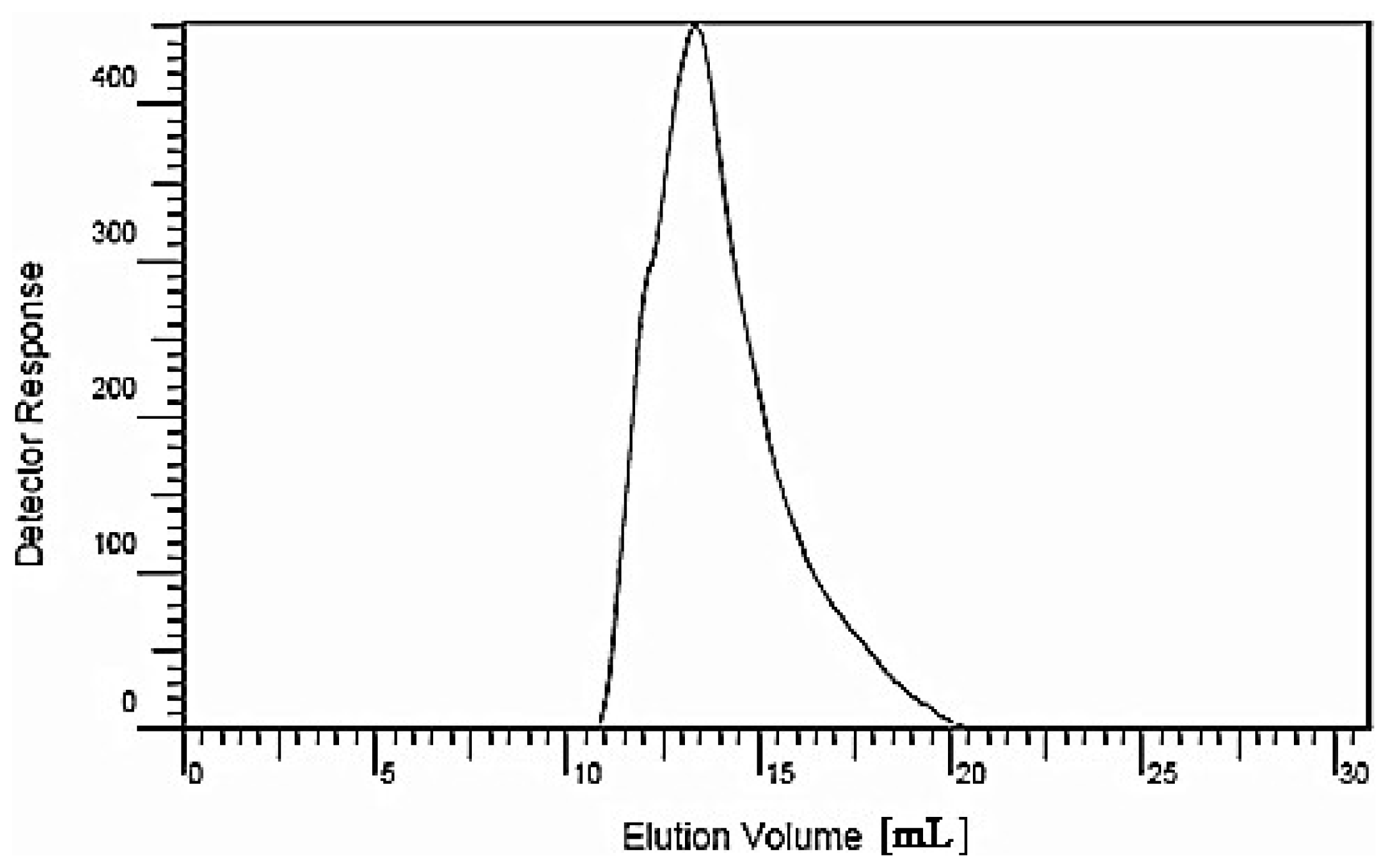
2.1. Isolation, Purification and Molecular Weight of the Polysaccharide
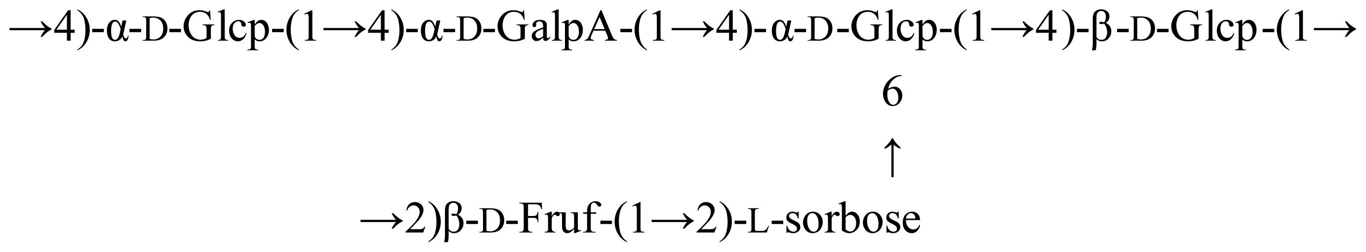
2.2. Chemical Structure
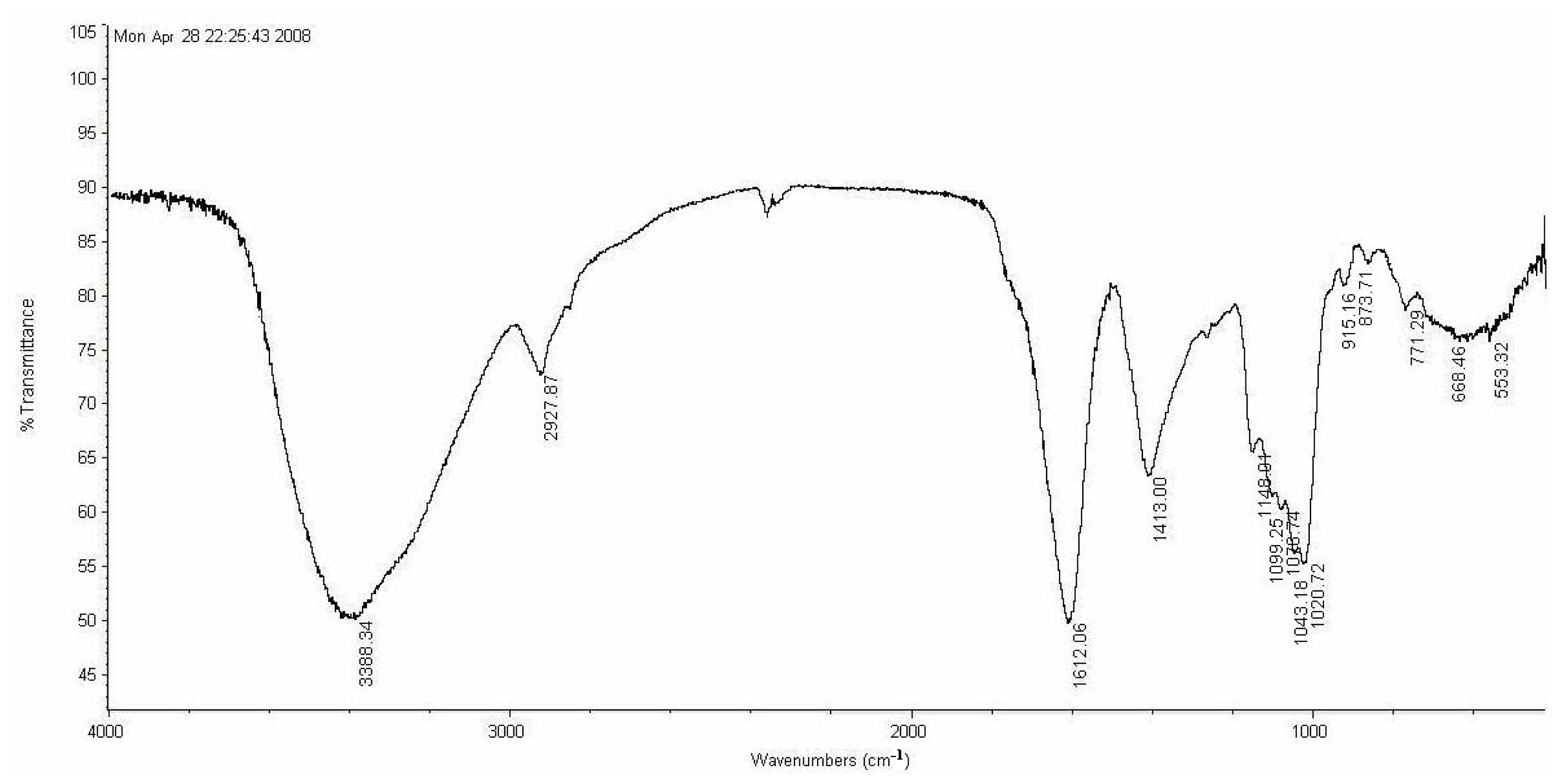
2.2.1. Fourier Transform Infrared (FT-IR) Spectrum Characterization
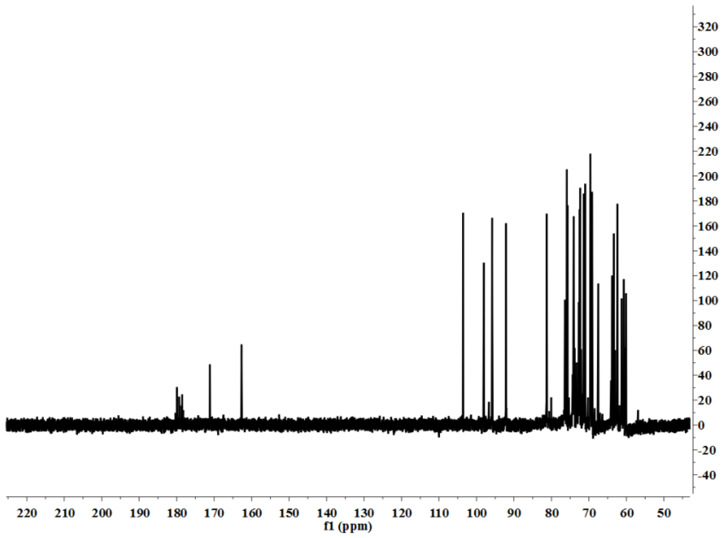
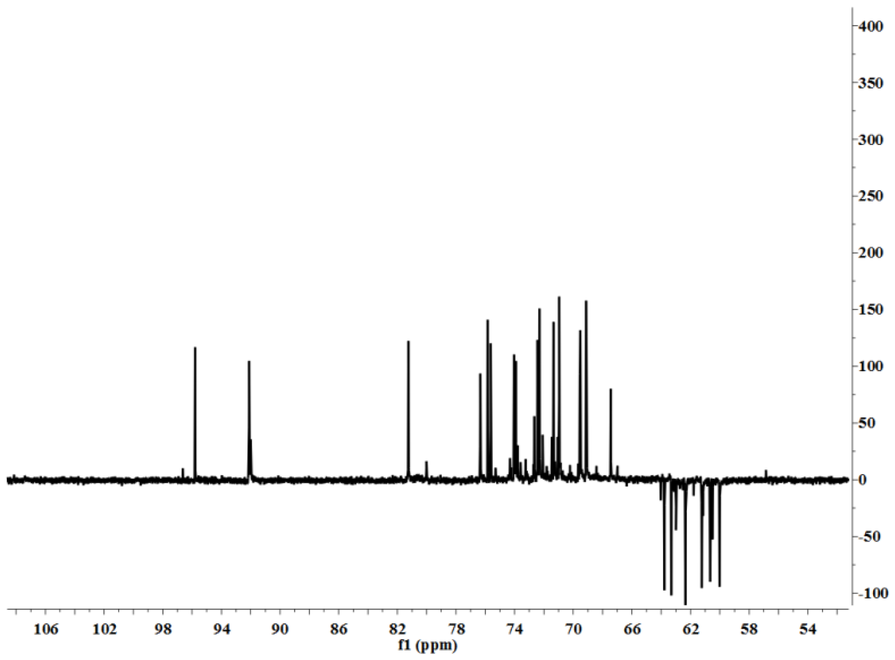
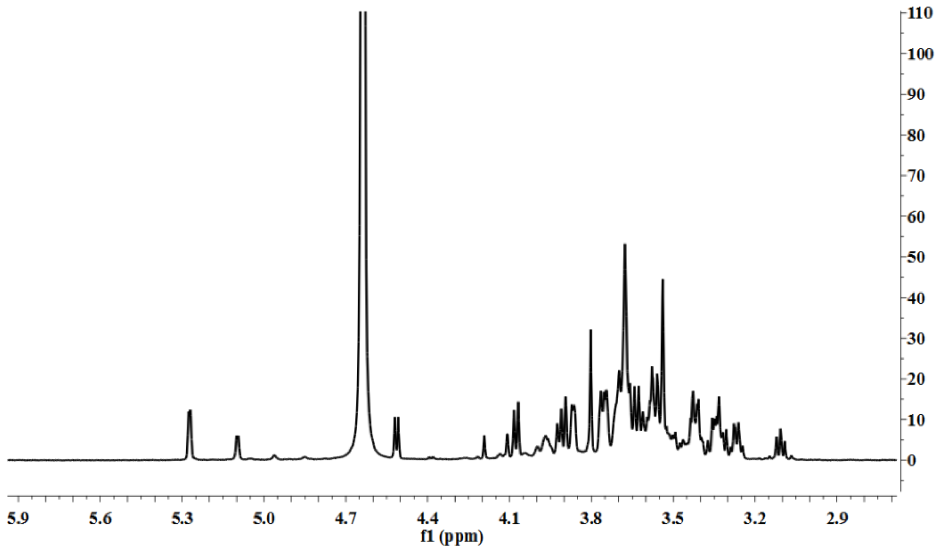
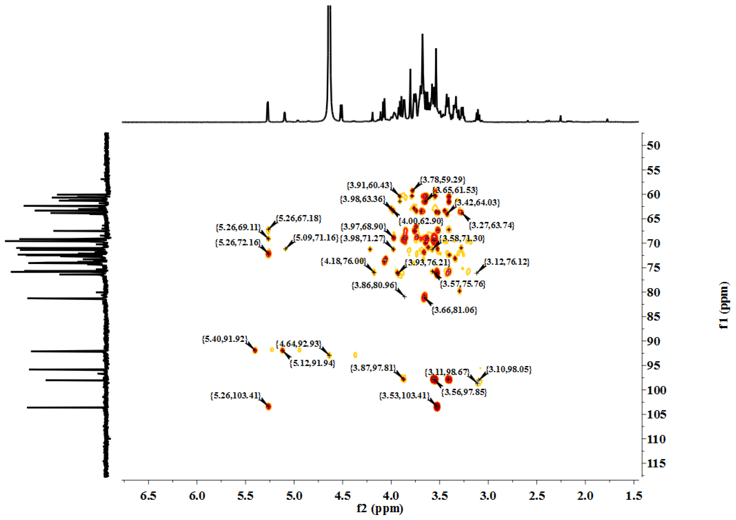
2.2.2. NMR Spectroscopic Analysis
2.3. Activity
2.3.1. In Vitro Inhibition of Tumor Cell Proliferation
2.3.2. Effect of LP1 on Macrophage Activity in Vitro
2.3.3. Immunomodulatory Activities in Vivo
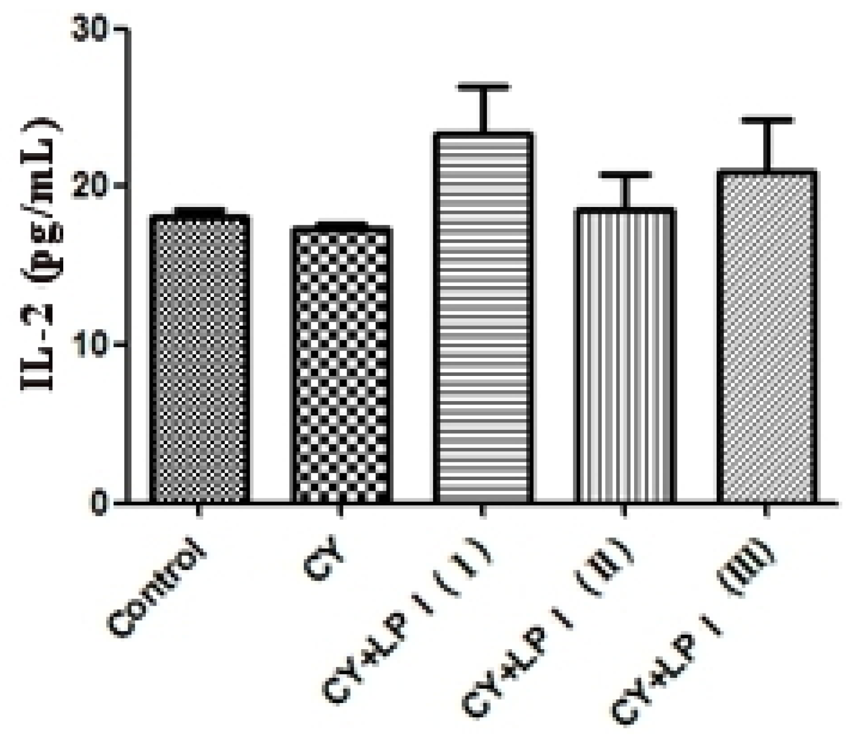
2.3.3.1. Effect of LP1 on the Stimulation of Cytokine Production
2.3.3.2. Effects on Macrophage Phagocytosis
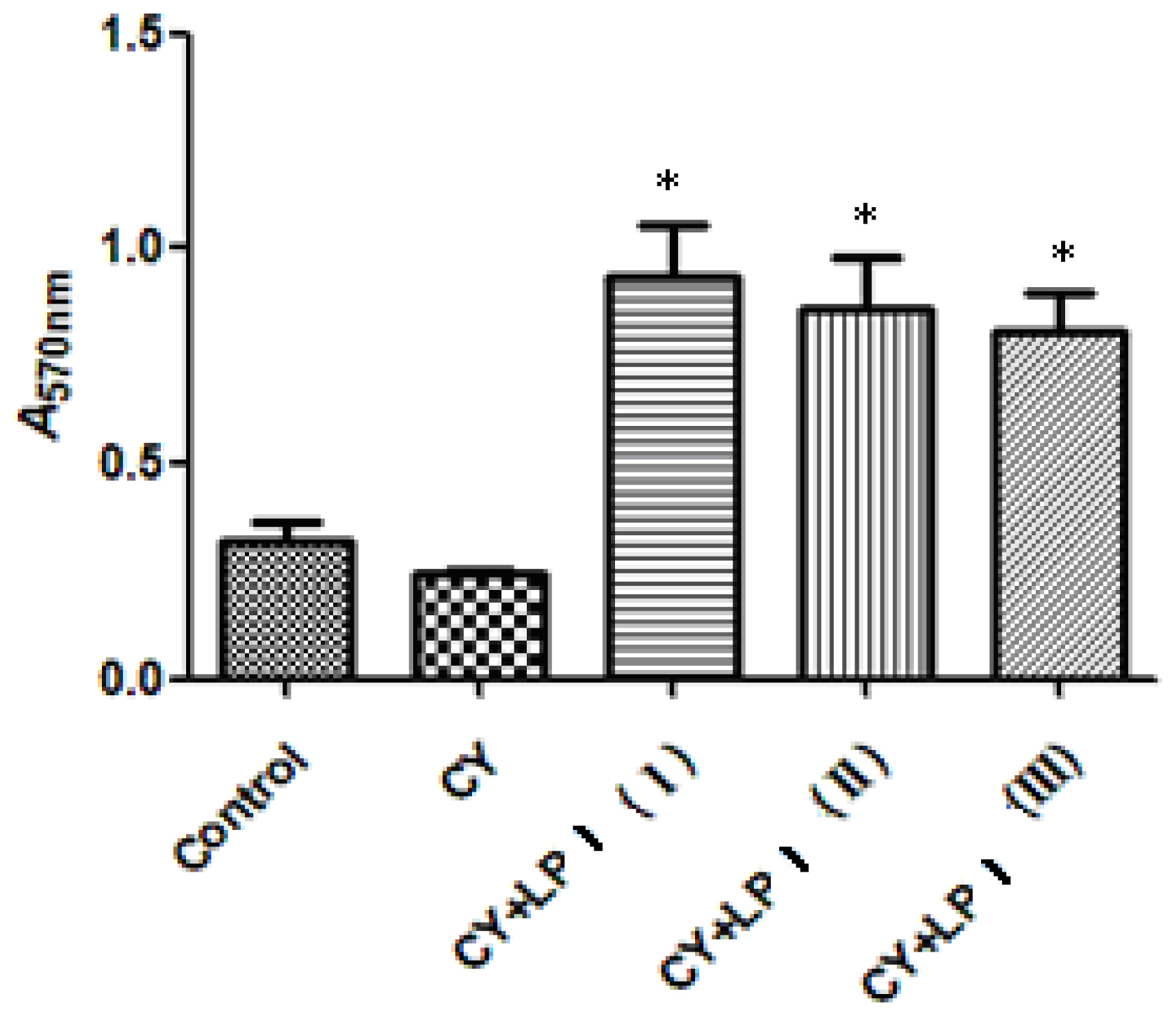
2.3.3.3. Effects of LP1 on Spleen Lymphocyte Proliferation
3. Experimental Section
3.1. Materials
3.2. Isolation and Purification of Polysaccharides
3.3. HPGPC to Determine the Molecular Weight of the Polysaccharide
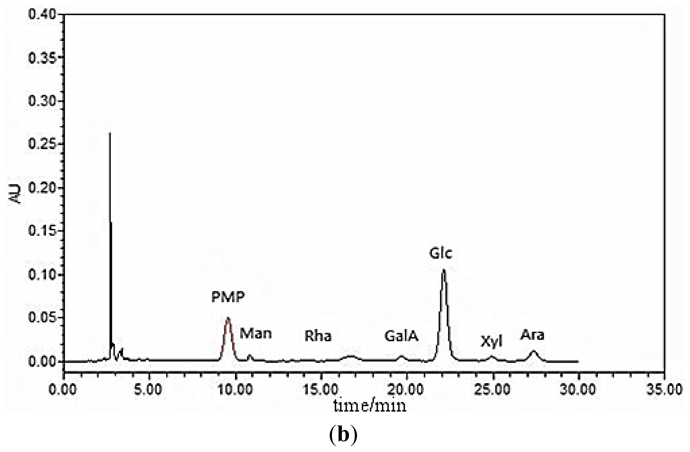
3.4. Monosaccharide Composition
3.5. Characterization
3.6. Biological Assays
3.6.1. Anti-Tumor Activity in Vitro
3.6.1.1. Cell Lines and Culture
3.6.1.2. Assay of the Inhibition of Tumor Cell Proliferation in Vitro
3.6.1.3. Effect of LP1 on the Proliferation of Macrophages in Vitro
3.6.2. Animal Immune Experiments
3.6.2.1. Chemicals and Animals
3.6.2.2. Cyclophosphamide-Induced Immunosuppression Mouse Model
3.6.2.3. Measurement of IL-2 and IFN-γ
3.6.2.4. Macrophage Phagocytosis Assay
3.6.2.5. Analysis of B- and T-Cell Proliferation
3.7. Statistics
4. Conclusions
Acknowledgments
Conflicts of Interest
References
- Yia, Y.; Zhang, M.W.; Liao, S.T.; Zhang, R.F.; Deng, Y.Y.; Wei, Z.C.; Tang, X.J.; Zhang, Y. Structural features and immunomodulatory activities of polysaccharides of longan pulp. Carbohydr. Polym. 2012, 87, 636–643. [Google Scholar]
- Park, S.J.; Park, D.H.; Kim, D.H.; Lee, S.; Yoon, B.H.; Jung, W.Y.; Lee, K.T.; Cheong, J.H.; Ryu, J.H. The memory-enhancing effects of Euphoria longan fruit extract in mice. J. Ethnopharmacol. 2010, 128, 160–165. [Google Scholar]
- Xu, L.Z.; Wang, H.G.; Geng, X.F.; Leng, P. The effect of ethanol extract pseudo-seed coat of Euphoria longan steud on pituitary-gonad axis in female rats. Chin. Med. 2002, 10, 57–59. [Google Scholar]
- Yang, B.; Zhao, M.M.; Shi, J.; Yang, N.; Jiang, Y.M. Effect of ultrasonic treatment on the recovery and DPPH radical scavenging activity of polysaccharides from longan fruit pericarp. Food Chem. 2008, 106, 685–690. [Google Scholar]
- Zhong, K.; Wang, Q.; He, Y.; He, X. Evaluation of radicals scavenging immunity-modulatory and antitumor activities of longan polysaccharides with ultrasonic extraction on in S180 tumor mice models. Int. J. Biol. Macromol. 2010, 47, 356–360. [Google Scholar]
- Zhang, X.H.; Liu, L.; Lin, C.W. Structural features antioxidant and immunological activity of a new polysaccharide (SP1) from sisal residue. Int. J. Biol. Macromol. 2013, 59, 184–191. [Google Scholar]
- Huang, Y.; Jiang, C.M.; Hu, Y.L.; Zhao, X.J.; Shi, C.; Yu, Y.; Liu, C.; Tao, Y.; Pan, H.R.; Feng, Y.B.; et al. Immunoenhancement effect of rehmannia glutinosa polysaccharide on lymphocyte proliferation and dendritic cell. Carbohydr. Polym. 2013, 96, 516–521. [Google Scholar]
- Jiang, G.X.; Prasad, K.N.; Jiang, Y.M.; Yang, B.; Jia, Y.X.; Sun, J. Extraction and structural identification of alkali-soluble polysaccharides of longan (Dimocarpus longan Lour) fruit pericarp. Innov. Food Sci. Emerg. Technol. 2009, 10, 638–642. [Google Scholar]
- Yang, B.; Zhao, M.M.; Prasad, K.N.; Jiang, G.X.; Jiang, Y.M. Effect of methylation on the structure and radical scavenging activity of polysaccharides from longan (Dimocarpus longan Lour) fruit pericarp. Food Chem 2010, 118, 364–368. [Google Scholar]
- Kardošov, A.; Machová, E. Antioxidant activity of medicinal plant polysaccharides. Fitoterapia 2006, 77, 367–373. [Google Scholar]
- Yi, Y.; Liao, S.T.; Zhang, M.W.; Shi, J.; Zhang, R.F.; Deng, Y.Y.; Wei, Z.C. Physicochemical characteristics and immunomodulatory activities of three polysaccharide-protein complexes of longan pulp. Molecules 2011, 16, 6148–6164. [Google Scholar]
- Li, S.G.; Wang, D.G.; Tian, W.; Wang, X.X.; Zhao, J.X.; Liu, Z.; Chen, R. Characterization and anti-tumor activity of a polysaccharide from Hedysarum polybotrys Hand-Mazz. Carbohydr. Polym 2008, 73, 344–350. [Google Scholar]
- Chen, J.; Zhang, L.; Yu, D.; Zhu, R. The chemical structure and solution properties of polysaccharides from Ganoderma lucidum mycelium. Chem. J. Chin. Univ. 2000, 21, 961–964. [Google Scholar]
- Englyst, H.N.; Quigley, M.E.; Hudson, G.J. Determination of dietary fiber as non-starch polysaccharides with gas-liquid chromatographic high-performance liquid chromatographic or spectrophotometric measurement of constituent sugars. Analyst 1994, 119, 1497–1509. [Google Scholar]
- Ruan, Z.; Su, J.; Dai, H.C.; Wu, M.C. Characterization and immunomodulating activities of polysaccharide from Lentinus edodes. Int. Immunopharmacol 2005, 5, 811–820. [Google Scholar]
- Cao, W.; Li, X.Q.; Liu, L.; Yang, T.H.; Li, C.; Fan, H.T.; Jia, M.; Lv, Z.G.; Mei, Q.B. Structure of an anti-tumor polysaccharide from Angelica sinensis (Oliv) Diels. Carbohydr. Polym 2006, 66, 149–159. [Google Scholar]
- Han, X.Q.; Chai, X.Y.; Jia, Y.M.; Han, C.X.; Tu, P.F. Structure elucidation and immunological activity of a novel polysaccharide from the fruit bodies of an edible mushroom Sarcodon aspratus (Berk) S Ito. Int. J. Biol. Macromol 2010, 47, 420–424. [Google Scholar]
- Borchers, A.T.; Stern, J.S.; Hackman, R.M.; Keen, C.L.; Gershwin, M.E. Mushrooms tumors and immunity. Proc. Soc. Exp. Biol. Med. 1999, 221, 281–293. [Google Scholar]
- Etaiw, S.E.H.; Sultan, A.S.; El-bendary, M.M. In vitro and in vivo antitumor activity of novel 3D-organotin supramolecular coordination polymers based on CuCN and pyridine bases. J. Organomet. Chem 2011, 696, 1668–1676. [Google Scholar]
- Bao, X.L.; Yuan, H.; Wang, C.Z.; Liu, J.J.; Lan, M.B. Antitumor and immunomodulatory activities of a polysaccharide from Artemisia argyi. Carbohydr. Polym 2013, 98, 1236–1243. [Google Scholar]
- Sun, X.; Gao, R.L.; Xiong, Y.K.; Huang, Q.C.; Xu, M. Antitumor and immunomodulatory effects of a water-solublepolysaccharide from Lilii bulbus in mice. Carbohydr. Polym 2014, 102, 543–549. [Google Scholar]
- Yang, C.; He, N.; Ling, X.; Ye, M.; Zhang, C.; Shao, W.; Yao, C.; Wang, Z.; Li, Q. The isolation and characterization of polysaccharides from longan pulp. Sep. Purif. Technol 2008, 63, 226–230. [Google Scholar]
- Shao, P.; Chen, X.X.; Sun, P.L. In vitro antioxidant and antitumor activities of different sulfatedpolysaccharides isolated from three algae. Int. J. Biol. Macromol. 2013, 62, 155–161. [Google Scholar]
- Miao, S.S.; Mao, X.H.; Pei, R.; Miao, S.P.; Xiang, C.; Lv, Y.J.; Yang, X.G.; Sun, J.; Jia, S.S.; Liu, Y.P. Antitumor activity of polysaccharides from Lepista sordida against laryngocarcinoma in vitro and in vivo. Int. J. Biol. Macromol. 2013, 60, 235–240. [Google Scholar]
- Zong, A.Z.; Cao, H.Z.; Wang, F.S. Anticancer polysaccharides from natural resources: A review of recent research. Carbohydr. Polym 2012, 90, 1395–1410. [Google Scholar]
- Kim, H.S.; Kim, Y.J.; Lee, H.K.; Ryu, H.S.; Kim, J.S.; Yoon, M.J.; Kang, J.S.; Hong, J.T.; Kim, Y.S.; Han, S.B. Activation of macrophages by polysaccharide isolated from Paecilomyces cicadae through toll-like receptor 4. Food Chem. Toxicol. 2012, 50, 3190–3197. [Google Scholar]
- Gopinath, V.K.; Musa, M.; Samsudin, A.R.; Sosroseno, W. Role of interleukin-1β and tumour necrosis factor-α on hydroxyapatite-induced phagocytosis by murine macrophages (RAW2647 cells). Br. J. Biomed. Sci 2006, 63, 176–178. [Google Scholar]
- Zirk, N.M.; Hashmi, S.F.; Ziegler, H.K. The polysaccharide portion of lipopolysaccharide regulates antigen-specific T-cell activation via effects on macrophage-mediated antigen processing. Infect. Immun 1999, 67, 319–326. [Google Scholar]
- Huang, M.; Mei, X.; Zhang, S. Mechanism of nitric oxide production in macrophages treated with medicinal mushroom extracts (review). Int. J. Med. Mushrooms 2011, 13, 1–6. [Google Scholar]
- Medzhitov, R.; Janeway, C. Innate immune recognition: Mechanisms and pathways. Immunol. Rev. 2000, 173, 89–97. [Google Scholar]
- Commins, S.P.; Borish, L.; Steinke, J.W. Immunologic messenger molecules: Cytokines interferons and chemokines. J. Allergy Clin. Immunol. 2010, 125, S53–S72. [Google Scholar]
- Lee, J.S.; Synytsya, A.; Kim, H.B.; Choi, D.J.; Lee, S.; Lee, J.S.; Kim, W.J.; Jang, S.J.; Park, Y.I. Purification characterization and immunomodulating activity of a pectic polysaccharide isolated from Korean mulberry fruit Oddi (Morus alba L). Int. Immunopharmacol. 2013, 17, 858–866. [Google Scholar]
- Adachi, Y.; Okazaki, M.; Ohno, N.; Yadomae, T. Enhancement of cytokine production by macrophages stimulated with (1→3)-β-d-glucan grifolan (GRN) isolated from Grifola frondosa. Biol. Pharm. Bull 1994, 17, 1554–1560. [Google Scholar]
- Derin, D.; Soydinc, H.O.; Guney, N.; Tas, F.; Çamlıca, H.; Duranyıldız, D.; Yasasever, V.; Topuz, E. Serum levels of apoptosis biomarkers survivin and TNF-α in nonsmall celllung cancer. Lung Cancer 2008, 59, 240–245. [Google Scholar]
- Yun, U.J.; Park, S.E.; Jo, Y.S.; Kim, J.; Shin, D.Y. DNA damage induces the IL-6/STAT3 signaling pathway which has anti-senescence and growth-promotingfunctions in human tumors. Cancer Lett. 2012, 323, 155–160. [Google Scholar]
- Wagley, Y.; Yoo, Y.C.; Seo, H.G.; Rhee, M.H.; Kim, T.H.; Kang, K.W.; Nah, S.Y.; Oh, J.W. The IL-6/sIL-6R treatment of a malignant melanoma cell line enhances susceptibility to TNF-α-induced apoptosis. Biochem. Biophys. Res. Commun. 2007, 354, 985–991. [Google Scholar]
- Suzanne, O.R. Immune surveillance: A balance between pro- and anti-tumor immunity. Curr. Opin. Genet. Dev. 2008, 18, 11–18. [Google Scholar]
- Li, X.; Jiao, L.; Zhang, X.; Tian, W.; Chen, S.; Zhang, L. Anti-tumor and immunomodulating activities of proteoglycans from mycelium of Phellinus nigricans and culture medium. Int. Immunopharmacol 2008, 8, 909–915. [Google Scholar]
- Xie, G.; Schepetkin, I.A.; Quinn, M.T. Immunomodulatory activity of acidic polysaccharides isolated from Tanacetum vulgare L. Int. Immunopharmacol. 2007, 7, 1639–1650. [Google Scholar]
- Sakurai, M.H.; Matsumoto, T.; Kiyohara, H.; Yamada, H. B-cell proliferation activity of pectic polysaccharide from a medicinal herb the roots of Bupleurum falcatum L and its structural requirement. Immunology 1999, 97, 540–547. [Google Scholar]
- Schepetkin, I.A.; Quinn, M.T. Botanical polysaccharides: Macrophage immunomodulation and therapeutic potential. Int. Immunopharmacol. 2006, 6, 317–333. [Google Scholar]
- Katsiari, C.G.; Liossis, S.C.; Sfikakis, P.P. The pathophysiologic role of monocytes and macrophages in systemic lupus erythematosus: A reappraisal. Arthritis Rheumatol 2010, 39, 491–503. [Google Scholar]
- Zhang, C.; Huang, K. Characteristic immunostimulation by MAP a polysaccharide isolated from the mucus of the loach Misgurnus anguillicaudatus. Carbohydr. Polym 2005, 59, 75–82. [Google Scholar]
- Mao, W.J.; Li, B.F.; Gu, Q.Q.; Fang, Y.C.; Xing, H.T. Preliminary studies on the chemical characterization and antihyperlipidemic activity of polysaccharide from the brown alga Sargassum fusiforme. Hydrobiologia 2004, 512, 263–266. [Google Scholar]
- Hoebe, K.; Janssen, E.M.; Kim, S.O.; Alexopoulou, L.; Flavell, R.A.; Han, J.; Beutler, B. Upregulation of costimulatory molecules induced by lipopolysaccharide and double-stranded RNA occurs by Trif-dependent and Trif-independent pathways. Nat. Immunol 2003, 4, 1223–1229. [Google Scholar]
- Yang, H.C. Animal Immunology; Chinese Agricultural University Press: Beijing, China, 2003; pp. 95–100. [Google Scholar]
- Mosmann, T.; Coffman, R. TH1 and TH2 cells: Different patterns of lymphokine secretion lead to different functional properties. Annu. Rev. Immunol 1989, 7, 145–173. [Google Scholar]
- Sun, Y.; Liu, J. Purification structure and immunobiological activity of a water-soluble polysaccharide from the fruiting body of Pleurotus ostreatus. Bioresour. Technol 2009, 100, 983–986. [Google Scholar]
- Alam, N.; Gupta, P.C. Structure of a water-soluble polysaccharide from the seeds of Cassia angustifolia. Planta Medica 1986, 52, 308–310. [Google Scholar]
- Dubois, M.; Gilles, K.A.; Hamilton, J.K.; Rebers, P.T.; Smith, F. Colorimetric method for determination of sugars and related substances. Anal. Chem 1956, 28, 350–356. [Google Scholar]
- Wang, L.; Huang, H.Y.; Wei, Y.Y.; Li, X.X.; Chen, Z.X. Characterization and anti-tumor activities of sulfated polysaccharide SRBPS2a obtained from defatted rice bran. Int. J. Biol. Macromol. 2009, 45, 427–431. [Google Scholar]
- Ang, A.S.W.; Cheung, R.C.F.; Dan, X.; Chan, Y.S.; Pan, W.; Ng, T.B. Purification and characterization of a glucosamine-binding antifungal lectin from Phaseolus vulgaris cv Chinese pinto beans with antiproliferative activity towards nasopharyngeal carcinoma cells. Appl. Biochem. Biotechnol 2014, 172, 672–686. [Google Scholar]
- Chen, W.; Zhang, W.; Shen, W.; Wang, K. Effects of the acid polysaccharide fraction isolated from a cultivated Cordyceps sinensis on macrophages in vitro. Cell. Immunol 2010, 262, 69–74. [Google Scholar]
- Belska, N.V.; Guriev, A.M.; Danilets, M.G.; Trophimova, E.S.; Uchasova, E.G.; Ligatcheva, A.A.; Belousov, M.V.; Agaphonov, V.I.; Golovchenko, V.G.; Yusubov, M.S.; et al. Water-soluble polysaccharide obtained from Acorus calamus L classically activates macrophages and stimulates Th1 response. Int. Immunopharmacol. 2010, 10, 933–942. [Google Scholar]
- Green, L.C.; Wagner, D.A.; Glogowski, J.; Skipper, P.L.; Wishnok, J.S.; Tannenbaum, S.R. Analysis of nitrate nitrite and [15N]nitrate in biological fluids. Anal. Biochem 1982, 126, 131–138. [Google Scholar]
- Mazzone, G.L.; Rigato, I.; Tiribelli, C. Unconjugated bilirubin modulates nitric oxide production via iNOS regulation. Biosci. Trends 2010, 4, 244–248. [Google Scholar]
- Babette, W.; Hannah, M.; Jennifer, K.; Zaahira, G.; Adriana, M.; Max, W.; Frank, B.; Stewart, J.; Jennifer, S.; Andrea, K. A whole blood in vitro cytokine release assay with aqueous monoclonal antibody presentation for the prediction of therapeutic protein induced cytokine release syndrome in humans. Cytokine 2012, 60, 828–837. [Google Scholar]
- Wang, J.P.; Lin, K.H.; Liu, C.Y.; Yu, Y.C.; Wu, P.T.; Chiu, C.C.; Su, C.L.; Chen, K.M.; Fang, K. Teroxirone inhibited growth of human non-small cell lung cancer cells by activating p53. Toxicol. Appl. Pharm 2013, 273, 110–120. [Google Scholar]
- Yi, L.; Su, Q. Molecular mechanisms for the anti-cancer effects of diallyl disulfide. Food Chem. Toxicol. 2013, 57, 362–370. [Google Scholar]
- Wang, D.Y.; Li, X.R.; Xu, L.X.; Hu, Y.L.; Zhang, B.K.; Liu, J.G. Immunologic synergism with IL-2 and effects of cCHMIs on mRNA expression of IL-2 and IFN-γ in chicken peripheral T lymphocyte. Vaccine 2006, 24, 7109–7114. [Google Scholar]
- Liu, Z.H.; Lv, C.L. Immunology Experimental Technology; Beijing Science Press: Beijing, China, 2002; p. 90. [Google Scholar]
- Fan, Y.P.; Hu, Y.L.; Wang, D.Y.; Liu, J.G.; Zhang, J.; Zhao, X.J.; Liu, X.; Liu, C.; Yuan, J.; Ruan, S.L. Effects of Astragalus polysaccharide liposome on lymphocyte proliferation in vitro and adjuvanticity in vivo. Carbohydr. Polym 2012, 88, 68–74. [Google Scholar]


| Glycosyl residues | H-1/C-1 | H-2/C-2 | H-3/C-3 | H-4/C-4 | H-5/C-5 | H-6a, H-6b/C-6 |
|---|---|---|---|---|---|---|
| →4)-α-d-Glcp-(1→ | 5.27/92.11 | 3.42/70.99 | 3.56/72.85 | 3.89/73.84 | 3.74/75.81 | 3.66,3.64/60.65 |
| 5.09/92.00 | 3.40/70.99 | 3.56/72.85 | 3.87/74.24 | 3.77/72.46 | 3.67, 3.57/60.55 | |
| →4,6)-β-d-Glcp-(1→ | 4.51/95.79 | 3.12/73.78 | 3.34/75.51 | 3.75/72.12 | 3.58/72.46 | 3.86,3.92/63.28 |
| →2)-β-d-Fruf-(1→ | 3.56,3.74/63.78 | –/103.59 | 4.07/76.23 | 3.92/74.57 | 3.74/81.27 | 3.64,3.59/62.99 |
| →2)-l-sorbose-(1→ | 3.56,3.66/64.04 | –/98.03 | 3.70/70.99 | 3.56/74.33 | 3.52/71.03 | 3.75,3.59/62.35 |
| Glycosyl residues | δH/δC atom | Observed connectivities |
|---|---|---|
| δH/δC atom | ||
| A→4)-α-d-Glcp-(1→ | 5.27 A-H1 | 72.12 B-C4 |
| A′→4)-α-d-Glcp-(1→ | 5.09 A′-H1 | 71.11 |
| B→4,6)-β-d-Glcp-(1→ | 4.51 B-H1 | – |
| C→2)-β-d-Fruf-(1→ | 103.41 C-C2 | 3.56 D-H1 |
| D→2)-l-sorbose-(1→ | 98.03 D-C2 | 3.56 D-H3 3.86 B-H6 |
| Group | Concentration (mg/L) | A570 nm | Inhibition (%) |
|---|---|---|---|
| Control | – | 0.799 ± 0.005 | 0 |
| 5-Fluorouracil (5-FU) | 5 | 0.350 ± 0.018 ** | 56.2 |
| LP1 | 40 | 0.480 ± 0.017 ** | 39.9 |
| 20 | 0.494 ± 0.007 ** | 38.2 | |
| 10 | 0.496 ± 0.005 ** | 37.9 | |
| 5 | 0.504 ± 0.002 ** | 36.9 |
| Group | Concentration (mg/L) | A570 nm | Inhibition (%) |
|---|---|---|---|
| Control | – | 0.610 ± 0.006 | 0 |
| 5-FU | 5 | 0.284 ± 0.007 ** | 53.4 |
| LP1 | 320 | 0.303 ± 0.008 ** | 50.3 |
| 160 | 0.328 ± 0.004 ** | 46.2 | |
| 80 | 0.330 ± 0.006 ** | 45.9 | |
| 40 | 0.355 ± 0.008 ** | 41.8 |
| Group | Concentration (μg/mL) | Phagocytic count (OD570 nm) |
|---|---|---|
| Blank control group | – | 0.184 ± 0.0071 |
| LPS-positive group | 10 | 0.306 ± 0.0167 * |
| LP1 | 25 | 0.293 ± 0.0128 * |
| 50 | 0.312 ± 0.0132 * | |
| 100 | 0.329 ± 0.0097 * |
| Group | Concentration (μg/mL) | NO production (μM) |
|---|---|---|
| Blank control group | – | 7.51 ± 0.781 |
| LPS-positive group | 10 | 25.05 ± 1.082 * |
| LP1 | 25 | 18.85 ± 1.012 * |
| 50 | 20.75 ± 0.976 * | |
| 100 | 22.85 ± 0.998 * |
| Group | Concentration (μg/mL) | TNF-α concentration (pg/mL) |
|---|---|---|
| Blank control group | – | 172.85 ± 17.09 |
| LPS-positive group | 10 | 560.65 ± 34.31 * |
| LP1 | 25 | 427.65 ± 22.64 * |
| 50 | 451.22 ± 26.98 * | |
| 100 | 497.65 ± 24.46 * |
| Group | Concentration (μg/mL) | IL-6 concentration (pg/mL) |
|---|---|---|
| Blank control group | – | 20.13 ± 1.69 |
| LPS-positive group | 10 | 59.55 ± 4.19 * |
| LP1 | 25 | 68.65 ± 3.64 # |
| 50 | 72.92 ± 4.08 # | |
| 100 | 78.85 ± 3.86 # |
| Group | Concentration (μg/mL) | IL-1β concentration (pg/mL) |
|---|---|---|
| Blank control group | – | 34.51 ± 2.89 |
| LPS-positive group | 10 | 64.35 ± 4.04 * |
| LP1 | 25 | 54.87 ± 3.12 * |
| 50 | 58.17 ± 2.88 * | |
| 100 | 64.14 ± 3.49 * |
| Group | A570 nm | Stimulus index 100% | |
|---|---|---|---|
| LPS stimulated | No LPS stimulation | ||
| Control | 0.321 ± 0.08 | 0.276 ± 0.10 | 116 |
| CY | 0.243 ± 0.03 | 0.226 ± 0.13 | 108 |
| CY + LP1 (I) | 0.932 ± 0.34 * | 0.637 ± 0.15 | 146 |
| CY + LP1 (II) | 0.856 ± 0.35 * | 0.681 ± 0.08 | 126 |
| CY + LP1 (III) | 0.810 ± 0.24 * | 0.673 ± 0.15 | 120 |
| Group | A570 nm | Stimulus index 100% | |
|---|---|---|---|
| ConA stimulated | No ConA stimulation | ||
| Control | 0.358 ± 0.05 | 0.276 ± 0.10 | 130 |
| CY | 0.263 ± 0.02 | 0.226 ± 0.13 | 116 |
| CY + LP1 (I) | 0.914 ± 0.13 * | 0.637 ± 0.15 | 143 |
| CY + LP1 (II) | 0.849 ± 0.28 * | 0.681 ± 0.08 | 125 |
| CY + LP1 (III) | 0.804 ± 0.21 * | 0.673 ± 0.15 | 119 |
© 2014 by the authors; licensee MDPI, Basel, Switzerland This article is an open access article distributed under the terms and conditions of the Creative Commons Attribution license (http://creativecommons.org/licenses/by/3.0/).
Share and Cite
Meng, F.-Y.; Ning, Y.-L.; Qi, J.; He, Z.; Jie, J.; Lin, J.-J.; Huang, Y.-J.; Li, F.-S.; Li, X.-H. Structure and Antitumor and Immunomodulatory Activities of a Water-Soluble Polysaccharide from Dimocarpus longan Pulp. Int. J. Mol. Sci. 2014, 15, 5140-5162. https://doi.org/10.3390/ijms15035140
Meng F-Y, Ning Y-L, Qi J, He Z, Jie J, Lin J-J, Huang Y-J, Li F-S, Li X-H. Structure and Antitumor and Immunomodulatory Activities of a Water-Soluble Polysaccharide from Dimocarpus longan Pulp. International Journal of Molecular Sciences. 2014; 15(3):5140-5162. https://doi.org/10.3390/ijms15035140
Chicago/Turabian StyleMeng, Fa-Yan, Yuan-Ling Ning, Jia Qi, Zhou He, Jiang Jie, Juan-Juan Lin, Yan-Jun Huang, Fu-Sen Li, and Xue-Hua Li. 2014. "Structure and Antitumor and Immunomodulatory Activities of a Water-Soluble Polysaccharide from Dimocarpus longan Pulp" International Journal of Molecular Sciences 15, no. 3: 5140-5162. https://doi.org/10.3390/ijms15035140
APA StyleMeng, F.-Y., Ning, Y.-L., Qi, J., He, Z., Jie, J., Lin, J.-J., Huang, Y.-J., Li, F.-S., & Li, X.-H. (2014). Structure and Antitumor and Immunomodulatory Activities of a Water-Soluble Polysaccharide from Dimocarpus longan Pulp. International Journal of Molecular Sciences, 15(3), 5140-5162. https://doi.org/10.3390/ijms15035140