Atomic Force Microscope Imaging of the Aggregation of Mouse Immunoglobulin G Molecules

Abstract
:Introduction
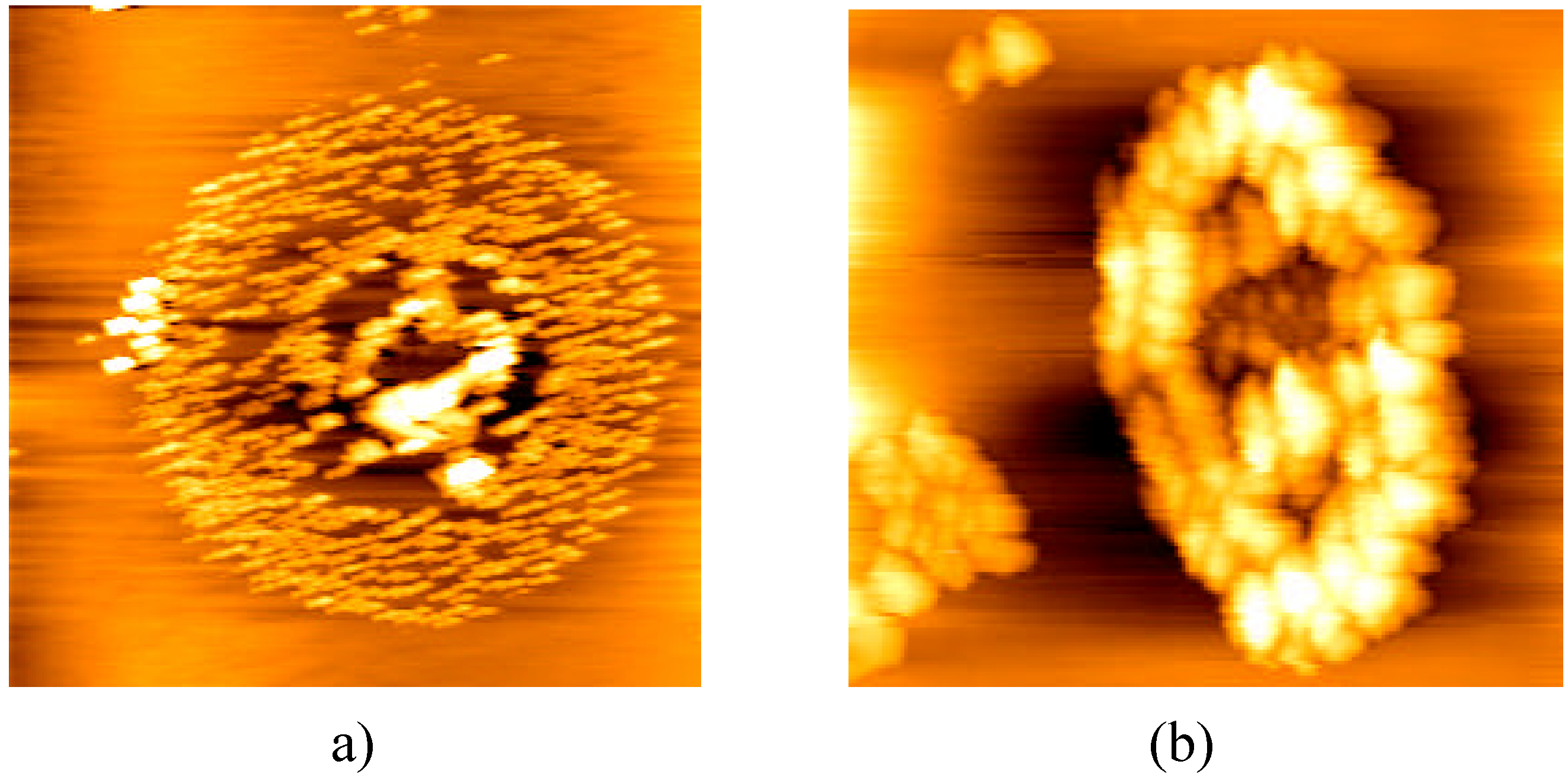
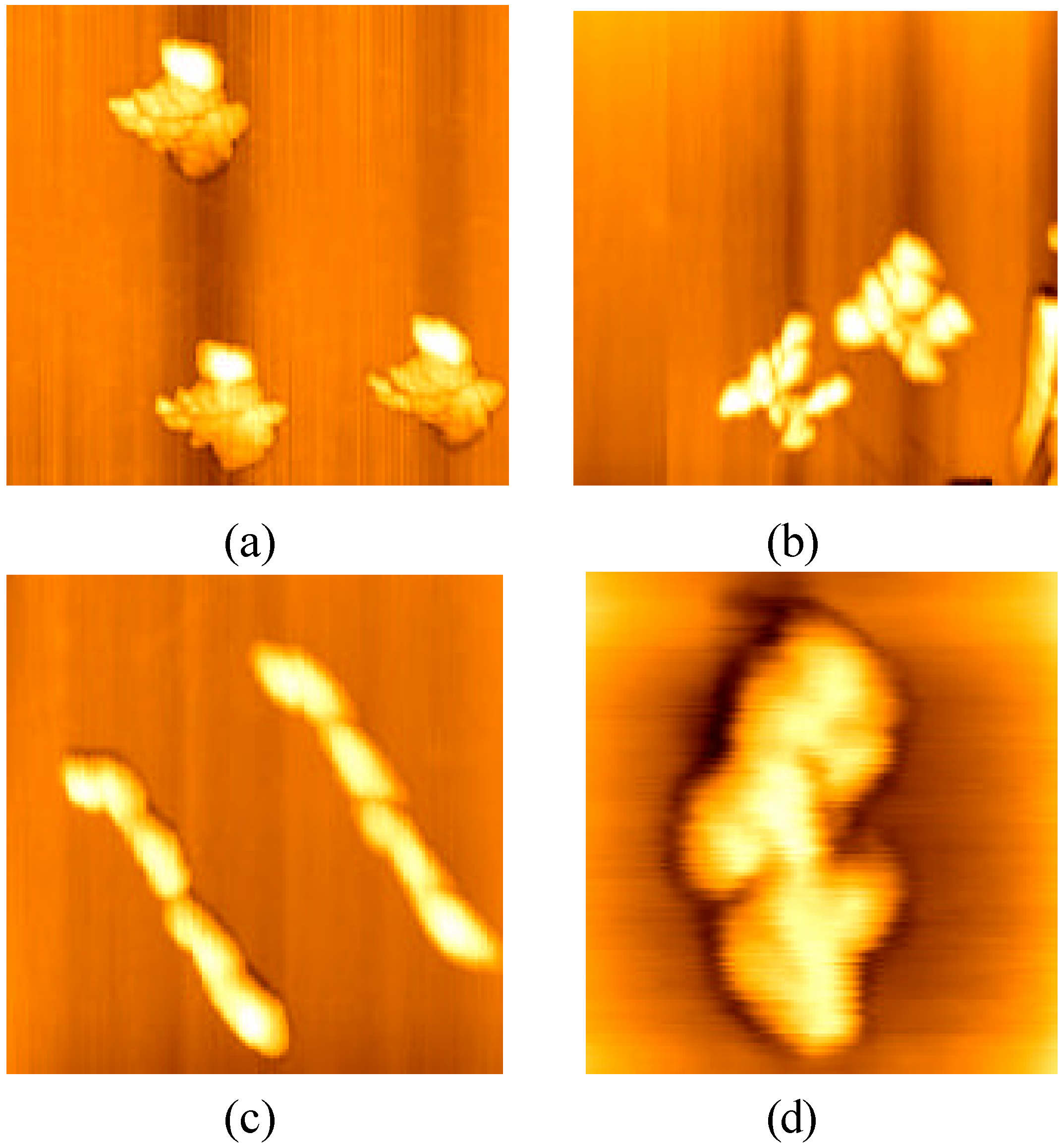
Results
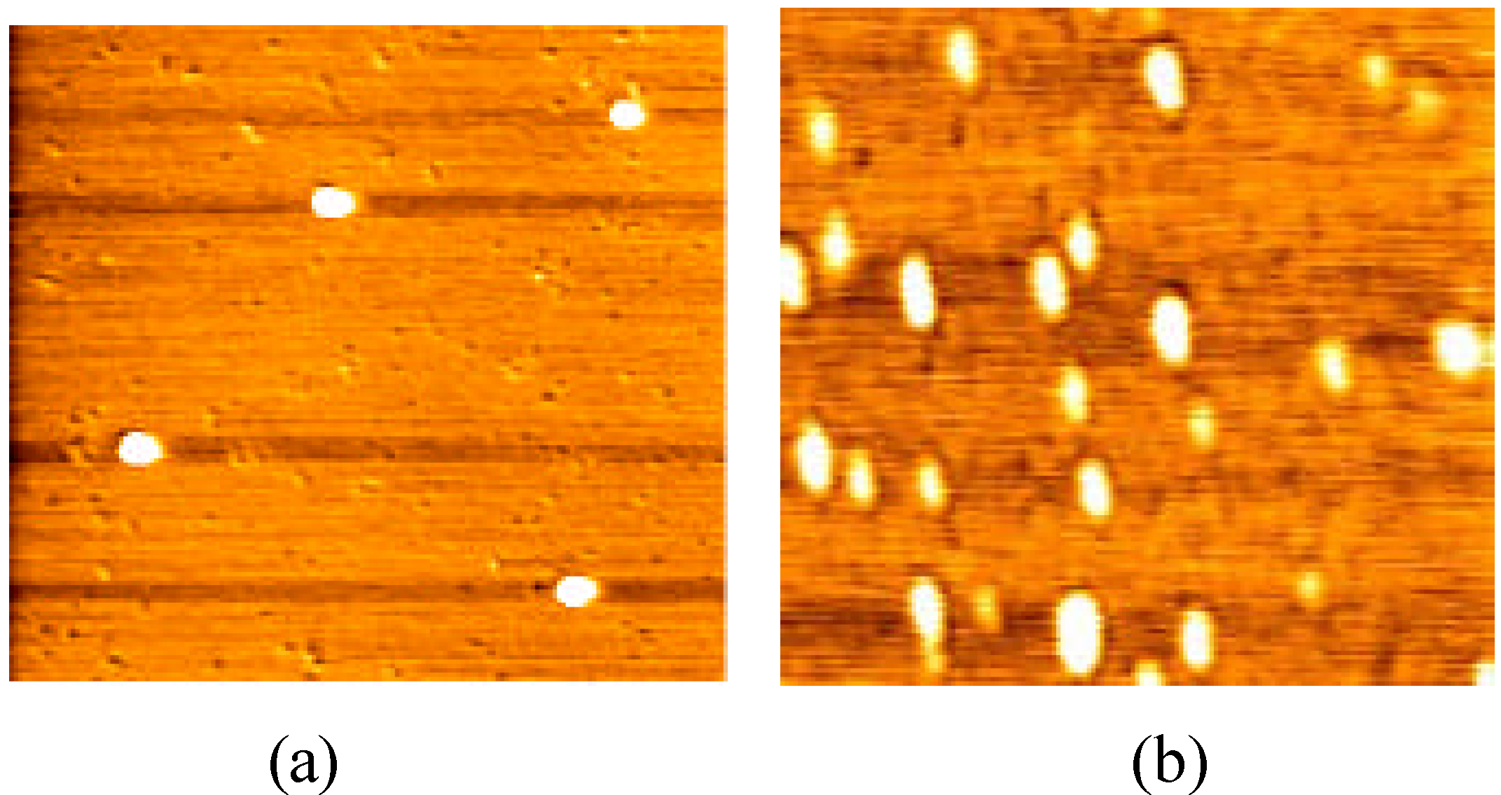
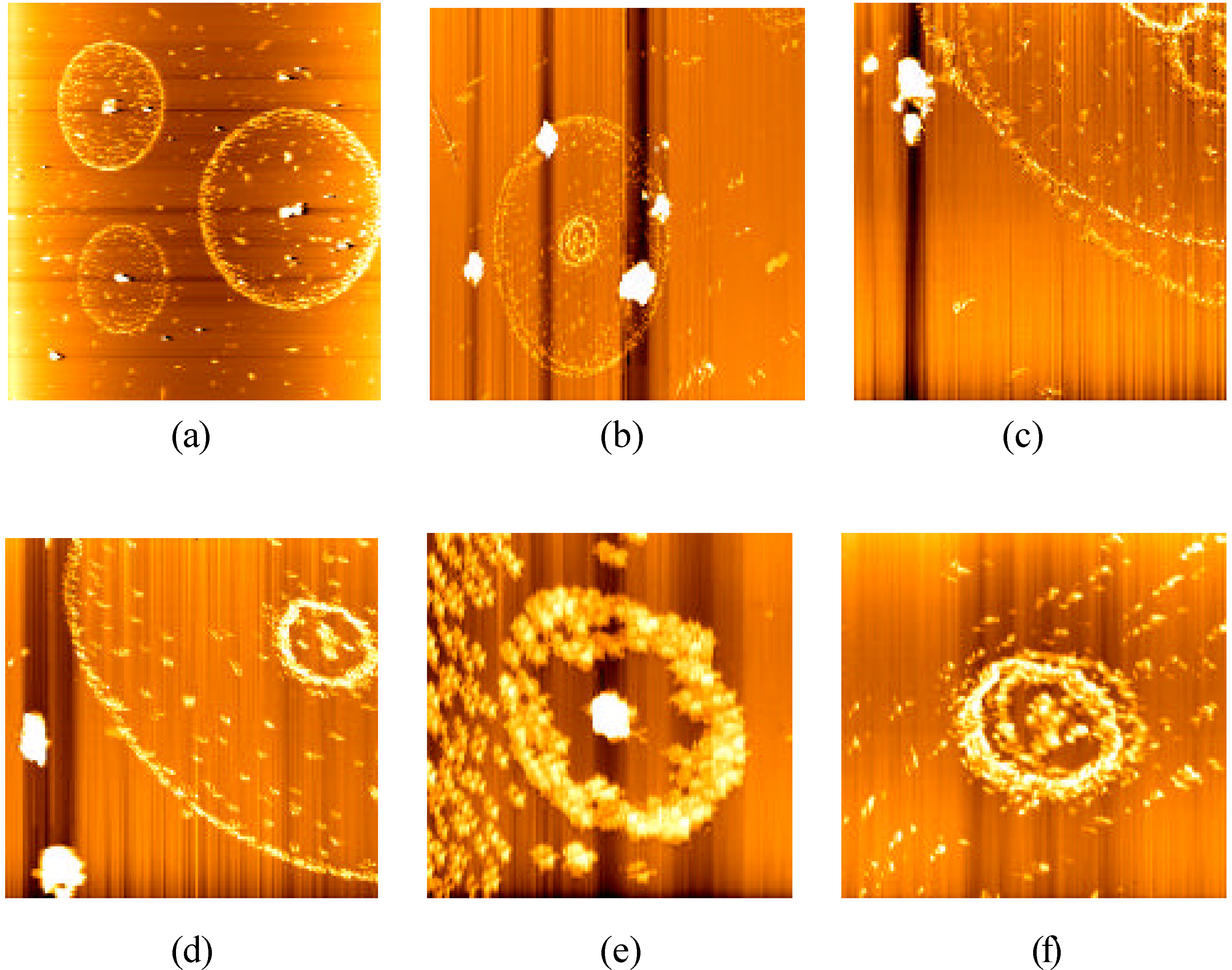


Discussion
Conclusions
Experimental
General
References and Notes
- Preininger, C.; Clausen-Schaumamn, H.; Ahluwalia, A.; De Rossi, D. Characterization of Ig G Langmuir-Blodgett films immobilized on funtionalized polymers. Talanta 2000, 52, 921–930. [Google Scholar] [PubMed]
- Disley, D.M.; Cullen, D.C.; You, H.-X.; Lowe, C.R. Covalent coupling of immunoglobulin G to self-assembled monolayers as a method for immobilizing the interfacial-recognition layer of a surface plasmon resonance immunosensor. Biosensors & Bioelectronics 1998, 13, 1213–1225. [Google Scholar] [CrossRef]
- Hansma, H.G.; Vesenka, J.; Siegerist, C.; Kelderman, G.; Morrett, H.; Shinsheimer, R.L.; Elings, V.; Bustamante, C.; Hansma, P.K. Reproducible Imaging and Dissection of Plasmid DNA Under Liquid With the Atomic Force Microscope. Science 1992, 256, 1180–1184. [Google Scholar]
- Drake, B.; Prater, C.B.; Weisenhorn, A.L.; Gould, S.A.C.; Albrecht, T.R.; Quate, C.F.; Cannell, D.S.; Hansma, H.G.; Hansma, P.K. Imaging crystals, polymers, and processes in water with the atomic force microscope. Science 1989, 243, 1586–1589. [Google Scholar]
- Keller, D. Reconstruction of STM and AFM images distorted by finite-size tips. Surf. Sci. 1991, 253, 353–364. [Google Scholar] [CrossRef]
- Alberts, B.; Bray, D.; Johnson, A.; Lewis, J.; Raff, M.; Roberts, K.; Walter, P. Essential Cell Biology; Garland Publishing, Inc.: New York & London, 1998; p. 157. [Google Scholar]
© 2003 by MDPI ( http://www.mdpi.org). Reproduction is permitted for noncommercial purposes.
Share and Cite
Cai, J.; Chen, Y.; Xu, Q.; Chen, Y.; Zhao, T.; Wang, X.; Xia, K. Atomic Force Microscope Imaging of the Aggregation of Mouse Immunoglobulin G Molecules. Molecules 2003, 8, 86-91. https://doi.org/10.3390/80100086
Cai J, Chen Y, Xu Q, Chen Y, Zhao T, Wang X, Xia K. Atomic Force Microscope Imaging of the Aggregation of Mouse Immunoglobulin G Molecules. Molecules. 2003; 8(1):86-91. https://doi.org/10.3390/80100086
Chicago/Turabian StyleCai, Jiye, Yao Chen, Qingcai Xu, Yong Chen, Tao Zhao, Xiaoyan Wang, and Ke Xia. 2003. "Atomic Force Microscope Imaging of the Aggregation of Mouse Immunoglobulin G Molecules" Molecules 8, no. 1: 86-91. https://doi.org/10.3390/80100086
APA StyleCai, J., Chen, Y., Xu, Q., Chen, Y., Zhao, T., Wang, X., & Xia, K. (2003). Atomic Force Microscope Imaging of the Aggregation of Mouse Immunoglobulin G Molecules. Molecules, 8(1), 86-91. https://doi.org/10.3390/80100086