1. Introduction
Epilepsy is a disorder of the central nervous system (CNS) in which the pathological state is exerted through repeated episodes of excessive neural activity, known as seizures. Through genome-wide association sequencing (GWAS) or exome sequencing studies, epilepsy-associated sequence variants in hundreds of genes have been identified [
1]. Dravet syndrome (DS) is caused by a monogenic mutation in
SCN1A, which encodes a voltage-gated sodium channel (Nav1.1). It is a clinically relevant epilepsy gene with thousands of genetic variants reported in different phenotypes. The most frequent phenotype is febrile seizures plus (FS+), and afebrile generalized tonic–clonic seizures [
2]. Most infants with DS exhibit normal development before experiencing an increase in the frequency of febrile and afebrile seizures during the first year. Eventually, symptoms progress to severe recurrent seizures, cognitive dysfunction, psychomotor impairment, and intellectual disability [
3,
4].
Although anti-seizure drugs (ASDs) such as valproate (VPA), clobazam (CLB), and stiripentol (STP) are used to alleviate seizures in DS, seizures remain poorly controlled [
5]. Despite the availability of >30 ASDs with broad categories of mechanisms, new ASDs with different mechanisms are needed to effectively reduce seizures while improving other comorbidities [
6]. DS is primarily characterized by the loss-of-function of the
SCN1A gene, which predominantly affects GABAergic inhibitory interneurons, leading to a global imbalance between excitation and inhibition in the brain. While current therapeutic strategies often focus on enhancing GABAergic neurotransmission, many patients remain refractory to these treatments. In the last decade, three ASDs were specifically available as therapeutic agents in DS: STP, cannabidiol (CBD), and fenfluramine (FFA). Although the most commonly used treatments are VPA and CLB in DS, these ASDs were escalated in efficacy as adjunctive therapy [
5]. STP received approval as a first therapy for the treatment of seizures associated with DS in 2017 in the EU and 2018 in the USA. STP is likely to have several mechanisms linked to anti-seizure properties, such as a positive allosteric modulator of GABAA receptors [
7,
8]. CBD was approved as an adjunctive treatment for seizures in DS and Lennox–Gastaut syndrome (LGS) in 2018 in the USA and in 2019 in the EU. Unlike other ASDs, CBD has distinct mechanisms in that it modulates intracellular calcium and inhibits adenosine cellular uptake [
9]. FFA, approved in 2020 in the EU and USA, is an add-on therapeutic agent for the treatment of seizures in patients with DS. Originally, FFA was used as a therapy for obesity until it was withdrawn because of increased cases of cardiac problems [
5,
10]. FFA acts variously on the serotonergic system compared with other ASDs: increasing serotonin (5-hydroxytryptamine, 5-HT) release, inhibiting the serotonin transporter, and agonist of serotonin receptors [
11]. In addition, FFA functions as a positive modulator of the sigma-1 receptor that could improve cognitive functions [
12]. Although the pharmacological profile of fenfluramine (FFA) has been a subject of ongoing debate, recent studies have shifted the consensus towards its role as a positive modulator of the sigma-1 receptor. While early zebrafish reports hypothesized a complex interaction with sigma-1 receptor [
13], current evidence supports that FFA’s agonistic activity at this receptor, alongside its serotonergic effects, contributes to its potent anti-seizure properties in SCN1A-deficient models [
12].
Several studies to generate and characterize the animal models of DS for drug screening have been reported. Zebrafish (
Danio rerio) has emerged as a promising in vivo model for biomedical research in the nervous system [
14,
15]. In addition, they are an attractive model for studying genetic diseases because approximately 70% of coding genes are conserved between humans and zebrafish [
16]. Since the mutation of the
SCN1A ortholog in zebrafish was reported, several studies have made efforts to find a small molecule for the treatment of DS [
17,
18,
19,
20,
21,
22]. The first reported
scn1lab mutant in zebrafish,
didys552, was generated by chemical mutagenesis and identified as a mutant with a point mutation [
23,
24]. FFA is selected as an anti-seizure compound by large-scale screening in a zebrafish model of DS [
18]. In addition, the zebrafish model of DS revealed that the anti-seizure effect of FFA not only acts as a 5-HT1D- and 5-HT2C-agonist, but likely also functions to block sigma-1 receptor [
13]. These studies demonstrated that the serotonergic system can be a good target to restore DS-associated seizures.
Previously, we generated a new
scn1lab knockout (KO) zebrafish using the CRISPR/Cas9 method to find a novel therapeutic agent for the treatment of DS [
25], and identified compound
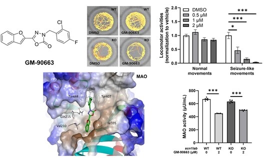
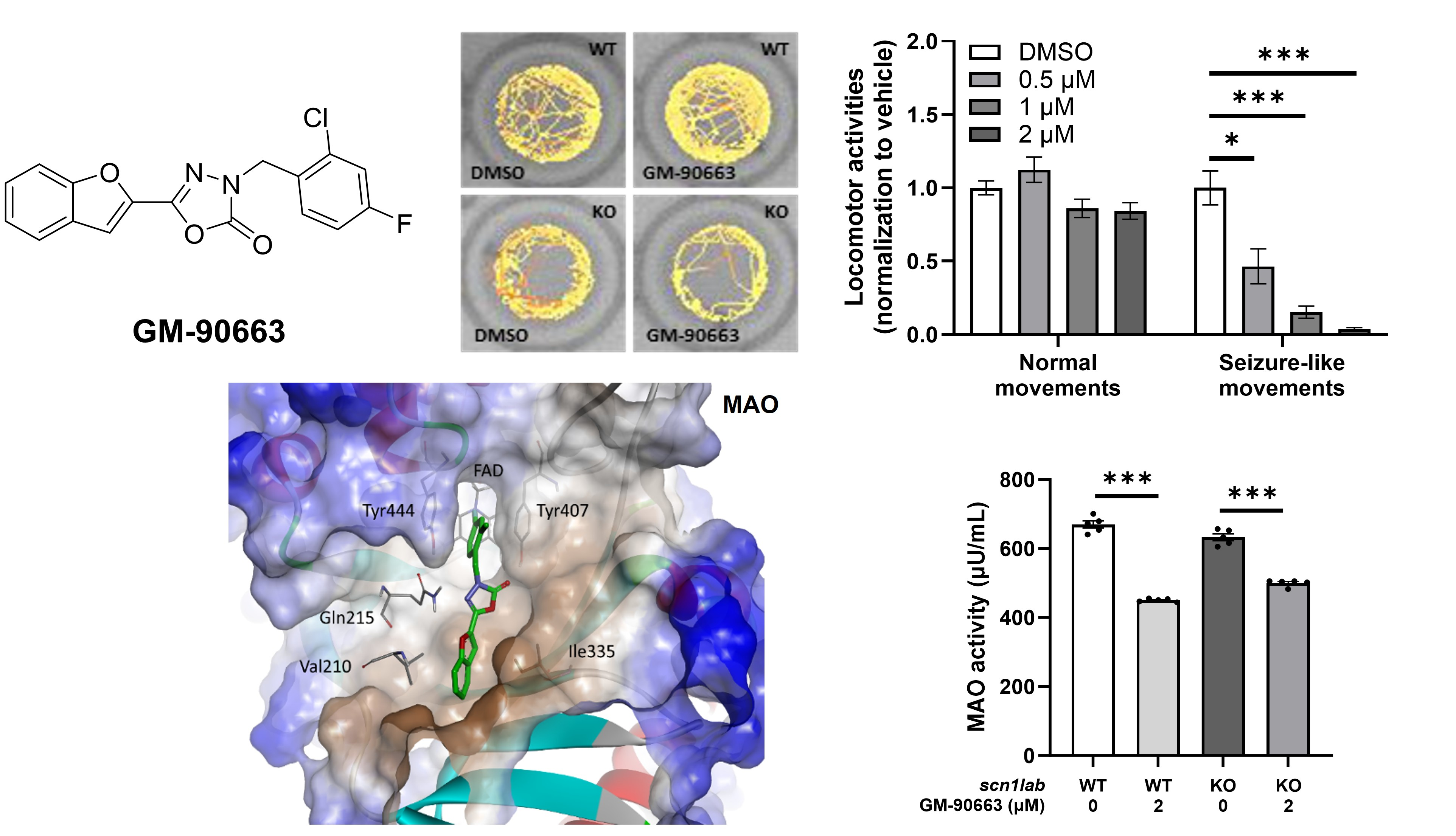
20e (3-(2-Chloro-4-fluorobenzyl)-5-(2-(trifluoromethyl)pyridin-4-yl)-1,3,4-oxadiazol-2(3H)-one), which demonstrated potent anti-seizure efficacy by upregulating TPH2 (tryptophan hydroxylase 2) expression. During the optimization, we synthesized diverse molecules. Among the synthesized compounds, we found 5-(Benzofuran-2-yl)-3-(2-chloro-4-fluorobenzyl)-1,3,4-oxadiazol-2(3H)-one (GM-90663), which alleviated Dravet syndrome with MOA inhibition as well as modulation of voltage-gated Na
+ channel. This situation prompted us report the synthesis, molecular docking and biological evaluation of new candidate for DS.
2. Materials & Methods
2.1. Materials
Commercially available reagents and solvents were utilized directly unless otherwise specified. The yields provided correspond to isolated products purified via crystallization or silica gel column chromatography. Proton nuclear magnetic resonance (1H NMR) data were acquired utilizing a JEOL JNM-ECS400 instrument (JEOL Ltd., Tokyo, Japan) operating at 400 MHz. Spectra were referenced internally to tetramethylsilane (TMS), with chemical shifts (δ) reported in ppm using CDCl3 as the deuterated solvent. Signal splitting patterns are designated as follows: s, singlet; d, doublet; t, triplet; q, quartet; dd, doublet of doublets; ddd, doublet of doublet of doublets; qd, quartet of doublets; dt, doublet of triplets; m, multiplet. For HPLC characterization, a Waters Agilent platform coupled with a PDA detector and a Waters SB-C18 analytical column (1.8 μm, 2.1 × 50 mm) was employed. Elution was carried out at a flow rate of 0.5 mL/min, employing a gradient of highly purified water containing 0.1% trifluoroacetic acid (Eluent A) and chromatographic-grade acetonitrile (Eluent B).
2.2. Procedure for the Synthesis of Compound GM-90663
Step 1. To a solution of ethyl benzofuran-2-carboxylate, 1 (1.25 g, 6.572 mmol) in EtOH (100 mL) was added hydrazine hydrate (2.303 g, 46.004 mmol, 7.0 equiv). Following 6 h of reflux, the mixture was concentrated in vacuo. The resulting residue was diluted with H2O and extracted with EtOAc (3 × 100 mL). The combined organic layers were dried over Na2SO4 and evaporated to afford benzofuran-2-carbohydrazide, 2 (880 mg, 4.995 mmol, 76%). 1H NMR (400 MHz, Chloroform-d) δ 7.80 (s, 1H), 7.69 (dd, J = 7.8, 1.3, 0.7 Hz, 1H), 7.54–7.48 (m, 2H), 7.43 (ddd, J = 8.5, 7.1, 1.3 Hz, 1H), 7.31 (ddd, J = 8.1, 7.1, 1.1 Hz, 1H), 4.10 (s, 2H).
Step 2. To a stirred solution of Benzofuran-2-carbohydrazide, 2 (880 mg, 4.995 mmol) and Et3N (2.089 mL, 14.985 mmol, 3.0 equiv) in THF (10 mL) at 0 °C, a solution of triphosgene (1.482 g, 4.995 mmol, 1.0 equiv) in THF was added dropwise. The reaction mixture was stirred at 0 °C for 1 h, then gradually warmed to room temperature and refluxed for 6 h. After cooling, the mixture was concentrated under reduced pressure. Purification of the resulting residue by silica gel column chromatography afforded 5-(benzofuran-2-yl)-1,3,4-oxadiazol-2(3H)-one, 3 (780 mg, 3.858 mmol, 77%). 1H NMR (400 MHz, DMSO-d6) δ 12.86 (s, 1H), 7.78 (dd, J = 7.8, 1.2 Hz, 1H), 7.73 (d, J = 8.3 Hz, 1H), 7.60 (d, J = 1.0 Hz, 1H), 7.48 (ddd, J = 8.5, 7.2, 1.4 Hz, 1H), 7.37 (dd, J = 7.9, 7.0 Hz, 1H).
Step 3. Under a nitrogen atmosphere, 5-(benzofuran-2-yl)-1,3,4-oxadiazol-2(3H)-one, 3 (150 mg, 0.742 mmol) and 2-chloro-4-fluorobenzyl bromide (164.65 mg, 0.742 mmol, 1.0 equiv) were dissolved in anhydrous DMF (2 mL). A suspension of NaH (60% in mineral oil, 17.81 mg, 0.742 mmol, 1.0 equiv) in DMF was added dropwise. After stirring at 60 °C for 4 h, the mixture was concentrated. The crude was partitioned between H2O (20 mL) and EtOAc (3 × 50 mL). The organic extracts were washed with brine and chromatographed on silica gel to yield 5-(benzofuran-2-yl)-3-(2-chloro-4-fluorobenzyl)-1,3,4-oxadiazol-2(3H)-one, GM-90663 (147.7 mg, 0.430 mmol, 58%). 1H NMR (400 MHz, Chloroform-d) δ 7.70–7.63 (m, 1H), 7.56 (d, J = 8.2 Hz, 1H), 7.47–7.38 (m, 2H), 7.36–7.28 (m, 2H), 7.26 (s, 1H), 7.18 (dd, J = 8.4, 2.6 Hz, 1H), 7.02 (td, J = 8.3, 2.6 Hz, 1H), 5.10 (s, 2H). LC-MS (m/z): 345.1 [M + H]+; HPLC purity 98.69%.
2.3. Maintenance of Zebrafish
Zebrafish embryos and larvae were reared under standard laboratory conditions as established by The Zebrafish Book [
26]. Briefly, fertilized eggs were collected after natural spawning and selected using a stereomicroscope to remove unfertilized eggs. Embryos were kept in embryonic medium (60 μg/L of sea salt; Sigma-Aldrich, St. Louis, MO, USA, cat# S9883) at 28 °C with a 14:10 h light/dark photoperiod. Embryonic medium was replaced daily until 6 days post-fertilization (dpf). Experiments involving zebrafish were performed in accordance with the NIH Guide for the Care and Use of Laboratory Animals (no. 8023, revised in 1996), and this study was approved by the Animal Care and Use Committee of the Korea Research Institute of Chemical Technology (2021-7B-04-02).
2.4. Behavioral Analysis in scn1lab Knockout (KO) Zebrafish Larvae
Larvae were obtained from the breeding of
scn1labkri111/+ (AB strain) adults [
25]. We utilized homozygous KO individuals, identified by their characteristic hyper-pigmentation, alongside age-matched wild-type siblings. To ensure data integrity, only larvae with normal morphological development were randomly assigned to experimental groups. Regarding the experimental timeline, all larvae were allowed to acclimatize to the testing environment during the morning hours. Behavioral assessments were consistently conducted in the afternoon to ensure stable physiological conditions and to minimize variability related to the animals’ circadian rhythms. At 6 days post-fertilization (dpf), individual larvae were transferred to 96-well plates and dark-acclimated for 30 min within a tracking chamber. GM-90663 was prepared as a 2 mM stock in dimethyl sulfoxide (DMSO) and subsequently diluted in embryo medium to final concentrations (0.5–2 μM). The final concentration of DMSO in all treatment and vehicle control groups was 0.1% (
v/
v), which has been reported to have no significant effect on zebrafish larval behavior or development. Larval activity was monitored for 30 min using the DanioVision system (Noldus, Wageningen, the Netherlands) equipped with EthoVision XT 15 software (Noldus). Seizure-like behaviors were categorized according to established criteria [
25,
27]. To evaluate color preference, 6-dpf larvae were pre-treated with either DMSO or GM-90663 in 6-well plates for 4 h. Following treatment, groups of 20 larvae were introduced into a dual-channel chamber (blue and yellow) containing 10 mL of embryo medium [
28]. A digital camera (HDR-CX130, Sony, Tokyo, Japan) recorded the distribution of larvae for 30 min. To quantify positional data, static images were captured from the video recordings at 2 min intervals. Preference scores for each color were determined by calculating the mean occupancy percentage of larvae within each respective channel over the recording period.
2.5. Electrophysiology
Neurons were obtained from hippocampal slices prepared from Sprague–Dawley rats (postnatal day 12–17, both sexes). Animals were anesthetized with ketamine (50 mg/kg, intraperitoneal) and subsequently decapitated. The brain was rapidly removed and sectioned transversely into 400 µm thick slices using a vibrating microslicer (Campden Instruments, Leicester, UK). To preserve tissue viability, slices containing the hippocampus were kept in an incubation medium (in mM; 124 NaCl, 3 KCl, 1.5 KH
2PO
4, 24 NaHCO
3, 2 CaCl
2, 1.3 MgSO
4, and 10 glucose) saturated with 95% O
2 and 5% CO
2 at room temperature (22–25 °C) for at least 1 h before the mechanical dissociation. For acute dissociation, individual slices were transferred into a 35 mm culture dish (Primaria 3801; Becton Dickinson, Rutherford, NJ, USA), which contained the standard external solution (in mM; 150 NaCl, 3 KCl, 2 CaCl
2, 1 MgCl
2, 10 glucose and 10 HEPES, a pH of 7.4 with Tris-base). The CA3 region of the hippocampus was visually identified under a stereomicroscope (SMZ-1; Nikon, Tokyo, Japan), and neurons were mechanically isolated as previously described [
29,
30]. Briefly, mechanical dissociation was performed using a vibration device (SI-10; K.T. Labs., Fukuoka, Japan) and a fire-polished glass pipette oscillating at approximately 50–60 Hz (0.3–0.5 mm) over the hippocampal CA3 region. Following this procedure, the slices were removed from the dish, and the acutely dissociated neurons were allowed to settle onto the bottom surface for approximately 15 min before further experimentation.
Whole-cell voltage-clamp recordings were performed using a patch-clamp amplifier (MultiClamp 700B; Molecular Devices, San Jose, CA, USA) following standard procedures [
31]. Unless otherwise specified, cells were held at −100 mV. Patch electrodes were fabricated from borosilicate glass capillaries (Narishige, Tokyo, Japan) using a programmable puller (Sutter Instrument, Novato, CA, USA) and had a resistance of 1.0–1.5 MΩ when filled with the internal solution. The pipette solution contained (in mM): 140 CsF, 10 CsCl, 2 EGTA, 2 Na
2-ATP, and 10 HEPES, adjusted to pH 7.2 with Tris base. Membrane currents were low-pass fil-tered at 3 kHz, digitized at 10 kHz, and acquired using pCLAMP 10.7 software (Molecular Devices). Capacitive transients and linear leak components were corrected using a P/4 subtraction protocol. Series (access) resistance was continuously monitored by applying brief hyperpolarizing voltage steps (10 mV, 30 ms), and recordings were excluded if the resistance varied by more than 10% during the experiment. To isolate voltage-gated Na
+ current (I
Na), recordings were conducted in a K
+-free external solution (in mM; 130 NaCl, 20 tetraethylammonium-Cl, 3 CsCl, 2 CaCl
2, 1 MgCl
2, 10 glucose, and 10 HEPES, and was adjusted to pH 7.4 with Tris-base). In addition, the bath solution was supplemented with 10 μM SR95531, 20 μM CNQX, 50 μM DL-APV, 100 μM Cd
2+ to suppress GABA
A receptors, ionotropic glutamate receptors, and voltage-gated Ca
2+ channels. Depolarizing voltage steps were delivered at 10 s intervals to allow complete recovery from Na
+ channel inactivation, unless stated otherwise. Pharmacological agents were applied using the “Y-tube system” for rapid solution exchange, as previously described [
32]. All experiments were performed at room temperature (22–25 °C).
Peak INa amplitude was determined as the difference between baseline and maximal inward current (pCLAMP 10.7). In a subset of experiments, the amplitude of TTX-R INa, was transformed into conductance (G) using the following equation: G = I/(V − ENa), where ENa is the Na+ equilibrium potential (+87.7 mV) calculated by the Nernst equation. The voltage-activation and voltage-inactivation relationships of TTX-R Na+ channels were fitted to the following Boltzmann equations, respectively; G/Gmax = 1/{1 + exp[(V50,activation − V)/k]} and I/Imax = 1 − 1/{1 + exp[(V50,inactivation − V)/k]}, where Gmax is the maximum conductance, Imax is the maximum current amplitude, V50,activation is the half-maximum potential for activation, V50,inactivation is the half-maximum potential for fast inactivation, and k is the slope factor. The kinetic data for the recovery from inactivation were fitted to the following equation: R(t) = A0 + Afast × [1 − exp(–t/τfast)] + Aintermediate × [1 − exp(–t/τintermediate)] + Aslow × [1 − exp(–t/τslow)], respectively, where R(t) is the amplitude ratio of INa at time t, and Afast, Aintermediate, and Aslow are the amplitude fractions of τfast, τintermediate, and τslow, respectively.
2.6. Quantitative Real-Time Polymerase Chain Reaction (qRT-PCR)
Following compound exposure, total RNA was extracted from 20-pooled larvae (WT or KO) using TRIzol (Invitrogen, cat# 15596026) reagent, following the standard manufacturer’s instructions. qRT-PCR was performed using 1-Step qRT-PCR kit (Thermo Scientific, Waltham, MA, USA, cat# AB-4106/A) with 7500/7500 Fast Real-Time PCR system (Applied Biosystems, Waltham, MA, USA,) under the manufacturer’s specified thermal conditions. Relative mRNA expression levels were determined via the 2
−ΔΔct method, with
β-actin serving as the internal reference gene for normalization. The specific primer sequences utilized are detailed in
Table 1.
2.7. Immunohistochemistry
Immunohistochemistry for phosphorylated Extracellular Signal-Regulated Kinases (pERK) was conducted as previously described [
33]. To preserve transient phosphorylation states, larvae were placed in 6-well plates with cell strainers (SPL, cat# 93100) to facilitate rapid immersion in fixative. Following a 4 h chemical treatment, samples were immediately fixed in a solution of 4% paraformaldehyde and 4% sucrose in phosphate-buffered saline (PBS) overnight at 4 °C. After fixation, tissues were rinsed with PBS containing 0.25% Triton X-100 (PBTx), and larval brains were carefully micro-dissected to optimize antibody accessibility. The tissues were then permeabilized with 1 mg/mL collagenase (Sigma-Aldrich, cat# C9891) in PBS for 1 h, followed by overnight blocking in PBTx supplemented with 2% normal goat serum and 2% DMSO at 4 °C. Brain tissues were incubated overnight with a primary rabbit anti-pERK antibody (Cell signaling Technology, Danvers, MA, USA, cat# 4370) and subsequently with a secondary antibody (Invitrogen, cat# A-11034). After final washes, the stained tissues were embedded in 1% low-melting-point agarose and visualized using a K1-Fluo confocal fluorescence microscopy (Nanoscope Systems, Daejeon, Republic of Korea).
2.8. LC-MS/MS
Comprehensive neurochemical analysis was conducted in zebrafish larvae (30-pooled) following exposure to GM-90663 in wild-type or KO. This analysis utilized an ultra-performance liquid chromatography (ACQUITY UPLC System, Waters Corporation, Milford, MA, USA) coupled with a Xevo TQ-S triple quadrupole mass spectrometer (LC-MS/MS, Waters Corporation), as per a previously reported method [
34]. Briefly, the zebrafish larvae were washed three times in ice-cold 0.1 M phosphate-buffered saline (PBS, pH 7.4, Corning, Corning, NY, USA), snap-frozen in liquid nitrogen. The samples were homogenized by ultrasonication in 100 μL of distilled water, extracted with an equal volume of methanol, vortexed, and centrifuged at 15,000 RPM at 4 °C for 20 min. The supernatant was then transferred to an LC-vial for the quantitative analysis of 31 neurochemicals using LC-MS/MS.
2.9. Molecular Modeling
The Schrodinger Suite was used for the molecular modeling study [
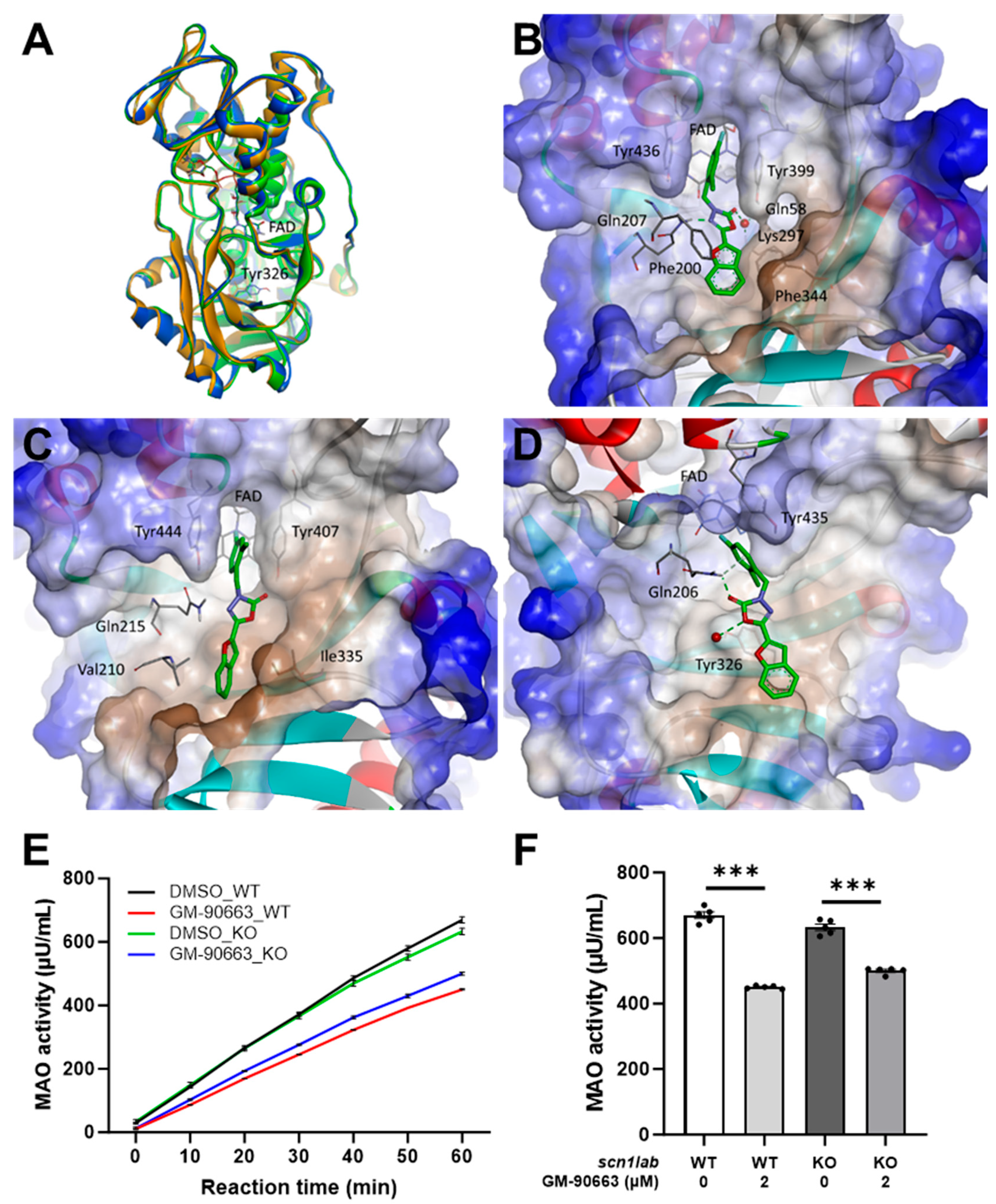
35]. The protein structural files of human MAO-A and MAO-B were obtained from the Protein Data Bank (PDB codes: 2Z5X and 2V5Z, respectively). Preparation of the protein models, including bond order assignment, addition of hydrogen atoms, and correction of missing loops and side chains, was performed by using the Protein Preparation Wizard of Schrodinger suite. The protein-ligand complexes were relaxed with Impref using the OPLS4 force field. Since the experimental structure of zebrafish MAO (zMAO) is not published yet, the AlphaFold prediction model (AF-Q6NSN2-F1;
https://alphafold.ebi.ac.uk/entry/Q6NSN2, accessed on 1 Auguest 2025) was employed as the protein model. Docking simulations were performed using the Glide docking program with the standard SP flexible ligand docking protocol and default settings. The ligand structures were docked into the energy grids of MAO proteins. The receptor grids for each MAO protein were generated using the Receptor Grid Generation module. The ligand binding sites were defined by the centroid of the crystal ligand with a cubit grid size of 22 Å. Robustness of protein structure and docking protocol was verified via reproduction of crystal ligand poses with a good root mean square displacement of 0.5 Å. The best-energy pose, having the lowest Glide score, was selected for the subsequent binding pose analysis and molecular dynamics simulation. The Desmond software with default settings was used in the classical MD simulations of the complexes for the interaction analysis. The structures of the best-scoring poses of protein-ligand complexes in each MAO protein were solvated in the SPC water model and the OPLS4 forcefield using orthorhombic periodic boundary conditions, with a 10 Å buffer distance and neutralizing Na
+ or Cl
− ions. Production simulations were run for 100 ns with constant temperature and pressure of 300 K and 1.01325 bar. After 100 ns simulations of each complex, the lowest-energy conformations were obtained from the last 10 ns trajectories. The sampling interval during the simulation was set to 100 ps, and the analysis of simulation results was performed using the SID and SEA programs in Maestro.
2.10. Monoamine Oxidase (MAO) Assay in Zebrafish Larvae
Assay of MAO activity was performed using a commercial kit (abcam, Cambridge, UK, cat# ab241031). Homogenates were prepared from 20-pooled larvae after exposure to GM-90663 in wild-type or KO. MAO activity was measured using a microplate reader (TECAN, Männedorf, Switzerland, M100PRO) as a fluorescence intensity (Ex/Em = 535/587 nm) and calculated according to the manufacturer’s protocol.
2.11. Statistical Analysis
Data are presented as mean ± standard error of mean (SEM). Statistical comparisons were performed via the non-parametric Mann–Whitney test using GraphPad Prism 9.4.0 and Microsoft Excel 2016. Electrophysiological data are presented as mean ± SEM, with normalization to control values where appropriate. Statistical comparisons were performed using paired two-tailed Student’s t-tests on absolute current amplitudes. A p-value of less than 0.05 was adopted as the criterion for statistical significance (* p < 0.05, ** p < 0.01, and *** p < 0.001).
4. Discussion
Fenfluramine (FFA), which recently gained approval as an adjunctive treatment for Dravet syndrome (DS), originally demonstrated its anticonvulsant properties by influencing serotonin pathways in the
didys552 zebrafish [
13,
21]. Interestingly, although it was previously pulled from the market as an anti-obesity medication, FFA successfully underwent drug repurposing to be authorized for DS management [
5]. In our earlier work, we established a novel zebrafish strain with a
scn1lab loss-of-function mutation to facilitate the discovery of emerging therapeutic agents for DS. Distinct from the
didys552 point mutation, the
kri111 strain expresses a prematurely truncated form of the s
cn1lab protein [
25]. However, the morphological phenotypes of the two mutants, such as hyperpigmentation and an uninflated swim bladder, are similar. Notably, behavioral phenotypes of the two mutants are identical, since the
kri111 mutant, like the
didys552 mutant, exhibited seizure-like movements [
17,
25]. Therefore, we concluded that new candidate screening for DS treatment could be available using the
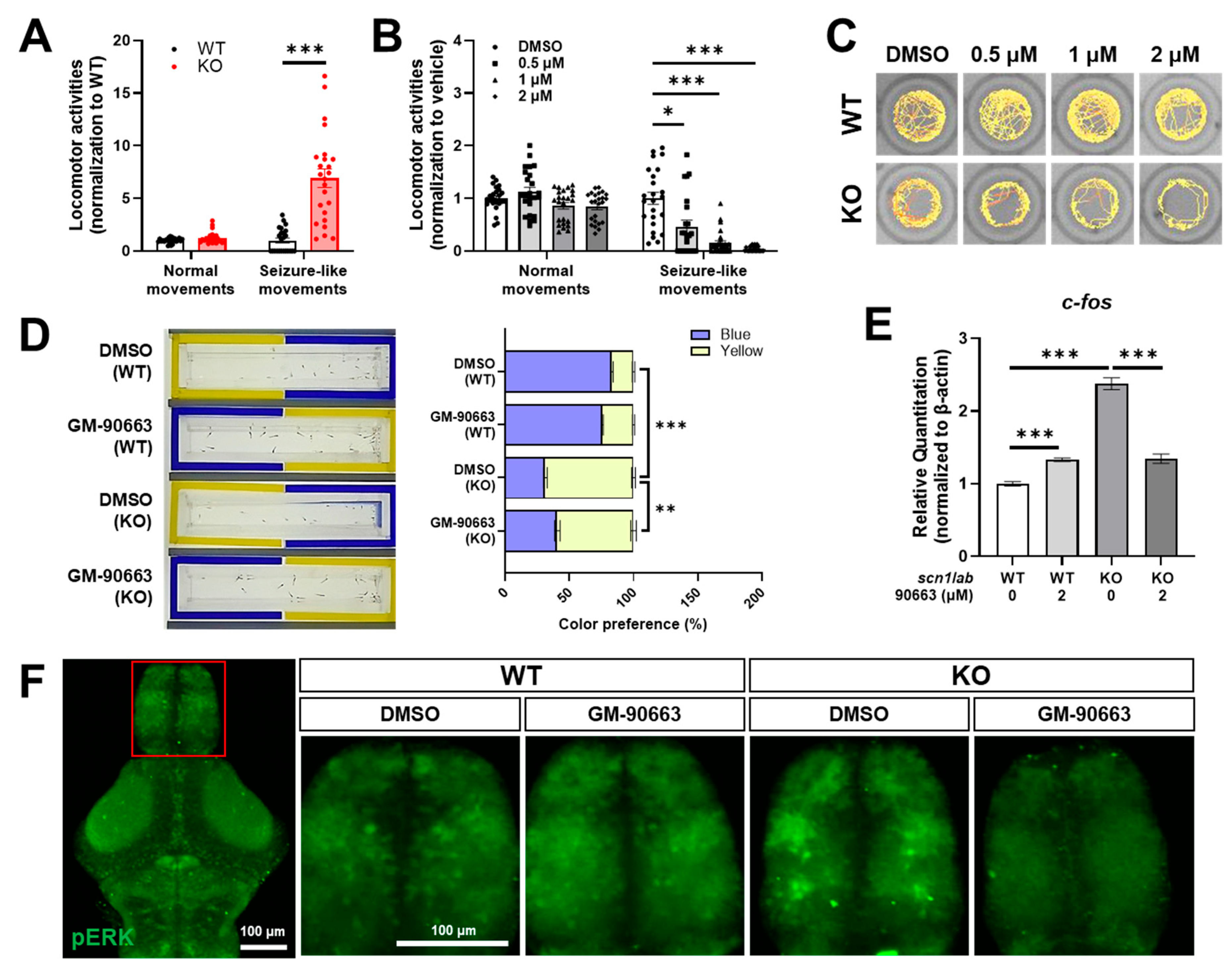
kri111 mutant. GM-90663 was identified as an optimized compound with improved anti-seizure efficacy in the
kri111 mutant. Although the ‘whirlpool-like’ circular behavioral pattern was not decreased, GM-90663 dose-dependently reduced hyper-activity in
scn1lab KO. Next, we employed LC-MS/MS-based neurochemical profiling to investigate the neurological effects of GM-90663 in both wild-type and
scn1lab KO. Our previous work has established this technology as a powerful tool for elucidating the mechanisms underlying the neuroprotective or neurotoxic effects of chemicals, utilizing various biological models, including zebrafish [
34,
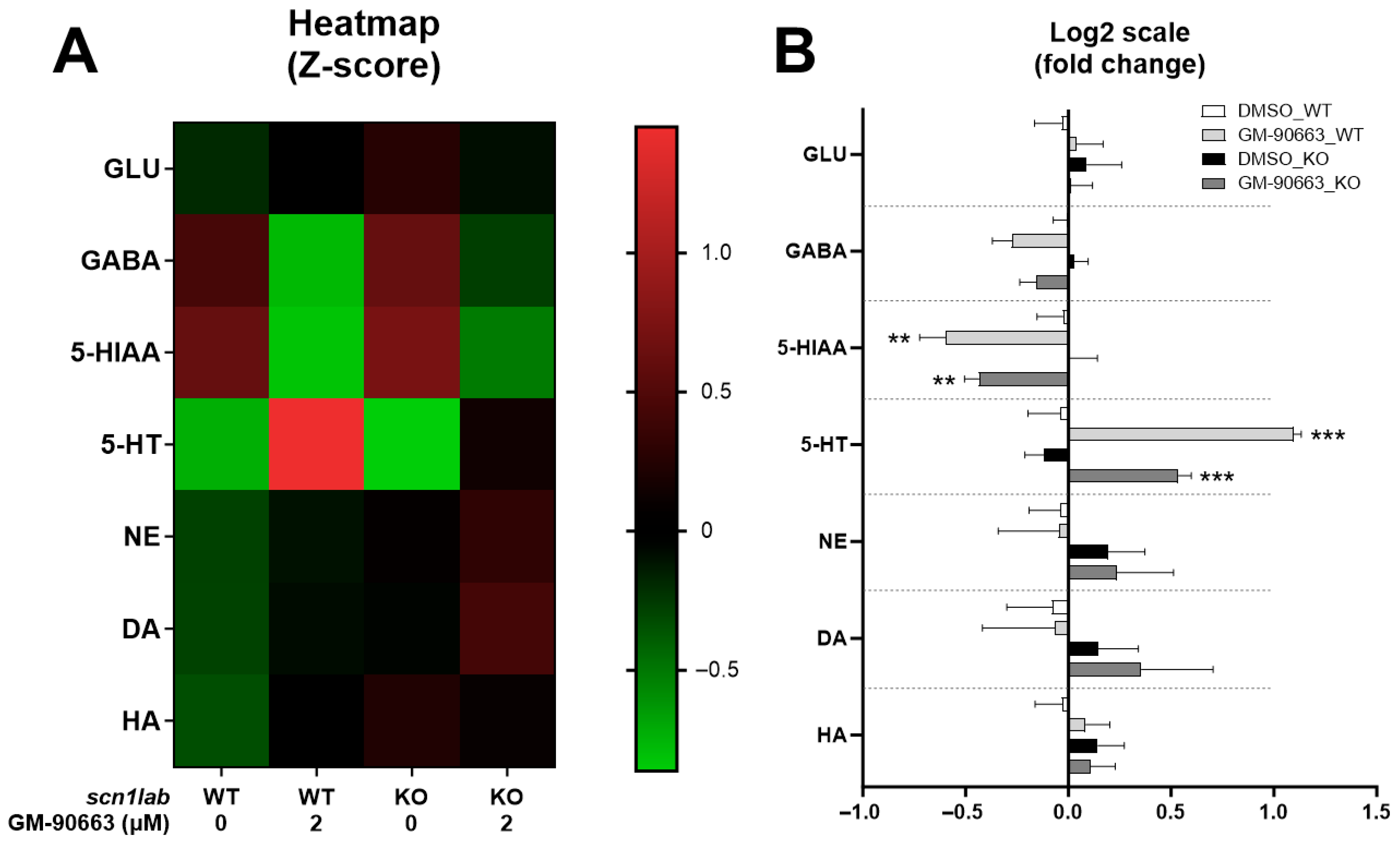
42]. We quantitatively analyzed a range of neurotransmitters, their precursors, and metabolites, comprising six categories of the nervous system: histaminergic, cholinergic, dopaminergic, serotonergic, kynurenergic, and GABAergic, in the zebrafish model. Neurochemical profiling revealed that GM-90663 treatment increased endogenous serotonin levels in both WT and
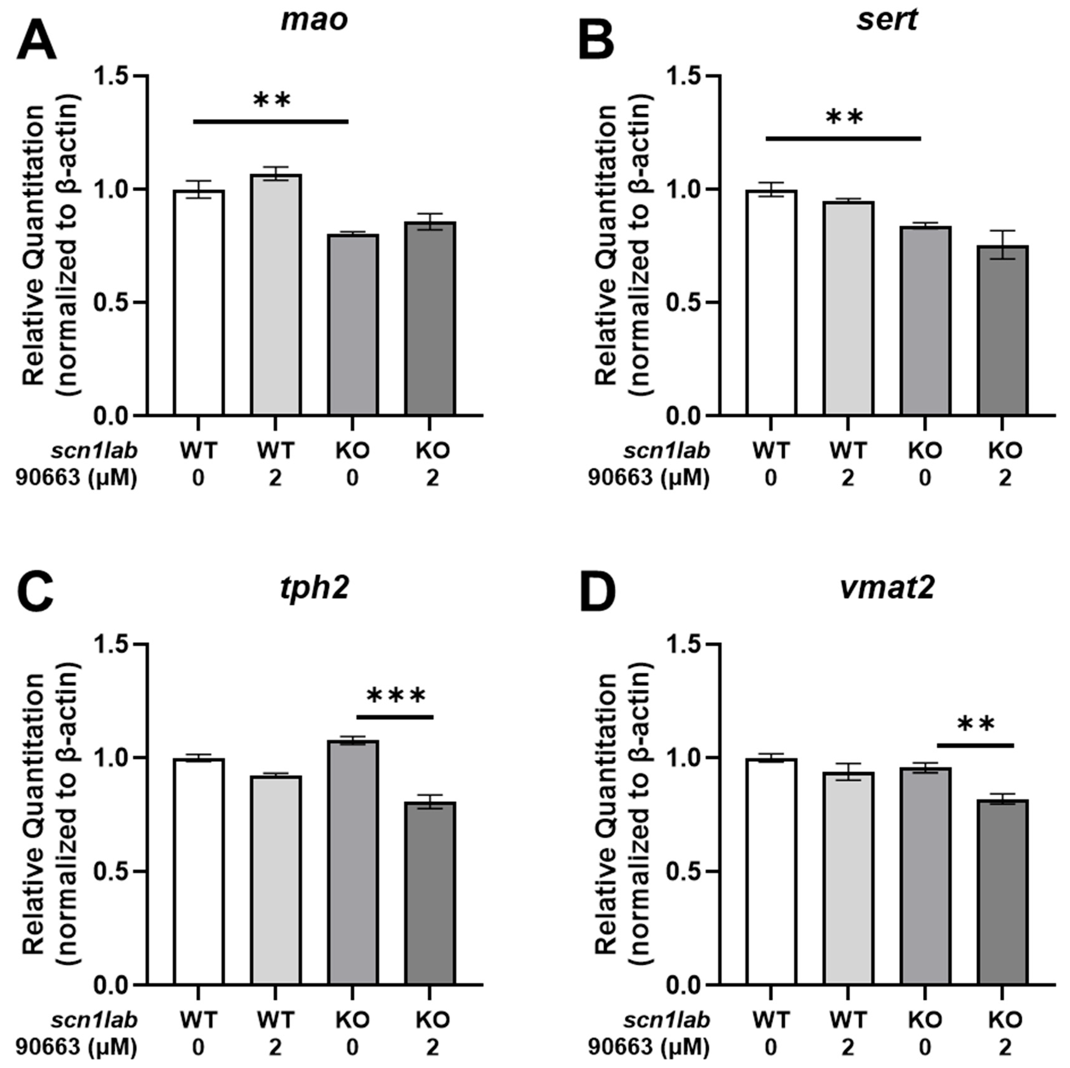
scn1lab KO larvae. The increase in serotonin levels induced by GM-90663 occurs independently of transcriptional changes in key serotonergic regulatory genes, such as
tph2,
sert,
vmat2, and
mao. Based on this result, we concluded that GM-90663 significantly inhibits MAO, impacting the metabolic conversion of serotonin to 5-HIAA.
Indeed, GM-90663 shows favorable docking poses in the binding sites of MAOs, with docking scores indicating strong binding affinity through docking simulation data. Especially, the ligand binding is stabilized by hydrogen bonds, stacking interactions, and hydrophobic interactions with specific residues in zMAO. In MAO-A, similar interactions are observed, with hydrogen bonds and hydrophobic interactions. Because the zMao model shares high sequence similarity (>70%) with human MAO-A and MAO-B, and its overall structure closely resembles that of human MAOs, we conclude that GM-90663 docking poses are also highly similar between human and zebrafish. MAO is an enzyme found primarily in the brain and other tissues, responsible for catalyzing the oxidation of monoamine neurotransmitters such as serotonin, dopamine, and norepinephrine. This enzyme plays a crucial role in the metabolism of neurotransmitters, regulating their levels in the brain and influencing various physiological processes, including mood, behavior, and cognition. Dysfunction of MAO has been implicated in various neurological and psychiatric disorders, making it an important target for pharmacological interventions [
43]. MAO inhibitors (MAOIs) are indeed used as drugs, primarily in the treatment of depression and certain other psychiatric disorders. Examples of MAOIs include phenelzine (Nardil), tranylcypromine (Parnate), and isocarvoxazid (Marplan). These drugs work by inhibiting the activity of MAO enzymes, thereby increasing the levels of neurotransmitters such as serotonin, dopamine, and norepinephrine in the brain, which can alleviate symptoms of depression and improve mood [
44]. In this study, we specifically targeted the MAO-A isoform for both computational docking and biological validation. This strategic selection was guided by the substrate preference of MAO-A, which exhibits a significantly higher affinity for 5-HT compared to MAO-B [
45]. Restoration of 5-HT levels in DS model has been clinically proven to reduce seizure frequency, MAO-A inhibition represents a more direct and mechanistically relevant intervention than MAO-B inhibition.
Serotonin is a neurotransmitter primarily found in the central nervous system (CNS) and gastrointestinal tract. It plays a key role in regulating mood, appetite, sleep, and various other physiological processes. In the brain, it acts as a neurotransmitter, transmitting signals between nerve cells and modulating neural activity. Although the metabolic signature for serotonergic neurochemistry in DS is not fully understood, numerous studies have highlighted serotonergic modulation—particularly targeting serotonin receptors 1A, 1D, 2A, 2C, and 3—as a promising therapeutic strategy for this condition [
19,
46]. Furthermore, FFA, which is approved for DS treatment, is known for its agonistic effects on serotonin receptors 1D, 2A, and 2C [
13].
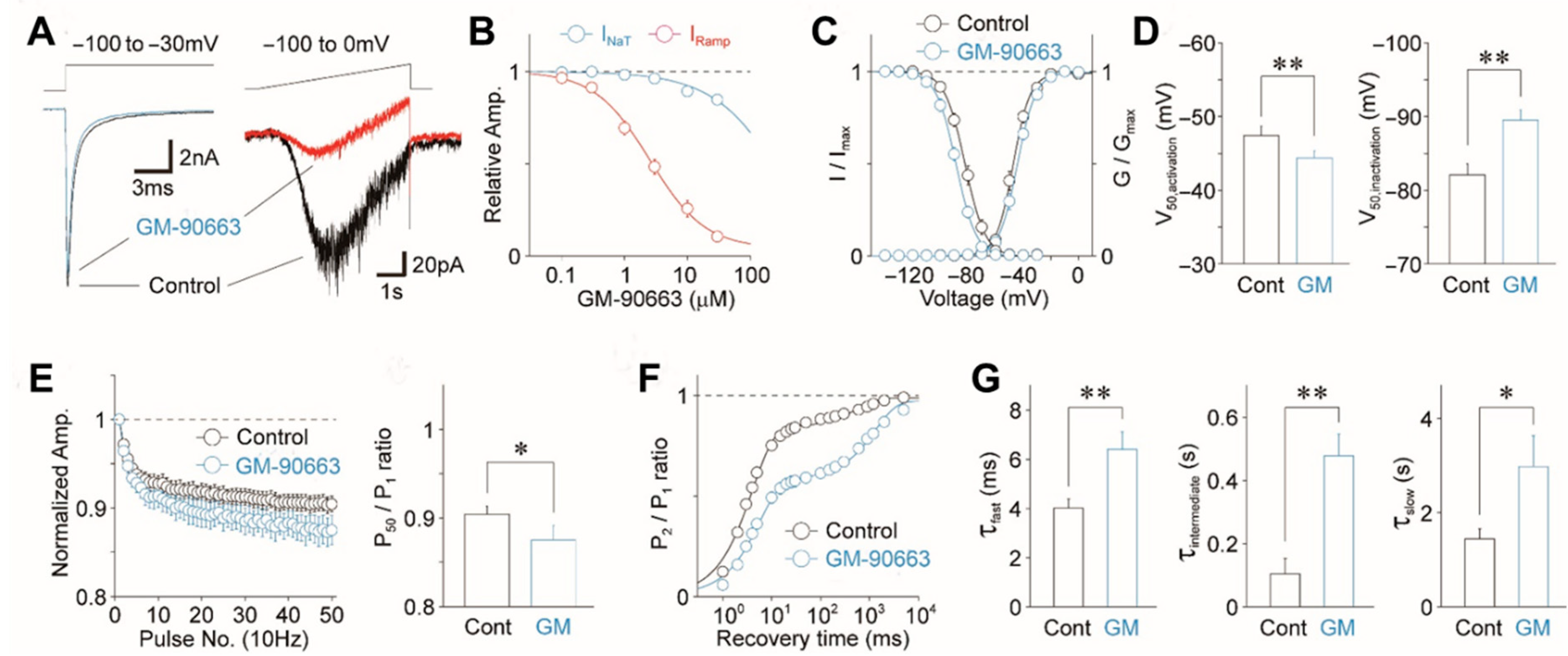
Beyond its neurochemical effects on the serotonergic system, GM-90663 directly regulates neuronal excitability by modulating voltage-gated Na+ channels. These electrophysiological properties suggest that GM-90663 selectively suppresses high-frequency repetitive firing and reduces the probability of action potential initiation, providing a robust biophysical mechanism for its anti-seizure efficacy in Dravet syndrome models. Unlike traditional Na+ channel blockers, which can sometimes exacerbate seizures in DS patients due to the loss of GABAergic interneuron function, GM-90663 offers a dual-action approach. By inhibiting MAO-A, it directly increases the synaptic availability of 5-HT, a neurotransmitter significantly depleted in DS models. This serotonergic enhancement, combined with its secondary modulation of ion channels, provides a synergistic anti-seizure effect that bypasses the limitations of mono-targeted therapies.
Interestingly, while GM-90663 significantly elevated synaptic serotonin levels via MAO-A inhibition, no significant changes were observed in total GABA levels. This finding is particularly noteworthy given that Dravet syndrome is fundamentally rooted in GABAergic dysfunction. The lack of alterations in GABA concentration suggests that the anti-seizure efficacy of GM-90663 is not mediated through a direct quantitative increase in GABAergic tone, but rather through a more targeted modulation of the serotonergic system and sodium channel kinetics. This distinction is crucial, as it indicates that GM-90663 operates via an alternative neuromodulatory pathway that can bypass the refractory nature of GABA-centered treatments in DS. Furthermore, the stable GABA levels reinforce the pharmacological selectivity of our compound, demonstrating that it does not cause a non-specific surge in all inhibitory neurotransmitters, thereby potentially reducing the risk of sedative side effects often associated with GABA-enhancing drugs.
The anti-seizure efficacy and mechanism of GM-90663 observed in the scn1lab KO zebrafish model provide significant insights into its translational potential for human Dravet syndrome. Given the high conservation of serotonergic pathways and voltage-gated sodium channels between zebrafish and mammals, our findings are likely to be generalizable to higher vertebrate models. Furthermore, the ability of GM-90663 to target MAO enzymatic component suggests it could serve as a versatile therapeutic intervention for patients with refractory epilepsy who do not respond to conventional mono-targeted drugs. Also, regarding the pharmacological profile of GM-90663, considering potential off-target effects is essential for translational development. Our results demonstrate that the anti-seizure efficacy occurs at concentrations significantly lower than those associated with general cytotoxicity or behavioral toxicity in zebrafish larvae. While further comprehensive profiling will be necessary in future studies, the lack of adverse effects on cardiac function and early development in our model indicates a high degree of safety and selectivity for the primary targets, MAO-A and sodium channels.
Taken together, our findings demonstrate that GM-90663 exhibits an anti-seizure effect with an MAO-A inhibition for DS treatment. Additionally, whole-cell patch-clamp results revealed that GM-90663 influences the function of voltage-gated Na+ channels in neurons, providing insights into its potential mechanisms of action and therapeutic effects in the context of seizure treatment.




 ,
,