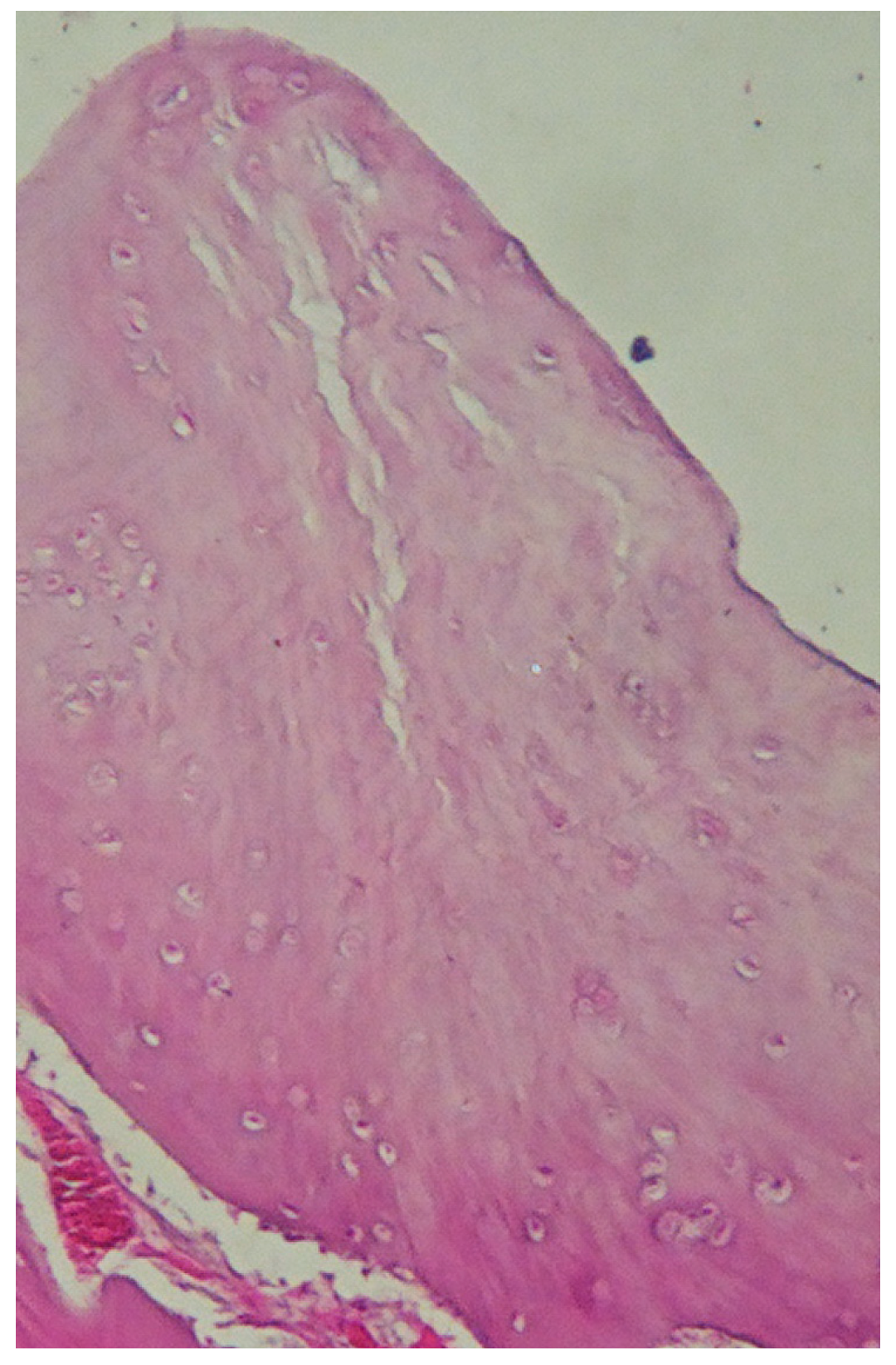
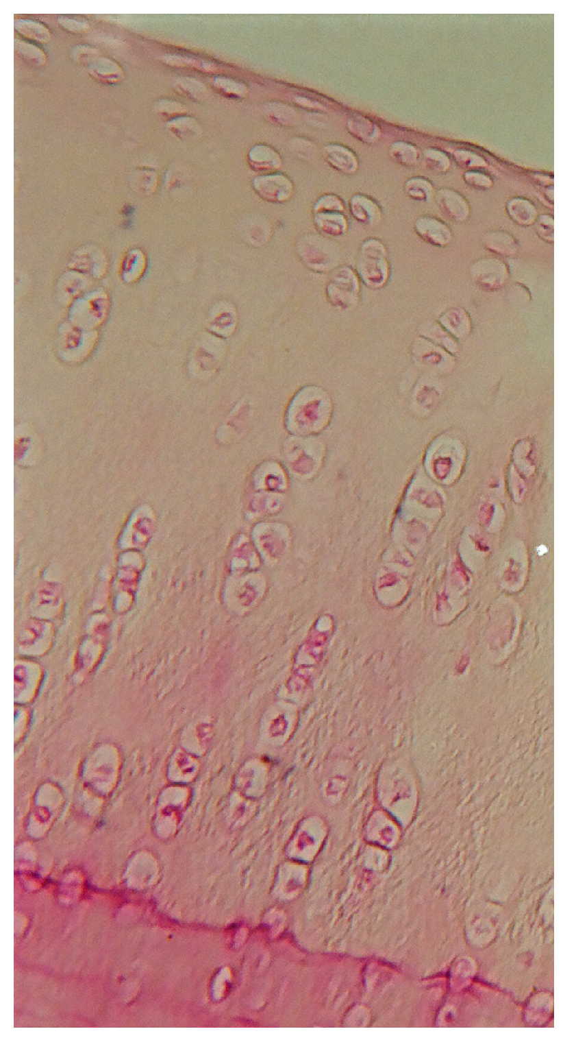
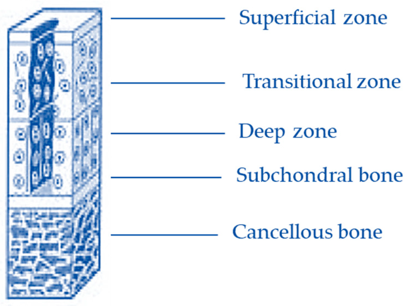
Autologous Chondrocyte Implantation on Polyethersulfone Scaffolds in a Rabbit Model of Grade III Lesions

 ,
,  ,
,
Abstract
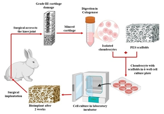
1. Introduction
2. Results and Discussion
2.1. Morphological Characterization of PES Scaffold
2.2. Number of Cells and Observation of Chondrocytes
2.3. Elemental Analysis
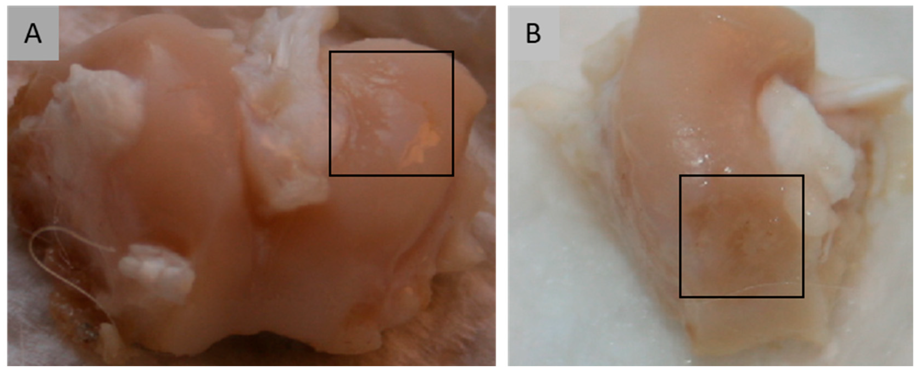
2.4. Macroscopic Assessment
2.4.1. Degree of Defect Repair
2.4.2. Macroscopic Appearance
2.4.3. Summary of Results in Macroscopic Assessment
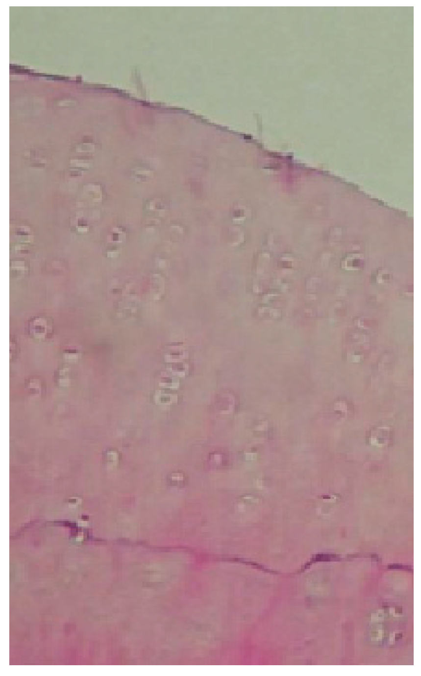
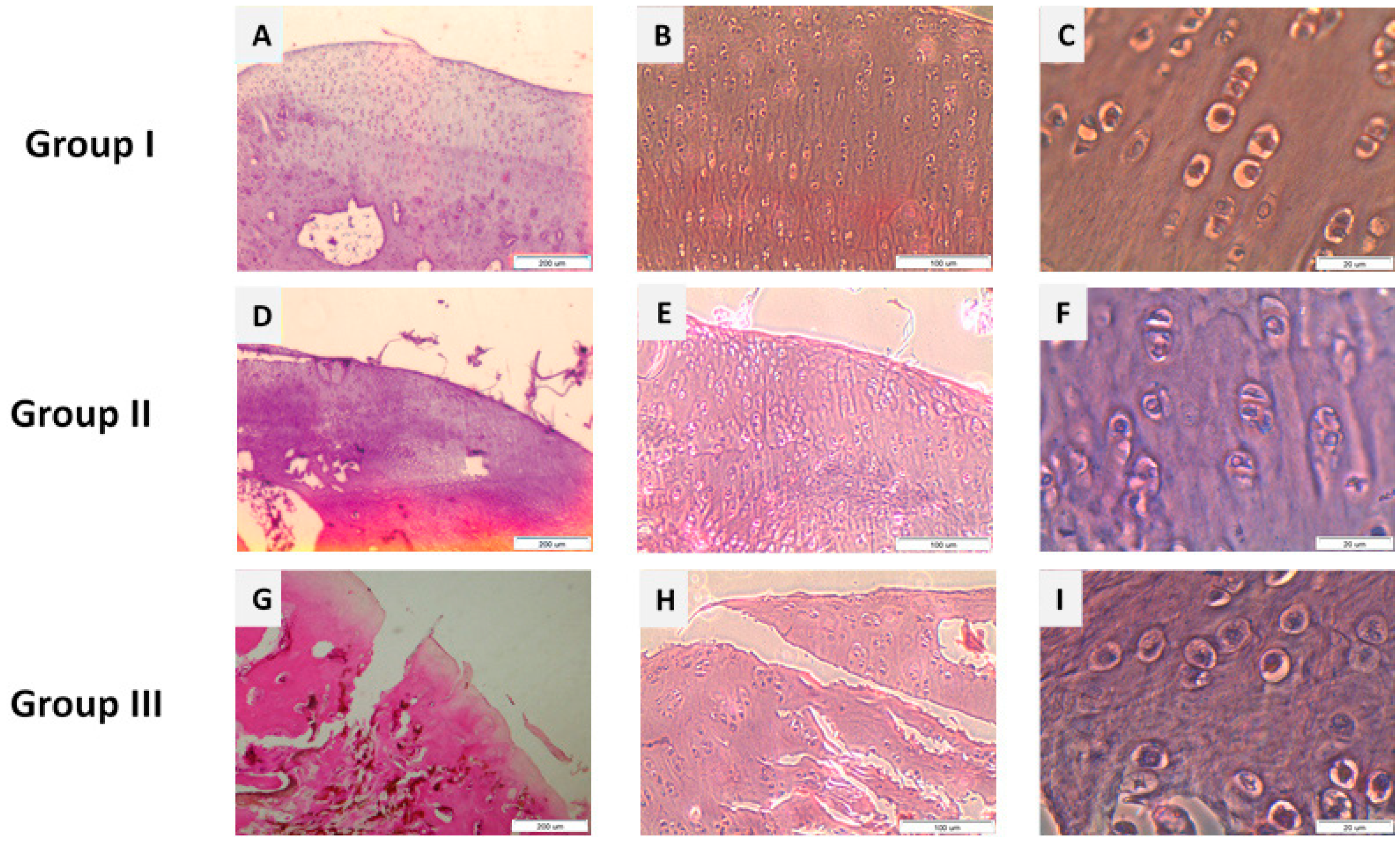
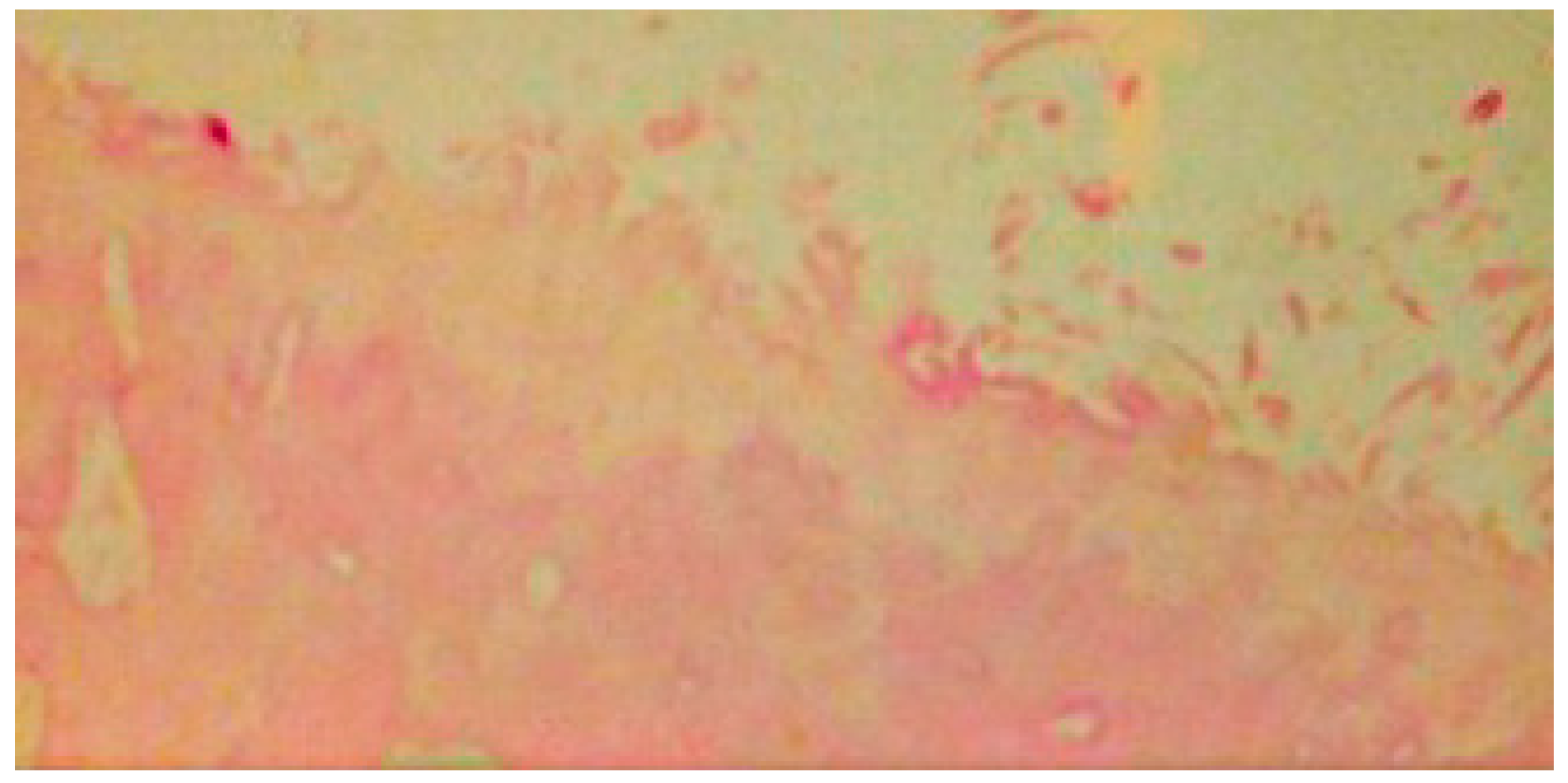
2.5. Microscopic Assessment
2.5.1. Nature of Predominant Tissue
2.5.2. Structural Characteristics
Surface Regularity
Structural Integrity
Thickness
Bonding to the Adjacent Cartilage
Stage of Degenerative Cellular Changes—Hypocellularity
Stage of Degenerative Cellular Changes—Degenerative Changes
Comparison of the Total Scores Achieved Across the Different Rabbit Groups
2.6. Limitations
3. Materials and Methods
3.1. Materials
3.1.1. Scaffolds
3.1.2. Rabbits
- Defect reaching the subchondral bone, with chondrocytes placed on a PES scaffold, in 25 knees.
- Defect reaching the subchondral bone and implantation of a PES scaffold without cells, in 25 knees.
- Defect reaching the subchondral bone without any implant, in 12 knees.
3.2. Methods
3.2.1. Methods for Chondrocyte Isolation and Cultivation
3.2.2. Procedure for Graft Implantation
3.2.3. Elemental Composition Analysis
3.2.4. Macroscopic Assessment
3.2.5. Microscopic Analysis
3.2.6. Statistical Evaluation
4. Conclusions
Author Contributions
Funding
Institutional Review Board Statement
Data Availability Statement
Acknowledgments
Conflicts of Interest
References
- Chang, L.; Marston, G.; Martin, A. Anatomy, Cartilage. In StatPearls; StatPearls Publishing: Treasure Island, FL, USA, 2022; pp. 2–4. [Google Scholar]
- Walter, S.G.; Ossendorff, R.; Schildberg, F.A. Articular cartilage regeneration and tissue engineering models: A systematic review. Arch. Orthop. Trauma Surg. 2018, 139, 305–316. [Google Scholar] [CrossRef] [Scilit] [PubMed]
- Nordberg, R.C.; Bielajew, B.J.; Takahashi, T.; Dai, S.; Hu, J.C.; Athanasiou, K.A. Recent advancements in cartilage tissue engineering innovation and translation. Nat. Rev. Rheumatol. 2024, 20, 323–346. [Google Scholar] [CrossRef] [Scilit] [PubMed]
- Karpiński, R.; Prus, A.; Baj, J.; Radej, S.; Prządka, M.; Krakowski, P.; Jonak, K. Articular Cartilage: Structure, Biomechanics, and the Potential of Conventional and Advanced Diagnostics. Appl. Sci. 2025, 15, 6896. [Google Scholar] [CrossRef] [Scilit]
- Loy, B.N.; Zimel, M.; Gowda, A.L.; Tooley, T.R.; Maerz, T.; Bicos, J.; Guettler, J. A Biomechanical and Structural Comparison of Articular Cartilage and Subchondral Bone of the Glenoid and Humeral Head. Orthop. J. Sports Med. 2018, 6, 2325967118785854. [Google Scholar] [CrossRef] [Scilit]
- Wasyłeczko, M.; Sikorska, W.; Chwojnowski, A. Review of synthetic and hybrid scaffolds in cartilage tissue engineering. Membranes 2020, 10, 348. [Google Scholar] [CrossRef] [Scilit]
- Lakin, B.A.; Snyder, B.D.; Grinstaff, M.W. Assessing Cartilage Biomechanical Properties: Techniques for Evaluating the Functional Performance of Cartilage in Health and Disease. Annu. Rev. Biomed. Eng. 2017, 19, 27–55. [Google Scholar] [CrossRef] [Scilit]
- Krakowski, P.; Rejniak, A.; Sobczyk, J.; Karpiński, R. Cartilage Integrity: A Review of Mechanical and Frictional Properties and Repair Approaches in Osteoarthritis. Healthcare 2024, 12, 1648. [Google Scholar] [CrossRef] [Scilit]
- Lee, D.H.; Kim, S.J.; Kim, S.A.; Ju, G.I. Past, present, and future of cartilage restoration: From localized defect to arthritis. Knee Surg. Relat. Res. 2022, 34, 1. [Google Scholar] [CrossRef] [Scilit]
- Kloppenburg, M.; Berenbaum, F. Osteoarthritis year in review 2019: Epidemiology and therapy. Osteoarthr. Cartil. 2020, 28, 242–248. [Google Scholar] [CrossRef] [Scilit]
- Leifer, V.P.; Katz, J.N.; Losina, E. The burden of OA-health services and economics. Osteoarthr. Cartil. 2022, 30, 10–16. [Google Scholar] [CrossRef] [Scilit]
- Jang, S.; Lee, K.; Ju, J.H. Recent updates of diagnosis, pathophysiology, and treatment on osteoarthritis of the knee. Int. J. Mol. Sci. 2021, 22, 2619. [Google Scholar] [CrossRef] [Scilit]
- Householder, N.A.; Raghuram, A.; Agyare, K.; Thipaphay, S.; Zumwalt, M. A Review of Recent Innovations in Cartilage Regeneration Strategies for the Treatment of Primary Osteoarthritis of the Knee: Intra-articular Injections. Orthop. J. Sports Med. 2023, 11, 23259671231155950. [Google Scholar] [CrossRef] [Scilit] [PubMed]
- Buckwalter, J.A.; Mankin, H.J.; Grodzinsky, A.J. Articular cartilage and osteoarthritis. Instr. Course Lect. 2005, 54, 465–480. [Google Scholar] [PubMed]
- Cong, B.; Sun, T.; Zhao, Y.; Chen, M. Current and Novel Therapeutics for Articular Cartilage Repair and Regeneration. Ther. Clin. Risk Manag. 2023, 19, 485–502. [Google Scholar] [CrossRef] [Scilit] [PubMed]
- Daou, F.; Cochis, A.; Leigheb, M.; Rimondini, L. Current advances in the regeneration of degenerated articular cartilage: A literature review on tissue engineering and its recent clinical translation. Materials 2022, 15, 31. [Google Scholar] [CrossRef] [Scilit]
- Zheng, M.H.; Willers, C.; Kirilak, L.; Yates, P.; Xu, J.; Wood, D.; Shimmin, A. Matrix-Induced Autologous Chondrocyte Implantation (MACI®): Biological and histological assessment. Tissue Eng. 2007, 13, 737–746. [Google Scholar] [CrossRef] [Scilit]
- Armiento, A.R.; Alini, M.; Stoddart, M.J. Articular fibrocartilage—Why does hyaline cartilage fail to repair? Adv. Drug Deliv. Rev. 2019, 146, 289–305. [Google Scholar] [CrossRef] [Scilit]
- Mirza, U.; Shubeena, S.; Shah, M.S.; Zaffer, B. Microfracture: A technique for repair of chondral defects. J. Entomol. Zool. Stud. 2018, 6, 1092–1097. [Google Scholar]
- Bs, C.S.; Kweon, C.Y. Classifications in Brief: Outerbridge Classification of Chondral Lesions. Clin. Orthop. Relat. Res. 2018, 476, 2101–2104. [Google Scholar] [CrossRef] [Scilit]
- Posadzy, M.; Desimpel, J.; Vanhoenacker, F. Staging of Osteochondral Lesions of the Talus: MRI and Cone Beam CT. J. Belg. Soc. Radiol. 2017, 101, 1. [Google Scholar] [CrossRef] [Scilit]
- Solanki, K.; Shanmugasundaram, S.; Shetty, N.; Kim, S.J. Articular cartilage repair & joint preservation: A review of the current status of biological approach. J. Clin. Orthop. Trauma 2021, 22, 101602. [Google Scholar] [CrossRef] [Scilit]
- Brittberg, M. Clinical articular cartilage repair—An up to date review. Ann. Jt. 2018, 3, 94. [Google Scholar] [CrossRef] [Scilit]
- Oztürk, A.; Ozdemir, M.R.; Ozkan, Y. Osteochondral autografting (mosaicplasty) in grade IV cartilage defects in the knee joint: 2- To 7-year results. Int. Orthop. 2006, 30, 200–204. [Google Scholar] [CrossRef] [Scilit] [PubMed]
- Smith, L.; Jakubiec, A.; Biant, L.; Tawy, G. The biomechanical and functional outcomes of autologous chondrocyte implantation for articular cartilage defects of the knee: A systematic review. Knee 2023, 44, 31–42. [Google Scholar] [CrossRef] [Scilit] [PubMed]
- Migliorini, F.; Eschweiler, J.; Prinz, J.; Weber, C.D.; Hofmann, U.K.; Hildebrand, F.; Maffulli, N. Autologous chondrocyte implantation in the knee is effective in skeletally immature patients: A systematic review. Knee Surg. Sports Traumatol. Arthrosc. 2023, 31, 2518–2525. [Google Scholar] [CrossRef] [Scilit]
- Schneider, S.; Kaiser, R.; Uterhark, B.; Holz, J.; Ossendorff, R.; Salzmann, G. Autologous surface repair: Autologous matrix-induced chondrogenesis and minced cartilage implantation. J. Cartil. Jt. Preserv. 2023, 3, 100111. [Google Scholar] [CrossRef] [Scilit]
- Peterson, L.; Minas, T.; Brittberg, M.; Nilsson, A.; Sjogren-jansson, E.; Lindahl, A. Two- to 9-Year Outcome After Autologous Chondrocyte Transplantation of the Knee. Clin. Orthop. Relat. Res. 2000, 374, 212–234. [Google Scholar] [CrossRef] [Scilit]
- Peterson, L.; Brittberg, M.; Kiviranta, I.; Akerlund, E.L.; Lindahl, A. Autologous Chondrocyte Transplantation. Biomechanics and long-term durability. Am. J. Sports Med. 2002, 30, 2–12. [Google Scholar] [CrossRef] [Scilit]
- Zhao, Z.; Fan, C.; Chen, F.; Sun, Y.; Xia, Y.; Ji, A.; Wang, D. Progress in Articular Cartilage Tissue Engineering: A Review on Therapeutic Cells and Macromolecular Scaffolds. Macromol. Biosci. 2019, 20, e1900278. [Google Scholar] [CrossRef] [Scilit]
- Ahmadi, F.; Giti, R.; Mohammadi-Samani, S.; Mohammadi, F. Biodegradable Scaffolds for Cartilage Tissue Engineering. Galen Med. J. 2017, 6, 70–80. [Google Scholar] [CrossRef] [Scilit]
- Kalkan, R.; Nwekwo, C.W.; Adali, T. The Use of Scaffolds in Cartilage Regeneration. Eukaryot. Gene Expr. 2018, 28, 343–348. [Google Scholar] [CrossRef] [Scilit] [PubMed]
- Armiento, A.R.; Stoddart, M.J.; Alini, M.; Eglin, D. Biomaterials for articular cartilage tissue engineering: Learning from biology. In Acta Biomaterialia; Acta Materialia Inc.: Albuquerque, NM, USA, 2018; Volume 65, pp. 1–20. [Google Scholar] [CrossRef] [Scilit]
- Jeuken, R.M.; Roth, A.K.; Peters, R.J.R.W.; van Donkelaar, C.C.; Thies, J.C.; van Rhijn, L.W.; Emans, P.J. Polymers in cartilage defect repair of the knee: Current status and future prospects. Polymers 2016, 8, 219. [Google Scholar] [CrossRef] [Scilit] [PubMed]
- Setayeshmehr, M.; Esfandiari, E.; Rafieinia, M.; Hashemibeni, B.; Taheri-Kafrani, A.; Samadikuchaksaraei, A.; Kaplan, D.L.; Moroni, L.; Joghataei, M.T. Hybrid and composite scaffolds based on extracellular matrices for cartilage tissue engineering. Tissue Eng.—Part B Rev. 2019, 25, 202–224. [Google Scholar] [CrossRef] [Scilit] [PubMed]
- Chen, S.; Chen, X.; Geng, Z.; Su, J. The horizon of bone organoid: A perspective on construction and application. Bioact. Mater. 2022, 18, 15–25. [Google Scholar] [CrossRef] [Scilit]
- Sun, C.; Kang, J.; Yang, C.; Zheng, J.; Su, Y.; Dong, E.; Liu, Y.; Yao, S.; Shi, C.; Pang, H.; et al. Additive manufactured polyether-ether-ketone implants for orthopaedic applications: A narrative review. Biomater. Transl. 2022, 3, 116–133. [Google Scholar] [CrossRef] [Scilit]
- Irawan, V.; Sung, T.C.; Higuchi, A.; Ikoma, T. Collagen Scaffolds in Cartilage Tissue Engineering and Relevant Approaches for Future Development. Tissue Eng. Regen. Med. 2018, 15, 673–697. [Google Scholar] [CrossRef] [Scilit]
- Zhang, Q.; Lu, H.; Kawazoe, N.; Chen, G. Pore size effect of collagen scaffolds on cartilage regeneration. Acta Biomater. 2014, 10, 2005–2013. [Google Scholar] [CrossRef] [Scilit]
- Rares, H.; Benea, C.; Earar, K.; Lattanzi, W.; Quercia, V.; Berce, C.; Mohan, A. Collagen Scaffold and Lipoaspirate Fluid—Derived Stem Cells for the Treatment of Cartilage Defects in a Rabbit Model. Rev. Chim. 2018, 69, 515–520. [Google Scholar]
- Gobbi, A.; Whyte, G.P. Long-term Clinical Outcomes of One-Stage Cartilage Repair in the Knee With Hyaluronic Acid—Based Scaffold Embedded With Mesenchymal Stem Cells Sourced From Bone Marrow Aspirate Concentrate. Am. J. Sports Med. 2019, 47, 1621–1628. [Google Scholar] [CrossRef] [Scilit]
- Lin, H.; Beck, A.M.; Fritch, M.R.; Tuan, R.S.; Deng, Y.; Kilroy, E.J.; Tang, Y.; Alexander, P.G. Optimization of photocrosslinked gelatin/hyaluronic acid hybrid scaffold for the repair of cartilage defect. J. Tissue Eng. Regen. Med. 2019, 13, 1418–1429. [Google Scholar] [CrossRef] [Scilit]
- Chen, S.; Chen, W.; Chen, Y.; Mo, X.; Fan, C. Chondroitin sulfate modified 3D porous electrospun nano fi ber scaffolds promote cartilage regeneration. Mater. Sci. Eng. C 2021, 118, 111312. [Google Scholar] [CrossRef] [Scilit]
- Zhou, F.; Zhang, X.; Cai, D.; Li, J.; Mu, Q.; Zhang, W.; Zhu, S. Silk fibroin-chondroitin sulfate scaffold with immuno-inhibition property for articular cartilage repair. Acta Biomater. 2017, 63, 64–75. [Google Scholar] [CrossRef] [Scilit] [PubMed]
- Filardo, G.; Drobnic, M.; Perdisa, F.; Kon, E.; Hribernik, M.; Marcacci, M. Fibrin glue improves osteochondral scaffold fi xation: Study on the human cadaveric knee exposed to continuous passive motion. Osteoarthr. Cartil. 2014, 22, 557–565. [Google Scholar] [CrossRef] [Scilit] [PubMed]
- Kim, J.; Hyung, T.; Lim, D.; Yeon, S.; Lee, Y.; Koh, Y.; Il, Y. Biochemical and Biophysical Research Communications Chondrogenic differentiation of human ASCs by stiffness control in 3D fi brin hydrogel. Biochem. Biophys. Res. Commun. 2020, 522, 213–219. [Google Scholar] [CrossRef] [Scilit]
- Demoor, M.; Ollitrault, D.; Gomez-Leduc, T.; Bouyoucef, M.; Hervieu, M.; Fabre, H.; Lafont, J.; Denoix, J.M.; Audigié, F.; Mallein-Gerin, F.; et al. Cartilage tissue engineering: Molecular control of chondrocyte differentiation for proper cartilage matrix reconstruction. Biochim. Biophys. Acta—Gen. Subj. 2014, 1840, 2414–2440. [Google Scholar] [CrossRef] [Scilit] [PubMed]
- Eltom, A.; Zhong, G.; Muhammad, A. Scaffold Techniques and Designs in Tissue Engineering Functions and Purposes: A Review. Adv. Mater. Sci. Eng. 2019, 2019, 3429527. [Google Scholar] [CrossRef] [Scilit]
- Janmohammadi, M.; Nourbakhsh, M.S. Electrospun polycaprolactone scaffolds for tissue engineering: A review. Int. J. Polym. Mater. Polym. Biomater. 2018, 68, 527–539. [Google Scholar] [CrossRef] [Scilit]
- Dao, T.T.; Vu, N.B.; Pham, L.H.; Bui, T.; Le, P.T.; Pham, P.V. In Vitro Production of Cartilage Tissue from Rabbit Bone Marrow-Derived Mesenchymal Stem Cells and Polycaprolactone Scaffold. In Tissue Engineering and Regenerative Medicine; Advances in Experimental Medicine and Biology; Springer: Cham, Switzerland, 2017; Volume 1084, pp. 45–60. [Google Scholar] [CrossRef] [Scilit]
- Baranowski, M.; Wasyłeczko, M.; Kosowska, A.; Plichta, A.; Kowalczyk, S.; Chwojnowski, A.; Bielecki, W.; Czubak, J. Regeneration of Articular Cartilage Using Membranes of Polyester Scaffolds in a Rabbit Model. Pharmaceutics 2022, 14, 1016. [Google Scholar] [CrossRef] [Scilit]
- Wasyłeczko, M.; Sikorska, W.; Przytulska, M.; Dulnik, J.; Chwojnowski, A. Polyester membranes as 3D scaffolds for cell culture. Desalination Water Treat. 2021, 214, 181–193. [Google Scholar] [CrossRef] [Scilit]
- Asghari, F.; Samiei, M.; Adibkia, K.; Akbarzadeh, A.; Davaran, S. Biodegradable and biocompatible polymers for tissue engineering application: A review. Artif. Cells Nanomed. Biotechnol. 2017, 45, 185–192. [Google Scholar] [CrossRef] [Scilit]
- Singhvi, M.S. Polylactic acid: Synthesis and biomedical applications. J. Appl. Microbiol. 2012, 127, 1612–1626. [Google Scholar] [CrossRef] [Scilit] [PubMed]
- Silva, D.; Kaduri, M.; Poley, M.; Adir, O.; Krinsky, N.; Shainsky-roitman, J.; Schroeder, A. Mini Review Biocompatibility, biodegradation and excretion of polylactic acid (PLA) in medical implants and theranostic systems. Chem. Eng. J. 2018, 340, 9–14. [Google Scholar] [CrossRef] [Scilit] [PubMed]
- Wasyłeczko, M.; Krysiak, Z.J.; Łukowska, E.; Gruba, M.; Sikorska, W.; Kruk, A.; Dulnik, J.; Czubak, J.; Chwojnowski, A. Three-dimensional scaffolds for bioengineering of cartilage tissue. Biocybern. Biomed. Eng. 2022, 42, 494–511. [Google Scholar] [CrossRef] [Scilit]
- Jakutowicz, T.; Wasyłeczko, M.; Płończak, M.; Wojciechowski, C.; Chwojnowski, A.; Czubak, J. Comparative Study of Autogenic and Allogenic Chondrocyte Transplants on Polyethersulfone Scaffolds for Cartilage Regeneration. Int. J. Mol. Sci. 2024, 25, 9075. [Google Scholar] [CrossRef] [Scilit]
- Mahboudi, H.; Soleimani, M.; Enderami, S.E.; Kehtari, M.; Hanaee- Ahvaz, H.; Ghanbarian, H.; Bandehpour, M.; Nojehdehi, S.; Mirzaei, S.; Kazemi, B. The effect of nanofibre-based polyethersulfone (PES) scaffold on the chondrogenesis of human induced pluripotent stem cells. Artif. Cells Nanomed. Biotechnol. 2018, 46, 1948–1956. [Google Scholar] [CrossRef] [Scilit]
- Dudziński, K.; Chwojnowski, A.; Gutowska, M.; Płończak, M.; Czubak, J.; Łukowska, E.; Wojciechowski, C. Three dimensional polyethersulphone scaffold for chondrocytes cultivation—The future supportive material for articular cartilage regeneration. Biocybern. Biomed. Eng. 2010, 30, 65–76. [Google Scholar]
- Plończak, M.; Czubak, J.; Hoser, G.; Chwojnowskl, A.; Kawiak, J.; Dudzińskp, K.; Czumińska, K. Repair of articular cartilage full thickness defects with cultured chondrocytes placed on polysulphonic membrane—Experimental studies in rabbits. Biocybern. Biomed. Eng. 2008, 28, 87–93. [Google Scholar]
- Irfan, M.; Idris, A. Overview of PES biocompatible/hemodialysis membranes: PES-blood interactions and modification techniques. Mater. Sci. Eng. C 2015, 56, 574–592. [Google Scholar] [CrossRef] [Scilit]
- Wasyłeczko, M.; Remiszewska, E.; Sikorska, W.; Dulnik, J.; Chwojnowski, A. Scaffolds for Cartilage Tissue Engineering from a Blend of Polyethersulfone and Polyurethane Polymers. Molecules 2023, 28, 3195. [Google Scholar] [CrossRef] [Scilit]
- Płończak, M.; Czubak, J. Culture of Human Autologous Chondrocytes on Polysulphonic Membrane—Preliminary Studies. Biocybern. Biomed. Eng. 2012, 32, 63–67. [Google Scholar] [CrossRef] [Scilit]
- Wasyłeczko, M.; Wojciechowski, C.; Chwojnowski, A. Polyethersulfone Polymer for Biomedical Applications and Biotechnology. Int. J. Mol. Sci. 2024, 25, 4233. [Google Scholar] [CrossRef] [Scilit] [PubMed]
- Płończak, M.; Wasyłeczko, M.; Jakutowicz, T.; Chwojnowski, A. Intraarticular Implantation of Autologous Chondrocytes Placed on Collagen or Polyethersulfone Scaffolds: An Experimental Study in Rabbits. Polymers 2023, 15, 2360. [Google Scholar] [CrossRef] [Scilit] [PubMed]
- Chung, C.; Burdick, J.A. Engineering cartilage tissue. Adv. Drug Deliv. Rev. 2008, 60, 243–262. [Google Scholar] [CrossRef] [Scilit] [PubMed]
- Ye, H.; Zhang, K.; Kai, D.; Li, Z.; Loh, X.J. Polyester elastomers for soft tissue engineering. Chem. Soc. Rev. 2018, 47, 4545–4580. [Google Scholar] [CrossRef] [Scilit]
- Daranarong, D.; Techaikool, P.; Intatue, W.; Daengngern, R.; Thomson, K.A.; Molloy, R.; Kungwan, N.; Foster, L.J.R.; Boonyawan, D.; Punyodom, W. Effect of surface modification of poly(L-lactide-co-ε-caprolactone) membranes by low-pressure plasma on support cell biocompatibility. Surf. Coat. Technol. 2016, 306, 328–335. [Google Scholar] [CrossRef] [Scilit]
- Guo, C.; Cai, N.; Dong, Y. Duplex surface modification of porous poly (lactic acid) scaffold. Mater. Lett. 2013, 94, 11–14. [Google Scholar] [CrossRef] [Scilit]
- He, Y.; Wang, W.R.; Ding, J.D. Effects of L-lactic acid and D,L-lactic acid on viability and osteogenic differentiation of mesenchymal stem cells. Chin. Sci. Bull. 2013, 58, 2404–2411. [Google Scholar] [CrossRef] [Scilit]
- Xu, Y.; Kim, C.; Saylor, D.M.; Koo, D. Polymer degradation and drug delivery in PLGA-based drug—Polymer applications: A review of experiments and theories. J. Biomed. Mater. Res. B Appl. Biomater. 2016, 105, 1692–1716. [Google Scholar] [CrossRef] [Scilit]
- Jiang, L.; Xu, L.; Ma, B.; Ding, H.; Tang, C. Effect of component and surface structure on poly(L-lactide-co-ε-Caprolactone) (PLCA) -based composite membrane. Compos. Part B 2019, 174, 107031. [Google Scholar] [CrossRef] [Scilit]
- Prasanna, S.; Narayan, B.; Rastogi, A.; Srivastava, P. Design and evaluation of chitosan/poly (L-lactide)/pectin based composite scaffolds for cartilage tissue regeneration. Int. J. Biol. Macromol. 2018, 112, 909–920. [Google Scholar] [CrossRef] [Scilit]
- Nofar, M.; Sacligil, D.; Carreau, P.J.; Kamal, M.R.; Heuzey, M.C. Poly (lactic acid) blends: Processing, properties and applications. Int. J. Biol. Macromol. 2019, 125, 307–360. [Google Scholar] [CrossRef] [Scilit]
- Wojciechowski, C.; Wasyłeczko, M.; Lewińska, D.; Chwojnowski, A. A Comprehensive Review of Hollow-Fiber Membrane Fabrication Methods across Biomedical, Biotechnological, and Environmental Domains. Molecules 2024, 29, 2637. [Google Scholar] [CrossRef] [Scilit] [PubMed]
- Sikorska, W.; Milner-Krawczyk, M.; Wasyłeczko, M.; Wojciechowski, C.; Chwojnowski, A. Biodegradation process of PSF-PUR blend hollow fiber membranes using escherichia coli Bacteria—Evaluation of changes in properties and porosity. Polymers 2021, 13, 1311. [Google Scholar] [CrossRef] [Scilit] [PubMed]
- Jafari, M.; Paknejad, Z.; Rad, M.R.; Motamedian, S.R.; Eghbal, M.J.; Nadjmi, N.; Khojasteh, A. Polymeric scaffolds in tissue engineering: A literature review. J. Biomed. Mater. Res.—Part B Appl. Biomater. 2017, 105, 431–459. [Google Scholar] [CrossRef] [Scilit] [PubMed]
- Sikorska, W.; Wojciechowski, C.; Przytulska, M.; Rokicki, G.; Wasyłeczko, M.; Kulikowski, J.L.; Chwojnowski, A. Polysulfone–polyurethane (PSf-PUR) blend partly degradable hollow fiber membranes: Preparation, characterization, and computer image analysis. Desalination Water Treat. 2018, 128, 383–391. [Google Scholar] [CrossRef] [Scilit]
- Medvedeva, E.V.; Grebenik, E.A.; Gornostaeva, S.N.; Telpuhov, V.I.; Lychagin, A.V.; Timashev, P.S.; Chagin, A.S. Repair of damaged articular cartilage: Current approaches and future directions. Int. J. Mol. Sci. 2018, 19, 2366. [Google Scholar] [CrossRef] [Scilit]
- Bistolfi, A.; Ferracini, R.; Galletta, C.; Tosto, F.; Sgarminato, V.; Digo, E.; Vernè, E.; Massè, A. Regeneration of articular cartilage: Scaffold used in orthopedic surgery. A short handbook of available products for regenerative joints surgery. Clin. Sci. Res. Rep. 2017, 1, 1–7. [Google Scholar] [CrossRef] [Scilit]
- Im, G.-I.; Kim, D.-Y.; Shin, J.-H.; Hyun, C.-W.; Cho, W.H. Repair of Cartilage Defect by Cultured Mesenchymal Stem Cell from Bone Marrow. J. Bone Jt. Surg. Br. 2001, 83, 289–294. [Google Scholar] [CrossRef]
- Belluzzi, E.; Todros, S.; Pozzuoli, A.; Ruggieri, P.; Carniel, E.L.; Berardo, A. Human Cartilage Biomechanics: Experimental and Theoretical Approaches towards the Identification of Mechanical Properties in Healthy and Osteoarthritic Conditions. Processes 2023, 11, 1014. [Google Scholar] [CrossRef] [Scilit]
- Wang, Y.; Shen, N.; Zhu, Z.; Liu, J.; Qi, X.; Liu, Z.; Zhu, Y.; Wang, X.; Long, Y.; Xiang, H. Electrospun 3D Nanofibrous Materials and Their Applications in Orthopaedics; Springer International Publishing: Berlin/Heidelberg, Germany, 2025; Volume 8. [Google Scholar]
- Lin, X.; Zhang, Y.; Li, J.; Oliver, B.G.; Wang, B.; Li, H.; Yong, K.T.; Li, J.J. Biomimetic multizonal scaffolds for the reconstruction of zonal articular cartilage in chondral and osteochondral defects. Bioact. Mater. 2025, 43, 510–549. [Google Scholar] [CrossRef] [Scilit]
- Hu, X.; Wu, Z.; Zhang, Z.; Yao, H.; Wang, D.A. Type II collagen scaffolds for tissue engineering. Commun. Mater. 2024, 5, 149. [Google Scholar] [CrossRef] [Scilit]
- Wu, X.; Cheng, X.; Kang, M.; Dong, R.; Zhao, J.; Qu, Y. Natural polysaccharide-based hydrogel bioprinting for articular cartilage repair. Front. Mater. 2024, 10, 1204318. [Google Scholar] [CrossRef] [Scilit]
- Guda, T.; Walker, J.A.; Wenke, J.C. Hydroxyapatite scaffold pore architecture effects in large bone defects in vivo. J. Biomater. Appl. 2023, 28, 388. [Google Scholar] [CrossRef] [Scilit] [PubMed]
- Im, G.; Ko, J.; Lee, J.H. Chondrogenesis of Adipose Stem Cells in a Porous Polymer Scaffold: Influence of the Pore Size. Cell Transplant. 2012, 21, 2397–2405. [Google Scholar] [CrossRef] [Scilit]
- O’Driscoll, S.W.; Marx, R.G.; Beaton, D.E.; Miura, Y.; Gallay, S.H.; Fitzsimmons, J.S. Validation of a simple histological-histochemical cartilage scoring system. Tissue Eng. 2001, 7, 313–320. [Google Scholar] [CrossRef] [Scilit]
- Metineren, H.; Dülgeroğlu, T.C. Regenerative effect of platelet-rich fibrin on articular cartilage defects in an experimental rat model. Eur. Res. J. 2018, 5, 299–305. [Google Scholar] [CrossRef] [Scilit]
- Frenkel, S.R.; Toolan, B.; Menche, D.; Pitman, M.I.; Pachence, J.M. Chondrocyte transplantation using a collagen bilayer matrix for cartilage repair. J. Bone Jt. Surg.—Ser. B 1997, 79, 831–836. [Google Scholar] [CrossRef]
- Chwojnowski, A.; Dudziński, K. Method for Producing Semipermeable Polysulfone and Polyethersulfone Membranes and Their Applications. PL 211793 B1, 2012. [Google Scholar]
- Cai, H.; Wang, P.; Xu, Y.; Yao, Y.; Liu, J.; Li, T.; Sun, Y.; Liang, J.; Fan, Y.; Zhang, X. BMSCs-assisted injectable Col i hydrogel-regenerated cartilage defect by reconstructing superficial and calcified cartilage. Regen. Biomater. 2019, 7, 35–45. [Google Scholar] [CrossRef] [Scilit]



| Number of PES Scaffold | Tissue Mass Content [mg] |
|---|---|
| 1 | 0.29 |
| 2 | 0.19 |
| 3 | 0.26 |
| 4 | 0.22 |
| Observation Period | Group | Number of Knees with Rating | Sum of Points | |||
|---|---|---|---|---|---|---|
| 0 | 1 | 2 | 3 | |||
| 12 weeks | I | 1 | 7 | 8 | ||
| II | 1 | 1 | 5 | 1 | 8 | |
| III | 2 | 2 | 4 | |||
| 25 weeks | I | 2 | 6 | 8 | ||
| II | 1 | 1 | 4 | 2 | 8 | |
| III | 2 | 2 | 4 | |||
| 52 weeks | I | 1 | 8 | 9 | ||
| II | 1 | 2 | 6 | 9 | ||
| III | 1 | 3 | 4 | |||
| Time | 12 Weeks | 25 Weeks | 52 Weeks | ||||||
|---|---|---|---|---|---|---|---|---|---|
| Group of rabbits | I | II | III | I | II | III | I | II | III |
| Average | 6 | 4.2 | 1.2 | 6.2 | 4 | 1.7 | 6.2 | 5.6 | 2.2 |
| SD | 1.3 | 1.8 | 0.8 | 1.2 | 1.6 | 0.8 | 1.6 | 1.6 | 0.8 |
| Median | 6 | 4 | 1.5 | 6 | 4 | 1.5 | 6 | 6 | 2.5 |
| Compared Groups | Statistical Significance of Differences Among Groups Shaded Fields Indicate Statistically Significant Results (p ≤ 0.05) | ||
|---|---|---|---|
| After 12 Weeks | After 25 Weeks | After 52 Weeks | |
| I/II | 0.046 | 0.021 | 0.427 |
| Groups | Statistical Significance Level of Differences Between Observation Periods. Shaded Fields Indicate Statistically Significant Results (p ≤ 0.05) | ||
|---|---|---|---|
| 12 Weeks → 25 Weeks | 12 Weeks → 52 Weeks | 25 Weeks → 52 Weeks | |
| I | 0.834 | 0.564 | 0.773 |
| II | 1.000 | 0.102 | 0.061 |
| Time | 12 Weeks | 25 Weeks | 52 Weeks | ||||||
|---|---|---|---|---|---|---|---|---|---|
| Group of rabbits | I | II | III | I | II | III | I | II | III |
| Average | 10.5 | 8.3 | 5.2 | 13 | 12 | 4 | 14.2 | 14.3 | 4.5 |
| SD | 4.7 | 2.2 | 2.2 | 3.8 | 4.2 | 1.8 | 3.9 | 1.5 | 2 |
| Median | 11 | 8.5 | 5 | 14 | 13.5 | 4 | 15 | 14 | 4.5 |
| Observation Period | Group | Number of Knees with Rating | Sum of Points | |||
|---|---|---|---|---|---|---|
| 0–6 | 7–10 | 11–14 | 15–18 | |||
| 12 weeks | I | 1 | 2 | 3 | 2 | 8 |
| II | 2 | 4 | 2 | 8 | ||
| III | 3 | 1 | 4 | |||
| 25 weeks | I | 1 | 1 | 2 | 4 | 8 |
| II | 1 | 1 | 5 | 1 | 8 | |
| III | 4 | 4 | ||||
| 52 weeks | I | 1 | 1 | 7 | 9 | |
| II | 5 | 4 | 9 | |||
| III | 3 | 1 | 4 | |||
| Compared Groups | Statistical Significance of Differences Among Groups Shaded Fields Indicate Statistically Significant Results (p ≤ 0.05) | ||
|---|---|---|---|
| After 12 Weeks | After 25 Weeks | After 52 Weeks | |
| I/II | 0.141 | 0.564 | 0.331 |
| Groups | Statistical Significance Level of Differences Between Observation Periods. Shaded Fields Indicate Statistically Significant Results (p ≤ 0.05) | ||
|---|---|---|---|
| 2 Weeks → 25 Weeks | 12 Weeks → 52 Weeks | 25 Weeks → 52 Weeks | |
| I | 0.294 | 0.043 | 0.441 |
| II | 0.031 | 0.001 | 0.163 |
Disclaimer/Publisher’s Note: The statements, opinions and data contained in all publications are solely those of the individual author(s) and contributor(s) and not of MDPI and/or the editor(s). MDPI and/or the editor(s) disclaim responsibility for any injury to people or property resulting from any ideas, methods, instructions or products referred to in the content. |
© 2026 by the authors. Licensee MDPI, Basel, Switzerland. This article is an open access article distributed under the terms and conditions of the Creative Commons Attribution (CC BY) license.
Share and Cite
Płończak, M.; Wasyłeczko, M.; Jakutowicz, T.; Chwojnowski, A.; Czubak, J. Autologous Chondrocyte Implantation on Polyethersulfone Scaffolds in a Rabbit Model of Grade III Lesions. Molecules 2026, 31, 1302. https://doi.org/10.3390/molecules31081302
Płończak M, Wasyłeczko M, Jakutowicz T, Chwojnowski A, Czubak J. Autologous Chondrocyte Implantation on Polyethersulfone Scaffolds in a Rabbit Model of Grade III Lesions. Molecules. 2026; 31(8):1302. https://doi.org/10.3390/molecules31081302
Chicago/Turabian StylePłończak, Maciej, Monika Wasyłeczko, Tomasz Jakutowicz, Andrzej Chwojnowski, and Jarosław Czubak. 2026. "Autologous Chondrocyte Implantation on Polyethersulfone Scaffolds in a Rabbit Model of Grade III Lesions" Molecules 31, no. 8: 1302. https://doi.org/10.3390/molecules31081302
APA StylePłończak, M., Wasyłeczko, M., Jakutowicz, T., Chwojnowski, A., & Czubak, J. (2026). Autologous Chondrocyte Implantation on Polyethersulfone Scaffolds in a Rabbit Model of Grade III Lesions. Molecules, 31(8), 1302. https://doi.org/10.3390/molecules31081302