


Research Progress on the Biological Function, Disease-Driving Mechanism and Clinical Targeting Strategies of G3BP2

Abstract
1. Structure, Expression, Regulation, and Mechanism of G3BP2
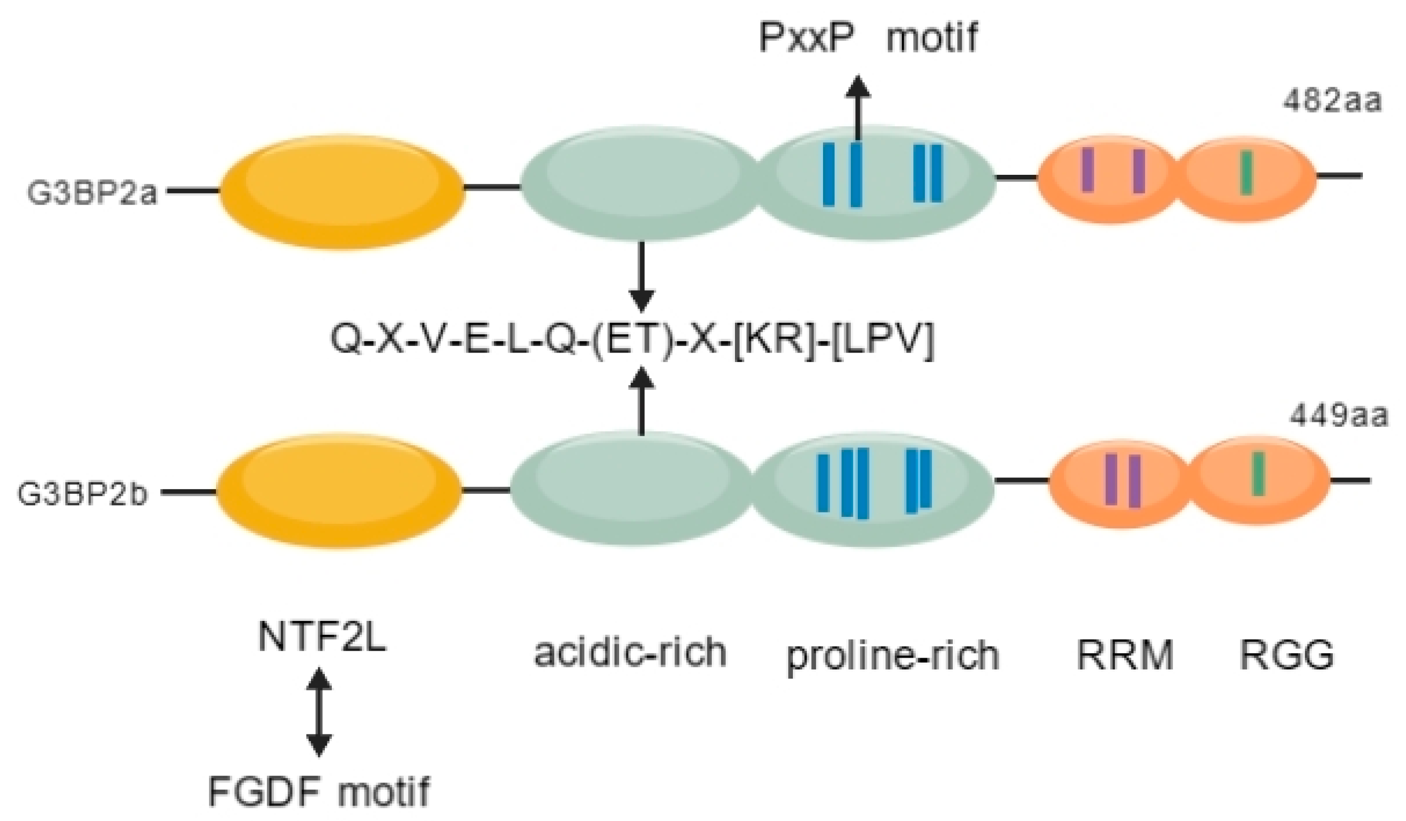
1.1. Structure of G3BP2
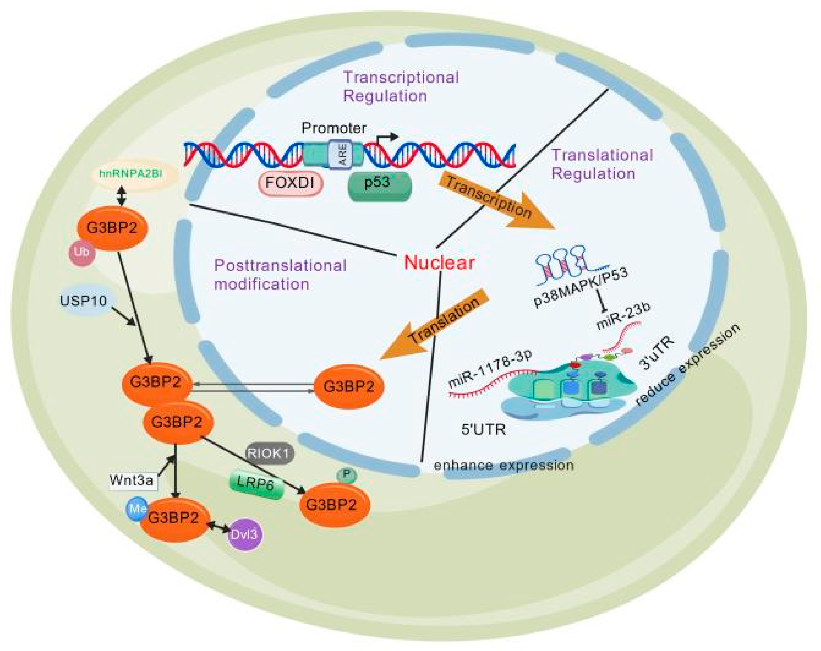
1.2. Expression Regulation of G3BP2
1.2.1. Transcriptional Regulation of G3BP2
1.2.2. Translational Regulation of G3BP2
1.2.3. Post-Translational Modification
Phosphorylation
Polyubiquitination
Arginine Methylation
1.2.4. Regulation of G3BP2 Subcellular Localization
1.3. Mechanism of G3BP2
1.3.1. RNA Binding-Mediated Transcript Stability
1.3.2. Regulation of Protein Subcellular Localization
1.3.3. Cytosolic Localization Regulation of TWIST1
1.3.4. Core Role of SG Assembly
1.3.5. The Relationship Between G3BP1 and G3BP2
2. Disease Expression of G3BP2 (Table 1)
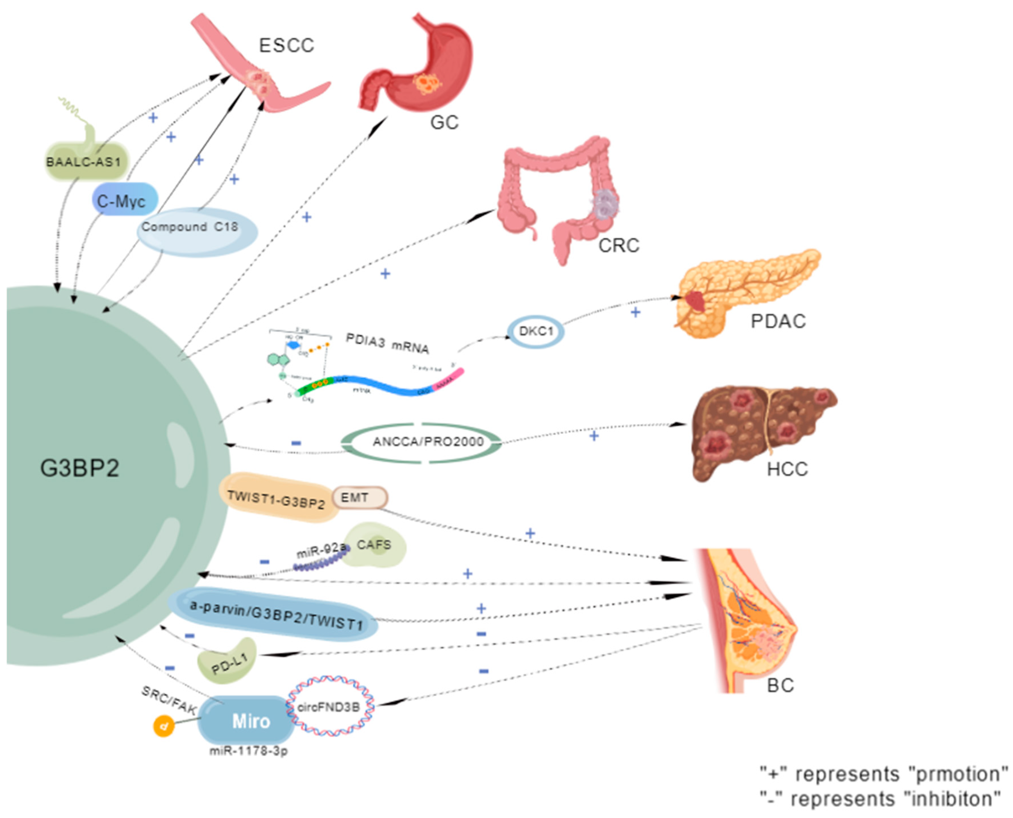
2.1. Expression of G3BP2 in Tumors
2.1.1. Esophageal Squamous Cell
2.1.2. Gastric Cancer (GC)
2.1.3. Colorectal Cancer (CRC)
2.1.4. Pancreatic Ductal Adenocarcinoma (PDAC)
2.1.5. Hepatocellular Carcinoma (HCC)
2.1.6. Breast Cancer (BC)
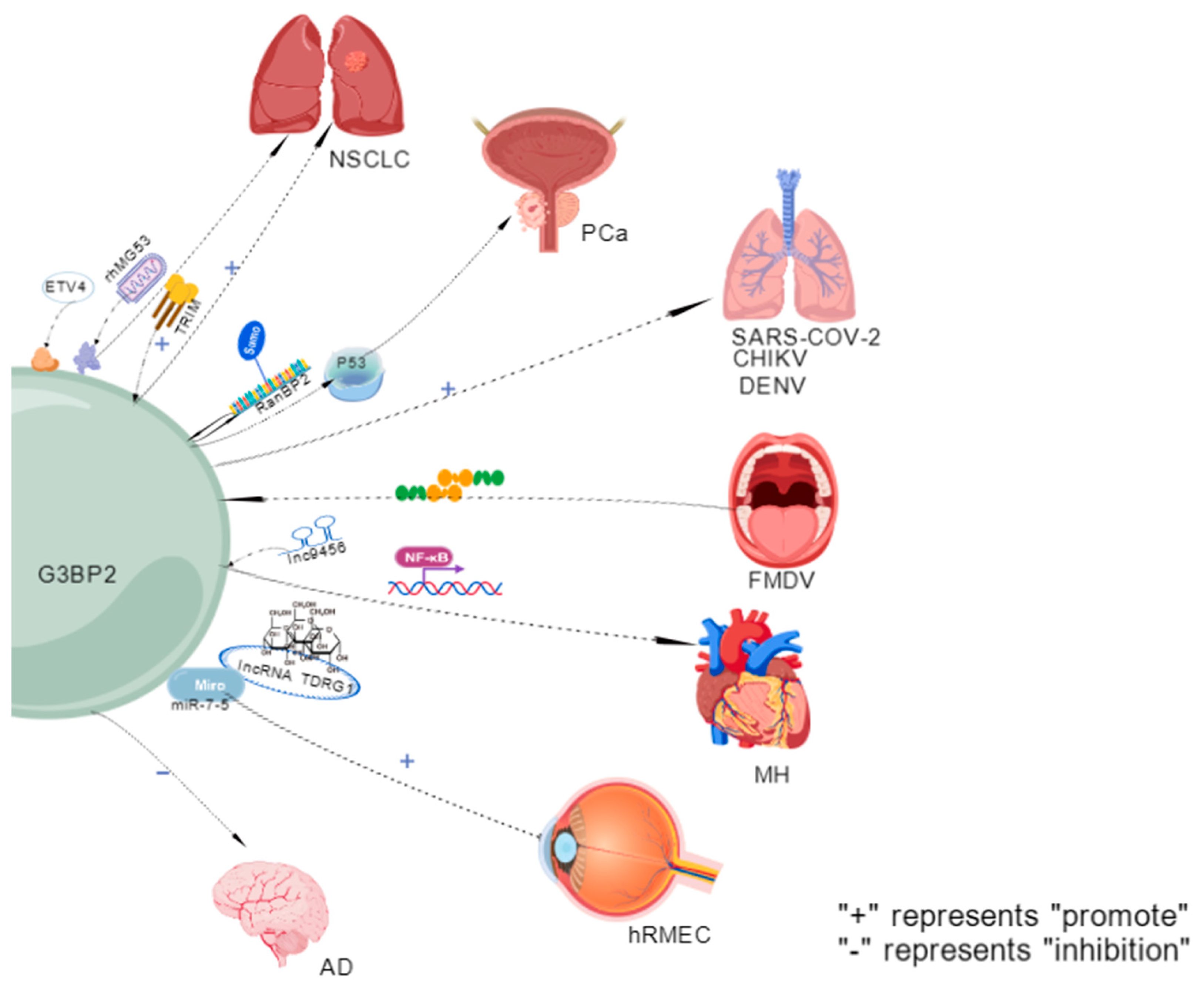
2.1.7. Non-Small-Cell Lung Cancer (NSCLC)
2.1.8. PCa
2.2. Expression of G3BP2 in Viral Infections
2.2.1. SARS-CoV-2
2.2.2. Chikungunya Virus (CHIKV)
2.2.3. Foot-and-Mouth Disease Virus
2.2.4. Dengue Virus (DENV)
2.3. G3BP2 Expression in Cardiovascular and Cerebrovascular Diseases
2.3.1. Myocardial Hypertrophy
2.3.2. Damage to Retinal Microvascular Endothelial Cells in the Human Eye
2.3.3. Alzheimer’s Disease (AD)
2.4. G3BP2 Expression in Other Diseases

| Classification | Disease | Pathway | Mechanism | Is There any Clinical Sample Verification? | Cell Line /Animals | Result | References |
|---|---|---|---|---|---|---|---|
| Tumor | Esophageal squamous cell carcinoma (SCC) | Long non-coding RNAlncRNA LINC01554 | LINC01554 stabilizes G3BP2 by inhibiting its ubiquitination degradation pathway, which forms a feedback loop to maintain its continuous high expression in ESCC. | Yes | Both | This markedly enhances ESCC cell migration and invasion, driving tumor progression. | [34,62] |
| Together with c-Myc and BAALC-AS1, forms a feedback loop | It may also provide novel therapeutic targets for ESCC and facilitate the development of new treatment strategies. | ||||||
| Gastric cancer (GC) | TM4SF1-AS1 is associated with multiple stress granule (SG) associated-related proteins, including G3BP2. | SG formation is promoted by sequestering RACK1, a stress-responsive MAPK pathway activator, in GC cells. | Yes | Cell line | G3BP2 upregulation correlates significantly with disease severity; carcinogenic pathway-altered protein subgroups may drive human gastric cancer (GC) progression. | [66,67] | |
| Helicobacter pylori infection induces distinct gastric proteome alterations. | |||||||
| Colorectal cancer (CRC) | hsa_circRNA_001676 regulates; The miR-556-3p/G3BP2 axis | Accelerate the proliferation, migration and stem cell-like transformation of CRC | Yes | Cell line | G3BP2 is a key factor for CRC cell Since the content in cell is too long, we added horizontal line for each row, please confirm. proliferation, migration and stemness maintenance; its depletion inhibits these malignant phenotypes. | [70] | |
| Pancreatic ductal adenocarcinoma (PDAC) | G3BP2 binds to PDIA3 mRNA and recruits it to stress granules. | Promotes DKC1 expression by enhancing its mRNA stability and reducing translation efficiency. | Yes | Cell line | - | [72] | |
| Hepatocellular carcinoma (HCC) | ANCCA/PRO2000 | Inhibits ERO1L and G3BP2 expression. | Yes | Cell line | Partially enhances hepatocellular carcinoma (HCC) cell migration. | [74,75] | |
| Breast cancer (BC) | TWIST1-G3BP2 mechanical transduction. | Drives EMT, invasion and metastasis by responding to tumor microenvironment signals. | Yes | Both | Regulates EMT, invasion and metastasis. | [5,31,43,79,80,81,82,83] | |
| Exosomes derived from Cafa contain miR-92a, which reduces the expression of G3BP2. | Promotes breast cancer cell migration and invasion. | SART3 mRNA stable upregulation enhances pluripotency factor Oct-4/Nanog expression, driving tumorigenesis; it markedly inhibits breast cancer (BC) cell proliferation, migration and invasion. | |||||
| Excessive expression of the circBACH1/miR-217 axis. | The miR-217 directly targets and represses G3BP2, while PTX-induced exosomal circBACH1 interacts with miR-217 to abrogate its suppression of G3BP2 and upregulate G3BP2 expression. | Inhibits BC malignant phenotypes | |||||
| circFNDC3B sequesters miR-1178-3p through sponge adsorption. | Inhibits G3BP2 expression, thereby blocking SRC/FAK signaling pathway phosphorylation. | - | |||||
| Non-small-cell lung cancer (NSCLC) | - | TRIM domain interacts with G3BP2. | Yes | Both | Potentially inhibits H1299 cell proliferation, migration and invasion | [30,85] | |
| Recruits the lysosome-TSC2 complex to inhibit mTORC1 activity | ETV4/TSC2/mTORC1 axis regulates key glycolysis and protein synthesis components | ||||||
| Recombinant human MG53 (rhMG53) induces G3BP2 nuclear translocation and inhibits stress granule formation. | Inhibits NSCLC cell proliferation and enhances cellular sensitivity to cisplatin. | ||||||
| Prostate cancer (PCa) | - | TRIM25 overexpression regulates p53 nuclear export by interacting with G3BP2. | Yes | Both | It promotes the proliferation and survival of cancer cells. | [9,88] | |
| Knockdown of G3BP2. | Inhibits tumor growth and increases p53 accumulation in the nucleus. | ||||||
| Driven by androgens, it translocates to the cytoplasm via p53. | Thereby indicating a risk of poor prognosis and hormone-resistant transformation. | ||||||
| Viral infection | Infection with SARS-CoV-2 | G3BP1/G3BP2 (RAS GTPase-activating protein SH3 domain-binding protein 1/2) is a key host factor that interacts with the nucleocapsid (N) protein of SARS-CoV-2. | G3BP2 is a key host factor interacting with the SARS-CoV-2 nucleocapsid (N) protein. | Yes | Both | It potentially enhances SARS-CoV-2 replication. | [93] |
| Chikungunya virus (CHIKV) | - | Simultaneous absence of G3BP1 and G3BP2 reduces CHIKV RNA levels, protein expression, and progeny titers. | Yes | Cell line | Chikungunya virus (CHIKV) also relies on G3BPs for efficient replication. | [97] | |
| Foot-and-mouth disease virus | It can be achieved by targeting the scaffold proteins G3BP1 and G3BP2, and using their L protease to cleave these proteins. | Antagonizing SG formation modulates the global stress response to evade host antiviral defense. | Yes | Cell line | It facilitates the formulation of future control measures against FMDV infection. | [100] | |
| Dengue virus (DENV) | - | Dengue virus (DENV) RNA binds to P-body (PB)/stress granule (SG) regulatory proteins, including DDX6, G3BP1, G3BP2, Caprin1, and USP10. | Yes | Cell line | DENV-2 3′ UTR acts as an assembly platform for PB/SG-associated proteins; DDX6 assembly on the 3′ UTR is essential for viral replication. | [102] | |
| Cardiovascular and cerebrovascular diseases | Myocardial hypertrophy | Lnc9456 interacts physically with G3BP2. | Promotes NF-κB nuclear translocation. | Yes | Both | This further activates the hypertrophy-related signaling cascade. | [35,107] |
| The hypertrophy response of neonatal rat cardiomyocytes (NRCMs) induced by overexpression of G3BP2 or isoproterenol (ISO) | It is significantly inhibited by the NF-κB inhibitor PDTC (50 μmol/L) or p65 knockdown. | ||||||
| The microvascular endothelial cells of the human retina | The lncRNA TDRG1 competes for binding with miR-7-5p | Increases G3BP2 transcriptional levels. | Yes | Cell line | strengthens high glucose (HG)-induced damage to human retinal microvascular endothelial cells (hRMECs). | [110] | |
| Alzheimer’s disease (AD) | - | G3BP2 directly binds to the Tau protein and inhibits its pathological aggregation. | Yes | Cell line | The absence of G3BP2 significantly accelerates the Tau pathological process. | [46,113] | |
| G3BP2 blocks Tau aggregation by masking its microtubule-binding region (MTBR) | |||||||
| Other diseases | G3BP2 is involved in immune responses, mRNA transport, and stress granule assembly | - | Yes | Mouse | Mouse spermatogenesis and male fertility are indispensable. | [114] | |
| The interaction between PGRMC1/PGRMC2 and G3BP2 | - | Yes | Cell line | Dynamic regulation of the rate of SIGC cell cycle entry. | [115] | ||
| G3BP2 elevates CD8+ T cell percentage | Yes | Cell line | Promotion of psoriasis development | [116] | |||
| miR-363 is indispensable in the polarization of M1 macrophages | Released from M1 macrophages via exosomes | - | - | Induces chondrocyte apoptosis and inflammation; this axis is a promising target for cartilage degeneration and OA prevention | [117] | ||
| miR-363 inhibits chondrocyte damage by targeting and repressing G3BP2 |
3. Clinical Treatment Transformation of G3BP2 (Table 2)
3.1. Role of G3BP2 in Clinical Diagnosis
3.2. Clinical Therapeutic Value of G3BP2
3.2.1. BC
3.2.2. Head and Neck Squamous Cell Carcinoma
3.2.3. Lung Cancer
3.2.4. Pancreatic Ductal Carcinoma
3.2.5. PCa
3.2.6. CRC
3.2.7. Oral Cancer
3.2.8. Atherosclerosis
3.2.9. Others
| Classification | Disease | Mechanism | Cell Line /Animals | Treatment | Clinical Trial | References |
|---|---|---|---|---|---|---|
| Biomarker | AD | The synergistic interaction of PSF and G3BP2 in the cell nucleus | Animals | Yes | [120] | |
| - | Depletion of PGRMC1, PGRMC2, or G3BP2 increases NF-κB transcriptional activity. | Animals | The cell cycle progresses at an accelerated pace. | Yes | [121] | |
| Therapy | BC | G3BP2 upregulation enhances breast cancer (BC) cell migration, stemness, and paclitaxel (PTX) resistance; its downregulation abrogates these phenotypes, highlighting a critical role of G3BP2 in BC progression. | Cell line | The circBACH1/miR-217/G3BP2 axis-mediated paclitaxel (PTX) resistance and breast cancer (BC) progression identifies novel therapeutic targets. | Yes | [82] |
| Head and neck squamous cell carcinoma | The PRMT5-USP7-G3BP2 regulatory axis drives tumorigenesis via lipid metabolic reprogramming. | Cell line | It identifies potential therapeutic targets for metabolic therapy in head and neck squamous cell carcinoma (HNSCC). | Yes | [124] | |
| Lung cancer | TRIM72 modulates G3BP2 activity. | - | This suppresses lung cancer progression. | [30] | ||
| PDAC | G3BP2-mediated SGs | - | Protective therapeutic targets for PDAC. | [72] | ||
| PCa | It forms a complex with the tumor suppressor p53, thereby inhibiting the regulation of cell growth by the p53 signaling pathway. | - | USP10 upregulates G3BP2 expression to act as a key mediator of PCa carcinogenesis. | [27] | ||
| Acting synergistically with the AR signaling, they jointly promote tumor development. | ||||||
| CRC | Phosphorylated YBX1 translocates to the nucleus to initiate G3BP2 transcription and activate the MAPK signaling pathway. Eps8l2 knockout suppressed CRC tumorigenesis in the AOM/DSS model. | Animals | EPS8L2-YBX1-G3BP2 axis: a driver of CRC progression and a novel therapeutic target. | Yes | [132] | |
| Oral cancer | - | G3BP2 impairs radiotherapy efficacy in oral cancer. | [11] | |||
| Atherosclerosis | In ApoE−/− atherosclerotic mice, G3BP2 peptide antigen administration or G3BP2 knockdown significantly reduces early atherosclerotic plaques | Animals | It represents a potential therapeutic target for atherosclerosis therapy. | Yes | [135] | |
| Anti-cancer immunotherapy | Three core findings: immune escape regulation, clinical relevance, targeted therapeutic potential:
| - | It identifies a direct target for blocking immune escape. | Yes | [80] | |
| New substrate of CRBN | Highly selective and potent phenylpyridineamine degraders target uncharacterized novel substrates (KDM4B, G3BP2, VCL), which lack the canonical CRBN β-hairpin motif | - | Marked expansion of the novel CRBN substrate repertoire defined by canonical IMiDs. | [136] |
4. Summary and Outlook
Author Contributions
Funding
Institutional Review Board Statement
Informed Consent Statement
Data Availability Statement
Acknowledgments
Conflicts of Interest
References
- Letunic, I.; Khedkar, S.; Bork, P. SMART: Recent updates, new developments and status in 2020. Nucleic Acids Res. 2021, 49, D458–D460. [Google Scholar] [CrossRef] [PubMed]
- Duchesne, M.; Schweighoffer, F.; Parker, F.; Clerc, F.; Frobert, Y.; Thang, M.N.; Tocqué, B. Identification of the SH3 domain of GAP as an essential sequence for Ras-GAP-mediated signaling. Science 1993, 259, 525–528. [Google Scholar] [CrossRef] [PubMed]
- Kennedy, D.; French, J.; Guitard, E.; Ru, K.; Tocque, B.; Mattick, J. Characterization of G3BPs: Tissue specific expression, chromosomal localisation and rasGAP(120) binding studies. J. Cell. Biochem. 2001, 84, 173–187. [Google Scholar] [CrossRef] [PubMed]
- Paschal, B.M.; Gerace, L. Identification of NTF2, a cytosolic factor for nuclear import that interacts with nuclear pore complex protein p62. J. Cell Biol. 1995, 129, 925–937. [Google Scholar] [CrossRef]
- Wei, S.C.; Fattet, L.; Tsai, J.H.; Guo, Y.; Pai, V.H.; Majeski, H.E.; Chen, A.C.; Sah, R.L.; Taylor, S.S.; Engler, A.J.; et al. Matrix stiffness drives epithelial-mesenchymal transition and tumour metastasis through a TWIST1-G3BP2 mechanotransduction pathway. Nat. Cell Biol. 2015, 17, 678–688. [Google Scholar] [CrossRef]
- Dreyfuss, G.; Kim, V.N.; Kataoka, N. Messenger-RNA-binding proteins and the messages they carry. Nat. Rev. Mol. Cell Biol. 2002, 3, 195–205. [Google Scholar] [CrossRef]
- Cléry, A.; Blatter, M.; Allain, F.H. RNA recognition motifs: Boring? Not quite. Curr. Opin. Struct. Biol. 2008, 18, 290–298. [Google Scholar] [CrossRef]
- Burd, C.G.; Dreyfuss, G. Conserved structures and diversity of functions of RNA-binding proteins. Science 1994, 265, 615–621. [Google Scholar] [CrossRef]
- Ashikari, D.; Takayama, K.; Tanaka, T.; Suzuki, Y.; Obinata, D.; Fujimura, T.; Urano, T.; Takahashi, S.; Inoue, S. Androgen induces G3BP2 and SUMO-mediated p53 nuclear export in prostate cancer. Oncogene 2017, 36, 6272–6281. [Google Scholar] [CrossRef]
- Aurilio, G.; Cimadamore, A.; Mazzucchelli, R.; Lopez-Beltran, A.; Verri, E.; Scarpelli, M.; Massari, F.; Cheng, L.; Santoni, M.; Montironi, R. Androgen Receptor Signaling Pathway in Prostate Cancer: From Genetics to Clinical Applications. Cells 2020, 9, 2653. [Google Scholar] [CrossRef]
- Lin, C.H.; Lee, H.H.; Chang, W.M.; Lee, F.P.; Chen, L.C.; Lu, L.S.; Lin, Y.F. FOXD1 Repression Potentiates Radiation Effectiveness by Downregulating G3BP2 Expression and Promoting the Activation of TXNIP-Related Pathways in Oral Cancer. Cancers 2020, 12, 2690. [Google Scholar] [CrossRef]
- Jin, G.; Zhang, Z.; Wan, J.; Wu, X.; Liu, X.; Zhang, W. G3BP2: Structure and function. Pharmacol. Res. 2022, 186, 106548. [Google Scholar] [CrossRef]
- Zhao, B.; Li, H.; Liu, J.; Han, P.; Zhang, C.; Bai, H.; Yuan, X.; Wang, X.; Li, L.; Ma, H.; et al. MicroRNA-23b Targets Ras GTPase-Activating Protein SH3 Domain-Binding Protein 2 to Alleviate Fibrosis and Albuminuria in Diabetic Nephropathy. J. Am. Soc. Nephrol. 2016, 27, 2597–2608. [Google Scholar] [CrossRef]
- Carney, E.F. Diabetic nephropathy: MiR-23b protects against fibrosis in diabetic nephropathy. Nat. Rev. Nephrol. 2016, 12, 197. [Google Scholar] [PubMed]
- Zhao, H.; He, Y. MiR-124-3p Suppresses the Dysfunction of High Glucose-Stimulated Endothelial Cells by Targeting G3BP2. Front. Genet. 2021, 12, 723625. [Google Scholar] [CrossRef] [PubMed]
- Shuang, O.; Zhou, J.; Cai, Z.; Liao, L.; Wang, Y.; Wang, W.; Xu, M. EBF1-mediated up-regulation of lncRNA FGD5-AS1 facilitates osteosarcoma progression by regulating miR-124-3p/G3BP2 axis as a ceRNA. J. Orthop. Surg. Res. 2022, 17, 332. [Google Scholar] [CrossRef] [PubMed]
- Esteller, M. Non-coding RNAs in human disease. Nat. Rev. Genet. 2011, 12, 861–874. [Google Scholar] [CrossRef]
- Matsui, M.; Corey, D.R. Non-coding RNAs as drug targets. Nat. Rev. Drug Discov. 2017, 16, 167–179. [Google Scholar] [CrossRef]
- Guillén-Boixet, J.; Kopach, A.; Holehouse, A.S.; Wittmann, S.; Jahnel, M.; Schlüßler, R.; Kim, K.; Trussina, I.; Wang, J.; Mateju, D.; et al. RNA-Induced Conformational Switching and Clustering of G3BP Drive Stress Granule Assembly by Condensation. Cell 2020, 181, 346–361.e17. [Google Scholar] [CrossRef]
- Chen, Y.; Zhou, S.; Wan, K.; Yu, L.; Zhao, C.; Deng, H.; Ou, Q.; Qin, J.; Hu, J.; Hou, Z. RIOK1 mediates p53 degradation and radioresistance in colorectal cancer through phosphorylation of G3BP2. Oncogene 2022, 41, 3433–3444. [Google Scholar] [CrossRef]
- Bikkavilli, R.K.; Malbon, C.C. Wnt3a-stimulated LRP6 phosphorylation is dependent upon arginine methylation of G3BP2. J. Cell Sci. 2012, 125, 2446–2456. [Google Scholar]
- Fattet, L.; Jung, H.Y.; Matsumoto, M.W.; Aubol, B.E.; Kumar, A.; Adams, J.A.; Chen, A.C.; Sah, R.L.; Engler, A.J.; Pasquale, E.B.; et al. Matrix Rigidity Controls Epithelial-Mesenchymal Plasticity and Tumor Metastasis via a Mechanoresponsive EPHA2/LYN Complex. Dev. Cell 2020, 54, 302–316.e7. [Google Scholar] [CrossRef] [PubMed]
- Kobayashi, T.; Winslow, S.; Sunesson, L.; Hellman, U.; Larsson, C. PKCα binds G3BP2 and regulates stress granule formation following cellular stress. PLoS ONE 2012, 7, e35820. [Google Scholar] [CrossRef] [PubMed]
- Kedersha, N.; Panas, M.D.; Achorn, C.A.; Lyons, S.; Tisdale, S.; Hickman, T.; Thomas, M.; Lieberman, J.; McInerney, G.M.; Ivanov, P.; et al. G3BP-Caprin1-USP10 complexes mediate stress granule condensation and associate with 40S subunits. J. Cell Biol. 2016, 212, 845–860. [Google Scholar] [CrossRef] [PubMed]
- Onomoto, K.; Sakai, M.; Watanabe, M.; Fukao, A.; Sakamura, Y.; Miyao, M.; Tomohiro, T.; Yamashita, A.; Fujiwara, T.; Takahashi, T.; et al. TRBP modulates RLR signaling by inhibiting PKR-mediated antiviral stress granule formation. Sci. Rep. 2025, 15, 20678. [Google Scholar] [CrossRef]
- Zhai, C.; Sun, X.; Zhang, S.; Xing, L. ROBO1 enhanced esophageal carcinoma cell radioresistance through accelerating G3BP2-mediated eIF3A degradation. Cell Death Dis. 2025, 16, 256. [Google Scholar] [CrossRef]
- Takayama, K.I.; Suzuki, T.; Fujimura, T.; Takahashi, S.; Inoue, S. Association of USP10 with G3BP2 Inhibits p53 Signaling and Contributes to Poor Outcome in Prostate Cancer. Mol. Cancer Res. 2018, 16, 846–856. [Google Scholar] [CrossRef]
- Zhang, J.; Li, J.; Luo, Y.; Mahony, T.J.; Gao, J.; Wang, Y.; Yang, X.; Yang, F.; Ou, X.; Zhang, J.; et al. LINC2781 enhances antiviral immunity against coxsackievirus B5 infection by activating the JAK-STAT pathway and blocking G3BP2-mediated STAT1 degradation. mSphere 2025, 10, e0006225. [Google Scholar] [CrossRef]
- Wang, X.; Fan, X.; Zhang, J.; Wang, F.; Chen, J.; Wen, Y.; Wang, L.; Li, T.; Li, H.; Gu, H.; et al. hnRNPA2B1 represses the disassembly of arsenite-induced stress granules and is essential for male fertility. Cell Rep. 2024, 43, 113769. [Google Scholar] [CrossRef]
- Li, H.; Lin, P.H.; Gupta, P.; Li, X.; Zhao, S.L.; Zhou, X.; Li, Z.; Wei, S.; Xu, L.; Han, R.; et al. MG53 suppresses tumor progression and stress granule formation by modulating G3BP2 activity in non-small cell lung cancer. Mol. Cancer 2021, 20, 118. [Google Scholar] [CrossRef]
- Gupta, N.; Badeaux, M.; Liu, Y.; Naxerova, K.; Sgroi, D.; Munn, L.L.; Jain, R.K.; Garkavtsev, I. Stress granule-associated protein G3BP2 regulates breast tumor initiation. Proc. Natl. Acad. Sci. USA 2017, 114, 1033–1038. [Google Scholar] [CrossRef]
- Fu, Q.; Song, X.; Liu, Z.; Deng, X.; Luo, R.; Ge, C.; Li, R.; Li, Z.; Zhao, M.; Chen, Y.; et al. miRomics and Proteomics Reveal a miR-296-3p/PRKCA/FAK/Ras/c-Myc Feedback Loop Modulated by HDGF/DDX5/β-catenin Complex in Lung Adenocarcinoma. Clin. Cancer Res. 2017, 23, 6336–6350. [Google Scholar] [CrossRef] [PubMed]
- Yang, Y.; Ma, Y.; Gao, H.; Peng, T.; Shi, H.; Tang, Y.; Li, H.; Chen, L.; Hu, K.; Han, A. A novel HDGF-ALCAM axis promotes the metastasis of Ewing sarcoma via regulating the GTPases signaling pathway. Oncogene 2021, 40, 731–745. [Google Scholar] [CrossRef] [PubMed]
- Zheng, Y.; Wu, J.; Deng, R.; Lin, C.; Huang, Y.; Yang, X.; Wang, C.; Yang, M.; He, Y.; Lu, J.; et al. G3BP2 regulated by the lncRNA LINC01554 facilitates esophageal squamous cell carcinoma metastasis through stabilizing HDGF transcript. Oncogene 2022, 41, 515–526. [Google Scholar] [CrossRef] [PubMed]
- Hong, H.Q.; Lu, J.; Fang, X.L.; Zhang, Y.H.; Cai, Y.; Yuan, J.; Liu, P.Q.; Ye, J.T. G3BP2 is involved in isoproterenol-induced cardiac hypertrophy through activating the NF-κB signaling pathway. Acta Pharmacol. Sin. 2018, 39, 184–194. [Google Scholar] [CrossRef]
- Prigent, M.; Barlat, I.; Langen, H.; Dargemont, C. IkappaBalpha and IkappaBalpha/NF-kappa B complexes are retained in the cytoplasm through interaction with a novel partner, RasGAP SH3-binding protein 2. J. Biol. Chem. 2000, 275, 36441–36449. [Google Scholar] [CrossRef]
- Kim, M.M.; Wiederschain, D.; Kennedy, D.; Hansen, E.; Yuan, Z.M. Modulation of p53 and MDM2 activity by novel interaction with Ras-GAP binding proteins (G3BP). Oncogene 2007, 26, 4209–4215. [Google Scholar] [CrossRef]
- Carter, S.; Bischof, O.; Dejean, A.; Vousden, K.H. C-terminal modifications regulate MDM2 dissociation and nuclear export of p53. Nat. Cell Biol. 2007, 9, 428–435. [Google Scholar] [CrossRef]
- Rusiecki, R.; Witkowski, J.; Jaszczewska-Adamczak, J. MDM2-p53 Interaction Inhibitors: The Current State-of-Art and Updated Patent Review (2010–Present). Recent Pat. Anticancer Drug Discov. 2019, 14, 324–369. [Google Scholar] [CrossRef]
- Güttler, T.; Görlich, D. Ran-dependent nuclear export mediators: A structural perspective. EMBO J. 2011, 30, 3457–3474. [Google Scholar] [CrossRef]
- Werner, A.; Flotho, A.; Melchior, F. The RanBP2/RanGAP1*SUMO1/Ubc9 complex is a multisubunit SUMO E3 ligase. Mol. Cell 2012, 46, 287–298. [Google Scholar] [CrossRef] [PubMed]
- Neve, R.M.; Chin, K.; Fridlyand, J.; Yeh, J.; Baehner, F.L.; Fevr, T.; Clark, L.; Bayani, N.; Coppe, J.P.; Tong, F.; et al. A collection of breast cancer cell lines for the study of functionally distinct cancer subtypes. Cancer Cell 2006, 10, 515–527. [Google Scholar] [CrossRef] [PubMed]
- Sun, Y.; Ding, Y.; Guo, C.; Liu, C.; Ma, P.; Ma, S.; Wang, Z.; Liu, J.; Qian, T.; Ma, L.; et al. α-Parvin promotes breast cancer progression and metastasis through interaction with G3BP2 and regulation of TWIST1 signaling. Oncogene 2019, 38, 4856–4874. [Google Scholar] [CrossRef] [PubMed]
- Prentzell, M.T.; Rehbein, U.; Cadena Sandoval, M.; De Meulemeester, A.S.; Baumeister, R.; Brohée, L.; Berdel, B.; Bockwoldt, M.; Carroll, B.; Chowdhury, S.R.; et al. G3BPs tether the TSC complex to lysosomes and suppress mTORC1 signaling. Cell 2021, 184, 655–674.e27. [Google Scholar] [CrossRef]
- Rehbein, U.; Prentzell, M.T.; Cadena Sandoval, M.; Heberle, A.M.; Henske, E.P.; Opitz, C.A.; Thedieck, K. The TSC Complex-mTORC1 Axis: From Lysosomes to Stress Granules and Back. Front. Cell Dev. Biol. 2021, 9, 751892. [Google Scholar] [CrossRef]
- Wang, C.; Terrigno, M.; Li, J.; Distler, T.; Pandya, N.J.; Ebeling, M.; Tyanova, S.; Hoozemans, J.J.M.; Dijkstra, A.A.; Fuchs, L.; et al. Increased G3BP2-Tau interaction in tauopathies is a natural defense against Tau aggregation. Neuron 2023, 111, 2660–2674.e9. [Google Scholar] [CrossRef]
- Sanders, D.W.; Kedersha, N.; Lee, D.S.W.; Strom, A.R.; Drake, V.; Riback, J.A.; Bracha, D.; Eeftens, J.M.; Iwanicki, A.; Wang, A.; et al. Competing Protein-RNA Interaction Networks Control Multiphase Intracellular Organization. Cell 2020, 181, 306–324.e28. [Google Scholar] [CrossRef]
- Yang, P.; Mathieu, C.; Kolaitis, R.M.; Zhang, P.; Messing, J.; Yurtsever, U.; Yang, Z.; Wu, J.; Li, Y.; Pan, Q.; et al. G3BP1 Is a Tunable Switch that Triggers Phase Separation to Assemble Stress Granules. Cell 2020, 181, 325–345.e28. [Google Scholar] [CrossRef]
- Somasekharan, S.P.; Gleave, M. SARS-CoV-2 nucleocapsid protein interacts with immunoregulators and stress granules and phase separates to form liquid droplets. FEBS Lett. 2021, 595, 2872–2896. [Google Scholar] [CrossRef]
- Huang, C.; Chen, Y.; Dai, H.; Zhang, H.; Xie, M.; Zhang, H.; Chen, F.; Kang, X.; Bai, X.; Chen, Z. UBAP2L arginine methylation by PRMT1 modulates stress granule assembly. Cell Death Differ. 2020, 27, 227–241. [Google Scholar] [CrossRef]
- Lee, A.K.; Klein, J.; Fon Tacer, K.; Lord, T.; Oatley, M.J.; Oatley, J.M.; Porter, S.N.; Pruett-Miller, S.M.; Tikhonova, E.B.; Karamyshev, A.L.; et al. Translational Repression of G3BP in Cancer and Germ Cells Suppresses Stress Granules and Enhances Stress Tolerance. Mol. Cell 2020, 79, 645–659.e9. [Google Scholar] [CrossRef]
- Lai, R.H.; Chow, Y.H.; Lin, Y.W.; Chung, N.H.; Nien, S.W.; Juang, J.L. Hyperglycemia facilitates EV71 replication: Insights into miR-206-mediated regulation of G3BP2 promoting EV71 IRES activity. Theranostics 2024, 14, 2706–2718. [Google Scholar] [CrossRef]
- Mukhopadhyay, C.; Zhou, P. Role(s) of G3BPs in Human Pathogenesis. J. Pharmacol. Exp. Ther. 2023, 387, 100–110. [Google Scholar] [CrossRef] [PubMed]
- Atlas, R.; Behar, L.; Elliott, E.; Ginzburg, I. The insulin-like growth factor mRNA binding-protein IMP-1 and the Ras-regulatory protein G3BP associate with tau mRNA and HuD protein in differentiated P19 neuronal cells. J. Neurochem. 2004, 89, 613–626. [Google Scholar] [CrossRef] [PubMed]
- Angenstein, F.; Evans, A.M.; Settlage, R.E.; Moran, S.T.; Ling, S.C.; Klintsova, A.Y.; Shabanowitz, J.; Hunt, D.F.; Greenough, W.T. A receptor for activated C kinase is part of messenger ribonucleoprotein complexes associated with polyA-mRNAs in neurons. J. Neurosci. 2002, 22, 8827–8837. [Google Scholar] [CrossRef] [PubMed]
- Irvine, K.; Stirling, R.; Hume, D.; Kennedy, D. Rasputin, more promiscuous than ever: A review of G3BP. Int. J. Dev. Biol. 2004, 48, 1065–1077. [Google Scholar] [CrossRef]
- Winslow, S.; Leandersson, K.; Larsson, C. Regulation of PMP22 mRNA by G3BP1 affects cell proliferation in breast cancer cells. Mol. Cancer 2013, 12, 156. [Google Scholar] [CrossRef]
- Taniuchi, K.; Nishimori, I.; Hollingsworth, M.A. The N-terminal domain of G3BP enhances cell motility and invasion by posttranscriptional regulation of BART. Mol. Cancer Res. 2011, 9, 856–866. [Google Scholar] [CrossRef]
- Taniuchi, K.; Nishimori, I.; Hollingsworth, M.A. Intracellular CD24 inhibits cell invasion by posttranscriptional regulation of BART through interaction with G3BP. Cancer Res. 2011, 71, 895–905. [Google Scholar] [CrossRef]
- Siegel, R.L.; Giaquinto, A.N.; Jemal, A. Cancer statistics, 2024. CA Cancer J. Clin. 2024, 74, 12–49. [Google Scholar] [CrossRef]
- Zeng, H.; Chen, W.; Zheng, R.; Zhang, S.; Ji, J.S.; Zou, X.; Xia, C.; Sun, K.; Yang, Z.; Li, H.; et al. Changing cancer survival in China during 2003–15: A pooled analysis of 17 population-based cancer registries. Lancet Glob. Health 2018, 6, e555–e567. [Google Scholar] [CrossRef]
- Zhang, H.; Wang, Y.; Zhang, W.; Wu, Q.; Fan, J.; Zhan, Q. BAALC-AS1/G3BP2/c-Myc feedback loop promotes cell proliferation in esophageal squamous cell carcinoma. Cancer Commun. 2021, 41, 240–257. [Google Scholar] [CrossRef] [PubMed]
- Zeng, R.; Gou, H.; Lau, H.C.H.; Yu, J. Stomach microbiota in gastric cancer development and clinical implications. Gut 2024, 73, 2062–2073. [Google Scholar] [CrossRef] [PubMed]
- Sundar, R.; Nakayama, I.; Markar, S.R.; Shitara, K.; van Laarhoven, H.W.M.; Janjigian, Y.Y.; Smyth, E.C. Gastric cancer. Lancet 2025, 405, 2087–2102. [Google Scholar] [CrossRef] [PubMed]
- Mott, T.; Gray, C. Gastric Cancer: Rapid Evidence Review. Am. Fam. Physician 2025, 111, 140–145. [Google Scholar]
- Noto, J.M.; Rose, K.L.; Hachey, A.J.; Delgado, A.G.; Romero-Gallo, J.; Wroblewski, L.E.; Schneider, B.G.; Shah, S.C.; Cover, T.L.; Wilson, K.T.; et al. Carcinogenic Helicobacter pylori Strains Selectively Dysregulate the In Vivo Gastric Proteome, Which May Be Associated with Stomach Cancer Progression. Mol. Cell. Proteom. 2019, 18, 352–371. [Google Scholar] [CrossRef]
- Kitajima, H.; Maruyama, R.; Niinuma, T.; Yamamoto, E.; Takasawa, A.; Takasawa, K.; Ishiguro, K.; Tsuyada, A.; Suzuki, R.; Sudo, G.; et al. TM4SF1-AS1 inhibits apoptosis by promoting stress granule formation in cancer cells. Cell Death Dis. 2023, 14, 424. [Google Scholar] [CrossRef]
- Biller, L.H.; Schrag, D. Diagnosis and Treatment of Metastatic Colorectal Cancer: A Review. JAMA 2021, 325, 669–685. [Google Scholar] [CrossRef]
- Thanikachalam, K.; Khan, G. Colorectal Cancer and Nutrition. Nutrients 2019, 11, 164. [Google Scholar] [CrossRef]
- Hao, Q.; Zhang, M.; Wu, Y.; Guo, Y.; Zheng, Y.; Wu, L.; Feng, L.; Wang, Z. Hsa_circRNA_001676 accelerates the proliferation, migration and stemness in colorectal cancer through regulating miR-556-3p/G3BP2 axis. Sci. Rep. 2023, 13, 18353. [Google Scholar] [CrossRef]
- Osei-Bordom, D.C.; Serifis, N.; Brown, Z.J.; Hewitt, D.B.; Lawal, G.; Sachdeva, G.; Cloonan, D.J.; Pawlik, T.M. Pancreatic ductal adenocarcinoma: Emerging therapeutic strategies. Surg. Oncol. 2022, 43, 101803. [Google Scholar] [CrossRef]
- Xing, F.L.; Li, B.R.; Fang, Y.J.; Liang, C.; Liu, J.; Wang, W.; Xu, J.; Yu, X.J.; Qin, Y.; Zhang, B. G3BP2 promotes tumor progression and gemcitabine resistance in PDAC via regulating PDIA3-DKC1-hENT in a stress granules-dependent manner. Acta Pharmacol. Sin. 2025, 46, 474–488. [Google Scholar] [CrossRef] [PubMed]
- Zheng, J.; Wang, S.; Xia, L.; Sun, Z.; Chan, K.M.; Bernards, R.; Qin, W.; Chen, J.; Xia, Q.; Jin, H. Hepatocellular carcinoma: Signaling pathways and therapeutic advances. Signal Transduct. Target. Ther. 2025, 10, 35. [Google Scholar] [CrossRef] [PubMed]
- Hwang, S.Y.; Danpanichkul, P.; Agopian, V.; Mehta, N.; Parikh, N.D.; Abou-Alfa, G.K.; Singal, A.G.; Yang, J.D. Hepatocellular carcinoma: Updates on epidemiology, surveillance, diagnosis and treatment. Clin. Mol. Hepatol. 2025, 31, S228–S254. [Google Scholar] [CrossRef] [PubMed]
- Huang, J.; Yang, J.; Lei, Y.; Gao, H.; Wei, T.; Luo, L.; Zhang, F.; Chen, H.; Zeng, Q.; Guo, L. An ANCCA/PRO2000-miR-520a-E2F2 regulatory loop as a driving force for the development of hepatocellular carcinoma. Oncogenesis 2016, 5, e229. [Google Scholar] [CrossRef]
- Jiang, B.; Bao, L.; He, S.; Chen, X.; Jin, Z.; Ye, Y. Deep learning applications in breast cancer histopathological imaging: Diagnosis, treatment, and prognosis. Breast Cancer Res. 2024, 26, 137. [Google Scholar] [CrossRef]
- Heater, N.K.; Warrior, S.; Lu, J. Current and future immunotherapy for breast cancer. J. Hematol. Oncol. 2024, 17, 131. [Google Scholar] [CrossRef]
- Heijink, A.M.; Everts, M.; Honeywell, M.E.; Richards, R.; Kok, Y.P.; de Vries, E.G.E.; Lee, M.J.; van Vugt, M. Modeling of Cisplatin-Induced Signaling Dynamics in Triple-Negative Breast Cancer Cells Reveals Mediators of Sensitivity. Cell Rep. 2019, 28, 2345–2357.e5. [Google Scholar] [CrossRef]
- Sheng, Z.; Wang, X.; Ding, X.; Zheng, Y.; Guo, A.; Cui, J.; Ma, J.; Duan, W.; Dong, H.; Zhang, H.; et al. Exosomal miRNA-92a derived from cancer-associated fibroblasts promote invasion and metastasis in breast cancer by regulating G3BP2. Cell Signal. 2024, 119, 111182. [Google Scholar] [CrossRef]
- Zhang, Y.; Yue, C.; Krichevsky, A.M.; Garkavtsev, I. Repression of the stress granule protein G3BP2 inhibits immune checkpoint molecule PD-L1. Mol. Oncol. 2021, 19, 558–571. [Google Scholar] [CrossRef]
- Alshaker, H.; Wang, Q.; Brewer, D.; Pchejetski, D. Transcriptome-Wide Effects of Sphingosine Kinases Knockdown in Metastatic Prostate and Breast Cancer Cells: Implications for Therapeutic Targeting. Front. Pharmacol. 2019, 10, 303. [Google Scholar] [CrossRef] [PubMed]
- Xia, W.; Chen, W.; Ni, C.; Meng, X.; Wu, J.; Yang, Q.; Tang, H.; Yuan, H.; Fang, S. Chemotherapy-induced exosomal circBACH1 promotes breast cancer resistance and stemness via miR-217/G3BP2 signaling pathway. Breast Cancer Res. 2023, 25, 85. [Google Scholar] [CrossRef] [PubMed]
- Liu, H.; Bi, J.; Dong, W.; Yang, M.; Shi, J.; Jiang, N.; Lin, T.; Huang, J. Invasion-related circular RNA circFNDC3B inhibits bladder cancer progression through the miR-1178-3p/G3BP2/SRC/FAK axis. Mol. Cancer 2018, 17, 161. [Google Scholar] [CrossRef] [PubMed]
- Hendriks, L.E.L.; Remon, J.; Faivre-Finn, C.; Garassino, M.C.; Heymach, J.V.; Kerr, K.M.; Tan, D.S.W.; Veronesi, G.; Reck, M. Non-small-cell lung cancer. Nat. Rev. Dis. Primers 2024, 10, 71. [Google Scholar] [CrossRef]
- Liu, B.; Zhang, J.; Meng, X.; Xie, S.M.; Liu, F.; Chen, H.; Yao, D.; Li, M.; Guo, M.; Shen, H.; et al. HDAC6-G3BP2 promotes lysosomal-TSC2 and suppresses mTORC1 under ETV4 targeting-induced low-lactate stress in non-small cell lung cancer. Oncogene 2023, 42, 1181–1195. [Google Scholar] [CrossRef]
- Boixareu, C.; Taha, T.; Venkadakrishnan, V.B.; de Bono, J.; Beltran, H. Targeting the tumour cell surface in advanced prostate cancer. Nat. Rev. Urol. 2025, 22, 569–589. [Google Scholar] [CrossRef]
- Basseri, S.; Perlis, N.; Ghai, S. Focal therapy for prostate cancer. Abdom. Radiol. 2025, 50, 757–769. [Google Scholar] [CrossRef]
- Takayama, K.I.; Suzuki, T.; Tanaka, T.; Fujimura, T.; Takahashi, S.; Urano, T.; Ikeda, K.; Inoue, S. TRIM25 enhances cell growth and cell survival by modulating p53 signals via interaction with G3BP2 in prostate cancer. Oncogene 2018, 37, 2165–2180. [Google Scholar] [CrossRef]
- Wilson, A.; McCormick, C. Reticulophagy and viral infection. Autophagy 2025, 21, 3–20. [Google Scholar] [CrossRef]
- McCormick, C.; Khaperskyy, D.A. Translation inhibition and stress granules in the antiviral immune response. Nat. Rev. Immunol. 2017, 17, 647–660. [Google Scholar] [CrossRef]
- Jackson, C.B.; Farzan, M.; Chen, B.; Choe, H. Mechanisms of SARS-CoV-2 entry into cells. Nat. Rev. Mol. Cell Biol. 2022, 23, 3–20. [Google Scholar] [CrossRef] [PubMed]
- Hu, B.; Guo, H.; Zhou, P.; Shi, Z.L. Characteristics of SARS-CoV-2 and COVID-19. Nat. Rev. Microbiol. 2021, 19, 141–154. [Google Scholar] [CrossRef] [PubMed]
- Xu, D.; Biswal, M.; Zhang, Q.; Light, C.; Wu, Y.; Ye, C.; Martínez-Sobrido, L.; Song, J.; Hai, R. Beyond Stress Granules: G3BP1 and G3BP2 Redundantly Suppress SARS-CoV-2 Infection. Viruses 2025, 17, 912. [Google Scholar] [CrossRef] [PubMed]
- Freppel, W.; Silva, L.A.; Stapleford, K.A.; Herrero, L.J. Pathogenicity and virulence of chikungunya virus. Virulence 2024, 15, 2396484. [Google Scholar] [CrossRef]
- Bartholomeeusen, K.; Daniel, M.; LaBeaud, D.A.; Gasque, P.; Peeling, R.W.; Stephenson, K.E.; Ng, L.F.P.; Ariën, K.K. Chikungunya fever. Nat. Rev. Dis. Primers 2023, 9, 17. [Google Scholar] [CrossRef]
- Scholte, F.E.; Tas, A.; Albulescu, I.C.; Žusinaite, E.; Merits, A.; Snijder, E.J.; van Hemert, M.J. Stress granule components G3BP1 and G3BP2 play a proviral role early in Chikungunya virus replication. J. Virol. 2015, 89, 4457–4469. [Google Scholar] [CrossRef]
- Fros, J.J.; Domeradzka, N.E.; Baggen, J.; Geertsema, C.; Flipse, J.; Vlak, J.M.; Pijlman, G.P. Chikungunya virus nsP3 blocks stress granule assembly by recruitment of G3BP into cytoplasmic foci. J. Virol. 2012, 86, 10873–10879. [Google Scholar] [CrossRef]
- Aslam, M.; Alkheraije, K.A. The prevalence of foot-and-mouth disease in Asia. Front. Vet. Sci. 2023, 10, 1201578. [Google Scholar] [CrossRef]
- Tabár, L.; Bozó, R.; Dean, P.B.; Ormándi, K.; Puchkova, O.; Oláh-Németh, O.; Németh, I.B.; Veréb, Z.; Yen, M.F.; Chen, L.S.; et al. Does Diffusely Infiltrating Lobular Carcinoma of the Breast Arise from Epithelial-Mesenchymal Hybrid Cells? Int. J. Mol. Sci. 2023, 24, 10752. [Google Scholar] [CrossRef]
- Visser, L.J.; Medina, G.N.; Rabouw, H.H.; de Groot, R.J.; Langereis, M.A.; de Los Santos, T.; van Kuppeveld, F.J.M. Foot-and-Mouth Disease Virus Leader Protease Cleaves G3BP1 and G3BP2 and Inhibits Stress Granule Formation. J. Virol. 2019, 93, e00922-18. [Google Scholar] [CrossRef]
- Sinha, S.; Singh, K.; Ravi Kumar, Y.S.; Roy, R.; Phadnis, S.; Meena, V.; Bhattacharyya, S.; Verma, B. Dengue virus pathogenesis and host molecular machineries. J. Biomed. Sci. 2024, 31, 43. [Google Scholar] [CrossRef] [PubMed]
- Ward, A.M.; Bidet, K.; Yinglin, A.; Ler, S.G.; Hogue, K.; Blackstock, W.; Gunaratne, J.; Garcia-Blanco, M.A. Quantitative mass spectrometry of DENV-2 RNA-interacting proteins reveals that the DEAD-box RNA helicase DDX6 binds the DB1 and DB2 3’ UTR structures. RNA Biol. 2011, 8, 1173–1186. [Google Scholar] [CrossRef] [PubMed]
- Levy, D.; Garrison, R.J.; Savage, D.D.; Kannel, W.B.; Castelli, W.P. Prognostic implications of echocardiographically determined left ventricular mass in the Framingham Heart Study. N. Engl. J. Med. 1990, 322, 1561–1566. [Google Scholar] [CrossRef] [PubMed]
- Artham, S.M.; Lavie, C.J.; Milani, R.V.; Patel, D.A.; Verma, A.; Ventura, H.O. Clinical impact of left ventricular hypertrophy and implications for regression. Prog. Cardiovasc. Dis. 2009, 52, 153–167. [Google Scholar] [CrossRef]
- Gjesdal, O.; Bluemke, D.A.; Lima, J.A. Cardiac remodeling at the population level--risk factors, screening, and outcomes. Nat. Rev. Cardiol. 2011, 8, 673–685. [Google Scholar] [CrossRef]
- Bazgir, F.; Nau, J.; Nakhaei-Rad, S.; Amin, E.; Wolf, M.J.; Saucerman, J.J.; Lorenz, K.; Ahmadian, M.R. The Microenvironment of the Pathogenesis of Cardiac Hypertrophy. Cells 2023, 12, 1780. [Google Scholar] [CrossRef]
- Wan, J.; Zhang, Z.; Wu, C.; Tian, S.; Zang, Y.; Jin, G.; Sun, Q.; Wang, P.; Luan, X.; Yang, Y.; et al. Astragaloside IV derivative HHQ16 ameliorates infarction-induced hypertrophy and heart failure through degradation of lncRNA4012/9456. Signal Transduct. Target. Ther. 2023, 8, 414. [Google Scholar] [CrossRef]
- Teo, Z.L.; Tham, Y.C.; Yu, M.; Chee, M.L.; Rim, T.H.; Cheung, N.; Bikbov, M.M.; Wang, Y.X.; Tang, Y.; Lu, Y.; et al. Global Prevalence of Diabetic Retinopathy and Projection of Burden through 2045: Systematic Review and Meta-analysis. Ophthalmology 2021, 128, 1580–1591. [Google Scholar] [CrossRef]
- Tan, X.; Xie, T.H.; Wei, T.T.; Zhu, L.; Yao, Y. Post-translational modifications in diabetic retinopathy: A comprehensive review of mechanisms, crosstalk and clinical prospects. Exp. Eye Res. 2025, 259, 110534. [Google Scholar] [CrossRef]
- Xie, S.; Liu, Z.; Luo, T.; Chen, Y.; Zeng, L.; Li, X. LncRNA TDRG1 facilitates high glucose-induced retinal microvascular endothelial cell injury via miR-7-5p/G3BP2. Vitr. Cell. Dev. Biol. Anim. 2025, 61, 729–739. [Google Scholar] [CrossRef]
- Breijyeh, Z.; Karaman, R. Comprehensive Review on Alzheimer’s Disease: Causes and Treatment. Molecules 2020, 25, 5789. [Google Scholar] [CrossRef] [PubMed]
- Twarowski, B.; Herbet, M. Inflammatory Processes in Alzheimer’s Disease-Pathomechanism, Diagnosis and Treatment: A Review. Int. J. Mol. Sci. 2023, 24, 6518. [Google Scholar] [CrossRef] [PubMed]
- Ghosh, A.; Vande Velde, C. A new way to mitigate pathological Tau? Neuron 2023, 111, 2617–2619. [Google Scholar] [CrossRef] [PubMed]
- Yun, D.; Zhou, L.; Shi, J.; Li, X.; Wu, X.; Sun, F. G3BP2, a stress granule assembly factor, is dispensable for spermatogenesis in mice. PeerJ 2022, 10, e13532. [Google Scholar] [CrossRef]
- Peluso, J.J.; Griffin, D.; Liu, X.; Horne, M. Progesterone receptor membrane component-1 (PGRMC1) and PGRMC-2 interact to suppress entry into the cell cycle in spontaneously immortalized rat granulosa cells. Biol. Reprod. 2014, 91, 104. [Google Scholar] [CrossRef]
- Gao, S.; Fan, H.; Wang, T.; Chen, J. Identification of psoriasis-associated immune marker G3BP2 through single-cell RNA sequencing and meta analysis. Immunology 2024, 173, 730–747. [Google Scholar] [CrossRef]
- Si, W.; Wei, H.; Chen, W.; Chen, B.; Zhou, Y.; Zhang, H. Exosomal microRNA-363 mediates the destructive effect of M1 macrophages on chondrocytes by repressing G3BP2. Exp. Cell Res. 2024, 442, 114276. [Google Scholar] [CrossRef]
- Durinikova, E.; Buzo, K.; Arena, S. Preclinical models as patients’ avatars for precision medicine in colorectal cancer: Past and future challenges. J. Exp. Clin. Cancer Res. 2021, 40, 185. [Google Scholar] [CrossRef]
- Miserocchi, G.; Bocchini, M.; Cortesi, M.; Arienti, C.; De Vita, A.; Liverani, C.; Mercatali, L.; Bravaccini, S.; Ulivi, P.; Zanoni, M. Combining preclinical tools and models to unravel tumor complexity: Jump into the next dimension. Front. Immunol. 2023, 14, 1171141. [Google Scholar] [CrossRef]
- Takayama, K.I.; Suzuki, T.; Sato, K.; Saito, Y.; Inoue, S. Cooperative nuclear action of RNA-binding proteins PSF and G3BP2 to sustain neuronal cell viability is decreased in aging and dementia. Aging Cell 2024, 23, e14316. [Google Scholar] [CrossRef]
- Peluso, J.J.; Pru, C.A.; Liu, X.; Kelp, N.C.; Pru, J.K. Progesterone receptor membrane component 1 and 2 regulate granulosa cell mitosis and survival through a NFΚB-dependent mechanism. Biol. Reprod. 2019, 100, 1571–1580. [Google Scholar] [CrossRef] [PubMed]
- Tabár, L.; Dean, P.B.; Lee Tucker, F.; Yen, A.M.; Chang, R.W.; Hsu, C.Y.; Smith, R.A.; Duffy, S.W.; Chen, T.H. Breast cancers originating from the major lactiferous ducts and the process of neoductgenesis: Ductal Adenocarcinoma of the Breast, DAB. Eur. J. Radiol. 2022, 153, 110363. [Google Scholar] [CrossRef] [PubMed]
- Johnson, D.E.; Burtness, B.; Leemans, C.R.; Lui, V.W.Y.; Bauman, J.E.; Grandis, J.R. Head and neck squamous cell carcinoma. Nat. Rev. Dis. Primers 2020, 6, 92. [Google Scholar] [CrossRef] [PubMed]
- Wang, N.; Li, T.; Liu, W.; Lin, J.; Zhang, K.; Li, Z.; Huang, Y.; Shi, Y.; Xu, M.; Liu, X. USP7- and PRMT5-dependent G3BP2 stabilization drives de novo lipogenesis and tumorigenesis of HNSC. Cell Death Dis. 2023, 14, 182. [Google Scholar] [CrossRef]
- Wathoni, N.; Puluhulawa, L.E.; Joni, I.M.; Muchtaridi, M.; Mohammed, A.F.A.; Elamin, K.M.; Milanda, T.; Gozali, D. Monoclonal antibody as a targeting mediator for nanoparticle targeted delivery system for lung cancer. Drug Deliv. 2022, 29, 2959–2970. [Google Scholar] [CrossRef]
- Zhu, X.; Yu, Z.; Feng, L.; Deng, L.; Fang, Z.; Liu, Z.; Li, Y.; Wu, X.; Qin, L.; Guo, R.; et al. Chitosan-based nanoparticle co-delivery of docetaxel and curcumin ameliorates anti-tumor chemoimmunotherapy in lung cancer. Carbohydr. Polym. 2021, 268, 118237. [Google Scholar] [CrossRef]
- Mathieu, L.N.; Larkins, E.; Sinha, A.K.; Mishra-Kalyani, P.S.; Jafri, S.; Kalavar, S.; Ghosh, S.; Goldberg, K.B.; Pazdur, R.; Beaver, J.A.; et al. FDA Approval Summary: Atezolizumab as Adjuvant Treatment following Surgical Resection and Platinum-Based Chemotherapy for Stage II to IIIA NSCLC. Clin. Cancer Res. 2023, 29, 2973–2978. [Google Scholar] [CrossRef]
- Halbrook, C.J.; Lyssiotis, C.A.; Pasca di Magliano, M.; Maitra, A. Pancreatic cancer: Advances and challenges. Cell 2023, 186, 1729–1754. [Google Scholar] [CrossRef]
- Desai, K.; McManus, J.M.; Sharifi, N. Hormonal Therapy for Prostate Cancer. Endocr. Rev. 2021, 42, 354–373. [Google Scholar] [CrossRef]
- Cornford, P.; van den Bergh, R.C.N.; Briers, E.; Van den Broeck, T.; Brunckhorst, O.; Darraugh, J.; Eberli, D.; De Meerleer, G.; De Santis, M.; Farolfi, A.; et al. EAU-EANM-ESTRO-ESUR-ISUP-SIOG Guidelines on Prostate Cancer—2024 Update. Part I: Screening, Diagnosis, and Local Treatment with Curative Intent. Eur. Urol. 2024, 86, 148–163. [Google Scholar] [CrossRef]
- Underwood, P.W.; Ruff, S.M.; Pawlik, T.M. Update on Targeted Therapy and Immunotherapy for Metastatic Colorectal Cancer. Cells 2024, 13, 245. [Google Scholar] [CrossRef]
- Duan, Y.; Li, P.; Yang, Y.; Wu, G.; Xing, H.; Chen, H.; Zhao, L.; Liu, L.; Sun, X.; Jin, S.; et al. EPS8L2 drives colorectal cancer cell proliferation and migration via YBX1-dependent activation of G3BP2 transcription. Cell Death Dis. 2025, 16, 605. [Google Scholar] [CrossRef]
- Chamoli, A.; Gosavi, A.S.; Shirwadkar, U.P.; Wangdale, K.V.; Behera, S.K.; Kurrey, N.K.; Kalia, K.; Mandoli, A. Overview of oral cavity squamous cell carcinoma: Risk factors, mechanisms, and diagnostics. Oral Oncol. 2021, 121, 105451. [Google Scholar] [CrossRef]
- Cheng, J.; Huang, H.; Chen, Y.; Wu, R. Nanomedicine for Diagnosis and Treatment of Atherosclerosis. Adv. Sci. 2023, 10, e2304294. [Google Scholar] [CrossRef]
- Li, T.; Safitri, M.; Zhang, K.; Wang, Y.; Huang, L.; Zhu, Y.; Daniel, R.; Wu, L.J.; Qiu, J.; Wang, G. Downregulation of G3BP2 reduces atherosclerotic lesions in ApoE−/− mice. Atherosclerosis 2020, 310, 64–74. [Google Scholar] [CrossRef]
- Steger, M.; Nishiguchi, G.; Wu, Q.; Schwalb, B.; Shashikadze, B.; McGowan, K.; Actis, M.; Aggarwal, A.; Shi, Z.; Price, J.; et al. Unbiased mapping of cereblon neosubstrate landscape by high-throughput proteomics. Nat. Commun. 2025, 16, 7773. [Google Scholar] [CrossRef]
Disclaimer/Publisher’s Note: The statements, opinions and data contained in all publications are solely those of the individual author(s) and contributor(s) and not of MDPI and/or the editor(s). MDPI and/or the editor(s) disclaim responsibility for any injury to people or property resulting from any ideas, methods, instructions or products referred to in the content. |
© 2026 by the authors. Licensee MDPI, Basel, Switzerland. This article is an open access article distributed under the terms and conditions of the Creative Commons Attribution (CC BY) license.
Share and Cite
Chen, Y.; Deng, Q.; Yang, L.-L.; Jiang, A.-L.; Zhang, R.; Yan, Q.-B.; Wu, Y.-K. Research Progress on the Biological Function, Disease-Driving Mechanism and Clinical Targeting Strategies of G3BP2. Molecules 2026, 31, 622. https://doi.org/10.3390/molecules31040622
Chen Y, Deng Q, Yang L-L, Jiang A-L, Zhang R, Yan Q-B, Wu Y-K. Research Progress on the Biological Function, Disease-Driving Mechanism and Clinical Targeting Strategies of G3BP2. Molecules. 2026; 31(4):622. https://doi.org/10.3390/molecules31040622
Chicago/Turabian StyleChen, Yao, Qi Deng, Li-Ling Yang, Ai-Ling Jiang, Rong Zhang, Qi-Bing Yan, and Yong-Kang Wu. 2026. "Research Progress on the Biological Function, Disease-Driving Mechanism and Clinical Targeting Strategies of G3BP2" Molecules 31, no. 4: 622. https://doi.org/10.3390/molecules31040622
APA StyleChen, Y., Deng, Q., Yang, L.-L., Jiang, A.-L., Zhang, R., Yan, Q.-B., & Wu, Y.-K. (2026). Research Progress on the Biological Function, Disease-Driving Mechanism and Clinical Targeting Strategies of G3BP2. Molecules, 31(4), 622. https://doi.org/10.3390/molecules31040622

