Article

Erinacenones M–Y, Part 2: Thirteen New Isoindolinone Alkaloids with α-Glucosidase Inhibitory Activity from Liquid Cultures of the Edible and Medicinal Mushroom Hericium erinaceus


1
College of Food Science and Engineering, Tarim University, Alar 843300, China
2
Anhui Province Key Laboratory of Bioactive Natural Products, School of Pharmacy, Anhui University of Chinese Medicine, Hefei 230012, China
*
Author to whom correspondence should be addressed.
Molecules 2026, 31(18), 3333; https://doi.org/10.3390/molecules31183333 (registering DOI)
Submission received: 10 August 2026 / Revised: 11 September 2026 / Accepted: 18 September 2026 / Published: 20 September 2026
(This article belongs to the Section Natural Products Chemistry)

Abstract

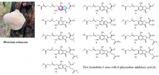
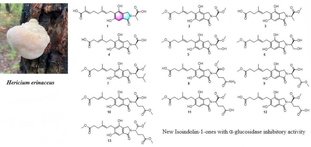
As a continuation of our previous study on erinacenones A–L (Part 1), thirteen previously undescribed isoindolin-1-ones, erinacenones M–Y (113), were isolated from the fermentation broth of Hericium erinaceus. Their structures were elucidated by extensive spectroscopic analyses (1D and 2D NMR, HRESIMS), and the absolute configurations of 113 were determined by comparing their specific rotations with those of related phthalimidines (1423). Compound 11 showed the highest α-glucosidase inhibitory activity among the tested compounds, with 31.5% inhibition at 50 μM. These findings significantly enrich the chemical diversity of isoindolinone metabolites from H. erinaceus and provide a foundation for future structure–activity relationship studies.

Graphical Abstract

1. Introduction

Diabetes mellitus is one of the most prevalent chronic metabolic diseases worldwide, posing a major threat to human health. According to the International Diabetes Federation (IDF), the global number of patients reached 536 million in 2021 and is projected to rise to 783 million by 2045, with nearly half of cases remaining undiagnosed [1,2]. Type 2 diabetes (T2D) accounts for the vast majority of cases. Although commonly used clinical drugs such as acarbose, an α-glucosidase inhibitor, effectively control postprandial hyperglycemia, they are often associated with gastrointestinal side effects [3]. Hence, identifying alternative inhibitors with high efficacy and low toxicity from natural sources has attracted considerable interest.
Fungi are a vital source of bioactive natural products. Hericium erinaceus, a medicinal and edible fungus, has been used in traditional Chinese medicine for over a millennium to treat gastric disorders [4] and produces diterpenoids, sterols, polysaccharides, and aromatic compounds. Among these, isoindolinone alkaloids are distinguished by their unique aromatic skeleton and have been reported to exhibit nerve growth factor-stimulating [5,6,7,8], cytotoxic [9,10,11], and anticoagulant activities [12]. Our previous work on aromatic metabolites from H. erinaceus led to the isolation of erinacenones A–L, which exhibited cytotoxic activities against cancer cell lines [13].
Recently, their hypoglycemic potential has been unveiled. Lee et al. [14] isolated 4-[3′,7′-dimethyl-2′,6′-octadienyl]-2-formyl-3-hydroxy-5-methyoxybenzylalcohol, hericene A, and hericene D from the fruiting bodies, all of which showed potent α-glucosidase inhibition; Miyazawa et al. [15] also reported similar activity for isohericerin and N-dephenylethyl isohericerin. Our previous work isolated multiple isoindolinone monomers and dimers from H. erinaceus fermentation and confirmed their cytotoxic and α-glucosidase inhibitory activities [13,16,17].
Nevertheless, research on the antihyperglycemic activity of aromatic compounds from this fungus remains limited, with unclear structure–activity relationships, and most existing studies have focused on known compounds, highlighting the need to explore new structural types. To address this, we continued our investigation of the aromatic constituents from H. erinaceus and isolated thirteen previously undescribed isoindolinone alkaloids, named erinacenones M–Y (113), from the same culture batch as that referenced in our previous study [13]. Although the compounds in this study were obtained from the same fermentation batch as our previous report [13], they were not co-isolated previously owing to co-elution with known analogs and the complexity of stereochemical determination (requiring semisynthesis and optical rotation comparison). Therefore, we optimized the chromatographic separation and employed combined spectroscopic and chemical derivatization to characterize the structures, aiming to discover novel hypoglycemic lead compounds and to provide a scientific basis for developing functional products derived from this fungus.

2. Results and Discussion

Structural Elucidation of the Previously Undescribed Compounds

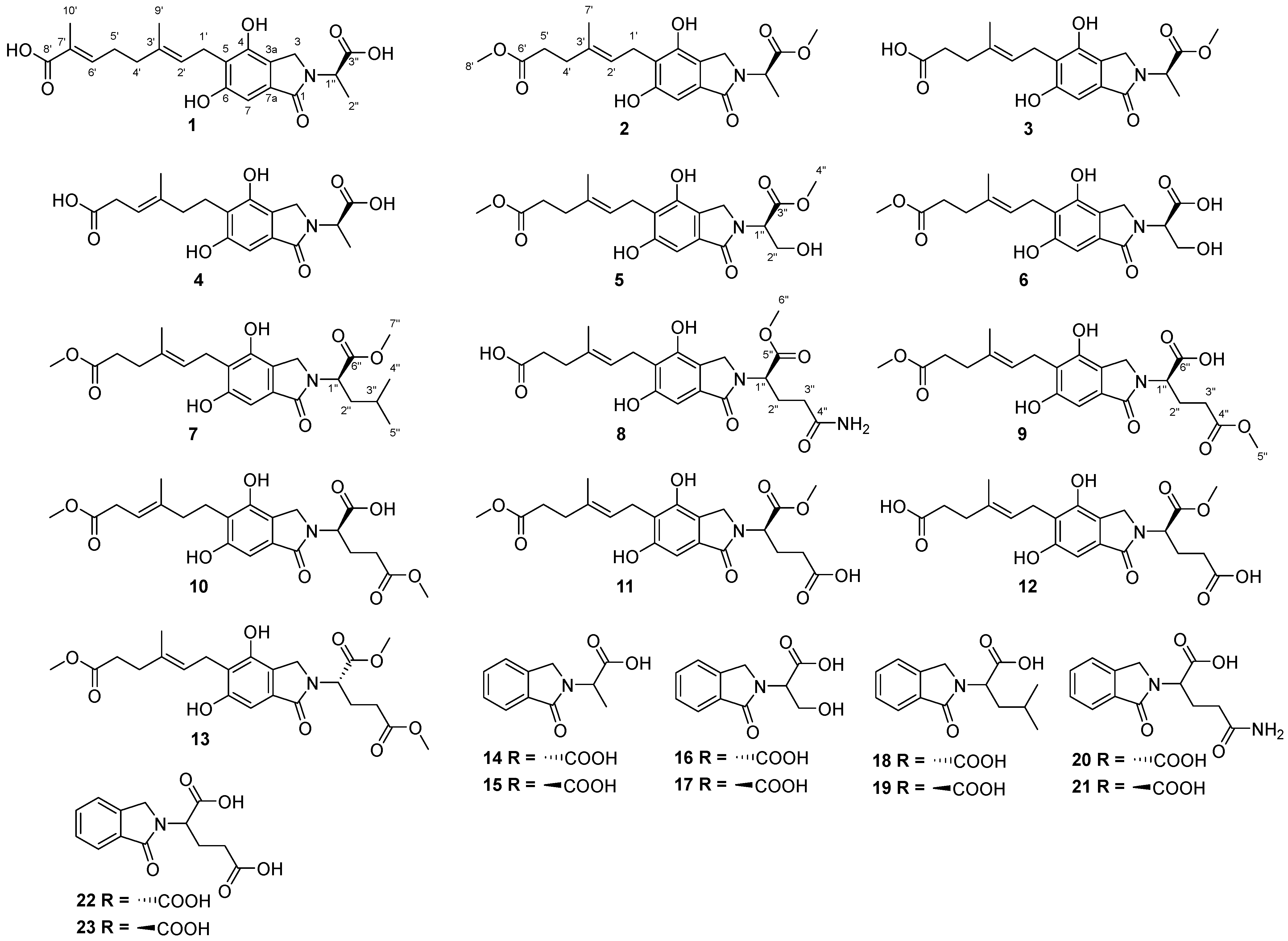
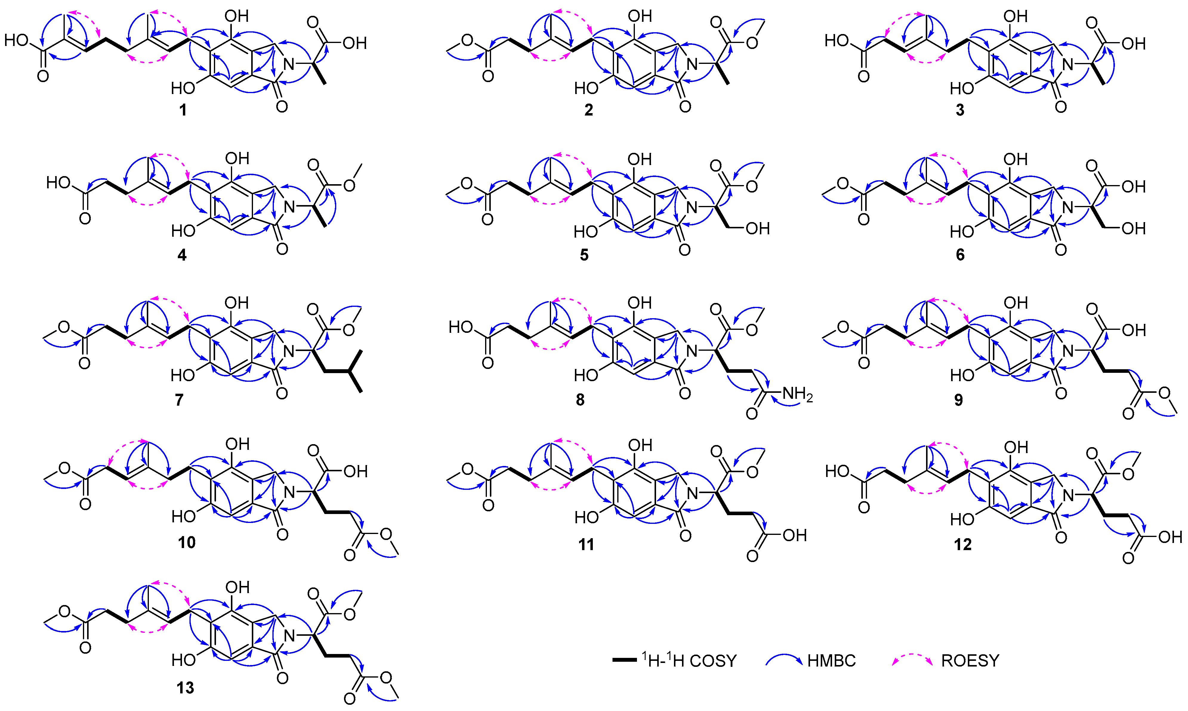
Compound 1 was obtained as a yellow oil, and the molecular ion peak at m/z 404.17075 [M+H]+ obtained by HRESIMS revealed that it had the molecular formula C21H25NO7. The 1H NMR spectrum (Table 1) of 1 showed three methyl groups (δH 1.81, 1.72 and 1.55), three methines, including an aromatic proton (δH 6.75, s), and two trisubstituted olefinic protons (δH 6.67, t, J = 7.2 Hz; 5.29, t, J = 7.2 Hz). In the 13C NMR spectrum (Table 1), 21 carbon signals were observed, including three methyls (δC 16.3, 15.8, and 12.4), four methylenes (δC 46.3, 39.4, 28.2, and 23.6), four methines (one aromatic at δC 101.9; two olefinic at δC 143.4, 124.6), and ten quaternary carbons (three carbonyl at δC 171.6, 173.5, and 172.1; five aromatic at δC 157.9, 151.7, 131.4, 121.8, and 121.1; two olefinic at δC 134.6, 129.2. The 1H and 13C NMR data for 1 showed that it shared the same substructure of (2E,6E)-8-(4,6-dihydroxy-1-oxoisoindolin-5-yl)-2,6-dimethylocta-2,6-dienoic acid [18], except for the presence of an additional methyl group (δH 1.55, t, J = 7.3 Hz; δC 15.8), a methine (δH 4.98, overlapped; δC 51.8), together with a carboxylic acid (δC 173.5) in 1. The HMBC correlations from H-1″ to C-1/C-3/C-3″, as well as the COSY correlations between H-1″ and H-2″, established that the 2-substituted propanoic acid moiety was linked to the nitrogen atom. The ROESY correlations of H-2′/H-4′ and H-5′/H-10′ confirmed the E configurations for two double bonds. Thus, the structure of 1 was designated as erinacenone M in Figure 1.
Compound 2 was obtained as a yellow oil, and the molecular ion peak at m/z 392.17026 [M+H]+ obtained by HRESIMS revealed that it had the molecular formula C20H25NO7. The 1H NMR spectrum (Table 1) of 2 showed four methyl groups, including two oxygenated methyl groups (δH 3.73 and 3.55), an aromatic proton (δH 6.75, s), and a trisubstituted olefinic proton (δH 5.28, t, J = 7.2 Hz). In the 13C NMR spectrum (Table 1), 20 carbon signals were observed, including four methyls (two oxygenated at δC 52.9, 51.9; one olefinic at δC 16.1; one aliphatic at δC 15.8), four methylenes (δC 46.4, 36.0, 33.9, and 23.6), three methines (one aromatic at δC 101.8; one olefinic at δC 124.5; other at δC 51.2), and nine quaternary carbons (three carbonyl at δC 175.8, 173.5, and 171.6; five aromatic at δC 158.2, 151.7, 131.3, 121.7, and 120.9; one olefinic at δC 134.3). The 1H and 13C NMR data for 2 showed that it shared the same substructure as (E)-methyl 6-(4,6-dihydroxy-1-oxoisoindolin-5-yl)-4-methylhex-4-enoate [18], except for the presence of two additional methyl groups [an oxygenated (δH 3.73, s; δC 52.9), (δH 1.59, d, J = 7.4 Hz; δC 15.8)], a methine (δH 4.99, dd, J = 14.8, 7.4 Hz; δC 51.2), together with a carboxylic acid (δC 173.5) in 2. The HMBC correlations from H-1″ to C-1/C-3/C-3″, from H-4″ to C-3″, as well as the COSY correlations between H-1″ and H-2″, established the methyl 2-substituted propanoate moiety was linked to the nitrogen atom. The ROESY correlations of H-1′/H-7′ and H-2′/H-4′ assigned the E configuration to the double bond. Thus, the structure of 2 was designated as erinacenone N.
Compounds 3 and 4 were both obtained as yellow oils, with HRESIMS analysis revealing molecular ion peaks at m/z 400.13681 [M+Na]+ (C19H23NO7) and m/z 386.12094 [M+Na]+ (C18H21NO7), respectively. The 1H and 13C NMR data of 3 and 4 indicated that the presence of (E)-methyl 6-(4,6-dihydroxy-1-oxoisoindolin-5-yl)-4-methylhex-4-enoic acid moiety [18] in their structures, including one olefinic methyl (3: δH 1.81, s; δC 54.7; 4: δH 1.75, s), four methylenes [3: δH 4.37, m; δC 46.4; δH 3.42, d, J = 7.2 Hz; δC 23.6; δH 2.27, m; δC 36.8; δH 2.21, m; δC 39.7; 4: δH 4.50, d, J = 16.5 Hz, 4.33, d, J = 16.5 Hz; δC 46.4; δH 2.97, t, J = 7.5 Hz; δC 35.2; δH 2.83, m; δC 23.3; δH 2.23, m; δC 39.7], two methines [one aromatic (3: δH 6.75, s; δC 101.9; 4: δH 6.73, s; δC 101.7); one olefinic (3: δH 5.29, t, J = 6.9 Hz; δC 123.7; 4: δH 5.23, t, J = 6.8 Hz; δC 117.8)], and eight quaternary carbons [two carbonyl (3: δC -, 171.6; 4: δC 177.6, 171.7); five aromatic (3: δC 158.2, 151.7, 131.3, 121.9, and 121.0; 4: δC 158.0, 151.9, 131.8, 122.1, and 121.2); one olefinic (3: δC 135.3; 4: δC 139.8)]. Compounds 3 and 4 contain methyl 2-substituted propanoate and 2-substituted propanoic acid moiety, respectively, as confirmed above by HMBC data analysis and 1H-1H COSY. The HMBC correlations from H-1″ to C-1/C-3 confirmed the linkage of the methyl 2-substituted propanoate moiety with the nitrogen atom in 3 (Figure 2). ROESY correlations of H-1′/H-7′ and H-2′/H-4′ assigned the E configuration to the double bond. Thus, the structure of 3 was designated as erinacenone O. In a similar way, 4 was identified through analysis of its 2D NMR data (Figure 2). Thus, the structure of 4 was designated as erinacenone P in Figure 1.
Compound 5 was obtained as a yellow oil, and its molecular formula was determined to be C20H25NO8 (m/z 430.14725 [M+Na]+, calculated for C20H25NO8Na, 430.14724) through the analysis of HRESIMS. The NMR spectra (Table 2) of 5 were similar to those of erinacerin G [18], except for the presence of an additional oxygenated methyl group (δH 3.75, s; δC 52.9), a methylene (δH 4.14, dd, J = 11.8, 7.3 Hz; 4.05, dd, J = 11.8, 3.9 Hz; δC 61.6), a methine (δH 5.02, dd, J = 7.3, 3.9 Hz; δC 58.2), together with a carboxylic acid (δC 171.2). The 1H–1H COSY correlation between H-1″ and H-2″, in combination with the HMBC correlations from H-1″ to C-1/C-3/C-3″ and from H-4″ to C-3″, confirmed the linkage of the methyl 2-(hydroxymethyl)-substituted-propanoate moiety to the nitrogen atom. The ROESY correlations of H-1′/H-7′ and H-2′/H-4′ assigned the E configuration of the double bond. Consequently, the structure of 5 was named erinacenone Q.
Compound 6 was obtained as a yellow oil, and its molecular formula was determined to be C19H23NO8 (m/z 394.14969 [M+H]+, calculated for C19H24NO8, 394.14964) through the analysis of HRESIMS. The NMR spectra (Table 2) of 6 showed that it was structurally similar to 5, with the only difference being the absence of a methyl group, which was supported by HRESIMS data. The configuration of the double bond between C-2′ and C-3′ was determined as described for compound 5. Consequently, the structure of 6 was named erinacenone R.
Compound 7 was obtained as a yellow oil, and its molecular formula was determined to be C23H31NO7 (m/z 434.21725 [M+H]+, calculated for C23H32NO7, 434.21733) through the analysis of HRESIMS. The 1H and 13C NMR spectra (Table 2) of 7 were similar to those of erinacerin G, except for the presence of three additional methyl groups [an oxygenated (δH 3.72, s; δC 52.9); other (δH 0.98, d, J = 6.2 Hz; δC 23.4); (δH 0.96, d, J = 6.2 Hz; δC 21.4)], a methylene (δH 1.94, m; 1.83, m; δC 39.4), two methine [(δH 5.05, dd, J = 11.4, 4.5 Hz; δC 53.6); (δH 1.46, m; δC 26.1)], together with a carboxylic acid (δC 173.4). The HMBC correlations from H-1″ to C-1/C-3/C-6″, from H-7″ to C-6″, as well as the COSY correlations between H-2″ and H-1″/H-3″, H-3″ and H-4″/H-5″, established that the methyl 2-isopropyl-2-substituted-propanoate moiety was linked to the nitrogen atom. The configuration of the double bond between C-2′ and C-3′ was determined as described for compound 6. Consequently, the structure of 7 was named erinacenone S.
Compound 8 was obtained as a yellow oil, and its molecular formula was determined to be C21H26N2O8 (m/z 435.17612 [M+H]+, calculated for C21H27N2O8, 435.17619) through the analysis of HRESIMS. The 1H and 13C NMR data (Table 2) of 8 were similar to those of erinacerin G, except for the presence of an additional oxygenated methyl group (δH 3.74, s; δC 53.0), two methylenes (δH 2.45, m; 2.21, overlapped; δC 26.4; δH 2.27, m; 2.23, overlapped; δC 32.7), a methine (δH 4.95, dd, J = 10.6, 4.8 Hz; δC 55.2), as well as two carboxylic acids (δC 177.2, 172.5). The HMBC correlations from H-1″ to C-1/C-3/C-5″, from H-2″ to C-4″, from H-6″ to C-5″, as well as the COSY correlations between H-2″ and H-1″/H-3″, established that the methyl 5-carbamoyl-2-substituted pentanoate moiety was linked to the nitrogen atom. The configuration of the double bond between C-2′ and C-3′ was determined as described for compound 7. Consequently, the structure of 8 was named erinacenone T.
Compounds 9 and 10 were isolated as yellow oils. They possessed the same molecular formula of C22H27NO9, with nine degrees of unsaturation, as established by the HRESIMS and NMR data analyses. The NMR data (Table 3 and Table 4) indicated structural similarity to erinacerin G, with both compounds sharing the (E)-methyl 6-(4,6-dihydroxy-1-oxoisoindolin-5-yl)-4-methylhex-4-enoate moiety, but differing only in the nitrogen-linked moiety. A comparison of the NMR data of 9 with those obtained for erinacerin G revealed the presence of an additional oxygenated methyl group (δH 3.56, s; δC 52.2), two methylenes (δH 2.46, m; 2.19, m; δC 26.3; δH 2.37, overlapped; δC 31.8), a methine (δH 4.89, overlapped; δC 55.7), as well as two carboxylic acids (δC 174.8, 172.2) in 9. The HMBC correlations from H-1″ to C-1/C-3/C-6″, from H-3″ to C-4″, from H-6″ to C-4″, as well as the COSY correlations between H-2″ and H-1″/H-3″, established that the methyl 5-(methoxycarbonyl)-2-substituted pentanoic acid moiety was present in the molecule. The double bond configuration between C-2′ and C-3′ was determined as described in compound 8. As a result, the structure of 9 was elucidated as erinacenone U. In a similar way, the structure of 10 was determined as erinacenone V after a thorough analysis of its HMBC and ROESY spectra (Figure 2). Compounds 9 and 10 are a pair of double-bond positional isomers.
Compound 11 was isolated as a yellow oil. Based on the HRESIMS at m/z 450.17581 [M+H]+, the molecular formula was determined to be C22H27NO9, with nine degrees of unsaturation. The NMR spectra (Table 3 and Table 4) of 11 revealed a structural similarity to 9, distinguished primarily by the relocation of a methyl group. This change was confirmed by an HMBC correlation from H-6″ to C-5″. ROESY correlations of H-1′/H-7′ and H-2′/H-4′ assigned the E configuration to the double bond. As a result, the structure of 11 was named erinacenone W.
Compound 12 was isolated as a yellow oil. Based on the HRESIMS at m/z 458.14215 [M+Na]+, the molecular formula of 12 was determined to be C21H25NO9, with nine degrees of unsaturation. The primary difference between the NMR spectra (Table 3 and Table 4) of 12 and 11 was that the methyl carboxylate of C-6″ was converted into a carboxylic acid. This change was confirmed by HRESIMS data. ROESY correlations of H-1′/H-7′ and H-2′/H-4′ assigned the E configuration to the double bond. As a result, the structure of 12 was named erinacenone X.
Compound 13 was isolated as a yellow oil. Based on the HRESIMS at m/z 464.19147 [M+H]+, the molecular formula was determined to be C23H29NO9, with nine degrees of unsaturation. Comparison of the 1H and 13C NMR spectra (Table 3 and Table 4) of 13 with those of 11 showed that the primary distinction was the presence of an extra methyl group, which was further confirmed by the HMBC correlations from H-5″ to C-4″. ROESY correlations of H-1′/H-7′ and H-2′/H-4′ assigned the E configuration to the double bond. As a result, the structure of 13 was named erinacenone Y.
To determine the absolute configurations of compounds 113, phthalimidines 1423 were synthesized by the reaction of o-phthalaldehyde with the enantiomerically pure α-amino acids (L/D-alanine, L/D-glutamic acid, L/D-serine, L/D-leucine, and L/D-glutamine). Comparative analysis of the specific optical rotations of compounds 113 and their corresponding synthetic phthalimidines 1423 revealed that compounds 112 showed (+)-specific optical rotations, indicating the R configuration at C-1″. In contrast, compound 13 showed (−)-specific optical rotation, confirming an S configuration at C-1″.

3. Materials and Methods

3.1. General Experimental Procedures

Optical rotations were measured on a JASCO P-2000 Automatic polarimeter (JASCO, Tokyo, Japan). CD spectra were recorded on a JASCO J-1500 CD Spectrometer (JASCO, Tokyo, Japan), and the UV spectra were obtained on an Analytik Jena SPECORD S600 UV spectrophotometer (Analytik Jena, Jena, Germany). NMR spectra were obtained on Bruker Avance 600 MHz and Bruker Avance III 500 spectrometers (Bruker, Ettlingen, Germany) with tetramethylsilane as an internal standard at room temperature. HRESIMS was recorded on a Q Exactive Orbitrap mass spectrometer (Thermo Scientific, Cambridge, MA, USA). Preparative HPLC was performed on an Agilent 1260 liquid chromatography system with DAD detector, using Zorbax SB-C18 column (5 µm, 9.4 × 150 mm) (Agilent, Santa Clara, CA, USA). Preparative HPLC was performed on a Hanbon NP7005C liquid chromatography system, using Zorbax SB-C18 column (5 µm, 9.4 × 150 mm) (Agilent, Santa Clara, CA, USA). On a PuriFlash 450 equipment (Interchim, Montluçon, France), medium pressure liquid chromatography (MPLC) was carried out. Silica gel (80–100, 300–400 mesh) (Qingdao Marine Chemical Factory, Qingdao, China) and Sephadex LH-20 (20–45 μm) (GE Healthcare, Uppsala, Sweden) were used for column chromatography (CC). TLC was carried out using GF254 plates (Qingdao Marine Chemical Factory, Qingdao, China).

3.2. Fungal Material

The fungus H. erinaceus was collected in Shangri-La County, Yunnan Province, China, in August 2007 and identified by Prof. Zang from Kunming Institute of Botany, Chinese Academy of Sciences. A voucher specimen (No. KU855351.1) has been deposited at South-Central Minzu University, Wuhan. H. erinaceus was cultured on slants of potato dextrose agar (20% potato, 2% glucose, 0.3% KH2PO4, 0.15% MgSO4, 0.01% citric acid, pH 6.5) at 25 °C for 7 days. Agar plugs (1 cm2) were inoculated in liquid medium (glucose 5%, yeast extract 0.4%, peptone 0.15%, KH2PO4 0.05%, MgSO4 0.05%, pH 6.5) and then cultured for 30 days at 25 °C on a rotary shaker at 150 rpm.

3.3. Extraction and Isolation

A 100 L culture broth filtrate was concentrated to 10 L. Mycelium was extracted with acetone, and the extract was concentrated under reduced pressure to dryness. The combined extracts were extracted with EtOAc, then they were concentrated under reduced pressure to give a crude extract (110.98 g), which was fractionated by silica gel CC eluting with a gradient of CHCl3–MeOH (v/v 1:0–0:1) to obtain six fractions, A–F. Fraction B was separated by MPLC (MeOH–H2O, 5:1, 20:1, 30:1, 45:1, 65:1, 75:1, 85:1, 100:1; 4 L for each step) to offer twelve subfractions (B1–B12). Subfraction B6 was subjected to silica gel CC (v/v 1:0–0:1) to yield subfractions (B6A–B6P). Subfraction B6L was purified by HPLC (MeCN–H2O, 34% isocratic, 4 mL/min) to give compounds 5 (tR = 9.93 min, 9.8 mg), 6 (tR = 9.92 min, 1.8 mg), 7 (tR = 10.13 min, 9.0 mg), and 9 (tR = 10.61 min, 2.6 mg). Fraction C was separated by MPLC (MeOH–H2O, 5:1, 20:1, 30:1, 45:1, 65:1, 75:1, 85:1, 100:1; 4 L for each step) into twenty subfractions (C1–C20). Subfraction C6 was further separated by silica gel CC (CHCl3–MeOH from v/v 1:0–0:1) to yield ten subfractions (C6A–C6J). Compound 11 (tR = 9.90 min, 1.1 mg) was obtained from subfraction C6D by HPLC (MeCN–H2O, 32% isocratic, 4 mL/min). Subfraction C10 was separated using silica gel CC (v/v 1:0–0:1) to get eleven subfractions (C10A–C10K). Compound 4 (tR = 9.84 min, 28.3 mg) was purified from subfraction C10A by HPLC (MeCN–H2O, 20% isocratic, 4 mL/min). Subfraction C11 was separated by silica gel CC (v/v 1:0–0:1) to get fifteen subfractions (C11A–C11O). Compound 8 was purified from subfraction C11J by HPLC (MeCN–H2O, 28% isocratic, 4 mL/min). Subfraction C11L was subjected to Sephadex LH-20 (MeOH) and further purified by HPLC (MeCN–H2O, 25% isocratic, 4 mL/min) to yield compounds 12 (tR = 8.69 min, 9.1 mg) and 10 (tR = 9.61 min, 1.0 mg). Subfraction C12 was separated by silica gel CC (v/v 1:0–0:1) to obtain nine subfractions (C12A–C12I). Fraction F was separated by MPLC (MeOH–H2O, 5:1, 20:1, 30:1, 45:1, 65:1, 75:1, 85:1, 100:1; 4 L for each step) to offer twenty-three subfractions (F1–F23). Subfraction F8 was subjected to silica gel CC (v/v 1:0–0:1) to obtain eight subfractions (F8A–F8H). Subfraction F8B was further separated on HPLC (MeCN–H2O, 13% isocratic, 4 mL/min) to give compound 13 (tR = 6.43 min, 1.1 mg). Subfraction F10 was separated using silica gel CC (v/v 1:0–0:1) to get subfractions (F10A–F10L). Compounds 2 (tR = 5.45 min, 16.2 mg) and 3 (tR = 6.28 min, 5.2 mg) were purified from subfraction F10E by HPLC (MeCN–H2O, 10% isocratic, 4 mL/min). Compound 1 (tR = 7.58 min, 17.3 mg) was obtained from subfraction F10G by HPLC (MeCN–H2O, 20% isocratic, 4 mL/min).

3.4. Characterization Data

3.4.1. Erinacenone M (1)

yellow oil; [α]25D +55.5 (c 0.02, MeOH); UV (MeOH) λmax (log ε) 215 (4.62), 260 (4.11), 300 (3.47) nm; 1H (600 MHz) and 13C NMR (150 MHz) data (CD3OD), see Table 1; HRESIMS m/z 404.17075 (calcd for C21H26NO7 [M+H]+, 404.17038).

3.4.2. Erinacenone N (2)

yellow oil; [α]25D +37.7 (c 0.03, MeOH); UV (MeOH) λmax (log ε) 220 (4.51), 260 (4.22), 300 (3.62) nm; 1H (500 MHz) and 13C NMR (125 MHz) data (CD3OD), see Table 1; HRESIMS m/z 392.17026 (calcd for C20H26NO7 [M+H]+, 392.17038).

3.4.3. Erinacenone O (3)

yellow oil; [α]25D +63.1 (c 0.02, MeOH); UV (MeOH) λmax (log ε) 215 (4.50), 260 (4.03), 300 (3.46) nm; 1H (600 MHz) and 13C NMR (150 MHz) data (CD3OD), see Table 1; HRESIMS m/z 400.13681 (calcd for C19H23NO7 [M+Na]+, 400.13667).

3.4.4. Erinacenone P (4)

yellow oil; [α]25D +48.2 (c 0.03, MeOH); UV (MeOH) λmax (log ε) 220 (4.46), 260 (4.19), 300 (3.60) nm; 1H (600 MHz) and 13C NMR (150 MHz) data (CD3OD), see Table 1; HRESIMS m/z 386.12094 (calcd for C18H21NO7Na [M+Na]+, 386.12102).

3.4.5. Erinacenone Q (5)

yellow oil; [α]25D +23.1 (c 0.03, MeOH); UV (MeOH) λmax (log ε) 215 (4.44), 260 (4.03), 300 (3.42) nm; 1H (500 MHz) and 13C NMR (125 MHz) data (CD3OD), see Table 2; HRESIMS m/z 430.14725 (calcd for C20H25NO8Na [M+Na]+, 430.14724).

3.4.6. Erinacenone R (6)

yellow oil; [α]25D +37.5 (c 0.02, MeOH); UV (MeOH) λmax (log ε) 215 (4.53), 260 (4.13), 300 (3.52) nm; 1H (500 MHz) and 13C NMR (125 MHz) data (CD3OD), see Table 2; HRESIMS m/z 394.14969 (calcd for C19H24NO8 [M+H]+, 394.14964).

3.4.7. Erinacenone S (7)

yellow oil; [α]25D +12.5 (c 0.03, MeOH); UV (MeOH) λmax (log ε) 220 (4.47), 260 (4.26), 300 (3.66) nm; 1H (600 MHz) and 13C NMR (150 MHz) data (CD3OD), see Table 2; HRESIMS m/z 434.21725 (calcd for C23H32NO7 [M+H]+, 434.21733).

3.4.8. Erinacenone T (8)

yellow oil; [α]25D+11.5 (c 0.03, MeOH); UV (MeOH) λmax (log ε) 210 (4.50), 260 (4.23), 300 (3.67) nm; 1H (600 MHz) and 13C NMR (150 MHz) data (CD3OD), see Table 2; HRESIMS m/z 435.17612 (calcd for C21H27N2O8 [M+H]+, 435.17619).

3.4.9. Erinacenone U (9)

yellow oil; [α]25D +6.6 (c 0.03, MeOH); UV (MeOH) λmax (log ε) 215 (4.47), 260 (4.13), 300 (3.50) nm; 1H (500 MHz) and 13C NMR (125 MHz) data (CD3OD), see Table 3 and Table 4; HRESIMS m/z 450.17572 (calcd for C22H28NO9 [M+H]+, 450.17586).

3.4.10. Erinacenone V (10)

yellow oil; [α]25D +7.7 (c 0.02, MeOH); UV (MeOH) λmax (log ε) 215 (4.52), 260 (4.10), 300 (3.48) nm; 1H (500 MHz) and 13C NMR (125 MHz) data (CD3OD), see Table 3 and Table 4; HRESIMS m/z 450.17578 (calcd for C22H28NO9 [M+H]+, 450.17586).

3.4.11. Erinacenone W (11)

yellow oil; [α]25D +10.0 (c 0.03, MeOH); UV (MeOH) λmax (log ε) 215 (4.55), 260 (4.22), 300 (3.63) nm; 1H (600 MHz) and 13C NMR (150 MHz) data (CD3OD), see Table 3 and Table 4; HRESIMS m/z 450.17581 (calcd for C22H28NO9 [M+H]+, 450.17586).

3.4.12. Erinacenone X (12)

yellow oil; [α]25D +14.3 (c 0.02, MeOH); UV (MeOH) λmax (log ε) 215 (4.51), 260 (4.09), 300 (3.47) nm; 1H (600 MHz) and 13C NMR (150 MHz) data (CD3OD), see Table 3 and Table 4; HRESIMS m/z 458.14215 (calcd for C21H25NO9Na [M+Na]+, 458.14215).

3.4.13. Erinacenone Y (13)

yellow oil; [α]25D −19.0 (c 0.05, MeOH); UV (MeOH) λmax (log ε) 225 (4.48), 260 (4.33), 300 (3.81) nm; 1H (600 MHz) and 13C NMR (150 MHz) data (CD3OD), see Table 3 and Table 4; HRESIMS m/z 464.19147 (calcd for C23H30NO9 [M+H]+, 464.19151).
Synthesis of Phthalimidines (1423). L-Alanine (0.5 mmol) was added to a solution of phathalic dicarboxaldehyde (0.5 mmol) in 10 mL of acetonitrile. The mixture was heated under relux for 16 h; then the reaction mixture was filtered while hot. The solvent was allowed to cool to yield the desired phathalimidine 14 as crystalline solids (92 mg). Using the same method, compounds 15 (71 mg), 16 (8 mg), 17 (53 mg), 18 (98 mg), 19 (55 mg), 20 (112 mg), 21 (82 mg), 22 (65 mg), and 23 (48 mg) were synthesized from D-alanine, L-glutamic acid, D-glutamic acid, L-serine, D-serine, L-leucine, D-leucine, L-glutamine, and D-glutamine, respectively.
Compound 14: colorless microcrystalline powder; [α]25D +16.537 (c 0.08, MeOH); 1H NMR (CD3OD, 600 MHz): 7.79 (1H, d, J = 7.6 Hz, H-7), 7.63 (1H, td, J = 7.6, 1.0 Hz, H-5), 7.60 (1H, d, J = 7.5 Hz, H-4), 7.51 (1H, t, J = 7.4 Hz, H-6), 5.02 (1H, dd, J = 15.0, 7.5 Hz, H-1′), 4.62 (1H, d, J = 17.2 Hz, H-3), 4.57 (1H, d, J = 17.2 Hz, H-3), 1.63 (3H, d, J = 7.5 Hz, H-2′); 13C NMR (CD3OD, 150 MHz): 174.7 (C-3′), 171.0 (C-1), 143.8 (C-3a), 133.2 (C-5), 133.0 (C-7a), 129.2 (C-6), 124.4 (C-4), 124.3 (C-7), 51.0 (C-1′), 48.6 (C-3), 15.9 (C-2′); HRESIMS m/z 206.08122 (calcd for C11H12NO3 [M+H]+, 206.08117).
Compound 15: colorless microcrystalline powder; [α]25D −26.527 (c 0.08, MeOH); 1H (600 MHz) and 13C NMR (150 MHz) data of 15 are identical with those of 14; HRESIMS m/z 206.08127 (calcd for C11H12NO3 [M+H]+, 206.08117).
Compound 16: colorless microcrystalline powder; [α]25D +43.206 (c 0.09, MeOH); 1H NMR (CD3OD, 600 MHz): 7.80 (1H, d, J = 7.6 Hz, H-7), 7.63 (1H, td, J = 7.6, 1.0 Hz, H-5), 7.60 (1H, d, J = 7.5 Hz, H-4), 7.51 (1H, t, J = 7.5 Hz, H-6), 5.06 (1H, dd, J = 7.6, 3.8 Hz, H-1′), 4.74 (1H, d, J = 17.4 Hz, H-3), 4.68 (1H, d, J = 17.4 Hz, H-3), 4.17 (1H, dd, J = 11.8, 7.6 Hz, H-2′), 4.10 (1H, dd, J = 11.8, 3.8 Hz, H-2′); 13C NMR (CD3OD, 150 MHz): 172.2 (C-3′), 171.7 (C-1), 144.2 (C-3a), 133.2 (C-5), 132.8 (C-7a), 129.1 (C-6), 124.3 (C-4, 7), 61.5 (C-2′), 58.1 (C-1′), 49.9 (C-3); HRESIMS m/z 222.07600 (calcd for C11H12NO4 [M+H]+, 222.07608).
Compound 17: colorless microcrystalline powder; [α]25D −53.101 (c 0.08, MeOH); 1H (600 MHz) and 13C NMR (150 MHz) data of 17 are identical with those of 16; HRESIMS m/z 222.07605 (calcd for C11H12NO4 [M+H]+, 222.07608).
Compound 18: colorless microcrystalline powder; [α]25D −3.731 (c 0.13, MeOH); 1H NMR (CD3OD, 600 MHz): 7.79 (1H, d, J = 7.6 Hz, H-7), 7.64 (1H, td, J = 7.6, 1.0 Hz, H-5), 7.61 (1H, d, J = 7.5 Hz, H-4), 7.52 (1H, t, J = 7.5 Hz, H-6), 5.07 (1H, dd, J = 11.6, 4.3 Hz, H-1′), 4.68 (1H, d, J = 17.2 Hz, H-3), 4.48 (1H, d, J = 17.2 Hz, H-3), 1.99 (1H, m, H-2′); 1.88 (1H, m, H-2′); 1.48 (1H, m, H-3′); 0.99 (6H, d, J = 6.6 Hz, H-4′, 5′); 13C NMR (CD3OD, 150 MHz): 174.6 (C-6′), 171.5 (C-1), 143.8 (C-3a), 133.2 (C-5), 132.8 (C-7a), 129.2 (C-6), 124.4 (C-4, 7), 53.5 (C-1′), 48.1 (C-3), 39.4 (C-2′), 26.3 (C-3′), 23.5 (C-5′), 21.3 (C-4′); HRESIMS m/z 248.12804 (calcd for C14H18NO3 [M+H]+, 248.12812).
Compound 19: colorless microcrystalline powder; [α]25D +6.02 (c 0.03, MeOH); 1H (600 MHz) and 13C NMR (150 MHz) data of 19 are identical with those of 18; HRESIMS m/z 248.12801 (calcd for C14H18NO3 [M+H]+, 248.12812).
Compound 20: colorless microcrystalline powder; [α]25D −11.588 (c 0.11, MeOH); 1H NMR (CD3OD, 600 MHz): 7.79 (1H, d, J = 7.6 Hz, H-7), 7.64 (1H, td, J = 7.6, 1.0 Hz, H-5), 7.60 (1H, d, J = 7.5 Hz, H-4), 7.51 (1H, t, J = 7.5 Hz, H-6), 4.97 (1H, dd, J = 10.8, 4.5 Hz, H-1′), 4.63 (1H, d, J = 17.1 Hz, H-3), 4.56 (1H, d, J = 17.1 Hz, H-3), 2.49 (1H, m, H-2′), 2.30 (1H, m, H-3′), 2.25 (1H, m, H-3′), 2.23 (1H, m, H-2′); 13C NMR (CD3OD, 150 MHz): 177.3 (C-4′), 173.5 (C-5′), 171.7 (C-1), 143.9 (C-3a), 133.3 (C-5), 132.7 (C-7a), 129.2 (C-6), 124.4 (C-4, 7), 55.1 (C-1′), 48.6 (C-3), 33.1 (C-3′), 26.5 (C-2′); HRESIMS m/z 263.10266 (calcd for C13H15N2O4 [M+H]+, 263.10263).
Compound 21: colorless microcrystalline powder; [α]25D +6.822 (c 0.03, MeOH); 1H (600 MHz) and 13C NMR (150 MHz) data of 21 are identical with those of 20; HRESIMS m/z 263.10263 (calcd for C13H15N2O4 [M+H]+, 263.10263).
Compound 22: colorless microcrystalline powder; [α]25D −29.073 (c 0.06, MeOH); 1H NMR (CD3OD, 600 MHz): 7.80 (1H, d, J = 7.6 Hz, H-7), 7.64 (1H, td, J = 7.6, 1.0 Hz, H-5), 7.60 (1H, d, J = 7.5 Hz, H-4), 7.52 (1H, t, J = 7.5 Hz, H-6), 5.02 (1H, dd, J = 11.0, 4.7 Hz, H-1′), 4.62 (1H, d, J = 17.1 Hz, H-3), 4.54 (1H, d, J = 17.1 Hz, H-3), 2.48 (1H, m, H-2′), 2.37 (2H, m, H-3′), 2.23 (1H, m, H-2′); 13C NMR (CD3OD, 150 MHz): 176.0 (C-4′), 173.5 (C-5′), 171.7 (C-1) 143.8 (C-3a), 133.3 (C-5), 132.7 (C-7a), 129.2 (C-6), 124.4 (C-4, 7), 55.0 (C-1′), 48.7 (C-3), 31.6 (C-3′), 25.9 (C-2′); HRESIMS m/z 264.08667 (calcd for C13H14NO5 [M+H]+, 264.08665).
Compound 23: colorless microcrystalline powder; [α]25D +23.427 (c 0.09, MeOH); 1H (600 MHz) and 13C NMR (150 MHz) data of 23 are identical with those of 22; HRESIMS m/z 264.08667 (calcd for C13H14NO5 [M+H]+, 264.08665).

3.5. α-Glucosidase Inhibitory Assay

The inhibitory effect on α-glucosidase was evaluated using a modified protocol based on earlier reports [19,20]. Briefly, each well of a 96-well plate received 125 µL of phosphate buffer (50 mM, pH 6.8), 25 µL of 10 mM p-nitrophenyl-α-D-glucopyranoside, and 25 µL of the test compound solution, yielding final concentrations of 1.25, 2.5, 5, 10, 12.5, 25, and 50 µM. After a 10 min pre-incubation at 37 °C, the enzymatic reaction was initiated by adding 25 µL of α-glucosidase (0.25 U/mL, from Saccharomyces cerevisiae). The plate was then incubated at 37 °C for 30 min, and the reaction was terminated by the addition of 50 µL of 1 M Na2CO3. The absorbance of the liberated p-nitrophenol was read at 405 nm using a microplate reader. Acarbose was included as the positive control, and a blank sample (without enzyme) was prepared by substituting the enzyme with an equal volume of buffer. The inhibition percentage was calculated as: Inhibition (%) = [(ODcontrol − ODcontrol blank) − (ODtest − ODtest blank)]/(ODcontrol − ODcontrol blank) × 100%. All experiments were performed in triplicate, and the data are presented as mean ± SD.

4. Conclusions

This study utilized the same batch of fermented material from H. erinaceus (preliminary results reported in our previous study [13]) to isolate and identify thirteen new isoindolin-1-one alkaloids, designated as erinacenones M–Y (113), further enriching the structural diversity of isoindolinone skeletons from this edible fungus. The α-glucosidase inhibitory activities of compounds 113 were evaluated at concentrations ranging from 1.25 to 50 µM. The inhibition percentages at 50 µM are summarized in Table 5. Among them, compound 11 was identified as a modest α-glucosidase inhibitor, warranting further structural optimization and more comprehensive biological evaluations. This work provides new chemical and pharmacological evidence for the development of functional products derived from H. erinaceus. However, the current study remains limited to enzyme-level assays, with unclear mechanisms and binding modes, and the insufficient yields of certain minor components preclude in-depth evaluation. To address these bottlenecks, subsequent research will focus on large-scale fermentation to produce sufficient quantities of active compounds for in vivo efficacy validation, combined with molecular docking and molecular dynamics simulations to elucidate the inhibition mechanism, alongside targeted isolation of trace constituents. Ultimately, these efforts aim to establish a foundation for the development of H. erinaceus-based health foods or novel antidiabetic drugs.

Supplementary Materials

The following supporting information can be downloaded at: https://www.mdpi.com/article/10.3390/molecules31183333/s1, Figures S1–S91: HRESIMS, HNMR, CNMR, COSY, HSQC, HMBC, ROESY of compounds 113. Figures S92–S111: HRESIMS, HNMR, CNMR of compounds 1423 (synthesis of phthalimidines). Table S1: Cytotoxicity of compounds (in IC50, μM).

Author Contributions

L.-L.Y., investigation (isolation and identification of all compounds), writing—original draft; J.-K.L., conceptualization, supervision, project administration, funding acquisition, writing—review and editing. All authors have read and agreed to the published version of the manuscript.

Funding

This work was financially supported by the National Natural Science Foundation of China (No. 22177138).

Institutional Review Board Statement

Not applicable.

Informed Consent Statement

Not applicable.

Data Availability Statement

The original contributions presented in this study are included in the article/Supplementary Materials. Further inquiries can be directed to the corresponding author.

Conflicts of Interest

The authors declare no conflict of interest.

References

  1. Sun, H.; Saeedi, P.; Karuranga, S.; Pinkepank, M.; Ogurtsova, K.; Duncan, B.B.; Stein, C.; Basit, A.; Chan, J.C.N.; Mbanya, J.C.; et al. IDF diabetes Atlas: Global, regional and country-level diabetes prevalence estimates for 2021 and projections for 2045. Diabetes Res. Clin. Pract. 2022, 183, 109119. [Google Scholar] [CrossRef] [Scilit] [PubMed]
  2. Saeedi, P.; Petersohn, I.; Salpea, P.; Malanda, B.; Karuranga, S.; Unwin, N.; Colagiuri, S.; Guariguata, L.; Motala, A.A.; Ogurtsova, K.; et al. Global and regional diabetes prevalence estimates for 2019 and projections for 2030 and 2045: Results from the International Diabetes Federation Diabetes Atlas, 9th edition. Diabetes Res. Clin. Pract. 2019, 157, 107843. [Google Scholar] [CrossRef] [Scilit] [PubMed]
  3. Patil, P.; Mandal, S.; Tomar, S.K.; Anand, S. Food protein-derived bioactive peptides in management of type 2 diabetes. Eur. J. Nutr. 2015, 54, 863–880. [Google Scholar] [CrossRef] [Scilit] [PubMed]
  4. Bhandari, D.R.; Shen, T.; Römpp, A.; Zorn, H.; Spengler, B. Analysis of cyathane-type diterpenoids from Cyathus striatus and Hericium erinaceus by high-resolution MALDI MS imaging. Anal. Bioanal. Chem. 2014, 406, 695–704. [Google Scholar] [CrossRef] [Scilit] [PubMed]
  5. Kawagishi, H.; Ando, M.; Sakamoto, H.; Yoshida, S.; Ojima, F.; Ishiguro, Y.; Ukai, N.; Furukawa, S. Hericenone C, D and E, stimulators of nerve growth factor (NGF) synthesis, from the mushroom Hericium erinaceum. Tetrahedron Lett. 1991, 32, 4561–4564. [Google Scholar] [CrossRef] [Scilit]
  6. Kawagishi, K.; Ando, M.; Shinba, K.; Sakamoto, H.; Yoshida, S.; Ojima, F.; Ishiguro, Y.; Ukai, N.; Furukawa, S. Chromans, hericenone F, G and H from the mushroom Hericium erinaceum. Phytochemistry 1993, 32, 175–178. [Google Scholar] [CrossRef] [Scilit]
  7. Kawagishi, H.; Shimada, A.; Shirai, R.; Okamoto, K.; Ojima, F.; Sakamoto, H.; Ishiguro, Y.; Furukawa, S. Erinacines A, B and C, strong stimulators of nerve growth factor (NGF)-synthesis, from the mycelia of Hericium erinaceum. Tetrahedron Lett. 1994, 35, 1569–1572. [Google Scholar] [CrossRef] [Scilit]
  8. Kawagishi, H.; Shimada, A.; Hosokawa, S.; Mori, H.; Sakamoto, H.; Ishiguro, Y.; Sakemi, S.; Bordner, J.; Kojima, N.; Furukawa, S. Erinacines E, F, and G, stimulators of nerve growth factor (NGF)-synthesis, from the mycelia of Hericium erinaceum. Tetrahedron Lett. 1996, 37, 7399–7402. [Google Scholar] [CrossRef] [Scilit]
  9. Kim, K.H.; Noh, H.J.; Choi, S.U.; Lee, K.R. Isohericenone, a new cytotoxic isoindolinone alkaloid from Hericium erinaceum. J. Antibiot. 2012, 65, 575. [Google Scholar] [CrossRef] [Scilit] [PubMed]
  10. Li, W.; Zhou, W.; Kim, E.J.; Shim, S.H.; Kang, H.K.; Kim, Y.H. Isolation and identification of aromatic compounds in Lion’s Mane Mushroom and their anticancer activities. Food. Chem. 2015, 170, 336–342. [Google Scholar] [CrossRef] [Scilit] [PubMed]
  11. Wang, X.L.; Xu, K.P.; Long, H.P.; Zou, H.; Cao, X.Z.; Zhang, K.; Hu, J.Z.; He, S.J.; Zhu, G.Z.; He, X.A.; et al. New isoindolinones from the fruiting bodies of Hericium erinaceum. Fitoterapia 2016, 111, 58–65. [Google Scholar] [CrossRef] [Scilit] [PubMed]
  12. Mori, K.; Kikuchi, H.; Obara, Y.; Iwashita, M.; Azumi, Y.; Kinugasa, S.; Inatomi, S.; Oshima, Y.; Nakahata, N. Inhibitory effect of hericenone B from Hericium erinaceus on collagen-induced platelet aggregation. Phytomedicine 2010, 17, 1082–1085. [Google Scholar] [CrossRef] [Scilit] [PubMed]
  13. Yuan, L.L.; Liu, J.K. Erinacenones A-L: Twelve New Isoindolinone Alkaloids from the Edible and Medicinal Mushroom Hericium erinaceus. Molecules 2024, 29, 4901. [Google Scholar] [CrossRef] [Scilit] [PubMed]
  14. Lee, S.K.; Ryu, S.H.; Turk, A.; Yeon, S.W.; Jo, Y.H.; Han, Y.K.; Hwang, B.Y.; Lee, K.Y.; Lee, M.K. Characterization of α-glucosidase inhibitory constituents of the fruiting body of lion’s mane mushroom (Hericium erinaceus). J. Ethnopharmacol. 2020, 262, 113197. [Google Scholar] [CrossRef] [Scilit] [PubMed]
  15. Miyazawa, M.; Takahashi, T.; Horibe, I.; Ishikawa, R. Two new aromatic compounds and a new D-arabinitol ester from the mushroom Hericium erinaceum. Tetrahedron 2012, 68, 3786. [Google Scholar] [CrossRef] [Scilit]
  16. Chen, L.; Li, Z.H.; Yao, J.N.; Peng, Y.L.; Huang, R.; Feng, T.; Liu, J.K. Isoindolinone-containing meroterpenoids with α-glucosidase inhibitory activity from mushroom Hericium caput-medusae. Fitoterapia 2017, 122, 107–114. [Google Scholar] [CrossRef] [Scilit] [PubMed]
  17. Jiang, M.Y.; Feng, T.; Liu, J.K. N-Containing compounds of macromycetes. Nat. Prod. Rep. 2011, 28, 783. [Google Scholar] [CrossRef] [Scilit] [PubMed]
  18. Wang, K.; Bao, L.; Qi, Q.Y.; Zhao, F.; Ma, K.; Pei, Y.F.; Liu, H.W. Erinacerins C–L, Isoindolin-1-ones with α-Glucosidase Inhibitory Activity from Cultures of the Medicinal Mushroom Hericium erinaceus. J. Nat. Prod. 2015, 78, 146–154. [Google Scholar] [CrossRef] [Scilit] [PubMed]
  19. Yuan, L.L.; Shi, B.B.; Feng, T.; Huang, R.; Li, Z.H.; Chen, H.P.; Liu, J.K. α-Glucosidase inhibitory phenylpropanoid-dihydrochalcone hybrids from the leaves of medicinal plant Malus hupehensis (Pamp.) Rehder. Phytochemistry 2022, 204, 113421. [Google Scholar] [CrossRef] [Scilit] [PubMed]
  20. Li, W.; Wei, K.; Fu, H.W.; Koike, K. Structure and Absolute Configuration of Clerodane Diterpene Glycosides and a Rearranged Cadinane Sesquiterpene Glycoside from the Stems of Tinospora sinensis. J. Nat. Prod. 2007, 70, 1971–1976. [Google Scholar] [CrossRef] [Scilit] [PubMed]
Figure 1. Structures of compounds 123.
Molecules 31 03333 g001
Figure 2. Key 2D NMR correlations for 113.
Molecules 31 03333 g002
Table 1. 1H NMR and 13C NMR spectroscopic data for compounds 14.
No.1 2 3 4
1171.6 171.6 171.6 171.7
346.34.50, d (16.7)
4.37, d (16.7)
46.44.37, d (1.3)46.44.37, m46.44.50, d (16.5)
4.33, d (16.5)
3a131.4 131.3 131.3 131.8
4151.7 151.7 151.7 151.9
5121.8 121.7 121.9 122.1
6157.9 158.2 158.2 158.0
7101.96.75, s101.86.75, s101.96.75, s101.76.73, s
7a121.1 120.9 121.0 121.2
1′23.63.41, m23.63.41, d (7.2)23.63.42, d (7.2)23.32.83, m
2′124.65.29, t (7.2)124.55.28, t (7.2)123.75.29, t (6.9)39.72.23, m
3′134.6 134.3 135.3 139.8
4′39.42.09, t (7.2)36.02.26, t (7.6)36.82.27, m117.85.23, t (6.8)
5′28.22.27, dd (14.6, 7.2)33.92.39, m39.72.21, m35.22.97, t (7.5)
6′143.46.67, t (7.2)175.8 overlapped 177.6
7′129.2 16.11.80, s16.31.81, s16.41.57, s
8′172.1 51.93.55, s
9′16.31.81, s
10′12.41.72, s
1″51.84.98, overlapped51.24.99, dd (14.8, 7.4)51.24.99, dd (14.8, 7.4)52.24.89, overlapped
2″15.81.55, d (7.3)15.81.59, d (7.4)15.81.59, d (7.4)16.51.57, d (7.4)
3″173.5 173.5 173.6 177.2
4″ 52.93.73, s52.93.73, s
Note: All spectra were measured in methanol-d4. The NMR spectra of compound 2 were recorded at 500 MHz (1H) and 125 MHz (13C), whereas compounds 1, 3 and 4 were measured at 600 MHz (1H) and 150 MHz (13C).
Table 2. 1H NMR and 13C NMR spectroscopic data for compounds 58.
No.5 6 7 8
1172.2 172.3 172.0 172.2
347.84.51, d (5.5)47.64.58, d (16.7)
4.44, d (16.7)
46.24.43, d (16.5)
4.26, d (16.5)
46.54.38, d (16.4)
4.33, d (16.4)
3a131.1 131.8 131.1 131.0
4151.7 151.6 151.7 151.8
5121.8 121.4 121.8 122.0
6158.1 157.9 158.2 158.2
7101.86.76, s101.86.76, s102.06.76, s102.06.75, s
7a121.5 121.3 120.9 121.0
1′23.63.42, d (7.1)23.63.41, d (7.0)23.63.41, d (7.2)23.63.42, d (6.9)
2′124.65.29, t (6.8)124.75.28, t (7.0)124.55.29, t (7.2)123.95.30, t (7.2)
3′134.3 134.1 134.3 135.0
4′36.02.26, t (7.5)36.02.26, t (7.6)36.02.26, t (7.6) 36.42.26, overlapped
5′33.92.40, m33.92.39, (m)33.92.39, m35.12.32, m
6′175.8 175.8 175.8 179.2
7′16.11.80, s16.11.80, s16.11.80, s16.31.81, s
8′51.93.55, s52.03.56, s51.93.55, s
1″58.25.02, dd (7.3, 3.9)59.54.90, overlapped53.65.05, dd (11.4, 4.5)55.24.95, dd (10.6, 4.8)
2″61.64.14, dd (11.8, 7.3)
4.05, dd (11.8, 3.9)
62.14.08, m39.41.94, m
1.83, m
26.42.45, m
2.21, overlapped
3″171.2 174.8 26.11.46, m32.72.27, m
2.23, overlapped
4″52.93.75, s 21.40.96, d (6.2)177.2
5″ 23.40.98, d (6.2)172.5
6″ 173.4 53.03.74, s
7″ 52.93.72, s
Note: All spectra were measured in methanol-d4. The NMR spectra of compound 5, 6 were recorded at 500 MHz (1H) and 125 MHz (13C), whereas compounds 7, 8 were measured at 600 MHz (1H) and 150 MHz (13C).
Table 3. 13C NMR spectroscopic data for compounds 913.
No.910111213
1172.2172.2172.2172.2172.1
346.446.346.646.446.5
3a131.3131.3131.1131.5130.9
4151.7151.8151.7151.7151.7
5121.6122.2121.8121.6121.9
6158.1158.2158.2158.0158.2
7101.9101.8101.9101.9101.9
7a120.9120.9120.9121.0120.9
1′23.623.423.623.623.6
2′124.639.6124.5124.2124.5
3′134.2140.7134.3134.5134.3
4′36.0116.836.036.036.0
5′33.934.333.934.233.9
6′175.8174.8175.8177.9175.8
7′16.116.416.116.216.1
8′51.952.151.9 51.9
1″55.755.855.256.355.0
2″26.326.326.526.525.8
3″31.831.832.831.931.3
4″174.8174.7177.7174.9174.6
5″52.252.2172.7174.952.2
6″172.2174.752.952.1172.4
7″ 53.0
Note: All spectra were measured in methanol-d4. The 13C NMR spectra of compound 9, 10 were recorded at 125 MHz, whereas compounds 11, 12 and 13 were measured at 150 MHz.
Table 4. 1H NMR spectroscopic data for compounds 913.
No.910111213
34.44, d (16.4)
4.28, d (16.4)
4.44, d (16.4)
4.27, d (16.4)
4.39, d (16.4)
4.33, d (16.4)
4.46, d (16.5)
4.26, d (16.5)
4.36, d (16.4)
4.29, d (16.4)
76.75, s6.74, s6.75, s6.75, s6.75, s
1′3.41, d (7.0)2.82, m3.41, d (7.1)3.42, d (6.9)3.41, d (7.1)
2′5.28, t (7.0)2.24, t (8.1)5.28, t (7.2)5.30, t (7.1)5.28, t (7.1)
4′2.26, t (7.6)5.25, t (7.1)2.26, overlapped2.25, m2.26, t (7.5)
5′2.39, overlapped3.03, d (7.2)2.39, m2.34, overlapped2.39, overlapped
7′1.80, s1.74, s1.80, s1.81, s1.80, s
8′3.55, s3.55, s3.55, s 3.55, s
1″4.89, overlapped4.89, overlapped4.98, dd (10.6, 4.8)4.85, overlapped4.98, dd (10.8, 4.6)
2″2.46, m
2.19, m
2.45, m
2.19, m
2.43, m
2.19, m
2.47, m
2.17, m
2.44, m
2.22, m
3″2.37, overlapped2.37, m2.28, overlapped2.36, overlapped2.38, overlapped
5″3.56, s3.63, s 3.57, s
6″ 3.73, s3.56, s
7″ 3.73, s
Note: Not detected. All spectra were measured in methanol-d4. The 1H NMR spectra of compound 9, 10 were recorded at 500 MHz, whereas compounds 11, 12 and 13 were measured at 600 MHz.
Table 5. α-glucosidase inhibitory activity of compounds.
CompoundInhibition (%) at 50 µMCompoundInhibition (%) at 50 µM
18.3 ± 1.285.2 ± 1.5
26.3 ± 0.899.8 ± 1.7
35.6 ± 1.0107.6 ± 1.4
44.1 ± 1.21131.5 ± 1.2
58.8 ± 1.5123.3 ± 2.1
610.7 ± 2.01313.5 ± 0.7
712.2 ± 1.3Acarbose a5 ± 1.1
a Positive control.
Disclaimer/Publisher’s Note: The statements, opinions and data contained in all publications are solely those of the individual author(s) and contributor(s) and not of MDPI and/or the editor(s). MDPI and/or the editor(s) disclaim responsibility for any injury to people or property resulting from any ideas, methods, instructions or products referred to in the content.