Review

Unlocking the Anticancer Potential of Patchouli Leaves: Molecular Mechanisms and Translational Perspectives

1
College of Life Sciences, Jilin Agricultural University, Changchun 130118, China
2
Key Laboratory of Soybean Molecular Design Breeding, Northeast Institute of Geography and Agroecology, Chinese Academy of Sciences, Changchun 130102, China
3
School of Food and Biology, Changchun Vocational University, Changchun 130033, China
*
Authors to whom correspondence should be addressed.
Molecules 2026, 31(16), 2870; https://doi.org/10.3390/molecules31162870
Submission received: 9 July 2026 / Revised: 7 August 2026 / Accepted: 16 August 2026 / Published: 17 August 2026

Abstract

Cancer remains one of the leading causes of global mortality, with its incidence continuing to rise due to population growth, aging, lifestyle factors, and environmental exposures. Despite significant advances in early diagnosis and therapeutic strategies, the clinical management of cancer is still hindered by drug resistance, limited selectivity, and treatment-related toxicity. Consequently, increasing attention has been directed toward natural products as sources of novel anticancer agents with improved efficacy and reduced adverse effects. Pogostemon cablin (patchouli), a medicinal plant widely used in traditional medicine, has emerged as a promising candidate owing to its diverse bioactive constituents and broad pharmacological properties. This review systematically summarizes and critically evaluates current evidence on the anticancer potential of patchouli leaves, with particular emphasis on molecular mechanisms and translational relevance. Based on available experimental and preclinical studies, patchouli and its major phytochemicals exhibit notable anticancer activity against a wide range of malignancies, including endometrial, ovarian, liver, skin, nasopharyngeal, prostate, hematological, colorectal, and lung cancers. Mechanistically, these effects are primarily associated with the modulation of apoptosis, cell cycle regulation, oxidative stress, and key oncogenic signaling pathways, as well as potential synergistic interactions with conventional chemotherapeutic agents. Overall, this review highlights the therapeutic promise of patchouli leaves as a source of anticancer agents, identifies current knowledge gaps, and outlines future research directions to facilitate their development and clinical translation.

1. Introduction

Cancer remains one of the leading causes of morbidity and mortality worldwide, with an estimated 19.3 million new cases and nearly 10.0 million deaths reported in 2020. In Indonesia alone, approximately 396,941 new cancer cases and 234,131 cancer-related deaths were recorded during the same year [1]. Despite advances in cancer-specific treatment strategies, conventional therapies, including surgery, radiotherapy, chemotherapy, hormone therapy, and targeted therapy, remain the primary treatment modalities [2,3]. However, these approaches are frequently associated with serious adverse effects, including bone marrow suppression, gastrointestinal disorders, cardiotoxicity, hepatotoxicity, nephrotoxicity, and the development of therapeutic resistance, all of which limit treatment efficacy and compromise patients’ quality of life [2,4,5,6]. Consequently, there is an increasing demand for safer, more effective therapeutic agents that target multiple pathways involved in cancer progression.
Natural products have long served as an important source of anticancer drug discovery because of their structural diversity, broad biological activities, and relatively favorable safety profiles [7,8]. Approximately 40% of approved commercial drugs and 64.9% of currently available anticancer agents are derived from natural products or their derivatives [9,10]. Consequently, medicinal plants have attracted considerable attention as sources of bioactive compounds that modulate multiple molecular targets involved in tumor initiation, proliferation, metastasis, and apoptosis [11].
Pogostemon cablin (patchouli) is a medicinal plant widely cultivated in the Philippines, Malaysia, India, and China and has been used in traditional medicine for centuries. The dried aerial parts (Pogostemonis Herba) have traditionally been employed to treat fever, headache, nausea, diarrhea, gastrointestinal disorders, chronic weakness, and fatigue [12]. In addition to its traditional medicinal use, patchouli is widely used in pharmaceutical, cosmetic, and aromatherapy applications due to its antioxidant, anti-inflammatory, antimicrobial, and skin-protective properties [13,14,15].
Phytochemical investigations have revealed that patchouli leaves contain a diverse array of bioactive constituents, including terpenoids, flavonoids, lignans, phytosterols, pyrones, glycosides, aldehydes, alcohols, and organic acids [16,17,18]. Among these, patchouli essential oil (PEO) is the most extensively studied phytochemical fraction and contains several biologically active constituents that have attracted increasing research interest [19,20,21]. Emerging evidence indicates that patchouli extracts and their major phytochemicals possess antioxidant, anti-inflammatory, antiproliferative, pro-apoptotic, and other pharmacological properties relevant to cancer prevention and treatment, highlighting patchouli as a promising source of novel anticancer agents [12,22,23,24,25,26,27,28].
This review summarizes the current evidence regarding the anticancer potential of patchouli, with particular emphasis on its bioactive constituents and the molecular mechanisms underlying their anticancer activities. It further discusses the therapeutic potential of patchouli-derived compounds across different cancer types, identifies current knowledge gaps, and highlights future research directions to facilitate the development and clinical translation of patchouli-based anticancer therapies.

2. Materials and Methods

A comprehensive literature search was conducted using PubMed (U.S. National Library of Medicine, Bethesda, MD, USA; https://pubmed.ncbi.nlm.nih.gov/, accessed on 7 August 2026), Web of Science (Clarivate Analytics, Philadelphia, PA, USA; https://www.webofscience.com/wos/, accessed on 7 August 2026), Scopus (Elsevier B.V., Amsterdam, The Netherlands; https://www.scopus.com, accessed on 7 August 2026), Google Scholar (Google LLC, Mountain View, CA, USA; https://scholar.google.com/, accessed on 7 August 2026), and China National Knowledge Infrastructure (CNKI) (Tsinghua Tongfang Knowledge Network Technology Co., Ltd., Beijing, China; https://www.cnki.net, accessed on 7 August 2026) to retrieve peer-reviewed studies published up to June 2026 regarding the anticancer potential of P. cablin. Search terms included “Pogostemon cablin”, “patchouli”, “patchoulol”, “pogostone”, “pachypodol”, “cancer”, “anticancer”, “apoptosis”, “cell cycle”, “metastasis”, and “molecular mechanism”, alongside additional related keywords, which were logically combined using standard Boolean operators. Eligible publications comprised original research articles that investigated the anticancer effects of P. cablin extracts, essential oil, or isolated bioactive compounds, as well as relevant phytochemical and molecular mechanistic studies. Duplicate records, conference abstracts, editorial materials, and studies with insufficient experimental evidence were excluded. The remaining eligible literature was critically assessed and systematically synthesized to summarize current research progress and highlight prospective future research directions.

3. Taxonomic and Morphological Features of Pogostemon Cablin

P. cablin (Blanco) Benth., commonly known as patchouli, belongs to the family Lamiaceae, a major angiosperm lineage comprising 12 subfamilies, approximately 240 genera, and nearly 7200 recognized species [29,30]. The genus Pogostemon comprises around 80 species, primarily found in Southeast Asia [12,31].
Patchouli, a tropical dicotyledonous plant species, is believed to have originated in the Philippines. However, it is now widely cultivated throughout Southeast Asia, South America, and parts of West Africa [32,33]. The plant thrives in warm, humid conditions, with optimal growth at temperatures between 24 °C and 28 °C, a relative humidity of around 75%, annual rainfall of 2000–3000 mm, and altitudes from sea level to 1200 m [12,34].
Morphologically, P. cablin is a robust, bushy perennial herb well-adapted to warm, humid environments. Plants typically reach heights of 1.0–1.2 m and possess erect stems with elliptical, petiolate leaves measuring approximately 0.1 m in length and 0.02 m in width [12]. The leaves display shallow lobes with crenate-serrate margins and an obtuse apex [33]. Numerous epidermal trichomes, concentrated mainly on the abaxial leaf surface and along the veins, constitute the primary sites of PEO accumulation [12]. The plant develops a deeply penetrating, highly branched root system and produces small, pale pinkish-white flowers [35] while its leaves, flowers, and seeds exhibit a characteristic aromatic profile [36].

4. Phytochemical Composition of Patchouli Leaves

Recent research has primarily focused on the patchouli plant as a whole, whereas detailed investigations of leaf-specific phytochemistry remain limited [12,37]. Nevertheless, patchouli leaves are recognized as chemically complex matrices that contain both volatile constituents (monoterpenes and sesquiterpenes) and non-volatile constituents (glycosides, flavonoids, organic acids, secondary metabolites, and phenolic compounds) (Figure 1) [35,38].
PEO represents the most extensively studied volatile fraction of the leaves and is well known for its broad pharmacological activities [39]. More than 150 compounds have been identified in PEO (Table 1) [40], with sesquiterpenes constituting the dominant class, alongside ketones, alcohols, and related derivatives [39,41]. Major constituents include patchoulene, patchoulol, pogostone, pogostol, bulnesene, caryophyllene, guaiene, and norpatchoulenol, among which patchoulene and patchoulol occur at the highest levels and serve as key indicators of oil quality, bioactivity, and commercial value [42,43]. The characteristic aroma and biological properties of PEO are primarily attributed to patchoulol, pogostone, patchoulene, and other sesquiterpene hydrocarbons such as guaiene and seychellene [37,44]. Additional volatile components, including germacrene-B, cardinene, daucosterol, and minor monoterpenes, have been identified through GC and GC-MS analyses, together with oxygenated sesquiterpenes such as β-caryophyllene, α-humulene, germacrene-D, elemene, patchoulene isomers, and caryophyllene oxide [45,46].
In addition to volatile constituents, patchouli leaves contain a diverse array of non-volatile compounds, with more than 50 components identified to date [57]. These include flavonoids, lignans, terpenoids, glycosides, organic acids, aldehydes, sterols, and triterpenoids (Table 2) [58,59]. Phytochemical analyses of the aerial parts, particularly the leaves, have revealed numerous flavones and related compounds, such as 4′,5-dihydroxy-7-methoxyflavone, 5,7-dihydroxy-3′,4’-dimethoxyflavone, 5-hydroxy-4′,7-dimethoxyflavone, 5-hydroxy-3′,4′,7-trimethoxyflavone, 4’,5,7-trihydroxyflavone, 5-hydroxy-3’,4’,7-trimethoxyflavanone, licochalcone A, 3,5-dihydroxy-4’,7-dimethoxyflavone, and ombuin [12]. Other notable non-volatile constituents include tilianin, stigmasterol, retusine, tschimganical A, dibutyl phthalate, and pachypodol, the latter receiving increasing attention due to its reported bioactivities [60,61].
Patchouli leaves contain prominent non-volatiles, including pedicularioside G, isocrenatoside, glycosides, verbascoside, actinosides, campeoside, and cablinoside epimers A and B [57,68]. Extensive solvent extraction combined with chromatographic and spectroscopic techniques has enabled the isolation of additional flavonoid glycosides, sterols, organic acids, and triterpenoids [69,70,71]. Advanced analytical approaches, including HPLC-Q-TOF-MS, HPLC, TLC, HSCCC, and preparative HPLC, have further facilitated the comprehensive characterization and quantification of these compounds [65,72,73]. Collectively, these findings underscore the exceptional chemical diversity of patchouli leaves and provide a robust phytochemical basis for elucidating their biological and anticancer potential.

5. Pharmacological Activities

Patchouli leaves exhibit a wide range of pharmacological effects attributable to their chemically diverse bioactive constituents. Substantial evidence supports their antimicrobial potential, including antibacterial, antifungal, and antiviral activities, with reported in vitro antiviral activity against pathogens such as the influenza virus and human immunodeficiency virus (HIV) [74,75,76]. In addition, these leaves demonstrate notable gastrointestinal protective effects, including antiemetic activity, regulation of intestinal function, and protection against peptic ulcers and diarrhea [77,78].
Documented cardiovascular and hematological activities include modulation of fibrinolysis and blood coagulation, as well as antihypertensive and antithrombotic effects [37,79]. Patchouli leaves also exhibit multiple protective and therapeutic properties, including antioxidant, analgesic, insecticidal, anti-inflammatory, and aphrodisiac effects, and have been used in dermatological treatments, aromatherapy, and as aphrodisiacs [80,81]. Emerging evidence further indicates roles in metabolic regulation, including anti-obesity effects, modulation of gut microecology, and antidiabetic activity, highlighting their systemic pharmacological significance [82,83].
Collectively, these findings suggest that patchouli leaves exert broad biological effects at cellular, tissue, and systemic levels. Consequently, growing attention has been directed to their potential roles in cancer prevention and therapy, warranting further investigation of their anticancer properties.

6. Anticancer Activity

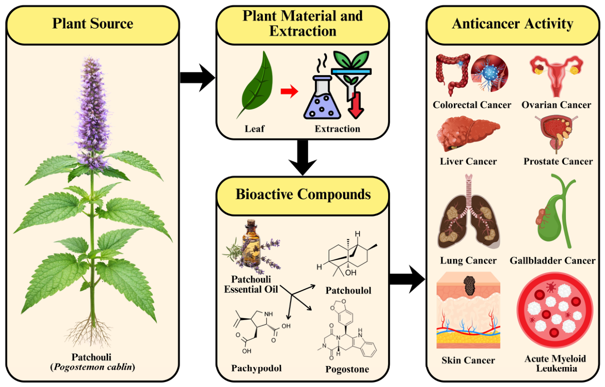
Emerging experimental evidence has highlighted patchouli leaves as a promising source of anticancer agents. Several bioactive compounds, such as patchoulol, pachypodol, and pogostone, have shown significant anticancer effects across various cancer types, including liver, colorectal, lung, prostate, and skin cancers. Their anticancer activity is primarily mediated through antiproliferative and apoptosis-inducing mechanisms, offering a mechanistic basis for the tumor-suppressive effects observed in preclinical models [72,84]. The reported anticancer effects of patchouli-derived bioactive compounds across different cancer models are systematically compiled in Table 3, while an overview of these bioactive compounds and their associated anticancer activities is summarized in Figure 2.

6.1. Anticancer Activity Against Endometrial Cancer

Endometrial cancer is the sixth most frequently diagnosed malignancy among women worldwide. It is the most prevalent gynecological cancer in developed countries. Due to increasing life expectancy, lifestyle changes, and associated risk factors, the global incidence of endometrial cancer has been steadily rising in recent years. In 2021 alone, approximately 97,370 new cases of endometrial cancer were reported worldwide [99].
Experimental studies have shown that P. cablin aqueous extract (PCAE) exhibits significant anticancer activity in endometrial cancer models, particularly in human Ishikawa endometrial cancer cells. The MTT assay was used to evaluate cell proliferation after PCAE treatment, while flow cytometry (FACSCalibur) was employed to analyze DNA content, cell-cycle distribution, and apoptosis-related changes. Furthermore, the expression and activation of apoptosis-associated proteins, including caspase-3 (CASP3), CASP9, and apoptosis-inducing factor (AIF), were assessed by Western blot analysis [90].
Treatment with PCAE (0, 1, 2, and 4 mg/mL) resulted in a dose-dependent suppression of Ishikawa cell proliferation and a marked increase in apoptosis. These effects were accompanied by increased CASP3 activity, CASP9 activation, and AIF involvement, collectively indicating the engagement of intrinsic apoptotic pathways. Further gene expression profiling (GEP) revealed that, in addition to suppressing endometrial cancer cell proliferation, patchouli-derived constituents exerted broader antitumor regulatory effects in established endometrial cancer cells [90].
These findings collectively demonstrate that PCAE demonstrates pronounced anticancer effects against endometrial cancer by inhibiting tumor cell proliferation and triggering intrinsic apoptosis. This is achieved through coordinated activation of CASP-dependent pathways and apoptosis-inducing factor-mediated signaling, underscoring its potential as a promising plant-derived therapeutic agent for the management of endometrial cancer.

6.2. Anticancer Activity Against Ovarian Cancer

Ovarian cancer is the third most prevalent malignancy and is the deadliest cancer affecting the female reproductive system. Around 70% of patients receive their diagnosis at advanced stages of the disease (FIGO stages III–IV), often with distant metastasis [100]. The elevated mortality associated with ovarian cancer is largely attributable to late-stage diagnosis and the absence of reliable early detection methods [101,102]. Even with the standard-of-care treatment, which includes optimal cytoreductive surgery followed by adjuvant chemotherapy, most patients eventually develop recurrent, chemotherapy-resistant disease, leading to a global 5-year survival rate of merely 30–40% [103].
Recent studies indicate that pogostone, a major bioactive constituent of patchouli, exerts potent anticancer effects in ovarian cancer models. Its activity was evaluated in OVCAR-3 human ovarian cancer cells treated with pogostone at the IC50 concentration of 90 µg/mL for 24 and 48 h. Apoptotic responses and cell viability were assessed using the MTT assay and flow cytometry, respectively. In contrast, PCR and a CASP3 colorimetric assay were employed to examine gene expression associated with apoptosis and cell-cycle regulation, as well as CASP3 protein levels [96].
Pogostone exposure resulted in a marked increase in apoptosis in OVCAR-3 cells at both time points. Molecular analyses revealed significant upregulation of tumor suppressor genes, including PTEN and DACT1, as well as apoptosis-related genes CASP8, CASP9, and CASP3. A significant elevation in the ratio of pro-apoptotic BAX to anti-apoptotic BCL-2 was observed. In contrast, key cell-cycle regulators, such as CCND1 and CDK4, were significantly downregulated. These transcriptional changes were supported by increased CASP3 protein expression, as confirmed by the colorimetric assay [96].
Collectively, these results demonstrate that pogostone effectively suppresses ovarian cancer cell proliferation and promotes apoptosis by coordinating the activation of tumor suppressor signaling, caspase-dependent apoptotic pathways, and the disruption of cell-cycle regulatory mechanisms, underscoring its promise as a plant-derived therapeutic candidate for ovarian cancer treatment.

6.3. Anticancer Activity Against Liver Cancer

Hepatocellular carcinoma is the predominant form of primary liver cancer and the sixth most frequently diagnosed malignancy worldwide. Despite this, therapeutic options for liver cancer remain limited, with nearly one million new cases and over 800,000 deaths reported annually [104,105]. Primary liver cancers encompass several pathological subtypes, including hepatocellular carcinoma, cholangiocarcinoma, and mixed hepatocellular-cholangiocarcinoma, with hepatocellular carcinoma being the most dominant, accounting for approximately 75–85% of cases globally. The incidence of hepatocellular carcinoma is notably high among patients with chronic liver diseases, such as hepatic fibrosis and cirrhosis associated with hepatitis B or C viral infection, metabolic liver disorders, alcohol abuse, particularly nonalcoholic fatty liver disease, and prolonged exposure to dietary carcinogens like aflatoxins and aristolochic acid. In China, the proportion of hepatocellular carcinoma among primary liver cancers is even higher, reaching nearly 93% [106,107,108].
Recent studies have further clarified the antitumor effects and underlying mechanisms of PEO against hepatocellular carcinoma through both in vitro and in vivo investigations. PEO showed significant inhibitory effects on hepatocellular carcinoma cell proliferation while exhibiting substantially lower cytotoxicity toward normal cells, particularly normal hepatocytes, indicating a favorable selectivity index. Mechanistically, PEO induced G0/G1 phase cell cycle arrest via both p53-dependent and -independent pathways involving key cell cycle-associated proteins. Concurrently, PEO activated the FAS-FASL-CASP8 signaling cascade, initiating extrinsic apoptotic pathways, while also increasing the BAX/BCL-2 ratio and promoting mitochondrial-mediated intrinsic apoptosis. These pro-apoptotic effects were closely linked to excessive intracellular reactive oxygen species (ROS) generation induced by PEO [92].
Notably, PEO demonstrated synergistic anticancer effects when combined with sorafenib, resulting in enhanced inhibition of hepatocellular carcinoma cell proliferation by modulating the Akt/mTOR signaling pathway and significant suppression of xenograft tumor growth. In animal models, PEO significantly reduced tumor burden, prolonged survival, and downregulated angiogenic signaling by suppressing the vascular endothelial growth factor (VEGF)/vascular endothelial growth factor receptor (VEGFR) axis, while inducing apoptosis in tumor tissues in vivo. Importantly, no significant systemic, physiological, or pathological toxicity was observed in treated animals. Collectively, these findings demonstrate that PEO (0–200 µg/mL) effectively suppresses hepatocellular carcinoma progression by inducing cell cycle arrest and activating multiple apoptotic pathways in vivo and in vitro. The low toxicity profile of PEO toward normal tissues further suggests a reduced risk of adverse effects, highlighting its potential for development as a therapeutic agent or functional food for the chemoprevention of hepatocellular carcinoma [92].
Beyond the essential oil constituents, individual phytochemicals isolated from patchouli significantly enhance its anti-liver-cancer activity. Pogostone, a major bioactive compound, has demonstrated concentration- and time-dependent cytotoxicity against liver cancer cell lines. Cytotoxicity assessments using the MTT assay, lactate dehydrogenase (LDH) release assay, and trypan blue exclusion test have shown notable reductions in cell viability and membrane integrity following pogostone treatment. Apoptotic induction was confirmed by diphenylamine assays, Annexin V-FITC staining, and real-time PCR. Pogostone significantly downregulated the expression of the anti-apoptotic gene BCL-2 while markedly upregulating pro-apoptotic markers, including BAX, p53, and CASP3, thereby promoting apoptosis primarily by modulating the BAX/BCL-2 ratio [93].
Moreover, flavonoids derived from patchouli, such as pachypodol, along with other natural antioxidants and phytochemicals, have recently been suggested as complementary anticancer agents due to their antiproliferative and pro-apoptotic properties [109,110]. Notably, pachypodol has shown significant cytotoxicity against HepG2 liver cancer cells, with an IC50 of 0.55 mg/mL, highlighting its potential as a promising candidate for further research in liver cancer therapeutics [111].
These findings collectively demonstrate that constituents of patchouli, including PEO, pogostone, and flavonoids such as pachypodol, exhibit significant anticancer effects against hepatocellular carcinoma. They achieve this by coordinating cell-cycle arrest, activating both extrinsic and intrinsic apoptotic pathways, modulating oxidative stress, and suppressing angiogenic signaling. This highlights their strong potential as promising plant-derived agents for the prevention and therapeutic intervention of liver cancer.

6.4. Anticancer Activity Against Gallbladder Cancer

Gallbladder cancer is a highly aggressive malignancy of the biliary tract with pronounced geographic variation, exhibiting particularly great extent in India, South America, and East Asia [112,113,114]. Global epidemiological data suggest an annual incidence of approximately 1–2 cases per 100,000 individuals, with a higher prevalence observed in women than in men [115]. Owing to its asymptomatic early course, gallbladder cancer is frequently diagnosed at advanced stages, resulting in poor clinical outcomes. Moreover, standard therapeutic modalities, including chemotherapy and radiotherapy, provide limited efficacy, posing substantial challenges to improving patient prognosis [116].
Recent experimental evidence demonstrates that pogostone exerts notable anticancer activity against gallbladder carcinoma. In the human gallbladder cancer cell line SGC-996, pogostone markedly inhibited cell proliferation and colony formation. These effects were mechanistically associated with activation of apoptosis-related pathways, as indicated by increased expression of CASP9, CASP3, and poly (ADP-ribose) polymerase-1 (PARP-1), together with an elevated BAX/BCL-2 ratio. In parallel, pogostone significantly suppressed cell-cycle progression by downregulating cyclins A, B, and D1 [91].
Collectively, these findings indicate that pogostone effectively inhibits gallbladder cancer cell growth by concurrently inducing apoptosis and arresting cell-cycle progression. This underscores its potential as a promising plant-derived anticancer agent for gallbladder cancer therapy.

6.5. Anticancer Activity Against Colorectal Cancer

Colorectal cancer is the third most commonly diagnosed malignancy and the second leading cause of cancer-related mortality worldwide [117]. Its occurrence is closely linked to Western lifestyle factors, such as excessive alcohol consumption, obesity, and a high intake of red and processed meats [118]. Individuals with chronic inflammatory bowel diseases, such as ulcerative colitis and Crohn’s disease, face a significantly heightened risk of colorectal cancer and thus require vigilant clinical monitoring [119]. Approximately 5% of colorectal cancer cases stem from hereditary syndromes associated with disease development, such as hereditary non-polyposis colorectal cancer (HNPCC) and familial adenomatous polyposis (FAP), while an additional ~20% of cases are linked to familial clustering. The majority of colorectal cancer cases (~75%) occur sporadically [118].
Patchouli has demonstrated significant anticancer activity against colorectal cancer in both in vivo and in vitro models. Treatment with patchouli extracts (0–80 μg/mL) significantly induced apoptosis and suppressed colorectal cancer cell proliferation, as evidenced by MTT-based assays. These effects were associated with G0/G1 cell-cycle arrest and modulation of apoptosis-related proteins. Notably, co-treatment with patchouli and 5-fluorouracil produced synergistic antiproliferative effects. In vivo studies further confirmed that patchouli inhibited tumor growth by inducing apoptosis without causing significant systemic toxicity, indicating a favorable safety profile [72].
Patchoulol has been identified as a principal contributor to patchouli’s anticancer effects in colorectal cancer. In HCT116 and SW480 cells, patchoulol inhibited proliferation and induced apoptosis in a dose-dependent manner by upregulating p21 and downregulating cyclin D1 and CDK4, consistent with cell-cycle arrest. Additionally, patchoulol suppressed histone deacetylase 2 activity and reduced c-Myc expression, while enhancing nuclear factor kappa B (NF-κB) transcriptional activity through p65 nuclear translocation, indicating coordinated regulation of cell-cycle control, epigenetic modulation, and apoptotic signaling [87].
Further studies have corroborated these findings in additional colorectal cancer models. In HT-29, LoVo, and Caco-2 cells, patchoulol (100 μM) markedly inhibited proliferation and induced G1-phase arrest, accompanied by suppression of β-catenin-dependent transcriptional activity. Consistently, oral administration of patchoulol (25–50 mg/kg) significantly reduced tumor number and burden in vivo in a dose-dependent manner [120]. Similar chemopreventive effects were observed in inflammation-associated colorectal tumorigenesis models, where patchoulol significantly reduced tumor formation in ApcMin/+ mice treated with dextran sulfate sodium, and showed antiproliferative and cell-cycle-arresting effects in Caco-2 cells [88].
Recent evidence has revealed olfactory receptor-mediated signaling as a novel mechanism underlying the anticancer activity of P. cablin. In an azoxymethane/dextran sulfate sodium-induced colorectal cancer mouse model and in CT26 and HCT116 colorectal cancer cells, patchouli oil significantly suppressed tumor progression by upregulating the ectopically expressed olfactory receptor OR13G1, activating the CaMKK2/AMPK signaling pathway, and inducing ferroptosis. Among the constituents of patchouli oil, patchouli alcohol (PA) was identified as the principal bioactive compound responsible for these antitumor effects. These findings identify the OR13G1–CaMKK2–AMPK axis and ferroptotic cell death as previously unrecognized mechanisms contributing to patchouli’s colorectal anticancer activity and further support PA as a promising natural therapeutic candidate for colorectal cancer [121].
In addition to essential oil constituents, patchouli leaf-derived flavonoids further contribute to its anticancer potential. Pachypodol exhibited antiproliferative activity against human Caco-2 colon cancer cells, with moderate cytotoxicity in brine shrimp lethality assays and measurable in vitro growth inhibition, suggesting a modest but significant anticancer effect [89].
These findings collectively demonstrate that bioactive constituents derived from patchouli, particularly crude extracts, patchoulol, and flavonoids like pachypodol, effectively inhibit the progression of colorectal cancer. They achieve this by integrating regulation of cell cycle arrest, apoptotic signaling, epigenetic modulation, inflammatory responses, and oncogenic signaling pathways, highlighting their strong potential as therapeutic and chemopreventive agents for colorectal cancer.

6.6. Anticancer Activity Against Lung Cancer

Lung cancer remains the most prevalent and deadly malignancy worldwide. It is broadly classified by cellular origin into small-cell lung cancer (SCLC) and non-small-cell lung cancer (NSCLC), with NSCLC accounting for the majority of cases [122,123,124]. Major histological subtypes include squamous cell carcinoma, adenocarcinoma, and neuroendocrine tumors such as large-cell neuroendocrine carcinoma, carcinoid tumors, and SCLC [122,125].
Patchoulol has demonstrated pronounced antitumor activity against NSCLC, particularly in A549 cell models, under both in vivo and in vitro conditions. It significantly inhibited tumor cell proliferation by inducing G1/S cell cycle arrest and stimulating mitochondrial-dependent apoptotic signaling, as evidenced by activation of CASP9 and CASP3. These effects were confirmed through multiple assays, including cell viability analysis, Hoechst 33,342 staining, TUNEL assays, and xenograft tumor suppression. Mechanistically, patchoulol exerted its anticancer effects by inhibiting the EGFR-ERK/MAPK signaling pathway, a central regulator of NSCLC proliferation. Reversal of these effects by exogenous epidermal growth factor further confirmed the dependence of patchoulol-induced apoptosis on EGFR pathway inhibition. Importantly, patchoulol displayed selective cytotoxicity toward cancer cells, with minimal toxicity observed in normal human cell lines, indicating a favorable safety profile [3].
In addition to apoptosis, patchoulol has been shown to induce autophagy-associated cell death in A549 cells. Treatment resulted in suppressed proliferation, accumulation of autophagic vesicles, and elevated LC3-II/LC3-I ratios, accompanied by reduced p62 expression, collectively indicating activation of autophagic pathways. However, the upstream molecular mechanisms governing patchoulol-induced autophagy remain unclear and warrant further investigation [126].
Patchoulol has also exhibited potent activity against drug-resistant NSCLC phenotypes. In both parental A549 and vincristine-resistant A549/V16 cells, patchoulol inhibited proliferation and colony formation while inducing G0/G1-phase arrest and apoptosis. These effects were mediated by ROS-induced DNA damage, activation of checkpoint kinases CHK1 and CHK2, and regulation of the p53/p21 and CDK2/cyclin E1 pathways. Apoptosis was primarily driven by intrinsic mitochondrial signaling, as evidenced by BAX upregulation and CASP activation. Notably, patchoulol reduced multidrug resistance and cancer stem cell-associated traits by downregulating P-glycoprotein (P-gp) and stemness markers such as CD44 and CD133. Moreover, combination treatment with patchoulol and cisplatin produced synergistic anticancer effects [94].
Collectively, these findings demonstrate that patchoulol exerts multifaceted anticancer effects against NSCLC by inducing cell-cycle arrest, mitochondrial apoptosis, and autophagy, while concurrently suppressing EGFR-driven oncogenic signaling, multidrug resistance, and cancer stem cell-associated phenotypes. These mechanisms underscore its strong therapeutic potential for both chemosensitive and drug-resistant lung cancer.

6.7. Anticancer Activity Against Nasopharyngeal Cancer

Nasopharyngeal carcinoma is a malignancy originating from the nasopharyngeal epithelium [127,128]. In the advanced stages of nasopharyngeal carcinoma, resistance to conventional chemotherapy and radiotherapy often develops, significantly limiting therapeutic efficacy. This diminished treatment responsiveness is mainly attributed to impaired apoptotic signaling in tumor cells, particularly due to the overexpression of the anti-apoptotic protein BCL-2. As a result, BCL-2 has become a crucial therapeutic target for restoring apoptosis and overcoming treatment resistance in nasopharyngeal carcinoma [95].
To discover potential natural inhibitors of BCL-2, bioactive compounds from patchouli were sourced from the KNApSAcK database and systematically assessed through molecular docking, followed by molecular dynamics simulations. Computational analyses pinpointed apigenin 7-(6″-p-coumarylglucoside), apigenin, and luteolin as promising candidates with significant inhibitory potential against BCL-2, based on their binding affinities and interaction profiles. Among these, apigenin 7-(6″-p-coumarylglucoside) showed the strongest predicted binding affinity (−9.9 kcal/mol), while luteolin and apigenin each exhibited binding energies of −7.2 kcal/mol. All three flavonoids were predicted to bind within the inhibitory pocket of BCL-2, primarily stabilized by hydrophobic interactions and van der Waals forces [95].
However, subsequent molecular dynamics simulations indicated that apigenin 7-(6″-p-coumarylglucoside) exhibited unstable binding over time, suggesting limited conformational persistence under dynamic physiological conditions. In contrast, luteolin and apigenin demonstrated more stable interaction profiles with BCL-2, suggesting a greater potential as functionally relevant inhibitors [95].
These findings collectively indicate that flavonoids derived from patchouli hold significant promise as alternative or complementary therapeutic agents for addressing radiotherapy- and chemotherapy-resistant nasopharyngeal carcinoma. Notably, luteolin and apigenin stand out as promising candidates for modulating apoptosis-related signaling by inhibiting BCL-2, thus meriting further experimental validation and translational exploration in nasopharyngeal carcinoma treatment strategies.

6.8. Anticancer Activity Against Skin Cancer

Skin cancer represents a major global health concern, with hundreds of thousands of new melanoma cases and more than 1.5 million non-melanoma cases reported annually worldwide [129,130]. Although melanoma occurs less frequently than non-melanoma skin cancers, it accounts for a disproportionately high mortality rate due to its aggressive behavior, strong metastatic capacity, and rapid dissemination to vital organs [131].
Patchoulol has demonstrated marked antitumor activity in both in vitro and in vivo melanoma models. In murine B16F10 melanoma cells, patchoulol selectively suppressed cell proliferation in a dose-(0–100 μM) and time-dependent manner, while inducing G0/G1 phase cell cycle arrest, and triggered apoptosis, as evidenced by chromatin condensation, DNA fragmentation, and apoptotic body formation. These effects were mechanistically associated with downregulation of cyclin D1 and CDK4, driven by activation of both intrinsic and extrinsic apoptotic pathways [98].
Beyond its antiproliferative effects, patchoulol significantly suppressed melanoma cell migration and invasiveness by modulating epithelial–mesenchymal transition-related markers, including upregulation of E-cadherin and downregulation of vimentin, phosphorylated SMAD2/3, and matrix metalloproteinases MMP-2/9. In animal models, patchoulol markedly reduced tumor growth and, when combined with cisplatin, produced synergistic inhibition of colony formation and cell migration while attenuating chemoresistance [98].
Collectively, these findings identify patchoulol as a promising natural anticancer agent against melanoma, exerting multitarget effects through coordinated regulation of cell-cycle control, apoptotic signaling, metastatic pathways, and drug resistance.

6.9. Anticancer Activity Against Prostate Cancer

Prostate cancer is the most commonly diagnosed malignancy in men and a leading cause of cancer-related mortality, with more than 1.1 million new cases reported annually worldwide [132]. Owing to its prolonged clinical course and marked heterogeneity, prostate cancer is classified according to tumor localization, metastatic status, and androgen sensitivity, encompassing localized prostate cancer, metastatic hormone-sensitive prostate cancer, and metastatic castration-resistant prostate cancer [133,134,135].
Castration-resistant prostate cancer (CRPC) represents an advanced and aggressive disease stage that is unresponsive to androgen-deprivation and conventional hormonal therapies. Patchoulol has demonstrated significant anticancer activity against CRPC in both in vivo and in vitro models at concentrations of 0–100 μg/mL. In human CRPC cell lines (DU145 and PC-3), patchoulol markedly suppressed cell proliferation, migration, and invasion in a dose-dependent manner and strongly induced apoptosis. These effects were associated with mitochondrial membrane depolarization, increased levels of cleaved CASP3 and PARP, upregulation of the pro-apoptotic protein BAX, accompanied by downregulation of anti-apoptotic proteins and proliferative markers, including BCL-2, Ki67, and MCL-1 [97].
Mechanistic analyses revealed that patchoulol enhanced expression of the NF-κB inhibitor IκBα while reducing NF-κB p65 transcriptional activity. Inhibition of NF-κB signaling was accompanied by decreased expression of metastasis- and angiogenesis-related factors, including MMP-2, MMP-7, MMP-9, and VEGF. Notably, patchoulol disrupted NF-κB p65 binding to the MCL-1 promoter, thereby suppressing MCL-1 transcription. Functional studies confirmed that p65 silencing potentiated patchoulol-induced apoptosis, whereas MCL-1 overexpression attenuated cell death. Consistent with these findings, patchoulol significantly inhibited tumor growth in mouse xenograft models [97].
Collectively, these results demonstrate that patchoulol exerts potent anticancer effects against CRPC by suppressing NF-κB-mediated survival signaling, inhibiting metastatic and angiogenic pathways, and activating mitochondria-dependent apoptotic mechanisms, highlighting its promise as a therapeutic candidate for advanced prostate cancer.

6.10. Anticancer Activity Against Acute Myeloid Leukemia

Leukemia encompasses a diverse group of hematological malignancies marked by the unchecked proliferation of abnormal blood cells, typically originating in the bone marrow [136]. It continues to rank among the most prevalent cancers worldwide, placing a substantial burden on global health, especially in developing countries, due to premature mortality, while clinical outcomes are comparatively better managed in developed regions [137,138]. Among the various leukemia subtypes, acute myeloid leukemia is a highly aggressive malignancy arising from the myeloid lineage, associated with poor prognosis and limited therapeutic options [139].
The anticancer potential of patchouli extract and its underlying mechanisms have been explored in HL-60 human acute myeloid leukemia cells. Results from the MTT assay showed that patchouli extract, at concentrations ranging from 0 to 200 μg/mL, significantly inhibited HL-60 cell proliferation in a dose-dependent manner. This was accompanied by notable morphological changes, such as membrane blebbing and cell shrinkage. Cell cycle analysis indicated that the treatment induced G0/G1-phase arrest, while apoptosis was triggered in both dose- and time-dependent manners, as evidenced by increased DNA fragmentation and apoptotic body formation. Mechanistically, patchouli extract promoted G0/G1-phase accumulation by enhancing phosphorylation of the retinoblastoma protein (p-Rb), upregulating the cyclin-dependent kinase (CDK) inhibitor p21, and downregulating key cell cycle-regulatory proteins. Concurrently, the extract activated both extrinsic and intrinsic apoptotic pathways, ultimately leading to leukemic cell death [85].
Beyond whole extracts, the primary sesquiterpene component, patchoulol, has shown significant antiproliferative and pro-apoptotic effects in human leukemia MV4-11 cells. Treatment with patchoulol significantly reduced cell proliferation, with MV4-11 cells showing the greatest sensitivity among the tested cancer cell lines, including HCT116, HepG2, A549, A375, 4T1, and THP-1, as well as human embryonic kidney 293A cells. Apoptotic indicators, such as nuclear shrinkage and chromatin condensation, were evident following patchoulol exposure, and flow cytometric analysis confirmed a dose-dependent increase in apoptotic cell populations. Mechanistic studies further revealed that patchoulol-induced apoptosis was associated with downregulation of NF-κB signaling and phosphorylated pyruvate kinase M2 (p-PKM2), along with increased CASP3 protein expression, indicating activation of a caspase-dependent apoptotic pathway [86].
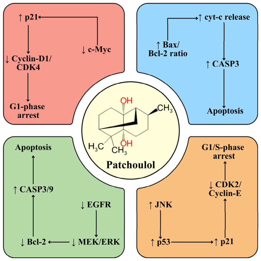
These findings collectively demonstrate that extracts and phytochemicals derived from patchouli leaves exhibit broad-spectrum anticancer activity against a wide range of malignancies, including endometrial, ovarian, liver, gallbladder, colorectal, lung, nasopharyngeal, skin, and prostate cancers, as well as acute myeloid leukemia. Despite differences among cancer types, patchouli-derived bioactive compounds consistently exert their anticancer effects through several conserved molecular mechanisms, including induction of apoptosis, cell-cycle arrest, suppression of proliferative and survival signaling pathways (e.g., PI3K/Akt, MAPK, and NF-κB), modulation of oxidative stress, and inhibition of tumor growth and metastasis. Among these constituents, patchoulol is the most extensively investigated compound, with consistent evidence from both in vitro and in vivo studies demonstrating its ability to regulate cell-cycle progression and activate both intrinsic and extrinsic apoptotic pathways. As summarized in Figure 3, patchoulol modulates key oncogenic signaling networks involving cell-cycle regulators, mitochondrial apoptosis, and growth factor-mediated pathways, providing a unified mechanistic framework for the anticancer activities of patchouli-derived phytochemicals. Collectively, these findings identify patchoulol as a representative lead compound and highlight the promise of patchouli-derived bioactive constituents as multi-target anticancer agents worthy of further mechanistic, pharmacological, and translational investigation.

7. Challenges and Limitations

Despite the growing interest in the anticancer potential of patchouli leaves, several challenges continue to hinder their translational development. A major constraint is the pronounced phytochemical variability associated with geographical origin, climate, soil conditions, cultivation practices, harvesting stage, and genetic diversity among cultivars. This variability may lead to inconsistent profiles and concentrations of bioactive compounds, resulting in differences in pharmacological activity and therapeutic efficacy [17]. Such heterogeneity complicates reproducibility, quality control, and standardization, while the molecular mechanisms underlying several reported pharmacological effects remain insufficiently elucidated [59]. Moreover, robust and large-scale clinical trials validating the therapeutic potential of patchouli-derived products remain scarce [140].
Methodological limitations further restrict progress. The absence of standardized extraction and processing protocols can lead to substantial variations in phytochemical yield, purity, and quality, as extraction techniques and distillation parameters strongly influence the composition of patchouli-derived preparations [141]. The high sesquiterpene content of patchouli leaves may also require prolonged distillation, reducing processing efficiency and emphasizing the need for optimized or innovative extraction strategies [38]. In addition, weaknesses in experimental design, including small sample sizes, inadequate controls, insufficient biological replication, and reliance on conventional analytical approaches, may compromise data reliability and overlook minor but biologically relevant constituents [23]. Product quality is another important concern, because contamination with heavy metals, pesticide residues, microorganisms, or adulterants may pose safety risks and limit the scalability and sustainability of patchouli-derived medicinal products [38].
Regulatory and safety considerations represent additional barriers. The absence of harmonized quality-control standards and the variability of regulatory frameworks governing herbal products contribute to inconsistencies in product efficacy, safety, and market acceptance [142]. Regulatory approval requires extensive pharmacological, toxicological, and clinical evaluation, making commercialization costly and time-consuming [143]. Comprehensive safety assessments addressing potential adverse effects, long-term toxicity, dose-dependent responses, and interactions with conventional drugs are therefore essential for ensuring regulatory compliance and establishing confidence in patchouli-derived therapeutic products [144].
Limited bioavailability is also a significant challenge affecting the therapeutic use of patchouli phytochemicals. Poor aqueous solubility, incomplete gastrointestinal absorption, rapid metabolism, and differences in compound composition among plant parts and developmental stages may substantially influence systemic exposure and biological efficacy [145]. Extraction methods, formulation strategies, dosing conditions, and interindividual variability may further affect clinical outcomes, while determining effective and safe dosing regimens requires extensive pharmacological validation [20]. Strategies that improve solubility, stability, absorption, and pharmacokinetic consistency are therefore essential for maximizing the therapeutic potential of patchouli-derived compounds [146].
Although the anticancer potential of PA has been widely demonstrated in preclinical studies, its clinical translation remains constrained by limited pharmacokinetic, formulation, and long-term safety data. One of the major pharmaceutical challenges is PA’s poor aqueous solubility, which limits its oral bioavailability. Several formulation strategies have consequently been investigated to improve its solubility, dissolution, stability, and systemic exposure. Solid dispersions formulated with Eudragit achieved prolonged supersaturation and maintained higher dissolved PA concentrations, thereby improving its dissolution profile [147]. Similarly, poloxamer-based solid dispersion systems containing Poloxamers 188 and 407 enhanced PA solubility and dissolution while offering a relatively simple, rapid, cost-effective, and potentially scalable preparation process [148]. β-Cyclodextrin inclusion complexes have also been developed to improve PA stability and dissolution characteristics, as demonstrated using differential scanning calorimetry (DSC), Fourier-transform infrared spectroscopy (FT-IR), powder X-ray diffraction (PXRD), and scanning electron microscopy (SEM) [149].
Available pharmacokinetic evidence indicates that PA follows a two-compartment open model with linear elimination kinetics in experimental animals. Following intravenous administration in rats, PA displayed relatively rapid systemic disposition, whereas oral administration resulted in measurable plasma exposure, with parameters such as the time to maximum plasma concentration (Cmax), area under the concentration–time curve (AUC), Time to reach Cmax (Tmax), and elimination half-life characterized using gas chromatographic and gas chromatography–mass spectrometry (GC-MS) methods [150,151]. Preliminary metabolism studies have additionally identified hydroxylated metabolites of PA in rabbit liver, providing initial evidence concerning its metabolic fate [152]. However, these investigations were conducted predominantly in healthy or non-cancer animal models. Consequently, the effects of tumors, altered cancer metabolism, anticancer co-medications, and disease-associated physiological changes on PA absorption, distribution, metabolism, and elimination remain largely unknown.
Available toxicological studies suggest that PA has relatively low acute toxicity in experimental animals. High maximum tolerated doses following oral administration and reported median lethal dose (LD50) values indicate a comparatively favorable short-term safety profile. Nevertheless, the available evidence is mainly limited to acute or short-duration toxicity assessments, and poor PA solubility has complicated the determination of precise toxicity thresholds in some studies. Comprehensive evaluations of repeated-dose and chronic toxicity, reproductive and developmental toxicity, genotoxicity, immunotoxicity, carcinogenicity, allergic reactions, local irritation, organ-specific toxicity, and potential herb–drug interactions remain insufficient [153]. These limitations prevent definitive conclusions regarding the long-term safety of PA and other patchouli-derived bioactive constituents.
Importantly, the currently available formulation, pharmacokinetic, metabolism, and toxicity data were generated mainly in non-cancer settings and therefore cannot be directly extrapolated to anticancer treatment. Cancer-specific information concerning plasma half-life, tumor accumulation, biodistribution, pharmacologically active concentrations, therapeutic windows, optimal dosing regimens, and interactions with chemotherapy remains scarce. Moreover, existing delivery approaches have primarily focused on improving solubility and oral absorption rather than achieving tumor-selective accumulation or controlled release. The development of targeted delivery systems, including nanoparticle-, liposome-, polymer-, or ligand-based formulations, may improve the stability, bioavailability, tumor targeting, and therapeutic index of PA; however, their efficacy and safety require systematic validation in relevant cancer models [12].
Overall, the therapeutic development of patchouli leaves remains limited by phytochemical variability, non-standardized methodologies, regulatory challenges, inconsistent product quality, poor bioavailability, insufficient cancer-specific pharmacokinetic evidence, and incomplete long-term safety evaluation. Addressing these barriers will require standardized cultivation and extraction procedures, rigorous chemical characterization, well-designed pharmacological studies, cancer-specific biodistribution and pharmacokinetic investigations, advanced targeted formulation strategies, and comprehensive repeated-dose and long-term toxicological assessments. Interdisciplinary collaboration among phytochemists, pharmacologists, formulation scientists, toxicologists, oncologists, and regulatory authorities will be essential for facilitating the successful preclinical and clinical development of patchouli-derived anticancer agents.

8. Future Perspectives

Future research on patchouli leaves is expected to be driven by advances in analytical, biotechnological, and translational approaches to realize their anticancer potential fully. Developments in high-resolution analytical platforms, such as MS and HPLC, have enabled more comprehensive characterization of phytochemical profiles and will continue to improve understanding of the bioactive constituents responsible for therapeutic effects [39]. Simultaneously, the increasing application of omics technologies, including proteomics, metabolomics, transcriptomics, and genomics, provides powerful tools for elucidating the biosynthetic pathways and regulatory networks governing the production of therapeutically important compounds [154]. When integrated with genetic and metabolic engineering strategies, these approaches may enable the targeted enhancement of key metabolites and support the sustainable production of patchouli-derived bioactive compounds for pharmaceutical and industrial applications [155].
From a therapeutic perspective, patchouli represents a promising but underexplored source of anticancer agents [156]. Although numerous bioactive compounds have been identified, systematic isolation, validation, and structure–activity relationship analyses of the principal anticancer constituents remain limited. This highlights the need for bioactivity-guided fractionation, compound standardization, and integrative phytochemical investigations to identify the compounds primarily responsible for the observed anticancer effects [157,158]. Furthermore, while antiproliferative, pro-apoptotic, and antimetastatic activities have been reported in experimental models, the precise molecular mechanisms, particularly those involving oncogenic signaling pathways, apoptosis, cell-cycle regulation, oxidative stress, ferroptosis, autophagy, and modulation of the tumor microenvironment, require further validation using well-characterized cellular and animal models [12]. The potential synergistic or chemosensitizing interactions between patchouli-derived compounds and conventional anticancer drugs should also be systematically evaluated to determine whether these compounds can enhance therapeutic efficacy, reduce drug resistance, or permit lower chemotherapy doses.
To facilitate clinical translation, future studies should prioritize cancer-specific pharmacokinetic and pharmacodynamic investigations together with the development of advanced targeted delivery systems and comprehensive long-term safety evaluations. Particular emphasis should be placed on characterizing the absorption, distribution, metabolism, elimination, tumor biodistribution, and therapeutic window of major bioactive compounds under clinically relevant conditions. The integration of pharmacokinetic and pharmacodynamic analyses with efficacy and safety assessments will be essential for optimizing therapeutic dosing, enhancing bioavailability and tumor targeting, minimizing off-target toxicity, and accelerating the successful translation of promising preclinical findings into well-designed clinical trials and ultimately clinical application.
Progress in this field will require coordinated efforts across multiple research areas. Comprehensive phytochemical profiling of different plant parts, cultivars, geographical origins, and developmental stages will be essential for capturing chemical diversity and identifying optimal sources of bioactive compounds [58]. Standardized cultivation, harvesting, extraction, processing, and quality-control procedures should be established to ensure reproducibility across studies. Expanding mechanistic investigations will be necessary to validate pharmacological targets and signaling pathways [159], while carefully designed preclinical studies should precede well-controlled clinical trials evaluating efficacy, safety, pharmacokinetics, tolerability, and potential interactions in humans [160].
Ultimately, interdisciplinary collaboration among phytochemists, pharmacologists, oncologists, toxicologists, formulation scientists, and bioinformaticians will accelerate the translation of patchouli leaf-derived compounds into clinically useful anticancer therapeutics and facilitate their development as novel anticancer drugs or effective adjuvant therapies [57,154].

9. Conclusions

This review provides a comprehensive synthesis of the current evidence regarding the anticancer potential of P. cablin, highlighting its phytochemical composition and the molecular mechanisms reported in experimental cancer models. Patchouli leaves contain diverse volatile and non-volatile bioactive constituents, including sesquiterpenes, flavonoids, glycosides, phytosterols, phenolic acids, and organic acids. Among these, patchoulol, pogostone, pachypodol, and other phytochemicals have received the greatest attention because of their reported anticancer activities. Collectively, the available evidence demonstrates that patchouli-derived extracts and bioactive compounds exhibit antiproliferative, pro-apoptotic, cell cycle-regulating, and anti-metastatic activities across multiple cancer cell lines and several animal models.
Experimental studies suggest that these effects are mediated through modulation of multiple molecular pathways, including NF-κB, PI3K/AKT, AKT/mTOR, EGFR/ERK, oxidative stress, mitochondrial apoptosis, and epigenetic regulation. Several studies have also reported enhanced chemosensitivity and reduced drug resistance when patchouli-derived compounds were combined with conventional anticancer agents. However, these findings are derived predominantly from in vitro experiments and preclinical animal models and should therefore be interpreted with caution.
Despite these promising experimental observations, the clinical translation of patchouli-derived compounds remains at a very early stage. Evidence regarding pharmacokinetics, bioavailability, formulation strategies, long-term toxicity, drug interactions, and clinical efficacy is still limited. Moreover, phytochemical variability, the absence of standardized extraction procedures, and the lack of rigorous clinical trials remain major barriers to therapeutic development. Consequently, the current evidence is insufficient to support clinical application or therapeutic recommendations.
Overall, the available literature indicates that patchouli is a promising source of bioactive molecules for future anticancer drug discovery. Nevertheless, well-designed pharmacokinetic studies, comprehensive toxicological evaluations, standardized formulations, and carefully conducted clinical trials are essential before the therapeutic value of patchouli-derived compounds can be established. This review provides a foundation for future research by summarizing the current evidence and identifying the key knowledge gaps that should be addressed to facilitate the translational development of patchouli-derived anticancer agents.

Author Contributions

E.M. and L.G. conceptualized the manuscript; E.M. prepared the initial draft; E.M., L.Q., F.Y., N.L., L.G. and C.Z. reviewed and revised the manuscript. All authors have read and agreed to the published version of the manuscript.

Funding

This research was supported by the Biological Breeding-National Science and Technology Major Project of China (2023ZD0403203), the National Natural Science Foundation of China (U21A20215 and 32488102), and Jilin Province Science and Technology Department (20230203162SF).

Institutional Review Board Statement

Not Applicable.

Informed Consent Statement

Not Applicable.

Data Availability Statement

No new data were created or analyzed in this study. Data sharing is not applicable to this article.

Acknowledgments

We thank the editors and reviewers for their critical comments on the manuscript.

Conflicts of Interest

The authors declare no conflicts of interest.

Abbreviations

The following abbreviations are used in this manuscript:
AIFApoptosis-inducing factor
CASP3/8/9Caspase-3/-8/-9
CDKCyclin-dependent kinase
CRPCCastration-resistant prostate cancer
DACT1Dishevelled-binding antagonist of β-catenin 1
DNADeoxyribonucleic acid
EGFREpidermal growth factor receptor
FAPFamilial adenomatous polyposis
FIGOInternational Federation of Gynecology and Obstetrics
GCGas chromatography
GC-MSGas chromatography–mass spectrometry
GEPGene expression profiling
HNPCCHereditary non-polyposis colorectal cancer
HPLCHigh-performance liquid chromatography
HPLC-Q-TOF-MSHigh-performance liquid chromatography–quadrupole time-of-flight mass spectrometry
HSCCCHigh-speed counter-current chromatography
LDHLactate dehydrogenase
MCL-1Myeloid cell leukemia-1
MMPMatrix metalloproteinase
MSMass spectrometry
MTT3-(4,5-Dimethylthiazol-2-yl)-2,5-diphenyltetrazolium bromide
NF-κBNuclear factor kappa B
NSCLCNon-small-cell lung cancer
PARP-1Poly(ADP-ribose) polymerase-1
PCAEPogostemon cablin aqueous extract
PCRPolymerase chain reaction
PEOPatchouli essential oil
PI3K/AKTPhosphoinositide 3-kinase/Protein kinase B signaling pathway
PTENPhosphatase and tensin homolog
ROSReactive oxygen species
SCLCSmall-cell lung cancer
TLCThin-layer chromatography
TUNELTerminal deoxynucleotidyl transferase dUTP nick-end labeling
VEGFVascular endothelial growth factor

References

  1. Andinata, B.; Bachtiar, A.; Oktamianti, P.; Partahi, J.R.; Dini, M.S.A. A Comparison of Cancer Incidences Between Dharmais Cancer Hospital and GLOBOCAN 2020: A Descriptive Study of Top 10 Cancer Incidences. Indones. J. Cancer 2023, 17, 119–122. [Google Scholar] [CrossRef] [Scilit]
  2. Tuy-on, T.; Itharat, A.; Maki, P.; Thongdeeying, P.; Pipatrattanaseree, W.; Ooraikul, B. In Vitro Cytotoxic Activity against Breast, Cervical, and Ovarian Cancer Cells and Flavonoid Content of Plant Ingredients Used in a Selected Thai Traditional Cancer Remedy: Correlation and Hierarchical Cluster Analysis. Evid.-Based Complement. Altern. Med. 2020, 2020, 8884529. [Google Scholar] [CrossRef] [Scilit] [PubMed]
  3. Lu, X.; Yang, L.; Lu, C.; Xu, Z.; Qiu, H.; Wu, J.; Wang, J.; Tong, J.; Zhu, Y.; Shen, J. Molecular Role of EGFR-MAPK Pathway in Patchouli Alcohol-Induced Apoptosis and Cell Cycle Arrest on A549 Cells In Vitro and In Vivo. BioMed Res. Int. 2016, 2016, 4567580. [Google Scholar] [CrossRef] [Scilit] [PubMed]
  4. Sari, S.L.; Indra, R.L.; Lestari, R.F. Korelasi Persepsi Tentang Efek Samping Kemoterapi Dengan Kualitas Hidup Pasien Kanker Payudara. J. Cakrawala Promkes 2019, 1, 40–47. [Google Scholar] [CrossRef] [Scilit]
  5. Brechbiel, M.W. Targeted α-Therapy. Cancer Biother. Radiopharm. 2020, 35, 397. [Google Scholar] [CrossRef] [Scilit] [PubMed]
  6. Azzahra, S.F.; Harnelly, E.; Subianto, M.; Kusuma, W.A.; Sari, W. Identification and Analysis of Potential Breast Anticancer Agents in Pogostemon Cablin through Network Pharmacology. J. Penelit. Pendidik. IPA 2024, 10, 107–119. [Google Scholar] [CrossRef] [Scilit]
  7. Shanmugam, M.K.; Lee, J.H.; Chai, E.Z.P.; Kanchi, M.M.; Kar, S.; Arfuso, F.; Dharmarajan, A.; Kumar, A.P.; Ramar, P.S.; Looi, C.Y.; et al. Cancer Prevention and Therapy through the Modulation of Transcription Factors by Bioactive Natural Compounds. Semin. Cancer Biol. 2016, 40–41, 35–47. [Google Scholar] [CrossRef] [Scilit] [PubMed]
  8. Bishayee, A.; Sethi, G. Bioactive Natural Products in Cancer Prevention and Therapy: Progress and Promise. Semin. Cancer Biol. 2016, 40–41, 1–3. [Google Scholar] [CrossRef] [Scilit] [PubMed]
  9. Newman, D.J.; Cragg, G.M. Natural Products as Sources of New Drugs over the Nearly Four Decades from 01/1981 to 09/2019. J. Nat. Prod. 2020, 83, 770–803. [Google Scholar] [CrossRef] [Scilit] [PubMed]
  10. Dutta, S.; Mahalanobish, S.; Saha, S.; Ghosh, S.; Sil, P.C. Natural Products: An Upcoming Therapeutic Approach to Cancer. Food Chem. Toxicol. 2019, 128, 240–255. [Google Scholar] [CrossRef] [Scilit] [PubMed]
  11. Wang, Z.; Qi, F.; Cui, Y.; Zhao, L.; Sun, X.; Tang, W.; Cai, P. An Update on Chinese Herbal Medicines as Adjuvant Treatment of Anticancer Therapeutics. Biosci. Trends 2018, 12, 220–239. [Google Scholar] [CrossRef] [Scilit] [PubMed]
  12. Mrisho, I.I.; Musazade, E.; Chen, H.; Zhao, H.; Xing, J.; Li, X.; Han, J.; Cai, E. Unlocking the Therapeutic Potential of Patchouli Leaves: A Comprehensive Review of Phytochemical and Pharmacological Insights. Plants 2025, 14, 1034. [Google Scholar] [CrossRef] [Scilit] [PubMed]
  13. Lin, R.-F.; Feng, X.-X.; Li, C.-W.; Zhang, X.-J.; Yu, X.-T.; Zhou, J.-Y.; Zhang, X.; Xie, Y.-L.; Su, Z.-R.; Zhan, J.Y.-X. Prevention of UV Radiation-Induced Cutaneous Photoaging in Mice by Topical Administration of Patchouli Oil. J. Ethnopharmacol. 2014, 154, 408–418. [Google Scholar] [CrossRef] [Scilit] [PubMed]
  14. Labarrade, F.; Perrin, A.; Ferreira, Y.; Oberto, G.; Gondran, C.; Morel, C.; Cucumel, K. Soothing Effect of Pogostemon Cablin Extract (Patchouli), via Cannabinoid Receptor 2 Modulation in Human Skin. J. Cosmet. Sci. 2020, 71, 425–437. [Google Scholar] [PubMed]
  15. Sadeghi-Nejad, B.; Deokule, S.S. Antidermatophytic Activity of Pogostemon Parviflorus Benth. Iran. J. Pharm. Res. IJPR 2010, 9, 279–285. [Google Scholar] [CrossRef] [Scilit]
  16. Mubarak, M.; Yunus, R.; Supiati, S.; Suwarni, S. Larvicidal And Repellent Potential Of Patchouli Extract (Pogostemon Cablin) Varieties Of Southeast Sulawesi For Aedes Aegypti Vector. Egypt. J. Chem. 2022, 66, 89–98. [Google Scholar] [CrossRef] [Scilit]
  17. Lallo, S.; Hasmiranti, A.; Usmar; Aminullah; Ismail; Hardianti, B. Effect of the Growth Environment on Patchouli (Pogostemon Cablin Benth.) Oil Character at Southeast Sulawesi, Indonesia. Med. Plants-Int. J. Phytomed. Relat. Ind. 2020, 12, 77–81. [Google Scholar] [CrossRef] [Scilit]
  18. Jadid, N.; Datus Soleha, I.; Esti Rahayu, A.; Puspaningtyas, I.; Anita Sari, S.; Rahmawati, M.; Aunurohim; Hidayati, D. Exogenous Methyl Jasmonate (MeJA) Altered Phytochemical Composition and Enhanced the Expression of PatAACT Gene of in Vitro Culture-Derived Patchouli Var. Sidikalang (Pogostemon cablin Benth.). J. King Saud Univ.-Sci. 2024, 36, 103301. [Google Scholar] [CrossRef] [Scilit]
  19. Ermaya, D.; Widodo, Y.; Teguh, D. Isolation and Utilization of Patchouli Oil (Pogostemon cablin Benth) as an Anti-Bacterial Alternative. IOP Conf. Ser. Earth Environ. Sci. 2022, 1012, 012015. [Google Scholar] [CrossRef] [Scilit]
  20. Muhammad, S.; H. P. S., A.K.; Abd Hamid, S.; Danish, M.; Marwan, M.; Yunardi, Y.; Abdullah, C.K.; Faisal, M.; Yahya, E.B. Characterization of Bioactive Compounds from Patchouli Extracted via Supercritical Carbon Dioxide (SC-CO2) Extraction. Molecules 2022, 27, 6025. [Google Scholar] [CrossRef] [Scilit] [PubMed]
  21. Umar Ijaz, M.; Azmat, R.; Karataş Yeni, D.; Ashraf, A.; Mariappan, V. Pachypodol, a Plant Flavonoid, Mitigates Cisplatin-Induced Hepatotoxicity Through Anti-Oxidant, Anti-Inflammatory and Anti-Apoptotic Mechanisms. J. Food Nutr. Res. 2023, 11, 513–518. [Google Scholar] [CrossRef] [Scilit]
  22. Wu, Z.; Zeng, H.; Zhang, L.; Pu, Y.; Li, S.; Yuan, Y.; Zhang, T.; Wang, B. Patchouli Alcohol: A Natural Sesquiterpene Against Both Inflammation and Intestinal Barrier Damage of Ulcerative Colitis. Inflammation 2020, 43, 1423–1435. [Google Scholar] [CrossRef] [Scilit] [PubMed]
  23. Srivastava, S.; Lal, R.K.; Singh, V.R.; Rout, P.K.; Padalia, R.C.; Yadav, A.K.; Bawitlung, L.; Bhatt, D.; Maurya, A.K.; Pal, A.; et al. Chemical Investigation and Biological Activities of Patchouli (Pogostemon cablin (Blanco) Benth) Essential Oil. Ind. Crops Prod. 2022, 188, 115504. [Google Scholar] [CrossRef] [Scilit]
  24. Khairan, K.; Amanda, R.; Hasbi, S.Y.; Diah, M.; Hasballah, K. Perspective Study of Pogostemon Cablin Benth as an Aphrodisiac. Malacca Pharm. 2023, 1, 62–70. [Google Scholar] [CrossRef] [Scilit]
  25. de Sousa, D.P.; de Assis Oliveira, F.; Arcanjo, D.D.R.; da Fonsêca, D.V.; Duarte, A.B.S.; de Oliveira Barbosa, C.; Ong, T.P.; Brocksom, T.J. Essential Oils: Chemistry and Pharmacological Activities—Part II. Biomedicines 2024, 12, 1185. [Google Scholar] [CrossRef] [Scilit] [PubMed]
  26. Tian, Y.; Wang, L.; Fan, X.; Zhang, H.; Dong, Z.; Tao, T. β-Patchoulene Alleviates Cognitive Dysfunction in a Mouse Model of Sepsis Associated Encephalopathy by Inhibition of Microglia Activation through Sirt1/Nrf2 Signaling Pathway. PLoS ONE 2023, 18, e0279964. [Google Scholar] [CrossRef] [Scilit] [PubMed]
  27. Green, M. Aromatherapy in the Treatment of Sexual Dysfunction and Relationship Problems (DRAFT). In Integrative sexual health; Castellanos, M.M., Ed.; Oxford University Press: Oxford, UK, 2018; Volume 1, pp. 471–497. [Google Scholar]
  28. Albuquerque, E.L.D.; Lima, J.K.A.; Souza, F.H.O.; Silva, I.M.A.; Santos, A.A.; Araújo, A.P.A.; Blank, A.F.; Lima, R.N.; Alves, P.B.; Bacci, L. Insecticidal and Repellence Activity of the Essential Oil of Pogostemon Cablin against Urban Ants Species. Acta Trop. 2013, 127, 181–186. [Google Scholar] [CrossRef] [Scilit] [PubMed]
  29. Ferreira, D.L.; de Mendonça, M.S.; Araujo, M.G.P.d.; Lescano, B.F.M.; Simão, M.O.d.A.R. Leaf Anatomy and Histochemistry of Oriza (Pogostemon cablin Beth., LAMIACEAE): Medicinal Plant Used in Community Arari Region, Itacoatiara, Amazonas. Int. J. Adv. Eng. Res. Sci. 2020, 7, 27–37. [Google Scholar] [CrossRef] [Scilit]
  30. Pérez-González, C.; Pérez-Ramos, J.; Alberto Méndez-Cuesta, C.; Serrano-Vega, R.; Martell-Mendoza, M.; Pérez-Gutiérrez, S. Cytotoxic Activity of Essential Oils of Some Species from Lamiaceae Family. In Cytotoxicity-Definition, Identification, and Cytotoxic Compounds; IntechOpen: London, UK, 2019; pp. 29–43. [Google Scholar] [CrossRef] [Scilit]
  31. Dongare, P.; Dhande, S.; Kadam, V. A Review on Pogostemon Patchouli. Res. J. Pharmacogn. Phytochem. 2014, 6, 41–47. [Google Scholar]
  32. Lubis, A.; Mandang, T.; Hermawan, W. Sutrisno Study of the Physical and Mechanical Characteristics of Patchouli Plants. AIMS Agric. Food 2021, 6, 525–537. [Google Scholar] [CrossRef] [Scilit]
  33. Kumar, A.; Sharma, N.; Chanotiya, C.S.; Lal, R.K. The Pharmacological Potential and the Agricultural Significance of the Aromatic Crop Patchouli (Pogostemon Cablin Benth.): A Review. Ecol. Front. 2024, 44, 1109–1118. [Google Scholar] [CrossRef] [Scilit]
  34. Yao, G.; Drew, B.T.; Yi, T.-S.; Yan, H.-F.; Yuan, Y.-M.; Ge, X.-J. Phylogenetic Relationships, Character Evolution and Biogeographic Diversification of Pogostemon s.l. (Lamiaceae). Mol. Phylogenet. Evol. 2016, 98, 184–200. [Google Scholar] [CrossRef] [Scilit] [PubMed]
  35. Singh, S.; Agrawal, N. Exploring the Pharmacological Potential and Bioactive Components of Pogostemon Cablin (Blanco) Benth, Traditional Chinese Medicine. Pharmacol. Res.-Mod. Chin. Med. 2024, 10, 100382. [Google Scholar] [CrossRef] [Scilit]
  36. Li, Y.-C.; Peng, S.-Z.; Chen, H.-M.; Zhang, F.-X.; Xu, P.-P.; Xie, J.-H.; He, J.-J.; Chen, J.-N.; Lai, X.-P.; Su, Z.-R. Oral Administration of Patchouli Alcohol Isolated from Pogostemonis Herba Augments Protection against Influenza Viral Infection in Mice. Int. Immunopharmacol. 2012, 12, 294–301. [Google Scholar] [CrossRef] [Scilit] [PubMed]
  37. Ijaz, M.U.; Akbar, A.; Anwar, H.; Inam, S.; Ashraf, A.; Riaz, M. Patchouli. In Essentials of Medicinal and Aromatic Crops; Springer: Berlin/Heidelberg, Germany, 2023; pp. 249–279. [Google Scholar]
  38. Jain, P.L.B.; Patel, S.R.; Desai, M.A. Patchouli Oil: An Overview on Extraction Method, Composition and Biological Activities. J. Essent. Oil Res. 2022, 34, 1–11. [Google Scholar] [CrossRef] [Scilit]
  39. Pandey, S.K.; Bhandari, S.; Sarma, N.; Begum, T.; Munda, S.; Baruah, J.; Gogoi, R.; Haldar, S.; Lal, M. Essential Oil Compositions, Pharmacological Importance and Agro Technological Practices of Patchouli (Pogostemon Cablin Benth.): A Review. J. Essent. Oil-Bear. Plants 2021, 24, 1212–1226. [Google Scholar] [CrossRef] [Scilit]
  40. Lima Santos, L.; Brandão, L.B.; Pena da Costa, A.L.; Martins, R.L.; Rodrigues, A.B.L.; Lobato, A.A.; Moreira da Silva de Almeida, S.S. Bioinsecticidal and Pharmacological Activities of the Essential Oil of Pogostemon Cablin Benth Leaves: A Review. Pharmacogn. Rev. 2022, 16, 139–145. [Google Scholar] [CrossRef] [Scilit]
  41. Souhoka, F.A.; Al Aziz, A.Z.; Nazudin, N. Patchouli Oil Isolation and Identification of Chemical Components Using GC-MS. Indones. J. Chem. Res. 2020, 8, 108–113. [Google Scholar] [CrossRef] [Scilit]
  42. Cano-Reinoso, D.M.; Purwanto, Y.A.; Budiastra, I.W.; Sutrisno; Kuroki, S.; Widodo, S.; Kamanga, B.M. Determination of α-Guaiene and Azulene Chemical Content in Patchouli Aromatic Oil (Pogostemon cablin Benth.) from Indonesia by near-Infrared Spectroscopy. Indian J. Nat. Prod. Resour. 2021, 12, 256–262. [Google Scholar] [CrossRef] [Scilit]
  43. Kusumaningrum, H.P.; Ni’mah, R.S.; Nurlindasari, Y.; Imrani, A.; Arrois, S.; Purnomo, E.; Arfiansyah, F.R.; Setyowati, E.; Loka, B.D. Patchouli Oil Analysis of Chemical Components Using FT-IR and GCMS Method. AIP Conf. Proc. 2023, 2738, 040026. [Google Scholar] [CrossRef] [Scilit]
  44. Singh, S.; Pathak, S.; Sharma, N.; Agrawal, N. Patchouli (Pogostemon Cablin Benth.): A Comprehensive Review on Medicinal Aromatic Plant with Industrial Utilization. In Advances in Medicinal and Aromatic Plants; Apple Academic Press: New York, NY, USA, 2024; pp. 131–162. [Google Scholar]
  45. Fahmi, Z.; Mudasir, M.; Rohman, A. Attenuated Total Reflectance-FTIR Spectra Combined with Multivariate Calibration and Discrimination Analysis for Analysis of Patchouli Oil Adulteration. Indones. J. Chem. 2019, 20, 1. [Google Scholar] [CrossRef] [Scilit]
  46. Swamy, M.; Sinniah, U. A Comprehensive Review on the Phytochemical Constituents and Pharmacological Activities of Pogostemon Cablin Benth.: An Aromatic Medicinal Plant of Industrial Importance. Molecules 2015, 20, 8521–8547. [Google Scholar] [CrossRef] [Scilit] [PubMed]
  47. Hanh, N.P.; Binh, N.Q.; Thuy, D.T.T.; Ogunwande, I.A. Chemical Constituents and Antimicrobial Activity of Essential Oils from Pogostemon Cablin (Blanco) Benth and Coriandrum Sativum L from Vietnam. Am. J. Essent. Oil Nat. Prod. 2022, 10, 10–15. [Google Scholar]
  48. Li, P.; Yin, Z.-Q.; Li, S.-L.; Huang, X.-J.; Ye, W.-C.; Zhang, Q.-W. Simultaneous Determination of Eight Flavonoids and Pogostone in Pogostemon Cablin by High Performance Liquid Chromatography. J. Liq. Chromatogr. Relat. Technol. 2014, 37, 1771–1784. [Google Scholar] [CrossRef] [Scilit]
  49. Akhila, A.; Sharma, P.K.; Thakur, R.S. Biosynthetic Relationships of Patchouli Alcohol, Seychellene and Cycloseychellene in Pogostemon Cablin. Phytochemistry 1988, 27, 2105–2108. [Google Scholar] [CrossRef] [Scilit]
  50. Rakotonirainy, O.; Gaydou, E.M.; Faure, R.; Bombarda, I. Sesquiterpenes from Patchouli (Pogostemon cablin) Essential Oil. Assignment of the Proton and Carbon-13 NMR Spectra. J. Essent. Oil Res. 1997, 9, 321–327. [Google Scholar] [CrossRef] [Scilit]
  51. Karimi, A. Characterization and Antimicrobial Activity of Patchouli Essential Oil Extracted from Pogostemon cablin [Blanco] Benth. [Lamiaceae]. Adv. Environ. Biol. 2014, 8, 2301–2309. [Google Scholar]
  52. Silva, M.A.S.; Ehlert, P.A.D.; Ming, L.C.; Marques, M.O.M. Composition and Chemical Variation during Daytime of Constituents of the Essential Oil of Pogostemon Pachouli Pellet Leaves. Acta Hortic. 2004, 629, 145–147. [Google Scholar] [CrossRef] [Scilit]
  53. Buré, C.M.; Sellier, N.M. Analysis of the Essential Oil of Indonesian Patchouli (Pogostemon cabin Benth.) Using GC/MS (EI/CI). J. Essent. Oil Res. 2004, 16, 17–19. [Google Scholar] [CrossRef] [Scilit]
  54. Hu, L.F.; Li, S.P.; Cao, H.; Liu, J.J.; Gao, J.L.; Yang, F.Q.; Wang, Y.T. GC-MS Fingerprint of Pogostemon cablin in China. J. Pharm. Biomed. Anal. 2006, 42, 200–206. [Google Scholar] [CrossRef] [Scilit] [PubMed]
  55. Bunrathep, S.; Lockwood, G.B.; Songsak, T.; Ruangrungsi, N. Chemical Constituents from Leaves and Cell Cultures of Pogostemon cablin and Use of Precursor Feeding to Improve Patchouli Alcohol Level. ScienceAsia 2006, 32, 293–296. [Google Scholar] [CrossRef]
  56. Tsai, Y.C.; Hsu, H.C.; Yang, W.C.; Tsai, W.J.; Chen, C.C.; Watanabe, T. α-Bulnesene, a PAF Inhibitor Isolated from the Essential Oil of Pogostemon cablin. Fitoterapia 2007, 78, 7–11. [Google Scholar] [CrossRef] [Scilit] [PubMed]
  57. Isnaini, N.; Khairan, K.; Faradhilla, M.; Sufriadi, E.; Prajaputra, V.; Ginting, B.; Muhammad, S.; Lufika, R.D. A Study of Essential Oils from Patchouli (Pogostemon cablin Benth.) and Its Potential as an Antivirus Agent to Relieve Symptoms of COVID-19. J. Patchouli Essent. Oil Prod. 2022, 1, 27–35. [Google Scholar] [CrossRef] [Scilit]
  58. Verma, R.S.; Padalia, R.C.; Chauhan, A.; Singh, V.R. Chemical Composition of Leaves, Inflorescence, Whole Aerial-Parts and Root Essential Oils of Patchouli {Pogostemon cablin (Blanco) Benth.}. J. Essent. Oil Res. 2019, 31, 319–325. [Google Scholar] [CrossRef] [Scilit]
  59. van Beek, T.A.; Joulain, D. The Essential Oil of Patchouli, Pogostemon Cablin: A Review. Flavour Fragr. J. 2018, 33, 6–51. [Google Scholar] [CrossRef] [Scilit]
  60. Kim, E.K.; Kim, J.H.; Jeong, S.; Choi, Y.W.; Choi, H.J.; Kim, C.Y.; Kim, Y.M. Pachypodol, a Methoxyflavonoid Isolated from Pogostemon Cablin Bentham Exerts Antioxidant and Cytoprotective Effects in HepG2 Cells: Possible Role of ERK-Dependent Nrf2 Activation. Int. J. Mol. Sci. 2019, 20, 4082. [Google Scholar] [CrossRef] [Scilit] [PubMed]
  61. Junren, C.; Xiaofang, X.; Mengting, L.; Qiuyun, X.; Gangmin, L.; Huiqiong, Z.; Guanru, C.; Xin, X.; Yanpeng, Y.; Fu, P.; et al. Pharmacological Activities and Mechanisms of Action of Pogostemon Cablin Benth: A Review. Chin. Med. 2021, 16, 5. [Google Scholar] [CrossRef] [Scilit] [PubMed]
  62. Itokawa, H.; Suto, K.; Takeya, K. Studies on a Novel P-Coumaroyl Glucoside of Apigenin and on Other Flavonoids Isolated from Patchouli (Labiatae). Chem. Pharm. Bull. 1981, 29, 254–256. [Google Scholar] [CrossRef] [Scilit]
  63. Chen, J.; Han, L.; Li, G.; Sun, L.; Qian, F.; Chen, K.; Zhang, L.; Li, Y. Lignans and Phenylethanoid Glycosides from the Aerial Parts of Pogostemon Cablin. Chem. Biodivers. 2022, 19, e202200889. [Google Scholar] [CrossRef] [Scilit] [PubMed]
  64. Kim, K.H.; Beemelmanns, C.; Clardy, J.; Cao, S. A New Antibacterial Octaketide and Cytotoxic Phenylethanoid Glycosides from Pogostemon cablin (Blanco) Benth. Bioorg. Med. Chem. Lett. 2015, 25, 2834–2836. [Google Scholar] [CrossRef] [Scilit] [PubMed]
  65. Amakura, Y.; Yoshimura, M.; Mouri, C.; Mikage, M.; Kawahara, N.; Goda, Y.; Yoshida, T. Convenient TLC-Based Identification Test for the Crude Drug “Pogostemoni Herba”. Yakugaku Zasshi 2008, 128, 1833–1837. [Google Scholar] [CrossRef] [Scilit] [PubMed]
  66. Xie, B.; Wu, X.F.; Luo, H.T.; Huang, X.L.; Huang, F.; Zhang, Q.Y.; Zhou, X.; Wu, H.Q. Chemical Profiling and Quality Evaluation of Pogostemon Cablin Benth by Liquid Chromatography Tandem Mass Spectrometry Combined with Multivariate Statistical Analysis. J. Pharm. Biomed. Anal. 2022, 209, 114526. [Google Scholar] [CrossRef] [Scilit] [PubMed]
  67. Xie, S.; Li, L.; Zhan, B.; Shen, X.; Deng, X.; Tan, W.; Fang, T. Pogostone Enhances the Antibacterial Activity of Colistin against MCR-1-Positive Bacteria by Inhibiting the Biological Function of MCR-1. Molecules 2022, 27, 2819. [Google Scholar] [CrossRef] [Scilit] [PubMed]
  68. Huang, X.; Li, P.; Yin, Z.; Lu, J.; Lin, L.; Wang, Y.; Ye, W.; Zhang, Q. Cablinosides A and B, Two Glycosidic Phenylacetic Acid Derivatives from the Leaves of Pogostemon Cablin. Chem. Biodivers. 2019, 16, e1900137. [Google Scholar] [CrossRef] [Scilit] [PubMed]
  69. +Huang, L.; Mu, S.; Zhang, J.; Deng, B.; Song, Z.; Hao, X. Chemical Constituents from Involatile Moiety of Pogostemon Cablin. China J. Chin. Mater. Medica 2009, 34, 410–413. [Google Scholar]
  70. Li, K.; Zhang, H.; Xie, H.; Liang, Y.; Wang, X.; Ito, Y. Preparative Isolation and Purification of Five Flavonoids from Pogostemon Cablin Benth by High-Speed Countercurrent Chromatography and Preparative High-Performance Liquid Chromatography. J. Liq. Chromatogr. Relat. Technol. 2011, 34, 1617–1629. [Google Scholar] [CrossRef] [Scilit] [PubMed]
  71. Zhou, Q.-M.; Peng, C.; Li, X.-H.; Guo, L.; Xiong, L.; Lin, D.-S. Study on constituents of the aerial parts of Pogostemon cablin. J. Chin. Med. Mater. 2013, 36, 915–918. [Google Scholar]
  72. Chien, J.-H.; Lee, S.-C.; Chang, K.-F.; Huang, X.-F.; Chen, Y.-T.; Tsai, N.-M. Extract of Pogostemon Cablin Possesses Potent Anticancer Activity against Colorectal Cancer Cells In Vitro and In Vivo. Evid.-Based Complement. Altern. Med. 2020, 2020, 9758156. [Google Scholar] [CrossRef] [Scilit] [PubMed]
  73. Xie, Y.C.; Tang, F. Protective Effect of Pogostemon Cablin on Membrane Fluidity of Intestinal Epithelia Cell in Ischemia/Reperfusion Rats after Ischemia/Reperfusion. Chin. J. Integr. Tradit. West. Med. 2009, 29, 639–641. [Google Scholar]
  74. Beşirik, N.Ş.; Göger, G. Antimicrobial Evaluation of the Patchouli (Pogostemon cablin Benth.) Leaf Essential Oil Combination with Standard Antimicrobial Compounds. Int. J. Second. Metab. 2023, 10, 385–393. [Google Scholar] [CrossRef] [Scilit]
  75. Krithika, S.; Sadiq, M.; Munuswamy Ramanujam, G.; Maruthapillai, A. Trimethoxy Flavone, Pachypodol Containing Pogostemon Cablin Leaf Extract Shows Broad Spectrum Antimicrobial Activity. Mater. Today Proc. 2022, 50, 297–300. [Google Scholar] [CrossRef] [Scilit]
  76. Adhayani, L.; Suhartono, S.; Amalia, A. Aceh Patchouli Oil (Pogostemon cablin Benth) as Antibacterial and Antibiofilm: Mechanism, Challenges, and Opportunities. AIP Conf. Proc. 2023, 2480, 050005. [Google Scholar] [CrossRef] [Scilit]
  77. Chen, X.-Y.; Chen, H.-M.; Liu, Y.-H.; Zhang, Z.-B.; Zheng, Y.-F.; Su, Z.-Q.; Zhang, X.; Xie, J.-H.; Liang, Y.-Z.; Fu, L.-D.; et al. The Gastroprotective Effect of Pogostone from Pogostemonis Herba against Indomethacin-Induced Gastric Ulcer in Rats. Exp. Biol. Med. 2016, 241, 193–204. [Google Scholar] [CrossRef] [Scilit] [PubMed]
  78. Han, H.; Gao, M.; Wang, F.; Luo, Z.; Jiang, X.; Qiu, Y.; Su, J.; Duan, X.; Luo, S.; Tang, S.; et al. Protective Effects of Patchouli Alcohol against DSS-Induced Ulcerative Colitis. Sci. Rep. 2024, 14, 16745. [Google Scholar] [CrossRef] [Scilit] [PubMed]
  79. Ojangba, T.; Boamah, S.; Miao, Y.; Guo, X.; Fen, Y.; Agboyibor, C.; Yuan, J.; Dong, W. Comprehensive Effects of Lifestyle Reform, Adherence, and Related Factors on Hypertension Control: A Review. J. Clin. Hypertens. 2023, 25, 509–520. [Google Scholar] [CrossRef] [Scilit] [PubMed]
  80. Reddy, V.P. Oxidative Stress in Health and Disease. Biomedicines 2023, 11, 2925. [Google Scholar] [CrossRef] [Scilit] [PubMed]
  81. Klran, T.R.; Otlu, O.; Karabulut, A.B. Oxidative Stress and Antioxidants in Health and Disease. J. Lab. Med. 2023, 47, 1–11. [Google Scholar] [CrossRef] [Scilit]
  82. Leong, W.; Huang, G.; Liao, W.; Xia, W.; Li, X.; Su, Z.; Liu, L.; Wu, Q.; Wong, V.K.W.; Law, B.Y.K.; et al. Traditional Patchouli Essential Oil Modulates the Host’s Immune Responses and Gut Microbiota and Exhibits Potent Anti-Cancer Effects in ApcMin /+ Mice. Pharmacol. Res. 2022, 176, 106082. [Google Scholar] [CrossRef] [Scilit] [PubMed]
  83. Pandey, S.K.; Gogoi, R.; Bhandari, S.; Sarma, N.; Begum, T.; Munda, S.; Lal, M. A Comparative Study on Chemical Composition, Pharmacological Potential and Toxicity of Pogostemon Cablin Linn., (Patchouli) Flower and Leaf Essential Oil. J. Essent. Oil-Bear. Plants 2022, 25, 160–179. [Google Scholar] [CrossRef] [Scilit]
  84. Alves-Silva, J.M.; Romane, A.; Efferth, T.; Salgueiro, L. North African Medicinal Plants Traditionally Used in Cancer Therapy. Front. Pharmacol. 2017, 8, 383. [Google Scholar] [CrossRef] [Scilit] [PubMed]
  85. Chien, J.; Huang, X.; Lai, W.; Chang, K.; Li, C.; Chen, S.; Wu, C.; Li, K.; Tsai, N. Pogostemon Cablin Extract as an Anticancer Agent on Human Acute Myeloid Leukemia. Food Sci. Nutr. 2021, 9, 3209–3218. [Google Scholar] [CrossRef] [Scilit] [PubMed]
  86. Yang, Y.T.; He, B.X.; He, Y.L.; Peng, C.; Xiong, L.; Cao, Z.X. Patchouli Alcohol Induces Apoptosis of MV4-11 Leukemia Cells by PKM2 and NF-ΚB. Chin. J. Exp. Tradit. Med. Formulae 2016, 22, 99–103. [Google Scholar]
  87. Jeong, J.B.; Choi, J.; Lou, Z.; Jiang, X.; Lee, S.-H. Patchouli Alcohol, an Essential Oil of Pogostemon Cablin, Exhibits Anti-Tumorigenic Activity in Human Colorectal Cancer Cells. Int. Immunopharmacol. 2013, 16, 184–190. [Google Scholar] [CrossRef] [Scilit] [PubMed]
  88. Lee, J.; Lee, H.-S.; Lee, S.-H. Preventive Activity of Patchouli Alcohol Against Colorectal Cancer and Diabetes. J. Med. Food 2023, 26, 255–261. [Google Scholar] [CrossRef] [Scilit] [PubMed]
  89. Ali, H.-A.; Chowdhury, A.K.A.; Rahman, A.K.M.; Borkowski, T.; Nahar, L.; Sarker, S.D. Pachypodol, a Flavonol from the Leaves of Calycopteris Floribunda, Inhibits the Growth of CaCo 2 Colon Cancer Cell Line in Vitro. Phytother. Res. 2008, 22, 1684–1687. [Google Scholar] [CrossRef] [Scilit] [PubMed]
  90. Tsai, C.-C.; Chang, Y.-H.; Chang, C.-C.; Cheng, Y.-M.; Ou, Y.-C.; Chien, C.-C.; Hsu, Y.-C. Induction of Apoptosis in Endometrial Cancer (Ishikawa) Cells by Pogostemon Cablin Aqueous Extract (PCAE). Int. J. Mol. Sci. 2015, 16, 12424–12435. [Google Scholar] [CrossRef] [Scilit] [PubMed]
  91. Wu, Y.; Huang, Y.; Dong, J. Patchoulenone Inhibits the Proliferation of Human Gallbladder Carcinoma SGC-996 Cells. Tumori 2017, 37, 50–57. [Google Scholar]
  92. Huang, X.-F.; Sheu, G.-T.; Chang, K.-F.; Huang, Y.-C.; Hung, P.-H.; Tsai, N.-M. Pogostemon Cablin Triggered ROS-Induced DNA Damage to Arrest Cell Cycle Progression and Induce Apoptosis on Human Hepatocellular Carcinoma in vitro and in vivo. Molecules 2020, 25, 5639. [Google Scholar] [CrossRef] [Scilit] [PubMed]
  93. Rezazadeh, D.; Asadi, S.; Nemati, H.; Jalili, C. The Effect of Pogostone on Viability, Membrane Integrity, and Apoptosis of Liver Cancer Cells. World Cancer Res. J. 2022, 9, e2436. [Google Scholar] [CrossRef]
  94. Liang, C.; Chang, K.; Huang, Y.; Huang, X.; Sheu, G.; Kuo, C.; Hsiao, C.; Tsai, N. Patchouli Alcohol Induces G0 /G1 Cell Cycle Arrest and Apoptosis in Vincristine-Resistant Non-Small Cell Lung Cancer through ROS-Mediated DNA Damage. Thorac. Cancer 2023, 14, 2007–2017. [Google Scholar] [CrossRef] [Scilit] [PubMed]
  95. Syahraini, A.; Harnelly, E.; Hermanto, F.E. Pro-Apoptosis Activity of Pogostemon Cablin Benth. Against Nasopharyngeal Carcinoma through the BCL-2 Inhibition Signaling Pathway: A Computational Investigation. Makara J. Sci. 2023, 27, 6. [Google Scholar] [CrossRef] [Scilit]
  96. Homayoun, M.; Sajedi, N.; Soleimani, M. In Vitro Evaluation of the Pogostone Effects on the Expression of PTEN and DACT1 Tumor Suppressor Genes, Cell Cycle, and Apoptosis in Ovarian Cancer Cell Line. Res. Pharm. Sci. 2022, 17, 164–175. [Google Scholar] [CrossRef] [Scilit] [PubMed]
  97. Cai, J.; Zhao, J.; Gao, P.; Xia, Y. Patchouli Alcohol Suppresses Castration-Resistant Prostate Cancer Progression by Inhibiting NF-ΚB Signal Pathways. Transl. Androl. Urol. 2022, 11, 528–542. [Google Scholar] [CrossRef] [Scilit] [PubMed]
  98. Chang, K.; Lai, H.; Lee, S.; Huang, X.; Huang, Y.; Chou, T.; Hsiao, C.; Tsai, N. The Effects of Patchouli Alcohol and Combination with Cisplatin on Proliferation, Apoptosis and Migration in B16F10 Melanoma Cells. J. Cell. Mol. Med. 2023, 27, 1423–1435. [Google Scholar] [CrossRef] [Scilit] [PubMed]
  99. Ma, F.-F.; Ma, R.-H.; Zhang, W.-W.; Zhang, X.-X.; Zhong, Q.; Thakur, K.; Zhang, J.-G.; Simal-Gandara, J.; Cao, H. Neferine Induces Apoptosis and Inhibits Metastasis in Human Endometrial Cancer Cells by Down-Regulating MiR-1266–5p. Food Biosci. 2025, 65, 106072. [Google Scholar] [CrossRef] [Scilit]
  100. Cortez, A.J.; Tudrej, P.; Kujawa, K.A.; Lisowska, K.M. Advances in Ovarian Cancer Therapy. Cancer Chemother. Pharmacol. 2018, 81, 17–38. [Google Scholar] [CrossRef] [Scilit] [PubMed]
  101. Tossetta, G.; Fantone, S.; Montanari, E.; Marzioni, D.; Goteri, G. Role of NRF2 in Ovarian Cancer. Antioxidants 2022, 11, 663. [Google Scholar] [CrossRef] [Scilit] [PubMed]
  102. Tossetta, G.; Inversetti, A. Ovarian Cancer: Advances in Pathophysiology and Therapies. Int. J. Mol. Sci. 2023, 24, 8930. [Google Scholar] [CrossRef] [Scilit] [PubMed]
  103. Wang, L.; Wang, X.; Zhu, X.; Zhong, L.; Jiang, Q.; Wang, Y.; Tang, Q.; Li, Q.; Zhang, C.; Wang, H.; et al. Drug Resistance in Ovarian Cancer: From Mechanism to Clinical Trial. Mol. Cancer 2024, 23, 66. [Google Scholar] [CrossRef] [Scilit] [PubMed]
  104. Lin, Q.; Jin, L.; Peng, R. New Progress in Zebrafish Liver Tumor Models: Techniques and Applications in Hepatocellular Carcinoma Research. Int. J. Mol. Sci. 2025, 26, 780. [Google Scholar] [CrossRef] [Scilit] [PubMed]
  105. Oh, J.H.; Jun, D.W. The Latest Global Burden of Liver Cancer: A Past and Present Threat. Clin. Mol. Hepatol. 2023, 29, 355–357. [Google Scholar] [CrossRef] [Scilit] [PubMed]
  106. Yang, J.D.; Hainaut, P.; Gores, G.J.; Amadou, A.; Plymoth, A.; Roberts, L.R. A Global View of Hepatocellular Carcinoma: Trends, Risk, Prevention and Management. Nat. Rev. Gastroenterol. Hepatol. 2019, 16, 589–604. [Google Scholar] [CrossRef] [Scilit] [PubMed]
  107. Roquito, T.; Colaço, M.; Costa, J.P.; Borges, O. Curcumin-Encapsulated Glucan Nanoparticles as an Oxidative Stress Modulator against Human Hepatic Cancer Cells. Colloids Surf. B Biointerfaces 2025, 245, 114326. [Google Scholar] [CrossRef] [Scilit] [PubMed]
  108. Perz, J.F.; Armstrong, G.L.; Farrington, L.A.; Hutin, Y.J.F.; Bell, B.P. The Contributions of Hepatitis B Virus and Hepatitis C Virus Infections to Cirrhosis and Primary Liver Cancer Worldwide. J. Hepatol. 2006, 45, 529–538. [Google Scholar] [CrossRef] [Scilit] [PubMed]
  109. Chikara, S.; Nagaprashantha, L.D.; Singhal, J.; Horne, D.; Awasthi, S.; Singhal, S.S. Oxidative Stress and Dietary Phytochemicals: Role in Cancer Chemoprevention and Treatment. Cancer Lett. 2018, 413, 122–134. [Google Scholar] [CrossRef] [Scilit] [PubMed]
  110. Singh, S.; Sharma, B.; Kanwar, S.S.; Kumar, A. Lead Phytochemicals for Anticancer Drug Development. Front. Plant Sci. 2016, 7, 1667. [Google Scholar] [CrossRef] [Scilit] [PubMed]
  111. Huong, D.T.; Luong, D.V.; Thao, T.T.P.; Sung, T.V. A New Flavone and Cytotoxic Activity of Flavonoid Constituents Isolated from Miliusa Balansae (Annonaceae). ChemInform 2005, 36, 627–629. [Google Scholar] [CrossRef] [Scilit]
  112. Zhang, G.; Lan, H.; Wu, J.; Sheng, X.; Huang, L.; Zhou, M.; Hu, J. Ganoderic Acid a Potentiates Cisplatin’s Cytotoxicity on Gallbladder Cancer Cells by Promoting DNA Damage and Inhibiting Cell Stemness. World J. Surg. Oncol. 2025, 23, 148. [Google Scholar] [CrossRef] [Scilit] [PubMed]
  113. Ugai, T.; Sasamoto, N.; Lee, H.-Y.; Ando, M.; Song, M.; Tamimi, R.M.; Kawachi, I.; Campbell, P.T.; Giovannucci, E.L.; Weiderpass, E.; et al. Is Early-Onset Cancer an Emerging Global Epidemic? Current Evidence and Future Implications. Nat. Rev. Clin. Oncol. 2022, 19, 656–673. [Google Scholar] [CrossRef] [Scilit] [PubMed]
  114. Yuan, L.; Zhang, Y.; Xu, J.; Liu, F.; Liang, Y.; Wang, B.; Wu, M.; Fan, Z.; Yang, L. Polycyclic Aromatic Hydrocarbons Promote Tumorigenesis of Gallbladder Cancer via Aryl Hydrocarbon Receptor-HEGBC Positive Feedback Axis. iScience 2025, 28, 112505. [Google Scholar] [CrossRef] [Scilit] [PubMed]
  115. Papier, K.; Fensom, G.K.; Knuppel, A.; Appleby, P.N.; Tong, T.Y.N.; Schmidt, J.A.; Travis, R.C.; Key, T.J.; Perez-Cornago, A. Meat Consumption and Risk of 25 Common Conditions: Outcome-Wide Analyses in 475,000 Men and Women in the UK Biobank Study. BMC Med. 2021, 19, 53. [Google Scholar] [CrossRef] [Scilit] [PubMed]
  116. Hickman, L.; Contreras, C. Gallbladder Cancer. Surg. Clin. N. Am. 2019, 99, 337–355. [Google Scholar] [CrossRef] [Scilit] [PubMed]
  117. Hayashi, M.; Nakamura, K.; Harada, S.; Tanaka, M.; Kobayashi, A.; Saito, H.; Tsuji, T.; Yamamoto, D.; Moriyama, H.; Kinoshita, J.; et al. GLUT1 Inhibition by BAY-876 Induces Metabolic Changes and Cell Death in Human Colorectal Cancer Cells. BMC Cancer 2025, 25, 716. [Google Scholar] [CrossRef] [Scilit] [PubMed]
  118. Ali, N.; Akbar, R.; Saleem, A.; Ali, A.; Ali, A. AI Based Natural Inhibitor Targeting RPS20 for Colorectal Cancer Treatment Using Integrated Computational Approaches. Sci. Rep. 2025, 15, 24906. [Google Scholar] [CrossRef] [Scilit] [PubMed]
  119. Freeman, H.-J. Colorectal Cancer Risk in Crohn’s Disease. World J. Gastroenterol. 2008, 14, 1810–1811. [Google Scholar] [CrossRef] [Scilit] [PubMed]
  120. Lee, J.; Lee, S.-H. Essential Oil Compound, Patchouli Alcohol, Represses Tumor Formation in Vivo and Causes G1 Arrest by Downregulating β-Catenin Transcriptional Activity in Colon Cancer Cells (FS13-02-19). Curr. Dev. Nutr. 2019, 3, nzz030.FS13-02-19. [Google Scholar] [CrossRef] [Scilit]
  121. Zhao, H.; Jin, M.; Yu, S.; Zhao, C.; Yang, J.; Du, H.; Mao, Z.; Cao, L.; Huang, S.; Bai, L.; et al. Patchouli Oil and Its Main Component Patchouli Alcohol Inhibit Colorectal Cancer by Activating Ectopic Olfactory Receptor. Phytomedicine 2026, 158, 158408. [Google Scholar] [CrossRef] [Scilit] [PubMed]
  122. Aftabi, S.; Barzegar Behrooz, A.; Cordani, M.; Rahiman, N.; Sadeghdoust, M.; Aligolighasemabadi, F.; Pistorius, S.; Alavizadeh, S.H.; Taefehshokr, N.; Ghavami, S. Therapeutic Targeting of TGF-β in Lung Cancer. FEBS J. 2025, 292, 1520–1557. [Google Scholar] [CrossRef] [Scilit] [PubMed]
  123. Teleb, E.K.; Mehanna, R.A.; Assem, N.M.; Houssen, M.E. Antitumor Effects of Dauricine on Sorafenib-Treated Human Lung Cancer Cell Lines via Modulation of HIF-1α Signaling Pathways. Med. Oncol. 2025, 42, 157. [Google Scholar] [CrossRef] [Scilit] [PubMed]
  124. Li, Y.; Yan, B.; He, S. Advances and Challenges in the Treatment of Lung Cancer. Biomed. Pharmacother. 2023, 169, 115891. [Google Scholar] [CrossRef] [Scilit] [PubMed]
  125. Quan, R.; Han, L.; Wu, S.; Li, X. Sinonasal Small-Cell Carcinoma Combined Concurrently with Small-Cell Lung Carcinoma: Case Report and Literature Review. Front. Immunol. 2025, 16, 1457907. [Google Scholar] [CrossRef] [Scilit] [PubMed]
  126. Yang, L.; Jia, R.; Ji, Q.; Wen, H.; Li, Q. Patchouli Alcohol Induces Autophagy in Human Lung Adenocarcinoma Cells A549 via Increasing Ratio of LC3 Protein II/I and down-Regulating P62. Precis. Med. Res. 2019, 1, 26–31. [Google Scholar] [CrossRef] [Scilit]
  127. Petersson, F. Nasopharyngeal Carcinoma: A Review. Semin. Diagn. Pathol. 2015, 32, 54–73. [Google Scholar] [CrossRef] [Scilit] [PubMed]
  128. Wong, K.C.W.; Hui, E.P.; Lo, K.-W.; Lam, W.K.J.; Johnson, D.; Li, L.; Tao, Q.; Chan, K.C.A.; To, K.-F.; King, A.D.; et al. Nasopharyngeal Carcinoma: An Evolving Paradigm. Nat. Rev. Clin. Oncol. 2021, 18, 679–695. [Google Scholar] [CrossRef] [Scilit] [PubMed]
  129. Suriyaamporn, P.; Pornpitchanarong, C.; Charoenying, T.; Dechsri, K.; Ngawhirunpat, T.; Opanasopit, P.; Pamornpathomkul, B. Artificial Intelligence-Driven Hydrogel Microneedle Patches Integrating 5-Fluorouracil Inclusion Complex-Loaded Flexible Pegylated Liposomes for Enhanced Non-Melanoma Skin Cancer Treatment. Int. J. Pharm. 2025, 669, 125072. [Google Scholar] [CrossRef] [Scilit] [PubMed]
  130. Arnold, M.; Singh, D.; Laversanne, M.; Vignat, J.; Vaccarella, S.; Meheus, F.; Cust, A.E.; de Vries, E.; Whiteman, D.C.; Bray, F. Global Burden of Cutaneous Melanoma in 2020 and Projections to 2040. JAMA Dermatol. 2022, 158, 495–503. [Google Scholar] [CrossRef] [Scilit] [PubMed]
  131. Khan, N.H.; Mir, M.; Qian, L.; Baloch, M.; Ali Khan, M.F.; Rehman, A.-; Ngowi, E.E.; Wu, D.-D.; Ji, X.-Y. Skin Cancer Biology and Barriers to Treatment: Recent Applications of Polymeric Micro/Nanostructures. J. Adv. Res. 2022, 36, 223–247. [Google Scholar] [CrossRef] [Scilit] [PubMed]
  132. Aziz, N.; Nabi, W.; Khan, M.; Bin Gulzar, A.H.; Rath, S.; Ansab, M.; Kumar, D.; Bhat, A. Trends and Disparities in Prostate Cancer Mortality in the United States (1999–2020). Clin. Genitourin. Cancer 2025, 23, 102298. [Google Scholar] [CrossRef] [Scilit] [PubMed]
  133. Desai, K.; McManus, J.M.; Sharifi, N. Hormonal Therapy for Prostate Cancer. Endocr. Rev. 2021, 42, 354–373. [Google Scholar] [CrossRef] [Scilit] [PubMed]
  134. Alabi, B.R.; Liu, S.; Stoyanova, T. Current and Emerging Therapies for Neuroendocrine Prostate Cancer. Pharmacol. Ther. 2022, 238, 108255. [Google Scholar] [CrossRef] [Scilit] [PubMed]
  135. Ge, R.; Wang, Z.; Montironi, R.; Jiang, Z.; Cheng, M.; Santoni, M.; Huang, K.; Massari, F.; Lu, X.; Cimadamore, A.; et al. Epigenetic Modulations and Lineage Plasticity in Advanced Prostate Cancer. Ann. Oncol. 2020, 31, 470–479. [Google Scholar] [CrossRef] [Scilit] [PubMed]
  136. Bose, P.; Bandyopadhyay, S. A Comprehensive Assessment and Classification of Acute Lymphocytic Leukemia. Math. Comput. Appl. 2024, 29, 45. [Google Scholar] [CrossRef] [Scilit]
  137. Miranda-Filho, A.; Piñeros, M.; Ferlay, J.; Soerjomataram, I.; Monnereau, A.; Bray, F. Epidemiological Patterns of Leukaemia in 184 Countries: A Population-Based Study. Lancet Haematol. 2018, 5, e14–e24. [Google Scholar] [CrossRef] [Scilit] [PubMed]
  138. Zeidan, A.M.; Mahmoud, D.; Kucmin-Bemelmans, I.T.; Alleman, C.J.; Hensen, M.; Skikne, B.; Smith, B.D. Economic Burden Associated with Acute Myeloid Leukemia Treatment. Expert Rev. Hematol. 2016, 9, 79–89. [Google Scholar] [CrossRef] [Scilit] [PubMed]
  139. Al-Bayati, A.; Al-Bayti, A.; Husain, V. A Short Review about Chronic Myeloid Leukemia. J. Life Bio Sci. Res. 2023, 4, 15–19. [Google Scholar] [CrossRef] [Scilit]
  140. Zaizen, K.; Kusano, Y.; Kabayahi, T. Study about a Novel Odor-active Compound in Patchouli Oil. In Proceedings of the 58th Symposium on the Chemistry of Terpenes, Essential Oils, and Aromatics (TEAC2014), Wakayama, Japan, 20–22 September 2014. [Google Scholar]
  141. Peter, K.V.; Sharangi, A.B. Medicinal Plants: Future Thrust Areas and Research Directions. In Medicinal Plants; Apple Academic Press: Palm Bay, FL, USA, 2022; pp. 521–536. [Google Scholar] [CrossRef] [Scilit]
  142. Istiningrum, R.B.; Saepuloh, A.; Jannah, W.; Aji, D.W. Measurement Uncertainty of Ester Number, Acid Number and Patchouli Alcohol of Patchouli Oil Produced in Yogyakarta. AIP Conf. Proc. 2017, 1823, 020080. [Google Scholar] [CrossRef] [Scilit]
  143. Mortimer, S.; Reeder, M. Botanicals in Dermatology: Essential Oils, Botanical Allergens, and Current Regulatory Practices. Dermatitis 2016, 27, 317–324. [Google Scholar] [CrossRef] [Scilit] [PubMed]
  144. Barbieri, C.; Borsotto, P. Essential Oils: Market and Legislation. In Potential of Essential Oils; Intechopen: London, UK, 2018; pp. 107–127. [Google Scholar] [CrossRef] [Scilit]
  145. Nour, A.H.; Modather, R.H.; Yunus, R.M.; Elnour, A.A.M.; Ismail, N.A. Characterization of Bioactive Compounds in Patchouli Oil Using Microwave-Assisted and Traditional Hydrodistillation Methods. Ind. Crops Prod. 2024, 208, 117901. [Google Scholar] [CrossRef] [Scilit]
  146. de Matos, S.P.; Lucca, L.G.; Koester, L.S. Essential Oils in Nanostructured Systems: Challenges in Preparation and Analytical Methods. Talanta 2019, 195, 204–214. [Google Scholar] [CrossRef] [Scilit] [PubMed]
  147. Chen, Y.L.; Bin Liao, J.; Liang, Y.Z.; Xie, J.H.; Wu, Q.; Lai, X.P.; Chen, J.N.; Su, Z.R.; Lin, Z.X. Characterization of Solid Dispersions of Patchouli Alcohol with Different Polymers: Effects on the Inhibition of Reprecipitation and the Improvement of Dissolution Rate. Drug Dev. Ind. Pharm. 2015, 41, 436–444. [Google Scholar] [CrossRef] [Scilit] [PubMed]
  148. Suneetha, A.; Manasa, K.; Lakshmi Sindhura, S. Stability Indicating RP-HPLC Method for Determination and Validation of Lurasidone HCL in Bulk and Pharmaceutical Dosage Forms. J. Pharm. Res. 2015, 14, 15–19. [Google Scholar] [CrossRef] [Scilit]
  149. Xu, F.; Yang, Q.; Wu, L.; Qi, R.; Wu, Y.; Li, Y.; Tang, L.; Guo, D.; Liu, B. Investigation of Inclusion Complex of Patchouli Alcohol with β-Cyclodextrin. PLoS ONE 2017, 12, e0169578. [Google Scholar] [CrossRef] [Scilit] [PubMed]
  150. Zhang, R.; Yan, P.; Li, Y.; Xiong, L.; Gong, X.; Peng, C. A Pharmacokinetic Study of Patchouli Alcohol after a Single Oral Administration of Patchouli Alcohol or Patchouli Oil in Rats. Eur. J. Drug Metab. Pharmacokinet. 2016, 41, 441–448. [Google Scholar] [CrossRef] [Scilit] [PubMed]
  151. Yang, F.; Xu, L.; Zou, Z.; Yang, S. Pharmacokinetics of patchouli alcohol and patchouli alcohol in patchouli oil after iv administrated to rats. Acta Pharm. Sin. 2004, 39, 726–729. [Google Scholar]
  152. Bang, L.; Ourisson, G.; Teisseire, P. Hydroxylation of Patchoulol by Rabbits. Hemi-Synthesis of nor-Patchoulenol, the Odour Carrier of Patchouli Oil. Tetrahedron Lett. 1975, 16, 2211–2214. [Google Scholar] [CrossRef] [Scilit]
  153. He, J.; Peng, S.; Xie, Q.; Li, Y.; Su, J.; Lai, X.; Su, Z. Acute Toxicity of Patchouli Alcohol. Shizhen Tradit. Chin. Med. 2012, 23, 2. [Google Scholar]
  154. Sahana, S.; Sarkar, J.; Mandal, S.; Chatterjee, I.; Dhar, S.; Datta, S.; Mondal, S. Multi-Omics Approaches: Transforming the Landscape of Natural Product Isolation. Funct. Integr. Genom. 2025, 25, 132. [Google Scholar] [CrossRef] [Scilit] [PubMed]
  155. Kusuma, H.S.; Mahfud, M. The Extraction of Essential Oils from Patchouli Leaves (Pogostemon Cablin Benth) Using a Microwave Air-Hydrodistillation Method as a New Green Technique. RSC Adv. 2017, 7, 1336–1347. [Google Scholar] [CrossRef] [Scilit]
  156. Quintero-Rincón, P.; Caballero-Gallardo, K.; Olivero-Verbel, J. Natural Anticancer Agents: Prospection of Medicinal and Aromatic Plants in Modern Chemoprevention and Chemotherapy. Nat. Prod. Bioprospect. 2025, 15, 25. [Google Scholar] [CrossRef] [Scilit] [PubMed]
  157. Śliwińska, A.A.; Tomiczak, K. Advancing the Potential of Polyscias Fruticosa as a Source of Bioactive Compounds: Biotechnological and Pharmacological Perspectives. Molecules 2025, 30, 3460. [Google Scholar] [CrossRef] [Scilit] [PubMed]
  158. Breaud, C.; Saunier, C.; Baghdikian, B.; Mabrouki, F.; Bertolotti, M.; Royer, M.; Silland, P.; Maresca, M.; Garaev, E.; Cavalier, J.-F.; et al. Bioactive Compounds Discovery from French Guiana Plant Extracts Through Antitubercular Screening and Molecular Networking. Plants 2025, 14, 3028. [Google Scholar] [CrossRef] [Scilit] [PubMed]
  159. Hu, G.; Peng, C.; Xie, X.; Zhang, S.; Cao, X. Availability, Pharmaceutics, Security, Pharmacokinetics, and Pharmacological Activities of Patchouli Alcohol. Evid.-Based Complement. Altern. Med. 2017, 2017, 4850612. [Google Scholar] [CrossRef] [Scilit] [PubMed]
  160. Thakur, A. Therapeutic Potential of Pogostemon Cablin Herb: A Comprehensive Review. Pharm. Pat. Anal. 2022, 11, 213–224. [Google Scholar] [CrossRef] [Scilit] [PubMed]
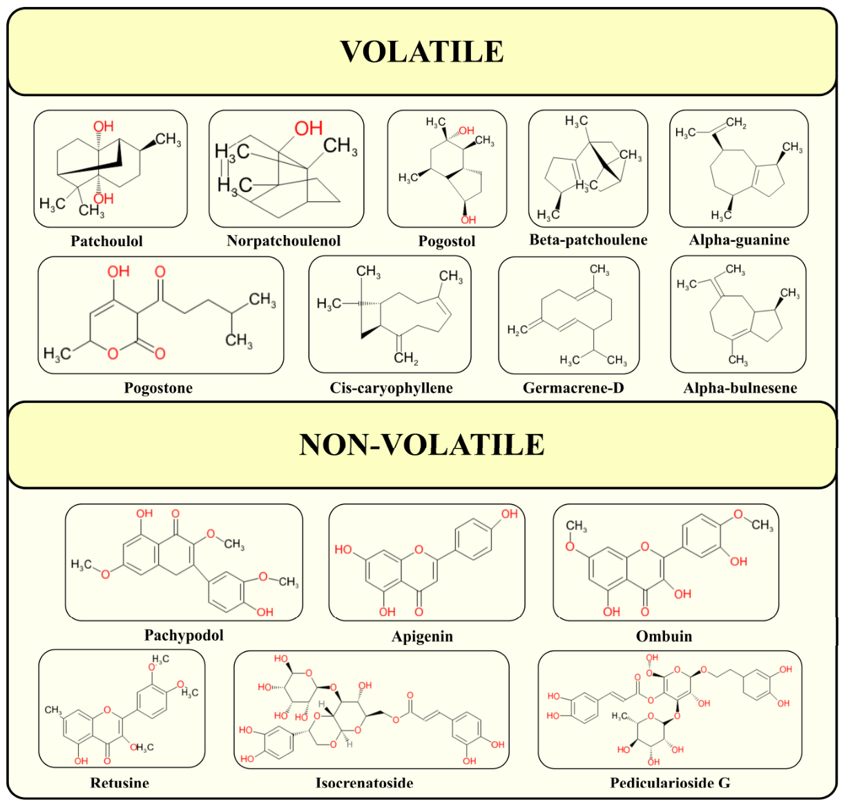
Figure 1. Chemical structures of representative volatile and non-volatile constituents in patchouli leaves. Volatile compounds are primarily sesquiterpenes and their oxygenated derivatives, whereas non-volatile compounds mainly comprise flavonoids and phenylethanoid glycosides.
Molecules 31 02870 g001
Figure 2. Overview of patchouli-derived bioactive compounds and their anticancer activities. This schematic illustration summarizes the source, extraction process, and anticancer potential of patchouli. The leaves of patchouli serve as the primary plant material for extraction, yielding bioactive constituents such as PEO and its major compounds, including patchoulol, pogostone, and pachypodol. These phytochemicals exhibit broad-spectrum anticancer activity against various malignancies, including colorectal, ovarian, liver, prostate, lung, gallbladder, and skin cancers, as well as acute myeloid leukemia, underscoring the therapeutic potential of patchouli-derived compounds as natural anticancer agents.
Molecules 31 02870 g002
Figure 3. Proposed molecular mechanisms underlying the anticancer activity of patchoulol. This schematic illustrates the primary signaling pathways that patchoulol modulates in cancer cells. Patchoulol induces cell cycle arrest at the G1 and G1/S phases by downregulating c-MYC, cyclin D1/CDK4, and CDK2/cyclin E, while simultaneously upregulating the cell cycle inhibitor p21, partially mediated through JNK-p53 signaling. At the same time, patchoulol promotes apoptosis by activating intrinsic mitochondrial pathways, as indicated by elevated BAX/BCL-2 ratios, mitochondrial cytochrome c release, and activation of CASP9 and CASP3. Additionally, patchoulol inhibits EGFR-MEK/ERK signaling, reducing BCL-2 expression, and further enhancing caspase-dependent apoptotic responses. Together, these coordinated effects on cell cycle regulation and apoptotic signaling pathways contribute to patchoulol’s broad-spectrum anticancer activity.
Molecules 31 02870 g003
Table 1. Representative volatile phytochemical constituents identified in P. cablin (patchouli) leaves.
Compound NameChemical ClassMolecular FormulaExtraction TechniqueAnalytical IdentificationReferences
(−)-camphorMonoterpenesC10H16OSteam distillationGC × GC–TOF-MS[46]
β-pineneC10H16 [46,47,48]
β-phellandreneC10H16 [46]
α-elemenoneSesquiterpenesC15H22O[46]
pogostolC15H26O[47]
(−)-α-selineneC15H24GC-MS[48]
α-bulneseneC15H21[46,47]
α-humuleneC15H24[47]
β-caryophylleneC15H24[46,47,48]
β-selineneC15H24[47,48]
cis-β-elemeneC15H24[47]
cycloseychelleneC15H24[47]
seychelleneC15H24[47,49,50]
trans-caryophylleneC15H24[51]
zizanalC15H22O[47]
δ-elemeneC15H24[47,52]
γ-gurjuneneC15H24[48,53]
α-, β-, δ-guaieneC15H24GC-MS, GC[47,54]
germacrene DC15H24[47,53]
limoneneC10H16[47]
patchouli alcoholC15H26O[47]
δ-patchouleneC15H24GC-MS, GC, NMR[47,54]
β-elemeneC15H24GC-MS, NMR[52]
α-, β-patchouleneC15H24GC-TOF-MS[47,55]
globulolC15H26OGC-TOF-MS, GC[56]
nortetrapatchoulolC14H24OGC-TOF-MS, GC-MS[47]
norpatchuolenolAlcoholsC14H22OGC-MS[47]
3-iso-thujopsanoneKetonesC15H24O[47]
pogostoneC12H16O4[46,47]
Table 2. Representative non-volatile phytochemical constituents identified in P. cablin (patchouli) leaves.
Compound NameChemical ClassMolecular FormulaExtraction TechniqueAnalytical IdentificationReferences
pachypodolFlavonoidsC16H12O6Ultrasonic-assisted 70% ethanol extractionGC[46,62]
3,5-dihydroxy-7,4′-dimethoxy-flavoneC17H14O6Reflux extraction with 70% aqueous ethanolHPLC-DAD[48]
4′,5,7-trihydroxyflavoneC15H10O5[48]
4′,5-dihydroxy-3,3′,7-trimethoxyflavoneC18H18O7[48]
5-hydroxy-3,3′,4′,7-tetramethoxyflavoneC18H18O6[48]
5-hydroxy-3,4′,7-trimethoxyflavoneC17H16O6[48]
5-hydroxy-7,3′,4′-trimethoxyflavanoneC17H16O6[48]
apigeninC15H10O5Ultrasonic-assisted 70% ethanol extraction[46,62]
rhamnetinC16H12O7[62]
isocrenatosideGlycosidesC29H34O15Maceration with 50% aqueous ethanolGC[46,54]
7R-campeoside IIC29H36O16HPLC[63]
7S-campeoside IIC29H36O16[63]
verbascosideC29H36O15[64]
2″,3″-O-acetylmartynosideC35H46O17HPLC-DAD[63]
3′-methoxyisocrenatosideC30H34O16[63]
actinosideC36H44O20[57]
osmanthuside BC29H36O13[63]
pedicularioside GC17H26O11[64]
crenatosidesC29H34O15TLC, HPLC[65]
acteosideC29H36O15TLC, HPLC, HPLC-Q-TOF-MS[65,66]
isoacteosidesC29H36015[65,67]
epifriedelinolTriterpenoidsC30H52OReflux extraction with petroleum ether and chloroformNMR, IR, MS, UV[46]
friedelinC30H50O[46]
methyl oleanolateC31H50O3[46]
oleanolic acidC30H48O3[46]
cytosporone VOther compoundsC13H16O5Ultrasonic-assisted 70% ethanol extractionHPLC[46]
cytosporone WC12H15O5[63]
isolariketoesterC33H39O10[63]
lariketoesterC33H39O10[63]
Table 3. Anticancer activities of patchouli-derived extracts and phytochemicals across different cancer models.
Cancer TypeCompoundExperimental ModelStudy TypeMolecular Target/
Signaling
Pathway
Dosage &
Duration
Reference
Acute myeloid leukemiaPCAEHL-60 cellsIn vitrop21, p-Rb, cyclins; extrinsic and intrinsic apoptosis pathways (caspase activation)0–200 µg/mL, 24–72 h[85]
PatchoulolMV4-11 cellsIn vitroPKM2, NF-κB, caspase-325–100 µM, 24–48 h[86]
Colorectal cancerPCAEHCT116 cellsIn vitro + in vivoG0/G1 cell-cycle arrest, apoptosis-related proteins; synergistic effect with 5-FU10–80 µg/mL (in vitro) and 30 mg/kg (in vivo)[72]
PatchoulolHT-29In vitroHDAC2, c-Myc, NF-κB, p21, cyclin D1, CDK450–100 µM, 24–48 h[87]
Caco-2 cellsIn vitro + in vivoG1-phase arrest, AMPK, Akt phosphorylation, glucose uptake25–100 µM, 24 h[88]
PachypodolCaco-2 cellsIn vitroCytotoxicity against CaCo-2 colon cancer cells (IC50 = 185.6 μM); no
specific molecular
target investigated
IC50 ≈ 185 µM, 48 h[89]
Endometrial cancerPCAEIshikawa cells In vitroCaspase-3, caspase-9, AIF-mediated apoptosis0–4 mg/mL, 24–48 h[90]
Gallbladder
cancer
PogostoneSGC-996 cellsIn vitroMitochondrial apoptosis (Bax/Bcl-2, caspase-9/caspase-3, PARP), cyclin D1, cyclin A, cyclin B1, S-phase arrest25–100 µg/mL, 24–48 h[91]
Liver cancerPEOHepG2 cellsIn vitro + in vivoROS-mediated DNA damage, p53, Fas/FasL/caspase-8, Bax/Bcl-2, Akt/mTOR, VEGF/VEGFR0–200 µg/mL (in vitro) and 25–50 mg/kg (in vivo)[92]
PogostoneHepG2 cellsIn vitroBax/Bcl-2, p53, caspase-310–100 µg/mL, 24–48 h[93]
Lung cancerPatchoulolA549 cells In vitro + in vivoEGFR/MAPK, mitochondrial apoptosis (caspase-9/caspase-3)50–100 µg/mL and 25 mg/kg[3]
A549 cells, and NSCLC cellsIn vitroROS–CHK1/CHK2, p53/p21, CDK2/cyclin E1, Bax/caspase-9/caspase-3, P-glycoprotein, CD44, CD133150–300 µM, 24 h[94]
Nasopharyngeal cancerApigeninNPC cell linesIn silico (molecular docking & molecular dynamics)BCL-2 inhibition (apigenin, rhamnetin, apigenin-7-(6″-p-coumarylglucoside))20–80 µM [95]
Ovarian cancerPogostoneOVCAR-3 cellsIn vitroPTEN, DACT1, Bax/Bcl-2, caspase-3, caspase-8, caspase-9, CCND1, CDK4IC50 ≈ 90 µg/mL, 24–48 h[96]
Prostate cancerPatchoulolDU145, and PC-3 cellsIn vitro + in vivo (xenograft)NF-κB/IκBα/p65, Mcl-1, Bax/Bcl-2, cleaved caspase-3, PARP, MMP-2, MMP-7, MMP-9, VEGF25–100 µg/mL and 30 mg/kg[97]
Skin cancerPatchoulolB16F10 cellsIn vitro + in vivoTGF-β/Smad2/3, E-cadherin, vimentin, MMP-2, MMP-9, G0/G1 cell-cycle arrest, apoptosis0–100 µM and 20 mg/kg[98]
Disclaimer/Publisher’s Note: The statements, opinions and data contained in all publications are solely those of the individual author(s) and contributor(s) and not of MDPI and/or the editor(s). MDPI and/or the editor(s) disclaim responsibility for any injury to people or property resulting from any ideas, methods, instructions or products referred to in the content.