Article

Whole-Cell Fungal Biotransformation of para-Hydroxycinnamic Acids Mediated by Phenolic Acid Decarboxylase, Carboxylic Acid Reductase and Alcohol Dehydrogenase


1
Department of Food Chemistry and Biocatalysis, Wrocław University of Environmental and Life Sciences, Norwida 25, 50-375 Wrocław, Poland
2
Istituto di Scienze e Tecnologie Chimiche “Giulio Natta”, Consiglio Nazionale delle Ricerche (SCITEC-CNR), Via Bassini 6, 20133 Milano, Italy
3
Plant Research Department, Nuclear Research Center, Egyptian Atomic Energy Authority, Cairo 11787, Egypt
4
Dipartimento di Chimica, Materiali ed Ingegneria Chimica “Giulio Natta”, Politecnico di Milano, Piazza Leonardo da Vinci 32, 20133 Milano, Italy
*
Authors to whom correspondence should be addressed.
Molecules 2026, 31(15), 2609; https://doi.org/10.3390/molecules31152609
Submission received: 18 June 2026 / Revised: 21 July 2026 / Accepted: 24 July 2026 / Published: 27 July 2026

Abstract

Microbial biotransformation of para-hydroxycinnamic acids (pHCAs) such as para-coumaric, caffeic, ferulic and sinapic acids into vinylphenols is catalyzed by phenolic acid decarboxylases (PADs), while reduction to their corresponding aldehydes and alcohols is mediated by carboxylic acid reductases (CARs) and alcohol dehydrogenases (ADHs), respectively. The present study systematically evaluated a diverse set of endophytic and basidiomycetes fungi as whole-cell biocatalysts for the transformation of pHCAs into their corresponding vinylphenols and/or aldehydes and alcohols. Twenty-three fungal strains were screened for their PAD, CAR and ADH activities. Based on ultra-high-performance liquid chromatography–diode array detector (UHPLC-DAD) analysis, fourteen strains were selected for preparative-scale biotransformations across all four substrates. Previous literature largely emphasizes enzyme activity or single substrates, seldom covering all four pHCAs. The strain Umbelopsis sp. JAR-T demonstrated promising biotransformation of para-coumaric acid and ferulic acid to 4-vinylphenol (28% isolated yield) and 4-vinylguaiacol (40% isolated yield), respectively, with minimal by-product formation. The results highlight endophytic fungi as largely untapped and versatile biocatalysts for pHCA biotransformation and establish whole-cell fungal systems as robust, non-recombinant alternatives to engineered platforms. This integrated screening-to-preparative workflow provides a scalable framework for the production of value-added compounds with potential applications in the food, cosmetic and pharmaceutical industries.

1. Introduction

para-Hydroxycinnamic acids (pHCAs) are a class of naturally abundant, simple phenolic acids possessing a single aromatic ring as their core with a C6-C3 phenylpropanoid structure [1]. Produced as specialized metabolites (traditionally termed secondary metabolites) in plants and fungi, pHCAs protect against UV, defend against pathogens and influence flavor, smell and color of plants [2]. The most abundant natural pHCAs are para-coumaric acid (4-hydroxycinnamic acid, pCA), caffeic acid (3,4-dihydroxycinnamic acid, CA), ferulic acid (4-hydroxy-3-methoxycinnamic acid, FA) and sinapic acid (3,5-dimethoxy-4-hydroxycinnamic acid, SA) [3]. These compounds are synthesized inside the plastids and cytosol of plants through the shikimate and phenylpropanoid pathways, where phenylalanine serves as the precursor for the synthesis of pCA, CA, FA and SA along with lignin [4,5]. Although these acids are present in nature, their alcohols, aldehydes and vinyl derivatives remain scarce and relatively understudied. This scarcity has directed interest in sustainable production strategies, especially biological routes capable of selective structural modification.
Microbial transformation employs whole cells of microorganisms or isolated enzymes to structurally modify phytochemicals and other organic compounds via biochemical reactions, facilitating the efficient production of valuable compounds. Compared to conventional chemical synthesis, biotransformation avoids harsh reaction environments and minimizes unwanted effects [6]. This environmentally friendly approach supports green chemistry for converting pHCAs into value-added derivatives for pharmaceutical and industrial use. Typically, enzymes present in microorganisms that aid in the pHCAs biotransformation include Phenolic Acid Decarboxylase (PAD), which mediates the decarboxylation of pHCAs to form their respective vinyl derivatives, and Carboxylic Acid Reductase (CAR) which catalyzes the ATP- and NAD(P)H-dependent reduction of pHCAs into their corresponding aldehydes and thereby to their alcohols, by means of Alcohol Dehydrogenase (ADH) catalysis. This microbial route bypasses the CoA activation and cinnamoyl-CoA reductase (CCR)-mediated reduction steps present in the canonical plant pathway, as fungal CARs directly reduce free hydroxycinnamic acids to aldehydes. These compounds are considered high-value products owing to their limited commercial availability and applications across broad range industries [7]. Figure 1 illustrates the biotransformation pathways of pHCAs.
Vinyl derivatives of pHCAs have diverse applications in addition to their food and pharmaceutical applications. 4-vinylphenol is used in semiconductors, photoresists and photolithography [8,9]; 4-vinylcatechol is utilized in the synthesis of polystyrenes, plastics and styrene-butadiene rubbers [9,10]; 4-vinylguaiacol is employed as a precursor in biocatalytic reactions to produce valuable food flavor compounds, such as ethylguaiacol, acetovanillone and vanillin, and its market value is approximately 30–40 times higher than FA, making it economically attractive [7]; and 4-vinylsyringol exhibits antioxidant and antimutagenic properties with potential applications in food processing, cosmetics and pharmaceuticals [11]. Beyond vinyl derivatives, hydroxycinnamyl alcohols are important intermediates in lignin biosynthesis, contributing to plant structure, water transport and pathogen defense; and they serve as valuable precursors for the sustainable production of polymers, resins, specialty chemicals and fragrance compounds, including those used in jasmine, gardenia and rose aromas. Their corresponding hydroxycinnamaldehydes are key intermediates in the synthesis of fine chemicals, flavors, fragrances and bioactive molecules, while also functioning as key metabolites in engineered microbial pathways [12,13]. Despite their high commercial value and broad applicability, the availability of these compounds remains limited because they are typically present in low concentrations in natural sources, while chemical synthesis often requires harsh reaction conditions and lacks the selectivity needed to produce natural-label products. Consequently, biotransformation of pHCAs into these derivatives offers a sustainable route to high-value chemicals for food, pharmaceutical, cosmetic, polymer and fine chemical applications, while increasing the economic value of renewable biomass-derived feedstocks.
Previous studies have reported biotransformation of one or more pHCAs through recombinant or enzyme-based strategies [9,14,15] as well as exploiting wild-type-based bacterial biotransformations [16]. Such investigations utilize bacteria engineered to express PAD, TAL (Tyrosine ammonia-lyase) and related decarboxylases to selectively produce high-titer single products. Few studies also involved yeasts and fungi, still focusing on enzyme activity in recombinant hosts with single-substrate transformation [17,18]. While effective for pathway optimization, these systems often require extensive genetic modification and external precursors, increasing process complexity. In contrast, whole-cell biocatalysts provide a self-sustaining catalytic environment in which enzymes remain in their native intracellular context, enabling cofactor regeneration and reducing the need for enzyme purification or supplementation [6]. Although isolated enzymes generally offer higher reaction specificity and simpler process control, whole-cell systems are particularly advantageous for multistep biotransformations involving sequential or competing enzymatic activities, making them attractive platforms for the conversion of pHCAs into structurally diverse derivatives. Fungi are known to inherently possess complex and compartmentalized metabolic networks, effective in catalyzing reduction, decarboxylation and oxidation reactions within a single cell. The handful of reports investigating ascomycetes [6,18,19] and basidiomycetes [20,21,22] have also focused on enzyme activity or single-substrate transformation, rather than exploring all four pHCA substrates or pathways leading to vinyl derivatives, aldehydes and alcohols. Compared with commonly used commercial fungal biocatalysts, endophytic fungi represent a promising resource. These fungi reside asymptomatically within plant tissues [23], where they are exposed to phenylpropanoid metabolites, suggesting a natural adaptation for metabolizing pHCAs and related aromatic compounds. In spite of this ecological relevance, they are largely unexplored for whole-cell biotransformation of pHCAs. One study explores the potential of endophyte Phomopsis liquidambari in the remediation of sinapic acid by converting it into 4-vinylsyringol [24]. While pCA, CA and FA are relatively studied, the gap in research is particularly evident with SA owing to its structural complexity. Beyond the study involving P. liquidambari, recent work has demonstrated sinapic acid conversion to 4-vinylsyringol using recombinant systems based on Neolentinus lepideus [22]. These isolated studies highlight the lack of systematic investigation into sinapic acid metabolism, particularly within native, whole-cell fungal systems.
The present study aimed to investigate a diverse set of endophytic and basidiomycete fungi as whole-cell biocatalysts for the transformation of pCA, CA, FA and SA. This study provides a comprehensive assessment of fungal PAD, CAR and ADH activities across multiple substrates by integrating screening-scale studies with preparative-scale biotransformation, ultra-high-performance liquid chromatography–diode array detector (UHPLC-DAD) analysis, flash purification and NMR-based structural confirmation. This study establishes a robust framework to develop sustainable, non-recombinant bioprocesses for the valorization of pHCAs.

2. Results

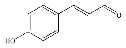
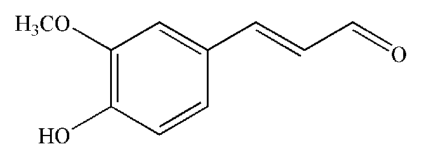
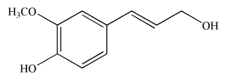
Biotransformation of para-hydroxycinnamic acids (pHCAs) using whole cells of microorganisms is a significant area of research beneficial in bioremediation, valorization of lignin and sustainable production of value-added phenolic compounds. In particular, fungi possess numerous enzymatic systems that aid in decarboxylation, reduction and other transformation reactions. Within this cluster, phenolic acid decarboxylase (PAD), carboxylic acid reductase (CAR) and alcohol dehydrogenase (ADH) activities gain particular emphasis, as they enable the conversion of pHCAs into vinylphenols, hydroxycinnamaldehydes and corresponding alcohols of industrial and biological relevance. The structures of the substrates and expected products through PAD, CAR and ADH pathways are depicted in Table 1.

2.1. Biotransformation at Screening Scale

A total of sixteen endophytic fungi and seven basidiomycetes were evaluated as whole-cell biocatalysts for the biotransformation of four pHCAs: p-coumaric acid (pCA), caffeic acid (CA), ferulic acid (FA) and sinapic acid (SA). These preliminary screening experiments were performed in Erlenmeyer flasks containing Sabouraud broth media, with each substrate added at a concentration of 50 mg per 100 mL of medium. Substrate conversion and product formation were monitored at regular time intervals initially using Gas Chromatography with Flame Ionization Detection (GC-FID) and Gas Chromatography–Mass Spectrometry (GC-MS). However, as the substrates underwent thermal degradation and did not yield reliable quantitative results, the analysis was performed using Ultra-High-Performance Liquid Chromatography–Diode Array Detector (UHPLC-DAD). As the same degradation pattern was detected for pure standards in the absence of fungal biomass, the phenomenon was attributed to thermal instability of the hydroxycinnamic acid substrates during GC analysis rather than to biological degradation. The fungal strains revealed distinctive, substrate-dependent biotransformation profiles. Based on dominant enzymatic routes, strains were classified according to PAD, CAR and ADH activity, or alternative oxidative and specialized metabolite pathways. Fourteen strains, comprising eleven endophytes and three basidiomycetes, exhibited consistent PAD and/or CAR and/or ADH activity towards at least one substrate and were selected for further scale-up to preparative-scale investigation (Table 2).
In the biotransformation of p-coumaric acid (1a), six strains (Trichoderma harzianum BUK-T, Coniochaeta velutina SW-B, Umbelopsis sp. JAR-T, Diaporthe sp. MOD-B, Diaporthe eres POP-L1 and Penicillium chrysogenum SOS-B2) formed 4-vinylphenol (1b), while five strains (Diaporthe sp. MOD-B, D. eres POP-L1, Trametes hirsuta d28, Phanerochaete chrysosporium KKP 784 and Laetiporus sulphureus AM 515) exhibited CAR and ADH activity. Two fungal strains, Diaporthe sp. MOD-B and D. eres POP-L1, uniquely displayed PAD, CAR and ADH activities, indicating metabolic branching between decarboxylation and reduction pathways. Biotransformation with caffeic acid (2a) exhibited higher selectivity with only three strains (T. harzianum BUK-T, Aspergillus westerdijkiae 17P and Umbelopsis sp. JAR-T) producing 4-vinylcatechol (2b) and five strains (Diaporthe sp. MOD-B, D. eres POP-L1, T. hirsuta d28, P. chrysosporium KKP 784 and L. sulphureus AM 515) showing CAR- and ADH-mediated product formation. In addition, Chaetomium cochliodes KLON-L1, based on GC-MS analysis, produced 4-vinyl-1-cyclohexene diepoxide. Biotransformation of ferulic acid (3a) yielded substantial metabolic diversity. Six strains (T. harzianum BUK-T, A. westerdijkiae 17P, Umbelopsis sp. JAR-T, Diaporthe sp. MOD-B, D. eres POP-L1 and P. chrysogenum SOS-B2) exhibited PAD activity, while seven strains (T. harzianum BUK-T, A. westerdijkiae 17P, D. eres POP-L1, Umbelopsis sp. 42, T. hirsuta d28, L. sulphureus AM 515 and P. chrysosporium KKP 784) demonstrated CAR and ADH activity. Several isolates, including T. harzianum BUK-T, A. westerdijkiae 17P and D. eres POP-L1, showed concurrent PAD, CAR and ADH activities, resulting in mixed product profiles. Additional oxidative metabolites, such as vanillic and isovanillic acids determined by GC-MS, were also detected in Aspergillus ochraceus ROB-L1 and Penicillium yarmokense JAR-L1 indicating the involvement of auxiliary metabolic pathways. Among all the pHCAs, biotransformation of sinapic acid (4a) was the most selective. Only two strains (T. harzianum BUK-T and P. yarmokense JAR-L1) showed PAD activity, while six strains (T. harzianum BUK-T, Umbelopsis sp. JAR-T, Diaporthe sp. MOD-B, D. eres POP-L1, L. sulphureus AM 515 and P. chrysosporium KKP 784) demonstrated CAR- and ADH-mediated reduction. T. harzianum BUK-T was the only strain to consistently exhibit PAD, CAR and ADH activity towards this substrate. For T. harzianum BUK-T and P. yarmokense JAR-L1, both strains were observed to produce additional metabolites such as syringic acid and acetosyringone beyond PAD, CAR and ADH pathways.
Overall, T. harzianum BUK-T exhibited the broadest substrate range and highest metabolic versatility in the screening study. Diaporthe sp. MOD-B and D. eres POP-L1 consistently demonstrated CAR and ADH activity and, in some cases, concurrent PAD activity. Basidiomycete strains, namely, T. hirsuta d28, P. chrysosporium KKP 784 and L. sulphureus AM 515, were predominantly associated with CAR- and ADH-mediated reduction. These findings provide a strong foundation for selecting fungal candidates for further preparative-scale biotransformation.

2.2. Biotransformation at Preparative Scale

Following preliminary screening, fourteen selected strains were evaluated under preparative-scale conditions to assess the scalability, efficiency and robustness of fungal whole-cell transformations of each pHCAs. Product formation and substrate depletion were quantified using UHPLC-DAD time-course analysis, and the conversion percentages for each substrate are summarized in Table 3, Table 4, Table 5 and Table 6.
Out of nine strains selected in the screening scale for biotransformation of p-coumaric acid (1a), three strains, namely, Umbelopsis sp. JAR-T, Diaporthe sp. MOD-B and P. chrysogenum SOS-B2, consistently produced 1b. Umbelopsis sp. JAR-T achieved almost 88% substrate conversion within two days and maintained >98% 1b abundance on the seventh day, indicating highly stable and dominant PAD activity. Although ADH-mediated reduction was observed in all three strains, it was subordinate to their PAD activity in the strains Umbelopsis sp. JAR-T and P. chrysogenum SOS-B2. Product formation of more than 50% of 1d was demonstrated by Diaporthe sp. MOD-B, indicating that this strain can efficiently utilize both PAD and ADH pathways.
In the case of caffeic acid (2a), nine strains were selected in the screening study. On the preparative scale, only C. cochliodes KLON-L1 produced a measurable amount of 2b (31%), and even with this strain, decarboxylation competed with the formation of 2d and other metabolites, resulting in a mixture of reaction products. Formation of product 2d was limited to transformations catalyzed by C. cochliodes KLON-L1 (11%) and Diaporthe sp. MOD-B (71%).
For ferulic acid (3a), PAD activity was more widely observed when compared with other substrates. With twelve strains shortlisted in screening, four strains, notably, A. westerdijkiae 17P (11%), Umbelopsis sp. JAR-T (100%), Diaporthe sp. MOD-B (22%) and P. chrysogenum SOS-B2 (32%), efficiently converted 3a into 3b in the preparative study. Among these, Umbelopsis sp. JAR-T demonstrated rapid, quantitative conversion with minimal side-product formation. Substrate 3a also supported strong ADH and CAR activity. A. westerdijkiae 17P (75% of 3d and 8% of 3c) and P. chrysogenum SOS-B2 (46% of 3d) produced substantial quantities of 3d, often exceeding 3b formation. This further supports 3a as a highly compliant substrate for decarboxylative, reductive and dehydrogenative fungal biotransformations.
Regarding sinapic acid (4a), of the seven strains selected in the screening study, only T. harzianum BUK-T mediated the formation of 4d (69%) as the dominant product of ADH activity.
The screening study identified a broad range of fungi capable of PAD-mediated decarboxylation, CAR-mediated reduction and ADH-mediated dehydrogenation of pHCAs. However, only a subset of these strains retained the stable and effective PAD, CAR and ADH pathways under preparative conditions. Under screening conditions, T. harzianum BUK-T and D. eres POP-L1 displayed high potential in transforming all four substrates under PAD and/or CAR/ADH pathways, yet, the same strains could not sustain scale-up, resulting in T. harzianum BUK-T demonstrating ADH activity only with 4a. All three basidiomycete strains, T. hirsuta d28, P. chrysosporium KKP784 and L. sulphureus AM 515, despite exhibiting promising CAR and ADH activities under screening scale, unfavorably showed no measurable results. While C. cochliodes KLON-L1 was chosen at the screening scale for its metabolite production ability, it demonstrated a significant level of PAD activity with 2a in the preparative scale. With 3a, although Umbelopsis sp. JAR-T and Umbelopsis sp. 42 belong to the same genus, their catalytic abilities via the PAD, CAR and ADH systems differ markedly. Umbelopsis sp. JAR-T exhibited PAD activity, converting 100% of 3a to 3b at the preparative scale, whereas Umbelopsis sp. 42 showed CAR and ADH activities only at the screening scale. In addition, Umbelopsis sp. JAR-T also demonstrated prominent PAD activity with 1a resulting in the formation of almost 100% of 1b. The comparison highlights the necessity of moving beyond analytical screening to identify fungal biocatalysts capable of efficient, scalable conversion of pHCAs. Compound 3a emerged as the most reliable substrate across both screening and preparative studies, while biotransformations with 2a and 4a remained challenging.
The reproducibility of the biotransformation was evaluated using Umbelopsis sp. JAR-T. The conversion of 1a to 1b and 3a to 3b reached 90.47 ± 4.81% and 93.54 ± 1.95%, respectively (mean ± SD, n = 3), after 7 and 2 days of incubation. Supplementary Figures S5 and S6 show the corresponding UHPLC-DAD chromatograms. The relatively low standard deviations observed for both transformations indicate good reproducibility among independent experiments and suggest consistent decarboxylation performance of the fungal whole-cell catalyst.
Quantitative assessment of recoverable products was achieved through purification of preparative-scale biotransformation extracts. With UHPLC-DAD guiding the product detection and flash chromatography determining final isolated yields, NMR analysis confirmed the product structures. In biotransformation of 1a, Umbelopsis sp. JAR-T showed near-complete PAD-driven conversion to 1b (Supplementary Figure S7) with an isolated yield of 28% after purification, identifying this strain as a highly efficient and selective PAD biocatalyst. In contrast, Diaporthe sp. MOD-B showed distributed metabolic flux between PAD and ADH pathways. Moreover, NMR analysis of the PAD-derived fraction also revealed the presence of ethylphenol. For biotransformation of 3a, Umbelopsis sp. JAR-T yielded a highly selective recovery of 3b (Supplementary Figure S8) with a 40% isolated yield, highlighting strong PAD activity and suitability for preparative-scale production. With A. westerdijkiae 17P and P. chrysogenum SOS-B2, coniferyl aldehyde-type structures were confirmed by NMR analysis, suggesting CAR as the predominant pathway.

3. Discussion

Most of the available literature studying pHCA biotransformation into its corresponding vinyl derivatives, alcohols and aldehydes has relied on isolated enzymes, recombinant microorganisms or wild-type bacterial strains. In particular, expression systems involving tyrosine ammonia-lyase (TAL), phenolic acid decarboxylase (PAD), carboxylic acid reductase (CAR) and related enzymes have been reported in bacterial host species like Escherichia coli [10,25,26], Bacillus [27,28,29,30,31] and Streptomyces [32,33]. Despite being effective in attaining high yield, these approaches predominantly require synthetic growth media and/or externally supplemented cofactors in addition to optimized expression systems and genetic modifications. Only a few studies [16,27,34] have directly employed bacteria in the biotransformation of one or more pHCAs into their respective vinyl derivatives. In line with these studies, the present work utilizes wild-type (non-GMO) fungal strains as biocatalysts.
Regardless of the comprehensive literature dealing with the PAD, CAR and ADH activities of bacteria, only a few studies have explored yeast [17,34,35,36] and fungal enzymes [19,21,22] as biocatalysts for the targeted biotransformation of pHCAs. Prevailing reports on fungal biotransformation consistently focus on recombinant enzymatic activity, typically employing purified enzymes instead of considering fungi as scalable, whole-cell production platforms. Furthermore, the existing studies have targeted ligninolytic and white-rot basidiomycetes, such as Neolentinus lepideus [22], Trametes versicolor [20], Schizophyllum commune [21], Phanerochaete chrysosporium [37] and Trametes hirsuta [37], as well as ascomycetes such as Isaria farinosa [18] and Aspergillus luchuensis [19] with investigations generally emphasizing individual substrates, specific enzymatic activities, or degradation pathways rather than systematic comparison of all four pHCAs and their corresponding vinyl, aldehyde and alcohol derivatives. Two research works show that the endophytic fungus Phomopsis liquidambari can metabolize both sinapic [24] and ferulic [38] acids through distinct pathways.
While 1a and 3a have been extensively investigated in microbial systems [17,18,19,25,34,39], with few systems exploring 2a [10,20,34,35,40], the research gap is remarkably noticeable in biotransformations involving 4a. Among the documented research, one study reports the conversion of 4a to yield low concentrations of 4b by employing the endophytic fungus, Phomopsis liquidambari [24]. Another study conducted recently described the biotransformation of 4a to 4b using recombinant Neolentinus lepideus [22]. These scarce available reports emphasize the lack of systematic investigation of 4a metabolism, specifically employing whole-cell fungi.
Addressing the gap in prior research, the present study establishes a strategy to utilize whole fungal cells as biocatalysts for the conversion of pHCAs into their corresponding vinyl derivatives, aldehydes and alcohols without genetic engineering. The observed product profiles are consistent with sequential decarboxylation (PAD) and reduction (CAR and ADH) reactions, although the specific enzymes responsible were not investigated in the present study. Whole-cell systems promote the constant intracellular regeneration of cofactors required for enzyme activity. Moreover, cellular compartmentalization in fungi enables the simultaneous functioning of multiple enzymatic pathways, thereby facilitating parallel decarboxylation and reduction processes within a single organism. Recombinant bacterial platforms often lack this metabolic integration because they are generally optimized for the production of a single dominant product [39,41]. The present work extends this narrow knowledge base by meticulously screening and scaling a diverse set of fungal strains belonging to endophytes and basidiomycetes, illustrating that several strains can achieve product selectivity and yields relative to those reported for the engineered bacterial systems.
The focus on endophytic fungi as whole-cell biocatalysts is another novel aspect of this study. Apart from the solitary report by Xie et al. [24], few studies have investigated endophytes for the production of vinyl derivatives or hydroxycinnamic alcohols. Since endophytes inhabit plant tissues, natural exposure to phenylpropanoid-derived metabolites is inevitable suggesting an intrinsic evolutionary adaptation for processing hydroxycinnamic acids and related compounds. This ecological hypothesis is supported by the strong PAD, CAR and ADH activities observed in the present study across diverse endophytic fungal species, establishing them as a largely untapped resource as biocatalysts.
Correspondingly, the results from UHPLC-DAD analysis and flash purification facilitated the quantification of biotransformation within whole-cell fungal systems. To verify the consistency of biotransformation performance, the selected high-yield strain Umbelopsis sp. JAR-T was grown in triplicate for the biotransformation of 1a and 3a. Comparable substrate conversion rates and product yields were observed across all replicates, indicating stable biotransformation capability. The attained yields did not differ substantially from those reported in recombinant bacterial systems [27,30,31], at the same time avoiding the operational and regulatory complexity associated with genetically modified organisms. Additionally, the wider metabolic profiles of endophytic strains A. westerdijkiae 17P and Diaporthe sp. MOD-B indicate the fungal capability in generating diverse products rather than targeting single-product reactions. Additionally, endophyte T. harzianum BUK-T was capable of converting 4a into 4d through the ADH pathway, albeit with limited efficiency.

4. Materials and Methods

The substrates for the biotransformations—para-coumaric acid (≥98%) and caffeic acid (≥98.5%)—were purchased from Glentham Life Sciences (Corsham, UK), ferulic acid (≥99%) was purchased from Sigma Aldrich (St. Louis, MO, USA) and sinapic acid (98.8%) was purchased from Ambeed (Arlington Heights, IL, USA). The pattern compounds of the expected products of PAD activity, namely, 4-vinylphenol (10wt% in propylene glycol), was purchased from Merck (Darmstadt, Germany) and 4-vinylsyringol (98.5%) was purchased from MedChemExpress (Monmouth Junction, NJ, USA). The compounds 4-vinylcatechol and 4-vinylguaiacol were obtained via chemical synthesis [42] and purified using bulb-to-bulb distillation. The pattern compounds of the expected products of CAR and ADH activity, namely, para-coumaryl alcohol (98.4%), caffeoyl alcohol (99.9%), sinapyl alcohol (98.7%) and sinapyl aldehyde (99.9%), were purchased from MedChemExpress. Coniferyl alcohol (98%) and coniferyl aldehyde (98%) were purchased from Merck. Analytical-grade solvents such as methanol, ethyl acetate and DMSO were obtained from Sigma Aldrich, while salts such as anhydrous sodium sulfate was purchased from Chempur (Piekary Śląskie, Poland).

4.1. Microorganisms and Culture Maintenance

A total of twenty-three fungal strains, comprising sixteen endophytes and seven basidiomycetes, were used in this study. All fungal strains were maintained on Sabouraud Dextrose (peptone: 10 g/L, glucose: 30 g/L and agar: 20 g/L) agar (SDA) plates and stored at 4 °C.
Endophytic strains used in the biotransformation include the following: Aspergillus ochraceus ROB-L1, Aspergillus westerdijkiae 17P, Aureobasidium pullulans POP-L2, Chaetomium cochliodes KLON-L1, Coniochaeta velutina SW-B, Diaporthe eres POP-L1, Diaporthe sp. MOD-B, Epicoccum nigrum COR-B, Monascus ruber M22, Penicillium chrysogenum SOS-B2, Penicillium yarmokense JAR-L1, Sphaeropsis sapinea BUK-L2, Trichoderma harzianum BUK-T, Umbelopsis isabellina COR-L1, Umbelopsis sp. 42 and Umbelopsis sp. JAR-T. These strains were previously isolated from Mokrzański forest plants in Wrocław, Poland [43] and procured from the fungal repository maintained by the Department of Food Chemistry and Biocatalysis, Wrocław University of Environmental and Life Sciences, under the voucher BioExplor22.
Basidiomycete strains including Inonotus radiatus AM 70, Laetiporus sulphureus AM 515, Phanerochaete chrysosporium KKP 784, Pholiota aurivella AM 522 and Trametes hirsuta d28 were sourced from the fungal culture collection of the Department of Food Chemistry and Biocatalysis at the Wrocław University of Environmental and Life Sciences. Agrocybe aegerita DSM 22459 and Pycnoporus cinnabarinus DSM 15225 were obtained from Istituto di Scienze e Tecnologie Chimiche -Consiglio Nazionale delle Ricerche (SCITEC-CNR) (Milano, Italy).

4.2. Growth Medium and Inoculum Preparation

Sabouraud Dextrose broth was used as the basal medium for both screening and preparative-scale biotransformation experiments. Pre-cultures were generated by transferring two actively growing mycelial plugs (5 mm) into 100 mL of broth in 250 mL Erlenmeyer flasks, followed by incubation at 28 °C with orbital shaking at 120 rpm for 5–7 days.

4.3. Screening-Scale Biotransformation

Screening-scale biotransformations were carried out in 250 mL Erlenmeyer flasks containing 100 mL sterile Sabouraud Dextrose broth. Flasks were inoculated with 2 plugs of freshly grown fungi from agar plates [44] and incubated for 5–7 days at 28 °C in a shaking incubator. After incubation, 50 mg of substrates (either pCA, CA, FA or SA) dissolved in 1 mL of DMSO [6] was added aseptically to the culture medium and the samples were extracted at different time points between 1 and 7 days using ethyl acetate to determine the substrate depletion and product formation by TLC (thin layer chromatography), GC-FID (gas chromatography—flame ionization detection), GC-MS (gas chromatography—mass spectrometry) and UHPLC-DAD.

4.4. Preparative-Scale Biotransformation

Based on the screening study, fourteen fungi exhibiting consistent PAD and/or CAR and/or ADH activity were selected for preparative-scale biotransformation which was carried out in 2000 mL Erlenmeyer flasks containing 400 mL Sabouraud Dextrose broth with 25% (v/v) pre-culture. After incubation for 5–7 days at 28 °C in a rotary shaker, 200 mg of each pHCA dissolved in 4 mL of DMSO was added to each fungal culture. Biotransformation progress was monitored by collecting samples at different time points within 1–7 days, extracted using ethyl acetate, dried over anhydrous Na2SO4, evaporated using a vacuum rotary evaporator (Heidolph, Schwabach, Germany), diluted and dissolved in methanol for UHPLC-DAD analysis.
Based on the progress, the entire reaction mixture was extracted thrice using ethyl acetate, and the organic phase was dried over anhydrous Na2SO4 and concentrated using a vacuum rotary evaporator to obtain crude products. The products were then purified using flash chromatography and the structures of the individual compounds were determined using NMR (Nuclear Magnetic Resonance) analysis.
The screening- and preparative-scale experiments were performed once for all fungal strains. To assess reproducibility, the highest-performing strain was subsequently evaluated in three independent biological replicates. Conversion values are reported as mean ± SD (n = 3).

4.5. Analytical Methods

4.5.1. Thin-Layer Chromatography (TLC)

The daily collected extracts were subjected to TLC fractionation (60 F254, Merck, Darmstadt, Germany) using hexane/ethyl acetate (1:1, v/v) as the eluent. The developed TLC plates were air-dried and examined using iodine vapor.

4.5.2. Gas Chromatography–Flame Ionization Detection (GC-FID)

Analyses were conducted using an Agilent 7890A GC-FID (Agilent Technologies, Santa Clara, CA, USA) platform equipped with a non-polar DB-5MS column (30 m × 0.32 mm × 0.25 µm). The analytical method employed an injection port temperature of 250 °C and a programmed oven temperature: an initial 60 °C isotherm, a ramp to 325 °C at 10 °C min−1, and a 1 min terminal hold. Constant-flow H2 was utilized as the carrier gas. Relative distributions of products and remaining substrates were determined through the integration and subsequent normalization of the resulting chromatograms.

4.5.3. Gas Chromatography–Mass Spectrometry (GC-MS)

Metabolite identification was performed using a GC-MS system, a Shimadzu QP 2020 Instrument (Shimadzu, Kyoto, Japan) equipped with column SH-Rxi-5SilMS (30 m × 0.25 mm × 0.25 µm). An amount of 1 μL of the sample was injected with splitless or split 10 modes, depending on sample concentration, at 260 °C. Helium was used as the carrier gas with a linear velocity of 36 cm·s−1. The GC initial temperature was 60 °C, held for 1 min, then ramped up to 325 °C at a rate of 10 °C ·min−1. The MS was operated in scan mode 40–600 m/z, with ion source and interface temperature 250 °C.
Compounds were tentatively identified by comparing mass spectra and retention indices with entries in the NIST mass spectral library and confirmed using authentic standards when available.

4.5.4. Ultra-High-Performance Liquid Chromatography (UHPLC)

Ultra-high-performance liquid chromatography (UHPLC) was executed using an Ultimate 3000 UHPLC+ Focused system (Thermo Scientific, Waltham, MA, USA). Chromatographic separation was achieved on an Acclaim RSLC Polar Advantage II C18 column (2.1 mm × 100 mm, 2.2 µm, Thermo Scientific, Waltham, MA, USA), maintained at a constant temperature of 28 °C. The mobile phase consisted of 0.1% formic acid (HCOOH) in water (Solvent A) and 0.1% HCOOH in acetonitrile (Solvent B). A flow rate of 0.7 mL min−1 was employed with the following gradient profile: 0 to 5.0 min, linear gradient from 98% to 2% A; 5.0 to 6.0 min, isocratic hold at 2% A; 6.0 to 6.2 min, return to 98% A; and 6.2 to 8.0 min, re-equilibration at 98% A. Detection was performed from λ = 210 to λ = 450 nm with a data acquisition frequency of 25 Hz.
Retention times of the compounds are tR para-coumaric acid: 3.65 min, tR caffeic acid: 3.26 min, tR ferulic acid: 3.56 min, tR sinapic acid: 3.47 min, tR 4-vinylphenol: 4.37 min, tR 4-vinylcatechol: 3.84 min, tR 4-vinylguaiacol: 4.19 min, tR 4-vinylsyringol: 4.02 min, tR p-coumaryl alcohol: 3.25 min, tR caffeoyl alcohol: 2.87 min, tR coniferyl alcohol: 3.22 min, tR coniferyl aldehyde: 3.63 min, tR sinapyl alcohol: 3.16 min and tR sinapyl aldehyde: 3.54 min. Representative UHPLC-DAD chromatograms are shown in Supplementary Figures S1–S4.

4.5.5. Flash Chromatography

Crude extracts were fractionated via automated flash chromatography utilizing a puriFlash® SX520 Plus system (Interchim, Montluçon, France). Separation was achieved on a silica gel stationary phase employing a stepwise gradient of hexane/ethyl acetate (from 9:1 to 1:1, v/v) as the mobile phase. Fractions were monitored by TLC and UHPLC-DAD, pooled based on chemical similarity, and concentrated to dryness. The isolated yields were recorded.

4.5.6. Nuclear Magnetic Resonance (NMR)

Structural confirmation of compounds was performed using 1H and 13C NMR spectroscopy (Bruker Advance DRX 600 (600 MHz) spectrometer (Billerica, MA, USA)). Spectra were recorded in CD3OD (Methanol-d4). Chemical shifts were reported in parts per million (ppm) relative to the residual solvent signal. The NMR spectra of all analyzed compounds are given as follows:
p-coumaric acid (1a) 1H NMR (600 MHz, CD3OD) δ 7.56 (d, J = 15.9 Hz, 1H, H-7), 7.44–7.38 (m, 2H, H-2, H-6), 6.80–6.74 (m, 2H, H-3, H-5), 6.24 (d, J = 15.9 Hz, 1H, H-8). 13C NMR (151 MHz, CD3OD) δ 171.1 (C-9), 161.1 (C-4), 146.6 (C-7), 131.1 (C-2, C-6), 127.3 (C-1), 116.8 (C-3, C-5), 115.6 (C-8).
caffeic acid (2a) 1H NMR (600 MHz, CD3OD) δ 7.49 (d, J = 15.9 Hz, 1H, H-7), 7.00 (d, J = 2.1 Hz, 1H, H-2), 6.90 (dd, J = 8.2, 2.0 Hz, 1H, H-6), 6.74 (d, J = 8.2 Hz, 1H, H-5), 6.18 (d, J = 15.9 Hz, 1H, H-8). 13C NMR (151 MHz, CD3OD) δ 171.1 (C-9), 149.5 (C-4), 147.0 (C-7), 146.8 (C-3), 127.8 (C-1), 122.8 (C-6), 116.5 (C-5), 115.6 (C-8), 115.1 (C-2).
ferulic acid (3a) 1H NMR (600 MHz, CD3OD) δ 7.56 (d, J = 15.9 Hz, 1H, H-7), 7.14 (d, J = 2.0 Hz, 1H, H-2), 7.02 (dd, J = 8.2, 1.9 Hz, 1H, H-6), 6.77 (d, J = 8.2 Hz, 1H, H-5), 6.27 (d, J = 15.9 Hz, 1H, H-8), 3.85 (s, 3H, OCH3). 13C NMR (151 MHz, CD3OD) δ 171.0 (C-9), 150.5 (C-4), 149.4 (C-3), 146.9 (C-7), 127.8 (C-1), 124.0 (C-6), 116.5 (C-5), 115.9 (C-8), 111.7 (C-2), 56.4 (OCH3).
sinapic acid (4a) 1H NMR (600 MHz, CD3OD) δ 7.55 (d, J = 15.9 Hz, 1H, H-7), 6.86 (s, 2H, H-2, H-6), 6.30 (d, J = 15.8 Hz, 1H, H-8), 3.84 (s, 6H, 2×OCH3). 13C NMR (151 MHz, CD3OD) δ 170.9 (C-9), 149.5 (C-3, C-5), 147.1 (C-7), 139.5 (C-4), 126.7 (C-1), 116.4 (C-8), 106.9 (C-2, C-6), 56.8 (2×OCH3).
4-vinylphenol (1b) 1H NMR (600 MHz, CD3OD) δ 7.24–7.18 (m, 2H, H-2, H-6), 6.71–6.67 (m, 2H, H-3, H-5), 6.58 (dd, J = 17.6, 10.9 Hz, 1H, H-7), 5.51 (dd, J = 17.6, 1.1 Hz, 1H, H-8trans), 4.98 (dd, J = 10.9, 1.1 Hz, 1H, H-8cis). 13C NMR (151 MHz, CD3OD) δ 158.5 (C-4), 137.9 (C-7), 128.6 (C-1), 128.4 (C-2, C-6), 116.2 (C-3, C-5), 110.8 (C-8).
4-vinylcatechol (2b) 1H NMR (600 MHz, CD3OD) δ 6.87 (d, J = 1.8 Hz, 1H, H-2), 6.72–6.66 (m, 2H, H-5, H-6), 6.52 (dd, J = 17.6, 10.9 Hz, 1H, H-7), 5.48 (dd, J = 17.6, 1.1 Hz, 1H, H-8trans), 4.97 (dd, J = 10.9, 1.0 Hz, 1H, H-8cis). 13C NMR (151 MHz, CD3OD) δ 146.6 (C-4), 146.4 (C-3), 138.0 (C-7), 131.2 (C-1), 119.6 (C-6), 116.2 (C-5), 113.6 (C-2), 110.8 (C-8).
4-vinylguaiacol (3b) 1H NMR (600 MHz, CD3OD) δ 6.97 (d, J = 1.9 Hz, 1H, H-2), 6.81 (dd, J = 8.2, 1.9 Hz, 1H, H-6), 6.70 (d, J = 8.1 Hz, 1H, H-5), 6.58 (dd, J = 17.6, 10.9 Hz, 1H, H-7), 5.54 (d, J = 18.6 Hz, 1H, H-8trans), 5.01 (d, J = 11.9 Hz, 1H, H-8cis), 3.82 (s, 3H, OCH3). 13C NMR (151 MHz, CD3OD) δ 149.0 (C-3), 147.7 (C-4), 138.1 (C-7), 131.3 (C-1), 120.8 (C-6), 116.1 (C-5), 111.1 (C-8), 110.2 (C-2), 56.3 (OCH3).
4-vinylsyringol (4b) 1H NMR (600 MHz, CD3OD) δ 6.67 (s, 2H, H-2, H-6), 6.58 (dd, J = 17.6, 10.9 Hz, 1H, H-7), 5.57 (dd, J = 17.6, 0.9 Hz, 1H, H-8trans), 5.04 (dd, J = 10.9, 0.8 Hz, 1H, H-8cis), 3.81 (s, 6H, 2×OCH3). 13C NMR (151 MHz, CD3OD) δ 149.3 (C-3, C-5), 138.3 (C-7), 136.8 (C-4), 130.3 (C-1), 111.5 (C-8), 104.7 (C-2, C-6), 56.7 (2×OCH3).
p-coumaryl alcohol (1d) 1H NMR (600 MHz, CD3OD) δ 7.20 (d, J = 8.6 Hz, 2H, H-2, H-6), 6.68 (d, J = 8.6 Hz, 2H, H-3, H-5), 6.46 (d, J = 15.8 Hz, 1H, H-7), 6.13 (dt, J = 15.8, 6.0 Hz, 1H, H-8), 4.14 (dd, J = 6.0, 1.4 Hz, 2H, H-9). 13C NMR (151 MHz, CD3OD) δ 158.3 (C-4), 131.9 (C-7), 130.0 (C-1), 128.7 (C-2, C-6), 126.7 (C-8), 116.3 (C-3, C-5), 63.9 (C-9).
caffeoyl alcohol (2d) 1H NMR (600 MHz, CD3OD) δ 6.83 (d, J = 1.9 Hz, 1H, H-2), 6.70–6.63 (m, 2H, H-5, H-6), 6.40 (d, J = 15.8 Hz, 1H, H-7), 6.08 (dt, J = 15.8, 6.0 Hz, 1H, H-8), 4.13 (dd, J = 6.0, 1.4 Hz, 2H, H-9). 13C NMR (151 MHz, CD3OD) δ 146.4 (C-3), 146.3 (C-4), 132.2 (C-7), 130.7 (C-1), 126.7 (C-8), 119.9 (C-6), 116.3 (C-5), 113.9 (C-2), 63.9 (C-9).
coniferyl alcohol (3d) 1H NMR (600 MHz, CD3OD) δ 6.94 (d, J = 1.9 Hz, 1H, H-2), 6.79 (dd, J = 8.1, 1.9 Hz, 1H, H-6), 6.68 (d, J = 8.1 Hz, 1H, H-5), 6.45 (d, J = 15.8 Hz, 1H, H-7), 6.14 (dt, J = 15.8, 5.9 Hz, 1H, H-8), 4.14 (dd, J = 5.9, 1.3 Hz, 2H, H-9), 3.80 (s, 3H, OCH3). 13C NMR (151 MHz, CD3OD) δ 149.0 (C-3), 147.5 (C-4), 132.0 (C-7), 130.6 (C-1), 127.0 (C-8), 120.9 (C-6), 116.2 (C-5), 110.5 (C-2), 63.9 (C-9), 56.3 (OCH3).
coniferyl aldehyde (3c) 1H NMR (600 MHz, CD3OD) δ 9.52 (d, J = 7.9 Hz, 1H, H-9), 7.51 (d, J = 15.7 Hz, 1H, H-7), 7.19 (d, J = 1.9 Hz, 1H, H-2), 7.11 (dd, J = 8.2, 2.0 Hz, 1H, H-6), 6.81 (d, J = 8.2 Hz, 1H, H-5), 6.59 (dd, J = 15.7, 7.9 Hz, 1H, H-8), 3.85 (s, 3H, OCH3). 13C NMR (151 MHz, CD3OD) δ 196.2 (C-9), 156.2 (C-7), 151.7 (C-4), 149.5 (C-3), 127.6 (C-1), 126.7 (C-8), 125.1 (C-6), 116.6 (C-5), 112.1 (C-2), 56.5 (OCH3).
sinapyl alcohol (4d) 1H NMR (600 MHz, CD3OD) δ 6.66–6.63 (m, 2H, H-2, H-6), 6.45 (d, J = 15.8 Hz, 1H, H-7), 6.17 (dt, J = 15.8, 5.9 Hz, 1H, H-8), 4.15 (dd, J = 5.9, 1.5 Hz, 2H, H-9), 3.80 (s, 6H, 2x OCH3). 13C NMR (151 MHz, CD3OD) δ 149.3 (C-3, C-5), 136.6 (C-4), 132.2 (C-7), 129.7 (C-1), 127.5 (C-8), 104.9 (C-2, C-6), 63.8 (C-9), 56.7 (2×OCH3).
sinapyl aldehyde (4c) 1H NMR (600 MHz, CD3OD) δ 9.54 (d, J = 7.8 Hz, 1H, H-9), 7.54 (d, J = 15.7 Hz, 1H, H-7), 6.95 (s, 2H, H-2, H-6), 6.69 (dd, J = 15.7, 7.8 Hz, 1H, H-8), 3.85 (s, 6H, 2x OCH3). 13C NMR (151 MHz, CD3OD) δ 196.1 (C-9), 156.4 (C-7), 149.6 (C-3, C-5), 140.7 (C-4), 127.1 (C-8), 126.5 (C-1), 107.6 (C-2, C-6), 56.9 (2×OCH3).

5. Conclusions

This study moves beyond enzyme-centric and recombinant strategies towards the functional exploitation of endophytic fungi and basidiomycetes as whole-cell biocatalysts in the biotransformation of pHCAs. One of the most extensive comparative investigations of fungal PAD, CAR and ADH activities across four pHCA substrates, including the sparsely researched sinapic acid, is presented, concurrently implementing endophytes as a prospective resource with relatively untapped potential for sustainable valorization of pHCAs. Although the conversion efficiency was low, endophytic T. harzianum BUK-T was capable of transforming sinapic acid into sinapyl alcohol via the ADH pathway. In addition, Umbelopsis sp. JAR-T demonstrated efficient biotransformation of p-coumaric acid and ferulic acid into 4-vinylphenol and 4-vinylguaiacol, respectively, via the PAD pathway, with isolated yields of 28% and 40%. Integration of preparative-scale recovery with UHPLC-DAD monitoring and flash chromatography purification, the investigation described in this work connects essential enzymatic reactions with translational bioprocess development, delivering a robust approach with a reduced environmental footprint for applications currently addressed by engineered microbial systems.

Supplementary Materials

The following supporting information can be downloaded at: https://www.mdpi.com/article/10.3390/molecules31152609/s1, Figure S1: UHPLC-DAD chromatograms of reference standards relevant to pCA biotransformation. Figure S2: UHPLC-DAD chromatograms of reference standards relevant to CA biotransformation. Figure S3: UHPLC-DAD chromatograms of reference standards relevant to FA biotransformation. Figure S4: UHPLC-DAD chromatograms of reference standards relevant to SA biotransformation. Figure S5: (a–c): UHPLC-DAD chromatograms of pCA biotransformation by Umbelopsis sp. JAR-T in triplicates. Figure S6: (a–c): UHPLC-DAD chromatograms of FA biotransformation by Umbelopsis sp. JAR-T in triplicates. Figure S7: UHPLC-DAD chromatogram of final product 4-vinylphenol biotransformed from para-coumaric acid by Umbelopsis sp. JAR-T. Figure S8: UHPLC-DAD chromatogram of final product 4-vinylguaiacol biotransformed from ferulic acid by Umbelopsis sp. JAR-T.

Author Contributions

A.B.: Data curation, formal analysis, investigation, methodology, visualization, writing—original draft, writing—editing; T.T. and J.Ł.: Methodology, software, validation, visualization; S.S., E.-S.R.E.-S., T.O. and E.B.: Methodology, resources, writing—review and editing; F.B.: Conceptualization, formal analysis, funding acquisition, methodology, validation, visualization, supervision, writing—review and editing. All authors have read and agreed to the published version of the manuscript.

Funding

The research and APC was financed by the Strategic Partnership project (no. BNI/PST/2023/1/00046/U/00001), co-financed by the Polish National Agency for Academic Exchange (NAWA).

Institutional Review Board Statement

This article does not contain any studies with human participants or animals performed by the authors.

Informed Consent Statement

Not applicable.

Data Availability Statement

The datasets used and/or analyzed during the current study are available from the corresponding authors on reasonable request.

Conflicts of Interest

The authors declare no conflicts of interest.

References

  1. Ceylan, F.D.; Günal-Köroğlu, D.; Saricaoglu, B.; Ozkan, G.; Capanoglu, E.; Calina, D.; Sharifi-Rad, J. Anticancer potential of hydroxycinnamic acids: Mechanisms, bioavailability, and therapeutic applications. Naunyn-Schmiedeberg’s Arch. Pharmacol. 2025, 398, 469–495. [Google Scholar]
  2. Taofiq, O.; González-Paramás, A.M.; Barreiro, M.F.; Ferreira, I.C. Hydroxycinnamic acids and their derivatives: Cosmeceutical significance, challenges and future perspectives, a review. Molecules 2017, 22, 281. [Google Scholar] [CrossRef] [PubMed]
  3. Coman, V.; Vodnar, D.C. Hydroxycinnamic acids and human health: Recent advances. J. Sci. Food Agric. 2020, 100, 483–499. [Google Scholar] [PubMed]
  4. Da Silva, A.P.G.; Sganzerla, W.G.; John, O.D.; Marchiosi, R. A comprehensive review of the classification, sources, biosynthesis, and biological properties of hydroxybenzoic and hydroxycinnamic acids. Phytochem. Rev. 2025, 24, 1061–1090. [Google Scholar]
  5. Khawula, S.; Gokul, A.; Niekerk, L.-A.; Basson, G.; Keyster, M.; Badiwe, M.; Klein, A.; Nkomo, M. Insights into the effects of hydroxycinnamic acid and its secondary metabolites as antioxidants for oxidative stress and plant growth under environmental stresses. Curr. Issues Mol. Biol. 2023, 46, 81–95. [Google Scholar] [CrossRef] [PubMed]
  6. Ibrahim, H.A.; Soliman, H.S.; Hamed, F.M.; Marrez, D.A.; Othman, S.M. Antibacterial activity of vanillic acid and catechol produced by microbial biotransformation of caffiec acid. J. Pharm. Sci. Res. 2020, 12, 740–743. [Google Scholar]
  7. Tinikul, R.; Chenprakhon, P.; Maenpuen, S.; Chaiyen, P. Biotransformation of plant-derived phenolic acids. Biotechnol. J. 2018, 13, 1700632. [Google Scholar] [CrossRef]
  8. Flanagin, L.W.; Singh, V.K.; Willson, C.G. Molecular model of phenolic polymer dissolution in photolithography. J. Polym. Sci. B Polym. Phys. 1999, 37, 2103–2113. [Google Scholar] [CrossRef]
  9. Wang, J.; Shen, X.; Rey, J.; Yuan, Q.; Yan, Y. Recent advances in microbial production of aromatic natural products and their derivatives. Appl. Microbiol. Biotechnol. 2018, 102, 47–61. [Google Scholar] [PubMed]
  10. Kang, S.Y.; Choi, O.; Lee, J.K.; Ahn, J.-O.; Ahn, J.S.; Hwang, B.Y.; Hong, Y.-S. Artificial de novo biosynthesis of hydroxystyrene derivatives in a tyrosine overproducing Escherichia coli strain. Microb. Cell Fact. 2015, 14, 78. [Google Scholar] [CrossRef] [PubMed]
  11. Nićiforović, N.; Abramovič, H. Sinapic acid and its derivatives: Natural sources and bioactivity. Compr. Rev. Food Sci. Food Saf. 2014, 13, 34–51. [Google Scholar] [PubMed]
  12. Liu, S.; Qi, Q.; Chao, N.; Hou, J.; Rao, G.; Xie, J.; Lu, H.; Jiang, X.; Gai, Y. Overexpression of artificially fused bifunctional enzyme 4CL1–CCR: A method for production of secreted 4-hydroxycinnamaldehydes in Escherichia coli. Microb. Cell Fact. 2015, 14, 118. [Google Scholar] [CrossRef] [PubMed]
  13. Liu, S.; Liu, J.; Hou, J.; Chao, N.; Gai, Y.; Jiang, X. Three steps in one pot: Biosynthesis of 4-hydroxycinnamyl alcohols using immobilized whole cells of two genetically engineered Escherichia coli strains. Microb. Cell Fact. 2017, 16, 104. [Google Scholar] [CrossRef] [PubMed]
  14. Lomascolo, A.; Odinot, E.; Villeneuve, P.; Lecomte, J. Challenges and advances in biotechnological approaches for the synthesis of canolol and other vinylphenols from biobased p-hydroxycinnamic acids: A review. Biotechnol. Biofuels Bioprod. 2023, 16, 173. [Google Scholar] [CrossRef] [PubMed]
  15. Grajales-Hernández, D.A.; Ruiz, M.A.A.; Contreras-Jácquez, V.; Mateos-Díaz, J.C. Biotransformation of phenolic acids from by-products using heterogeneous biocatalysts: One more step toward a circular economy. Curr. Opin. Green. Sustain. Chem. 2021, 32, 100550. [Google Scholar] [CrossRef]
  16. Sun, L.H.; Lv, S.W.; Yu, F.; Li, S.N.; He, L.Y. Biosynthesis of 4-vinylguaiacol from crude ferulic acid by Bacillus licheniformis DLF-17056. J. Biotechnol. 2018, 281, 144–149. [Google Scholar] [CrossRef] [PubMed]
  17. Huang, H.K.; Tokashiki, M.; Maeno, S.; Onaga, S.; Taira, T.; Ito, S. Purification and properties of phenolic acid decarboxylase from Candida guilliermondii. J. Ind. Microbiol. Biotechnol. 2012, 39, 55–62. [Google Scholar] [CrossRef] [PubMed]
  18. Linke, D.; Riemer, S.J.; Schimanski, S.; Nieter, A.; Krings, U.; Berger, R.G. Cold generation of smoke flavour by the first phenolic acid decarboxylase from a filamentous ascomycete–Isaria farinosa. Fungal Biol. 2017, 121, 763–774. [Google Scholar] [CrossRef] [PubMed]
  19. Maeda, M.; Tokashiki, M.; Tokashiki, M.; Uechi, K.; Ito, S.; Taira, T. Characterization and induction of phenolic acid decarboxylase from Aspergillus luchuensis. J. Biosci. Bioeng. 2018, 126, 162–168. [Google Scholar] [CrossRef] [PubMed]
  20. Winkler, M.; Winkler, C.K. Trametes versicolor carboxylate reductase uncovered. Monatsh. Chem. 2016, 147, 575–578. [Google Scholar] [CrossRef] [PubMed]
  21. Detering, T.; Mundry, K.; Berger, R.G. Generation of 4-vinylguaiacol through a novel high-affinity ferulic acid decarboxylase to obtain smoke flavours without carcinogenic contaminants. PLoS ONE 2020, 15, e0244290. [Google Scholar] [CrossRef] [PubMed]
  22. Odinot, E.; Bisotto-Mignot, A.; Frezouls, T.; Bissaro, B.; Navarro, D.; Record, E.; Cadoret, F.; Doan, A.; Chevret, D.; Fine, F.; et al. A New Phenolic Acid Decarboxylase from the Brown-Rot Fungus Neolentinus lepideus Natively Decarboxylates Biosourced Sinapic Acid into Canolol, a Bioactive Phenolic Compound. Bioengineering 2024, 11, 181. [Google Scholar] [CrossRef] [PubMed]
  23. Baskaran, A.; Wasilewicz, A.; Rollinger, J.M.; Grzelczyk, J.; Gałązka-Czarnecka, I.; Budryn, G.; Strzała, T.; Graf, T.N.; Oberlies, N.H.; Boratyński, F.; et al. Natural pigments from the endophyte Aspergillus westerdijkiae and evaluation of their bioactivities. Microb. Cell Fact. 2026, 25, 32. [Google Scholar] [CrossRef]
  24. Xie, X.G.; Huang, C.Y.; Fu, W.Q.; Dai, C.C. Potential of endophytic fungus Phomopsis liquidambari for transformation and degradation of recalcitrant pollutant sinapic acid. Fungal Biol. 2016, 120, 402–413. [Google Scholar] [CrossRef] [PubMed]
  25. Zhao, M.; Tao, Y.; Wu, X.; Xiao, Y. One-pot efficient biosynthesis of 4-hydroxyphenylacetic acid and its analogues from lignin-related p-coumaric and ferulic acids. ACS Sustain. Chem. Eng. 2021, 9, 6400–6409. [Google Scholar] [CrossRef]
  26. Salgado, J.M.; Rodríguez-Solana, R.; Curiel, J.A.; Rivas, B.d.L.; Muñoz, R.; Domínguez, J.M. Bioproduction of 4-vinylphenol from corn cob alkaline hydrolyzate in two-phase extractive fermentation using free or immobilized recombinant E. coli expressing pad gene. Enzym. Microb. Technol. 2014, 58, 22–28. [Google Scholar] [CrossRef]
  27. Girawale, S.D.; Meena, S.N.; Yadav, A.A.; Kodam, K.M. Biocatalysis at its finest: Bacillus paralicheniformis produces 4-vinylphenol with potent biological activities. Biocatal. Agric. Biotechnol. 2023, 52, 102842. [Google Scholar] [CrossRef]
  28. Degrassi, G.; Polverino De Laureto, P.; Bruschi, C.V. Purification and characterization of ferulate and p-coumarate decarboxylase from Bacillus pumilus. Appl. Environ. Microbiol. 1995, 61, 326–332. [Google Scholar] [CrossRef] [PubMed]
  29. Cavin, J.F.; Dartois, V.; Diviès, C. Gene cloning, transcriptional analysis, purification, and characterization of phenolic acid decarboxylase from Bacillus subtilis. Appl. Environ. Microbiol. 1998, 64, 1466–1471. [Google Scholar] [CrossRef] [PubMed]
  30. Jung, D.H.; Choi, W.; Choi, K.-Y.; Jung, E.; Yun, H.; Kazlauskas, R.J.; Kim, B.-G. Bioconversion of p-coumaric acid to p-hydroxystyrene using phenolic acid decarboxylase from B. amyloliquefaciens in biphasic reaction system. Appl. Microbiol. Biotechnol. 2013, 97, 1501–1511. [Google Scholar] [PubMed]
  31. Li, L.; Long, S.; Ding, S. Bioproduction of high-concentration 4-vinylguaiacol using whole-cell catalysis harboring an organic solvent-tolerant phenolic acid decarboxylase from Bacillus atrophaeus. Front. Microbiol. 2019, 10, 1798. [Google Scholar] [CrossRef] [PubMed]
  32. Fujiwara, R.; Noda, S.; Kawai, Y.; Tanaka, T.; Kondo, A. 4-Vinylphenol production from glucose using recombinant Streptomyces mobaraense expressing a tyrosine ammonia lyase from Rhodobacter sphaeroides. Biotechnol. Lett. 2016, 38, 1543–1549. [Google Scholar] [CrossRef] [PubMed]
  33. Noda, S.; Kawai, Y.; Tanaka, T.; Kondo, A. 4-Vinylphenol biosynthesis from cellulose as the sole carbon source using phenolic acid decarboxylase-and tyrosine ammonia lyase-expressing Streptomyces lividans. Bioresour. Technol. 2015, 180, 59–65. [Google Scholar] [CrossRef] [PubMed]
  34. Buron, N.; Coton, M.; Desmarais, C.; Ledauphin, J.; Guichard, H.; Barillier, D.; Coton, E. Screening of representative cider yeasts and bacteria for volatile phenol-production ability. Food Microbiol. 2011, 28, 1243–1251. [Google Scholar] [CrossRef] [PubMed]
  35. Edlin, D.A.; Narbad, A.; Gasson, M.J.; Dickinson, J.; Lloyd, D. Purification and characterization of hydroxycinnamate decarboxylase from Brettanomyces anomalus. Enzym. Microb. Technol. 1998, 22, 232–239. [Google Scholar] [CrossRef]
  36. Godoy, L.; Martínez, C.; Carrasco, N.; Ganga, M.A. Purification and characterization of a p-coumarate decarboxylase and a vinylphenol reductase from Brettanomyces bruxellensis. Int. J. Food Microbiol. 2008, 127, 6–11. [Google Scholar] [CrossRef] [PubMed]
  37. Patil, P.D.; Yadav, G.D. Comparative Studies of White-Rot Fungal Strains (Trametes hirsuta MTCC-1171 and Phanerochaete chrysosporium NCIM-1106) for Effective Degradation and Bioconversion of Ferulic Acid. ACS Omega 2018, 3, 14858–14868. [Google Scholar] [CrossRef] [PubMed]
  38. Xie, X.G.; Dai, C.C. Degradation of a Model Pollutant Ferulic Acid by the Endophytic Fungus Phomopsis liquidambari. Bioresour. Technol. 2015, 179, 35–42. [Google Scholar] [CrossRef] [PubMed]
  39. Williamson, J.J.; Bahrin, N.; Hardiman, E.M.; Bugg, T.D. Production of substituted styrene bioproducts from lignin and lignocellulose using engineered Pseudomonas putida KT2440. Biotechnol. J. 2020, 15, 1900571. [Google Scholar] [CrossRef]
  40. Landete, J.M.; Rodríguez, H.; Curiel, J.A.; Rivas, B.d.L.; Mancheño, J.M.; Muñoz, R. Gene cloning, expression, and characterization of phenolic acid decarboxylase from Lactobacillus brevis RM84. J. Ind. Microbiol. Biotechnol. 2010, 37, 617–624. [Google Scholar] [CrossRef] [PubMed]
  41. Gu, W.; Li, X.; Huang, J.; Duan, Y.; Meng, Z.; Zhang, K.-Q.; Yang, J. Cloning, sequencing, and overexpression in Escherichia coli of the Enterobacter sp. Px6-4 gene for ferulic acid decarboxylase. Appl. Microbiol. Biotechnol. 2011, 89, 1797–1805. [Google Scholar] [PubMed]
  42. Terpinc, P.; Polak, T.; Šegatin, N.; Hanzlowsky, A.; Ulrih, N.P.; Abramovič, H. Antioxidant properties of 4-vinyl derivatives of hydroxycinnamic acids. Food Chem. 2011, 128, 62–69. [Google Scholar] [CrossRef] [PubMed]
  43. El-Sayed, E.R.; Baskaran, A.; Pomarańska, O.; Mykhailova, D.; Dunal, A.; Dudek, A.; Satam, S.; Strzała, T.; Łyczko, J.; Olejniczak, T.; et al. Bioprospecting endophytic fungi of forest plants for bioactive metabolites with antibacterial, antifungal, and antioxidant potentials. Molecules 2024, 29, 4746. [Google Scholar] [CrossRef] [PubMed]
  44. Abdul Rahman, K.A.M.; Abdul Rahim, M.S.A.; Zarkasi, K.Z.; Ibrahim, D. Enhancement of Anti-MRSA Potential Produced by an Endophytic Fungus Ceratobasidium Ramicola IBRLCM127 via Submerged Fermentation System. Malays. J. Med. Health Sci. 2023, 19, 66–74. [Google Scholar] [CrossRef]
Figure 1. Products of PAD-, CAR- and ADH-mediated transformations of pHCA substrates: R1 = R2 = H (p-coumaric, 1), R1 = OH and R2 = H (caffeic, 2), R1 = OCH3 and R2 = H (ferulic, 3), R1 = R2 = OCH3 (sinapic, 4).
Molecules 31 02609 g001
Table 1. Expected products from biotransformations of pHCA through PAD, CAR and ADH pathways.
No.SubstrateStructurePAD PathwayCAR PathwayADH Pathway
ProductStructureProductStructureProductStructure
1.para-coumaric acid (pCA, 1a)Molecules 31 02609 i0014-vinylphenol
(4VP, 1b)
Molecules 31 02609 i002para-coumaryl aldehyde (1c)Molecules 31 02609 i003para-coumaryl alcohol (1d)Molecules 31 02609 i004
2.Caffeic acid
(CA, 2a)
Molecules 31 02609 i0054-vinylcatechol
(4VC, 2b)
Molecules 31 02609 i006caffeoyl aldehyde (2c)Molecules 31 02609 i007caffeoyl alcohol (2d)Molecules 31 02609 i008
3.Ferulic acid
(FA, 3a)
Molecules 31 02609 i0094-vinylguaiacol (4VG, 3b)Molecules 31 02609 i010coniferyl aldehyde (3c)Molecules 31 02609 i011coniferyl alcohol (3d)Molecules 31 02609 i012
4.Sinapic acid
(SA, 4a)
Molecules 31 02609 i0134-vinylsyringol
(4VS, 4b)
Molecules 31 02609 i014sinapyl aldehyde (4c)Molecules 31 02609 i015sinapyl alcohol (4d)Molecules 31 02609 i016
Table 2. Fungal strains selected for preparative-scale investigation based on their consistent PAD, CAR and/or ADH activity toward at least one substrate.
No.Fungal StrainPhenolic Acid Decarboxylase (PAD) ActivityCarboxylic Acid Reductase (CAR) ActivityAlcohol Dehydrogenase (ADH) ActivityOther Important Compounds
Endophytes
1.Aspergillus ochraceus ROB-L1NoNoNoYes—vanillic or isovanillic acid
2.Aspergillus westerdijkiae 17PYes (2b and 3b)Yes (3c)Yes (3d)No
3.Chaetomium cochliodes KLON-L1NoNoNoYes—4-vinyl 1-cyclohexene diepoxide
4.Coniochaeta velutina SW-BYes (1b)NoNoNo
5.Diaporthe eres POP-L1Yes (1b and 3b)Yes (2c, 3c and 4c)Yes (1d, 2d, 3d and 4d)No
6.Diaporte sp. MOD-BYes (1b and 3b)Yes (2c and 4c)Yes (1d, 2d and 4d)No
7.Penicillium chrysogenum SOS-B2Yes (1b and 3b)NoNoNo
8.Penicillium yarmokense JAR-L1Yes (4b)NoNoYes—vanillic or isovanillic acid, syringic acid, acetosyringone
9.Trichoderma harizanum BUK-TYes (1b, 2b, 3b and 4b)Yes (1c, 2c, 3c and 4c)Yes (1d, 2d, 3d and 4d)Yes—syringic acid, acetosyringone
10.Umbelopsis sp. 42NoNoYes (3d)No
11.Umbelopsis sp. JAR-TYes (1b, 2b and 3b)Yes (4c)Yes (4d)No
Basidiomycetes
12.Laetiporus sulphureus AM 515NoYes (1c, 2c, 3c and 4c)Yes (1d, 2d, 3d and 4d)No
13.Phanerochaete chrysosporium KKP 784NoYes (1c, 2c, 3c and 4c)Yes (1d, 2d, 3d and 4d)No
14.Trametes hirsuta d28NoYes (1c, 2c and 3c)Yes (1d, 2d and 3d)No
Table 3. Biotransformation of para-coumaric acid (pCA, 1a) by selected fungal strains. Values are expressed as percentages, where 1a represents the substrate and 1b–1d represent the corresponding biotransformation products. The percentage for 1a indicates the remaining substrate after biotransformation, whereas the percentages for 1b–1d indicate the yields of the respective biotransformation products.
StrainTime (Days)1a1b1c1dOther
Metabolites
Diaporthe sp. MOD-B11915-615
2-26-659
3-36-586
7-33-5116
P. chrysogenum SOS-B224147-12-
31486---
61578-7-
71379-8-
Umbelopsis sp. JAR-T16040---
21088-2-
3296-2-
7-98-2-
Table 4. Biotransformation of caffeic acid (CA, 2a) by selected fungal strains. Values are expressed as percentages, where 2a represents the substrate and 2b–2d represent the corresponding biotransformation products. The percentage for 2a indicates the remaining substrate after biotransformation, whereas the percentages for 2b–2d indicate the yields of the respective biotransformation products.
StrainTime (Days)2a2b2c2dOther
Metabolites
C. cochliodes KLON-L124931-119
34530-916
63526-732
73526-731
Diaporthe sp. MOD-B169--427
2---5941
3---7129
7---3763
Table 5. Biotransformation of ferulic acid (FA, 3a) by selected fungal strains. Values are expressed as percentages, where 3a represents the substrate and 3b–3d represent the corresponding biotransformation products. The percentage for 3a indicates the remaining substrate after biotransformation, whereas the percentages for 3b–3d indicate the yields of the respective biotransformation products.
StrainTime (Days)3a3b3c3dOther
Metabolites
A. westerdijkiae 17P1888-4-
2859-6-
5-118756
Diaporthe sp. MOD-B11522-1251
2-14-1076
3-4--96
P. chrysogenum SOS-B216315-22-
25032-18-
52628-46-
Umbelopsis sp. JAR-T1694---
2-100---
3-100---
7-100---
Table 6. Biotransformation of sinapic acid (SA, 4a) by selected fungal strains. Values are expressed as percentages, where 4a represents the substrate and 4b–4d represent the corresponding biotransformation products. The percentage for 4a indicates the remaining substrate after biotransformation, whereas the percentages for 4b–4d indicate the yields of the respective biotransformation products.
StrainTime (Days)4a4b4c4dOther
Metabolites
T. harzianum BUK-T2---4753
3---5248
6---6931
7---6832
Disclaimer/Publisher’s Note: The statements, opinions and data contained in all publications are solely those of the individual author(s) and contributor(s) and not of MDPI and/or the editor(s). MDPI and/or the editor(s) disclaim responsibility for any injury to people or property resulting from any ideas, methods, instructions or products referred to in the content.