


Abstract
Aging is the dominant risk factor for most chronic diseases, yet the mechanisms driving this relationship remain poorly integrated across biological scales. Existing frameworks have catalogued key hallmarks of aging but do not explain how these processes converge to produce organism-level decline and multimorbidity. A systems-level framework is introduced in which aging is conceptualized as progressive destabilization of interacting regulatory networks. Mitochondrial quality control, nutrient-sensing pathways, and chronic inflammatory signaling form a putative high-centrality network core: mitochondria coordinate redox balance, bioenergetics, and transcriptional adaptation, while NAD+-dependent signaling and NLRP3 inflammasome activation propagate perturbations across regulatory layers. This architecture provides a mechanistic basis for the convergence of neurodegenerative, cardiovascular, metabolic, and oncological phenotypes as emergent consequences of shared network instability. Reframing the hallmarks as coupled network nodes shifts the explanatory focus from isolated mechanisms to system-level resilience and non-linear dynamics. This narrative and conceptual review integrates evidence across mitochondrial biology, metabolic signaling, and inflammatory pathways to develop these arguments, with explicit acknowledgment that the proposed framework is hypothesis-generating rather than formally validated. Interventions targeting high-centrality nodes, including mTOR modulation, NAD+ restoration, mitophagy activation, and anti-inflammatory strategies, may exert system-wide effects by reconfiguring network dynamics rather than correcting individual pathways. This perspective suggests that biomarker-stratified, network-calibrated interventions may offer a broader systems-level therapeutic rationale than single-pathway approaches.
1. Introduction
Aging is the dominant risk factor for most chronic diseases, yet its biological organization remains difficult to reconcile across molecular, cellular, and systemic scales. Existing frameworks, including the hallmarks of aging [1,2] and the geroscience hypothesis [3], have catalogued key mechanistic processes [4,5,6], but have not explained how these processes interact to produce organism-level decline and multimorbidity.
A central limitation of prevailing models is their predominantly modular structure. Although individual mechanisms such as genomic instability, mitochondrial dysfunction, and chronic inflammation are well characterized, their integration is often treated descriptively rather than mechanistically. Systems biology and network medicine emphasize that physiological states emerge from interactions among interconnected components rather than isolated lesions [7,8,9], yet existing frameworks describe the hallmarks without explaining how they converge into organism-level decline.
Here, a complementary framework is proposed in which aging is understood as progressive destabilization of interacting regulatory networks. Physiological function depends not only on the integrity of individual components but on the stability of their interactions across scales. Aging therefore reflects a progressive loss of network coordination, manifest empirically as non-linear accumulation of health deficits [10,11,12,13] and the structured clustering of chronic conditions characteristic of multimorbidity [14].
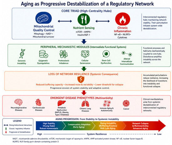
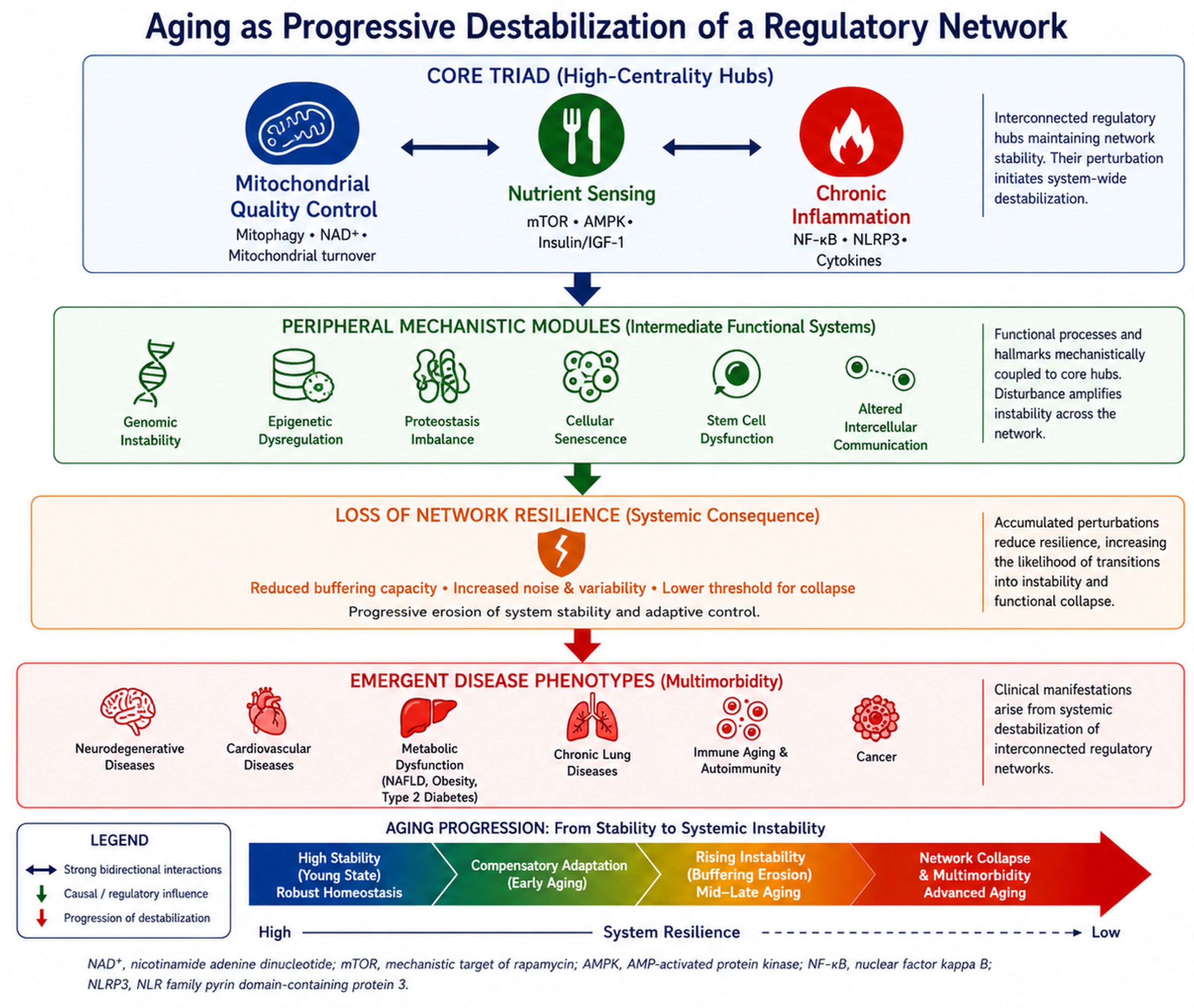
Within this architecture, mitochondrial quality control, nutrient sensing, and chronic inflammation form a proposed high-centrality regulatory core. These three modules are designated putative dominant hubs on the basis of their pleiotropic connectivity in the literature reviewed here: each is reported to directly regulate or be regulated by the majority of remaining hallmarks, a pattern consistent with, though not formally established as, disproportionate destabilizing potential relative to more peripheral nodes (Figure 1). Mitochondria act as signaling hubs coordinating redox balance and transcriptional adaptation [15,16], while age-associated alterations in mitophagy, mitochondrial turnover, and NAD+ (nicotinamide adenine dinucleotide)-dependent signaling extend across regulatory layers [15,17,18,19,20,21]. In parallel, inflammatory signaling both responds to and reinforces mitochondrial dysfunction, including through activation of the NLRP3 (NLR family pyrin domain-containing protein 3) inflammasome [22,23,24], establishing bidirectional coupling between metabolic stress and immune activation [25].
Figure 1.
Aging as progressive destabilization of a regulatory network. The figure illustrates aging as a systems-level process organized into four hierarchical tiers. The Core Triad comprises three putative high-centrality hubs: mitochondrial quality control, nutrient sensing, and chronic inflammation. These interact through strong bidirectional regulatory connections and collectively maintain network stability; their perturbation is proposed to initiate system-wide destabilization. Peripheral Mechanistic Modules represent intermediate functional systems, including genomic instability, epigenetic dysregulation, proteostasis imbalance, cellular senescence, stem cell dysfunction, and altered intercellular communication, which are mechanistically coupled to the core triad through which network perturbations are proposed to propagate across the network. Loss of Network Resilience represents a systemic consequence of accumulated perturbation, characterized by reduced buffering capacity, increased noise and variability, and a progressively lower threshold for functional collapse. Emergent Disease Phenotypes represent the clinical manifestations proposed to arise from this systemic destabilization, including neurodegenerative diseases, cardiovascular diseases, metabolic dysfunction (including non-alcoholic fatty liver disease, obesity, and type 2 diabetes), chronic lung diseases, immune aging and autoimmunity, and cancer. The horizontal arrow at the bottom of the figure depicts the progression of aging from high stability (robust homeostasis in the young state) through compensatory adaptation, rising instability with buffering erosion, to network collapse and multimorbidity in advanced aging, accompanied by a corresponding decline in system resilience. Arrows are limited to three types: bidirectional arrows indicate strong reciprocal interactions among core triad components; single-headed arrows indicate causal or regulatory influence from one tier to the next; and downward arrows indicate progression of destabilization through the hierarchy. NAD+, nicotinamide adenine dinucleotide; mTOR, mechanistic target of rapamycin; AMPK, AMP-activated protein kinase; NF-κB, nuclear factor kappa B; NLRP3, NLR family pyrin domain-containing protein 3. The network structure depicted is conceptual and heuristic, derived from convergence across experimental and translational literature; it does not represent a formally validated network topology.
This framework extends existing models in three key ways. First, it reframes network stability—rather than individual hallmark integrity—as the primary determinant of aging dynamics. Second, it identifies mitochondrial regulation as the operational hub linking metabolic and inflammatory layers. Third, it incorporates non-linear systems properties: feedback loops, bistability, and threshold effects—as exemplified by mitohormesis [26,27]—mean that identical perturbations produce qualitatively different outcomes depending on network state. This has direct implications for therapeutic calibration and for individual variability in aging trajectories. The triangular functional core proposed here should be regarded as a heuristic organizing framework derived from convergence across experimental and translational literature, rather than as a formally validated network topology. This distinction is addressed explicitly in the Limitations section. This article constitutes a narrative and conceptual review. The literature was identified through searches of PubMed and Web of Science conducted iteratively between May and June 2026 using terms including aging, network medicine, mitochondrial dysfunction, NAD+ metabolism, NLRP3 inflammasome, hallmarks of aging, and multimorbidity, with no date restriction on publication year. In practice, priority was given to studies reporting a direct molecular or cellular pathway linking a candidate process to mitochondrial quality control, nutrient sensing, or chronic inflammatory signaling, and when findings extended beyond a single model system to human tissue, human biomarker data, or clinical outcomes. This was not applied as a formal a priori protocol but reflects, in retrospect, the basis on which sources were selected; in descending order, interventional human trials, human biomarker-level or observational data, and mechanistic preclinical studies were favored, with primary research articles and seminal reviews prioritized over secondary commentary. Studies focusing exclusively on single molecular targets without broader network or systemic implications, and animal studies lacking translational relevance to human aging biology, were generally not prioritized.
2. Integrative Biology as a Multi-Scale Framework
Molecular mechanisms that are well-characterized in isolation routinely fail to predict organism-level phenotypes. This is not a gap in mechanistic knowledge—it reflects the emergent nature of biological networks. Network medicine conceptualizes disease as a perturbation of functional modules rather than isolated lesions [7,8]. Multi-scale integration, as used here, denotes the capacity of molecular events to propagate through cellular, tissue, and systemic layers, a property absent from pathway-level models but essential for understanding how localized dysfunction generates systemic disease.
Fusion and fission regulate organelle integrity and metabolic adaptability [17,28,29]. Mitophagy selectively removes damaged mitochondria, constraining propagation of oxidative and inflammatory stress signals [15,16,17,30,31,32]. Mitochondrial stress also activates the UPRmt, linking organelle dysfunction to nuclear transcriptional remodeling [15,17,18].
NAD+-dependent signaling, including SIRT1-mediated deacetylation of PGC-1α, couples bioenergetics to mitochondrial biogenesis and broader transcriptional control [15,19,20,21], and age-related mitochondrial decline in humans is associated with insulin resistance [33,34,35], a pattern consistent with, though not sufficient on its own to establish, systemic metabolic coupling.
Mitochondrial dysfunction activates the NLRP3 inflammasome [22], creating bidirectional feedback between metabolic stress and immune activation [36,37] that amplifies both. Mitochondria, nutrient-sensing pathways, and inflammatory signaling thus constitute a densely interconnected triad whose collective perturbation—rather than the failure of any single component—underlies the systemic character of biological aging (Figure 1). Within this triad, mitochondria are labelled an operational hub because they execute the downstream bioenergetic and redox consequences of upstream signals; nutrient sensing is labelled a regulatory integrator because it directly senses and adjusts mitochondrial and inflammatory activity in response to metabolic state; and chronic inflammation is labelled an amplifier hub because it converts localized mitochondrial or metabolic perturbation into systemic, self-reinforcing signalling.
3. Hallmarks of Aging as an Interconnected Network
The hallmarks of aging can be reinterpreted as dynamically interacting network nodes rather than independent processes [1,2]. Genomic and epigenomic regulation is tightly coupled to metabolic state through NAD+-dependent enzymes and mitonuclear signaling [19,20,38,39,40]. PARP1 (poly (ADP-ribose) polymerase 1) and sirtuins compete for the same NAD+ pool—the former during DNA damage repair, the latter during transcriptional regulation. NAD+ depletion thus creates direct metabolic coupling between genome maintenance and mitochondrial signaling, linking two hallmarks otherwise treated as independent. Consequently, genome maintenance and epigenetic remodeling are inseparable from cellular bioenergetics and mitochondrial signaling.
Loss of proteostasis intersects with mitochondrial quality control, as reduced mitophagy permits accumulation of dysfunctional organelles that amplify oxidative and inflammatory stress [16,22]. Reduced mitophagy thus bridges proteostatic collapse, metabolic inflexibility, and immune activation within a single mechanistic chain. Deregulated nutrient sensing further modulates autophagy and mitochondrial biogenesis; pharmacological inhibition of mTOR extends lifespan in model organisms [41], underscoring the systemic influence of nutrient-sensing nodes.
Cellular senescence contributes to inflammatory amplification through the SASP (senescence-associated secretory phenotype), which in turn impairs mitochondrial function in neighboring cells and induces paracrine senescence, establishing a self-reinforcing cycle that progressively expands network instability [1,2,42]. Senolytic strategies attenuate age-related phenotypes in preclinical models [43], supporting the role of senescence as a network amplifier rather than a static cellular endpoint. In parallel, altered intercellular communication and stem cell exhaustion reflect downstream consequences of chronic inflammatory signaling and metabolic constraints, reinforcing the view that hallmarks operate as an interacting system rather than as a list of parallel pathways. The hallmarks do not merely co-occur—they are coupled, and perturbation of any one propagates through the network according to connectivity, not causal proximity (Table 1).
Table 1.
Relationship of canonical aging hallmarks to the triangular functional core. For each hallmark, the primary functional relationship to mitochondrial quality control, nutrient sensing, and chronic inflammation is indicated, along with the principal mechanistic rationale and key supporting references. Upstream: the hallmark drives dysfunction within the core. Downstream: the hallmark arises as a consequence of core perturbation. Bidirectional: the hallmark both influences and is influenced by core nodes. Emergent: the hallmark arises from convergent interactions across multiple core nodes. This classification is an interpretive synthesis of the literature reviewed here and is not derived from formal network modeling or quantitative centrality analysis.
4. Mitochondrial Regulation of Network Stability
Mitochondria occupy a uniquely central position in the aging network [44,45]. No other organelle simultaneously governs energy metabolism, redox signaling, apoptosis, innate immune activation, and epigenetic reprogramming—a functional breadth that makes mitochondrial dysfunction inherently propagative across regulatory modules. Balanced fusion–fission dynamics maintain structural integrity and metabolic flexibility [17,28,29], while impaired dynamics contribute to cardiovascular dysfunction and reduced stress tolerance [46,47]. Mitochondrial plasticity is therefore not a secondary feature but a determinant of systemic stability.
Reduced mitophagy efficiency permits accumulation of dysfunctional mitochondria, amplifying redox imbalance and inflammatory signaling [15,16,17,22,30,31,32]. Because mitophagy constrains propagation of mitochondrial stress signals, its decline effectively increases noise within cellular regulatory circuits and reduces buffering capacity. Coordination between mitophagy and mitochondrial biogenesis is required to maintain organelle homeostasis [16,48,49], and disruption of this balance accelerates functional decline.
Age-associated NAD+ decline impairs mitonuclear communication, decoupling energetic state from transcriptional and stress-response programs [15,19,20,21,38]—a shift that propagates mitochondrial dysfunction into proteostatic and immune modules. Adaptive signaling pathways such as the UPRmt further link organelle stress to nuclear transcriptional remodeling [18].
Mitohormesis illustrates non-linear redox regulation, whereby moderate mitochondrial stress can induce adaptive resilience, whereas excessive stress promotes dysfunction [26,27]. A key mediator of this non-linearity is mitochondrial ROS production. At physiological levels, ROS act as redox signalling molecules that regulate adaptive responses; sustained overproduction drives oxidative damage and accelerates hallmark progression. Comparative analyses across species show that mitochondrial H2O2 production rate per unit oxygen consumed correlates inversely with maximum lifespan across species, positioning mitochondrial ROS dynamics as a quantitative effector of biological aging [50]. Effective interventions must therefore calibrate redox signalling dynamics rather than indiscriminately suppress oxidative pathways. Exercise and caloric restriction are the most robustly validated interventions for network recalibration: both activate mitohormetic programmes, improve mitochondrial quality control, reduce inflammatory tone, and extend healthspan across multiple model systems. Low-dose mitochondrial uncouplers can partially mimic these responses, but the breadth and durability of exercise- and caloric restriction-induced effects remain unmatched by pharmacological approaches.
Secondary mitochondrial dysfunction, defined here as mitochondrial impairment arising downstream of other processes such as inflammation, metabolic dysregulation, or proteostatic stress, rather than from primary defects in mitochondrial or mtDNA-encoded components, is widely observed in metabolic and cardiovascular disease [33,34,35,44,46,51,52,53]. Mitochondrial dysfunction therefore occupies a topologically privileged position in the aging network. As the node through which metabolic, proteostatic, and inflammatory signals converge, its progressive failure reconfigures the entire regulatory landscape rather than disrupting a single functional domain. At the cellular level, this progression follows a recognizable pathological sequence: impaired mitochondrial quality control increases steady-state ROS exposure and oxidative macromolecular damage, which activates the NLRP3 inflammasome and drives a senescence-associated secretory phenotype (SASP); the resulting chronic cytokine output promotes extracellular matrix remodeling and fibrotic change in affected tissues, ultimately manifesting as organ-level functional decline and the degenerative disease phenotypes discussed in Section 5.
5. Age-Related Diseases as Emergent Network Phenotypes
Neurodegenerative diseases exhibit mitochondrial dysfunction, impaired mitophagy, and inflammatory activation [54,55]. In Alzheimer’s disease, mitochondrial bioenergetic deficits and altered dynamics precede overt neuronal loss [54,55], suggesting that metabolic instability contributes to disease initiation rather than merely reflecting downstream damage. Deregulated nutrient sensing amplifies this vulnerability: mTOR hyperactivation impairs autophagy and mitophagy, while AMPK hypoactivation reduces metabolic flexibility. Activation of the NLRP3 inflammasome links mitochondrial stress to neuroinflammation [23], and chronic cytokine release from activated microglia creates a self-reinforcing inflammatory loop that further compromises neuronal bioenergetics [56]. This temporal precedence has mechanistic weight: it positions mitochondrial dysfunction as a driver of neurodegeneration rather than a consequence—an important distinction for identifying early intervention windows.
Cardiovascular aging involves impaired mitochondrial energetics, altered substrate utilization, and increased inflammatory signaling [33,46,47,57]. Reduced mitochondrial flexibility compromises cardiac stress responses, while chronic low-grade inflammation promotes vascular remodeling and plaque instability [36,58]. Deregulated nutrient sensing further impairs cardiac autophagy through mTOR activation and reduces metabolic flexibility through AMPK suppression, limiting the heart’s response to energetic stress. The clinical efficacy of anti-inflammatory therapy in reducing cardiovascular events [59] supports the interpretation that inflammatory signaling operates as a modifiable high-centrality node within aging-associated networks.
Type 2 diabetes reflects integrated mitochondrial, metabolic, and inflammatory disturbances [33,35,51,60]. Mitochondrial dysfunction contributes to insulin resistance, while chronic inflammation exacerbates metabolic dysregulation. These interdependencies exemplify the network destabilization model: in type 2 diabetes, no single causal pathway is sufficient to explain the full phenotype, and effective intervention may require simultaneous modulation of multiple high-centrality nodes rather than sequential correction of individual pathways.
Cancer represents a distinct pathological network outcome within this systems framework: it engages the same core nodes highlighted here, including mTOR signaling, mitochondrial metabolism, and NF-κB-related inflammation, but channels their dysregulation toward adaptive rewiring of metabolic circuitry rather than network collapse, exploiting the reduced regulatory precision characteristic of aged tissue [61,62]. Functional mitochondria support biosynthetic demands and redox balance in tumor cells [62,63], illustrating how oncogenesis actively co-opts the same metabolic nodes that aging-associated instability otherwise fails to suppress. The distinction between permissive instability and functional collapse highlights the context-dependence of network-level outcomes.
Multimorbidity thus reflects overlapping network failures rather than coincidental co-occurrence of independent diseases. As regulatory hubs fail, the probability of multiple simultaneous disease phenotypes increases according to network topology, not pathological independence—a prediction with direct implications for how multi-morbid patients are stratified and treated.
6. Network-Oriented Therapeutic Modulation
The network destabilization model has a direct therapeutic implication: interventions should target nodes with the highest systemic connectivity, where modulation propagates across regulatory layers rather than correcting isolated deficits. Interventions targeting nutrient-sensing pathways represent one of the most extensively studied strategies in this regard. Pharmacological inhibition of mTOR extends lifespan in multiple model organisms [41,64,65,66,67] and modulates immune function in older adults [41,68,69]. Rather than acting through a single downstream pathway, mTOR inhibition influences autophagy, mitochondrial turnover, protein synthesis, and metabolic adaptation, illustrating how modulation of a high-centrality node can propagate across regulatory layers. This pleiotropic reach carries a calibration requirement: mild mTOR inhibition promotes clearance, but excessive suppression impairs tissue repair—a dose-sensitivity the network framework explicitly predicts.
Metformin similarly targets nutrient sensing and mitochondrial metabolism through activation of AMP-activated protein kinase (AMPK) and indirect mTOR modulation [69,70]. Its established cardiometabolic benefits in type 2 diabetes highlight the translational potential of metabolic reprogramming, although long-term effects on multimorbidity and global resilience remain under investigation.
Mitochondrial-targeted strategies focus on restoring organelle quality and signaling competence. Urolithin A has been shown to induce mitophagy in model organisms [71] and to improve mitochondrial biomarkers in humans [72], while NAD+ precursors increase systemic NAD+ levels and modulate metabolic parameters in humans [73,74,75,76,77], an effect so far established only at the biomarker level, with clinical endpoints such as reduced multimorbidity or extended healthspan not yet demonstrated. Spermidine extends lifespan and reduces cardiovascular risk through autophagy-dependent mechanisms [78,79]: it inhibits EP300 acetyltransferase, triggering histone hypoacetylation and autophagy induction [80], and activates mitophagy to restore mitochondrial quality. These effects reduce NLRP3 inflammasome activation and lower circulating inflammatory markers. Dietary spermidine intake is associated with reduced mortality, improved cardiac outcomes, and signals of cognitive benefit [79,81,82], further supporting the importance of mitochondrial and proteostatic integration.
Cellular senescence represents another network amplifier amenable to modulation. Senolytics provide a direct test of the network hypothesis: if senescent cells amplify instability via the SASP, their elimination should produce system-wide effects disproportionate to cell numbers removed—a prediction supported preclinically [43,83,84] and now entering human trials. Anti-inflammatory therapy targeting IL-1β (interleukin-1β) has also demonstrated a reduction in cardiovascular events [59], providing proof-of-principle that inflammatory nodes are clinically modifiable. Alpha-ketoglutarate (AKG), an endogenous TCA cycle intermediate whose levels decline with age, extends lifespan and compresses morbidity in mice while suppressing chronic inflammatory cytokines [85,86]; human evidence remains limited to an early-phase clinical trial protocol, and the mouse findings should not yet be assumed to translate to comparable effects in humans. Its mechanisms are multi-nodal: AKG is an obligate cofactor for TET DNA demethylases and Jumonji C-domain histone demethylases, linking mitochondrial metabolic state to epigenetic gene regulation; it also suppresses NF-κB-mediated inflammation through prolyl hydroxylase-dependent inhibition of IKKβ [87]. Together, these effects span mitochondrial function, epigenetic regulation, and chronic inflammation. Pyrroloquinoline quinone (PQQ), a redox-active o-quinone compound found in diverse foods, stimulates mitochondrial biogenesis through SIRT1/PGC-1α activation and attenuates inflammatory markers [88,89], supporting its candidacy as a multi-node intervention targeting both mitochondrial quality control and chronic inflammation; clinical evidence remains exploratory, derived from a single small trial, and requires replication before any clinical recommendation can be made.
Because aging networks are non-linear and context-dependent, interventions targeting high-centrality nodes may require calibrated modulation rather than maximal inhibition. The interventions discussed here are included as mechanistic exemplars illustrating modulation of distinct high-centrality nodes rather than as an exhaustive therapeutic inventory. A summary is provided in Table 2.
Table 2.
Therapeutic interventions targeting high-centrality nodes and closely associated downstream processes in the aging network. The table presents a representative, non-exhaustive selection of interventions chosen on the basis of mechanistic relevance to the three core nodes (mitochondrial quality control, nutrient sensing, and chronic inflammation), as well as to mechanistically linked downstream processes (autophagy, cellular senescence) through which several interventions act, and availability of at least preliminary human data. Target Node indicates the regulatory module or closely associated process primarily modulated. Principal Mechanism summarizes the dominant molecular action. Human Evidence reflects the current level of clinical support, ranging from randomized controlled trials (rapamycin/mTOR inhibitors, anti-IL-1β therapy, Coenzyme Q10) to biomarker-level clinical observations (NAD+ precursors, urolithin A) and epidemiological associations (spermidine); fisetin evidence remains predominantly preclinical. This classification of target nodes is an interpretive synthesis of the literature reviewed here and is not derived from formal network modeling. Abbreviations: mTOR, mechanistic target of rapamycin; mTORC1, mTOR complex 1; AMPK, AMP-activated protein kinase; NAD+, nicotinamide adenine dinucleotide; NF-κB, nuclear factor kappa B; NLRP3, NLR family pyrin domain-containing protein 3.
7. Limitations and Conceptual Boundaries
Although the network destabilization framework offers integrative explanatory power, several limitations require consideration. Biological networks are inherently context-dependent, and the relative centrality of regulatory nodes varies across tissues, genetic backgrounds, and environmental exposures. Aging trajectories display substantial heterogeneity, and network-based analyses of health deficits reveal non-linear accumulation patterns rather than uniform progression. Identical interventions targeting mitochondrial quality, nutrient sensing, or inflammatory signaling may therefore produce divergent effects depending on baseline network configuration and individual resilience. A key conceptual limitation is that the network destabilization framework, as presented here, remains qualitative. The designation of mitochondrial quality control, nutrient sensing, and inflammatory signaling as high-centrality nodes is supported by extensive empirical evidence but has not been formally validated by quantitative network analysis of aging-associated interactomes. Future work employing graph-theoretic methods on multi-omics datasets will be necessary to test whether these nodes meet formal criteria of hub-centrality and to identify additional high-centrality nodes that the present framework may overlook [92,93].
Network dynamics are intrinsically non-linear. Moderate mitochondrial stress may activate adaptive responses consistent with mitohormesis [26,27], whereas excessive or chronic stress promotes dysfunction. Similarly, pharmacological modulation of nutrient-sensing pathways such as mTOR can extend lifespan in model organisms [65], yet excessive suppression may compromise immune competence or tissue repair. Translational limitations further constrain interpretation. NAD+ augmentation consistently increases systemic NAD+ levels and modulates metabolic parameters [73,74,75,76,77], but whether these changes translate into reduced multimorbidity, improved functional capacity, or extended healthspan remains unestablished. Adequately powered, long-duration trials are needed. Similarly, mTOR modulation influences immune parameters in older adults [68], but durable effects on mortality and multimorbidity remain insufficiently established.
Finally, while the triangular functional core emphasizes mitochondrial quality control, nutrient sensing, and inflammatory signaling as high-centrality hubs, not all age-related pathologies can be reduced exclusively to these modules. The network destabilization framework should therefore complement mechanistic specificity rather than replace it.
8. Conceptual Synthesis and Future Directions
The convergence of mitochondrial dysfunction, metabolic inflexibility, and inflammatory amplification across neurodegenerative, cardiovascular, metabolic, and oncological diseases points to shared regulatory architecture rather than disease-specific pathology. These phenotypes arise from the progressive failure of shared hubs, not independent pathological cascades—a perspective that opens new angles for both research design and therapeutic strategy.
Translation of network-based aging biology into clinical practice will likely require rethinking trial design. Traditional randomized trials typically evaluate single agents in heterogeneous populations using static endpoints. Precision geroscience approaches may benefit from stratification based on mitochondrial function, metabolic flexibility, inflammatory tone, or composite resilience indices rather than chronological age alone [94,95]. Incorporation of longitudinal systems-level biomarkers and adaptive trial designs may improve the detection of clinically meaningful effects.
Several testable predictions follow. First, network instability should manifest as correlated changes across three domains: mitochondrial function (NAD+/NADH ratio, membrane potential proxies), metabolic flexibility (respiratory quotient variability, ketone body production capacity), and inflammatory tone (IL-6 (interleukin-6), CRP (C-reactive protein), NLRP3 activation markers). Composite indices capturing this inter-domain coupling may outperform single biomarkers in predicting age-related decline. Second, interventions targeting high-centrality nodes may produce system-wide effects that exceed those of pathway-specific modulation, detectable as correlated improvements across multiple hallmark-associated biomarkers rather than isolated endpoint changes. Third, individual variability in aging trajectories may reflect quantifiable differences in baseline network configuration and resilience capacity, suggesting that composite network-state indices could help stratify individuals by biological rather than chronological age and predict differential responses to intervention. Future studies integrating multi-omics data with network analysis may help operationalize these predictions and bring the framework closer to formal quantitative validation. Viewing aging as progressive destabilization of interconnected regulatory networks provides a mechanistic framework linking molecular hallmarks with multimorbidity and may support the development of more integrative therapeutic strategies. Formal validation of this framework will require graph-theoretical analysis of longitudinal multi-omics and clinical datasets to test the centrality, connectivity, and predictive value of the proposed core nodes.
Funding
This research received no external funding.
Institutional Review Board Statement
Not applicable.
Informed Consent Statement
Not applicable.
Data Availability Statement
No new data were created or analyzed in this study. Data sharing is not applicable.
Conflicts of Interest
The author declares no conflicts of interest.
References
- López-Otín, C.; Blasco, M.A.; Partridge, L.; Serrano, M.; Kroemer, G. The hallmarks of aging. Cell 2013, 153, 1194–1217. [Google Scholar] [CrossRef] [Scilit] [PubMed]
- López-Otín, C.; Kroemer, G. Hallmarks of aging: An expanding universe. Cell 2023, 186, 243–278. [Google Scholar] [CrossRef] [Scilit] [PubMed]
- Kennedy, B.K.; Berger, S.L.; Brunet, A.; Campisi, J.; Cuervo, A.M.; Epel, E.S.; Franceschi, C.; Lithgow, G.J.; Morimoto, R.I.; Pessin, J.E.; et al. Geroscience: Linking aging to chronic disease. Cell 2014, 159, 709–713. [Google Scholar] [CrossRef] [Scilit] [PubMed]
- Kirkwood, T.B.L. Understanding the odd science of aging. Cell 2005, 120, 437–447. [Google Scholar] [CrossRef] [Scilit] [PubMed]
- Kenyon, C.J. The genetics of ageing. Nature 2010, 464, 504–512. [Google Scholar] [CrossRef] [Scilit] [PubMed]
- Kaeberlein, M.; Rabinovitch, P.S.; Martin, G.M. Healthy aging: The ultimate preventative medicine. Science 2015, 350, 1191–1193. [Google Scholar] [CrossRef] [Scilit] [PubMed]
- Kitano, H. Systems biology: A brief overview. Science 2002, 295, 1662–1664. [Google Scholar] [CrossRef] [Scilit] [PubMed]
- Barabási, A.L.; Gulbahce, N.; Loscalzo, J. Network medicine: A network-based approach to human disease. Nat. Rev. Genet. 2011, 12, 56–68. [Google Scholar] [CrossRef] [Scilit] [PubMed]
- Loscalzo, J.; Barabasi, A.L. Systems biology and the future of medicine. Rev. Syst. Biol. Med. 2011, 3, 619–627. [Google Scholar] [CrossRef] [Scilit] [PubMed]
- Promislow, D.E.L.; Anderson, R.M.; Scheffer, M.; Crespi, B.; DeGregori, J.; Harris, K.; Horowitz, B.N.; Levine, M.E.; Riolo, M.A.; Schneider, D.S.; et al. Resilience integrates concepts in aging research. IScience 2022, 25, 104199. [Google Scholar] [CrossRef] [Scilit] [PubMed]
- Farrell, S.G.; Mitnitski, A.; Theou, O.; Rockwood, K.; Rutenberg, A.D. Probing the network structure of health deficits in human aging. Phys. Rev. E 2018, 98, 032302. [Google Scholar] [CrossRef] [Scilit]
- Rockwood, K.; Mitnitski, A. Frailty in relation to the accumulation of deficits. J. Gerontol. A 2007, 62, 722–727. [Google Scholar] [CrossRef] [Scilit] [PubMed]
- Mitnitski, A.; Rockwood, K. The rate of aging: The rate of deficit accumulation does not change over the adult life span. Biogerontology 2016, 17, 199–204. [Google Scholar] [CrossRef] [Scilit] [PubMed]
- Barnett, K.; Mercer, S.W.; Norbury, M.; Watt, G.; Wyke, S.; Guthrie, B. Epidemiology of multimorbidity and implications for health care, research, and medical education: A cross-sectional study. Lancet 2012, 380, 37–43. [Google Scholar] [CrossRef] [Scilit] [PubMed]
- Quirós, P.M.; Mottis, A.; Auwerx, J. Mitonuclear communication in homeostasis and stress. Nat. Rev. Mol. Cell Biol. 2016, 17, 213–226. [Google Scholar] [CrossRef] [Scilit] [PubMed]
- Mottis, A.; Herzig, S.; Auwerx, J. Mitocellular communication: Shaping health and disease. Science 2019, 366, 827–832. [Google Scholar] [CrossRef] [Scilit] [PubMed]
- Youle, R.J.; van der Bliek, A.M. Mitochondrial fission, fusion, and stress. Science 2012, 337, 1062–1065. [Google Scholar] [CrossRef] [Scilit] [PubMed]
- Shpilka, T.; Haynes, C.M. The mitochondrial UPR: Mechanisms, physiological functions and implications in ageing. Nat. Rev. Mol. Cell Biol. 2018, 19, 109–120. [Google Scholar] [CrossRef] [Scilit] [PubMed]
- Imai, S.I.; Guarente, L. NAD+ and sirtuins in aging and disease. Trends Cell Biol. 2014, 24, 464–471. [Google Scholar] [CrossRef] [Scilit] [PubMed]
- Verdin, E. NAD+ in aging, metabolism, and neurodegeneration. Science 2015, 350, 1208–1213. [Google Scholar] [CrossRef] [Scilit] [PubMed]
- Covarrubias, A.J.; Perrone, R.; Grozio, A.; Verdin, E. NAD+ metabolism and its roles in cellular processes during ageing. Nat. Rev. Mol. Cell Biol. 2021, 22, 119–141. [Google Scholar] [CrossRef] [Scilit] [PubMed]
- Zhou, R.; Yazdi, A.S.; Menu, P.; Tschopp, J. A role for mitochondria in NLRP3 inflammasome activation. Nature 2011, 469, 221–225. [Google Scholar] [CrossRef] [Scilit] [PubMed]
- Heneka, M.T.; Kummer, M.P.; Stutz, A.; Delekate, A.; Schwartz, S.; Vieira-Saecker, A.; Griep, A.; Axt, D.; Remus, A.; Tzeng, T.-C.; et al. NLRP3 is activated in Alzheimer’s disease and contributes to pathology in APP/PS1 mice. Nature 2013, 493, 674–678. [Google Scholar] [CrossRef] [Scilit] [PubMed]
- Toldo, S.; Abbate, A. The NLRP3 inflammasome in acute myocardial infarction. Nat. Rev. Cardiol. 2018, 15, 203–214. [Google Scholar] [CrossRef] [Scilit] [PubMed]
- Green, D.R.; Galluzzi, L.; Kroemer, G. Mitochondria and the autophagy-inflammation-cell death axis in organismal aging. Science 2011, 333, 1109–1112. [Google Scholar] [CrossRef] [Scilit] [PubMed]
- Ristow, M.; Zarse, K. How increased oxidative stress promotes longevity and metabolic health: The concept of mitochondrial hormesis (mitohormesis). Exp. Gerontol. 2010, 45, 410–418. [Google Scholar] [CrossRef] [Scilit] [PubMed]
- Yun, J.; Finkel, T. Mitohormesis. Cell Metab. 2014, 19, 757–766. [Google Scholar] [CrossRef] [Scilit] [PubMed]
- Chan, D.C. Fusion and fission: Interlinked processes critical for mitochondrial health. Annu. Rev. Genet. 2012, 46, 265–287. [Google Scholar] [CrossRef] [Scilit] [PubMed]
- Ong, S.B.; Hausenloy, D.J. Mitochondrial morphology and cardiovascular disease. Cardiovasc. Res. 2010, 88, 16–29. [Google Scholar] [CrossRef] [Scilit] [PubMed]
- Narendra, D.; Tanaka, A.; Suen, D.F.; Youle, R.J. Parkin is recruited selectively to impaired mitochondria and promotes their autophagy. J. Cell Biol. 2008, 183, 795–803. [Google Scholar] [CrossRef] [Scilit] [PubMed]
- Pickles, S.; Vigié, P.; Youle, R.J. Mitophagy and quality control mechanisms in mitochondrial maintenance. Curr. Biol. 2018, 28, 170–185. [Google Scholar] [CrossRef] [Scilit] [PubMed]
- Palikaras, K.; Lionaki, E.; Tavernarakis, N. Mechanisms of mitophagy in cellular homeostasis, physiology and pathology. Nat. Cell Biol. 2018, 20, 1013–1022. [Google Scholar] [CrossRef] [Scilit] [PubMed]
- Petersen, K.F.; Befroy, D.; Dufour, S.; Dziura, J.; Ariyan, C.; Rothman, D.L.; DiPietro, L.; Cline, G.W.; Shulman, G.I. Mitochondrial dysfunction in the elderly: Possible role in insulin resistance. Science 2003, 300, 1140–1142. [Google Scholar] [CrossRef] [Scilit] [PubMed]
- Short, K.R.; Bigelow, M.L.; Kahl, J.; Singh, R.; Coenen-Schimke, J.; Raghavakaimal, S.; Nair, K.S. Decline in skeletal muscle mitochondrial function with aging in humans. Proc. Natl. Acad. Sci. USA 2005, 102, 5618–5623. [Google Scholar] [CrossRef] [Scilit] [PubMed]
- Lowell, B.B.; Shulman, G.I. Mitochondrial dysfunction and type 2 diabetes. Science 2005, 307, 384–387. [Google Scholar] [CrossRef] [Scilit] [PubMed]
- Ferrucci, L.; Fabbri, E. Inflammageing: Chronic inflammation in ageing, cardiovascular disease, and frailty. Nat. Rev. Cardiol. 2018, 15, 505–522. [Google Scholar] [CrossRef] [Scilit] [PubMed]
- Franceschi, C.; Bonafè, M.; Valensin, S.; Olivieri, F.; De Luca, M.; Ottaviani, E.; De Benedictis, G. Inflamm-aging: An evolutionary perspective on immunosenescence. Ann. N. Y. Acad. Sci. 2000, 908, 244–254. [Google Scholar] [CrossRef] [Scilit] [PubMed]
- Gomes, A.P.; Price, N.L.; Ling, A.J.Y.; Moslehi, J.J.; Montgomery, M.K.; Rajman, L.; White, J.P.; Teodoro, J.S.; Wrann, C.D.; Hubbard, B.P.; et al. Declining NAD+ induces a pseudohypoxic state disrupting nuclear-mitochondrial communication during aging. Cell 2013, 155, 1624–1638. [Google Scholar] [CrossRef] [Scilit] [PubMed]
- Canto, C.; Auwerx, J. NAD+ as a signaling molecule modulating metabolism. Cold Spring Harb. Symp. Quant. Biol. 2011, 76, 291–298. [Google Scholar] [CrossRef] [Scilit] [PubMed]
- López-Otín, C.; Galluzzi, L.; Freije, J.M.P.; Madeo, F.; Kroemer, G. Metabolic control of longevity. Cell 2016, 166, 802–821. [Google Scholar] [CrossRef] [Scilit] [PubMed]
- Johnson, S.C.; Rabinovitch, P.S.; Kaeberlein, M. mTOR is a key modulator of ageing and age-related disease. Nature 2013, 493, 338–345. [Google Scholar] [CrossRef] [Scilit] [PubMed]
- Campisi, J. Cellular senescence: Putting the paradoxes in perspective. Curr. Opin. Genet. Dev. 2011, 21, 107–112. [Google Scholar] [CrossRef] [Scilit] [PubMed]
- Baker, D.J.; Wijshake, T.; Tchkonia, T.; LeBrasseur, N.K.; Childs, B.G.; van de Sluis, B.; Van Deursen, J.M. Clearance of p16Ink4a-positive senescent cells delays ageing-associated disorders. Nature 2011, 479, 232–236. [Google Scholar] [CrossRef] [Scilit] [PubMed]
- Sun, N.; Youle, R.J.; Finkel, T. The mitochondrial basis of aging. Mol. Cell 2016, 61, 654–666. [Google Scholar] [CrossRef] [Scilit] [PubMed]
- Ziada, A.S.; Smith, M.S.R.; Côté, H.C.F. Updating the free radical theory of aging. Front. Cell Dev. Biol. 2020, 8, 575645. [Google Scholar] [CrossRef] [Scilit] [PubMed]
- Dorn, G.W., 2nd. Mitochondrial dynamics in heart disease. Biochim. Biophys. Acta 2013, 1833, 233–241. [Google Scholar] [CrossRef] [Scilit] [PubMed]
- Brown, D.A.; Perry, J.B.; Allen, M.E.; Sabbah, H.N.; Stauffer, B.L.; Shaikh, S.R.; Cleland, J.G.F.; Colucci, W.S.; Butler, J.; Voors, A.A.; et al. Mitochondrial function as a therapeutic target in heart failure. Nat. Rev. Cardiol. 2017, 14, 238–250. [Google Scholar] [CrossRef] [Scilit] [PubMed]
- Palikaras, K.; Lionaki, E.; Tavernarakis, N. Coordination of mitophagy and mitochondrial biogenesis during ageing in C. elegans. Nature 2015, 521, 525–528. [Google Scholar] [CrossRef] [Scilit] [PubMed]
- Scarpulla, R.C. Metabolic control of mitochondrial biogenesis through the PGC-1 family regulatory network. Biochim. Biophys. Acta 2011, 1813, 1269–1278. [Google Scholar] [CrossRef] [Scilit] [PubMed]
- Gómez, J.; Mota-Martorell, N.; Jové, M.; Pamplona, R.; Barja, G. Mitochondrial ROS production, oxidative stress and aging within and between species: Evidences and recent advances on this aging effector. Exp. Gerontol. 2023, 174, 112134. [Google Scholar] [CrossRef] [Scilit] [PubMed]
- Kujoth, G.C.; Hiona, A.; Pugh, T.D.; Someya, S.; Panzer, S.K.; Wohlgemuth, S.E.; Hofer, T.; Seo, A.Y.; Sullivan, R.; Jobling, W.A.; et al. Mitochondrial DNA mutations, oxidative stress, and apoptosis in mammalian aging. Science 2005, 309, 481–484. [Google Scholar] [CrossRef] [Scilit] [PubMed]
- Trifunovic, A.; Wredenberg, A.; Falkenberg, M.; Spelbrink, J.N.; Rovio, A.T.; Bruder, C.E.; Bohlooly-Y, M.; Gidlöf, S.; Oldfors, A.; Wibom, R.; et al. Premature ageing in mice expressing defective mitochondrial DNA polymerase. Nature 2004, 429, 417–423. [Google Scholar] [CrossRef] [Scilit] [PubMed]
- Wang, W.; Zhao, F.; Ma, X.; Perry, G.; Zhu, X. Mitochondria dysfunction in the pathogenesis of Alzheimer’s disease: Recent advances. Mol. Neurodegener. 2020, 15, 30. [Google Scholar] [CrossRef] [Scilit] [PubMed]
- Kerr, J.S.; Adriaanse, B.A.; Greig, N.H.; Mattson, M.P.; Cader, M.Z.; Bohr, V.A.; Fang, E.F. Mitophagy and Alzheimer’s disease: Cellular and molecular mechanisms. Trends Neurosci. 2017, 40, 151–166. [Google Scholar] [CrossRef] [Scilit] [PubMed]
- Yao, J.; Irwin, R.W.; Zhao, L.; Nilsen, J.; Hamilton, R.T.; Brinton, R.D. Mitochondrial bioenergetic deficit precedes Alzheimer’s pathology in female mouse model of Alzheimer’s disease. Proc. Natl. Acad. Sci. USA 2009, 106, 14670–14675. [Google Scholar] [CrossRef] [Scilit] [PubMed]
- Deczkowska, A.; Keren-Shaul, H.; Weiner, A.; Colonna, M.; Schwartz, M.; Amit, I. Disease-associated microglia: A universal immune sensor of neurodegeneration. Cell 2018, 173, 1073–1081. [Google Scholar] [CrossRef] [Scilit] [PubMed]
- Zhou, B.; Tian, R. Mitochondrial dysfunction in pathophysiology of heart failure. J. Clin. Investig. 2018, 128, 3716–3726. [Google Scholar] [CrossRef] [Scilit] [PubMed]
- Libby, P.; Ridker, P.M.; Hansson, G.K. Progress and challenges in translating the biology of atherosclerosis. Nature 2011, 473, 317–325. [Google Scholar] [CrossRef] [Scilit] [PubMed]
- Ridker, P.M.; Everett, B.M.; Thuren, T.; MacFadyen, J.G.; Chang, W.H.; Ballantyne, C.; Fonseca, F.; Nicolau, J.; Koenig, W.; Anker, S.D.; et al. Antiinflammatory therapy with canakinumab for atherosclerotic disease. N. Engl. J. Med. 2017, 377, 1119–1131. [Google Scholar] [CrossRef] [Scilit] [PubMed]
- Samuel, V.T.; Shulman, G.I. Mechanisms for Insulin Resistance: Common Threads and Missing Links. Cell 2012, 148, 852–871. [Google Scholar] [CrossRef] [Scilit] [PubMed]
- Hanahan, D.; Weinberg, R.A. Hallmarks of cancer: The next generation. Cell 2011, 144, 646–674. [Google Scholar] [CrossRef] [Scilit] [PubMed]
- Pavlova, N.N.; Thompson, C.B. The emerging hallmarks of cancer metabolism. Cell Metab. 2016, 23, 27–47. [Google Scholar] [CrossRef] [Scilit] [PubMed]
- Vyas, S.; Zaganjor, E.; Haigis, M.C. Mitochondria and cancer. Cell 2016, 166, 555–566. [Google Scholar] [CrossRef] [Scilit] [PubMed]
- Harrison, D.E.; Strong, R.; Sharp, Z.D.; Nelson, J.F.; Astle, C.M.; Flurkey, K.; Nadon, N.L.; Wilkinson, J.E.; Frenkel, K.; Carter, C.S.; et al. Rapamycin fed late in life extends lifespan in genetically heterogeneous mice. Nature 2009, 460, 392–395. [Google Scholar] [CrossRef] [Scilit] [PubMed]
- Blagosklonny, M.V. Rapamycin for longevity: Opinion article. Aging 2019, 11, 8048–8067. [Google Scholar] [CrossRef] [Scilit] [PubMed]
- Kaeberlein, M. Rapamycin and aging: When, for how long, and how much? J. Genet. Genom. 2014, 41, 459–463. [Google Scholar] [CrossRef] [Scilit] [PubMed]
- Mannick, J.B.; Del Giudice, G.; Lattanzi, M.; Valiante, N.M.; Praestgaard, J.; Huang, B.; Lonetto, M.A.; Maecker, H.T.; Kovarik, J.; Carson, S.; et al. mTOR inhibition improves immune function in the elderly. Sci. Transl. Med. 2014, 6, 268ra179. [Google Scholar] [CrossRef] [Scilit] [PubMed]
- Mannick, J.B.; Morris, M.; Hockey, H.P.; Roma, G.; Beibel, M.; Kulmatycki, K.; Watkins, M.; Shavlakadze, T.; Zhou, W.; Quinn, D.; et al. TORC1 inhibition enhances immune function and reduces infections in the elderly. Sci. Transl. Med. 2018, 10, eaaq1564. [Google Scholar] [CrossRef] [Scilit] [PubMed]
- Barzilai, N.; Crandall, J.P.; Kritchevsky, S.B.; Espeland, M.A. Metformin as a tool to target aging. Cell Metab. 2016, 23, 1060–1065. [Google Scholar] [CrossRef] [Scilit] [PubMed]
- UK Prospective Diabetes Study (UKPDS) Group. Effect of intensive blood-glucose control with metformin on complications in overweight patients with type 2 diabetes (UKPDS 34). Lancet 1998, 352, 854–865. [Google Scholar] [CrossRef] [Scilit]
- Ryu, D.; Mouchiroud, L.; Andreux, P.A.; Katsyuba, E.; Moullan, N.; Nicolet-dit-Félix, A.A.; Williams, E.G.; Jha, P.; Lo Sasso, G.; Huzard, D.; et al. Urolithin A induces mitophagy and prolongs lifespan in C. elegans and increases muscle function in rodents. Nat. Med. 2016, 22, 879–888. [Google Scholar] [CrossRef] [Scilit] [PubMed]
- Andreux, P.A.; Blanco-Bose, W.; Ryu, D.; Burdet, F.; Ibberson, M.; Aebischer, P.; Auwerx, J.; Singh, A.; Rinsch, C. The mitophagy activator urolithin A is safe and induces a molecular signature of improved mitochondrial and cellular health in humans. Nat. Metab. 2019, 1, 595–603. [Google Scholar] [CrossRef] [Scilit] [PubMed]
- Trammell, S.A.J.; Schmidt, M.S.; Weidemann, B.J.; Redpath, P.; Jaksch, F.; Dellinger, R.W.; Li, Z.; Abel, E.D.; Migaud, M.E.; Brenner, C. Nicotinamide riboside is uniquely and orally bioavailable in mice and humans. Nat. Commun. 2016, 7, 12948. [Google Scholar] [CrossRef] [Scilit] [PubMed]
- Dollerup, O.L.; Trammell, S.A.J.; Hartmann, B.; Holst, J.J.; Christensen, B.; Møller, N.; Gillum, M.P.; Treebak, J.T.; Jessen, N. Effects of nicotinamide riboside on endocrine pancreatic function and incretin hormones in nondiabetic men with obesity. J. Clin. Endocrinol. Metab. 2019, 104, 5703–5714. [Google Scholar] [CrossRef] [Scilit] [PubMed]
- Elhassan, Y.S.; Kluckova, K.; Fletcher, R.S.; Schmidt, M.S.; Garten, A.; Doig, C.L.; Cartwright, D.M.; Oakey, L.; Burley, C.V.; Jenkinson, N.; et al. Nicotinamide riboside augments the aged human skeletal muscle NAD+ metabolome and induces transcriptomic and anti-inflammatory signatures. Cell Rep. 2019, 28, 1717–1728. [Google Scholar] [CrossRef] [Scilit] [PubMed]
- Yoshino, J.; Mills, K.F.; Yoon, M.J.; Imai, S.I. Nicotinamide mononucleotide, a key NAD+ intermediate, treats the pathophysiology of diet- and age-induced diabetes in mice. Cell Metab. 2011, 14, 528–536. [Google Scholar] [CrossRef] [Scilit] [PubMed]
- Eisenberg, T.; Abdellatif, M.; Schroeder, S.; Primessnig, U.; Stekovic, S.; Pendl, T.; Harger, A.; Schipke, J.; Zimmermann, A.; Schmidt, A.; et al. Cardioprotection and lifespan extension by the natural polyamine spermidine. Nat. Med. 2016, 22, 1428–1438. [Google Scholar] [CrossRef] [Scilit] [PubMed]
- Mizushima, N.; Levine, B. Autophagy in human diseases. N. Engl. J. Med. 2020, 383, 1564–1576. [Google Scholar] [CrossRef] [Scilit] [PubMed]
- Eisenberg, T.; Knauer, H.; Schauer, A.; Büttner, S.; Ruckenstuhl, C.; Carmona-Gutierrez, D.; Ring, J.; Schroeder, S.; Magnes, C.; Antonacci, L.; et al. Induction of autophagy by spermidine promotes longevity. Nat. Cell Biol. 2009, 11, 1305–1314. [Google Scholar] [CrossRef] [Scilit] [PubMed]
- Pietrocola, F.; Lachkar, S.; Enot, D.; Niso-Santano, M.; Bravo-San Pedro, J.M.; Sica, V.; Izzo, V.; Maiuri, M.C.; Madeo, F.; Mariño, G.; et al. Spermidine induces autophagy by inhibiting the acetyltransferase EP300. Cell Death Differ. 2015, 22, 509–516. [Google Scholar] [CrossRef] [Scilit] [PubMed]
- Kiechl, S.; Pechlaner, R.; Willeit, P.; Notdurfter, M.; Paulweber, B.; Willeit, K.; Werner, P.; Ruckenstuhl, C.; Iglseder, B.; Weger, S.; et al. Higher spermidine intake is linked to lower mortality: A prospective population-based study. Am. J. Clin. Nutr. 2018, 108, 371–380. [Google Scholar] [CrossRef] [Scilit] [PubMed]
- Wirth, M.; Benson, G.; Schwarz, C.; Köbe, T.; Grittner, U.; Schmitz, D.; Sigrist, S.J.; Bohlken, J.; Stekovic, S.; Madeo, F.; et al. The effect of spermidine on memory performance in older adults at risk for dementia: A randomized controlled trial. Cortex 2018, 109, 181–188. [Google Scholar] [CrossRef] [Scilit] [PubMed]
- Xu, M.; Pirtskhalava, T.; Farr, J.N.; Weigand, B.M.; Palmer, A.K.; Weivoda, M.M.; Inman, C.L.; Ogrodnik, M.B.; Hachfeld, C.M.; Fraser, D.G.; et al. Senolytics improve physical function and increase lifespan in old age. Nat. Med. 2018, 24, 1246–1256. [Google Scholar] [CrossRef] [Scilit] [PubMed]
- Kirkland, J.L.; Tchkonia, T. Senolytic drugs: From discovery to translation. J. Intern. Med. 2020, 288, 518–536. [Google Scholar] [CrossRef] [Scilit] [PubMed]
- Asadi Shahmirzadi, A.; Edgar, D.; Liao, C.Y.; Hsu, Y.M.; Lucanic, M.; Asadi Shahmirzadi, A.; Wiley, C.D.; Gan, G.; Kim, D.E.; Kasler, H.G.; et al. Alpha-ketoglutarate, an endogenous metabolite, extends lifespan and compresses morbidity in aging mice. Cell Metab. 2020, 32, 447–456. [Google Scholar] [CrossRef] [Scilit] [PubMed]
- Sandalova, E.; Goh, J.; Lim, Z.X.; Lim, Z.M.; Barardo, D.; Dorajoo, R.; Kennedy, B.K.; Maier, A.B. Alpha-ketoglutarate supplementation and BiologicaL agE in middle-aged adults (ABLE)—Intervention study protocol. GeroScience 2023, 45, 2897–2907. [Google Scholar] [CrossRef] [Scilit] [PubMed]
- Liu, S.; Yang, J.; Wu, Z. The regulatory role of α-ketoglutarate metabolism in macrophages. Mediat. Inflamm. 2021, 2021, 5577577. [Google Scholar] [CrossRef] [Scilit] [PubMed]
- Saihara, K.; Kamikubo, R.; Ikemoto, K.; Uchida, K.; Akagawa, M. Pyrroloquinoline quinone, a redox-active o-quinone, stimulates mitochondrial biogenesis by activating the SIRT1/PGC-1α signaling pathway. Biochemistry 2017, 56, 6615–6625. [Google Scholar] [CrossRef] [Scilit] [PubMed]
- Baltic, S.; Nedeljkovic, D.; Todorovic, N.; Ranisavljev, M.; Korovljev, D.; Cvejic, J.; Ostojic, J.; LeBaron, T.W.; Timmcke, J.; Stajer, V.; et al. The impact of six-week dihydrogen-pyrroloquinoline quinone supplementation on mitochondrial biomarkers, brain metabolism, and cognition in elderly individuals with mild cognitive impairment: A randomized controlled trial. J. Nutr. Health Aging 2024, 28, 100287. [Google Scholar] [CrossRef] [Scilit] [PubMed]
- Yousefzadeh, M.J.; Zhu, Y.; McGowan, S.J.; Angelini, L.; Fuhrmann-Stroissnigg, H.; Xu, M.; Ling, Y.Y.; Melos, K.I.; Pirtskhalava, T.; Inman, C.L.; et al. Fisetin is a senotherapeutic that extends health and lifespan. eBioMedicine 2018, 36, 18–28. [Google Scholar] [CrossRef] [Scilit] [PubMed]
- Mortensen, S.A.; Rosenfeldt, F.; Kumar, A.; Dolliner, P.; Filipiak, K.J.; Pella, D.; Alehagen, U.; Steurer, G.; Littarru, G.P. The effect of coenzyme Q10 on morbidity and mortality in chronic heart failure: Results from Q-SYMBIO: A Randomized Double-Blind Trial. JACC Heart Fail. 2014, 2, 641–649. [Google Scholar] [CrossRef] [Scilit] [PubMed]
- Gross, B.; Ehlert, J.; Gladyshev, V.N.; Loscalzo, J.; Barabási, A.L. Network-driven discovery of repurposable drugs targeting hallmarks of aging. bioRxiv 2025. [Google Scholar] [CrossRef] [Scilit]
- Chaurasiya, M.; Prasad, G. Identification of ageing-related hub genes in humans, mouse and rat based on bioinformatics analysis. Ann. Neurosci. 2025, 32, 238–248. [Google Scholar] [CrossRef] [Scilit] [PubMed]
- Levine, M.E.; Lu, A.T.; Quach, A.; Chen, B.H.; Assimes, T.L.; Bandinelli, S.; Hou, L.; Baccarelli, A.A.; Stewart, J.D.; Li, Y.; et al. An epigenetic biomarker of aging for lifespan and healthspan. Aging 2018, 10, 573–591. [Google Scholar] [CrossRef] [Scilit] [PubMed]
- Horvath, S. DNA methylation age of human tissues and cell types. Genome Biol. 2013, 14, R115. [Google Scholar] [CrossRef] [Scilit] [PubMed]

Disclaimer/Publisher’s Note: The statements, opinions and data contained in all publications are solely those of the individual author(s) and contributor(s) and not of MDPI and/or the editor(s). MDPI and/or the editor(s) disclaim responsibility for any injury to people or property resulting from any ideas, methods, instructions or products referred to in the content. |
© 2026 by the author. Licensee MDPI, Basel, Switzerland. This article is an open access article distributed under the terms and conditions of the Creative Commons Attribution (CC BY) license.