Review

Hepatoprotective Effect of Kaempferol—A Review

1
Department of Pharmacology, Medical University of Lublin, Radziwiłłowska 11, 20-080 Lublin, Poland
2
Department of Comprehensive Paediatric and Adult Dentistry, Medical University of Lublin, Chodżki 6, 20-093 Lublin, Poland
3
Department of Thermal Technology and Food Process Engineering, University of Life Sciences in Lublin, Głęboka 31, 20-612 Lublin, Poland
4
Department of Physical Chemistry, Medical University of Lublin, Chodżki 4a, 20-093 Lublin, Poland
5
Department of Inorganic Chemistry, Medical University of Lublin, Chodźki 4a, 20-093 Lublin, Poland
*
Author to whom correspondence should be addressed.
Molecules 2025, 30(9), 1913; https://doi.org/10.3390/molecules30091913
Submission received: 30 March 2025 / Revised: 18 April 2025 / Accepted: 20 April 2025 / Published: 25 April 2025
(This article belongs to the Special Issue Bioactive Compounds in Food and Their Applications)

Abstract

Liver diseases, including chronic inflammation and related metabolic dysfunction-associated steatotic liver disease (MASLD), fibrosis and cirrhosis remain a growing global health burden. Currently, available pharmacotherapy for liver dysfunction has limited efficacy. Kaempferol, a naturally occurring flavonoid, has demonstrated significant hepatoprotective effects in preclinical models. This substance activates the SIRT1/AMPK signalling pathway, improves mitochondrial function, inhibits proinflammatory cytokine production via TLR4/NF-κB suppression and attenuates hepatic stellate cell activation by modulating the TGF-β/Smad pathway. In addition, kaempferol regulates the composition of the gut microbiota, thus improving bile acid metabolism and alleviating steatosis and fibrosis. This review presents an integrated analysis of recent in vitro and in vivo studies on the mode of action and utility of kaempferol in liver disease and hepatoprotection.

1. Introduction

The liver is an internal organ that accounts for approximately 2% of adult body weight. The liver plays a crucial role in many physiological processes, including nutrient metabolism, detoxification, carbohydrate, protein and lipid homeostasis, and regulation of the immune system [1,2]. Maintaining proper liver function is one of the most important issues in human health. To date, approximately 4% of deaths worldwide are caused by liver disease, accounting for approximately two million cases [3]. The most serious liver diseases are cirrhosis and its progression to liver cancer, which are characterized by high mortality. Heavy alcohol consumption, western dietary habits, viral infections and metabolic disorders are considered to be the main causes of liver dysfunction, which manifests as hepatic steatosis, fibrosis and ultimately cirrhosis and hepatocellular carcinoma (HCC) [4]. In addition to alcohol consumption, which is the main cause of cirrhosis, hepatic steatosis can also develop in people who do not drink at all. Metabolic-associated steatotic liver disease (MASLD), formerly known as non-alcoholic fatty liver disease (NAFLD), is also a very common burden, with an estimated global adult prevalence of 30%. It is also estimated that the prevalence of MASLD will continue to increase, particularly in Western countries [5]. MASLD is considered to be the first step in the development of steatohepatitis (approximately one-third of all MASLD cases will progress to metabolic-associated steatohepatitis, MASH) and ultimately to cirrhosis [6]. By 2024, there will be no targeted drugs for NAFLD. However, Resmetirom, the only drug currently approved for MASH, only alleviates the fibrotic scars in the course of MASLD/MASH without addressing the underlying causes [7,8]. On the other hand, many secondary plant metabolites, including polyphenols, are known to have pharmacological activity and may be useful in various diseases. An undoubted advantage of natural products is their abundance, relatively low cost of extraction from plant material and, in most cases, mild side effects, which make them interesting candidates for the alleviation and prevention of many diseases [9]. Polyphenols such as resveratrol, chlorogenic acid (CGA), quercetin or kaempferol are considered promising molecules in hepatoprotection, mainly due to their antioxidant, anti-inflammatory, cardioprotective and antihypertensive properties [10,11,12,13]. Kaempferol, a secondary plant metabolite belonging to the flavonoids, a subclass of polyphenols, has been widely used in traditional medicine as an antioxidant, anti-inflammatory, anti-obesity, anti-ageing, chemopreventive and anti-tumour agent, as well as an antidiabetic and antihypertensive compound [14]. Kaempferol’s potential for treating central nervous system disorders is shown in promising studies [15]. Its presence in common edible and medicinal plants such as the tea plant and green and leafy vegetables makes it readily available and affordable for use in common diseases [16]. This review summarises the current state of knowledge on the potential use of kaempferol in hepatoprotection, the molecular mechanisms underlying the efficacy of kaempferol in liver diseases, and future directions for the use of kaempferol, in particular, novel dosage forms and improved bioavailability.

2. General Overview of Liver Disease Pathophysiology

Liver disease encompasses a spectrum of conditions that share common pathophysiological mechanisms such as oxidative stress, chronic inflammation, lipid accumulation, hepatocyte death and fibrosis. These complex and interrelated processes can lead to progressive impairment of liver structure and function, which can manifest as cirrhosis and hepatocellular carcinoma (HCC) [17]. Due to the increasing prevalence of metabolic disorders such as diabetes mellitus, dyslipidaemia, or obesity, metabolic-associated steatotic liver disease (MASLD), formerly known as non-alcoholic fatty liver disease (NAFLD), is becoming a major concern [18]. MASLD, a term that encompasses a spectrum of symptoms, is caused by lipid accumulation in hepatocytes. The term NAFLD has been utilised to describe the histological spectrum from steatosis to steatohepatitis. Nevertheless, the utilisation of the term NAFLD is not without its limitations, as it is predicated on exclusionary confounding terminology and employs language that has the potential to engender stigma. In 2023, a consensus was reached that the acronym should be revised to MASLD, which signifies ‘metabolic dysfunction-associated steatotic liver disease’ [18]. It is primarily caused by alterations in carbohydrate or lipid metabolism, which are often associated with other metabolic diseases, so MASLD is often considered to be the hepatic manifestation of the metabolic syndrome [19]. Other possible mechanisms for the development of MASLD have been proposed, including an imbalance in the gut microbiota, sleep disturbances and thyroid hormone alterations [20,21,22]. MASLD is the first step, albeit fully reversible in the early stages, in the progression to metabolic dysfunction-associated steatohepatitis (MASH) with varying degrees of fibrosis. Further progression includes cirrhosis and HCC [23,24,25]. In general, fibrosis can be either metabolic or iatrogenic and is the result of untreated chronic hepatic inflammatory processes in which the liver constantly attempts to regenerate itself [26]. It is characterised by excessive deposition of extracellular matrix (ECM) proteins in response to chronic liver injury [27]. In addition to steatotic diseases such as MASLD or MASH, chronic liver injury can also be caused by viral infections (e.g., HBV and HCV), prolonged exposure to hepatotoxic substances (e.g., ethanol, acetaminophen) or bacterial toxins (mainly lipopolysaccharide, LPS) [28,29,30]. The development of fibrosis is a complex and multifaceted process involving intensive interactions between hepatocytes, hepatic stellate cells (HSCs) and immune cells (e.g., Kupffer cells). Many signalling pathways, including reactive oxygen species (ROS), platelet-derived growth factor (PDGF) and transforming growth factor-β (TGF-β), are involved in the activation of HSCs and their transformation into myofibroblasts (MFBs), which in turn express high levels of ECM proteins and alpha-smooth muscle actin (α-SMA) [31]. Excessive synthesis of ECM proteins, such as type I and III collagen, results in the development of fibrous scarring, which can ultimately disrupt proper liver function [32]. Many therapeutic approaches have been proposed, including synthetic small molecules (e.g., pegbelfermin), dietary supplementation, immunotherapeutics (e.g., simtuzumab) and even genetic regulation (e.g., non-coding RNAs), but none of them show sufficient efficacy to alleviate liver fibrosis [33]. On the other hand, it is well documented that plant secondary metabolites, including flavonoids, are widely used as therapeutic agents due to their high efficacy and relatively mild side effects. The presence of significant quantities of these substances has been well documented in a wide variety of common edible plants and medicinal herbs. In addition, a considerable amount of research has been carried out on the chemistry of flavonoids. In particular, the antioxidant and anti-inflammatory properties of flavonoids appear to be most relevant for their potential use as antifibrotic and hepatoprotective agents [9,34].

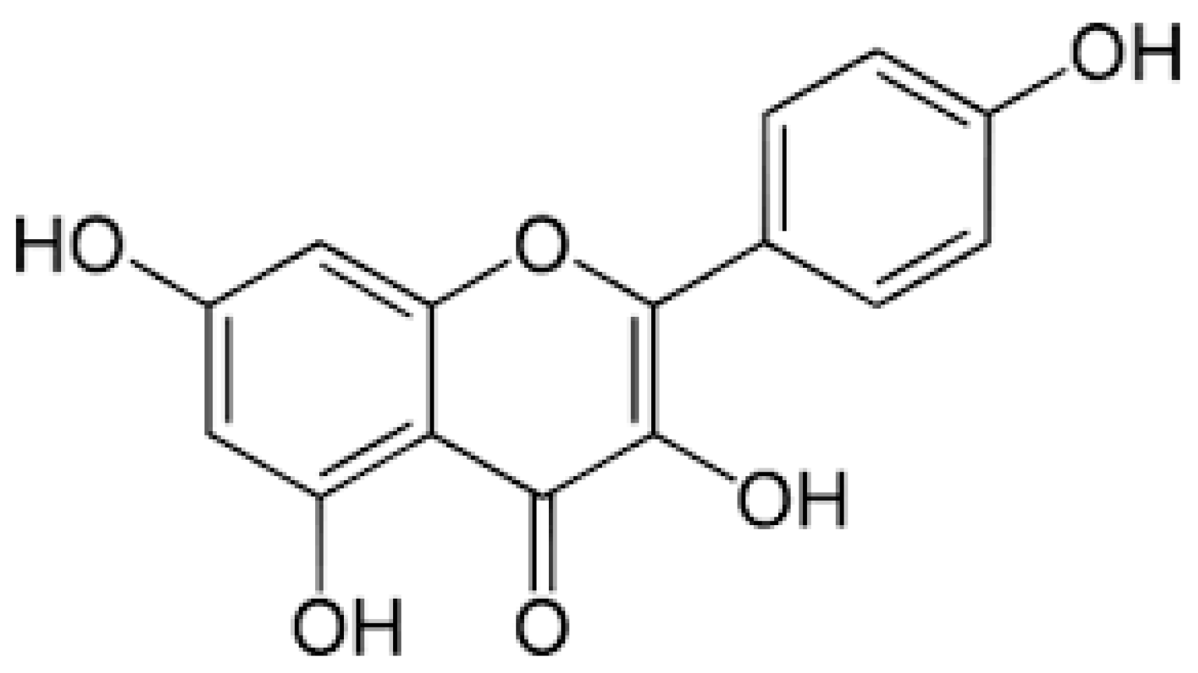
3. Sources and Biological Functions of Kaempferol

Kaempferol (3,5,7-trihydroxy-2-(4-hydroxyphenyl)-4H-chromen-4-one, molecular formula C15H10O6, molecular weight 286.24 g/mol) is a secondary plant metabolite that belongs to the larger group of flavonoids. They are polyphenolic molecules that share a basic structure of phenyl-benzo-γ-pyran, where aromatic ring A is condensed with heterocyclic ring C and linked to another aromatic ring B [35]. This group of compounds has strong antioxidant properties, mainly due to the phenolic hydroxyl groups attached to the aromatic rings [36]. In the realm of flavonoids, kaempferol, a member of the flavanol sub-group, has emerged as a subject of considerable scientific interest in recent times. This compound was first discovered in the tea plant (Camellia sinensis), but its name comes from Engelbert Kaempfer, a 17th-century German naturalist and physician who contributed significantly to the introduction of Japanese botanical knowledge in Europe [37]. Kaempferol has a polyphenolic structure with a carbonyl group at position 4 and four hydroxyl groups at positions 3, 4′, 5, and 7 (another name: 3,4′,5,7-tetrahydroxyflavone). The pure substance is a yellow crystalline powder with moderate solubility in water, but soluble in hot ethanol, ether, and hydroxide solutions [38]. The chemical structure of kaempferol is shown in Figure 1.
Kaempferol is widely distributed in various dietary and medicinal plants. It is one of the major flavanols that contribute to the beneficial effects of a plant-based diet [39]. Several vegetables, fruits and traditional medicinal herbs are particularly rich in kaempferol. The most significant amounts of kaempferol can be found in vegetables such as kale, spinach, onions, or beverages, especially black or green tea infusions [39]. Selected major sources of kaempferol are listed in Table 1.
Flavanols, such as kaempferol, have long been recognised as compounds with potent antioxidant properties [44], but a number of studies indicate that polyphenols (including kaempferol), rather than acting solely as direct antioxidants, appear to exert their beneficial effects primarily by modulating cellular signalling pathways and molecular mechanisms that regulate cell function in both healthy and pathological conditions [45]. For this reason, polyphenols have been extensively studied as potential therapeutic agents in various pathologies, including neurodegenerative, metabolic or inflammatory diseases, as well as in oncology as anticancer agents [38,41,46,47]. In addition, recent studies show promising results for the use of kaempferol as a preventive and antidotal agent against various natural and man-made toxins [48]. Kaempferol has also been investigated as an antiviral, antimicrobial, antiprotozoal and antifungal compound [48,49]. In this review, we will focus on the use of kaempferol in various metabolic disorders, especially those related to liver function.

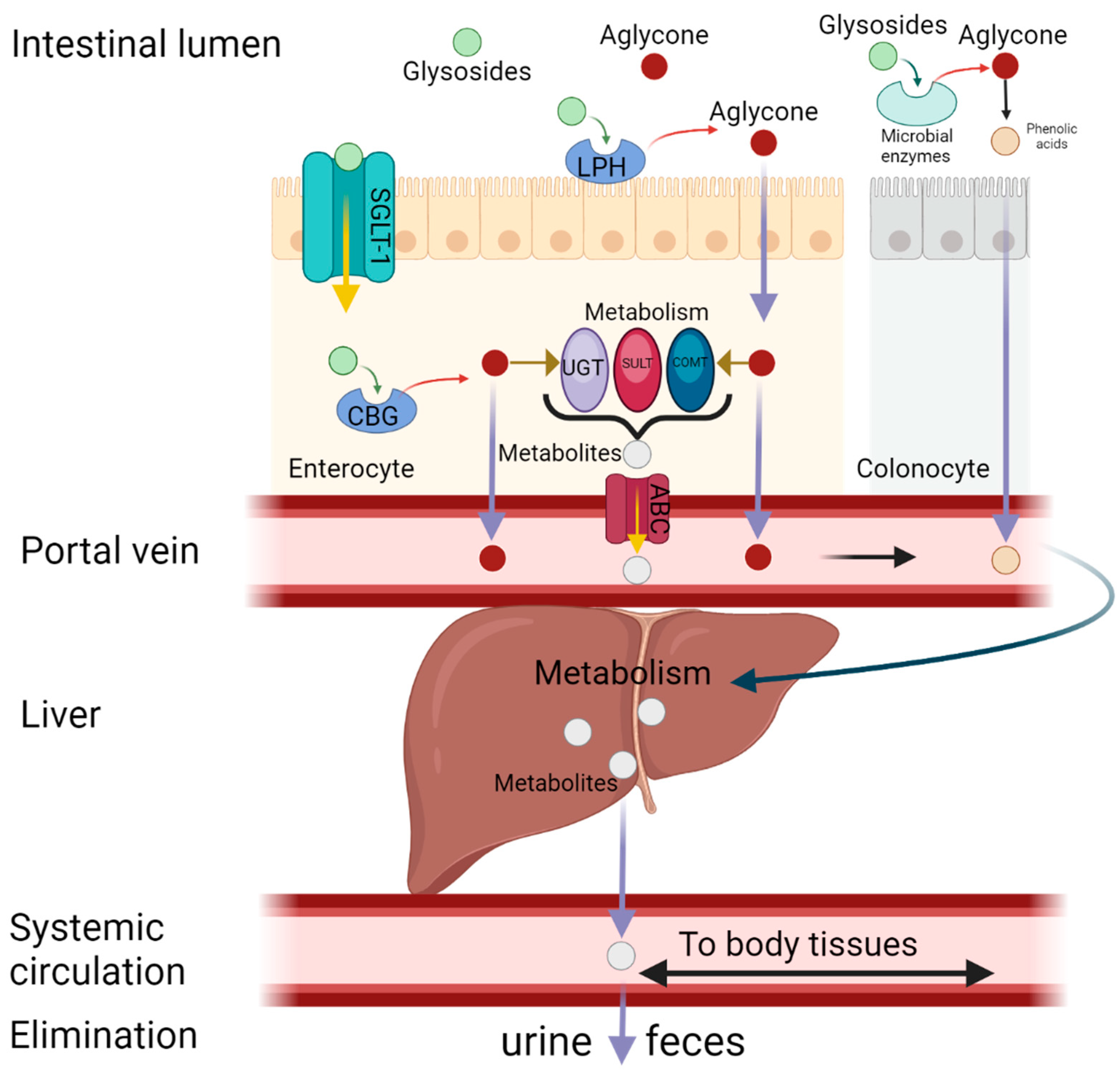
4. Metabolism and Bioavailability of Kaempferol

Flavonoids in general can occur in three chemical compositions: as aglycones, glycosides or methylated derivatives. Aglycone is a basic structure of any flavonoid, consisting of two benzene rings linked to a heterocyclic pyrene ring (the actual structure of kaempferol is shown in Figure 1). Glycosides consist of an aglycone and a sugar moiety, usually attached at the 3 or 7 position by a glycosidic linkage [50]. The most notable sugars that form glycosides with kaempferol include D-glucose, glucorhamnose, galactose, L-rhamnose, rutinose and arabinose [34]. There are examples of widely distributed kaempferol glycosides such as kaempferol-3-O-glucoside (astragalin) and kaempferol-3-O-rutinoside, found for example in tea and mulberry [51,52], or kaempferol-3-O-β-d-glucopyranoside-6-p-coumaril ester (tiliroside), found in lime or rose [53]. However, many other glycosides are found only in certain plant families or species and are therefore characteristic of these taxa and may be responsible for their unique properties [54]. Kaempferol in aglycon form has different physicochemical properties (e.g., lipophilicity, pKa, molecular size, solubility, etc.) than its glycosides, resulting in different pharmacokinetic properties, so the bioavailability of certain chemical forms of kaempferol may vary. Kaempferol in the aglycone form has lipophilic properties, whereas its glycosides become lipophobic when combined with sugars [55]. Nevertheless, either the hydrophobic aglycone or the hydrophilic glycosides of kaempferol are mainly supplied to the human body by the oral route, as they are very common phytochemicals present in many dietary products [39]. However, the bioavailability of the various chemical forms of oral kaempferol is low and has been calculated to be around 2% [46]. In the aglycon form, kaempferol is absorbed unchanged by passive diffusion into the enterocyte. Highly polar glycosides can be hydrolysed by the enzyme lactase-phlorizin hydrolase (LPH), which is present at the intestinal brush border. As a result, aglycon is released from its glycoside form and passively diffuses into the intestinal cells. The colonic microbiota, such as Escherichia coli, may also play an important role in the hydrolysis of kaempferol glycosides, but the cleaved aglycones in turn undergo further biotransformation to phenolic acids and are absorbed in this form [56,57]. However, glycosides can also be absorbed directly into the enterocyte via active transport mediated by sodium-dependent glucose transporter-1 (SGLT-1) and subsequently hydrolysed by the cytosolic enzyme β-glucosidase (CBG) [58]. Aglycones present in enterocytes may be transported directly into the portal circulation or may be metabolised prior to further distribution. Biotransformation in enterocytes involves both phase I (oxidation and O-demethylation) and phase II (sulphatation, glucuronidation, and methylation) reactions [59]. The main enzymes involved in phase II enzymes are uridine-5ʹ-diphosphate-glucuronosyltransferases (UGT), sulfotransferases (SULT) and catechol-O-methyltransferases (COMT) [56]. Unmetabolized aglycons and kaempferol metabolites are transported into the hepatic portal vein via passive diffusion and ATP-binding cassette (ABC), respectively [58]. The remaining kaempferol aglycones are metabolised in the liver during phase I and phase II metabolism and distributed to target organs, tissues and cells via the systemic circulation in the form of methyl, sulphur or glucuronide (mainly 7-O-glucuronide) [60]. These polar metabolites are mainly excreted by the kidneys in the urine or by the bile in the faeces. Approximately 2–2.5% of the total kaempferol ingested is excreted unchanged in the urine [61]. Extensive first-pass metabolism is probably responsible for the low bioavailability of kaempferol (approximately 2%), which limits its clinical use [46]. However, there are some modern approaches (nanoparticles, structural modifications, chimeric molecules) that could certainly be exploited to improve kaempferol bioavailability [62,63]. A schematic representation of the absorption, metabolism, distribution and excretion (ADME) of kaempferol is shown in Figure 2.

5. Effect of Kaempferol on Liver Condition

A substantial body of research has demonstrated the hepatoprotective properties of kaempferol. However, it is important to note that the preponderance of extant evidence derives from preclinical models [64,65,66]. Pretreatment with kaempferol in rats subjected to carbon tetrachloride administration has been shown to enhance liver enzyme activity and mitigate liver damage in rats treated with acetaminophen, an effect that is associated with an augmentation in sirtuin 1 (SIRT1) activity [66]. Recent research has highlighted the critical protective function of SIRT1 within the liver. The enzyme in question has been shown to influence a variety of physiological processes, including apoptosis, cel viability and antioxidant concentrations [67]. This NAD+-dependent deacetylase has been found to mediate these effects by deacetylation of transcription factors (e.g., NF-κB and STAT3) involved in the processes of inflammation and the maintenance of antioxidant potential, as well as fork-head transcription factors (FOXO and p53) and PGC-1 (which is involved in mitochondrial biogenesis). SIRT1 increases cell proliferation and survival by deacetylating multiple transcription factors. It also reduces oxidative stress and cellular inflammation as well as increases the production of ATP and mitochondrial biogenesis [68]. By increasing antioxidant levels and reducing inflammation and apoptosis in the liver, kaempferol has the capacity to provide comprehensive protection against oxidative damage to this organ [69].
As postulated by BinMowyna and AlFaris [70], the hepatoprotective effect of the compound is a consequence of the deacetylation of FOXO1. The suppression of the expression of a number of apoptotic genes has been observed to be accompanied by an increase in the expression of antiapoptotic and antioxidant genes (MnSOD and Bcl-2). This has been shown to lead to acetylation and inactivation of NF-κB p65, inhibition of p53 acetylation, nuclear translocation and a consequent increase in Bax synthesis. NF-κB p65 generally stimulates inflammation by upregulation of inflammatory cytokines and induces apoptosis through Bcl-2 suppression and Bax upregulation. Upregulation and activation of SIRT1 in the liver by kaempferol are shown in Figure 3 [68,71].
The hepatoprotective effects of kaempferol are attributed to its specific modulation of liver-related pathways, including SIRT1 activation, CYP2E1 inhibition, TLR4/NF-κB suppression and ALK5/Smad pathway interference. Unique mechanisms distinguish it from other flavonoids such as quercetin and genistein, and highlight its potential as a targeted therapeutic agent for liver disease. Quercetin shares some antioxidant and anti-inflammatory properties, but does not have the same level of specificity in modulating SIRT1 activity or inhibiting CYP2E1 [72]. Genistein, primarily known for its estrogenic effects, lacks the direct involvement in liver-specific pathways that kaempferol demonstrates [73]. These specific pathways highlight kaempferol’s potential as a therapeutic agent for liver diseases, and warrant further clinical investigation to fully elucidate its efficacy and safety.

5.1. Reduction of Hepatic Lipid Accumulation

Two mechanisms that regulate lipid levels are lipid storage and metabolism. An imbalance in either process can lead to the accumulation of lipids in the liver, potentially causing harm to health [74]. As demonstrated by in vitro studies conducted with oleic acid-induced HepG2 cells, treatment with kaempferol led to a considerable decrease in lipid storage [74]. In addition, results from an in vivo study of type 2 diabetes in mice substantiated that a dosage of kaempferol (50 mg/kg) led to a marked diminution of lipid accumulation and a significant amelioration of liver damage, which is a key area of concern. New research has shown that SIRT1 and AMPK (AMP-activated protein kinase) work together to form a positively regulated loop that reinforces each other [75]. Nevertheless, research has indicated that the progression of hepatic steatosis leads to the inhibition of SIRT1/AMPK [76]. The activation of SIRT1/AMPK signalling has been demonstrated to promote fatty acid oxidation (FAO) through the modulation of the proliferator-activated receptor gamma coactivator 1α (PGC1α), acetyl-CoA carboxylase (ACC) and carnitine palmitoyltransferase 1 (CPT1) pathways. In mice, decreased hepatic AMPK activity has been found to lower ACC phosphorylation, which inactivates ACC, thereby reducing fatty acid oxidation (FAO) and increasing lipogenesis [77]. Sterol regulatory element-binding proteins (SREBPs) are essential transcription factors that control the expression of genes responsible for lipid, cholesterol, fatty acid, and triglyceride production in liver tissues [78]. Research has shown that AMPK suppresses SREBP activation by promoting its deacetylation, which in turn lowers the expression of crucial lipogenic genes like fatty acid synthase (FASN) [79]. Kaempferol treatment caused activation of SIRT1 and AMPK, a significant improvement in the concentrations of fatty acid oxidation-activated PGC1α and a reduction in the concentrations of lipid synthesis-related proteins. In addition, the anti-lipid effect of kaempferol was blocked when SIRT1 or AMPK were silenced. This suggests that kaempferol could be a therapeutic agent for controlling lipid accumulation in the liver by targeting the SIRT1/AMPK signalling pathway.

5.2. Inhibition of Hepatic Inflammation

The liver is vulnerable to the toxic effects of chemicals, which can result in liver damage, fibrosis, and impaired function [71]. One such substance that has been shown to induce liver damage and trigger receptors such as TLR4 in hepatocytes and Kupffer cells is lipopolysaccharide (LPS) [80]. It has been demonstrated that this compound has the capacity to promote NF-κB phosphorylation and the production of pro-inflammatory cytokines such as TNF-α, IL-1 and IL-6 [81]. It has been demonstrated that LPS-induced hepatocellular injury can also lead to oxidative and nitrosative stress, resulting in the elevation of oxidants like ROS and RNS, depletion of endogenous antioxidants such as GSH and SOD, and elevation of MDA [82]. A multitude of cells activate a wide range of intracellular signalling cascades via two key adaptor molecules containing Toll/IL-1 receptor domains, TRIF and MyD88, to promote the activation of pro-inflammatory transcription factors including NF-κB and AP-1. This process results in the expression of genes that are involved in the inflammatory response, including iNOS, COX-2, cytokines, and chemokines [83]. It has been reported that kaempferol significantly decreases the expression level of TLR4 mRNA and protein to reduce NF-κB p65 phosphorylation in liver tissue. Kaempferol can also suppress the production and expression of COX-2, IL-1β, TNF-α, and IL-6 mRNA, which play key roles in inflammation. Additionally, it reduces the levels of NO and PGE2 while lowering iNOS mRNA expression in cases of acute liver injury. These findings indicate that kaempferol alleviates liver inflammation by inhibiting TLR4 and NF-κB activation, thereby reducing proinflammatory cytokine production. Moreover, it protects against hepatic nitrosative stress and helps restore normal liver function [84]. A mechanism of the protection of kaempferol against LPS-induced acute liver injury is illustrated in Figure 4 [71].
Persistent fat accumulation in the liver leads to excessive production of the extracellular matrix, surpassing its breakdown rate and ultimately causing progressive liver fibrosis [46]. Liu et al. [85] investigated this process using an in vitro model of oleic acid-treated HepG2 cells and an in vivo model of high-fat diet (HFD)-fed Sprague-Dawley (SD) rats. Their findings suggest that kaempferol (1 or 10 μM) can enhance NF-κB levels in the cytoplasm while reducing its presence in the nucleus, thereby lowering TNF-α and IL-6 levels- key factors in the pathophysiology of MASLD. Additionally, kaempferol (20, 40, or 60 μM) was shown to alleviate liver fat accumulation by modulating endoplasmic reticulum stress (ERS) and the expression of liver X receptor (LXR) and lysophosphatidylcholine acyltransferase 3 (LPACT3). These mechanisms are thought to play a crucial role in the development of hepatic steatosis and inflammation. Reducing liver inflammation may be achieved by suppressing the mRNA expression of inflammatory markers such as TNF-α and IL-6 [86].

5.3. Inhibition of Hepatic Oxidative Stress

It is imperative to note the intricate relationship between oxidative stress, inflammation and apoptosis. These processes are of particular significance within both physiological and pathological conditions [87]. Kaempferol has been shown to play a role in the treatment of diseases through its antioxidant potential. In a study on diabetic rats, it was observed that administration of kaempferol led to a significant improvement in glucose, insulin, and lipid peroxidation product levels in the plasma [88]. Furthermore, the levels of enzymatic and non-enzymatic antioxidative substances were found to be restored to nearly normal levels [89].
In a further study [90] the activity of SOD and the levels of MDA were determined in the liver to investigate the role of kaempferol in the oxidative response following haemorrhagic shock. The results obtained demonstrated that, in comparison to the sham group, the haemorrhagic shock groups exhibited significantly elevated levels of MDA. Conversely, SOD activity was reduced in the haemorrhagic shock groups in comparison to the control group. The injection of kaempferol after haemorrhagic shock had no effect on liver MDA levels and SOD activity when compared with the haemorrhagic shock group. However, when kaempferol was injected 12 h prior to the induction of haemorrhagic shock, increased SOD activity and decreased MDA levels in the liver were observed when compared with the haemorrhagic shock group. Furthermore, a significant decrease in plasma levels of IL-6 and TNF-α was observed, accompanied by the restoration of MDA, SOD, and MPO levels in the liver. Concurrently, an augmented expression of HO-1 was observed. Collectively, these observations suggest that kaempferol may possess the potential to mitigate the detrimental consequences of haemorrhagic shock in murine models. It is also notable that oxidative stress can be induced by excessive alcohol consumption. Ethanol is metabolised by alcohol dehydrogenase (ADH) and CYP2E1 to produce ROS. A recent study demonstrated a correlation between CYP2E1 activity and ethanol-induced liver damage and lipid peroxidation [91].
Elevated levels of liver enzymes (AST and ALT) have been shown to damage liver cells. These enzymes rely on pyridoxal phosphate (PLP) to function and play a key role in converting aspartate and ketoglutarate into glutamate and oxaloacetate. When liver cells are damaged, the levels of these enzymes can rise, indicating a loss of membrane integrity and impaired cellular function. Furthermore, it has been demonstrated that oxidative stress can induce Nrf2 in human hepatocyte cells by facilitating dissociation from Keap1 and subsequent translocation to the nucleus, where it binds to antioxidant response elements and activates target gene expression [92].
It has been demonstrated that kaempferol has the capacity to inhibit CYP2E1 at both the expression and activity levels, consequently leading to a reduction in ROS levels and liver damage. The significant decrease in serum AST and ALT levels is due to this inhibitory effect. The induction of reactive antioxidant enzymes (GSH and SOD) by this compound serves to remove lipid products (MDA) and ROS (H2O2) [93]. Furthermore, kaempferol has been shown to induce protective effects on liver structure by inhibiting hepatocyte apoptosis through the reduction of apoptosis-related proteins, including cytochrome c, Bax, Bcl-2, caspases:3, 8 and 9 [94].

5.4. Down-Regulation of Liver Fibrosis

The process of liver fibrosis is characterised by persistent or recurrent liver damage resulting from hepatotoxic substances such as alcohol, in addition to chronic liver diseases including alcoholic hepatitis, steatosis, and viral hepatitis, along with autoimmune disorders [95]. Cirrhosis, frequently associated with liver failure, represents the culmination of chronic liver necrosis and serves as the ultimate outcome of the aforementioned process [96]. Liver fibrogenesis is primarily driven by dysfunctional hepatic stellate cells (HSCs), which, under normal physiological conditions, act as stores of vitamin A in their inactive form. However, upon activation, these cells transform into myofibroblast-like cells, express α-SMA and produce large amounts of collagen. Accumulation of collagen results in the replacement of normal liver parenchyma by scar tissue, leading to liver fibrosis [97]. TGF-β is a regulatory cytokine that plays a critical role in the process of liver fibrosis, affecting HSC activation and proliferation, and ECM formation [98]. As demonstrated in the relevant literature, transforming growth factor-beta (TGF-β) has been shown to bind to its cognate receptor, namely TGF-β type II, thus resulting in the phosphorylation of Smad2 and Smad3. This process, in turn, has been observed to activate hepatic stellate cells (HSCs) and initiate the transcription of pro-fibrosis genes [99].
Kaempferol has been shown to be capable of inhibiting type I collagen expression in HSCs and reducing collagen density in liver tissue. It has been demonstrated that this can be achieved by reducing the phosphorylation of Smad2 and Smad3 by the serine/threonine kinase, attenuating α-SMA production, and inhibiting TGF-β-stimulated HSCs. In addition, it has been shown to bind specifically to ALK5 and further inhibit the TGF-β/Smad pathway. It may also act as an anti-fibrotic agent against fibrotic diseases [100].

5.5. Modulation of Gut Microbiota

Hepatic steatosis occurs when excess fat accumulates in the liver. This condition is frequently seen in individuals with obesity and type 2 diabetes. Extensive research involving both human and animal studies has shown a strong link between obesity and the gut microbiome [101,102].
Recent findings suggest that polyphenols, such as kaempferol, can influence gut microbiota composition, potentially improving metabolic disorders [103]. In the study conducted by Wang et al. [104] C57BL/6 J mice were fed a high-fat diet and given kaempferol (200 mg/kg) for eight weeks. The results showed a reduction in body weight and fat deposits in various white adipose tissues, including inguinal, epididymal, and perirenal fat. Additionally, kaempferol supplementation increased the Shannon index in faecal samples, indicating greater microbial diversity. The treatment also altered the gut microbiome by increasing the relative abundance of Bacteroidetes and Proteobacteria while reducing Firmicutes at the phylum level. At the genus level, there was an increase in Akkermansia, Bacteroides, and Lactobacillus populations. Another study extended the supplementation period to 16 weeks in obese C57BL/6 J mice and found that kaempferol helped counteract obesity-related changes in gut microbiota. The researchers observed an overall increase in microbial diversity in mice consuming a high-fat diet with kaempferol supplementation [105]. Results of kaempferol administration on on liver diseases in preclinical models and its mechanisms of action are presented in Table 2.

6. Safety of Kaempferol and Its Possible Interactions with Conventional Medicines

It should be noted that kaempferol exhibits a range of biological activities, some of which, depending on the circumstances, may be beneficial or detrimental. Kaempferol has been reported to have mutagenic and genotoxic properties in experiments on Drosophila melanogaster [121]. In vitro, kaempferol may induce chromosomal aberrations in V79 Chinese hamster cells, as I. Duarte Silva et al. have found [122]. This is thought to involve the biotransformation of kaempferol to quercetin by cytochromes P450 in the presence of metabolic activation systems. Moreover, it has been reported that kaempferol may result in a multitude of adverse consequences. For instance, as demonstrated by Lemos et al. [123], it was found that kaempferol could moderately inhibit the uptake of folic acid in human colon adenocarcinoma Caco-2 cells. This may have certain adverse impacts on folate-deficient individuals. It has been demonstrated that kaempferol exerts potent cytotoxic and anti-proliferative activities against several human cancer cells. A growing body of research has recently demonstrated that kaempferol exhibits selective toxicity towards cancer cells, while sparing normal cells. This finding serves to reinforce the notion that further investigation into kaempferol as an anticancer agent is warranted. For instance, the toxicity of kaempferol was significantly higher in HeLa cells than in normal HFF cells. This is manifested by the IC50 values of kaempferol HeLa cells being 45.63, 22.87 and 10.48 mM at 24, 48, and 72 h of treatment, respectively, as compared with 1079.0 and 707.0 mM for HFF cells at 48 and 72 h, respectively. At 24, 48 and 72 h, the IC50 values for HeLa cells were 45.63, 22.87 and 10.48 mM, compared with 1079.0 and 707.0 mM for HFF cells at 48 and 72 h. Mechanically, kaempferol induced cellular apoptosis and ageing by downregulating the PI3K/AKT and hTERT pathways [123]. The present result is consistent with the findings of Tu et al. [124], who demonstrated that kaempferol inhibited the proliferation of SiHa cervical cancer cells in a dose- and time-dependent manner. However, the study also revealed that kaempferol had minimal cytotoxic effects on normal kidney HK-2 cells. Examples of IC 50 for various cells are as follows: HepG2—30.0 μM (48 h), pancreatic cancer Mia PaCa-2—79.07 μM (48 h), lung cancer H460—50.0 μM (48 h), blood cancer HL60 cells- 250.60 μM (48 h), noncancer cells HFF—1079.00 μM (48 h). The findings of the studies indicated that kaempferol does not demonstrate significant toxicity towards normal cells, provided that the administered dose and the duration of administration are within reasonable limits.
When considering the potential interactions of kaempferol with conventional drugs, there are several factors to be taken into account, including its metabolic pathways and the manner in which it may affect drug-metabolising enzymes, particularly those located in the liver. The liver plays a pivotal role in the process of drug metabolism through the action of enzymes, including the cytochrome P450 (CYP) system. Kaempferol interacts with cytochrome P450 enzymes, including CYP3A4, which is key to drug metabolism. It has been demonstrated that drugs metabolised by this enzyme, including statins (e.g., atorvastatin) and certain immunosuppressants (e.g., cyclosporine), may experience increased plasma concentrations when co-administered with kaempferol, potentially resulting in adverse effects [125]. It may also inhibit CYP1A2, an enzyme that facilitates the metabolism of drugs such as theophylline and caffeine. There is some evidence to suggest that kaempferol can inhibit CYP2C9, which is involved in the metabolism of drugs like warfarin. This could increase the risk of bleeding if these medications are used concomitantly [126]. These interactions have the potential to result in alterations to drug levels in the bloodstream, either increasing toxicity or reducing the therapeutic efficacy of certain medications.
P-glycoprotein (P-gp) is a drug transporter that plays a role in limiting the absorption and enhancing the elimination of various drugs. It has been demonstrated that kaempferol is capable of inhibiting P-gp, which may consequently result in an enhancement of the bioavailability of drugs that are P-gp substrates. It has been hypothesised that this may result in an increased risk of adverse effects or toxicity in medications such as tacrolimus (utilised in the context of organ transplantation) or anticancer agents, including paclitaxel [127]. The influence of kaempferol on the expression of liver enzymes, particularly through the activation of nuclear receptors such as Pregnane X Receptor (PXR) and Aryl Hydrocarbon Receptor (AhR), suggests the potential for regulation of drug-metabolising enzymes [128,129]. This modulation has the potential to induce alterations in the hepatic metabolism of various pharmaceutical agents, which may necessitate dose recalibrations for patients receiving drugs that are substrates of these pathways [130].

7. Conclusions and Perspectives

Natural medicines are gaining worldwide recognition due to their excellent therapeutic efficacy and relatively low side effects. Kaempferol, a widely available natural compound, is present in many medicinal plants. This review provides an overview of its pharmacological effects on liver diseases, the molecular mechanisms involved, its dietary sources, and its pharmacokinetic properties. Various in vivo and in vitro studies have demonstrated that kaempferol exerts therapeutic effects on liver diseases by regulating metabolic functions and pathological processes. For instance, it can help reduce fat accumulation in the liver, decrease liver fibrosis, and support a healthy balance of intestinal flora, which plays a crucial role in its beneficial effects. In addition, the anti-inflammatory effect through the downregulation of pro-inflammatory cytokines is the common mechanism of action of kaempferol in the treatment of liver problems, including IL-6, IL-1β and TNF-α. Through multiple mechanisms and signalling pathways, these studies show that kaempferol has the potential to improve the symptoms of liver disease. It is evident that kaempferol is a natural compound that merits rigorous investigation. The present study, however, is not without its limitations. The research conducted on kaempferol in the context of liver disease researchers has been primarily confined to fundamental investigations, encompassing animal and cell experiments. Hence, the necessity for additional clinical studies arises, with the objective of substantiating the purported beneficial effects of kaempferol.
Natural compound safety assumes equal significance to its efficacy in the development of therapeutic drugs. While the majority of studies have shown that it is relatively non-toxic to normal cells under reasonable doses and times, the need for additional, longer-term clinical trials to better determine its safety in humans is crucial. Furthermore, there remains a paucity of data on the oral bioavailability of kaempferol, a key obstacle to its use in treating metabolic diseases. Enhancing the oral bioavailability of kaempferol through nanotechnology or structural modifications could help advance it from basic research to clinical use.
In summary, kaempferol holds significant promise as a treatment for liver diseases. However, further research, particularly clinical studies, is needed to provide stronger evidence for its proposed molecular mechanisms and targets. Additionally, well-structured, extensive, and long-term clinical trials are essential to assess its effectiveness and safety, ultimately supporting its transition into clinical practice and allowing more patients to benefit from its therapeutic potential.

Author Contributions

Conceptualisation, T.O., P.N. and A.O.; methodology, P.N., A.K. and A.O.; data collection, P.N., B.P. and A.K.; writing—original draft preparation, P.N., T.O. and A.O; supervision, A.O. All authors have read and agreed to the published version of the manuscript.

Funding

This research received no external funding.

Institutional Review Board Statement

Not applicable.

Informed Consent Statement

Not applicable.

Data Availability Statement

Not applicable.

Acknowledgments

Graphical Abstract and Figure 2, Figure 3 and Figure 4 were created in BioRender. License granted to Niziński, P. (2025) https://BioRender.com/38iavn6; https://BioRender.com/wpnlrf4; https://biorender.com/jx5npt5; https://www.biorender.com/ktyaw74.

Conflicts of Interest

The authors declare no conflicts of interest. The funders had no role in the design of the study; in the collection, analyses, or interpretation of data; in the writing of the manuscript; or in the decision to publish the results.

Abbreviations

The following abbreviations are used in this manuscript:
ABCATP-binding cassette
ACCacetyl-CoA carboxylase
ADH alcohol dehydrogenase
ADMEabsorption, distribution, metabolism, excretion
AhRAryl Hydrocarbon Receptor
ALK5activin receptor-like kinase 5
ALTalanine transferase
AMPKSIRT1/AMP-activated protein kinase
ASTaspartate aminotransferase
Baxbcl-2-like protein 4
Bcl-2B-cell CLL/lymphoma 2
CBGCGAcytosolic enzyme β-glucosidasechlorogenic acid
COMTcatechol-O-methyltransferase
COXcyclooxygenase
CPT1carnitine palmitoyltransferase 1
CYP2E1cytochrome P450 2E1
ECMextracellular matrix
ERSendoplasmic reticulum stress
FAOfatty acid oxidation
FASNfatty acid synthase
FOXOforkhead box protein
GCGglucagon
GIPgastric inhibitory polypeptide
GLP-1glucagon-like peptide 1
GSHglutathione
HCChepatocellular carcinoma
HBVhepatitis B virus
HCVhepatitis C virus
HFDhigh-fat diet
HO-1heme oxygenase 1
HSChematopoietic stem cells
ILinterleukin
iNOSinducible nitric oxide synthase
LPACT3lysophosphatidylcholine acyltransferase 3
LPHlactase-phlorizin hydrolase
LPSlipopolysaccharide
LXRliver X receptor
MASLDmetabolic dysfunction-associated steatotic liver disease
MDAmalondialdehyde
MFBs myofibroblasts
MnSODmanganese-dependent superoxide dismutase
MPOmyeloperoxidase
NAD+oxidized nicotinamide adenine dinucleotide
NAFLDnon-alcoholic fatty liver disease
NF-κBnuclear factor kappa-light-chain-enhancer of activated B cells
NOnitric oxide
P-gpP-glycoprotein
p53tumour protein p53
PDGFplatelet-derived growth factor
PGC-1peroxisome proliferator-activated receptor gamma coactivator 1
PGEProstaglandin E
PLPpyridoxal phosphate
PXRPregnane X Receptor
RNSreactive nitrogen species
ROSreactive oxygen species
SGLT-1sodium-dependent glucose transporter-1
SIRT1sirtuin 1
SODsuperoxide dismutase
SREBPssterol regulatory element-binding proteins
STAT3signal transducer and activator of transcription 3.
SULTsulfotransferase
TLR4toll-like receptor 4
TNF-αtumour necrosis factor-alpha
UGTuridine-5ʹ-diphosphate-glucuronosyltransferase
α-SMA alpha-smooth muscle actin

References

  1. Trefts, E.; Gannon, M.; Wasserman, D.H. The Liver. Curr. Biol. 2017, 27, R1147–R1151. [Google Scholar] [CrossRef] [PubMed]
  2. Kalra, A.; Yetiskul, E.; Wehrle, C.J.; Tuma, F. Physiology, Liver. In StatPearls; StatPearls Publishing: Treasure Island, FL, USA, 2025. [Google Scholar]
  3. Asrani, S.K.; Devarbhavi, H.; Eaton, J.; Kamath, P.S. Burden of Liver Diseases in the World. J. Hepatol. 2019, 70, 151–171. [Google Scholar] [CrossRef]
  4. Devarbhavi, H.; Asrani, S.K.; Arab, J.P.; Nartey, Y.A.; Pose, E.; Kamath, P.S. Global Burden of Liver Disease: 2023 Update. J. Hepatol. 2023, 79, 516–537. [Google Scholar] [CrossRef]
  5. Teng, M.L.; Ng, C.H.; Huang, D.Q.; Chan, K.E.; Tan, D.J.; Lim, W.H.; Yang, J.D.; Tan, E.; Muthiah, M.D. Global Incidence and Prevalence of Nonalcoholic Fatty Liver Disease. Clin. Mol. Hepatol. 2023, 29, S32–S42. [Google Scholar] [CrossRef]
  6. Heinemann, F.; Gross, P.; Zeveleva, S.; Qian, H.S.; Hill, J.; Höfer, A.; Jonigk, D.; Diehl, A.M.; Abdelmalek, M.; Lenter, M.C.; et al. Deep Learning-Based Quantification of NAFLD/NASH Progression in Human Liver Biopsies. Sci. Rep. 2022, 12, 19236. [Google Scholar] [CrossRef]
  7. Harrison, S.A.; Bedossa, P.; Guy, C.D.; Schattenberg, J.M.; Loomba, R.; Taub, R.; Labriola, D.; Moussa, S.E.; Neff, G.W.; Rinella, M.E.; et al. A Phase 3, Randomized, Controlled Trial of Resmetirom in NASH with Liver Fibrosis. N. Engl. J. Med. 2024, 390, 497–509. [Google Scholar] [CrossRef] [PubMed]
  8. Food and Drug Administration (FDA). Available online: https://www.fda.gov/news-events/press-announcements/fda-approves-first-treatment-patients-liver-scarring-due-fatty-liver-disease (accessed on 30 March 2025).
  9. Zhao, B.; Liu, K.; Liu, X.; Li, Q.; Li, Z.; Xi, J.; Xie, F.; Li, X. Plant-Derived Flavonoids Are a Potential Source of Drugs for the Treatment of Liver Fibrosis. Phytother. Res. 2024, 38, 3122–3145. [Google Scholar] [CrossRef]
  10. Dabeek, W.M.; Marra, M.V. Dietary Quercetin and Kaempferol: Bioavailability and Potential Cardiovascular-Related Bioactivity in Humans. Nutrients 2019, 11, 2288. [Google Scholar] [CrossRef] [PubMed]
  11. Markowska, J.; Kasprzak-Drozd, K.; Niziński, P.; Dragan, M.; Kondracka, A.; Gondek, E.; Oniszczuk, T.; Oniszczuk, A. Quercetin: A Promising Candidate for the Management of Metabolic Dysfunction-Associated Steatotic Liver Disease (MASLD). Molecules 2024, 29, 5245. [Google Scholar] [CrossRef]
  12. Ziółkiewicz, A.; Niziński, P.; Soja, J.; Oniszczuk, T.; Combrzyński, M.; Kondracka, A.; Oniszczuk, A. Potential of Chlorogenic Acid in the Management of Metabolic Dysfunction-Associated Steatotic Liver Disease (MASLD): Animal Studies and Clinical Trials—A Narrative Review. Metabolites 2024, 14, 346. [Google Scholar] [CrossRef]
  13. Kasprzak-Drozd, K.; Niziński, P.; Kasprzak, P.; Kondracka, A.; Oniszczuk, T.; Rusinek, A.; Oniszczuk, A. Does Resveratrol Improve Metabolic Dysfunction-Associated Steatotic Liver Disease (MASLD)? Int. J. Mol. Sci. 2024, 25, 3746. [Google Scholar] [CrossRef] [PubMed]
  14. Ashrafizadeh, M.; Tavakol, S.; Ahmadi, Z.; Roomiani, S.; Mohammadinejad, R.; Samarghandian, S. Therapeutic Effects of Kaempferol Affecting Autophagy and Endoplasmic Reticulum Stress. Phytother. Res. 2020, 34, 911–923. [Google Scholar] [CrossRef] [PubMed]
  15. Silva dos Santos, J.; Gonçalves Cirino, J.P.; de Oliveira Carvalho, P.; Ortega, M.M. The Pharmacological Action of Kaempferol in Central Nervous System Diseases: A Review. Front. Pharmacol. 2021, 11, 565700. [Google Scholar] [CrossRef]
  16. Zheng, Y.; Xie, L.; Yang, D.; Luo, K.; Li, X. Small-Molecule Natural Plants for Reversing Liver Fibrosis Based on Modulation of Hepatic Stellate Cells Activation: An Update. Phytomedicine 2023, 113, 154721. [Google Scholar] [CrossRef] [PubMed]
  17. Huang, T.D.; Behary, J.; Zekry, A. Non-Alcoholic Fatty Liver Disease: A Review of Epidemiology, Risk Factors, Diagnosis and Management. Intern. Med. J. 2020, 50, 1038–1047. [Google Scholar] [CrossRef]
  18. Rinella, M.E.; Lazarus, J.V.; Ratziu, V.; Francque, S.M.; Sanyal, A.J.; Kanwal, F.; Romero, D.; Abdelmalek, M.F.; Anstee, Q.M.; Arab, J.P.; et al. A Multisociety Delphi Consensus Statement on New Fatty Liver Disease Nomenclature. Hepatology 2023, 78, 1966–1986. [Google Scholar] [CrossRef]
  19. Lonardo, A.; Ballestri, S.; Marchesini, G.; Angulo, P.; Loria, P. Nonalcoholic Fatty Liver Disease: A Precursor of the Metabolic Syndrome. Dig. Liver Dis. 2015, 47, 181–190. [Google Scholar] [CrossRef]
  20. Rizzo, M.; Colletti, A.; Penson, P.E.; Katsiki, N.; Mikhailidis, D.P.; Toth, P.P.; Gouni-Berthold, I.; Mancini, J.; Marais, D.; Moriarty, P.; et al. Nutraceutical Approaches to Non-Alcoholic Fatty Liver Disease (NAFLD): A Position Paper from the International Lipid Expert Panel (ILEP). Pharmacol. Res. 2023, 189, 106679. [Google Scholar] [CrossRef]
  21. Katsiki, N.; Stoian, A.P.; Steiropoulos, P.; Papanas, N.; Suceveanu, A.-I.; Mikhailidis, D.P. Metabolic Syndrome and Abnormal Peri-Organ or Intra-Organ Fat (APIFat) Deposition in Chronic Obstructive Pulmonary Disease: An Overview. Metabolites 2020, 10, 465. [Google Scholar] [CrossRef]
  22. Zhao, M.; Chen, S.; Ji, X.; Shen, X.; You, J.; Liang, X.; Yin, H.; Zhao, L. Current Innovations in Nutraceuticals and Functional Foods for Intervention of Non-Alcoholic Fatty Liver Disease. Pharmacol. Res. 2021, 166, 105517. [Google Scholar] [CrossRef]
  23. Farrell, G.C.; Larter, C.Z. Nonalcoholic Fatty Liver Disease: From Steatosis to Cirrhosis. Hepatology 2006, 43, S99–S112. [Google Scholar] [CrossRef]
  24. Taylor, R.S.; Taylor, R.J.; Bayliss, S.; Hagström, H.; Nasr, P.; Schattenberg, J.M.; Ishigami, M.; Toyoda, H.; Wai-Sun Wong, V.; Peleg, N.; et al. Association Between Fibrosis Stage and Outcomes of Patients with Nonalcoholic Fatty Liver Disease: A Systematic Review and Meta-Analysis. Gastroenterology 2020, 158, 1611–1625.e12. [Google Scholar] [CrossRef] [PubMed]
  25. Simon, T.G.; Roelstraete, B.; Hagström, H.; Loomba, R.; Ludvigsson, J.F. Progression of Non-Alcoholic Fatty Liver Disease and Long-Term Outcomes: A Nationwide Paired Liver Biopsy Cohort Study. J. Hepatol. 2023, 79, 1366–1373. [Google Scholar] [CrossRef]
  26. Tanaka, M.; Miyajima, A. Liver Regeneration and Fibrosis after Inflammation. Inflamm. Regen. 2016, 36, 19. [Google Scholar] [CrossRef] [PubMed]
  27. Dhar, D.; Baglieri, J.; Kisseleva, T.; Brenner, D.A. Mechanisms of Liver Fibrosis and Its Role in Liver Cancer. Exp. Biol. Med. 2020, 245, 96–108. [Google Scholar] [CrossRef]
  28. Bai, Q.; Yan, H.; Sheng, Y.; Jin, Y.; Shi, L.; Ji, L.; Wang, Z. Long-Term Acetaminophen Treatment Induced Liver Fibrosis in Mice and the Involvement of Egr-1. Toxicology 2017, 382, 47–58. [Google Scholar] [CrossRef] [PubMed]
  29. Berumen, J.; Baglieri, J.; Kisseleva, T.; Mekeel, K. Liver Fibrosis: Pathophysiology and Clinical Implications. WIREs Mech. Dis. 2021, 13, e1499. [Google Scholar] [CrossRef]
  30. An, L.; Wirth, U.; Koch, D.; Schirren, M.; Drefs, M.; Koliogiannis, D.; Nieß, H.; Andrassy, J.; Guba, M.; Bazhin, A.V.; et al. The Role of Gut-Derived Lipopolysaccharides and the Intestinal Barrier in Fatty Liver Diseases. J. Gastrointest. Surg. 2022, 26, 671–683. [Google Scholar] [CrossRef]
  31. Dewidar, B.; Meyer, C.; Dooley, S.; Meindl-Beinker, N. TGF-β in Hepatic Stellate Cell Activation and Liver Fibrogenesis—Updated 2019. Cells 2019, 8, 1419. [Google Scholar] [CrossRef]
  32. Sultana, M.; Islam, M.A.; Khairnar, R.; Kumar, S. A Guide to Pathophysiology, Signaling Pathways, and Preclinical Models of Liver Fibrosis. Mol. Cell. Endocrinol. 2025, 598, 112448. [Google Scholar] [CrossRef]
  33. Zhang, C.-Y.; Liu, S.; Yang, M. Treatment of Liver Fibrosis: Past, Current, and Future. World J. Hepatol. 2023, 15, 755–774. [Google Scholar] [CrossRef]
  34. Kumar, S.; Pandey, A.K. Chemistry and Biological Activities of Flavonoids: An Overview. Sci. World J. 2013, 2013, 162750. [Google Scholar] [CrossRef]
  35. Panche, A.N.; Diwan, A.D.; Chandra, S.R. Flavonoids: An Overview. J. Nutr. Sci. 2016, 5, e47. [Google Scholar] [CrossRef] [PubMed]
  36. Hossen, M.J.; Uddin, M.B.; Ahmed, S.S.U.; Yu, Z.-L.; Cho, J.Y. Kaempferol: Review on Natural Sources and Bioavailability. In Kaempferol: Biosynthesis, Food Sources and Therapeutic Uses; Nova Science Publishers: New York, NY, USA, 2016; pp. 101–150. [Google Scholar]
  37. Periferakis, A. On the Dissemination of Acupuncture to Europe. JournalNX 2020, 6, 201–209. [Google Scholar]
  38. Jin, S.; Zhang, L.; Wang, L. Kaempferol, a Potential Neuroprotective Agent in Neurodegenerative Diseases: From Chemistry to Medicine. Biomed. Pharmacother. 2023, 165, 115215. [Google Scholar] [CrossRef] [PubMed]
  39. Li, S.; Wang, S.; Zhang, L.; Ka, Y.; Zhou, M.; Wang, Y.; Tang, Z.; Zhang, J.; Wang, W.; Liu, W. Research Progress on Pharmacokinetics, Anti-Inflammatory and Immunomodulatory Effects of Kaempferol. Int. Immunopharmacol. 2025, 152, 114387. [Google Scholar] [CrossRef]
  40. Miean, K.H.; Mohamed, S. Flavonoid (Myricetin, Quercetin, Kaempferol, Luteolin, and Apigenin) Content of Edible Tropical Plants. J. Agric. Food Chem. 2001, 49, 3106–3112. [Google Scholar] [CrossRef]
  41. Alam, W.; Khan, H.; Shah, M.A.; Cauli, O.; Saso, L. Kaempferol as a Dietary Anti-Inflammatory Agent: Current Therapeutic Standing. Molecules 2020, 25, 4073. [Google Scholar] [CrossRef] [PubMed]
  42. Matute, A.; Tabart, J.; Cheramy-Bien, J.-P.; Pirotte, B.; Kevers, C.; Auger, C.; Schini-Kerth, V.; Dommes, J.; Defraigne, J.-O.; Pincemail, J. Compared Phenolic Compound Contents of 22 Commercial Fruit and Vegetable Juices: Relationship to Ex-Vivo Vascular Reactivity and Potential In Vivo Projection. Antioxidants 2020, 9, 92. [Google Scholar] [CrossRef]
  43. Phenol Explorer. Available online: http://phenol-explorer.eu/contents/polyphenol/290#chromatography-after-hydrolysis (accessed on 19 March 2025).
  44. Heim, K.E.; Tagliaferro, A.R.; Bobilya, D.J. Flavonoid Antioxidants: Chemistry, Metabolism and Structure-Activity Relationships. J. Nutr. Biochem. 2002, 13, 572–584. [Google Scholar] [CrossRef]
  45. Vauzour, D.; Rodriguez-Mateos, A.; Corona, G.; Oruna-Concha, M.J.; Spencer, J.P.E. Polyphenols and Human Health: Prevention of Disease and Mechanisms of Action. Nutrients 2010, 2, 1106–1131. [Google Scholar] [CrossRef] [PubMed]
  46. Yao, Y.; Yu, Y.; Dai, S.; Zhang, C.; Xue, X.; Zhou, M.; Yao, C.; Li, Y. Kaempferol Efficacy in Metabolic Diseases: Molecular Mechanisms of Action in Diabetes Mellitus, Obesity, Non-Alcoholic Fatty Liver Disease, Steatohepatitis, and Atherosclerosis. Biomed. Pharmacother. 2024, 175, 116694. [Google Scholar] [CrossRef] [PubMed]
  47. Imran, M.; Rauf, A.; Shah, Z.A.; Saeed, F.; Imran, A.; Arshad, M.U.; Ahmad, B.; Bawazeer, S.; Atif, M.; Peters, D.G.; et al. Chemo-Preventive and Therapeutic Effect of the Dietary Flavonoid Kaempferol: A Comprehensive Review. Phytother. Res. 2019, 33, 263–275. [Google Scholar] [CrossRef]
  48. Hosseini, A.; Alipour, A.; Baradaran Rahimi, V.; Askari, V.R. A Comprehensive and Mechanistic Review on Protective Effects of Kaempferol against Natural and Chemical Toxins: Role of NF-κB Inhibition and Nrf2 Activation. BioFactors 2023, 49, 322–350. [Google Scholar] [CrossRef]
  49. Periferakis, A.; Periferakis, K.; Badarau, I.A.; Petran, E.M.; Popa, D.C.; Caruntu, A.; Costache, R.S.; Scheau, C.; Caruntu, C.; Costache, D.O. Kaempferol: Antimicrobial Properties, Sources, Clinical, and Traditional Applications. Int. J. Mol. Sci. 2022, 23, 15054. [Google Scholar] [CrossRef]
  50. Periferakis, A.; Periferakis, A.-T.; Troumpata, L.; Periferakis, K.; Scheau, A.-E.; Savulescu-Fiedler, I.; Caruntu, A.; Badarau, I.A.; Caruntu, C.; Scheau, C. Kaempferol: A Review of Current Evidence of Its Antiviral Potential. Int. J. Mol. Sci. 2023, 24, 16299. [Google Scholar] [CrossRef]
  51. Zhou, P.; Ma, Y.; Peng, J.; Hua, F. Kaempferol-3-O-Rutinoside: A Natural Flavonoid Glycosides with Multifaceted Therapeutic Potential. Neurochem. J. 2023, 17, 247–252. [Google Scholar] [CrossRef]
  52. Chen, J.; Zhong, K.; Qin, S.; Jing, Y.; Liu, S.; Li, D.; Peng, C. Astragalin: A Food-Origin Flavonoid with Therapeutic Effect for Multiple Diseases. Front. Pharmacol. 2023, 14, 1265960. [Google Scholar] [CrossRef] [PubMed]
  53. Goto, T.; Teraminami, A.; Lee, J.-Y.; Ohyama, K.; Funakoshi, K.; Kim, Y.-I.; Hirai, S.; Uemura, T.; Yu, R.; Takahashi, N.; et al. Tiliroside, a Glycosidic Flavonoid, Ameliorates Obesity-Induced Metabolic Disorders via Activation of Adiponectin Signaling Followed by Enhancement of Fatty Acid Oxidation in Liver and Skeletal Muscle in Obese-Diabetic Mice. J. Nutr. Biochem. 2012, 23, 768–776. [Google Scholar] [CrossRef]
  54. Calderón-Montaño, J.M.; Burgos-Morón, E.; Pérez-Guerrero, C.; López-Lázaro, M. A Review on the Dietary Flavonoid Kaempferol. Mini Rev. Med. Chem. 2011, 11, 298–344. [Google Scholar] [CrossRef]
  55. Hollman, P.C.; Bijsman, M.N.; van Gameren, Y.; Cnossen, E.P.; de Vries, J.H.; Katan, M.B. The Sugar Moiety Is a Major Determinant of the Absorption of Dietary Flavonoid Glycosides in Man. Free Radic. Res. 1999, 31, 569–573. [Google Scholar] [CrossRef] [PubMed]
  56. Murota, K.; Nakamura, Y.; Uehara, M. Flavonoid Metabolism: The Interaction of Metabolites and Gut Microbiota. Biosci. Biotechnol. Biochem. 2018, 82, 600–610. [Google Scholar] [CrossRef]
  57. Marín, L.; Miguélez, E.M.; Villar, C.J.; Lombó, F. Bioavailability of Dietary Polyphenols and Gut Microbiota Metabolism: Antimicrobial Properties. Biomed. Res. Int. 2015, 2015, 905215. [Google Scholar] [CrossRef]
  58. Williamson, G.; Kay, C.D.; Crozier, A. The Bioavailability, Transport, and Bioactivity of Dietary Flavonoids: A Review from a Historical Perspective. Compr. Rev. Food Sci. Food Saf. 2018, 17, 1054–1112. [Google Scholar] [CrossRef] [PubMed]
  59. Cassidy, A.; Minihane, A.-M. The Role of Metabolism (and the Microbiome) in Defining the Clinical Efficacy of Dietary Flavonoids. Am. J. Clin. Nutr. 2017, 105, 10–22. [Google Scholar] [CrossRef]
  60. Barve, A.; Chen, C.; Hebbar, V.; Desiderio, J.; Saw, C.L.-L.; Kong, A.-N. Metabolism, Oral Bioavailability and Pharmacokinetics of Chemopreventive Kaempferol in Rats. Biopharm. Drug Dispos. 2009, 30, 356–365. [Google Scholar] [CrossRef]
  61. Herrera, T.E.S.; Tello, I.P.S.; Mustafa, M.A.; Jamil, N.Y.; Alaraj, M.; Atiyah Altameem, K.K.; Alasheqi, M.Q.; Hamoody, A.-H.M.; Alkhafaji, A.T.; Shakir, M.N.; et al. Kaempferol: Unveiling Its Anti-Inflammatory Properties for Therapeutic Innovation. Cytokine 2025, 186, 156846. [Google Scholar] [CrossRef] [PubMed]
  62. Kazmi, I.; Al-Abbasi, F.A.; Afzal, M.; Altayb, H.N.; Nadeem, M.S.; Gupta, G. Formulation and Evaluation of Kaempferol Loaded Nanoparticles against Experimentally Induced Hepatocellular Carcinoma: In Vitro and In Vivo Studies. Pharmaceutics 2021, 13, 2086. [Google Scholar] [CrossRef] [PubMed]
  63. Ghosh, A.; Khanam, N.; Nath, D. Solid Lipid Nanoparticle: A Potent Vehicle of the Kaempferol for Brain Delivery through the Blood-Brain Barrier in the Focal Cerebral Ischemic Rat. Chem.-Biol. Interact. 2024, 397, 111084. [Google Scholar] [CrossRef]
  64. Wang, J.; Fang, X.; Ge, L.; Cao, F.; Zhao, L.; Wang, Z.; Xiao, W. Antitumor, Antioxidant and Anti-Inflammatory Activities of Kaempferol and Its Corresponding Glycosides and the Enzymatic Preparation of Kaempferol. PLoS ONE 2018, 13, e0197563. [Google Scholar] [CrossRef]
  65. Kashyap, D.; Sharma, A.; Tuli, H.S.; Sak, K.; Punia, S.; Mukherjee, T.K. Kaempferol—A Dietary Anticancer Molecule with Multiple Mechanisms of Action: Recent Trends and Advancements. J. Funct. Foods 2017, 30, 203–219. [Google Scholar] [CrossRef] [PubMed]
  66. Zang, Y.; Zhang, D.; Yu, C.; Jin, C.; Igarashi, K. Antioxidant and Hepatoprotective Activity of Kaempferol 3-O-β-d- (2,6-Di-O-α-l-Rhamnopyranosyl)Galactopyronoside against Carbon Tetrachloride-Induced Liver Injury in Mice. Food Sci. Biotechnol. 2017, 26, 1071–1076. [Google Scholar] [CrossRef]
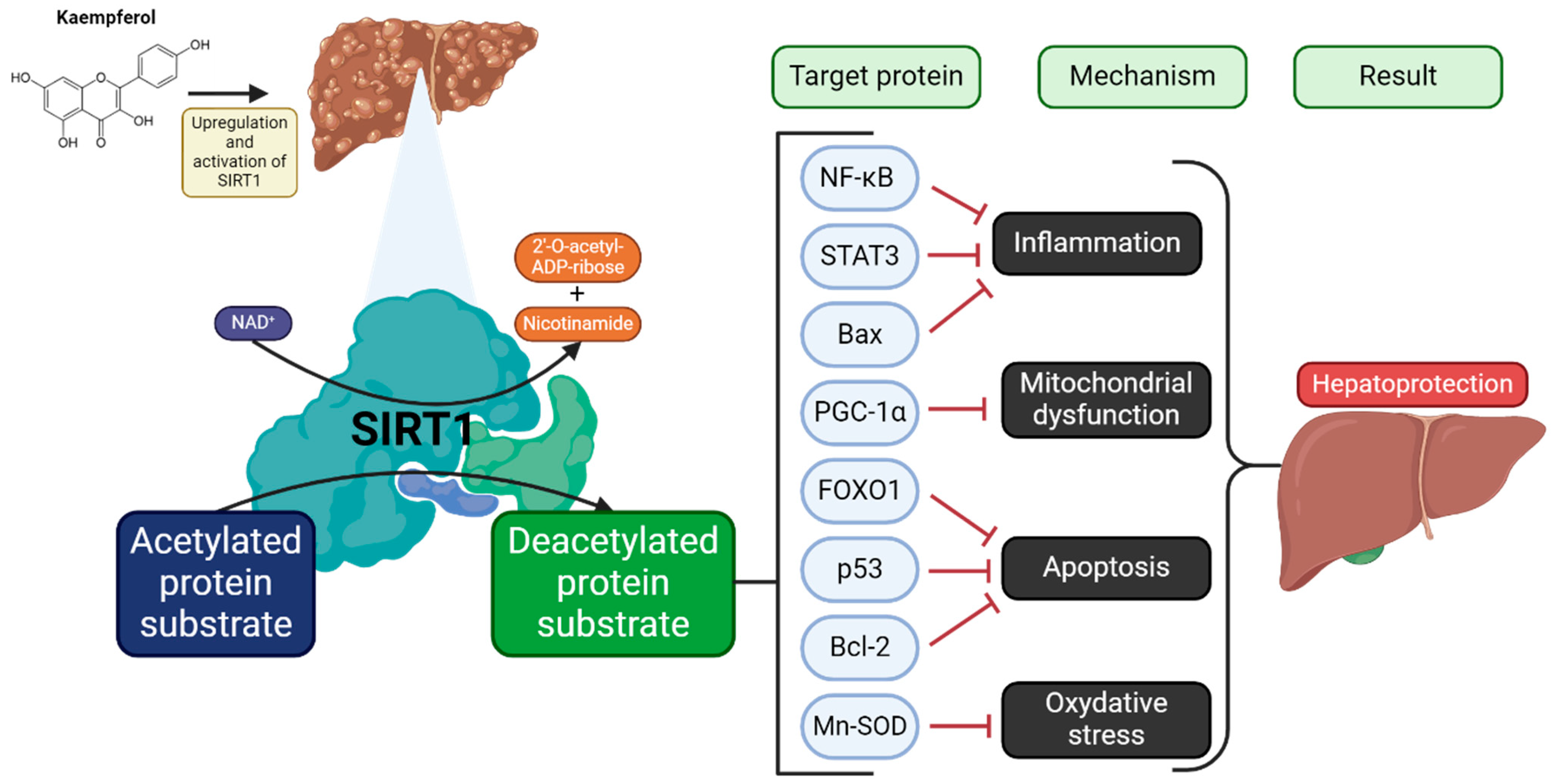
  67. Iside, C.; Scafuro, M.; Nebbioso, A.; Altucci, L. SIRT1 Activation by Natural Phytochemicals: An Overview. Front. Pharmacol. 2020, 11, 1225. [Google Scholar] [CrossRef] [PubMed]
  68. Yang, Y.; Liu, Y.; Wang, Y.; Chao, Y.; Zhang, J.; Jia, Y.; Tie, J.; Hu, D. Regulation of SIRT1 and Its Roles in Inflammation. Front. Immunol. 2022, 13, 831168. [Google Scholar] [CrossRef]
  69. Ren, J.; Lu, Y.; Qian, Y.; Chen, B.; Wu, T.; Ji, G. Recent Progress Regarding Kaempferol for the Treatment of Various Diseases. Exp. Ther. Med. 2019, 18, 2759–2776. [Google Scholar] [CrossRef] [PubMed]
  70. BinMowyna, M.N.; AlFaris, N.A. Kaempferol Suppresses Acetaminophen-Induced Liver Damage by Upregulation/Activation of SIRT1. Pharm. Biol. 2021, 59, 144–154. [Google Scholar] [CrossRef]
  71. Alkandahri, M.Y.; Pamungkas, B.T.; Oktoba, Z.; Shafirany, M.Z.; Sulastri, L.; Arfania, M.; Anggraeny, E.N.; Pratiwi, A.; Astuti, F.D.; Indriyani; et al. Hepatoprotective Effect of Kaempferol: A Review of the Dietary Sources, Bioavailability, Mechanisms of Action, and Safety. Adv. Pharmacol. Pharm. Sci. 2023, 2023, 1–16. [Google Scholar] [CrossRef]
  72. Pingili, R.B.; Challa, S.R.; Pawar, A.K.; Toleti, V.; Kodali, T.; Koppula, S. A Systematic Review on Hepatoprotective Activity of Quercetin against Various Drugs and Toxic Agents: Evidence from Preclinical Studies. Phytother. Res. 2020, 34, 5–32. [Google Scholar] [CrossRef]
  73. Domitrović, R.; Potočnjak, I. A Comprehensive Overview of Hepatoprotective Natural Compounds: Mechanism of Action and Clinical Perspectives. Arch. Toxicol. 2016, 90, 39–79. [Google Scholar] [CrossRef]
  74. Li, N.; Yin, L.; Shang, J.; Liang, M.; Liu, Z.; Yang, H.; Qiang, G.; Du, G.; Yang, X. Kaempferol Attenuates Nonalcoholic Fatty Liver Disease in Type 2 Diabetic Mice via the Sirt1/AMPK Signaling Pathway. Biomed. Pharmacother. 2023, 165, 115113. [Google Scholar] [CrossRef]
  75. Ruderman, N.B.; Julia Xu, X.; Nelson, L.; Cacicedo, J.M.; Saha, A.K.; Lan, F.; Ido, Y. AMPK and SIRT1: A Long-Standing Partnership? Am. J. Physiol.-Endocrinol. Metab. 2010, 298, E751–E760. [Google Scholar] [CrossRef] [PubMed]
  76. Xu, H.; Chen, G.-F.; Ma, Y.-S.; Zhang, H.-W.; Zhou, Y.; Liu, G.-H.; Chen, D.-Y.; Ping, J.; Liu, Y.-H.; Mou, X.; et al. Hepatic Proteomic Changes and Sirt1/AMPK Signaling Activation by Oxymatrine Treatment in Rats With Non-Alcoholic Steatosis. Front. Pharmacol. 2020, 11, 216. [Google Scholar] [CrossRef] [PubMed]
  77. Dzamko, N.; van Denderen, B.J.W.; Hevener, A.L.; Jørgensen, S.B.; Honeyman, J.; Galic, S.; Chen, Z.-P.; Watt, M.J.; Campbell, D.J.; Steinberg, G.R.; et al. AMPK Β1 Deletion Reduces Appetite, Preventing Obesity and Hepatic Insulin Resistance*. J. Biol. Chem. 2010, 285, 115–122. [Google Scholar] [CrossRef] [PubMed]
  78. Mohammadi, M.; Abbasalipourkabir, R.; Ziamajidi, N. Fish Oil and Chicoric Acid Combination Protects Better against Palmitate-Induced Lipid Accumulation via Regulating AMPK-Mediated SREBP-1/FAS and PPARα/UCP2 Pathways. Arch. Physiol. Biochem. 2020, 129, 1–9. [Google Scholar] [CrossRef]
  79. Shokri Afra, H.; Zangooei, M.; Meshkani, R.; Ghahremani, M.H.; Ilbeigi, D.; Khedri, A.; Shahmohamadnejad, S.; Khaghani, S.; Nourbakhsh, M. Hesperetin Is a Potent Bioactivator That Activates SIRT1-AMPK Signaling Pathway in HepG2 Cells. J. Physiol. Biochem. 2019, 75, 125–133. [Google Scholar] [CrossRef]
  80. Ding, Y.; Liu, P.; Chen, Z.-L.; Zhang, S.-J.; Wang, Y.-Q.; Cai, X.; Luo, L.; Zhou, X.; Zhao, L. Emodin Attenuates Lipopolysaccharide-Induced Acute Liver Injury via Inhibiting the TLR4 Signaling Pathway In Vitro and In Vivo. Front. Pharmacol. 2018, 9, 962. [Google Scholar] [CrossRef]
  81. Cheng, P.; Wang, T.; Li, W.; Muhammad, I.; Wang, H.; Sun, X.; Yang, Y.; Li, J.; Xiao, T.; Zhang, X. Baicalin Alleviates Lipopolysaccharide-Induced Liver Inflammation in Chicken by Suppressing TLR4-Mediated NF-κB Pathway. Front. Pharmacol. 2017, 8, 547. [Google Scholar] [CrossRef]
  82. Yang, Y.-Q.; Yan, X.-T.; Wang, K.; Tian, R.-M.; Lu, Z.-Y.; Wu, L.-L.; Xu, H.-T.; Wu, Y.-S.; Liu, X.-S.; Mao, W.; et al. Triptriolide Alleviates Lipopolysaccharide-Induced Liver Injury by Nrf2 and NF-κB Signaling Pathways. Front. Pharmacol. 2018, 9, 999. [Google Scholar] [CrossRef]
  83. Kobayashi, Y.; Iwata, A.; Suzuki, K.; Suto, A.; Kawashima, S.; Saito, Y.; Owada, T.; Kobayashi, M.; Watanabe, N.; Nakajima, H. B and T Lymphocyte Attenuator Inhibits LPS-Induced Endotoxic Shock by Suppressing Toll-like Receptor 4 Signaling in Innate Immune Cells. Proc. Natl. Acad. Sci. USA 2013, 110, 5121–5126. [Google Scholar] [CrossRef]
  84. Islam, M.S.; Yu, H.; Miao, L.; Liu, Z.; He, Y.; Sun, H. Hepatoprotective Effect of the Ethanol Extract of Illicium Henryi against Acute Liver Injury in Mice Induced by Lipopolysaccharide. Antioxidants 2019, 8, 446. [Google Scholar] [CrossRef]
  85. Liu, P.; Wu, P.; Yang, B.; Wang, T.; Li, J.; Song, X.; Sun, W. Kaempferol Prevents the Progression from Simple Steatosis to Non-Alcoholic Steatohepatitis by Inhibiting the NF-κB Pathway in Oleic Acid-Induced HepG2 Cells and High-Fat Diet-Induced Rats. J. Funct. Foods 2021, 85, 104655. [Google Scholar] [CrossRef]
  86. Xiang, H.; Shao, M.; Lu, Y.; Wang, J.; Wu, T.; Ji, G. Kaempferol Alleviates Steatosis and Inflammation During Early Non-Alcoholic Steatohepatitis Associated With Liver X Receptor α-Lysophosphatidylcholine Acyltransferase 3 Signaling Pathway. Front. Pharmacol. 2021, 12, 690736. [Google Scholar] [CrossRef] [PubMed]
  87. Alrumaihi, F.; Almatroodi, S.A.; Alharbi, H.O.A.; Alwanian, W.M.; Alharbi, F.A.; Almatroudi, A.; Rahmani, A.H. Pharmacological Potential of Kaempferol, a Flavonoid in the Management of Pathogenesis via Modulation of Inflammation and Other Biological Activities. Molecules 2024, 29, 2007. [Google Scholar] [CrossRef] [PubMed]
  88. Al Olayan, E.M.; Aloufi, A.S.; AlAmri, O.D.; El-Habit, O.H.; Abdel Moneim, A.E. Protocatechuic Acid Mitigates Cadmium-Induced Neurotoxicity in Rats: Role of Oxidative Stress, Inflammation and Apoptosis. Sci. Total Environ. 2020, 723, 137969. [Google Scholar] [CrossRef] [PubMed]
  89. Al-Numair, K.S.; Chandramohan, G.; Veeramani, C.; Alsaif, M.A. Ameliorative Effect of Kaempferol, a Flavonoid, on Oxidative Stress in Streptozotocin-Induced Diabetic Rats. Redox Rep. 2015, 20, 198–209. [Google Scholar] [CrossRef]
  90. Yang, Q.-S.; He, L.-P.; Zhou, X.-L.; Zhao, Y.; Shen, J.; Xu, P.; Ni, S.-Z. Kaempferol Pretreatment Modulates Systemic Inflammation and Oxidative Stress Following Hemorrhagic Shock in Mice. Chin. Med. 2015, 10, 6. [Google Scholar] [CrossRef]
  91. Cioarca-Nedelcu, R.; Atanasiu, V.; Stoian, I. Alcoholic Liver Disease-from Steatosis to Cirrhosis—A Biochemistry Approach. J. Med. Life 2021, 14, 594–599. [Google Scholar] [CrossRef]
  92. Renu, K.; Saravanan, A.; Elangovan, A.; Ramesh, S.; Annamalai, S.; Namachivayam, A.; Abel, P.; Madhyastha, H.; Madhyastha, R.; Maruyama, M.; et al. An Appraisal on Molecular and Biochemical Signalling Cascades during Arsenic-Induced Hepatotoxicity. Life Sci. 2020, 260, 118438. [Google Scholar] [CrossRef]
  93. Shih, T.-Y.; Young, T.-H.; Lee, H.-S.; Hsieh, C.-B.; Hu, O.Y.-P. Protective Effects of Kaempferol on Isoniazid- and Rifampicin-Induced Hepatotoxicity. AAPS J. 2013, 15, 753–762. [Google Scholar] [CrossRef]
  94. Dong, L.; Yin, L.; Quan, H.; Chu, Y.; Lu, J. Hepatoprotective Effects of Kaempferol-3-O-α-l-Arabinopyranosyl-7-O-α-l-Rhamnopyranoside on d-Galactosamine and Lipopolysaccharide Caused Hepatic Failure in Mice. Molecules 2017, 22, 1755. [Google Scholar] [CrossRef]
  95. Hu, C.; Zhao, L.; Duan, J.; Li, L. Strategies to Improve the Efficiency of Mesenchymal Stem Cell Transplantation for Reversal of Liver Fibrosis. J. Cell. Mol. Med. 2019, 23, 1657–1670. [Google Scholar] [CrossRef]
  96. Yoshiji, H.; Nagoshi, S.; Akahane, T.; Asaoka, Y.; Ueno, Y.; Ogawa, K.; Kawaguchi, T.; Kurosaki, M.; Sakaida, I.; Shimizu, M.; et al. Evidence-Based Clinical Practice Guidelines for Liver Cirrhosis 2020. J. Gastroenterol. 2021, 56, 593–619. [Google Scholar] [CrossRef] [PubMed]
  97. Caligiuri, A.; Gentilini, A.; Pastore, M.; Gitto, S.; Marra, F. Cellular and Molecular Mechanisms Underlying Liver Fibrosis Regression. Cells 2021, 10, 2759. [Google Scholar] [CrossRef]
  98. Targeting TGF-β Signal Transduction for Fibrosis and Cancer Therapy|Molecular Cancer|Full Text. Available online: https://molecular-cancer.biomedcentral.com/articles/10.1186/s12943-022-01569-x (accessed on 30 January 2025).
  99. Zhang, J.; Li, Y.; Liu, Q.; Huang, Y.; Li, R.; Wu, T.; Zhang, Z.; Zhou, J.; Huang, H.; Tang, Q.; et al. Sirt6 Alleviated Liver Fibrosis by Deacetylating Conserved Lysine 54 on Smad2 in Hepatic Stellate Cells. Hepatology 2021, 73, 1140–1157. [Google Scholar] [CrossRef] [PubMed]
  100. Xu, T.; Huang, S.; Huang, Q.; Ming, Z.; Wang, M.; Li, R.; Zhao, Y. Kaempferol Attenuates Liver Fibrosis by Inhibiting Activin Receptor-like Kinase 5. J. Cell. Mol. Med. 2019, 23, 6403–6410. [Google Scholar] [CrossRef]
  101. Chen, J.; Zhong, H.; Huang, Z.; Chen, X.; You, J.; Zou, T. A Critical Review of Kaempferol in Intestinal Health and Diseases. Antioxidants 2023, 12, 1642. [Google Scholar] [CrossRef]
  102. Duarte, L.; Gasaly, N.; Poblete-Aro, C.; Uribe, D.; Echeverria, F.; Gotteland, M.; Garcia-Diaz, D.F. Polyphenols and Their Anti-Obesity Role Mediated by the Gut Microbiota: A Comprehensive Review. Rev. Endocr. Metab. Disord. 2021, 22, 367–388. [Google Scholar] [CrossRef] [PubMed]
  103. Osborn, L.J.; Schultz, K.; Massey, W.; DeLucia, B.; Choucair, I.; Varadharajan, V.; Banerjee, R.; Fung, K.; Horak, A.J.; Orabi, D.; et al. A Gut Microbial Metabolite of Dietary Polyphenols Reverses Obesity-Driven Hepatic Steatosis. Proc. Natl. Acad. Sci. USA 2022, 119, e2202934119. [Google Scholar] [CrossRef]
  104. Wang, T.; Wu, Q.; Zhao, T. Preventive Effects of Kaempferol on High-Fat Diet-Induced Obesity Complications in C57BL/6 Mice. BioMed Res. Int. 2020, 2020, 4532482. [Google Scholar] [CrossRef]
  105. Bian, Y.; Lei, J.; Zhong, J.; Wang, B.; Wan, Y.; Li, J.; Liao, C.; He, Y.; Liu, Z.; Ito, K.; et al. Kaempferol Reduces Obesity, Prevents Intestinal Inflammation, and Modulates Gut Microbiota in High-Fat Diet Mice. J. Nutr. Biochem. 2022, 99, 108840. [Google Scholar] [CrossRef]
  106. Lu, Y.; Shao, M.; Zhang, C.; Xiang, H.; Wang, J.; Wu, T.; Ji, G. Kaempferol Attenuates Nonalcoholic Steatohepatitis by Regulating Serum and Liver Bile Acid Metabolism. Front. Pharmacol. 2022, 13, 946360. [Google Scholar] [CrossRef] [PubMed]
  107. Wei, T.; Xiong, F.; Wang, S.; Wang, K.; Zhang, Y.; Zhang, Q. Flavonoid Ingredients of Ginkgo Biloba Leaf Extract Regulate Lipid Metabolism through Sp1-Mediated Carnitine Palmitoyltranferase 1A up-Regulation. J. Biomed. Sci. 2014, 21, 87. [Google Scholar] [CrossRef]
  108. Fan, X.; Bai, J.; Hu, M.; Xu, Y.; Zhao, S.; Sun, Y.; Wang, B.; Hu, J.; Li, Y. Drug Interaction Study of Flavonoids toward OATP1B1 and Their 3D Structure Activity Relationship Analysis for Predicting Hepatoprotective Effects. Toxicology 2020, 437, 152445. [Google Scholar] [CrossRef] [PubMed]
  109. Singab, A.N.B.; Youssef, D.T.A.; Noaman, E.; Kotb, S. Hepatoprotective Effect of Flavonol Glycosides Rich Fraction from Egyptian Vicia Calcarata Desf. against CCl4-Induced Liver Damage in Rats. Arch. Pharm. Res. 2005, 28, 791–798. [Google Scholar] [CrossRef]
  110. Cho, S.S.; Yang, J.H.; Seo, K.H.; Shin, S.M.; Park, E.Y.; Cho, S.S.; Jo, G.U.; Eo, J.H.; Park, J.S.; Oh, D.S.; et al. Cudrania Tricuspidata Extract and Its Major Constituents Inhibit Oxidative Stress-Induced Liver Injury. J. Med. Food 2019, 22, 602–613. [Google Scholar] [CrossRef]
  111. Wang, M.; Sun, J.; Jiang, Z.; Xie, W.; Zhang, X. Hepatoprotective Effect of Kaempferol against Alcoholic Liver Injury in Mice. Am. J. Chin. Med. 2015, 43, 241–254. [Google Scholar] [CrossRef] [PubMed]
  112. Cai, F.-F.; Bian, Y.-Q.; Wu, R.; Sun, Y.; Chen, X.-L.; Yang, M.-D.; Zhang, Q.-R.; Hu, Y.; Sun, M.-Y.; Su, S.-B. Yinchenhao Decoction Suppresses Rat Liver Fibrosis Involved in an Apoptosis Regulation Mechanism Based on Network Pharmacology and Transcriptomic Analysis. Biomed. Pharmacother. 2019, 114, 108863. [Google Scholar] [CrossRef]
  113. Zhang, Q.; Cheng, G.; Qiu, H.; Zhu, L.; Ren, Z.; Zhao, W.; Zhang, T.; Liu, L. The P53-Inducible Gene 3 Involved in Flavonoid-Induced Cytotoxicity through the Reactive Oxygen Species-Mediated Mitochondrial Apoptotic Pathway in Human Hepatoma Cells. Food Funct. 2015, 6, 1518–1525. [Google Scholar] [CrossRef]
  114. Du, Y.-C.; Lai, L.; Zhang, H.; Zhong, F.-R.; Cheng, H.-L.; Qian, B.-L.; Tan, P.; Xia, X.-M.; Fu, W.-G. Kaempferol from Penthorum Chinense Pursh Suppresses HMGB1/TLR4/NF-κB Signaling and NLRP3 Inflammasome Activation in Acetaminophen-Induced Hepatotoxicity. Food Funct. 2020, 11, 7925–7934. [Google Scholar] [CrossRef]
  115. Lee, Y.-J.; Choi, H.-S.; Seo, M.-J.; Jeon, H.-J.; Kim, K.-J.; Lee, B.-Y. Kaempferol Suppresses Lipid Accumulation by Inhibiting Early Adipogenesis in 3T3-L1 Cells and Zebrafish. Food Funct. 2015, 6, 2824–2833. [Google Scholar] [CrossRef]
  116. Torres-Villarreal, D.; Camacho, A.; Castro, H.; Ortiz-Lopez, R.; de la Garza, A.L. Anti-Obesity Effects of Kaempferol by Inhibiting Adipogenesis and Increasing Lipolysis in 3T3-L1 Cells. J. Physiol. Biochem. 2019, 75, 83–88. [Google Scholar] [CrossRef]
  117. Gómez-Zorita, S.; Lasa, A.; Abendaño, N.; Fernández-Quintela, A.; Mosqueda-Solís, A.; Garcia-Sobreviela, M.P.; Arbonés-Mainar, J.M.; Portillo, M.P. Phenolic Compounds Apigenin, Hesperidin and Kaempferol Reduce in Vitro Lipid Accumulation in Human Adipocytes. J. Transl. Med. 2017, 15, 237. [Google Scholar] [CrossRef]
  118. Lee, B.; Kwon, M.; Choi, J.S.; Jeong, H.O.; Chung, H.Y.; Kim, H.-R. Kaempferol Isolated from Nelumbo Nucifera Inhibits Lipid Accumulation and Increases Fatty Acid Oxidation Signaling in Adipocytes. J. Med. Food 2015, 18, 1363–1370. [Google Scholar] [CrossRef]
  119. Zhang, X.; Hou, X.; Xu, C.; Cheng, S.; Ni, X.; Shi, Y.; Yao, Y.; Chen, L.; Hu, M.G.; Xia, D. Kaempferol Regulates the Thermogenic Function of Adipocytes in High-Fat-Diet-Induced Obesity via the CDK6/RUNX1/UCP1 Signaling Pathway. Food Funct. 2023, 14, 8201–8216. [Google Scholar] [CrossRef] [PubMed]
  120. Romero-Juárez, P.A.; Visco, D.B.; Manhães-de-Castro, R.; Urquiza-Martínez, M.V.; Saavedra, L.M.; González-Vargas, M.C.; Mercado-Camargo, R.; Aquino, J.d.S.; Toscano, A.E.; Torner, L.; et al. Dietary Flavonoid Kaempferol Reduces Obesity-Associated Hypothalamic Microglia Activation and Promotes Body Weight Loss in Mice with Obesity. Nutr. Neurosci. 2023, 26, 25–39. [Google Scholar] [CrossRef] [PubMed]
  121. Watson, W.A.F. The Mutagenic Activity of Quercetin and Kaempferol in Drosophila Melanogaster. Mutat. Res. Lett. 1982, 103, 145–147. [Google Scholar] [CrossRef]
  122. Silva, I.D.; Rodrigues, A.; Gaspar, J.; Mala, R.; Laires, A.; Rueff, J. Mutagenicity of Kaempferol in V79 Cells: The Role of Cytochromes P450. Teratog. Carcinog. Mutagen. 1996, 16, 229–241. [Google Scholar] [CrossRef]
  123. Lemos, C.; Peters, G.J.; Jansen, G.; Martel, F.; Calhau, C. Modulation of Folate Uptake in Cultured Human Colon Adenocarcinoma Caco-2 Cells by Dietary Compounds. Eur. J. Nutr. 2007, 46, 329–336. [Google Scholar] [CrossRef]
  124. Tu, L.-Y.; Bai, H.-H.; Cai, J.-Y.; Deng, S.-P. The Mechanism of Kaempferol Induced Apoptosis and Inhibited Proliferation in Human Cervical Cancer SiHa Cell: From Macro to Nano. Scanning 2016, 38, 644–653. [Google Scholar] [CrossRef]
  125. Goey, A.K.L.; Mooiman, K.D.; Beijnen, J.H.; Schellens, J.H.M.; Meijerman, I. Relevance of in Vitro and Clinical Data for Predicting CYP3A4-Mediated Herb-Drug Interactions in Cancer Patients. Cancer Treat. Rev. 2013, 39, 773–783. [Google Scholar] [CrossRef]
  126. Rendic, S.; Guengerich, F.P. Human Cytochrome P450 Enzymes 5-51 as Targets of Drugs and Natural and Environmental Compounds: Mechanisms, Induction, and Inhibition—Toxic Effects and Benefits. Drug Metab. Rev. 2018, 50, 256–342. [Google Scholar] [CrossRef] [PubMed]
  127. Nguyen, T.-T.-L.; Duong, V.-A.; Maeng, H.-J. Pharmaceutical Formulations with P-Glycoprotein Inhibitory Effect as Promising Approaches for Enhancing Oral Drug Absorption and Bioavailability. Pharmaceutics 2021, 13, 1103. [Google Scholar] [CrossRef] [PubMed]
  128. Jin, U.-H.; Park, H.; Li, X.; Davidson, L.A.; Allred, C.; Patil, B.; Jayaprakasha, G.; Orr, A.A.; Mao, L.; Chapkin, R.S.; et al. Structure-Dependent Modulation of Aryl Hydrocarbon Receptor-Mediated Activities by Flavonoids. Toxicol. Sci. 2018, 164, 205–217. [Google Scholar] [CrossRef] [PubMed]
  129. Dong, H.; Lin, W.; Wu, J.; Chen, T. Flavonoids Activate Pregnane x Receptor-Mediated CYP3A4 Gene Expression by Inhibiting Cyclin-Dependent Kinases in HepG2 Liver Carcinoma Cells. BMC Biochem. 2010, 11, 23. [Google Scholar] [CrossRef]
  130. Kitakaze, T.; Makiyama, A.; Nakai, R.; Kimura, Y.; Ashida, H. Kaempferol Modulates TCDD- and t-BHQ-Induced Drug-Metabolizing Enzymes and Luteolin Enhances This Effect. Food Funct. 2020, 11, 3668–3680. [Google Scholar] [CrossRef]
Figure 1. Chemical structure of kaempferol.
Molecules 30 01913 g001
Figure 2. An overview of kaempferol ADME processes in the body. After ingestion, kaempferol may be directly transported to the enterocyte in the form of aglycone or glucosides as well as prior to absorption sugar moiety can be cleaved. Phase I and phase II metabolism occur either in enterocytes or in hepatocytes, and in the form of methyl, sulfur or glucuronide metabolites, kaempferol is distributed to the target tissues and ultimately excreted mainly in the renal way. Abbreviations: ABC—ATP-binding cassette, CBG—cytosolic β-glucosidase, COMT—catechol-O-methyltransferase, LPH—lactase-phlorizin hydrolase, SGLT-1—sodium-dependent glucose transporter-1, SULT—sulfotransferase, UGT—uridine-5ʹ-diphosphate-glucuronosyltransferase. Created in BioRender.
Molecules 30 01913 g002
Figure 3. An overview of upregulation and activation of SIRT1 in the liver by kaempferol. For further explanations please see text above. Abbreviations: Bax—bcl-2-like protein 4, Bcl-2—B-cell CLL/lymphoma 2, FOXO1—Forkhead box protein O1, Mn-SOD—manganese-dependent superoxide dismutase, NF-κB—nuclear factor kappa-light-chain-enhancer of activated B cells, p53—tumour protein p53, PGC-1α—peroxisome proliferator-activated receptor gamma coactivator 1-alpha, SIRT1—sirtuin-1, STAT3—signal transducer and activator of transcription 3. Inhibition (⟞). Adopted and modified from figures by Yang et al. [68] and Alkandahri et al. [71]. Created in BioRender.
Molecules 30 01913 g003
Figure 4. An overview of kaempferol protection mechanism in LPS-induced acute liver injury. For further explanations please see text above. Inhibition (⟞). Adopted and modified from figures in Alkandahri et al. [71]. Created in BioRender.
Molecules 30 01913 g004
Table 1. Major dietary sources of kaempferol.
PlantScientific NameAmount References
Green chiliCapsicum annum39[40]
Plant extracts [mg/kg dry mass]Onion leavesAllium fistulosum832
Papaya shootsCarica papaya453
BrinjalSolanum melongena80
PumpkinCucurbita maxima371
SengkuangPachyrrhizus erosus37
CarrotDaucus carota140
White radishRaphanus sativus38
Daun turiSesbania grandifolia21
Lemon grassCymbopogon citratus178
Cekur manisSauropus androgynus323
PegagaHydrocotyle asiatica20
Bunga kantanPhaeomeria speciosa286
Black teaCamellia sinensis118
BeansPhaseolus vulgaris14[41]
BroccoliBrassica oleracea var. italica72
CauliflowerBrassica oleracea var. botrytis270
Plant-derived beverages [μg/mL]Lemon juiceCitrus limon1.9 [42]
Grapefruit juiceCitrus × paradisi1.1
Pineapple juice Ananas comosus1.2
Apple juiceMalus domestica1.0
Black teaCamellia sinensis11.4 [43]
Plants
[mg/100 g fresh weight]
SpinachSpinacia oleracea7.86[43]
Garden cressLepidium sativum13.00
BroccoliBrassica oleracea var. italica5.65
KaleBrassica oleracea var. sabellica5.65
OnionAllium cepa26.74
Rabbiteye blueberriesVaccinium virgatum2.36
Table 2. Role of kaempferol in regulation of liver diseases.
DiseasesModelDosesMechanism of Action/EffectRef.
MASLDMale ddY mice4.9 mg/kg↓ TBARS and TNF-α caused by CCl4 free radicals[66]
C57BLKS/J mice fed HFD87.5 µmol/kgRegulation of hepatic lipid accumulation (activation of the SIRT1/AMPK pathway)[74]
HFD-induced SD rats350 µmol/kgPrevention of advancement of simple fatty liver disease to non-alcoholic steatohepatitis (blocking the NF-κB pathway)[85]
HepG2 cells1 or 10 µM
C57BL/6 J mice fed HFD0.5 mL/100 gRegulation of BA metabolism in the serum and liver (enhancing CYP27A1 and NTCP expression) [106]
MASLDHepG2 cells50 mg/kg↓ fat buildup in the liver, ↑ NF-κB signalling pathway, ↑ mitochondrial beta-oxidation, ↑ expression of CPT1A[107]
Liver injuryBosentan-induced rat liver injury model and HEK-293 cells25 mg/kg and 1–150 µM↓ OATP1B1 transporter (keeping AST and ALT levels stable)[108]
Male swiss albino rats25 mg/kg↓ lipid peroxidation caused by CCl4 free radicals[109]
Mice and HepG2 cells250 and 500 mg/kg and 100, 200, and 400 µM↓ AA + Fe-induced ROS, ↓ glutathione depletion [110]
ALI mice model10 and 20 mg/kg↓ antioxidant defence activity, ↑ lipid peroxidation and oxidative stress [111]
Liver fibrosisL02, LX2, and rats20 µM↓ caspase-3 protein levels, ↑ p-ERK1/2, PI3K and Bcl-xL protein expression in L02 cells; ↑proliferation of LX2 cells, ↑ Bax and cleaved caspase-8.[112]
HSCs/CCl4-induced mouse model2–10 µmol/L↓ hyaluronan, ALT, AST and Smad2/3, ↓ collagen synthesis and HSC activation; ↑activin receptor-like kinase 5[100]
Liver cancerHepG2 cells10, 20, 40, and 80 µM↑ ROS production, ↑ cytochrome c ↑ PIG3 mRNA and protein, ↓ mitochondrial membrane potential, ↓ f Bax/Bcl-2 and caspase-9 and -3[113]
HepatotoxicityMale C57BL/6 mice30 and 60 mg/kg↓ ALT and AST, ↓ liver cell damage, ↑ antioxidant enzymes and apoptosis; ↓ NLRP3 and pro-inflammatory molecules[114]
ObesityWild-type zebrafish 7.5, 15, and 30 µM↑ adipogenesis [115]
The 3T3-L1 preadipocytes60 µM↑ lipolysis, ↓ adipogenesis[116]
Human mesenchymal fat cells 1, 10 or 25 µM↑ lipolysis, ↓ adipogenesis[117]
The 3T3-L1 preadipocytes2.5, 5, 10, 20 and 40 µM↓ adipogenic transcription factors, ↑ PPARα-mediated signalling of FAO[118]
C57BL/6 J male mice fed HFD43.75, 87.5, and 175 µmol/kgRegulation of adipocyte thermogenesis via the CDK6/RUNX1/UCP1 pathway[119]
C57BL/6 J male mice fed HFDa high-fat diet with 0.1% kaempferol ↑ intestinal barrier integrity, ↓ intestinal inflammation by inhibition of TLR4/NF-κB pathway [105]
C57BL/6 mice fed HFD350 µmol/kg↑ gut microbiota and ↓ the progression of insulin resistance.[104]
C57BL/6 mice fed HFD0.875 µmol/kgRegulation of physiological processes concerning energy balance and inflammation [120]
↑ upregulating/improving, ↓ downregulating/decreasing
Disclaimer/Publisher’s Note: The statements, opinions and data contained in all publications are solely those of the individual author(s) and contributor(s) and not of MDPI and/or the editor(s). MDPI and/or the editor(s) disclaim responsibility for any injury to people or property resulting from any ideas, methods, instructions or products referred to in the content.

