
Developing a Multi-Method Approach for Understanding Cellular Uptake and Biological Response: Investigating Co-Exposure of Macrophage-like Differentiated THP-1 Cells to Al2O3 and CeO2 Nanoparticles

 , , , , , , , , and
Abstract
1. Introduction
2. Results and Discussion
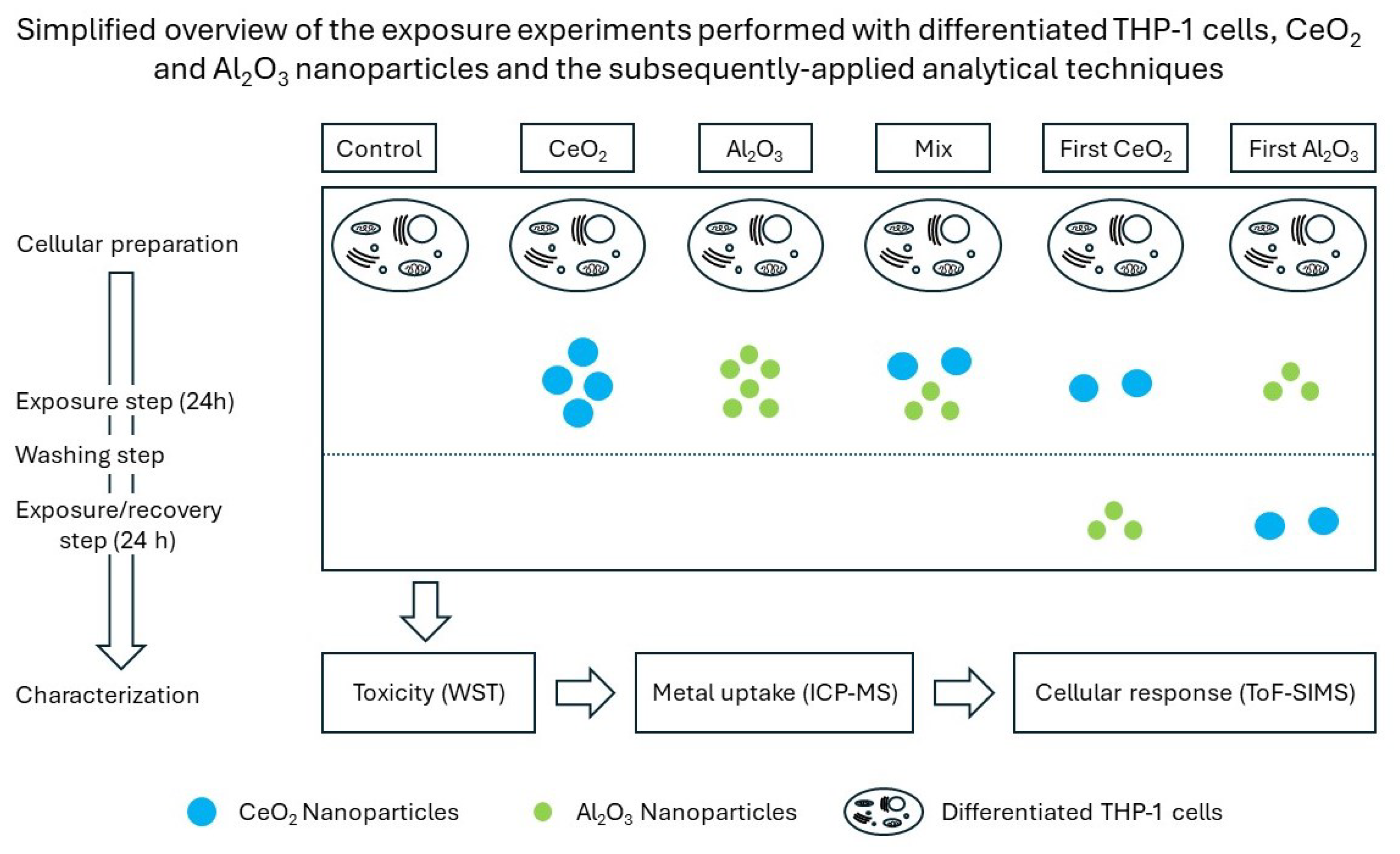
2.1. Experimental Design
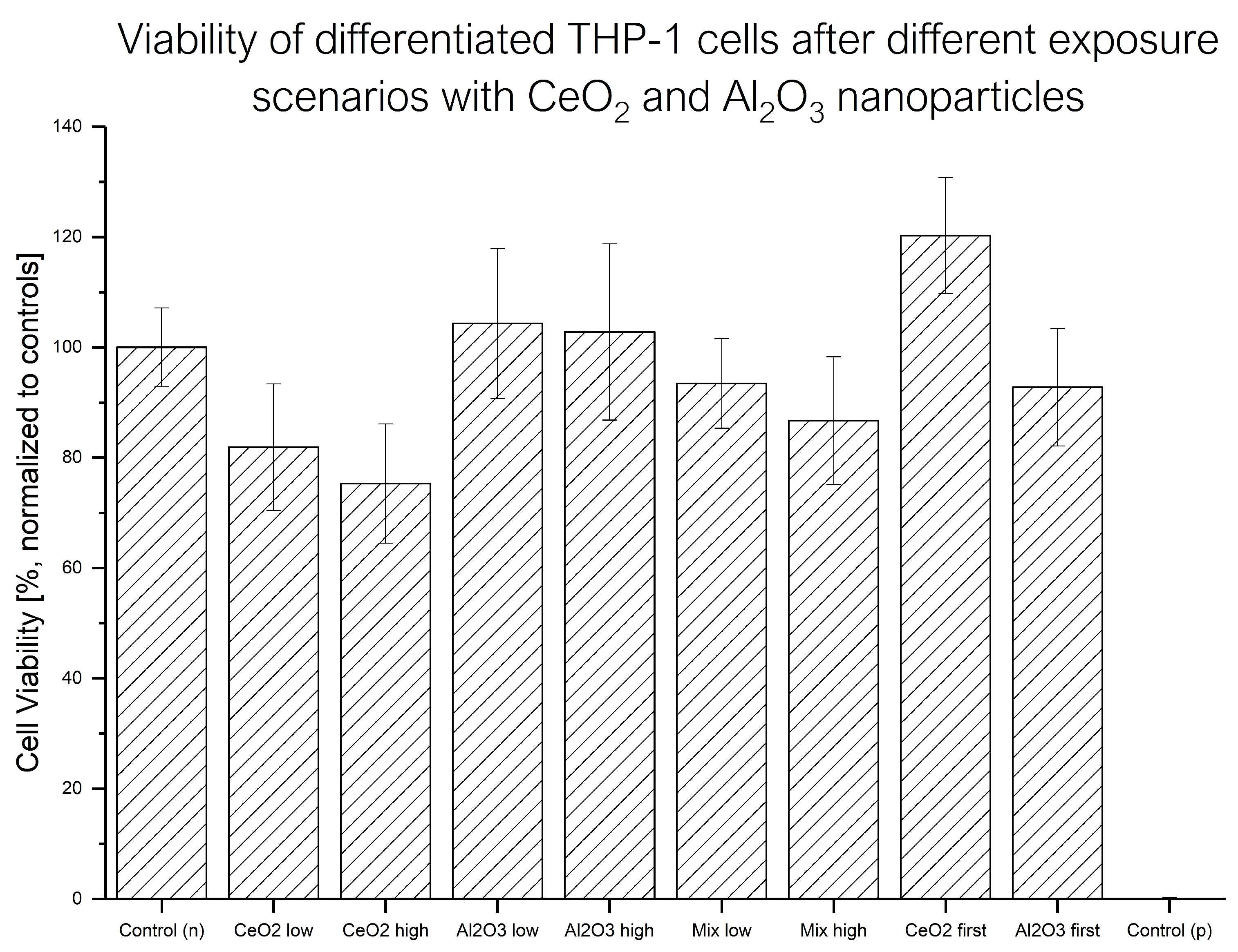
2.2. Viability Changes
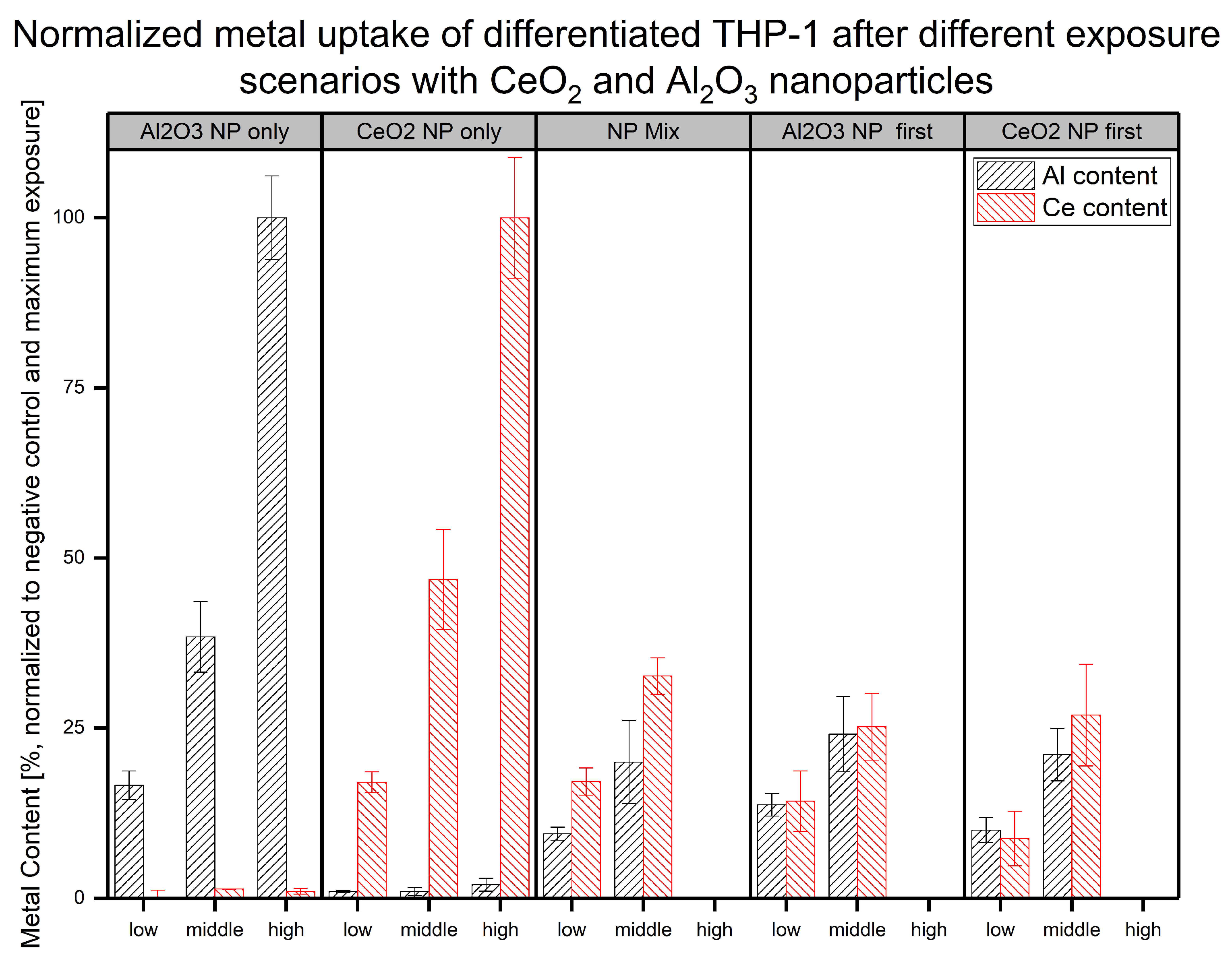
2.3. MW ICP-MS
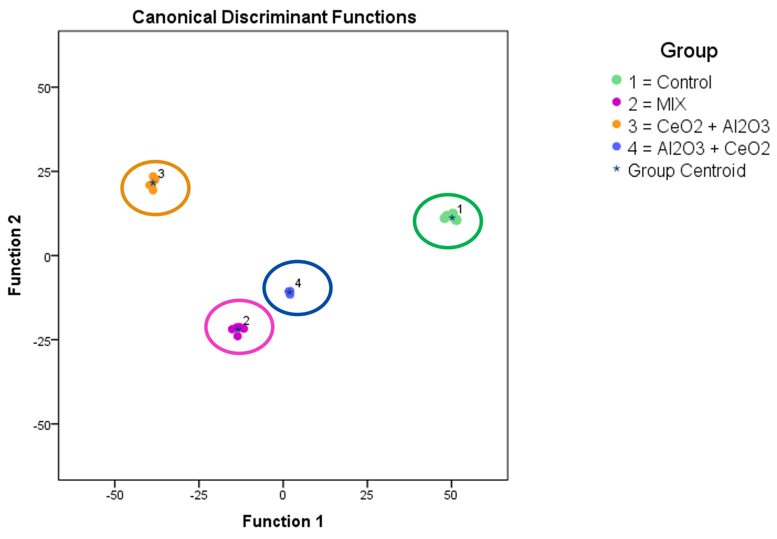
2.4. ToF-SIMS
3. Materials and Methods
3.1. Chemicals and Materials
3.2. Cell Culture
3.3. WST-1 Test
3.4. Mass Spectrometry
3.5. ToF-SIMS
4. Conclusions
Supplementary Materials
Author Contributions
Funding
Data Availability Statement
Acknowledgments
Conflicts of Interest
Abbreviations
| NM | nanomaterial |
| JRC | joint research centre |
| MW | microwave digestion |
| WST-1 | 4-[3-(4-Iodophenyl)-2-(4-nitro-phenyl)-2H-5-tetrazolio]-1,3-benzene sulfonate |
| spICP-MS | single-particle inductively coupled plasma–mass spectrometry |
| DI | direct injection |
| ToF-SIMS | time-of-flight–secondary ion mass spectrometry |
| NP | nanoparticle |
| PCA | principal component analysis |
| SE | standard error |
References
- Rathore, R.; Suhag, D.; Wan, F.; Thakur, A.; Thakur, P. Everyday Nanotechnology. In Integrated Nanomaterials and Their Applications; Suhag, D., Thakur, A., Thakur, P., Eds.; Springer Nature: Singapore, 2023; pp. 19–35. [Google Scholar] [CrossRef]
- Parashar, S.; Raj, S.; Srivastava, P.; Singh, A.K. Comparative toxicity assessment of selected nanoparticles using different experimental model organisms. J. Pharmacol. Toxicol. Methods 2024, 130, 107563. [Google Scholar] [CrossRef] [PubMed]
- Brame, J.A.; Poda, A.R.; Kennedy, A.J.; Steevens, J.A. EHS Testing of Products Containing Nanomaterials: What is Nano Release? Environ. Sci. Technol. 2015, 49, 11245–11246. [Google Scholar] [CrossRef] [PubMed]
- Zhu, M.; Nie, G.; Meng, H.; Xia, T.; Nel, A.; Zhao, Y. Physicochemical Properties Determine Nanomaterial Cellular Uptake, Transport, and Fate. Accounts Chem. Res. 2013, 46, 622–631. [Google Scholar] [CrossRef] [PubMed]
- El-Kady, M.M.; Ansari, I.; Arora, C.; Rai, N.; Soni, S.; Verma, D.K.; Singh, P.; Mahmoud, A.E.D. Nanomaterials: A comprehensive review of applications, toxicity, impact, and fate to environment. J. Mol. Liq. 2023, 370, 121046. [Google Scholar] [CrossRef]
- van Ravenzwaay, B.; Landsiedel, R.; Fabian, E.; Burkhardt, S.; Strauss, V.; Ma-Hock, L. Comparing fate and effects of three particles of different surface properties: Nano-TiO2, pigmentary TiO2 and quartz. Toxicol. Lett. 2009, 186, 152–159. [Google Scholar] [CrossRef]
- Sundaram, T.; Rajendran, S.; Natarajan, S.; Vinayagam, S.; Rajamohan, R.; Lackner, M. Environmental fate and transformation of TiO2 nanoparticles: A comprehensive assessment. Alex. Eng. J. 2025, 115, 264–276. [Google Scholar] [CrossRef]
- Liu, L.; Xu, Y.; Ma, Y.; Duan, F.; Wang, C.; Feng, J.; Yin, H.; Sun, L.; Li, P.; Li, Z.H. Fate of polystyrene micro- and nanoplastics in zebrafish liver cells: Influence of protein corona on transport, oxidative stress, and glycolipid metabolism. J. Hazard. Mater. 2025, 489, 137596. [Google Scholar] [CrossRef]
- Lin, P.C.; Lin, S.; Wang, P.C.; Sridhar, R. Techniques for physicochemical characterization of nanomaterials. Biotechnol. Adv. 2014, 32, 711–726. [Google Scholar] [CrossRef]
- Hachenberger, Y.U.; Rosenkranz, D.; Kromer, C.; Krause, B.C.; Dreiack, N.; Kriegel, F.L.; Koz’menko, E.; Jungnickel, H.; Tentschert, J.; Bierkandt, F.S.; et al. Nanomaterial Characterization in Complex Media—Guidance and Application. Nanomaterials 2023, 13, 922. [Google Scholar] [CrossRef]
- Wang, S.; Yu, H.; Wickliffe, J.K. Limitation of the MTT and XTT assays for measuring cell viability due to superoxide formation induced by nano-scale TiO2. Toxicol. In Vitro 2011, 25, 2147–2151. [Google Scholar] [CrossRef]
- Scimeca, J.; Verron, E. Nano-engineered biomaterials: Safety matters and toxicity evaluation. Mater. Today Adv. 2022, 15, 100260. [Google Scholar] [CrossRef]
- Franken, R.; Goede, H.; Shandilya, N.; Ge, C.; Kalkman, G.; Otto, M.; van Venrooij, B.; van Someren, E.; Fransman, W. 78 Nano Exposure Quantifier (NEQ)—A Quantitative tool for Assessing Exposure in the Workplace. Ann. Work. Expo Health 2023, 67, i57. [Google Scholar] [CrossRef]
- Trinh, T.X.; Kim, J. Status Quo in Data Availability and Predictive Models of Nano-Mixture Toxicity. Nanomaterials 2021, 11, 124. [Google Scholar] [CrossRef]
- Tralau, T.; Oelgeschläger, M.; Kugler, J.; Bloch, D.; Braeuning, A.; Burgdorf, T.; Marx-Stoelting, P.; Ritz, V.; Schmeisser, S.; Trubiroha, A.; et al. A prospective whole-mixture approach to assess risk of the food and chemical exposome. Nat. Food 2021, 2, 463–468. [Google Scholar] [CrossRef]
- Deng, R.; Lin, D.; Zhu, L.; Majumdar, S.; White, J.C.; Gardea-Torresdey, J.L.; Xing, B. Nanoparticle interactions with co-existing contaminants: Joint toxicity, bioaccumulation and risk. Nanotoxicology 2017, 11, 591–612. [Google Scholar] [CrossRef] [PubMed]
- Haase, A.; Tentschert, J.; Jungnickel, H.; Graf, P.; Mantion, A.; Draude, F.; Plendl, J.; Goetz, M.E.; Galla, S.; Masic, A.; et al. Toxicity of silver nanoparticles in human macrophages: Uptake, intracellular distribution and cellular responses. J. Phys. Conf. Ser. 2011, 304, 012030. [Google Scholar] [CrossRef]
- Sieg, H.; Braeuning, C.; Kunz, B.M.; Daher, H.; Kästner, C.; Krause, B.C.; Meyer, T.; Jalili, P.; Hogeveen, K.; Böhmert, L.; et al. Uptake and molecular impact of aluminum-containing nanomaterials on human intestinal CaCO2 cells. Nanotoxicology 2018, 12, 992–1013. [Google Scholar] [CrossRef] [PubMed]
- Hernández-Moreno, D.; Valdehita, A.; Conde, E.; Rucandio, I.; Navas, J.M.; Fernández-Cruz, M.L. Acute toxic effects caused by the co-exposure of nanoparticles of ZnO and Cu in rainbow trout. Sci. Total Environ. 2019, 687, 24–33. [Google Scholar] [CrossRef]
- Haghighat, F.; Kim, Y.; Sourinejad, I.; Yu, I.J.; Johari, S.A. Titanium dioxide nanoparticles affect the toxicity of silver nanoparticles in common carp (Cyprinus carpio). Chemosphere 2021, 262, 127805. [Google Scholar] [CrossRef]
- Abd-Elhakim, Y.M.; Hashem, M.M.; Abo-EL-Sooud, K.; Hassan, B.A.; Elbohi, K.M.; Al-Sagheer, A. Effects of Co-Exposure of Nanoparticles and Metals on Different Organisms: A Review. Toxics 2021, 9, 284. [Google Scholar] [CrossRef]
- da Silva, P.M.M.; de Alkimin, G.D.; Camparotto, N.G.; Prediger, P.; Nunes, B. Toxicological effects resulting from co-exposure to nanomaterials and to /beta-blocker pharmaceutical drug in the non-target macrophyte species Lemna minor. Environ. Pollut. 2023, 322, 121166. [Google Scholar] [CrossRef] [PubMed]
- Kriegel, F.L.; Krause, B.C.; Reichardt, P.; Singh, A.V.; Tentschert, J.; Laux, P.; Jungnickel, H.; Luch, A. The Vitamin A and D Exposure of Cells Affects the Intracellular Uptake of Aluminum Nanomaterials and Its Agglomeration Behavior: A Chemo-Analytic Investigation. Int. J. Mol. Sci. 2020, 21, 1278. [Google Scholar] [CrossRef] [PubMed]
- Guo, C.; Robertson, S.; Weber, R.J.M.; Buckley, A.; Warren, J.; Hodgson, A.; Rappoport, J.Z.; Ignatyev, K.; Meldrum, K.; Römer, I.; et al. Pulmonary toxicity of inhaled nano-sized cerium oxide aerosols in Sprague-Dawley rats. Nanotoxicology 2019, 13, 733–750. [Google Scholar] [CrossRef] [PubMed]
- Fernández-Bertólez, N.; Martínez, L.; Ramos-Pan, L.; Touzani, A.; Costa, C.; Laffon, B.; Valdiglesias, V. In vitro and in vivo assessment of nanoceria biocompatibility for their safe use in nervous system applications. J. Hazard. Mater. 2025, 486, 137041. [Google Scholar] [CrossRef]
- Gagnon, J.; Fromm, K.M. Toxicity and Protective Effects of Cerium Oxide Nanoparticles (Nanoceria) Depending on Their Preparation Method, Particle Size, Cell Type, and Exposure Route. Eur. J. Inorg. Chem. 2015, 2015, 4510–4517. [Google Scholar] [CrossRef]
- Pulido-Reyes, G.; Rodea-Palomares, I.; Das, S.; Sakthivel, T.S.; Leganes, F.; Rosal, R.; Seal, S.; Fernández-Piñas, F. Untangling the biological effects of cerium oxide nanoparticles: The role of surface valence states. Sci. Rep. 2015, 5, 15613. [Google Scholar] [CrossRef]
- Krause, B.C.; Kriegel, F.L.; Tartz, V.; Jungnickel, H.; Reichardt, P.; Singh, A.V.; Laux, P.; Shemis, M.; Luch, A. Combinatory Effects of Cerium Dioxide Nanoparticles and Acetaminophen on the Liver—A Case Study of Low-Dose Interactions in Human HuH-7 Cells. Int. J. Mol. Sci. 2021, 22, 6866. [Google Scholar] [CrossRef]
- Navrotsky, A. Energetics at the nanoscale: Impacts for geochemistry, the environment, and materials. MRS Bull. 2016, 41, 139–145. [Google Scholar] [CrossRef]
- Peydayesh, M.; Pauchard, M.; Bolisetty, S.; Stellacci, F.; Mezzenga, R. Ubiquitous aluminium contamination in water and amyloid hybrid membranes as a sustainable possible solution. Chem. Commun. 2019, 55, 11143–11146. [Google Scholar] [CrossRef]
- Roach, K.A.; Stefaniak, A.B.; Roberts, J.R. Metal nanomaterials: Immune effects and implications of physicochemical properties on sensitization, elicitation, and exacerbation of allergic disease. J. Immunotoxicol. 2019, 16, 87–124. [Google Scholar] [CrossRef]
- Krewski, D.; Yokel, R.A.; Nieboer, E.; Borchelt, D.; Cohen, J.; Harry, J.; Kacew, S.; Lindsay, J.; Mahfouz, A.M.; Rondeau, V. Human Health Risk Assessment for Aluminium, Aluminium Oxide, and Aluminium Hydroxide. J. Toxicol. Environ. Health Part B 2007, 10, 1–269. [Google Scholar] [CrossRef] [PubMed]
- Reches, Y. Nanoparticles as concrete additives: Review and perspectives. Constr. Build. Mater. 2018, 175, 483–495. [Google Scholar] [CrossRef]
- Jalili, P.; Huet, S.; Burel, A.; Krause, B.C.; Fontana, C.; Chevance, S.; Gauffre, F.; Guichard, Y.; Lampen, A.; Laux, P.; et al. Genotoxic impact of aluminum-containing nanomaterials in human intestinal and hepatic cells. Toxicol. In Vitro 2022, 78, 105257. [Google Scholar] [CrossRef]
- Singh, C.; Friedrichs, S.; Ceccone, G.; Gibson, P.; Jensen, K.; Levin, M.; Goenaga, I.H.; Carlander, D.; Rasmussen, K. Cerium Dioxide, NM-211, NM-212, NM-213. Characterisation and Test Item Preparation; Scientific analysis or review LB-NA-26649-EN-C (print); LB-NA-26649-EN-N (online); European Commission’s Joint Research Centre (JRC): Luxembourg, 2014. [Google Scholar]
- Cross, R.K.; Bossa, N.; Stolpe, B.; Loosli, F.; Sahlgren, N.M.; Clausen, P.A.; Delpivo, C.; Persson, M.; Valsesia, A.; Ponti, J.; et al. Reproducibility of methods required to identify and characterize nanoforms of substances. NanoImpact 2022, 27, 100410. [Google Scholar] [CrossRef]
- Krause, B.; Meyer, T.; Sieg, H.; Kästner, C.; Reichardt, P.; Tentschert, J.; Jungnickel, H.; Estrela-Lopis, I.; Burel, A.; Chevance, S.; et al. Characterization of aluminum, aluminum oxide and titanium dioxide nanomaterials using a combination of methods for particle surface and size analysis. RSC Adv. 2018, 8, 14377–14388. [Google Scholar] [CrossRef]
- Atran, A.A.; Ibrahim, F.A.; Hamdy, M.S. Functionalization and applications of the versatile CeO2 nanoparticles: A review. Inorg. Chem. Commun. 2024, 163, 112359. [Google Scholar] [CrossRef]
- Congreve, R.C.; Quezada, C.P.; Kokkarachedu, V. Aluminum Oxide Nanoparticles: Properties and Applications Overview. In Nanoparticles in Modern Antimicrobial and Antiviral Applications; Kokkarachedu, V., Sadiku, R., Eds.; Springer International Publishing: Cham, Switzerland, 2024; pp. 265–288. [Google Scholar] [CrossRef]
- Hameed, Z.R.; Zabbon, A.; Al-Bairuty, G. The effects of fenugreek seeds on the albino rat male reproductive system, MDA and SOD levels, and CD16 responses to Al2O3 NPs administration. Acta Sci.-Anim. Sci. 2025, 47, e71295. [Google Scholar] [CrossRef]
- Vital, N.; Pinhão, M.; Yamani, N.E.; Rundén-Pran, E.; Louro, H.; Dušinská, M.; Silva, M.J. Hazard Assessment of Benchmark Metal-Based Nanomaterials Through a Set of In Vitro Genotoxicity Assays. Adv. Exp. Med. Biol. 2022, 1357, 351–375. [Google Scholar] [CrossRef]
- Salmen, S.H.; Alharbi, S.A. Mitigating Pb-induced oxidative stress in rice plants by cerium oxide and iron oxide nanoparticles. S. Afr. J. Bot. 2024, 172, 544–555. [Google Scholar] [CrossRef]
- Murdock, R.C.; Braydich-Stolle, L.; Schrand, A.M.; Schlager, J.J.; Hussain, S.M. Characterization of Nanomaterial Dispersion in Solution Prior to In Vitro Exposure Using Dynamic Light Scattering Technique. Toxicol. Sci. 2007, 101, 239–253. [Google Scholar] [CrossRef]
- Llewellyn, S.V.; Conway, G.E.; Zanoni, I.; Jørgensen, A.K.; Shah, U.K.; Seleci, D.A.; Keller, J.G.; Kim, J.W.; Wohlleben, W.; Jensen, K.A.; et al. Understanding the impact of more realistic low-dose, prolonged engineered nanomaterial exposure on genotoxicity using 3D models of the human liver. J. Nanobiotechnol. 2021, 19, 193. [Google Scholar] [CrossRef] [PubMed]
- Fleit, H.B.; Kobasiuk, C.D. The Human Monocyte-Like Cell Line THP-1 Expresses FcγRI and FCγRII. J. Leukoc. Biol. 1991, 49, 556–565. [Google Scholar] [CrossRef] [PubMed]
- Brouwer, H.; Porbahaie, M.; Boeren, S.; Busch, M.; Bouwmeester, H. The in vitro gastrointestinal digestion-associated protein corona of polystyrene nano- and microplastics increases their uptake by human THP-1-derived macrophages. Part. Fibre Toxicol. 2024, 21, 4. [Google Scholar] [CrossRef]
- Kumar, V.; Sharma, N.; Maitra, S.S. In vitro and in vivo toxicity assessment of nanoparticles. Int. Nano Lett. 2017, 7, 243–256. [Google Scholar] [CrossRef]
- Leibrock, L.; Wagener, S.; Singh, A.V.; Laux, P.; Luch, A. Nanoparticle induced barrier function assessment at liquid-liquid and air-liquid interface in novel human lung epithelia cell lines. Toxicol. Res. 2019, 8, 1016–1027. [Google Scholar] [CrossRef]
- Brzicova, T.; Sikorova, J.; Milcova, A.; Vrbova, K.; Klema, J.; Pikal, P.; Lubovska, Z.; Philimonenko, V.; Franco, F.; Topinka, J.; et al. Nano-TiO2 stability in medium and size as important factors of toxicity in macrophage-like cells. Toxicol. In Vitro 2019, 54, 178–188. [Google Scholar] [CrossRef]
- Peters, R.; Elbers, I.; Undas, A.; Sijtsma, E.; Briffa, S.; Carnell-Morris, P.; Siupa, A.; Yoon, T.H.; Burr, L.; Schmid, D.; et al. Benchmarking the ACEnano Toolbox for Characterisation of Nanoparticle Size and Concentration by Interlaboratory Comparisons. Molecules 2021, 26, 5315. [Google Scholar] [CrossRef]
- Hachenberger, Y.U.; Rosenkranz, D.; Kriegel, F.L.; Krause, B.; Matschaß, R.; Reichardt, P.; Tentschert, J.; Laux, P.; Jakubowski, N.; Panne, U.; et al. Tackling Complex Analytical Tasks: An ISO/TS-Based Validation Approach for Hydrodynamic Chromatography Single Particle Inductively Coupled Plasma Mass Spectrometry. Materials 2020, 13, 1447. [Google Scholar] [CrossRef]
- Rosenkranz, D.; Kriegel, F.L.; Mavrakis, E.; Pergantis, S.A.; Reichardt, P.; Tentschert, J.; Jakubowski, N.; Laux, P.; Panne, U.; Luch, A. Improved validation for single particle ICP-MS analysis using a pneumatic nebulizer/microdroplet generator sample introduction system for multi-mode nanoparticle determination. Anal. Chim. Acta 2020, 1099, 16–25. [Google Scholar] [CrossRef]
- ISO/TS 19590:2017; Nanotechnologies—Size Distribution and Concentration of Inorganic Nanoparticles in Aqueous Media via Single Particle Inductively Coupled Plasma Mass Spectrometry. International Organization for Standardization: Geneva, Switzerland, 2017.
- DIN CEN ISO/TS 19590:2019-11; Nanotechnologien—Größenverteilung und Konzentration anorganischer Nanopartikel in wässrigen Medien durch Massenspektrometrie an Einzelpartikeln mit induktiv gekoppeltem Plasma (ISO/TS 19590:2017). German Institute for Standardization: Berlin, Germany, 2019.
- Laycock, A.; Clark, N.J.; Clough, R.; Smith, R.; Handy, R.D. Determination of metallic nanoparticles in biological samples by single particle ICP-MS: A systematic review from sample collection to analysis. Environ. Sci. Nano 2022, 9, 420–453. [Google Scholar] [CrossRef]
- Krause, B.C.; Kriegel, F.L.; Rosenkranz, D.; Dreiack, N.; Tentschert, J.; Jungnickel, H.; Jalili, P.; Fessard, V.; Laux, P.; Luch, A. Aluminum and aluminum oxide nanomaterials uptake after oral exposure—A comparative study. Sci. Rep. 2020, 10, 2698. [Google Scholar] [CrossRef]
- Massonnet, P.; Heeren, R.M.A. A concise tutorial review of TOF-SIMS based molecular and cellular imaging. J. Anal. At. Spectrom. 2019, 34, 2217–2228. [Google Scholar] [CrossRef]
- Graham, D.J.; Castner, D.G. Multivariate Analysis of ToF-SIMS Data from Multicomponent Systems: The Why, When, and How. Biointerphases 2012, 7, 49. [Google Scholar] [CrossRef]
- Jungnickel, H.; Jones, E.A.; Lockyer, N.P.; Oliver, S.G.; Stephens, G.M.; Vickerman, J.C. Application of TOF-SIMS with Chemometrics To Discriminate between Four Different Yeast Strains from the Species Candida glabrata and Saccharomyces cerevisiae. Anal. Chem. 2005, 77, 1740–1745. [Google Scholar] [CrossRef]
- Haase, A.; Arlinghaus, H.F.; Tentschert, J.; Jungnickel, H.; Graf, P.; Mantion, A.; Draude, F.; Galla, S.; Plendl, J.; Goetz, M.E.; et al. Application of Laser Postionization Secondary Neutral Mass Spectrometry/Time-of-Flight Secondary Ion Mass Spectrometry in Nanotoxicology: Visualization of Nanosilver in Human Macrophages and Cellular Responses. ACS Nano 2011, 5, 3059–3068. [Google Scholar] [CrossRef] [PubMed]
- Singh, A.V.; Maharjan, R.S.; Jungnickel, H.; Romanowski, H.; Hachenberger, Y.U.; Reichardt, P.; Bierkandt, F.; Siewert, K.; Gadicherla, A.; Laux, P.; et al. Evaluating Particle Emissions and Toxicity of 3D Pen Printed Filaments with Metal Nanoparticles As Additives: In Vitro and in Silico Discriminant Function Analysis. ACS Sustain. Chem. Eng. 2021, 9, 11724–11737. [Google Scholar] [CrossRef]
- Gupta, G.; Kaur, J.; Bhattacharya, K.; Chambers, B.J.; Gazzi, A.; Furesi, G.; Rauner, M.; Fuoco, C.; Orecchioni, M.; Delogu, L.G.; et al. Exploiting Mass Spectrometry to Unlock the Mechanism of Nanoparticle-Induced Inflammasome Activation. ACS Nano 2023, 17, 17451–17467. [Google Scholar] [CrossRef] [PubMed]
- Bennet, F.; Müller, A.; Radnik, J.; Hachenberger, Y.; Jungnickel, H.; Laux, P.; Luch, A.; Tentschert, J. Preparation of Nanoparticles for ToF-SIMS and XPS Analysis. JoVE 2020, 163, e61758. [Google Scholar] [CrossRef]
- Piwowar, A.; Fletcher, J.; Lockyer, N.; Vickerman, J. Investigating the effect of temperature on depth profiles of biological material using ToF-SIMS. Surf. Interface Anal. 2011, 43, 207–210. [Google Scholar] [CrossRef]
- Auwerx, J. The human leukemia cell line, THP-1: A multifacetted model for the study of monocyte-macrophage differentiation. Experientia 1991, 47, 22–31. [Google Scholar] [CrossRef]
- Tentschert, J.; Draude, F.; Jungnickel, H.; Haase, A.; Mantion, A.; Galla, S.; Thünemann, A.F.; Taubert, A.; Luch, A.; Arlinghaus, H.F. TOF-SIMS analysis of cell membrane changes in functional impaired human macrophages upon nanosilver treatment. Surf. Interface Anal. 2013, 45, 483–485. [Google Scholar] [CrossRef]
- Thompson, C.; Jungnickel, H.; Lockyer, N.; Stephens, G.; Vickerman, J. ToF-SIMS studies as a tool to discriminate between spores and vegetative cells of bacteria. Appl. Surf. Sci. 2004, 231–232, 420–423. [Google Scholar] [CrossRef]
- Basinas, I.; Jiménez, A.S.; Galea, K.S.; Tongeren, M.V.; Hurley, F. A Systematic Review of the Routes and Forms of Exposure to Engineered Nanomaterials. Ann. Work. Expo Health 2018, 62, 639–662. [Google Scholar] [CrossRef] [PubMed]
- Kuhlbusch, T.A.; Wijnhoven, S.W.; Haase, A. Nanomaterial exposures for worker, consumer and the general public. NanoImpact 2018, 10, 11–25. [Google Scholar] [CrossRef]


| Exposure Group | Control (p) | Low | High | Low | High | Mix Low | Mix High | First | First |
|---|---|---|---|---|---|---|---|---|---|
| Control (p) | |||||||||
| low | |||||||||
| high | |||||||||
| low | |||||||||
| high | |||||||||
| Mix low | |||||||||
| Mix high | |||||||||
| first | |||||||||
| first |
| Particle Number [a.u.] | Ionic Content [ng/mL] | Mean Size [nm] | ||||
|---|---|---|---|---|---|---|
| 12 h | 48 h | 12 h | 48 h | 12 h | 48 h | |
| O3 a | 442 ± 46 | 502 ± 42 | 1.37 ± 0.12 | 1.24 ± 0.09 | 183 ± 6 | 166 ± 3 |
| O3 b | 985 ± 64 | 932 ± 53 | 0.29 ± 0.04 | 0.26 ± 0.03 | 129 ± 1 | 128 ± 1 |
| CeO2 a | 2958 ± 41 | 2887 ± 72 | 1.39 ± 0.14 | 1.25 ± 0.11 | 67 ± 2 | 63 ± 2 |
| CeO2 b | 2531 ± 59 | 2423 ± 66 | 2.66 ± 0.20 | 2.20 ± 0.16 | 84 ± 2 | 80 ± 2 |
Disclaimer/Publisher’s Note: The statements, opinions and data contained in all publications are solely those of the individual author(s) and contributor(s) and not of MDPI and/or the editor(s). MDPI and/or the editor(s) disclaim responsibility for any injury to people or property resulting from any ideas, methods, instructions or products referred to in the content. |
© 2025 by the authors. Licensee MDPI, Basel, Switzerland. This article is an open access article distributed under the terms and conditions of the Creative Commons Attribution (CC BY) license (https://creativecommons.org/licenses/by/4.0/).
Share and Cite
Hachenberger, Y.U.; Krause, B.C.; Kriegel, F.L.; Reichardt, P.; Tentschert, J.; Jungnickel, H.; Bierkandt, F.S.; Laux, P.; Panne, U.; Luch, A. Developing a Multi-Method Approach for Understanding Cellular Uptake and Biological Response: Investigating Co-Exposure of Macrophage-like Differentiated THP-1 Cells to Al2O3 and CeO2 Nanoparticles. Molecules 2025, 30, 1647. https://doi.org/10.3390/molecules30071647
Hachenberger YU, Krause BC, Kriegel FL, Reichardt P, Tentschert J, Jungnickel H, Bierkandt FS, Laux P, Panne U, Luch A. Developing a Multi-Method Approach for Understanding Cellular Uptake and Biological Response: Investigating Co-Exposure of Macrophage-like Differentiated THP-1 Cells to Al2O3 and CeO2 Nanoparticles. Molecules. 2025; 30(7):1647. https://doi.org/10.3390/molecules30071647
Chicago/Turabian StyleHachenberger, Yves Uwe, Benjamin Christoph Krause, Fabian Lukas Kriegel, Philipp Reichardt, Jutta Tentschert, Harald Jungnickel, Frank Stefan Bierkandt, Peter Laux, Ulrich Panne, and Andreas Luch. 2025. "Developing a Multi-Method Approach for Understanding Cellular Uptake and Biological Response: Investigating Co-Exposure of Macrophage-like Differentiated THP-1 Cells to Al2O3 and CeO2 Nanoparticles" Molecules 30, no. 7: 1647. https://doi.org/10.3390/molecules30071647
APA StyleHachenberger, Y. U., Krause, B. C., Kriegel, F. L., Reichardt, P., Tentschert, J., Jungnickel, H., Bierkandt, F. S., Laux, P., Panne, U., & Luch, A. (2025). Developing a Multi-Method Approach for Understanding Cellular Uptake and Biological Response: Investigating Co-Exposure of Macrophage-like Differentiated THP-1 Cells to Al2O3 and CeO2 Nanoparticles. Molecules, 30(7), 1647. https://doi.org/10.3390/molecules30071647