Article

Manganese Phthalocyanine-Based Magnetic Core–Shell Composites with Peroxidase Mimetic Activity for Colorimetric Detection of Ascorbic Acid and Glutathione

Hubei Key Laboratory of Novel Reactor and Green Chemical Technology, Key Laboratory of Novel Biomass-Based Environmental and Energy Materials in Petroleum and Chemical Industry, School of Chemical Engineering and Pharmacy, Wuhan Institute of Technology, Wuhan 430072, China
*
Author to whom correspondence should be addressed.
Molecules 2025, 30(7), 1484; https://doi.org/10.3390/molecules30071484
Submission received: 17 February 2025 / Revised: 18 March 2025 / Accepted: 21 March 2025 / Published: 27 March 2025
(This article belongs to the Special Issue 30th Anniversary of Molecules—Recent Advances in Applied Chemistry)

Abstract

:
Ascorbic acid (AA) and glutathione (GSH) play a pivotal role in health assessment, drug development, and quality control of nutritional supplements. The development of a new and efficient method for their detection is highly desired. In this work, we fabricated magnetic core–shell nanocomposites (Fe3O4@MnPc-NDs) by a one-pot hydrothermal method with citric acid and manganese tetraamino phthalocyanine (MnTAPc) as precursors. Fe3O4@MnPc-NDs exhibited enhanced peroxidase activity compared to bare Fe3O4 nanoparticles, enabling catalytic oxidation of colorless 3,3′,5,5′-tetramethylbenzidine (TMB) to blue ox-TMB in the presence of H2O2. Leveraging the antioxidant properties of AA/GSH to reduce ox-TMB, a colorimetric assay achieved a low detection limit of 0.161 μM for AA and 0.188 μM for GSH with broad linear ranges. Moreover, this method displayed high specificity against 12 interfering substances and excellent recyclability (>90% activity after five cycles). Finally, the Fe3O4@MnPc-NDs could act as an efficient colorimetric sensor for accurately detecting AA in genuine VC tablets and GSH in whitening serums with high accuracy. Therefore, Fe3O4@MnPc-NDs exhibited great potential in bioassay applications, benefiting from their outstanding sensitivity and high recycling rate.

Graphical Abstract

1. Introduction

Ascorbic acid, commonly known as vitamin C (AA), holds a pivotal position as a vital vitamin and potent antioxidant in human nutrition, essential for disease prevention and enhancement of immune defenses [1,2,3]. Similar to AA, glutathione (GSH) plays an important role in providing immunity, enhancing antioxidant function, removing free radicals to delay aging [4,5,6,7], and reducing stress and a broad range of ailments, including Parkinson’s disease, Alzheimer’s disease, HIV, diabetes, and cancer [8,9,10,11,12]. However, it is commonly acknowledged that the inherent concentration of AA/GSH in the human body often falls short of fulfilling the requirements for optimal physiological functions, thereby posing a heightened risk of developing various diseases when their levels are depleted. Humans need to consume AA/GSH from their daily diet or functional foods to maintain health. Therefore, to provide guidance for selecting foods, it is of great significance to develop effective and quantitative methods for the determination of AA and GSH [13,14,15,16].
Based on their antioxidant effects, it is feasible to realize the detection of ascorbic acid and glutathione through specific oxidative reactions [17,18]. Among the diverse measurement techniques, colorimetric approaches harnessing peroxidase-mimicking activity have garnered significant attention due to their swiftness, cost-efficiency, and user-friendliness [19,20,21,22]. Horseradish peroxidase [23] is a natural peroxidase that has some inherent disadvantages, such as being time-consuming to extract, easy to inactivate, and difficult to preserve, thus leading to limited practical applications [24]. Therefore, it was imminent to develop potential peroxidase-mimicking enzymes to realize efficient detection. Fe3O4 exhibited catalytic activity that was analogous to that of horseradish peroxidase, demonstrating its potential as a peroxidase mimic for various applications [25,26,27]. Its preparation into nanoparticles endowed them with peroxidase properties as well as unique properties such as magnetism, low toxicity, and high biocompatibility, which were highly promising for applications [28,29]. Due to the distinctive macrocyclic conjugated aromatic structure, porphyrins have garnered extensive attention in numerous fields, including biomimetics, pharmaceutical science, medicinal chemistry, catalysis, materials chemistry, and coordination chemistry [30,31]. Metal porphyrin compounds were widely present in living organisms in nature and played an important role in life activities, among which horseradish peroxidase was composed of iron porphyrin. The structure of phthalocyanine was similar to porphyrin and belonged to porphyrin derivatives with better catalytic activity and stability [32,33]. Therefore, the combination of metal phthalocyanine with Fe3O4 might provide exceptional applications in the domains of artificial enzyme mimicry, biosensing platforms, and electrochemical catalysis.
The aim of this study is to develop a reusable magnetic core–shell nanocomposite (Fe3O4@MnPc-NDs) with enhanced peroxidase-like activity for the dual detection of AA and GSH in complex matrices. By integrating manganese phthalocyanine MnPc with Fe3O4 nanoparticles through a one-pot hydrothermal synthesis, we address the limitations of natural enzymes (e.g., instability and high cost) and existing nanozymes (e.g., low catalytic efficiency and poor specificity). Through systematic characterization of structural properties and kinetic analysis, this work elucidates the synergistic mechanism between MnPc and Fe3O4, which significantly improves the catalytic oxidation of TMB in the presence of H2O2. Furthermore, we establish a colorimetric platform leveraging the antioxidant properties of AA/GSH to reduce ox-TMB, achieving ultra-low detection limits of 0.161 μM for AA and 0.188 μM for GSH, respectively. Finally, the colorimetric method based on Fe3O4@MnPc-NDs was used to determine the AA content in vitamin C tablets and the GSH content in whitening serum with high precision and reliability.

2. Experimental Sections

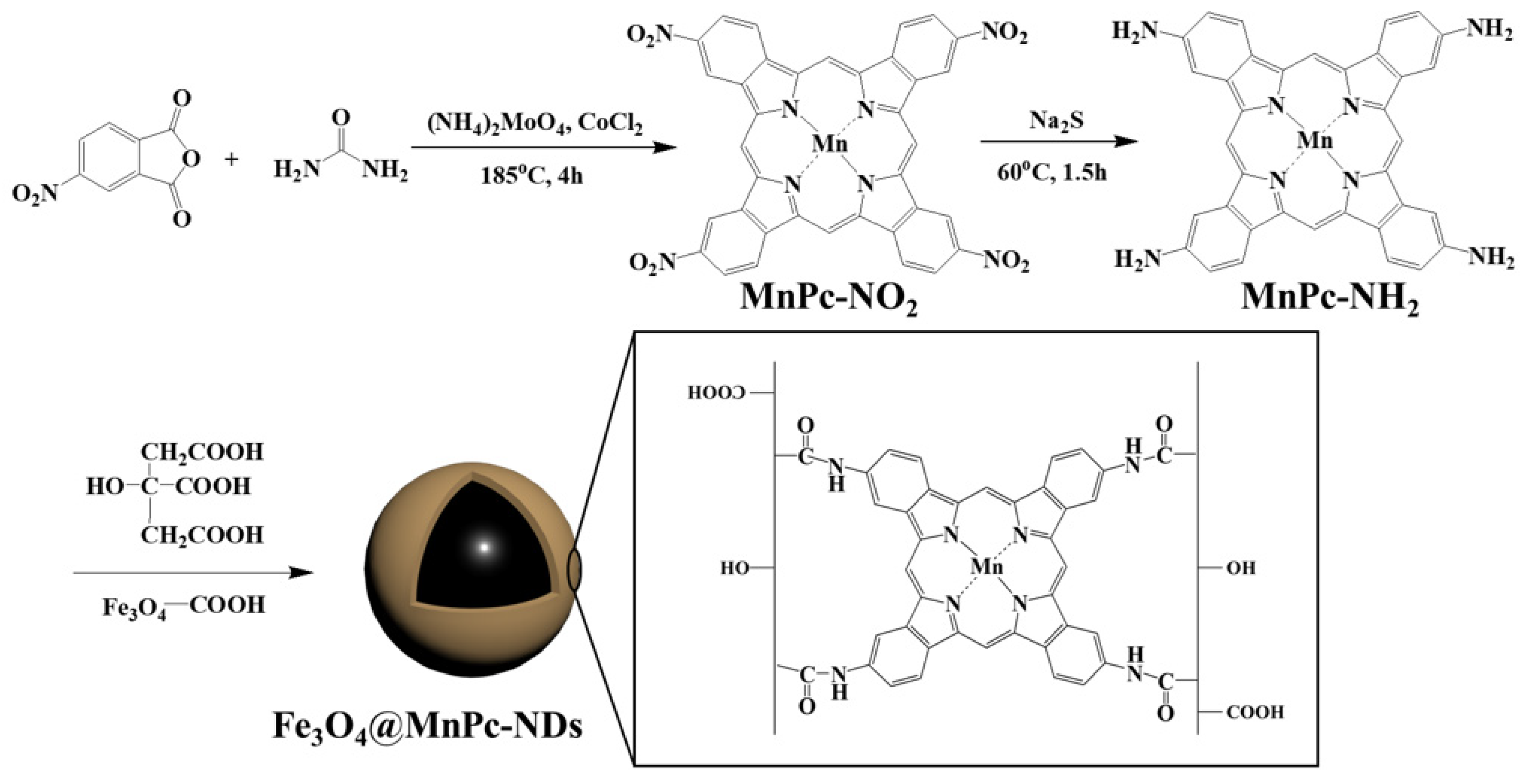
2.1. Synthesis of Manganese Tetranitro Phthalocyanine

As shown in Figure 1, a 6.0 g amount (100 mmol) of urea, 4.0 g (20 mmol) of 4-nitrophthalic anhydride, and 656 mg (5.2 mmol) of manganese chloride were dissolved in 30 mL of nitrobenzene solution. Afterward, 26 mg (0.02 mmol) of ammonium molybdate was added to the reaction mixture, which was then heated at 185 °C for 4 h under a nitrogen atmosphere. The reaction mixture was then cooled to room temperature and diluted with toluene. The suspension was centrifuged to collect the formed sediment, which was washed thoroughly with toluene, water, methanol/ether (1:9), and ethyl acetate/hexane (2:1) successively. Finally, the solid was dried under vacuum to obtain manganese tetranitro phthalocyanine (MnPc-NO2) in 80% yield.

2.2. Synthesis of Manganese Tetraamino Phthalocyanine

A 7.4 g amount (30.9 mmol) of sodium sulfide nonahydrate and 1.92 g (2.5 mmol) of manganese tetranitro phthalocyanine were dissolved in 50 mL of DMF. Under nitrogen atmosphere, the reaction mixture was heated to 60 °C and agitated for a period of 1.5 h. After cooling to ambient temperature, the reaction solution was diluted with 150 mL of water. The formed precipitate was then separated by centrifugation, followed by washing with methanol/ether mixture (1:9) and ethyl acetate successively. Finally, the solid was dried under vacuum to obtain manganese tetranitro phthalocyanine (MnPc-NH2) in 75% yield.

2.3. Synthesis of Magnetic Fe3O4 Nanoparticles

Magnetic Fe3O4 nanoparticles were fabricated using the solvothermal approach. Firstly, 0.75 g of PSSMA was dissolved in 20 mL of ethylene glycol and stirred for 15 min. Then 0.8 g of FeCl3·6H2O and 2.25 g of magnesium acetate were added sequentially and reacted for another 15 min. The reaction mixture was then carefully transferred to a hydrothermal reactor and heated at 200 °C for a duration of 10 h. Upon completion of the reaction, the resultant material was thoroughly rinsed three times using deionized water and anhydrous ethanol, to ensure the removal of impurities. Finally, the material was dried under vacuum for 12 h to yield the desired powder.

2.4. Synthesis of Fe3O4@MnPc-NDs

Firstly, 50 mg of Fe3O4 nanoparticles were dispersed by sonication in 10 mL of ultrapure water for 10 min. Then 15 mg of manganese tetraamino phthalocyanine and 150 mg of citric acid were sequentially added. After sonication for 10 min, the reaction solution was transferred to a hydrothermal reactor. After heated at 200 °C for 4 h, the resulting solid was separated and washed three times with deionized water and anhydrous ethanol, respectively. Finally, the desired composites were dried under vacuum to obtain the magnetic Fe3O4@MnPc-NDs with core–shell structure.

2.5. Peroxidase Like Catalytic Activity of Fe3O4@MnPc-NDs

To assess the peroxidase-like properties of Fe3O4@MnPc-NDs, the catalyzed oxidation of TMB by H2O2 was determined. In brief, TMB (5 mM, 50 μL) and Fe3O4@MnPc-NDs (1 mg/mL, 20 μL) were added into HAc-NaAc buffer (pH = 4.0, 2.8 μL). The combined solution was maintained at 25 °C for 7 min. Fe3O4@MnPc-NDs were then separated from the solution using an external magnet, followed by the measurement of the maximum absorbance at 652 nm.

2.6. Kinetic Measurements

The kinetic analysis of Fe3O4@MnPc-NDs was evaluated by Michaelis–Menten model [18]. In brief, different concentrations of H2O2 or TMB were added into a HAc-NaAc buffer solution (pH 3.8) containing Fe3O4@MnPc-NDs (1 µg/mL, 20 μL). The intensity of absorption at 652 nm was monitored at room temperature over time. k m is the Michaelis constant, v 0 and v m are the initial and maximum reaction velocities of the reaction, respectively, and s is the concentration of the substrate. The computation of this value is carried out using the Michaelis–Menten equation (Equation (1)).
1 v 0 = k m v m s + 1 v m
where v0 and vm represent the initial rate and maximum rate, respectively. km stands for the Michaelis–Menten constant and [s] stands for substrate (H2O2 or TMB) concentration.

2.7. Detection of H2O2 by the Colorimetric Method

A colorimetric detection method for H2O2 was established based on the relationship between the concentration of H2O2 and the absorbance of system at 652 nm. Specifically, 50 μL of different concentrations of H2O2, 20 μL of Fe3O4@MnPc-NDs suspension (1.0 mg/mL), and 50 μL of TMB solution (5 mM) were added to 2800 μL of HAc-NaAc buffer solution (pH 3.8), which was stirring at 25 °C for 7 min. Fe3O4@MnPc-NDs were then isolated from the solution by magnet, followed by the measurement of the absorbance at 652 nm. To further investigate the specificity of this method, interfering substances such as ascorbic acid (AA), citric acid (CA), dopamine (DA), Na+, K+, Ca2+, and glucose were selected as control samples.

2.8. Determination of AA and GSH by the Colorimetric Method

A colorimetric detection platform was devised based on the antioxidant activity of AA/GSH. Specifically, 20 μL of suspension containing Fe3O4@MnPc-NDs (1.0 mg/mL), 50 μL of H2O2 (0.1 M), 50 μL of TMB solution (5 mM), and 50 μL of AA (or GSH) of different concentrations were sequentially added to 2830 μL of HAc-NaAc buffer solution (pH 3.8). After stirring at 25 °C for 7 min, the absorbance at 652 nm was recorded by UV–vis absorption spectrum. The AA or GSH content was measured by observing the variation in absorbance at 652 nm. The selectivity of AA/GSH was determined by comparing the variations in absorbance at 652 nm between AA/GSH and twelve distinct interfering substances (Arg (arginine), His (histidine), Lys (lysine), Gly (glycine), Ala (alanine), Phe (phenylalanine), Val (valine), Mg2+, Zn2+, Na+, K+, and Ca2+). The concentration of AA/GSH was 60 μM, while the concentrations of the interfering substances were set at a level tenfold higher than that of AA/GSH.

2.9. Identification of AA and GSH Concentrations in Real Samples

To explore the practicality of the colorimetric method, a certain weight of vitamin C tablets was dissolved in water and further adjusted to the required volume to determine the amount of AA in vitamin C tablets. For accuracy verification, a standard AA solution was spiked into the diluted vitamin C tablet sample, and the absorbance variation at 652 nm upon vitamin C tablet addition was documented, respectively. Meanwhile, whitening serum was obtained from a nearby beauty shop and the spiked recovery method was employed to quantify the glutathione level in the sample.

3. Results and Discussion

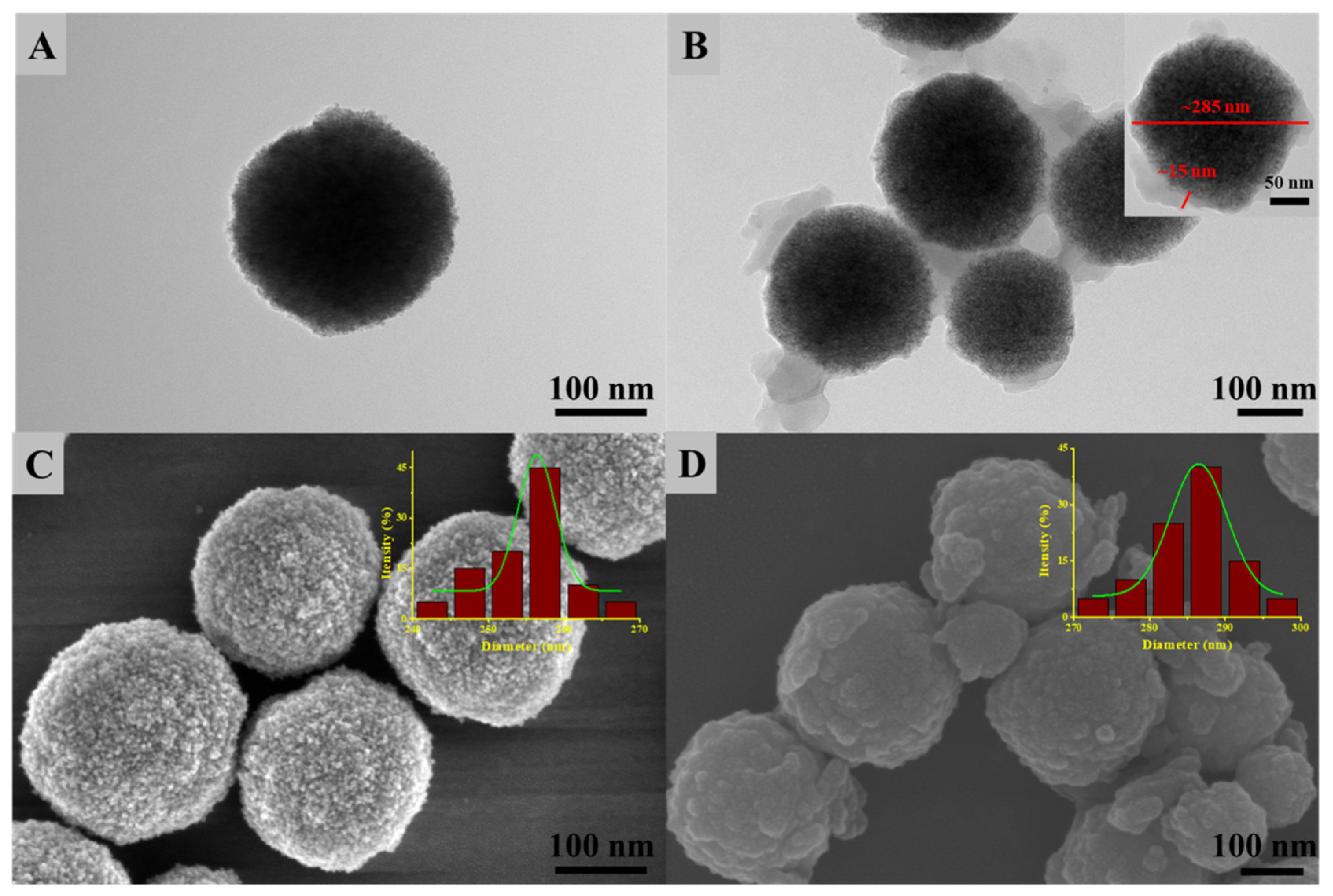
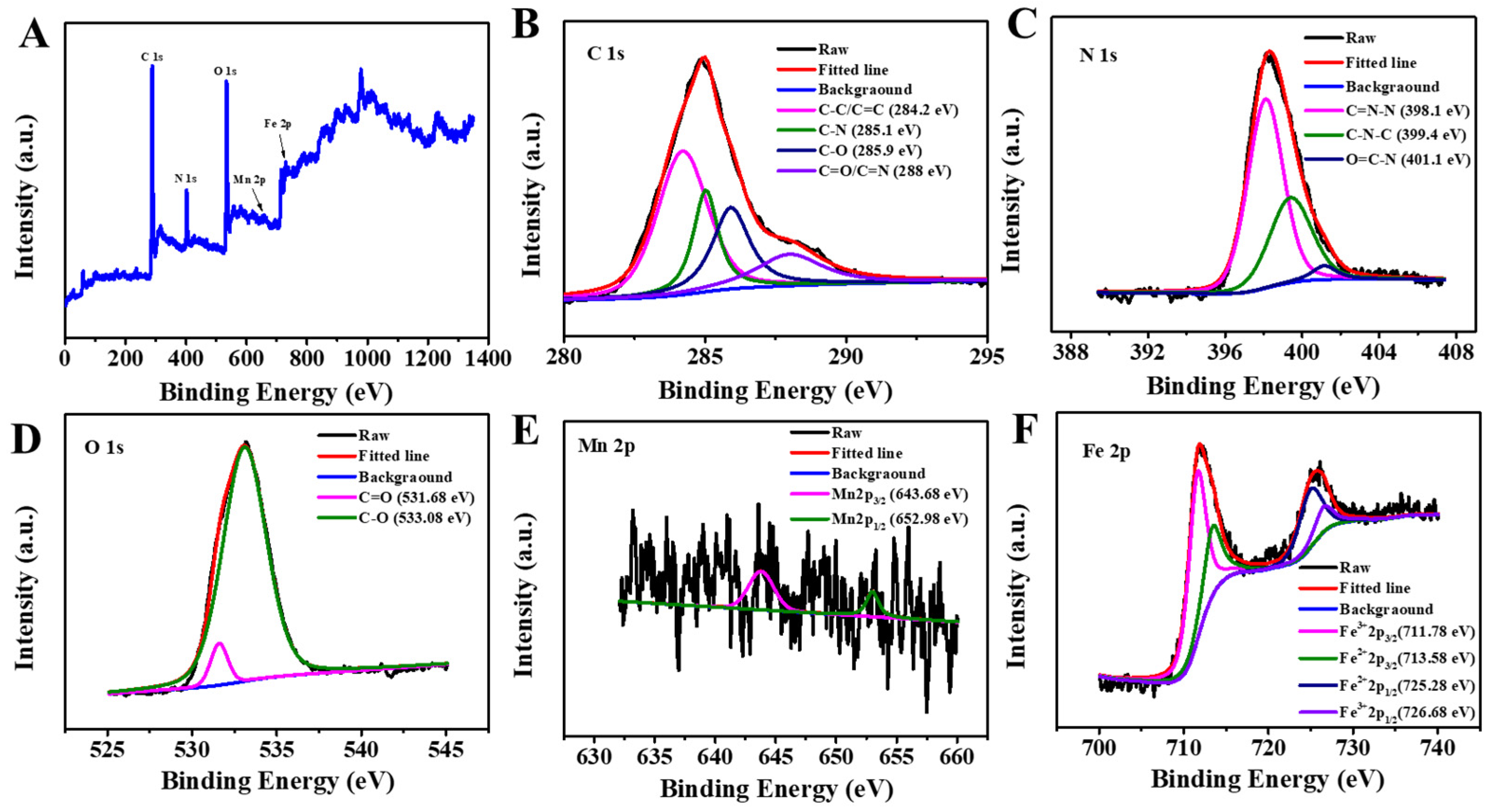
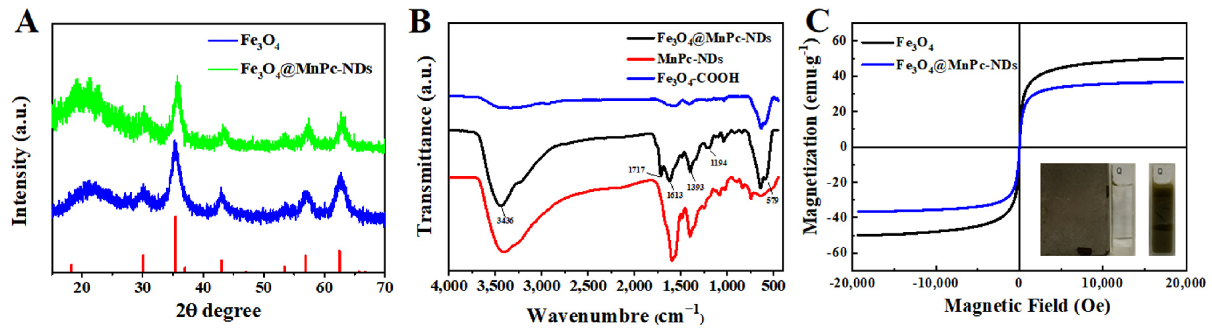
3.1. Characterization of Fe3O4@MnPc-NDs

The size and morphology of Fe3O4 nanospheres and Fe3O4@MnPc-NDs were characterized by scanning electron microscopy (SEM) and transmission electron microscopy (TEM). As indicated in Figure 2A, the size of Fe3O4 nanoparticles was about 257 nm. After functionalization with manganese phthalocyanine, the composites with core–shell structures were formed along with the increase in particle size to 285 nm (Figure 2B). The thickness of the shell layer in Fe3O4@MnPc-NDs was approximated to be 15 nm. FESEM (Figure 2C,D) demonstrated that the surface of Fe3O4@MnPc-NDs was rougher than that of bare Fe3O4 nanoparticles, further indicating the successful modification of phthalocyanine to the Fe3O4 surface.
The elemental composition of Fe3O4@MnPc-NDs was confirmed by XPS analysis. As indicated in Figure 3A, the distinct peaks at 285, 400, 531, 643, and 726 eV were observed, attributable to C 1s, N 1s, O 1s, Mn 2p, and Fe 2p, respectively. The XPS analysis at high resolution for C 1s orbital (Figure 3B) unveiled four discrete peaks positioned at 284.2, 285.1, 285.9, and 288.0 eV, each corresponding to C-C/C=C, C-N, C-O, and C=O/C=N bonds, respectively. The high-resolution XPS spectrum of N 1s (Figure 3C) showed C=N (398.1 eV) attributed to nitrogen of phthalocyanine ring, C-N (399.4 eV) attributed to the coordination of manganese and nitrogen, and O=C-N (401.1 eV) attributed to amide on the of nitrogen. The XPS analysis at high resolution for O 1s (Figure 3D) showed two peaks at 533.69 and 533.08 eV, attributed to C=O and C-O, respectively. The XPS spectra of Mn 2p could be fitted into two peaks at 652.98 and 643.68 eV, attributable to Mn 2p1/2 and Mn 2p3/2, respectively, indicating the presence of trivalent manganese ions in Fe3O4@MnPc-NDs (Figure 3E). Four peaks were observed in the high-resolution XPS spectra of Fe 2p (Figure 3F), matching Fe2+ 2p3/2 (713.58 eV), Fe2+ 2p1/2 (725.28 eV), Fe3+ 2p3/2 (711.78 eV), and Fe3+ 2p1/2 (726.68 eV) of Fe3O4 nanoparticles, respectively. Thus, the XPS spectrum clearly confirmed the presence of Fe and Mn elements in Fe3O4@MnPc-NDs in addition to C, N, and O.
The crystal structures of Fe3O4 nanoparticles and Fe3O4@MnPc-NDs were characterized through an X-ray diffraction (XRD) curve. As shown in Figure 4A, the XRD spectrum of Fe3O4 nanoparticles exhibited distinct diffraction peaks positioned at angles of 30.1°, 35.5°, 43.2°, 53.5°, 57.3°, and 62.9°, attributed to the characteristic lattice planes of (220), (311), (400), (422), (511), and (440), respectively, which were consistent with the reported data [34] (JCPDS No. 16-692). The XRD spectra of Fe3O4@MnPc-NDs were similar to that of Fe3O4 nanoparticles, indicating the functionalization of phthalocyanine did not affect the crystal structures of Fe3O4 nanoparticles. The functional groups on the surface were further characterized via infrared spectroscopy. As shown in Figure 4B, the characteristic peak at 579 cm−1 was ascribed to the Fe-O-Fe vibration in the Fe3O4 nanoparticles. The peaks at 3437, 1713, and 1613 cm−1 were assigned to the stretching vibrations of the O-H, C=O, and C=C, respectively. In addition, the peaks at 1393 and 1194 cm−1 were ascribed to the stretching vibrations of the C-N and C-O bonds, respectively. The magnetic properties of Fe3O4 nanoparticles and Fe3O4@MnPc-NDs were investigated using hysteresis loops. As indicated in Figure 4C, both Fe3O4 nanoparticles and Fe3O4@MnPc-NDs displayed obvious magnetic properties with saturation magnetization strengths of 49.46 and 36.37 emu/g, respectively. In addition, with the help of an external magnetic field, Fe3O4@MnPc-NDs dispersed in aqueous solution can be collected in a short time, indicating their excellent magnetism and ability for rapid magnetic separation.

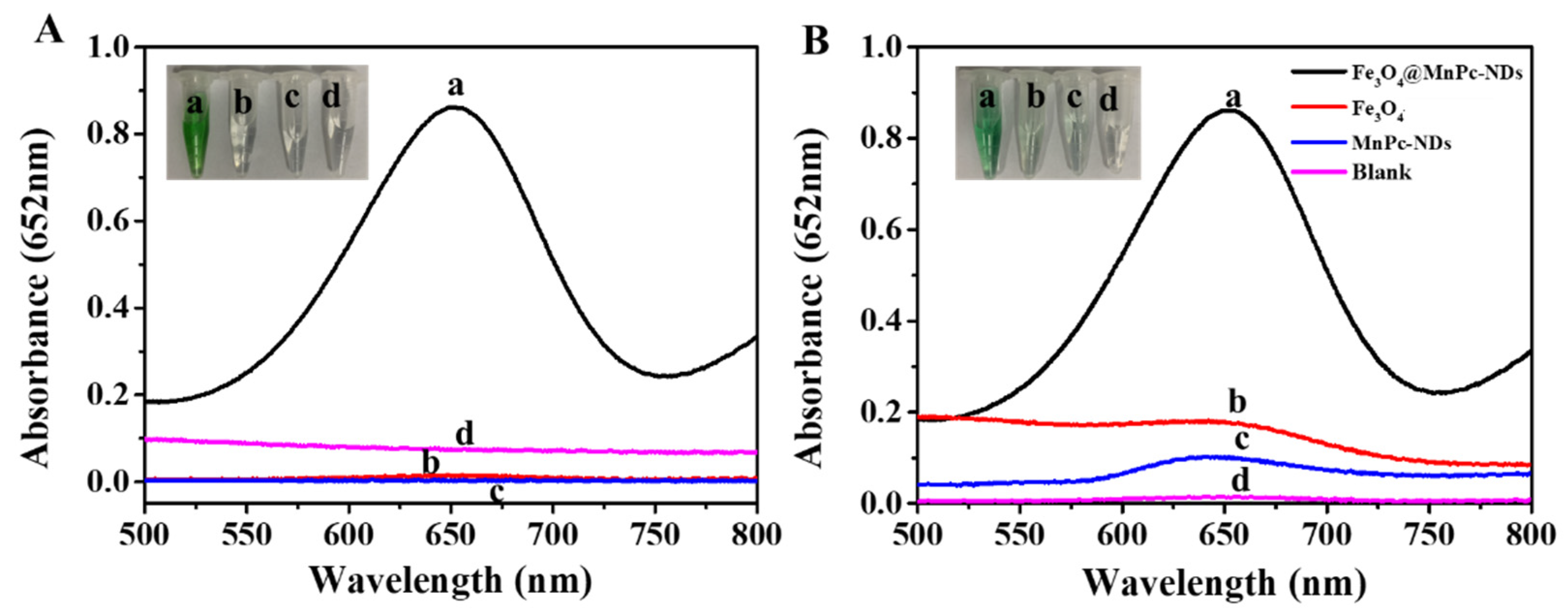
3.2. Peroxidase-Mimicking Activity of Fe3O4@MnPc-NDs

The peroxidase-mimicking activity of Fe3O4@MnPc-NDs was evaluated with TMB and H2O2 as substrates. The absorption spectrum of the system was recorded through UV–vis absorption spectroscopy. As depicted in Figure 5A, only the “TMB + H2O2 + Fe3O4@MnPc-NDs” system exhibited an obvious absorption peak at 652 nm by comparing with the control groups, along with the appearance of a distinct blue color in solution. Moreover, the structural advantages of Fe3O4@MnPc-NDs were illustrated by comparing the catalytic activities of raw materials and composites. As shown in Figure 5B, the UV–visible spectrum of Fe3O4@MnPc-NDs exhibited a significant absorption peak at 652 nm. Moreover, the absorption intensity at 652 nm of Fe3O4@MnPc-NDs was higher than that of other control samples (Fe3O4 nanoparticles and MnPc-NDs), indicating the higher catalytic activity after functionalization.

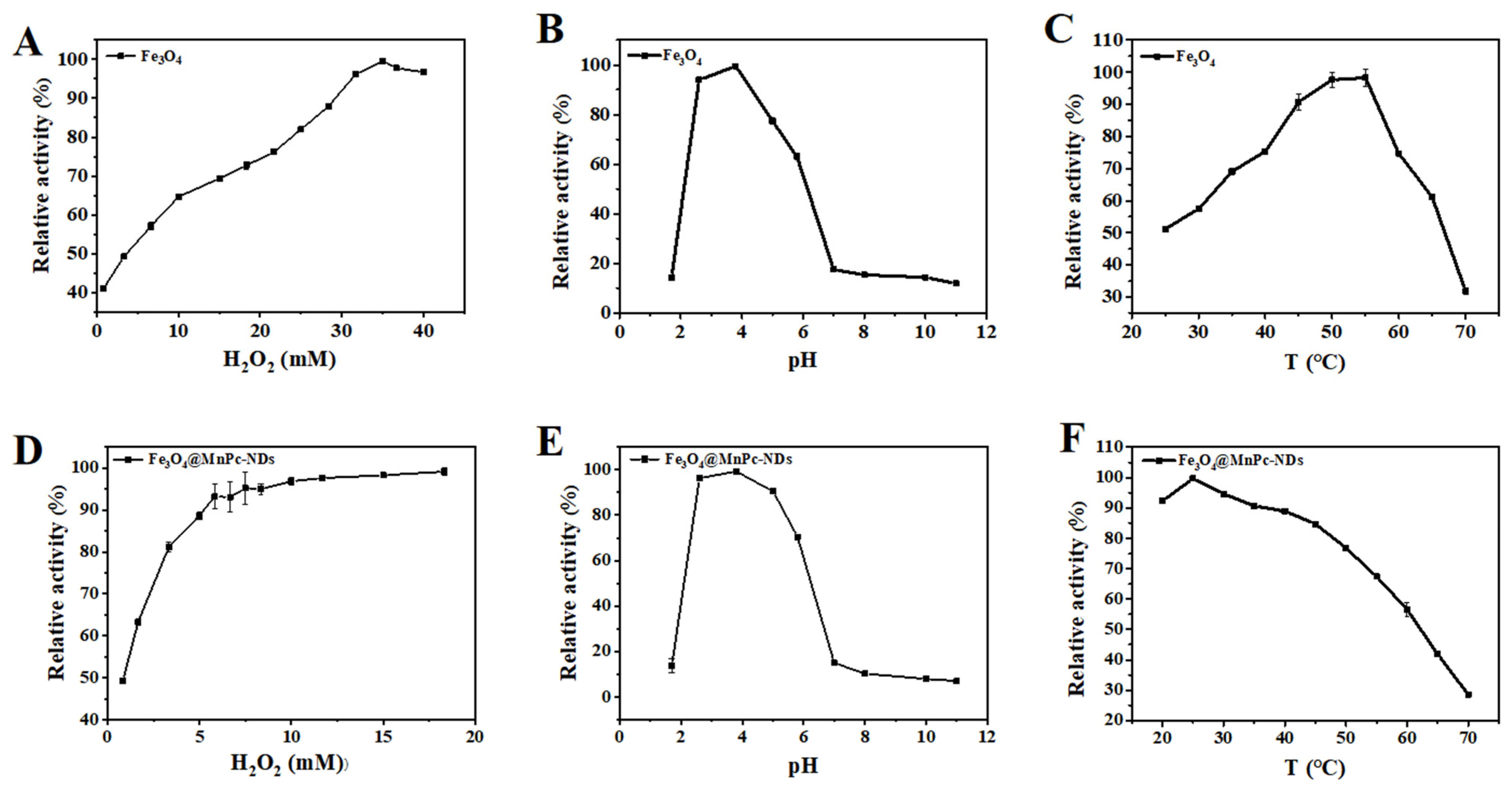
3.3. Optimization of the Catalyzed Conditions

Similar to other peroxidase-like artificial enzymes, the catalytic activity of Fe3O4@MnPc-NDs might also depend on the H2O2 concentration, pH values, and temperature. Accordingly, the investigations were conducted to examine the catalytic activity of Fe3O4 nanoparticles and Fe3O4@MnPc-NDs under varying experimental conditions, specifically encompassing H2O2 concentrations ranging from 0.8 to 13 mM, pH values spanning from 1.7 to 11, and temperatures ranging from 20 to 70 °C. As depicted in Figure 6A,D, the catalytic activity of two materials started to increase and then leveled off with the increase in H2O2 concentration. The best catalytic activity was achieved at an H2O2 concentration of 10 mM, lower than that of bare Fe3O4 nanoparticles (255 mM). As shown in Figure 6E, Fe3O4@MnPc-NDs exhibited the highest activity at pH 3.8, which was similar to that of Fe3O4 nanoparticles (Figure 6B). Moreover, a temperature of 25 °C was found to be the most favorable for the catalytic performance of Fe3O4@MnPc-NDs (Figure 6F), while a higher temperature (55 °C) was required for Fe3O4 nanoparticles to realize the maximum catalytic activity (Figure 6C). Therefore, Fe3O4@MnPc-NDs exhibited the maximum catalytic activity under the milder conditions compared with that of bare Fe3O4 nanoparticles, and H2O2 concentration of 10 mM, pH 3.8, and temperature 25 °C were chosen as the most suitable conditions for studying the catalytic activity of Fe3O4@MnPc-NDs.

3.4. Steady-State Kinetic Determination of Fe3O4@MnPc-NDs

To further understand the catalytic mechanism of Fe3O4@MnPc-NDs, a series of kinetic assessments were performed by incrementally adjusting the concentration of one substrate while maintaining a constant level of the other substrate throughout the entire series of measurements. As displayed in Figure 7A,C, the typical Michaelis–Menten curves for TMB and H2O2 were measured within their appropriate concentration ranges. The corresponding relationships between the reciprocals of initial velocity and substrate concentration were obtained (Figure 7B,D). The oxidation reaction of Fe3O4@MnPc-NDs upon TMB and H2O2 conformed to the typical Michaelis–Mente equation. To assess the key kinetic attributes of the enzyme, including the Michaelis–Menten constant (Km) and the maximum initial reaction rate (Vmax), the Lineweaver–Burk plot was utilized (Table 1). Km serves as an indicator of enzyme affinity for substrates. As the Km value decreases, the affinity of the enzyme toward its substrate increases. Compared with HRP and Fe3O4 nanoparticles, Fe3O4@MnPc-NDs exhibited lower Km values for H2O2, indicating that H2O2 can be detected at lower concentrations.

3.5. Detection of H2O2

Based on the peroxidase-mimicking properties of Fe3O4@MnPc-NDs, a simple colorimetric approach for the quantification of H2O2 was devised. As depicted in Figure 8A,C, a distinct enhancement in the absorbance of the mixture of Fe3O4@MnPc-NDs and TMB at 652 nm was observed, with the H2O2 concentration ranging from 0.2 to 15 mM. Based on the variation of absorbance and color change in solution, a linear calibration curve was obtained with the H2O2 concentration ranging from 20 to 250 μM (Figure 8B). According to the obtained linear equation, the detection limit (LOD) of H2O2 was calculated to be 4.7 μM, which was lower than the values previously reported in the literature (Table 2). In order to evaluate the influence of interfering substances, a range of potential interferences, including dopamine (DA), citric acid (CA), ascorbic acid (AA), glucose, as well as ions like Na+, K+, and Ca2+ were chosen. As depicted in Figure 8D, the solution color of Fe3O4@MnPc-NDs and TMB did not show any significant change after adding the interfering substances, even though their concentration was five times higher than that of H2O2, indicating the high selectivity and specificity for H2O2 detection.

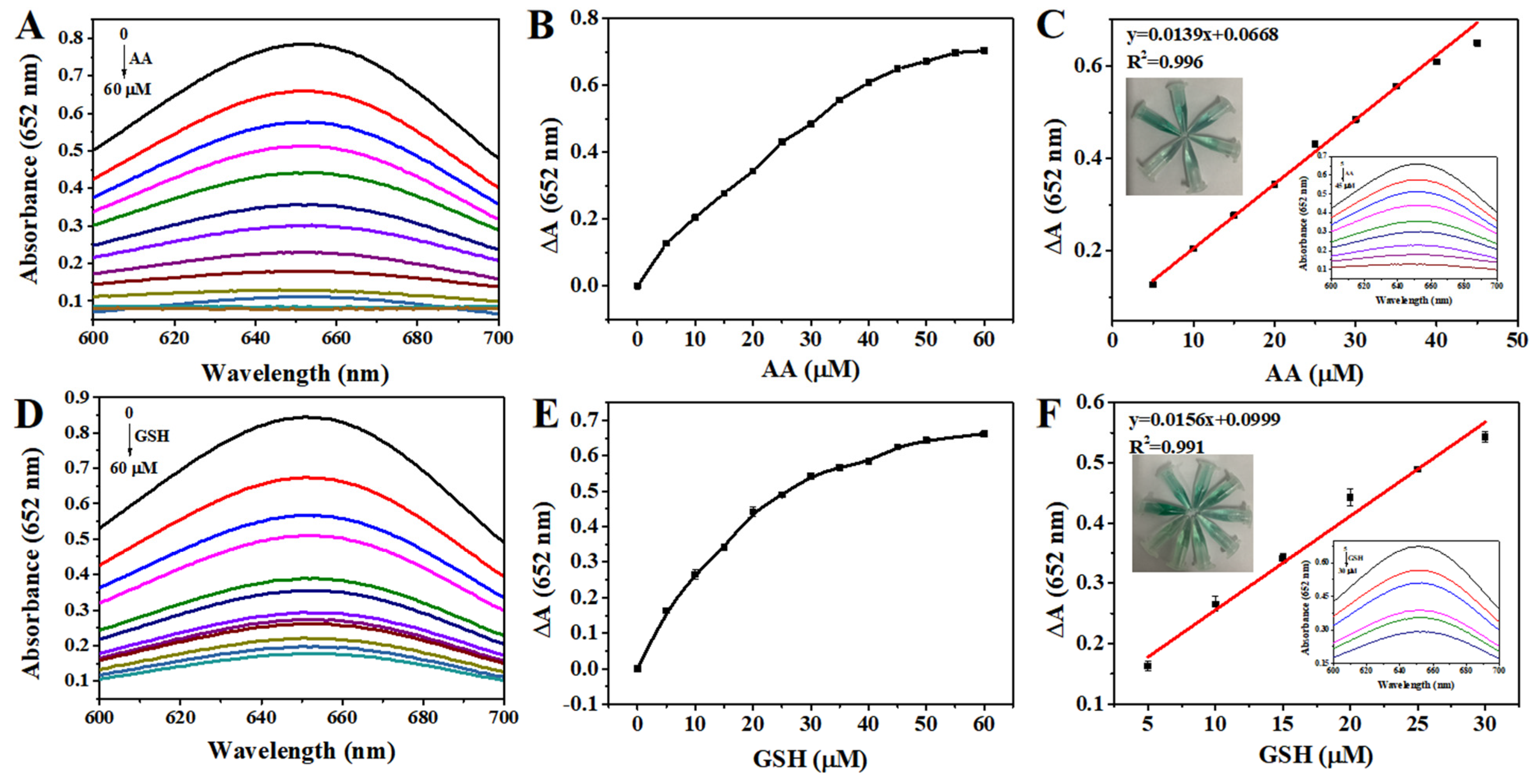
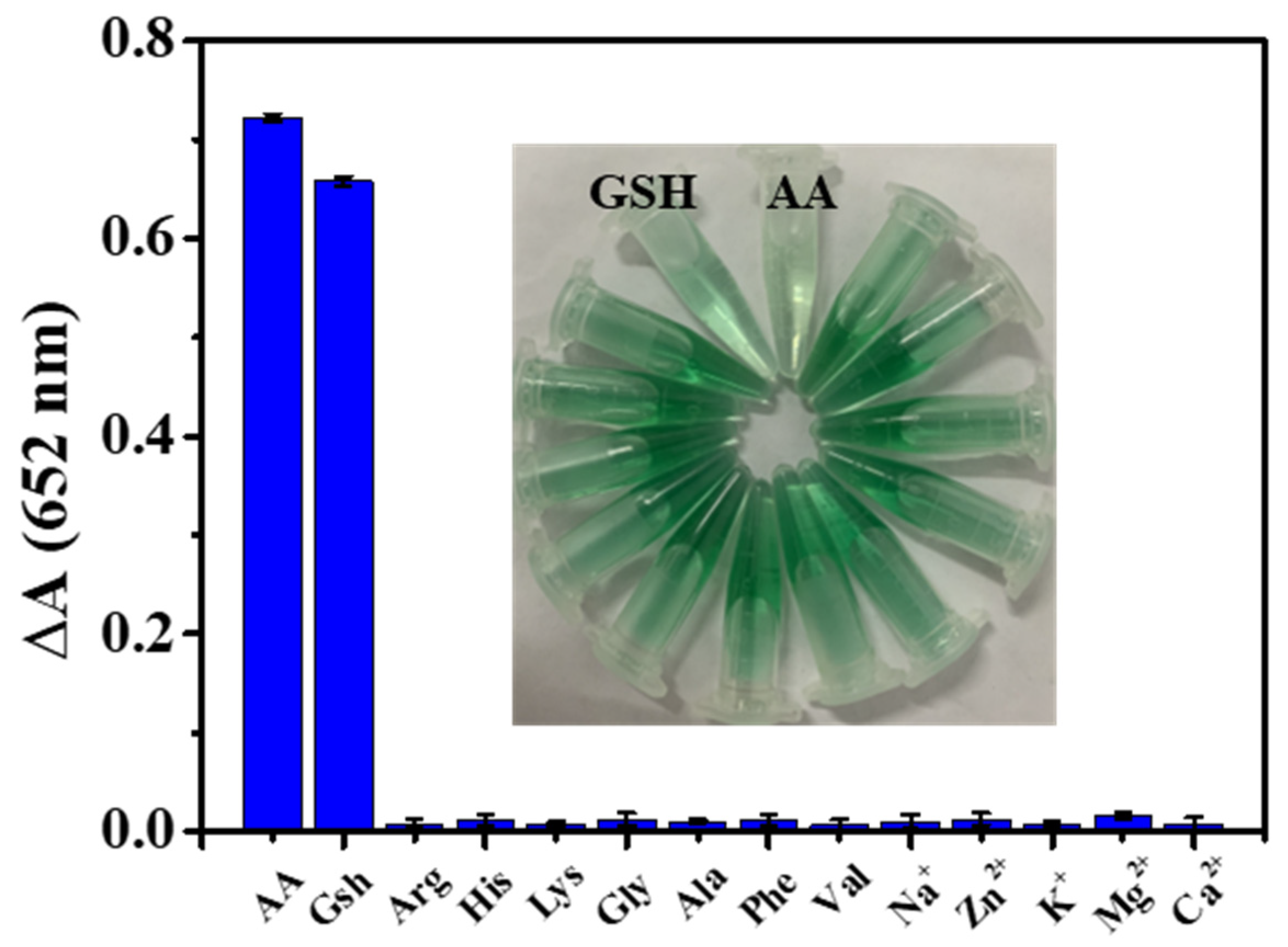
3.6. Detection of AA and GSH

Based on the antioxidant effects of AA and GSH, a colorimetric approach for detection was established using the system of Fe3O4@MnPc-NDs and TMB. As shown in Figure 9A, the absorbance of the sensing platform at 652 nm exhibited a gradual decline upon the addition of ascorbic acid and GSH (Figure 9A,B), along with the fading of the blue solution. Based on the variation curve of absorbance at 652 nm upon the AA (Figure 9C) or GSH (Figure 9D) concentration, a linear equation was obtained for AA (ΔA = 0.0139 [AA] + 0.0668 (R2 = 0.996)) and GSH (ΔA = 0.0156 [GSH] + 0.0999 (R2 = 0.991)) (Figure 9E,F). According to these equations, the detection limit was calculated to be 0.161 μM for AA, and 0.188 μM for GSH, which were comparable to those reported by colorimetric methods (Table 3). Using Arg (arginine), Ala (alanine), His (histidine), Lys (lysine), Gly (glycine), Phe (phenylalanine), Val (valine), Mg2+, Zn2+, Na+, K+, and Ca2+ as interfering substances, the selectivity study for AA/GSH determination was conducted based on Fe3O4@MnPc-NDs. As shown in Figure 10, even though the concentrations of numerous interfering substances escalated to tenfold that of AA/GSH, the solution color of Fe3O4@MnPc-NDs/TMB/H2O2 system did not exhibit any significant change, along with negligible alterations in absorbance at 652 nm.

3.7. Detection of Ascorbic Acid and Glutathione in Actual Samples

To evaluate the practical application of this colorimetric method, the ascorbic acid content in vitamin C was tested. Specifically, vitamin C was diluted to a certain concentration and then added to the Fe3O4@MnPc-NDs/TMB/H2O2 system. To further corroborate the precision of this method, a known concentration of AA was introduced into the diluted sample of vitamin C tablets. As evident from Table S1, the recovered quantities of AA in these samples were extremely close to the expected values, with recovery rates higher than 98% and the relative standard deviation (RSD) less than 5%. The colorimetric method was also used to detect the content of glutathione in the purchased whitening serum. As shown in Table S2, the sample recoveries were higher than 99%, with an RSD of less than 5%, indicating the high accuracy of this method for the analysis of real samples.

3.8. Recyclability Testing

To evaluate the recyclability of Fe3O4@MnPc-NDs, the recovered Fe3O4@MnPc-NDs were added into the “TMB + H2O2” system, and their catalytic activity was tested. As shown in Figure 11, after five catalytic cycles, Fe3O4@MnPc-NDs still possessed high peroxidase activity with catalytic activity around 90%, indicating the good reusability of Fe3O4@MnPc-NDs.

4. Conclusions

In summary, the Fe3O4@MnPc-NDs composites were effectively synthesized via the hydrothermal method. Fe3O4@MnPc-NDs demonstrated enhanced peroxidase-like activity compared with the bare Fe3O4 nanoparticles, which could oxidize the colorless TMB to blue ox-TMB in the presence of H2O2. Based on the inhibitory effect of AA/GSH on this catalytic process, a highly sensitive and rapid colorimetric sensor was developed, capable of determining AA and GSH with detection limits of 0.161 and 0.188 μM, respectively. This developed sensor exhibited high selectivity and exceptional reproducibility, enabling its application in determining AA levels in vitamin C tablets and GSH concentrations in whitening serum, indicating the practical potential of Fe3O4@MnPc-NDs in colorimetric assays.

Supplementary Materials

The following supporting information can be downloaded at: https://www.mdpi.com/article/10.3390/molecules30071484/s1. Table S1 Recovery of AA from vitamin C samples by labeling. Table S2 Standard addition and recovery of GSH in whitening essence.

Author Contributions

Methodology, J.Q., Y.P. and F.W.; Software, Y.P.; Validation, J.Q. and L.T.; Formal analysis, Y.P.; Investigation, L.T.; Data curation, L.T.; Writing—original draft, J.Q.; Writing—review & editing, F.W.; Supervision, F.W. All authors have read and agreed to the published version of the manuscript.

Funding

This work was supported by the Hubei Province Science and Technology Innovation Talent Plan Project (grant no. 2024DJC036) and Innovation Project of Key Laboratory of Novel Biomass-Based Environmental and Energy Materials in Petroleum and Chemical Industry (grant no. 2024BEEA06).

Institutional Review Board Statement

Not applicable.

Informed Consent Statement

Not applicable.

Data Availability Statement

The original contributions presented in this study are included in the article/Supplementary Materials. Further inquiries can be directed to the corresponding author.

Conflicts of Interest

The authors declare no conflict of interest.

References

  1. Zhong, B.; Zhao, L.; Yu, J.; Hou, Y.; Ai, N.; Lu, J.-J.; Ge, W.; Chen, X. Exogenous iron impairs the anti-cancer effect of ascorbic acid both in vitro and in vivo. J. Adv. Res. 2023, 46, 149–158. [Google Scholar] [CrossRef]
  2. Agathocleous, M.; Meacham, C.E.; Burgess, R.J.; Piskounova, E.; Zhao, Z.Y.; Crane, G.M.; Cowin, B.L.; Bruner, E.; Murphy, M.M.; Chen, W.N.; et al. Ascorbate regulates haematopoietic stem cell function and leukaemogenesis. Nature 2017, 549, 476–481. [Google Scholar] [CrossRef]
  3. Shenoy, N.; Creagan, E.; Witzig, T.; Levine, M. Ascorbic Acid in Cancer Treatment: Let the Phoenix Fly. Cancer Cell 2018, 34, 700–706. [Google Scholar] [CrossRef]
  4. Chang, J.; Qin, X.; Li, S.; He, F.; Gai, S.; Ding, H.; Yang, P. Combining Cobalt Ferrite Nanozymes with a Natural Enzyme to Reshape the Tumor Microenvironment for Boosted Cascade Enzyme-Like Activities. ACS Appl. Mater. Interfaces 2022, 14, 45217–45228. [Google Scholar] [CrossRef]
  5. Li, H.Q.; Ma, J.; Qin, Y.M.; Sun, X.; Pei, Z.G.; Yang, R.Q.; Li, Y.M.; Zhang, Q.H. Assessment of interactions between elemental carbon and metals in black carbon: Hydroxyl radical generation and glutathione depletion. J. Hazard. Mater. 2024, 470, 134223. [Google Scholar] [CrossRef]
  6. Pei, P.; Shen, W.H.; Zhou, H.L.; Sun, Y.C.; Zhong, J.; Liu, T.; Yang, K. Radionuclide labeled gold nanoclusters boost effective anti-tumor immunity for augmented radio-immunotherapy of cancer. Nano Today 2021, 38, 101144. [Google Scholar] [CrossRef]
  7. Liu, W.L.; Zhang, Y.H.; Wei, G.; Zhang, M.X.; Li, T.; Liu, Q.Y.; Zhou, Z.J.; Du, Y.; Wei, H. Integrated Cascade Nanozymes with Antisenescence Activities for Atherosclerosis Therapy. Angew. Chem. Int. Ed. 2023, 62, e202304465. [Google Scholar] [CrossRef]
  8. Charisis, S.; Ntanasi, E.; Stamelou, M.; Xiromerisiou, G.; Maraki, M.; Veskoukis, A.S.; Yannakoulia, M.; Kosmidis, M.H.; Anastasiou, C.A.; Giagkou, N.; et al. Plasma Glutathione and Prodromal Parkinson’s Disease Probability. Mov. Disord. 2022, 37, 200–205. [Google Scholar] [CrossRef]
  9. Guo, Y.X.; Liu, Y.R.; Zhao, S.H.; Xu, W.T.; Li, Y.Q.; Zhao, P.W.; Wang, D.; Cheng, H.Q.; Ke, Y.H.; Zhang, X. Oxidative stress-induced FABP5 S-glutathionylation protects against acute lung injury by suppressing inflammation in macrophages. Nat. Commun. 2021, 12, 7094. [Google Scholar] [CrossRef]
  10. Zhu, X.H.; Feng, T.T.; Chen, Y.D.; Xiao, Y.; Wen, W.; Zhang, X.H.; Wang, D.; Wang, S.F.; Liang, J.C.; Xiong, H.Y. GSH-depleted Cu-covalent organic frameworks for multimodal synergistic therapy against diabetic infections. Chem. Eng. J. 2024, 488, 150770. [Google Scholar] [CrossRef]
  11. Cheng, X.T.; Xu, H.D.; Ran, H.H.; Liang, G.L.; Wu, F.G. Glutathione-Depleting Nanomedicines for Synergistic Cancer Therapy. Acs Nano 2021, 15, 8039–8068. [Google Scholar] [CrossRef]
  12. Cano, A.; Ettcheto, M.; Chang, J.-H.; Barroso, E.; Espina, M.; Kühne, B.A.; Barenys, M.; Auladell, C.; Folch, J.; Souto, E.B.; et al. Dual-drug loaded nanoparticles of Epigallocatechin-3-gallate (EGCG)/Ascorbic acid enhance therapeutic efficacy of EGCG in a APPswe/PS1dE9 Alzheimer’s disease mice model. J. Control. Release 2019, 301, 62–75. [Google Scholar] [CrossRef]
  13. Xi, X.X.; Wang, J.H.; Wang, Y.Z.; Xiong, H.Y.; Chen, M.M.; Wu, Z.; Zhang, X.H.; Wang, S.F.; Wen, W. Preparation of Au/Pt/Ti3C2Cl2 nanoflakes with self-reducing method for colorimetric detection of glutathione and intracellular sensing of hydrogen peroxide. Carbon 2022, 197, 476–484. [Google Scholar] [CrossRef]
  14. Lai, X.; Shen, Y.; Gao, S.B.; Chen, Y.J.; Cui, Y.S.; Ning, D.X.; Ji, X.B.; Liu, Z.W.; Wang, L.G. The Mn-modified porphyrin metal-organic framework with enhanced oxidase-like activity for sensitively colorimetric detection of glutathione. Biosens. Bioelectron. 2022, 213, 114446. [Google Scholar] [CrossRef]
  15. Liao, L.H.; Lin, X.; Zhang, J.; Hu, Z.Y.; Wu, F.S. Facile preparation of carbon dots with multicolor emission for fluorescence detection of ascorbic acid, glutathione and moisture content. J. Lumin. 2023, 264, 120169. [Google Scholar] [CrossRef]
  16. Zhe, Y.; Wang, J.L.; Zhao, Z.Q.; Ren, G.Y.; Du, J.J.; Li, K.; Lin, Y.Q. Ascorbate oxidase-like nanozyme with high specificity for inhibition of cancer cell proliferation and online electrochemical DOPAC monitoring. Biosens. Bioelectron. 2023, 220, 114893. [Google Scholar] [CrossRef]
  17. Chen, M.M.; Zhu, J.L.; Yang, B.C.; Yao, X.X.; Zhu, X.X.; Liu, Q.Y.; Lyu, X.T. Novel synthesis of NiS/MMT/GO nanocomposites with enhanced peroxidase-like activity for sensitive colorimetric detection of glutathione in solution. Adv. Compos. Hybrid Mater. 2018, 1, 612–623. [Google Scholar] [CrossRef]
  18. Guo, D.; Li, C.; Liu, G.; Luo, X.; Wu, F. Oxidase Mimetic Activity of a Metalloporphyrin-Containing Porous Organic Polymer and Its Applications for Colorimetric Detection of Both Ascorbic Acid and Glutathione. Acs Sustain. Chem. Eng. 2021, 9, 5412–5421. [Google Scholar] [CrossRef]
  19. Wang, C.; Chen, L.; Wang, P.; Li, M.; Liu, D. A novel ultrasensitive electrochemiluminescence biosensor for glutathione detection based on poly-L-lysine as co-reactant and graphene-based poly(luminol/aniline) as nanoprobes. Biosens. Bioelectron. 2019, 133, 154–159. [Google Scholar] [CrossRef]
  20. Zeng, J.J.; Liao, L.H.; Lin, X.; Liu, G.Y.; Luo, X.G.; Luo, M.; Wu, F.S. Red-Emissive Sulfur-Doped Carbon Dots for Selective and Sensitive Detection of Mercury (II) Ion and Glutathione. Int. J. Mol. Sci. 2022, 23, 9213. [Google Scholar] [CrossRef]
  21. Fan, S.S.; Zhao, M.G.; Ding, L.J.; Li, H.; Chen, S.G. Preparation of Co3O4 crumpled graphene microsphere as peroxidase mimetic for colorimetric assay of ascorbic acid. Biosens. Bioelectron. 2017, 89, 846–852. [Google Scholar] [CrossRef]
  22. Zhang, J.J.; Zheng, W.S.; Jiang, X.Y. Ag+-Gated Surface Chemistry of Gold Nanoparticles and Colorimetric Detection of Acetylcholinesterase. Small 2018, 14, 1801680. [Google Scholar] [CrossRef]
  23. Li, W.; Khan, M.; Lin, L.; Zhang, Q.; Feng, S.; Wu, Z.; Lin, J.M. Monitoring H2O2 on the Surface of Single Cells with Liquid Crystal Elastomer Microspheres. Angew. Chem. Int. Ed. 2020, 59, 9282–9287. [Google Scholar] [CrossRef]
  24. Liu, Y.; Liu, X.; Guo, Z.; Hu, Z.; Xue, Z.; Lu, X. Horseradish peroxidase supported on porous graphene as a novel sensing platform for detection of hydrogen peroxide in living cells sensitively. Biosens. Bioelectron. 2017, 87, 101–107. [Google Scholar] [CrossRef]
  25. Zhang, L.L.; Wang, J.; Zhao, C.L.; Zhou, F.J.; Yao, C.; Song, C. Ultra-fast colorimetric detection of glutathione by magnetic Fe NPs with peroxidase-like activity. Sens. Actuators B-Chem. 2022, 361, 131750. [Google Scholar] [CrossRef]
  26. Huang, Y.; Gu, Y.Q.; Liu, X.Y.; Deng, T.T.; Dai, S.; Qu, J.F.; Yang, G.H.; Qu, L.L. Reusable ring-like Fe3O4Au nanozymes with enhanced peroxidase-like activities for colorimetric-SERS dual-mode sensing of biomolecules in human blood. Biosens. Bioelectron. 2022, 209, 114253. [Google Scholar] [CrossRef]
  27. Liu, C.; Zhao, X.P.; Wang, Z.C.; Zhao, Y.Y.; Li, R.F.; Chen, X.Y.; Chen, H.; Wan, M.N.; Wang, X.Q. Metal-organic framework-modulated Fe3O4 composite au nanoparticles for antibacterial wound healing via synergistic peroxidase-like nanozymatic catalysis. J. Nanobiotechnology 2023, 21, 427. [Google Scholar] [CrossRef]
  28. Zhang, Z.; Zhang, X.; Liu, B.; Liu, J. Molecular Imprinting on Inorganic Nanozymes for Hundred-fold Enzyme Specificity. J. Am. Chem. Soc. 2017, 139, 5412–5419. [Google Scholar] [CrossRef]
  29. Liu, S.X.; Yu, B.; Wang, S.; Shen, Y.Q.; Cong, H.L. Preparation, surface functionalization and application of Fe3O4 magnetic nanoparticles. Adv. Colloid Interface Sci. 2020, 281, 102165. [Google Scholar] [CrossRef]
  30. Matheu, R.; Gutierrez-Puebla, E.; Monge, M.Á.; Diercks, C.S.; Kang, J.; Prévot, M.S.; Pei, X.; Hanikel, N.; Zhang, B.; Yang, P.; et al. Three-Dimensional Phthalocyanine Metal-Catecholates for High Electrochemical Carbon Dioxide Reduction. J. Am. Chem. Soc. 2019, 141, 17081–17085. [Google Scholar] [CrossRef]
  31. Sun, Q.Q.; Liu, Q.; Gao, W.; Xing, C.W.; Shen, J.S.; Liu, X.; Kong, X.; Li, X.Y.; Zhang, Y.X.; Chen, Y.L. A high-performance photoelectrochemical sensor for the specific detection of H2O2 and glucose based on an organic conjugated microporous polymer. J. Mater. Chem. A 2021, 9, 26216–26225. [Google Scholar] [CrossRef]
  32. Chan, J.Y.M.; Kawata, T.; Kobayashi, N.; Ng, D.K.P. Boron(III) Carbazosubphthalocyanines: Core-Expanded Antiaromatic Boron(III) Subphthalocyanine Analogues. Angew. Chem. Int. Ed. 2019, 58, 2272–2277. [Google Scholar] [CrossRef]
  33. Zheng, B.D.; He, Q.X.; Li, X.S.; Yoon, J.; Huang, J.D. Phthalocyanines as contrast agents for photothermal therapy. Coord. Chem. Rev. 2021, 426, 213548. [Google Scholar] [CrossRef]
  34. Wang, Q.Q.; Zhang, X.P.; Huang, L.; Zhang, Z.Q.; Dong, S.J. One-Pot Synthesis of Fe3O4 Nanoparticle Loaded 3D Porous Graphene Nanocomposites with Enhanced Nanozyme Activity for Glucose Detection. Acs Appl. Mater. Interfaces 2017, 9, 7465–7471. [Google Scholar] [CrossRef]
  35. Gao, L.; Zhuang, J.; Nie, L.; Zhang, J.; Zhang, Y.; Gu, N.; Wang, T.; Feng, J.; Yang, D.; Perrett, S.; et al. Intrinsic peroxidase-like activity of ferromagnetic nanoparticles. Nat. Nanotechnol. 2007, 2, 577–583. [Google Scholar] [CrossRef]
  36. Darabdhara, G.; Sharma, B.; Das, M.R.; Boukherroub, R.; Szunerits, S. Cu-Ag bimetallic nanoparticles on reduced graphene oxide nanosheets as peroxidase mimic for glucose and ascorbic acid detection. Sens. Actuators B Chem. 2017, 238, 842–851. [Google Scholar] [CrossRef]
  37. Shi, Y.; Su, P.; Wang, Y.; Yang, Y. Fe3O4 peroxidase mimetics as a general strategy for the fluorescent detection of H2O2-involved systems. Talanta 2014, 130, 259–264. [Google Scholar] [CrossRef]
  38. Song, Y.; Qu, K.; Zhao, C.; Ren, J.; Qu, X. Graphene Oxide: Intrinsic Peroxidase Catalytic Activity and Its Application to Glucose Detection. Adv. Mater. 2010, 22, 2206–2210. [Google Scholar] [CrossRef]
  39. Xiao, F.; Li, Y.; Zan, X.; Liao, K.; Xu, R.; Duan, H. Growth of Metal-Metal Oxide Nanostructures on Freestanding Graphene Paper for Flexible Biosensors. Adv. Funct. Mater. 2012, 22, 2487–2494. [Google Scholar] [CrossRef]
  40. Han, L.; Li, C.; Zhang, T.; Lang, Q.; Liu, A. Au@Ag Heterogeneous Nanorods as Nanozyme Interfaces with Peroxidase-Like Activity and Their Application for One-Pot Analysis of Glucose at Nearly Neutral pH. ACS Appl. Mater. Interfaces 2015, 7, 14463–14470. [Google Scholar] [CrossRef]
  41. Yadav, P.K.; Singh, V.K.; Chandra, S.; Bano, D.; Kumar, V.; Talat, M.; Hasan, S.H. Green Synthesis of Fluorescent Carbon Quantum Dots from Azadirachta indica Leaves and Their Peroxidase-Mimetic Activity for the Detection of H2O2 and Ascorbic Acid in Common Fresh Fruits. ACS Biomater. Sci. Eng. 2018, 5, 623–632. [Google Scholar] [CrossRef] [PubMed]
  42. Luo, X.; Zhang, W.; Han, Y.; Chen, X.; Zhu, L.; Tang, W.; Wang, J.; Yue, T.; Li, Z. N,S co-doped carbon dots based fluorescent “on-off-on” sensor for determination of ascorbic acid in common fruits. Food Chem. 2018, 258, 214–221. [Google Scholar] [CrossRef] [PubMed]
  43. Gao, C.; Zhu, H.; Chen, J.; Qiu, H. Facile synthesis of enzyme functional metal-organic framework for colorimetric detecting H2O2 and ascorbic acid. Chin. Chem. Lett. 2017, 28, 1006–1012. [Google Scholar] [CrossRef]
  44. Liu, J.; Bao, C.; Zhong, X.; Zhao, C.; Zhu, L. Highly selective detection of glutathione using a quantum-dot-based OFF–ON fluorescent probe. Chem. Commun. 2010, 46, 2971–2973. [Google Scholar] [CrossRef]
  45. Dennany, L.; Gerlach, M.; O’Carroll, S.; Keyes, T.E.; Forster, R.J.; Bertoncello, P. Electrochemiluminescence (ECL) sensing properties of water soluble core-shell CdSe/ZnS quantum dots/Nafion composite films. J. Mater. Chem. 2011, 21, 13984–13990. [Google Scholar] [CrossRef]
  46. Li, S.; Wang, L.; Zhang, X.; Chai, H.; Huang, Y. A Co,N co-doped hierarchically porous carbon hybrid as a highly efficient oxidase mimetic for glutathione detection. Sens. Actuators B Chem. 2018, 264, 312–319. [Google Scholar] [CrossRef]
Figure 1. Schematic diagram of the formation of Fe3O4@MnPc-NDs with core–shell structure.
Molecules 30 01484 g001
Figure 2. (A) TEM images showcasing the morphology of Fe3O4 nanospheres (A), TEM of Fe3O4@MnPc-NDs (B); FESEM visuals revealing the surface details of Fe3O4-COOH nanospheres (C), TEM of Fe3O4@MnPc-NDs (D).
Molecules 30 01484 g002
Figure 3. (A) XPS spectrum of Fe3O4@MnPc-NDs; (B) C 1s spectrum of Fe3O4@MnPc-NDs; (C) N 1s spectrum of Fe3O4@MnPc-NDs; (D) O 1s spectrum of Fe3O4@MnPc-NDs; (E) Mn 2p spectrum of Fe3O4@MnPc-NDs; (F) Fe 2p spectrum of Fe3O4@MnPc-NDs.
Molecules 30 01484 g003
Figure 4. (A) XRD patterns of Fe3O4 and Fe3O4@MnPc-NDs; (B) FT-IR spectra of Fe3O4, MnPc-NDs, and Fe3O4@MnPc-NDs; and (C) magnetic hysteresis curve of Fe3O4 and Fe3O4@MnPc-NDs.
Molecules 30 01484 g004
Figure 5. (A) UV–vis absorption spectra of (a) Fe3O4@MnPc-NDs + TMB + H2O2, (b) TMB + H2O2 (substrate control), (c) HAc-NaAc buffer (background control), (d) Fe3O4@MnPc-NDs (nanoparticle control); (B) Catalytic activity comparison of different materials in TMB + H2O2 system: (a) Fe3O4@MnPc-NDs, (b) Fe3O4 nanoparticles, (c) MnPc-NDs, (d) blank (no catalyst). Reaction conditions: [TMB] = 0.083 mM, [H2O2] = 1.67 mM.
Molecules 30 01484 g005
Figure 6. Peroxidase catalytic activity of Fe3O4-COOH nanoparticles under (A) varying hydrogen peroxide concentrations, (B) different pH values, and (C) varying temperatures. Peroxidase catalytic activity of Fe3O4@MnPc-NDs under (D) varying hydrogen peroxide concentrations, (E) different pH values, and (F) varying temperatures.
Molecules 30 01484 g006
Figure 7. Steady-state kinetic assay of Fe3O4@MnPc-NDs: (A) 1.67 mM H2O2 with different TMB concentrations; and (C) 0.083 mM TMB with different H2O2 concentrations. Double-reciprocal plots are (B) and (D), corresponding to (A) and (C), respectively.
Molecules 30 01484 g007
Figure 8. (A) UV–vis absorption spectra of ox-TMB at 652 nm varying with H2O2 concentrations; (B) the linear calibration curve of H2O2 (illustrated with photos of their UV–vis absorption spectra and corresponding color changes); (C) UV–vis absorption spectra of ox-TMB across varying H2O2 concentrations; (D) the UV–vis absorption spectra and corresponding color changes in the Fe3O4@MnPc-NDs sensing system ox-TMB in the presence of different substances including H2O2, dopamine (DA), citric acid (CA), ascorbic acid (AA), Na+, K+, Ca2+ and glucose.
Molecules 30 01484 g008
Figure 9. (A,B) UV–vis absorption spectra overlay of ox-TMB at different AA/GSH concentrations; (C,D) UV–vis absorption changes in ox-TMB at different AA/GSH concentrations at 652 nm; (E,F) linear calibration curve of AA/GSH (illustrated with photos of corresponding color changes and their UV–vis absorption spectra).
Molecules 30 01484 g009
Figure 10. The UV–vis absorbance changes and corresponding color changes in the Fe3O4@MnPc-NDs sensing system ox-TMB in the presence of different substances including Arg (arginine), His (histidine), Lys (lysine), Gly (glycine), Ala (alanine), Phe (phenylalanine), Val (valine), Mg2+, Zn2+, Na+, K+ and Ca2+.
Molecules 30 01484 g010
Figure 11. The selectivity analysis of AA detection with different interferents based on Co-POP + TMB system. The catalytic activity of the Fe3O4@MnPc-NDs in five successive recycling catalysis. (The conversion of the catalytic activity with the first run as 100%).
Molecules 30 01484 g011
Table 1. Comparison of apparent steady-state kinetic parameters between Fe3O4@MnPc-NDs and other artificial enzyme mimics.
MaterialCatalytic SubstrateKm (mM)Vmax (10−8 M
S−1)
Reference
Fe3O4@MnPc-NDsTMB0.0373.78This work
H2O20.474.82
Fe3O4TMB0.0983.44[35]
H2O21549.78
HRPTMB0.43410[35]
H2O23.78.71
Cu-Ag/rGOTMB8.617.02[36]
H2O20.6344.26
Table 2. Comparison of detection limit and linear range for detecting H2O2 using different peroxidase mimetic enzymes.
MaterialLinear Range (μM)Detection Limit (μM)Reference
Fe3O4@MnPc-NDs20–2504.7This work
Fe3O45–1003[37]
Copper10–100010[38]
Pt-MnO2/GOP2–13,3305[39]
Au@Ag10–10,0006[40]
Table 3. Comparison of linear range and detection limit for detecting AA and GSH using different peroxidase mimetics.
CatalystDetectionLinear Range (µM)Detection Limit (μM)Reference
N-CQDsAA5–401.77[41]
N, S-CDsAA10–2004.69[42]
MIL-88AA2.57–10.101.03[43]
Fe3O4@MnPc-NDsAA5–450.161This work
QDsGSH5–2500.6[44]
Cd-Se/ZnS QDsGSH10–1801.5[45]
Co, N-HPCGSH0.05–300.036[46]
Fe3O4@MnPc-NDsGSH5–300.188This work
Disclaimer/Publisher’s Note: The statements, opinions and data contained in all publications are solely those of the individual author(s) and contributor(s) and not of MDPI and/or the editor(s). MDPI and/or the editor(s) disclaim responsibility for any injury to people or property resulting from any ideas, methods, instructions or products referred to in the content.

