Article

A Novel DLLME-Based Approach for the Spectrophotometric Determination of Mercury in Environmental Samples Using the Fe(II) Phthalocyanine Sensor

Department of Genetic and Bioengineering, Giresun University, 28200 Giresun, Türkiye
Molecules 2025, 30(21), 4192; https://doi.org/10.3390/molecules30214192
Submission received: 2 October 2025 / Revised: 22 October 2025 / Accepted: 22 October 2025 / Published: 27 October 2025
(This article belongs to the Section Analytical Chemistry)

Abstract

In the present investigation, a novel dispersive liquid–liquid microextraction (DLLME) method was developed for the spectrophotometric determination of Hg2+. Fe(II) phthalocyanine (Fe(II)Pc) was employed as the sensor, chloroform (300 µL) as the extraction solvent, and ethanol (700 µL) as the dispersive solvent. Following the formation of the Hg2+:Fe(II)Pc complex, the sample was centrifuged at 1000 rpm for 2 min. The aqueous phase was discarded, and the extraction phase was diluted to 250 µL with methanol and transferred into a 250 µL quartz cell for spectrophotometric measurement at 276 nm. The method exhibited a linear range of 1–20 µg/L, with limits of detection (LOD) and quantification (LOQ) calculated as 1.44 µg/L and 4.80 µg/L, respectively. The enrichment factor was determined to be 105, and the optimum pH for the procedure was 2.0.

1. Introduction

Mercury (Hg) is widely acknowledged as one of the most hazardous heavy metals and has been the focus of extensive research and regulatory attention worldwide because of its high toxicity and potential to cause severe ecological disruption. The adverse impacts of mercury on biological systems are governed by several determinants, such as its chemical speciation, level of exposure, route of entry, and host susceptibility [1,2]. In humans, major exposure routes comprise dietary consumption, occupational settings, domestic applications, and products containing mercury [3,4,5]. Mercury occurs in the environment in three major forms: elemental mercury (Hg0), inorganic species (Hg22+/Hg2+), and methylmercury (HgCH3+). Although its environmental concentrations are generally low, mercury has a strong potential for bioaccumulation through the food chain and, consequently, poses significant risks to human health and ecosystems, particularly in aquatic environments [6,7,8,9]. Populations with traditionally high seafood consumption are therefore more susceptible to one of the most critical routes of exposure [10,11,12]. Mercury exposure affects the renal, immune, and nervous systems, and at high concentrations, it may cause severe physiological damage [13,14,15,16,17,18]. Acute intoxication can provoke gastrointestinal disturbances and may even be life-threatening [19].
Although the severe toxicity of mercury is well recognized, mercury and its derivatives are still extensively utilized in various industrial processes and consumer products [20,21,22]. Reliable quantification of trace metals such as mercury in complex environmental matrices often necessitates an effective sample pre-treatment step [23,24,25,26]. Liquid-phase microextraction techniques such as immersed single-drop microextraction (I-SDME) [27], headspace single-drop microextraction (HS-SDME) [28], and hollow fiber-based liquid–liquid-liquid microextraction (HF-LLLME) [29] are commonly employed for this purpose. Among these methods, dispersive liquid–liquid microextraction (DLLME) has emerged as one of the most efficient sample preparation techniques for trace analysis of organic and inorganic pollutants in environmental and biological matrices [30,31,32,33,34]. Since its introduction in 2006, DLLME has gained widespread attention due to its simplicity, rapidity, low solvent consumption, and high preconcentration factor compared with conventional extraction methods [35]. In DLLME, a small volume of extraction solvent is rapidly dispersed into an aqueous sample with the aid of a disperser solvent, forming a fine cloudy solution that facilitates rapid mass transfer of the target analytes into the extraction phase. After phase separation, typically by centrifugation, the enriched analytes can be determined using various analytical instruments such as UV–Vis spectrophotometry, FAAS, ICP-MS, or HPLC [36,37,38,39,40,41]. DLLME is now considered a powerful and versatile technique for the determination of trace levels of heavy metals, including mercury, in complex water samples [25,42,43,44,45,46,47].
Spectroscopic methods have been validated and successfully applied for the analysis of mercury in real samples. Spectrophotometry [48], cold vapor atomic fluorescence spectroscopy (CV-AFS) [49], cold vapor atomic absorption spectrometry (CV-AAS) [50], inductively coupled plasma mass spectrometry (ICP-MS) [51], inductively coupled plasma optical emission spectrometry (ICP-OES) [52], and spectrofluorimetry [53,54,55,56] are among the techniques reported in the literature. Owing to its simplicity, low cost, ease of application, lack of need for specialized expertise, and time efficiency, UV–Vis spectroscopy can be readily integrated with the DLLME method. In this context, employing UV–Vis spectroscopy for the selective and sensitive determination of Hg2+ represents an effective approach [57,58,59,60].
In this study, an original dispersive liquid–liquid microextraction (DLLME) method was developed and validated for the spectrophotometric determination of Hg2+ in water samples, employing the Fe(II) phthalocyanine (Fe(II)Pc) as a sensor. In the DLLME procedure, the choice of chelating reagent is a critical parameter influencing extraction efficiency. Phthalocyanines are highly suitable compounds for the development of metal sensors [61,62]. The four peripheral side chains containing nitrile groups (soft bases) are expected to enhance the selective binding affinity toward mercury (Hg2+), which is classified as a soft acid. The central Fe(II) stabilizes the phthalocyanine by preserving the rigidity of the macrocycle, improving resistance to environmental conditions, and ensuring consistent spectroscopic responses, thereby making Fe(II)Pc a reliable sensor for Hg2+ detection.

2. Results and Discussion

2.1. Optimization of DLLME

2.1.1. Kind and Volume of Extractive/Disperser Solvents

In the DLLME procedure, the selection of the extractive solvent is of critical importance, as the extraction efficiency strongly depends on this factor. The extractive solvent should possess a high extraction capacity for the analyte, exhibit very low solubility in water, and have a density greater than that of water [63,64]. In this study, to optimize the extractive solvent kind, chloroform, dichloromethane, 1-butyl-3-methylimidazolium hexafluorophosphate, 1-hexyl-3-methylimidazolium hexafluorophosphate, 1-heptyl-3-methylimidazolium hexafluorophosphate, 1-methyl-3-octylimidazolium hexafluorophosphate, and 1-butyl-3-pentylimidazolium hexafluorophosphate were tested. The absorbance reached its maximum value when chloroform was employed as the extractive solvent (Figure 1).
In DLLME, determining the appropriate volume of extractive solvent is an important parameter, as it provides maximum extraction efficiency with minimal solvent consumption [65]. To determine the optimal extractive solvent volume for the DLLME method, enrichment factors obtained using different volumes of chloroform were compared. The optimal extractive solvent volume for this method was found to be 300 µL.
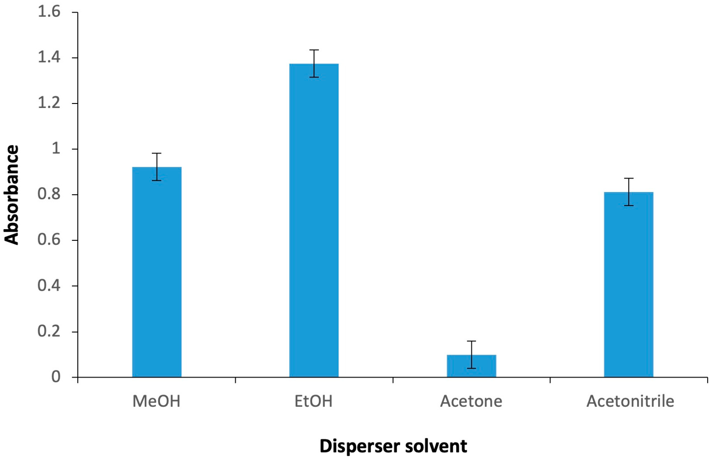
The choice of disperser solvent significantly affects both the stability of the cloudy phase and the enrichment factor achieved. The disperser solvent not only facilitates the rapid dispersion of the extraction solvent into the aqueous phase but also acts as a medium that bridges the polarity differences between the aqueous solution and the extraction solvent. This bridging role is essential for the efficient transfer of analytes into the extraction phase and for maintaining the reproducibility and effectiveness of the overall microextraction process [66,67]. To determine the most appropriate disperser solvent for the preconcentration of Hg2+, four solvents (acetone, acetonitrile, ethanol, and methanol), all of which are miscible with both aqueous and organic phases, were evaluated in this procedure. The experimental results demonstrated that a sedimented phase could be obtained with all four disperser solvents; however, the signal responses varied (Figure 2). The most notable finding was that the highest signal response was obtained with ethanol, which was therefore selected as the disperser solvent for the proposed DLLME procedure.
The disperser solvent volume plays a fundamental role in the formation of the cloudy phase within the microextraction tube, which represents a ternary system. When the volume of the disperser solvent is too low, the cloudy solution does not form efficiently, resulting in reduced extraction efficiency. Conversely, if the disperser solvent volume is excessively high, the solubility of the complex increases, which again diminishes the extraction efficiency [68]. To optimize the disperser solvent volume, enrichment factors were compared using different volumes of ethanol as disperser solvents. The optimal volume for the proposed method was established as 700 µL.

2.1.2. Effect of pH

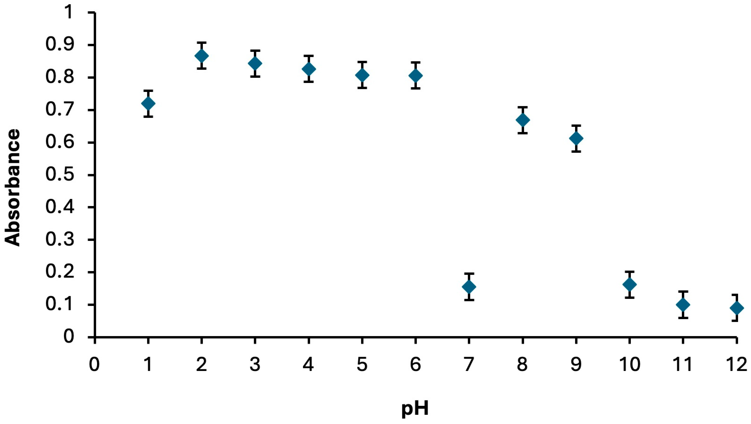
pH is a key parameter influencing both metal–sensor complex formation and the extraction process [69]. In the spectrophotometric determination of Hg2+, the optimum pH was established by preparing a series of Hg2+:Fe(II)Pc complexes at pH values ranging from 1.0 to 12.0. The maximum absorbance intensity was observed at pH 2 for the Fe(II)Pc sensor (Figure 3).

2.1.3. Role of Centrifuge Parameters

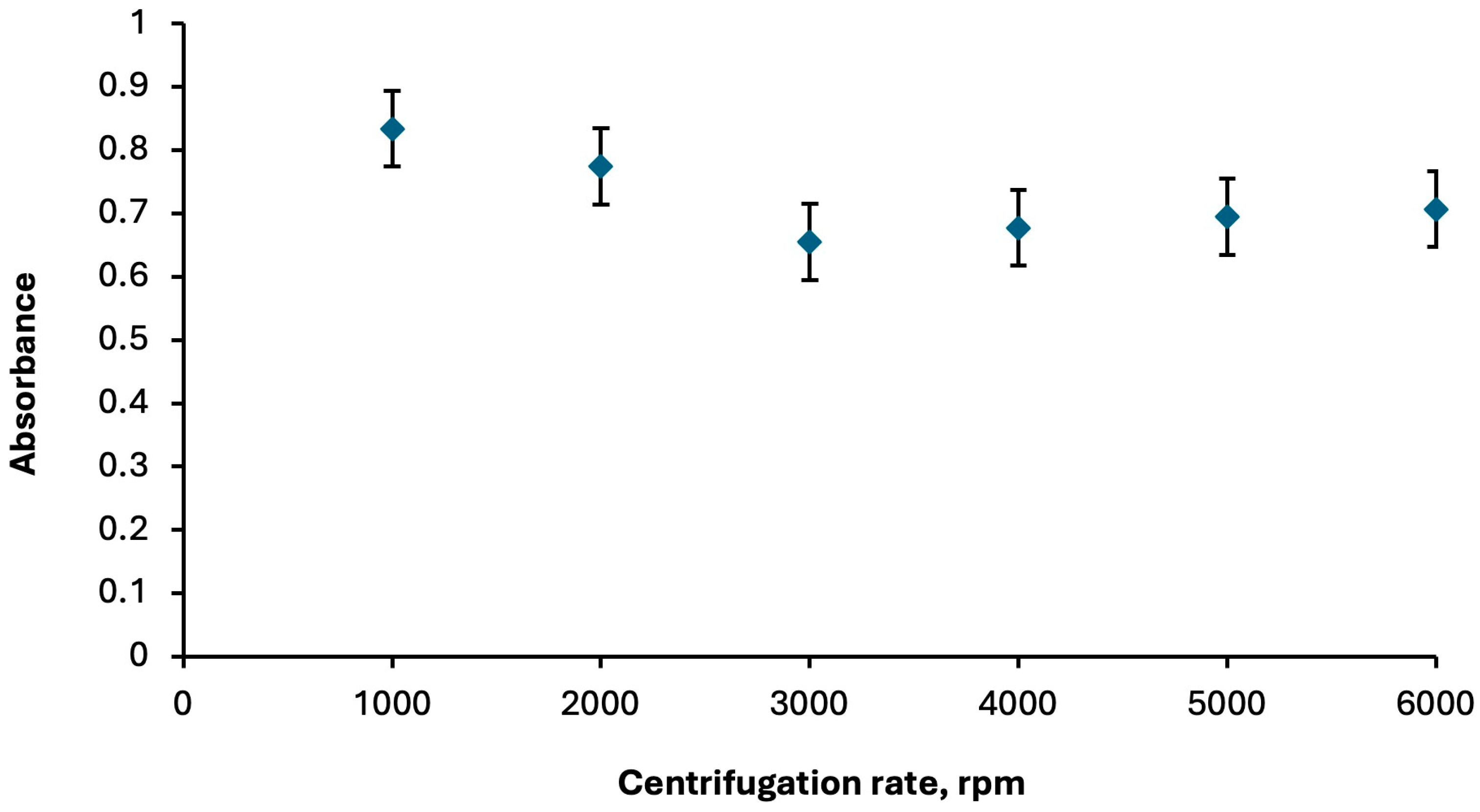
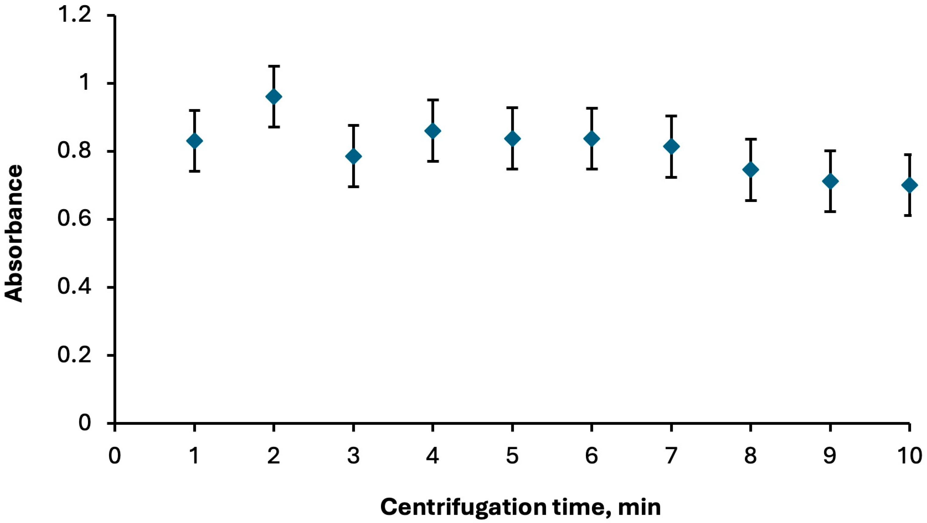
The centrifugation step in DLLME enables efficient phase separation of the cloudy solution formed during extraction. Appropriate centrifugation speed and time allow the extraction phase to settle completely, thereby enhancing enrichment, improving reproducibility, and ensuring that the analyte-rich phase can be readily collected for subsequent spectroscopic analysis. The centrifugation step was optimized by evaluating speeds between 1000 and 6000 rpm and times of 1 to 10 min. The best performance was achieved at 1000 rpm for 2 min (Figure 4 and Figure 5, respectively).

2.1.4. Compex Stability

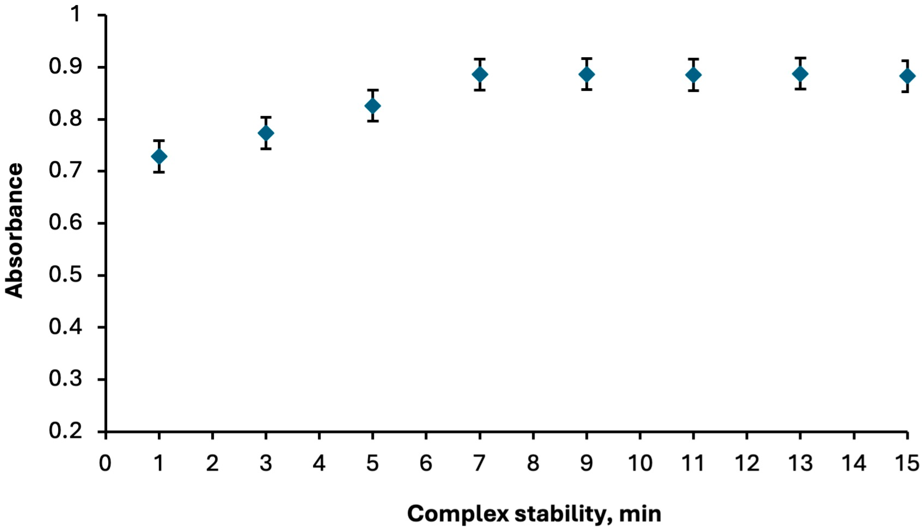
The absorbance variation in the Hg2+:Fe(II)Pc complex at 276 nm was monitored within the time interval of 1–15 min in order to evaluate its stability. The results revealed that the complex gradually increased in stability and reached a steady state after the seventh minute. Once equilibrium was established, the Hg2+:Fe(II)Pc complex remained stable for approximately 8 min. without exhibiting significant fluctuations in absorbance (Figure 6). This observation indicates that the formed complex possesses sufficient temporal stability under the applied experimental conditions, which is a critical factor for ensuring reliable spectrophotometric measurements in analytical applications.

2.1.5. Effect of Fe(II)Pc Sensor Concentration

The effect of sensor concentration on the absorbance was investigated by varying the Fe(II)Pc concentration in the range of 1.00 × 10⁻⁶ to 1.00 × 10⁻⁴ M, while keeping all other experimental parameters constant. The absorbance reached its highest value at 5.00 × 10⁻⁶ M, which was selected as the optimum sensor concentration for subsequent measurements.

2.2. Interferences

In order to assess the applicability of the proposed method to real samples, the potential interfering effects of various cations and anions were systematically examined, and their tolerance limits were established. For this purpose, solutions containing 5.00 × 10⁻⁶ M sensors and an equimolar amount of Hg2+ were spiked with different concentrations of foreign ions to prepare a series of mixtures. These mixtures were subjected to the proposed microextraction procedure, and the absorbance intensities measured at 276 nm were compared with those of a reference solution containing only the sensor and Hg2+. The tolerance limit was defined as the maximum concentration of a foreign ion that produced a deviation of less than 5% from the reference absorbance. The obtained values are summarized in Table 1.

2.3. Analytical Figures of Merit

The linear dynamic range of the proposed methods for the spectrophotometric determination of mercury was determined using spectrophotometric titration (Supplementary Figure S1) and was found to be 1.00–20.00 µg/L with a correlation coefficient (R2) of 0.9978 (Supplementary Figure S2). The limit of detection (LOD) and limit of quantification (LOQ) of the proposed DLLME method were estimated using the equations LOD = 3σ/S and LOQ = 10σ/S, where σ represents the standard deviation of the response and S is the slope of the calibration curve. In this study, σ was calculated from the standard deviation of absorbance values obtained from 11 blank samples. Based on these calculations, the LOD and LOQ were determined to be 1.44 µg/L and 4.80 µg/L, respectively. The enrichment factor, defined as the ratio of the slopes of the calibration curves for Hg2+ obtained before and after the extraction procedure, was calculated to be 105. The precision of the developed method was assessed under both intra-day and inter-day conditions by analyzing Hg2+ at a concentration level of 10 µg/L. The relative standard deviations (RSDs) were observed in the range of 1.27–2.68% for intra-day analyses and 1.09–3.01% for inter-day analyses (n:6) (Table 2).
Since the Hg2+ concentrations in the environmental water samples were below the detection limit of the proposed DLLME method, the analysis was carried out through spiking experiments based on the recovery of added Hg2+ ions into the sample matrices. For this purpose, different amounts of analyte ions were added to 25 mL portions of three different water samples, and the proposed microextraction procedure was applied using 5.00 mL of each sample. The results are presented in Table 3, showing recovery values ranging from 94.00% to 99.00%.

2.4. Comparison with Other Studies

Table 4 outlines selected reports addressing the determination of Hg2+ through the DLLME technique. Nemati et al. (2023) [5] developed a DES based DLLME method for Hg2+ extraction using 4,4′-bis(dimethylamino)thiobenzophenone (TMK) as a ligand and a coumarin–octanoic acid solvent system. TMK is a commonly used ligand for mercury determination. In contrast, the proposed method employs Fe(II)Pc for the first time in mercury determination. Although the TMK based method achieved good extraction efficiency, with an enrichment factor of 200, its performance strongly depends on the solvent composition, as conventional DES systems fail to stabilize the Hg–TMK complex. This solvent-specific limitation reduces the method’s applicability and increases its procedural complexity. Moreover, due to the negative influence of halide ions on the stability of the Hg2+–TMK complex, the addition of sodium sulfate (5.0% w/v) was required in their procedure. In contrast, the Fe(II)Pc based system developed in this study provides high extraction efficiency without the need for any additional chemical agents such as salts. In the study by Gharehbaghi et al. (2009) [70], an ionic liquid based DLLME method was developed for the spectrophotometric determination of Hg2+ using 4,4′-bis(dimethylamino)thiobenzophenone (TMK) as a chelating ligand and (1-hexyl-3-methylimmidazolium bis(trifluormethylsulfonyl)imid) [Hmim][Tf2N] as the extraction solvent. Although the method demonstrated acceptable analytical performance, the use of TMK, a widely employed ligand for mercury determination, reduces the originality of the approach. Furthermore, the addition of sodium nitrate (NaNO3) to improve extraction efficiency and sodium dodecyl sulfate (SDS) to prevent the adhesion of ionic liquid droplets to the test tube walls after centrifugation introduced additional preparation steps, thereby increasing the cost, time requirements, and environmental impact of the procedure. Additionally, the centrifugation time reported in their study was 4 min, whereas the proposed Fe(II)Pc based DLLME method achieves efficient phase separation within only 2 min, thereby enhancing practical applicability. In the study by Ripoll et al. (2023) [57], a NADES-based DLLME method was developed for the determination of inorganic and organic mercury species (MeHg+, EtHg+, PhHg+, and Hg2+). Although the method successfully achieved mercury speciation using a hydrophobic NADES system (decanoic acid:DL-menthol, 1:2 molar ratio), it exhibits several drawbacks in terms of operational efficiency and simplicity. In addition to the standard centrifugation step, a 3 min vortexing process was required to ensure NADES dispersion, thereby increasing the total analysis time. Adjusting the sample to pH 12 may also cause hydrolysis or transformation of mercury species, affecting extraction reliability. Furthermore, the use of dithizone as a conventional ligand and the incorporation of EDTA during liquid chromatography (LC) analysis reduce the method’s novelty, cost-effectiveness, and environmental compatibility. Compared with the proposed Fe(II)Pc based DLLME method, the longer centrifugation time and multi-step procedure make this approach less efficient in terms of time, resources, and practicality. Hossein-Poor-Zaryabi et al. (2014) [71] developed a DLLME–UV–Vis method for the determination of Hg2+ using dithizone as a chelating reagent, ethanol as a disperser solvent, and chloroform as the extraction solvent. The optimum extraction pH reported in their study was 1.80, which is consistent with the pH 2.00 condition employed in the Fe(II)Pc based DLLME method. Both methods operate under highly acidic conditions, ensuring that Hg2+ ions remain in soluble form and preventing the formation of mercury hydroxide precipitates. However, the dithizone-based method achieved a relatively low enrichment factor (39) compared to 105 obtained with the Fe(II)Pc based system, indicating significantly higher extraction efficiency and analytical sensitivity in the present study. Moreover, the use of dithizone, a conventional complexing reagent widely applied in mercury determination, limits the originality of the previous approach. In contrast, Fe(II)Pc has been employed for the first time as a sensing and extraction agent for Hg2+, providing enhanced selectivity, methodological innovation, and improved extraction efficiency. In the DLLME method developed by Çağlar et al. (2018) [72], the quaternized Fe(II)phthalocyanine (q-Fe(II)Pc) compound, the water-soluble form of the sensor used in present study, was employed as the sensing agent, and 1-heptyl-3-methylimidazolium and acetonitrile were used as the extraction and dispersive solvents, respectively. The Fe(II)Pc used in the present study exhibits a lipophilic nature which, compared to the ionic character of q-Fe(II)Pc, enables the formation of neutral and hydrophobic Hg2+:Fe(II)Pc complexes that efficiently transfer into the organic phase, thereby enhancing extraction efficiency. The previously reported method exhibited a linear range of 0.05–1.00 µg/mL (R2 = 0.9960) with LOD and LOQ values of 0.01 and 0.03 µg/mL, respectively. In contrast, the method developed in this study operates within a linear range of 1.00–20.00 µg/L (R2 = 0.9978) and provides LOD and LOQ values of 1.44 and 4.80 µg/L, respectively. Overall, these results indicate that the lipophilic Fe(II)Pc compound employed in this work offers superior extraction performance and analytical sensitivity compared to its water-soluble derivative, making it more suitable for DLLME-based preconcentration of Hg2+ ions.
Compared with these studies, the method developed in the present work, which employs Fe(II)Pc as a novel sensor together with chloroform and ethanol as the extraction and dispersive solvents, respectively, provides a narrower linear range (1.00–20 µg/L) but demonstrates satisfactory sensitivity with a LOD of 1.44 µg/L, LOQ of 4.80 µg/L, and a notably high enrichment factor of 105. Furthermore, the use of a phthalocyanine based ligand, which remains scarcely explored in DLLME, highlights the originality of the proposed method. Although the sensitivity parameters of some previous studies are superior, the present work contributes a unique approach by demonstrating the applicability of Fe(II)Pc as a selective sensor, thereby addressing an existing gap in DLLME based mercury determination.

3. Materials and Methods

3.1. Reagents and Instrumentation

Fe(II)Pc was synthesized in accordance with a previously published procedure [72]. Analytical grade reagents and solvents were procured from Merck (Darmstadt, Germany) and Sigma-Aldrich (Taufkirchen, Germany). A stock solution of Hg2+ was prepared from nitrate salts in methanol followed by acidification with HNO3. The Fe(II)Pc sensor solution was prepared daily by dissolving a precisely weighed amount of the compound in methanol. The pH required for extraction was controlled and adjusted with buffer systems composed of citric acid, NaOH, and HCl.
All details for instrumentation were given in Supplementary File.

3.2. DLLME Procedure

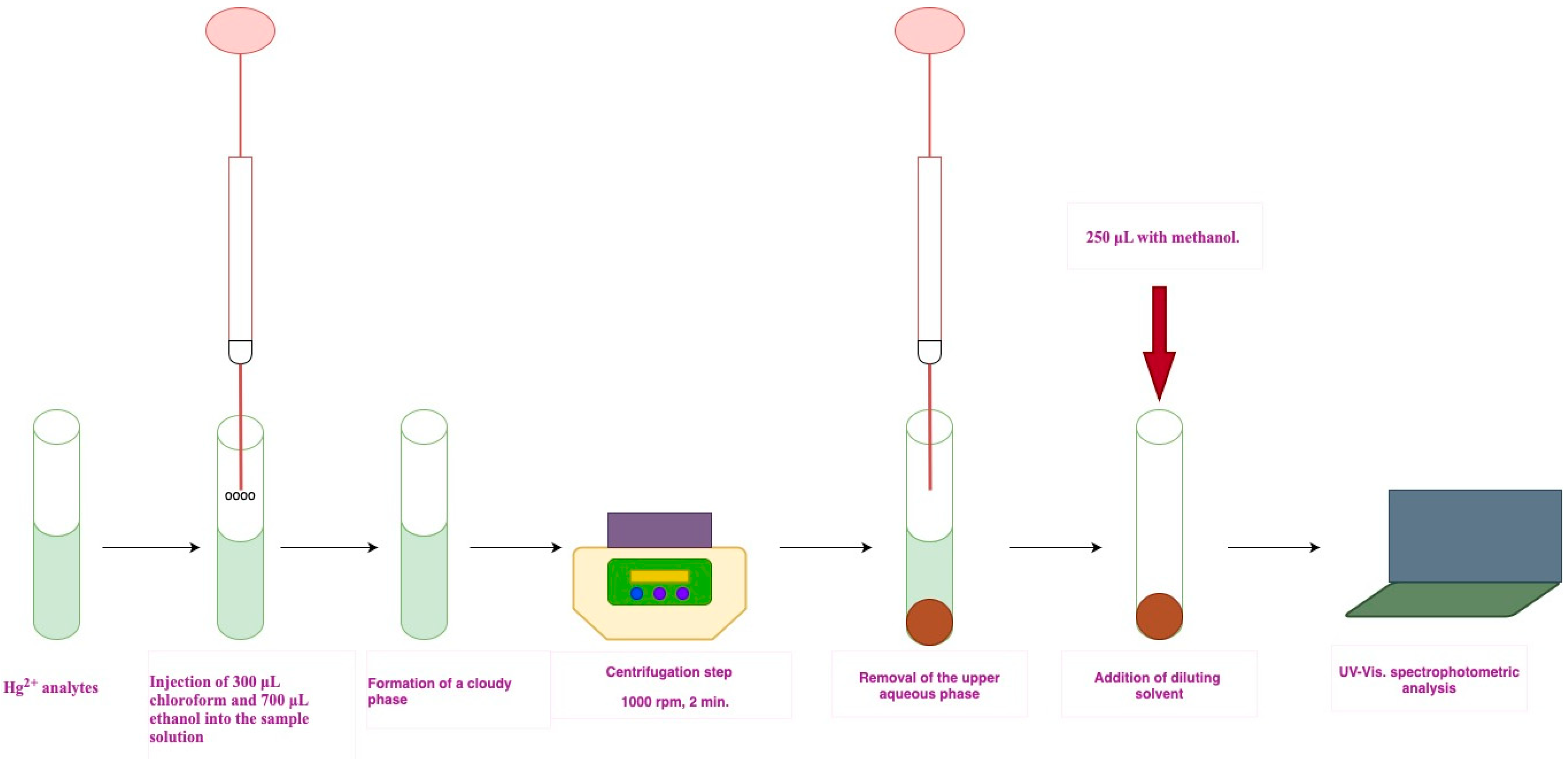
250 μL of Fe(II)Pc (5.00 × 10⁻⁶ M, methanol) was added as the sensor to 5 mL of an aqueous sample solution containing Hg2+ in a 10 mL conical glass tube. The pH was then adjusted to 2.00 using a citric acid–sodium hydroxide buffer. An extraction mixture consisting of 300 μL chloroform (as the extraction solvent) and 700 μL ethanol (as the dispersive solvent) was then rapidly injected into the sample solution with the aid of a 5 mL syringe. A fine cloudy solution was formed and subsequently centrifuged at 1000 rpm for 2 min. Following centrifugation, the aqueous phase was completely discarded, and the sedimented analyte-rich phase was diluted to 250 μL with methanol. Finally, absorbance measurements were carried out at 276 nm using a quartz cell (optical path length: 1 cm, volume: 250 μL) (Figure 7).

4. Conclusions

Mercury and its compounds, despite their well-documented toxicity, remain extensively employed in industrial applications, thereby necessitating reliable methods for their determination in environmental samples. Conventional DLLME based approaches often depend on advanced techniques such as HPLC, GC, ICP, and AAS, which are costly and require specialized expertise. In this study, a novel DLLME method integrated with spectrophotometry was developed for the determination of Hg2+ in water samples using Fe(II) phthalocyanine as a sensor. Phthalocyanine compounds are well recognized as excellent candidates for sensor design due to their structural stability and strong coordination ability [61]. A review of the literature indicates that reports utilizing phthalocyanine compounds as sensing or complexing agents in DLLME are scarce. Therefore, the use of a phthalocyanine derivative with a high capacity to form stable and selective sensor systems significantly enhances the novelty and originality of the present work. Furthermore, the proposed method aligns with the principles of green chemistry by minimizing organic solvent consumption and waste generation. Although it exhibits a relatively narrow linear range compared to instrumental techniques, it offers low LOD and LOQ values and a high enrichment factor. In addition, its implementation through a simple and accessible technique such as UV–Vis spectrophotometry makes it suitable for routine analytical applications.
The concentration of the Fe(II)Pc ligand to be used in the DLLME method for the spectrophotometric determination of Hg2+ was established as 5.00 × 10⁻⁶ M, and 276 nm, where the highest absorbance was observed, was selected as the working wavelength. The proposed method is extremely simple and rapid to apply. The rapid stabilization of the formed complex and the short centrifugation time are important factors contributing to their applicability.
The formation between Hg2+ and Fe(II)Pc complex was confirmed by a noticeable red shift in the UV–Vis spectrum, where the main Q-band moved from 718 nm to 728 nm upon the addition of Hg2+ ions (Supplementary Figure S3). This spectral response clearly indicates coordination between mercury ions and the nitrogen atoms of the phthalocyanine macrocycle [73]. The interaction causes electronic perturbation within the conjugated π-system, consistent with literature reports describing the affinity of soft metal ions such as Hg2+ for nitrogen donor sites [74]. Furthermore, Job’s plot analysis supported a 1:1 metal-to-ligand stoichiometry (Supplementary Figure S4), validating the proposed binding mechanism between Fe(II)Pc and Hg2+ [75].

Supplementary Materials

The following supporting information can be downloaded at: https://www.mdpi.com/article/10.3390/molecules30214192/s1.

Funding

This research was funded by the Scientific and Technological Research Council of Turkey (TUBITAK) under Grant No: 115Z076.

Institutional Review Board Statement

Not applicable.

Informed Consent Statement

Not applicable.

Data Availability Statement

The original contributions presented in this study are included in the article. Further inquiries can be directed to the corresponding author.

Conflicts of Interest

The author declare no conflict of interest.

References

  1. Gibb, H.; O’Leary, K.G. Mercury Exposure and Health Impacts among Individuals in the Artisanal and Small-Scale Gold Mining Community: A Comprehensive Review. Environ. Health Perspect. 2014, 122, 667–672. [Google Scholar] [CrossRef]
  2. Gao, Z.; Ma, X. Speciation Analysis of Mercury in Water Samples Using Dispersive Liquid-Liquid Microextraction Combined with High-Performance Liquid Chromatography. Anal. Chim. Acta 2011, 702, 50–55. [Google Scholar] [CrossRef] [PubMed]
  3. Driscoll, C.T.; Mason, R.P.; Chan, H.M.; Jacob, D.J.; Pirrone, N. Mercury as a Global Pollutant: Sources, Pathways, and Effects. Environ. Sci. Technol. 2013, 47, 4967–4983. [Google Scholar] [CrossRef]
  4. Mordukhovich, I.; Wright, R.O.; Hu, H.; Amarasiriwardena, C.; Baccarelli, A.; Litonjua, A.; Sparrow, D.; Vokonas, P.; Schwartz, J. Associations of Toenail Arsenic, Cadmium, Mercury, Manganese, and Lead with Blood Pressure in the Normative Aging Study. Environ. Health Perspect. 2012, 120, 98–104. [Google Scholar] [CrossRef]
  5. Nemati, I.; Faraji, M.; Jafarinejad, S.; Shirani, M. Development of a Deep Eutectic Solvent-Based Dispersive Liquid–Liquid Microextraction Coupled with Spectrophotometer Technique for Determination of Trace Amount of Hg(II) in Water Samples. Chem. Pap. 2023, 77, 909–919. [Google Scholar] [CrossRef]
  6. Houston, M.C. Role of Mercury Toxicity in Hypertension, Cardiovascular Disease, and Stroke. J. Clin. Hypertens 2011, 13, 621–627. [Google Scholar] [CrossRef]
  7. Al-Batanony, M.A.; Abdel-Rasul, G.M.; Ma, A.-S.; Al-Dalatony, M.M.; Allam, H.K.; Al-Batanony, M.A. Occupational Exposure to Mercury among Workers in a Fluorescent Lamp Factory, Quisna Industrial Zone, Egypt. Int. J. Occup. Environ. Med. 2013, 4, 149–156. [Google Scholar]
  8. Farina, M.; Avila, D.S.; Da Rocha, J.B.T.; Aschner, M. Metals, Oxidative Stress and Neurodegeneration: A Focus on Iron, Manganese and Mercury. Neurochem. Int. 2013, 62, 575–594. [Google Scholar] [CrossRef]
  9. Björkman, L.; Lundekvam, B.F.; Lægreid, T.; Bertelsen, B.I.; Morild, I.; Lilleng, P.; Lind, B.; Palm, B.; Vahter, M. Mercury in Human Brain, Blood, Muscle and Toenails in Relation to Exposure: An Autopsy Study. Environ. Health 2007, 6, 30. [Google Scholar] [CrossRef] [PubMed]
  10. Arantes, F.P.; Savassi, L.A.; Santos, H.B.; Gomes, M.V.T.; Bazzoli, N. Bioaccumulation of Mercury, Cadmium, Zinc, Chromium, and Lead in Muscle, Liver, and Spleen Tissues of a Large Commercially Valuable Catfish Species from Brazil. An. Acad. Bras Cienc. 2016, 88, 137–147. [Google Scholar] [CrossRef]
  11. Li, J.; Zhou, Q.; Yuan, G.; He, X.; Xie, P. Mercury Bioaccumulation in the Food Web of Three Gorges Reservoir (China): Tempo-Spatial Patterns and Effect of Reservoir Management. Sci. Total Environ. 2015, 527–528, 203–210. [Google Scholar] [CrossRef]
  12. Qu, P.; Pang, M.; Wang, P.; Ma, X.; Zhang, Z.; Wang, Z.; Gong, Y. Bioaccumulation of Mercury along Continuous Fauna Trophic Levels in the Yellow River Estuary and Adjacent Sea Indicated by Nitrogen Stable Isotopes. J. Hazard. Mater. 2022, 432, 128631. [Google Scholar] [CrossRef]
  13. Tchounwou, P.B.; Ayensu, W.K.; Ninashvili, N.; Sutton, D. Environmental Exposure to Mercury and Its Toxicopathologic Implications for Public Health. Environ. Toxicol. 2003, 18, 149–175. [Google Scholar] [CrossRef] [PubMed]
  14. Zulaikhah, S.T.; Wahyuwibowo, J.; Pratama, A.A. Mercury and Its Effect on Human Health: A Review of the Literature. Int. J. Public Health Sci. 2020, 9, 103–114. [Google Scholar] [CrossRef]
  15. Zahir, F.; Rizwi, S.J.; Haq, S.K.; Khan, R.H. Low Dose Mercury Toxicity and Human Health. Environ. Toxicol. Pharmacol. 2005, 20, 351–360. [Google Scholar] [CrossRef] [PubMed]
  16. Clarkson, T.W.; Magos, L.; Myers, G.J. Human Exposure to Mercury: The Three Modern Dilemmas. J. Trace Elements Exp. Med. 2003, 16, 321–343. [Google Scholar] [CrossRef]
  17. Burgess, N.M.; Meyer, M.W. Methylmercury Exposure Associated with Reduced Productivity in Common Loons. Ecotoxicology 2007, 17, 83–91. [Google Scholar] [CrossRef]
  18. Carrasco, L.; Barata, C.; García-Berthou, E.; Tobias, A.; Bayona, J.M.; Díez, S. Patterns of Mercury and Methylmercury Bioaccumulation in Fish Species Downstream of a Long-Term Mercury-Contaminated Site in the Lower Ebro River (NE Spain). Chemosphere 2011, 84, 1642–1649. [Google Scholar] [CrossRef]
  19. Mei, B.; Fong, W.; Siu, S.; Sai, J.; Tee, K.; Tam, S. Determination of Mercury in Whole Blood and Urine by Inductively Coupled Plasma Mass Spectrometry. J. Anal. Toxicol. 2007, 31, 281–287. [Google Scholar] [CrossRef]
  20. Teng, H.; Altaf, A.R. Elemental Mercury (Hg0) Emission, Hazards, and Control: A Brief Review. J. Hazard. Mater. Adv. 2022, 5, 100049. [Google Scholar] [CrossRef]
  21. Natasha; Shahid, M.; Khalid, S.; Bibi, I.; Bundschuh, J.; Khan Niazi, N.; Dumat, C. A Critical Review of Mercury Speciation, Bioavailability, Toxicity and Detoxification in Soil-Plant Environment: Ecotoxicology and Health Risk Assessment. Sci. Total Environ. 2020, 711, 134749. [Google Scholar] [CrossRef]
  22. Kim, K.H.; Kabir, E.; Jahan, S.A. A Review on the Distribution of Hg in the Environment and Its Human Health Impacts. J. Hazard. Mater. 2016, 306, 376–385. [Google Scholar] [CrossRef]
  23. Bidari, A.; Ganjali, M.R.; Assadi, Y.; Kiani, A.; Norouzi, P. Assay of Total Mercury in Commercial Food Supplements of Marine Origin by Means of DLLME/ICP-AES. Food Anal. Methods 2012, 5, 695–701. [Google Scholar] [CrossRef]
  24. Pinheiro, F.C.; Aguirre, M.Á.; Nóbrega, J.A.; Canals, A. Dispersive Liquid-Liquid Microextraction of Cd, Hg and Pb from Medicines Prior to ICP OES Determination According to the United States Pharmacopeia. Anal. Methods 2021, 13, 5670–5678. [Google Scholar] [CrossRef]
  25. Liang, P.; Yu, J.; Yang, E.; Mo, Y. Determination of Mercury in Food and Water Samples by Displacement-Dispersive Liquid-Liquid Microextraction Coupled with Graphite Furnace Atomic Absorption Spectrometry. Food Anal. Methods 2015, 8, 236–242. [Google Scholar] [CrossRef]
  26. Faraji, M. Novel Hydrophobic Deep Eutectic Solvent for Vortex Assisted Dispersive Liquid-Liquid Micro-Extraction of Two Auxins in Water and Fruit Juice Samples and Determination by High Performance Liquid Chromatography. Microchem. J. 2019, 150, 104130. [Google Scholar] [CrossRef]
  27. Bagheri, H.; Naderi, M. Immersed Single-Drop Microextraction-Electrothermal Vaporization Atomic Absorption Spectroscopy for the Trace Determination of Mercury in Water Samples. J. Hazard. Mater. 2009, 165, 353–358. [Google Scholar] [CrossRef]
  28. Martinis, E.M.; Wuilloud, R.G. Cold Vapor Ionic Liquid-Assisted Headspace Single-Drop Microextraction: A Novel Preconcentration Technique for Mercury Species Determination in Complex Matrix Samples. J. Anal. At. Spectrom. 2010, 25, 1432–1439. [Google Scholar] [CrossRef]
  29. Chen, B.; Wu, Y.; Guo, X.; He, M.; Hu, B. Speciation of Mercury in Various Samples from the Micro-Ecosystem of East Lake by Hollow Fiber-Liquid-Liquid-Liquid Microextraction-HPLC-ICP-MS. J. Anal. Spectrom. 2015, 30, 875–881. [Google Scholar] [CrossRef]
  30. Anthemidis, A.N.; Ioannou, K.I.G. Recent Developments in Homogeneous and Dispersive Liquid-Liquid Extraction for Inorganic Elements Determination. A Review. Talanta 2009, 80, 413–421. [Google Scholar] [CrossRef]
  31. Dadfarnia, S.; Haji Shabani, A.M. Recent Development in Liquid Phase Microextraction for Determination of Trace Level Concentration of Metals-A Review. Anal. Chim. Acta 2010, 658, 107–119. [Google Scholar] [CrossRef]
  32. Kamal El-Deen, A.; Elmansi, H.; Belal, F.; Magdy, G. Recent Advances in Dispersion Strategies for Dispersive Liquid–Liquid Microextraction from Green Chemistry Perspectives. Microchem. J. 2023, 191, 108807. [Google Scholar] [CrossRef]
  33. Dmitrienko, S.G.; Apyari, V.V.; Tolmacheva, V.V.; Gorbunova, M.V. Dispersive Liquid–Liquid Microextraction of Organic Compounds: An Overview of Reviews. J. Anal. Chem. 2020, 75, 1237–1251. [Google Scholar] [CrossRef]
  34. El-Deen, A.K.; Shimizu, K. Deep Eutectic Solvents as Promising Green Solvents in Dispersive Liquid–Liquid Microextraction Based on Solidification of Floating Organic Droplet: Recent Applications, Challenges and Future Perspectives. Molecules 2021, 26, 7406. [Google Scholar] [CrossRef]
  35. Rezaee, M.; Assadi, Y.; Milani Hosseini, M.R.; Aghaee, E.; Ahmadi, F.; Berijani, S. Determination of Organic Compounds in Water Using Dispersive Liquid-Liquid Microextraction. J. Chromatogr. A 2006, 1116, 1–9. [Google Scholar] [CrossRef] [PubMed]
  36. Pacheco-Fernández, I.; González-Martín, R.; e Silva, F.A.; Freire, M.G.; Pino, V. Insights into Coacervative and Dispersive Liquid-Phase Microextraction Strategies with Hydrophilic Media—A Review. Anal. Chim. Acta 2021, 1143, 225–249. [Google Scholar] [CrossRef] [PubMed]
  37. Magqira, B.; Mahlambi, P. Miniaturized Green Liquid-Phase Sample Pre-Concentration Methods for Pharmaceutical Drugs Assessment in Wastewater. Water Environ. J. 2025, 39, 143–167. [Google Scholar] [CrossRef]
  38. Andruch, V.; Balogh, I.S.; Kocúrová, L.; Šandrejová, J. Five Years of Dispersive Liquid-Liquid Microextraction. Appl. Spectrosc. Rev. 2013, 48, 161–259. [Google Scholar] [CrossRef]
  39. Leong, M.I.; Fuh, M.R.; Huang, S. Da Beyond Dispersive Liquid-Liquid Microextraction. J. Chromatogr. A 2014, 1335, 2–14. [Google Scholar] [CrossRef]
  40. Al-Saidi, H.M.; Emara, A.A.A. The Recent Developments in Dispersive Liquid-Liquid Microextraction for Preconcentration and Determination of Inorganic Analytes. J. Saudi Chem. Soc. 2014, 18, 745–761. [Google Scholar] [CrossRef]
  41. Rezaee, M.; Yamini, Y.; Faraji, M. Evolution of Dispersive Liquid-Liquid Microextraction Method. J. Chromatogr. A 2010, 1217, 2342–2357. [Google Scholar] [CrossRef]
  42. Fernández, E.; Vidal, L.; Martín-Yerga, D.; Blanco, M.D.C.; Canals, A.; Costa-García, A. Screen-Printed Electrode Based Electrochemical Detector Coupled with Ionic Liquid Dispersive Liquid-Liquid Microextraction and Microvolume Back-Extraction for Determination of Mercury in Water Samples. Talanta 2015, 135, 34–40. [Google Scholar] [CrossRef]
  43. Shirkhanloo, H.; Golbabaei, F.; Hassani, H.; Eftekhar, F.; Kian, M.J. Occupational Exposure to Mercury: Air Exposure Assessment and Biological Monitoring Based on Dispersive Ionic Liquid-Liquid Microextraction. Iran. J. Public Health 2014, 43, 793. [Google Scholar]
  44. Kiwanuka, M.J.; Nomngongo, P.N.; Mketo, N. Rapid and Greener Vortex-Assisted Deep Eutectic Solvent-Based Dispersive Liquid-Liquid Microextraction for Spectroscopic Determination of Hg in Fuels. Green Anal. Chem. 2025, 12, 100236. [Google Scholar] [CrossRef]
  45. Azhdari, N.; Ayazi, Z.; Habibi, B.; Rahimpour, E. Enhanced Mercury Detection through Ultrasound-Assisted Liquid–Liquid Microextraction and Digital Image Colorimetry Using Hydrophobic Deep Eutectic Solvents. Int. J. Environ. Sci. Technol. 2025, 22, 10067–10082. [Google Scholar] [CrossRef]
  46. Khan, M.; Soylak, M. Deep Eutectic Solvent Based Liquid-Liquid Microextraction of Mercury in Water, Hair and Fish with Spectrophotometric Determination: A Green Protocol. Anal. Lett. 2023, 56, 1161–1173. [Google Scholar] [CrossRef]
  47. Conchado-Amado, P.; Sánchez-Piñero, J.; Moreda-Piñeiro, J.; Turnes-Carou, I.; López-Mahía, P.; Muniategui-Lorenzo, S. Ultra-Trace Mercury Determination in Seawater after Vortex-Assisted Liquid-Liquid Micro-Extraction. Spectrochim. Acta Part B At. Spectrosc. 2023, 204, 106683. [Google Scholar] [CrossRef]
  48. Alzahrani, L.; El-Ghamry, H.A.; Saber, A.L.; Mohammed, G.I. Spectrophotometric Determination of Mercury(II) Ions in Laboratory and Zamzam Water Using Bis Schiff Base Ligand Based on 1,2,4-Triazole-3,5-Diamine and o-Vaniline. Arab. J. Chem. 2023, 16, 104418. [Google Scholar] [CrossRef]
  49. Liu, Y.M.; Zhang, F.P.; Jiao, B.Y.; Rao, J.Y.; Leng, G. Automated Dispersive Liquid-Liquid Microextraction Coupled to High Performance Liquid Chromatography—Cold Vapour Atomic Fluorescence Spectroscopy for the Determination of Mercury Species in Natural Water Samples. J. Chromatogr. A 2017, 1493, 1–9. [Google Scholar] [CrossRef]
  50. Leao, D.J.; Silva Junior, M.M.; Silva Junior, J.B.; De Oliveira, D.A.F.; Queiroz, A.F.S.; Ferreira, S.L.C. Ultrasound Assisted Extraction for the Determination of Mercury in Sediment Samples Employing Cold Vapour Atomic Absorption Spectrometry. Anal. Methods 2016, 8, 6554–6559. [Google Scholar] [CrossRef]
  51. Ma, S.; He, M.; Chen, B.; Deng, W.; Zheng, Q.; Hu, B. Magnetic Solid Phase Extraction Coupled with Inductively Coupled Plasma Mass Spectrometry for the Speciation of Mercury in Environmental Water and Human Hair Samples. Talanta 2016, 146, 93–99. [Google Scholar] [CrossRef]
  52. Rofouei, M.K.; Rezaei, A.; Masteri-Farahani, M.; Khani, H. Selective Extraction and Preconcentration of Ultra-Trace Level of Mercury Ions in Water and Fish Samples Using Fe 3O 4-Magnetite- Nanoparticles Functionalized by Triazene Compound Prior to Its Determination by Inductively Coupled Plasma-Optical Emission Spectrometry. Anal. Methods 2012, 4, 959–966. [Google Scholar] [CrossRef]
  53. Zavalishin, M.N.; Kiselev, A.N.; Isagulieva, A.K.; Shibaeva, A.V.; Kuzmin, V.A.; Morozov, V.N.; Zevakin, E.A.; Petrova, U.A.; Knyazeva, A.A.; Eroshin, A.V.; et al. Shedding Light on Heavy Metal Contamination: Fluorescein-Based Chemosensor for Selective Detection of Hg2+ in Water. Int. J. Mol. Sci. 2024, 25, 3186. [Google Scholar] [CrossRef]
  54. Sunitha, P.; Gowda, B.G.; Singh, P.; Vengatesh, G.; Varadaraju, K.; Abass, K.S.; Stupin, V.; Silina, E.; Kollur, S.P. Synthesis of Thiophene Derived Fluorescent Sensor for Hg2+ Ion Detection and Its Applications in Cell Imaging, Latent Fingerprint, and Real Water Analysis. Sci. Rep. 2025, 15, 33207. [Google Scholar] [CrossRef]
  55. Zavalishin, M.N.; Gamov, G.A. Fluorescein Acetone Hydrazone: A Highly Sensitive Probe for Hg2+ Ion Detection and Computational Insights. J. Mol. Struct. 2025, 1348, 1–24. [Google Scholar] [CrossRef]
  56. Li, T.; Zhang, X.; Sun, M.; Liu, S.; Liu, H.; Pu, X.; Meng, F. Development of a Highly Selective Near-Infrared Fluorescent Probe for Sensitive Detection of Hg2+ in Environmental and Biological Samples. Spectrochim. Acta A Mol. Biomol. Spectrosc. 2026, 345, 126784. [Google Scholar] [CrossRef]
  57. Ripoll, L.; Rayos, J.; Aguirre, M.Á.; Vidal, L.; Canals, A. Natural Deep Eutectic Solvent-Based Microextraction for Mercury Speciation in Water Samples. Anal. Bioanal. Chem. 2023, 415, 4435–4444. [Google Scholar] [CrossRef] [PubMed]
  58. Soylak, M.; Ahmed, H.E.H.; Khan, M. Switchable Hydrophilicity Solvent Based Microextraction of Mercury from Water, Fish and Hair Samples before Its Spectrophotometric Detection. Sustain. Chem. Pharm. 2023, 32, 101006. [Google Scholar] [CrossRef]
  59. Hayati, M.; Ramezani, M.; Rezanejade Bardajee, G.; Momeni Isfahani, T. Application of Robust Syringe-to-Syringe Dispersive Liquid-Phase Microextraction Method for Preconcentration and Determination of Mercury with the Aid of an Experimental Design. Sep. Sci. Technol. 2022, 57, 274–283. [Google Scholar] [CrossRef]
  60. El-Feky, H.H.; Mohammed, T.Y.; Amin, A.S.; Kassem, M.A. Rapid Extraction and Separation of Mercury in Water and Food Samples Based on Micelles and Azo-Thiazoles Complexation before Determination by UV-Vis Spectrophotometry. Anal. Methods Environ. Chem. J. 2023, 6, 19–36. [Google Scholar] [CrossRef]
  61. Liu, X.; Qi, C.; Bing, T.; Cheng, X.; Shangguan, D. Highly Selective Phthalocyanine-Thymine Conjugate Sensor for Hg2+ Based on Target Induced Aggregation. Anal. Chem. 2009, 81, 3699–3704. [Google Scholar] [CrossRef]
  62. Claessens, C.G.; Hahn, U.; Torres, T. Phthalocyanines: From Outstanding Electronic Properties to Emerging Applications. Chem. Rec. 2008, 8, 75–97. [Google Scholar] [CrossRef]
  63. Grau, J.; Azorín, C.; Benedé, J.L.; Chisvert, A.; Salvador, A. Use of Green Alternative Solvents in Dispersive Liquid-Liquid Microextraction: A Review. J. Sep. Sci. 2022, 45, 210–222. [Google Scholar] [CrossRef] [PubMed]
  64. Kokosa, J.M. Selecting an Extraction Solvent for a Greener Liquid Phase Microextraction (LPME) Mode-Based Analytical Method. TrAC Trends Anal. Chem. 2019, 118, 238–247. [Google Scholar] [CrossRef]
  65. Karimi, M.; Sereshti, H.; Samadi, S.; Parastar, H. Optimization of Dispersive Liquid-Liquid Microextraction and Improvement of Detection Limit of Methyl Tert-Butyl Ether in Water with the Aid of Chemometrics. J. Chromatogr. A 2010, 1217, 7017–7023. [Google Scholar] [CrossRef] [PubMed]
  66. Sereshti, H.; Karimi, M.; Samadi, S. Application of Response Surface Method for Optimization of Dispersive Liquid-Liquid Microextraction of Water-Soluble Components of Rosa Damascena Mill. Essential Oil. J. Chromatogr. A 2009, 1216, 198–204. [Google Scholar] [CrossRef]
  67. Fattahi, N.; Samadi, S.; Assadi, Y.; Hosseini, M.R.M. Solid-Phase Extraction Combined with Dispersive Liquid-Liquid Microextraction-Ultra Preconcentration of Chlorophenols in Aqueous Samples. J. Chromatogr. A 2007, 1169, 63–69. [Google Scholar] [CrossRef]
  68. Notardonato, I.; Avino, P. Dispersive Liquid–Liquid Micro Extraction: An Analytical Technique Undergoing Continuous Evolution and Development—A Review of the Last 5 Years. Separations 2024, 11, 203. [Google Scholar] [CrossRef]
  69. Laosuwan, M.; Mukdasai, S.; Srijaranai, S. A Simple in Syringe Low Density Solvent-Dispersive Liquid Liquid Microextraction for Enrichment of Some Metal Ions Prior to Their Determination by High Performance Liquid Chromatography in Food Samples. Molecules 2020, 25, 552. [Google Scholar] [CrossRef]
  70. Gharehbaghi, M.; Shemirani, F.; Baghdadi, M. Dispersive Liquid-Liquid Microextraction Based on Ionic Liquid and Spectrophotometric Determination of Mercury in Water Samples. Int. J. Environ. Anal. Chem. 2009, 89, 21–33. [Google Scholar] [CrossRef]
  71. Hossien-poor-Zaryabi, M.; Chamsaz, M.; Heidari, T.; Zavar, M.H.A.; Behbahani, M.; Salarian, M. Application of Dispersive Liquid-Liquid Micro-Extraction Using Mean Centering of Ratio Spectra Method for Trace Determination of Mercury in Food and Environmental Samples. Food Anal. Methods 2014, 7, 352–359. [Google Scholar] [CrossRef]
  72. Çağlar, Y.; Biyiklioglu, Z. Spectrophotometric Determination of Hg(II) in Water Samples by Dispersive Liquid Liquid Microextraction with Use Ionic Liquid after Derivatization with a Water Soluble Fe(II) Phthalocyanine. J. Incl. Phenom. Macrocycl. Chem. 2018, 90, 331–339. [Google Scholar] [CrossRef]
  73. Erzunov, D.; Tonkova, S.; Belikova, A.; Vashurin, A. Enhanced Visible Light Absorption and Photophysical Features of Novel Isomeric Magnesium Phthalocyaninates with Cyanophenoxy Substitution. Chemosensors 2022, 10, 503. [Google Scholar] [CrossRef]
  74. Sun, Q.Z.; Zhao, B.; Chai, L.Y.; Liu, H.; Jin, H.Z.; Liu, H. A 3D Nickel(II) Coordination Polymer Constructed by Mixed- Ligand Strategy: Synthesis, Crystal Structure and Sensing of Hg(II) Ion. Inorg. Nano-Met. Chem. 2022, 52, 711–717. [Google Scholar] [CrossRef]
  75. Çaǧlar, Y.; Gümrükçüoǧlu, N.; Saka, E.T.; Ocak, M.; Kantekin, H.; Ocak, Ü. Phthalocyanine-Based Fluorescent Chemosensor for the Sensing of Zn (II) in Dimethyl Sulfoxide-Acetonitrile. J. Incl. Phenom. Macrocycl. Chem. 2012, 72, 443–447. [Google Scholar] [CrossRef]
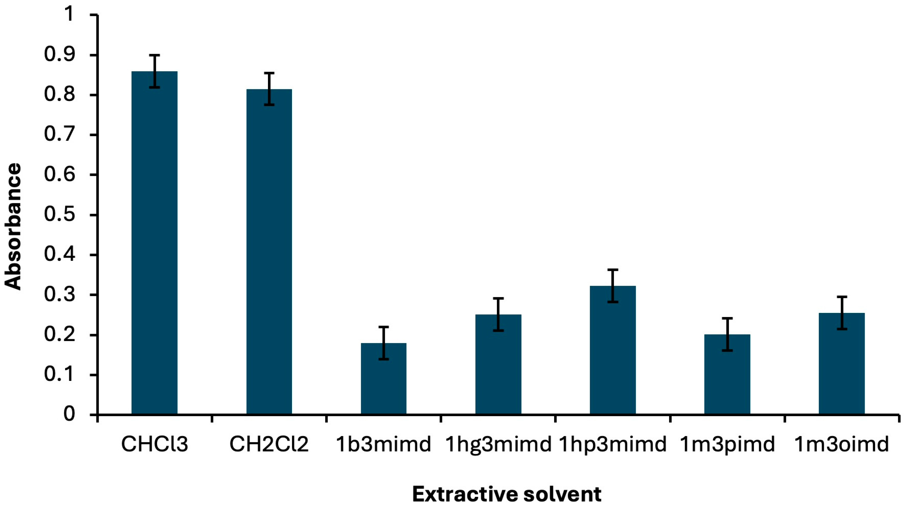
Figure 1. Absorbance changes observed with different extractive solvents. Extraction conditions: sample volume, 5.00 mL; disperser solvent, 700 μL ethanol; 250 µL of Fe(II)Pc (5.00 × 10⁻⁶ M); pH, 2.0; centrifugation conditions: 1000 rpm, 2 min. Error bars show the variability (±SD) for three replicate determinations (n = 3).
Molecules 30 04192 g001
Figure 2. Absorbance changes observed with different extractive solvents. Extraction conditions: sample volume, 5.00 mL; extractive solvent, 300 μL chloroform; 250 µL of Fe(II)Pc (5.00 × 10⁻⁶ M); pH, 2.0; centrifugation conditions: 1000 rpm, 2 min. Error bars show the variability (±SD) for three replicate determinations (n = 3).
Molecules 30 04192 g002
Figure 3. Absorbance changes observed with different extractive solvents. Extraction conditions: sample volume, 5.00 mL; extractive solvent, 300 μL chloroform; disperser solvent, 700 μL ethanol; 250 µL of Fe(II)Pc (5.00 × 10⁻⁶ M); centrifugation conditions: 1000 rpm, 2 min. Error bars show the variability (±SD) for three replicate determinations (n = 3).
Molecules 30 04192 g003
Figure 4. Optimization of centrifugation rate for phase separation. Extraction conditions: sample volume, 5.00 mL; extractive solvent, 300 μL chloroform; disperser solvent, 700 μL ethanol; 250 µL of Fe(II)Pc (5.00 × 10⁻⁶ M); pH, 2.0; centrifugation conditions: 2 min. Error bars show the variability (±SD) for three replicate determinations (n = 3).
Molecules 30 04192 g004
Figure 5. Optimization of centrifugation time for phase separation. Extraction conditions: sample volume, 5.00 mL; extractive solvent, 300 μL chloroform; disperser solvent, 700 μL ethanol; 250 µL of Fe(II)Pc (5.00 × 10⁻⁶ M); pH, 2.0; centrifugation conditions: 1000 rpm. Error bars show the variability (±SD) for three replicate determinations (n = 3).
Molecules 30 04192 g005
Figure 6. Time-dependent absorbance variation in the Hg2+:Fe(II)Pc (1:1) complex.
Molecules 30 04192 g006
Figure 7. Graphical representation of the proposed DLLME procedure.
Molecules 30 04192 g007
Table 1. Tolerance limits of interfering for the determination of Hg2+ (6.32 µg/L).
Foreign Ion AddedTolerance Limit (µg/L)
Ag+132
Cu2+119
Cr3+158
Co2+94
Pb2+51
Ni2+51
Cd2+299
Mn2+106
Zn2+427
NO3556
ClO4435
SO42−214
PO43−278
H2PO4223
Table 2. Analytical performance parameters for Hg2+ determination.
Parameters
Linear range, µg/L1.00–20.00
Correlation coefficient (R2)0.9991
LOD, µg/L1.44
LOQ, µg/L4.80
Enhancement factor,105
RSD, % (n = 6)1.27–2.68 a and 1.09–3.01 b
a Intra-day precision; b Inter-day precision.
Table 3. Accuracy evaluation of the DLLME method by recovery studies in water samples.
SampleHg2+ Amount µg/L (n:3)
AddedFinded ± SDRecovery
%
Black Sea 8.007.49 ± 0.1194.00
Tap water 8.007.92 ± 0.0499.00
Sera Lake8.008.17 ± 0.13102.12
Results are expressed as mean ± SD of three independent samples, each analyzed in triplicate.
Table 4. Comparative Assessment of the Developed DLLME Method and Other DLLME Strategies for Hg2+ Determination.
Extraction MethodLinear Range
(µg/L)
LOD
(µg/L)
Enrichment FactorRSD
(%)
Reference
DLLME1.00–100 0.302002.80–7.50[5]
IL-DLLME12.00–140 3.90 18.801.70[70]
DLLME a10.00–2003.00 -10.00–12.00 a[57]
DLLME0.50–1000.15392.60[71]
IL-DLLME0.050–1.00 b0.010 b1280.78[72]
DLLME1.00–201.441051.27–2.68 c and 1.09–3.01 dPresent study
a RSD (%) for the Hg2+; b The linear range was reported in µg/mL; c Intra-day precision; d Inter-day precision.
Disclaimer/Publisher’s Note: The statements, opinions and data contained in all publications are solely those of the individual author(s) and contributor(s) and not of MDPI and/or the editor(s). MDPI and/or the editor(s) disclaim responsibility for any injury to people or property resulting from any ideas, methods, instructions or products referred to in the content.