Article

Composite Material Formation Based on Biochar and Nickel (II)-Copper (II) Ferrites

by
Nina P. Shabelskaya
1,2,*,
Alexandr V. Vyaltsev
1,
Neonilla G. Sundukova
1,
Vera A. Baranova
1,
Sergej I. Sulima
3,
Elena V. Sulima
3,
Yulia A. Gaidukova
1,
Asatullo M. Radzhbov
1,
Elena V. Vasileva
1 and
Elena A. Yakovenko
1
1
Department of Ecology and Industrial Safety, Faculty of Technology, Platov South-Russian State Polytechnic University (NPI), 346428 Novocherkassk, Russia
2
Laboratory of Agrobiotechnology for Improving Soil Fertility and Agricultural Product Quality, Southern Federal University, 344006 Rostov-on-Don, Russia
3
Department of Chemical Technologies, Faculty of Technology, Platov South-Russian State Polytechnic University (NPI), 346428 Novocherkassk, Russia
*
Author to whom correspondence should be addressed.
Molecules 2025, 30(19), 3900; https://doi.org/10.3390/molecules30193900
Submission received: 6 September 2025 / Revised: 20 September 2025 / Accepted: 25 September 2025 / Published: 26 September 2025

Abstract

This paper studies the formation process of a composite material based on an organic substance, biochar from sunflower husks, and an inorganic substance, nickel (II)-copper (II) ferrites of the composition CuxNi1−xFe2O4 (x = 0.0; 0.5; 1.0). The obtained materials were characterized by X-ray phase analysis, scanning electron microscopy, and FTIR spectroscopy. It is shown that when replacing copper (II) cations with nickel (II) cations, the average parameters and volume of the unit cell gradually decrease, and the cation–anion distances in both the tetrahedral and octahedral spinel grids also decrease with regularity. The oxide materials were found to form a film on the surface of biochar, repeating its porous structure. The obtained materials exhibit high catalytic activity in the methyl orange decomposition reaction under the action of hydrogen peroxide in an acidic medium; the degradation of methyl orange in an aqueous solution occurs 30 min after the start of the reaction. This result may be associated with the formation of the Fenton system during the oxidation–reduction process. A significant increase in the reaction rate in the system containing mixed nickel–copper ferrite as a catalyst may be associated with the formation of a more defective structure due to the Jahn–Teller effect manifestation, which creates additional active centers on the catalyst surface.

1. Introduction

Nowadays, the expansion of industrial production has led to a continuous increase in environmental pollution. According to [1], more than 100,000 tons of synthetic dyes are produced each year, with azo dyes (compounds containing the bond –N=N–) accounting for almost 70% of the total volume of dye production [2]. The ingress of dyes into natural water bodies leads to a decrease in the flow of light, which is accompanied by a negative impact on aquatic and soil ecosystems, and interferes with the process of photosynthesis [3]. In addition, the toxic and carcinogenic properties of azo dyes pose a danger not only to humans but also to other living beings [3]. In this regard, an urgent task is to find effective methods for cleaning water resources from pollutants.
One of the promising methods of protecting water from dyes is purification using adsorption or oxidative destruction processes. Transition metal ferrites with a spinel structure are promising materials in many applications due to the successful combination of several properties, such as magnetic and dielectric.
Ferrites are a special type of ferrimagnetic material [4], which contains a grid of oxygen anions and cations of three- and two-charged metals distributed in it. The spinel formula can be represented as A2+B23+O4, where A and B represent metal cations. Depending on several factors, the spinel structure can be “normal” (all A2+ cations are located in tetrahedral grid positions, or A-positions, all B3+ cations are located in the centers of octahedra, or B-positions), “inverted” (all A2+ cations and half of the B3+ cations occupy octahedral sites, half of the B3+ cations are located in tetrahedral voids) [5,6].
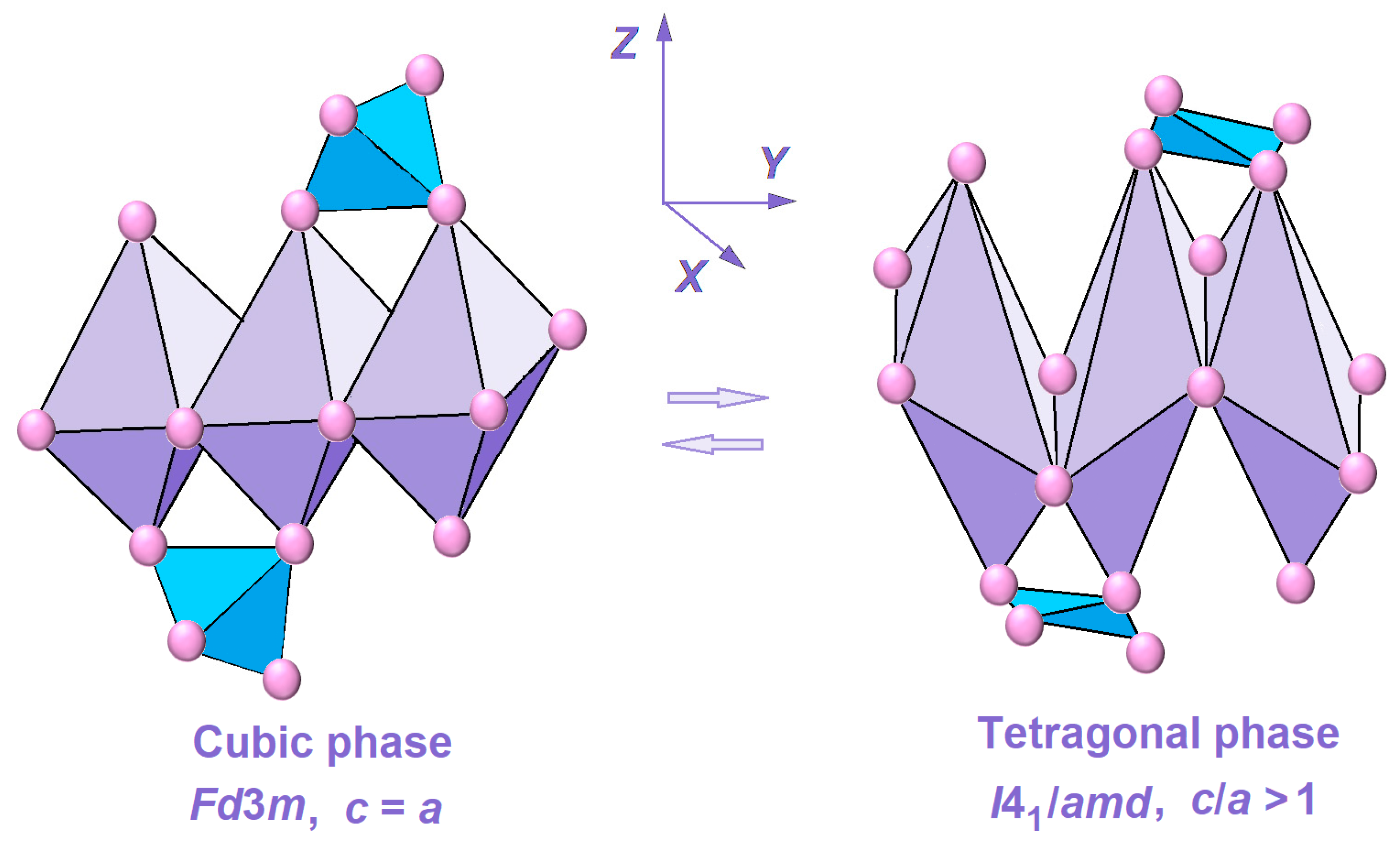
However, in nature, spinels are encountered most often with a partially inverted structure. Replacing the A2+ metal in the spinel composition can lead to a redistribution of A2+ and B3+ ions over the crystal grid nodes and, hence, to the appearance of new favorable properties [7]. In addition, several such compounds can experience crystal grid distortion due to the Jahn–Teller effect manifestation [8,9,10,11]. In this case, the crystal symmetry decreases, and a transition from the cubic (Fd3m) to the tetragonally distorted (I41/amd) phase occurs [12]. For example, copper (II) ferrite can exist in a cubic [13] or tetragonal [12] modification. Formation of areas with a defective structure can have a positive effect on the spinel’s catalytic and adsorption properties.
Spinel ferrites, due to their unique properties, have numerous potential applications in various industries, for example, in medicine for targeted drug delivery [14], as sensors for detecting hazardous substances [15,16,17], and in the processes of heat [18] and electric energy [19,20,21] transfer and accumulation. In recent years, ferrites have been successfully used as inexpensive, effective catalysts for hydrogen production [22], cyclic hydrocarbon oxidation [23], and dye decomposition [24,25,26,27].
Table 1 shows some data on the degradation of the methyl orange dye by the action of hydrogen peroxide in the presence of a catalyst.
The properties of ferrites with a spinel structure are significantly affected by the composition, production processes, and heat treatment mode. To obtain ferrites, both traditional methods, such as ceramic [36], coprecipitation method [37,38,39], and relatively new ones, such as hydrothermal [40,41], and sol–gel technology [4,25,26,42] are used.
Synthesis of organo-inorganic composite materials can obtain compounds with new properties characteristic of individual substances included in the composite [17,41]. Taking into account the above, the current work aimed to obtain composite materials based on an organic substance, biochar from sunflower husks, and an inorganic substance, nickel(II)-copper(II) ferrites of the composition of CuxNi1−xFe2O4, (x = 0.0; 0.5; 1.0), and to study their catalytic activity in the azo dye—methyl orange—decomposition reaction.

2. Results and Discussion

During the synthesis process, the formation of a porous gel-like substance was observed first, then its combustion occurred, and, ultimately, the samples became black powders. The resulting composite materials were studied using the X-ray phase analysis method.
Figure 1 shows the X-ray patterns of the synthesized materials.
The organic part of the composite material is X-ray amorphous. The inorganic part is copper (II) ferrite CuFe2O4, PDF Number 000-34-0425, tetragonal modification I41/amd (Figure 1 (a)), nickel (II) NiFe2O4, PDF Number 000-54-0964, cubic modification Fd3m (Figure 1 (c)), mixed nickel (II)-copper (II) ferrite (Figure 1 (b)). NiFe2O4 is characterized by peaks at the angles (2θ): 30.3; 35.7; 37.2; 43.3; 53.8; 57.35; 63.0, for which the parameter values (h, k, l) are respectively set to 220; 311; 222; 440; 422; 511; 440. CuFe2O4 is characterized by peaks at the angles (2θ): 30.32; 35.64; 38.88; 43.48; 53.84; 57.2; 63.0, for which the parameter values (h, k, l) are respectively set to 112; 211; 202; 220; 213; 312; 321; 400. Cu0.5Ni0.5Fe2O4 is characterized by peaks at the angles (2θ): 30.24; 35.56; 38.8; 43.24; 53.76; 56.92; 62.76, for which the parameter values (h, k, l) are respectively set to 112; 211; 202; 220; 213; 312; 321; 400. It is interesting to note that the 2θ angle values for mixed nickel (II)-copper (II) ferrite are not between those of pure nickel (II) ferrite and copper (II) ferrite but are usually smaller. This may be due to the greater defectiveness of the inorganic material being formed.
Table 2 presents data on the calculations of the unit cell parameters of ferrites (a, c), nm; the degree of tetragonality (c/a); the average cell parameter am, nm; the unit cell volume V, (nm)3; the crystallite size D, nm; the “anion-cation” distances (nm) in the tetrahedral (LA) and octahedral (LB) coordination of the spinel grid.
For the synthesized spinels in the composite material, the inversion parameter λ, grid deformation (ε), dislocation density (δ), and X-ray density (ρ) were calculated. The results are presented in Table 3. When compiling the chemical formulas, it was considered that nickel (II) and copper (II) cations tend to be located in the B-positions of the spinel structure [12], and for Ni2+ this preference is more pronounced. Fe3+ cations do not have a pronounced predisposition to occupy tetragonal or octahedral voids.
The obtained X-ray phase data analysis results show that when replacing copper (II) cations with nickel (II) cations, which have a smaller size (the radii of these cations are 0.080 and 0.074 nm, respectively), the average parameter and the volume of the unit cell gradually decrease. The cation–anion distances in both the tetrahedral and octahedral spinel grids also decrease with regularity.
However, an anomaly in the value of the grid parameter of a for mixed nickel–copper ferrite should be noted: it was less than the expected value. This experimental fact can be associated with a high degree of nickel-containing ferrite inversion and the formation of a more defective spinel structure due to the Jahn–Teller effect manifestation. In the latter case, the symmetry of the oxygen framework decreases, and areas with octahedra elongated along the z axis are formed. The formation of a distorted spinel structure under the influence of the Jahn–Teller effect is schematically shown in Figure 2.
The formation of a defect structure in mixed nickel–copper ferrite is accompanied by an increase in the X-ray density value (Table 3). For the same sample, the grid deformation and dislocation density values are 32.5 % and 18 % higher, respectively, compared to these values for NiFe2O4.
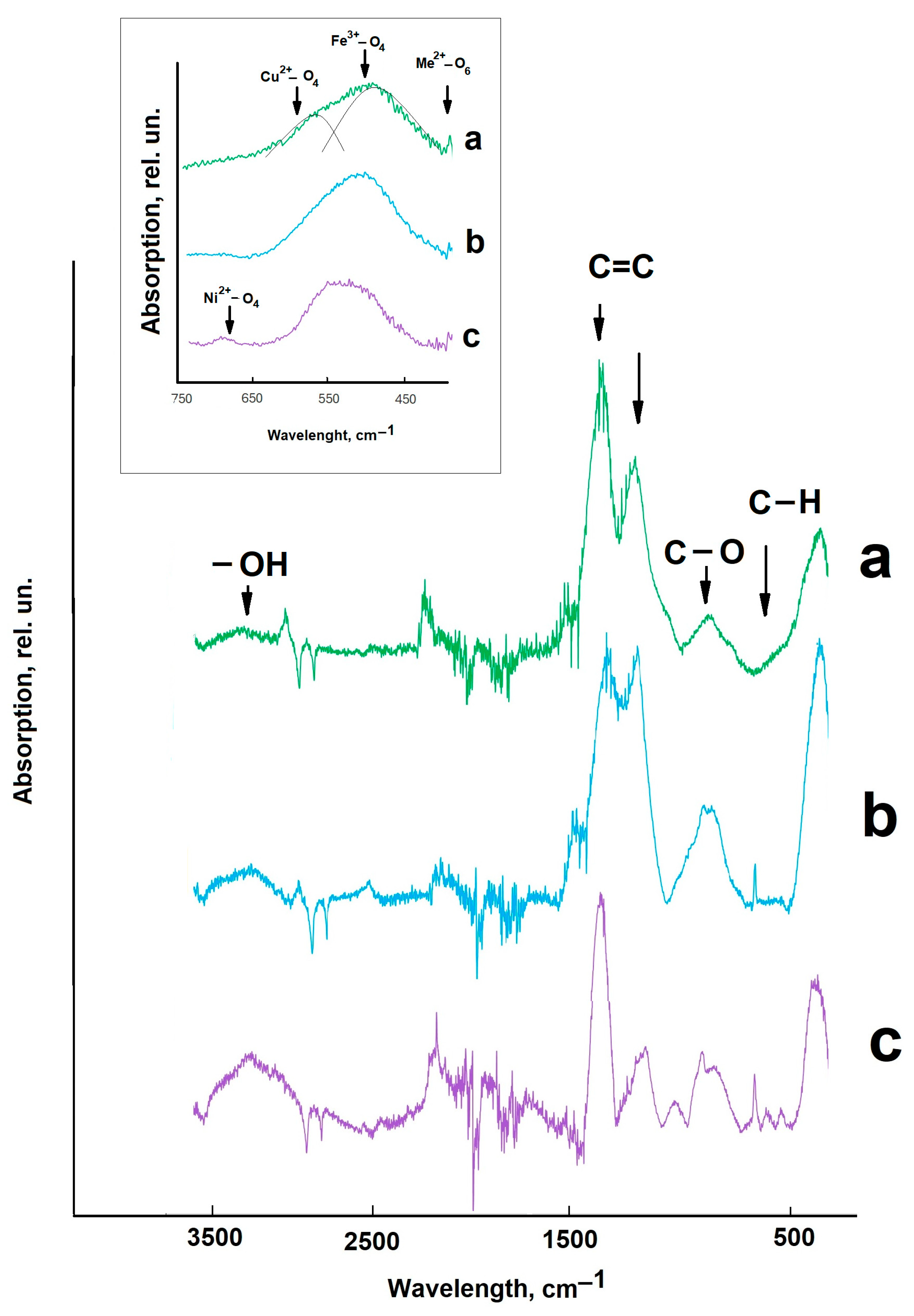
The FTIR spectroscopy data complement the results of the structural features analysis. Figure 3 shows the FTIR spectra of the synthesized composite materials; the recording was carried out in the wavelength range of 500–3500 cm−1.
In the range of 800–3500 cm−1, oscillations of the composite material organic component appear [17]. Peaks in the range of 3000–3500 cm−1 are associated with vibrations of -OH groups [17]. In the range of 1500–1700 cm−1, intense peaks of C=C bonds appear. The peaks in the range of 1000–1200 cm−1 and 800 cm−1 are associated with vibrations of C-O and C-H groups, respectively [43]. In the range of 500–800 cm−1, vibrations of the composite material oxide component appear [17,40] (in Figure 3, this region is highlighted in the insert). In this case, vibrations of the bivalent metal (Ni2+ or Cu2+) in the octahedral positions of the spinel grid are distinguished in the region of 450 cm−1.
The broad peak in the region of 550–600 cm−1 is associated with vibrations of Fe3+ in tetrahedral positions. In the region of the largest wavelengths of 650–750 cm−1, peaks of vibrations of two valence cations (Ni2+ or Cu2+) in tetrahedral coordination appear. It should be noted that for copper-containing composite materials, the broad peak in the region of 500–700 cm−1 is not symmetrical; it can be decomposed into several components, unlike the composite with NiFe2O4 (Figure 3, insert). Thus, the FTIR spectroscopy data also indicate the formation of a structure with increased defectiveness in mixed nickel–copper ferrite.
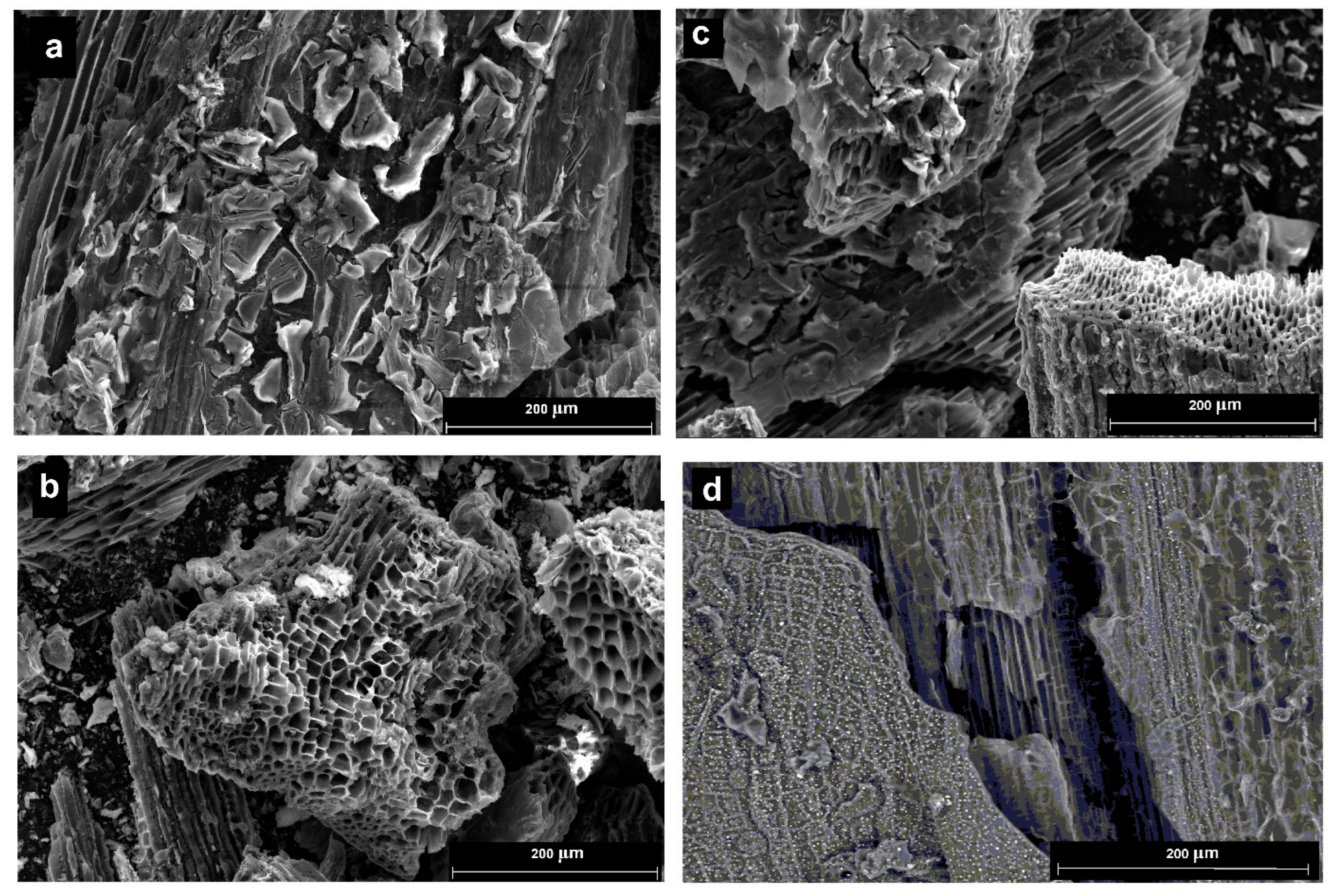
Figure 4 shows micrographs of the synthesized materials (a–c) and pure biochar (d).
Evidently, the oxide materials form a film on the biochar surface, repeating its porous structure. This can be useful for the catalytic properties of the materials, as materials with a developed surface are formed, which is important for catalysts. However, the value of the specific surface area decreases by about 20% (for the composite Cu0.5Ni0.5Fe2O4/biochar, it was 42.3 m2/g).
The synthesized materials were tested in the azo dye degradation reaction under hydrogen peroxide in an aqueous solution. To study the effect of the medium acidity on the destruction process course, the catalytic properties of the materials were studied at pH values of 1, 6, and 11. For this, 0.1 mL of sulfuric acid solution or 0.1 mL of sodium hydroxide solution with a concentration of 1 mol/L were introduced into the reaction system. If the process was carried out without introducing additional reagents, the pH of the solution was slightly acidic due to partial dissociation of hydrogen peroxide according to Reaction (1):
H2O2 = H+ + HO2.
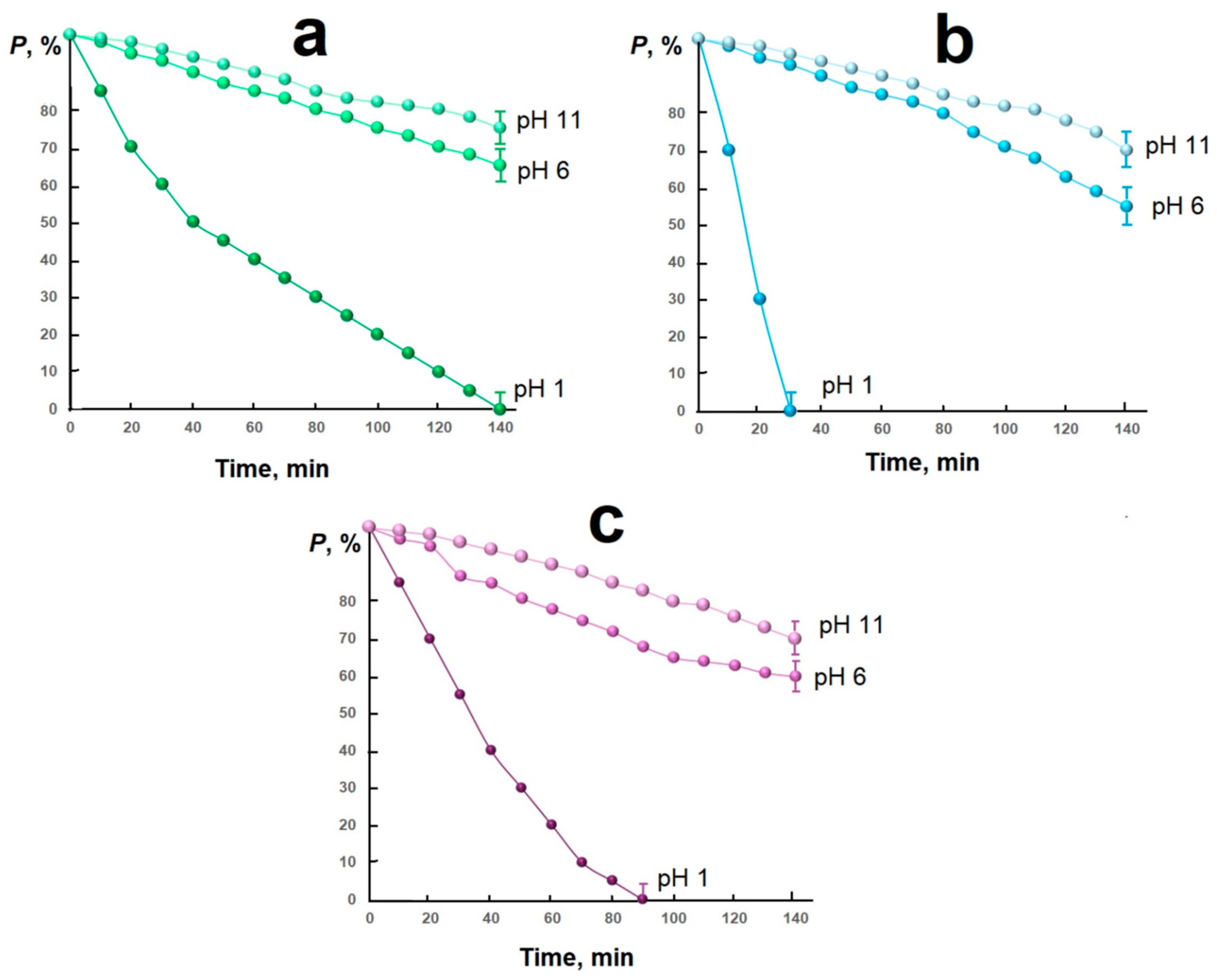
It was found that in a neutral and alkaline environment, it is not possible to completely purify the aqueous solution from the azo dye: even after 12 h, the concentration of methyl orange in all the conditions studied did not decrease below 50% (mass). The results of purifying the aqueous solution in an acidic environment are shown in Figure 5.
It was determined that the mixed nickel–copper ferrite exhibits exceptional activity in the methyl orange (MO, C14H14N3O3SNa) degradation reaction in an aqueous solution (see Table 1): complete destruction of the dye occurs 30 min after the start of the reaction. This result may be associated with the Fenton system formation during the oxidation–reduction process (Equations (2)–(6)):
CuFe2O4 + H2O2 → (CuFe+2Fe+3O4) + O0 + H2O+,
H2O+ →2H+ + O0,
2H+ + O0 = H2O,
2(CuFe+2Fe+3O4) + O0 + 2H+ → 2CuFe2O4 + H2O,
O0 + MO → (NOx + CO2 + H2O + …).
A more complete process of dye degradation in an acidic medium may be associated with an intermediate product formation based on copper (II) ferrite, which has a negative charge. Considering that in an acidic medium methyl orange exists in two modifications with an excess positive charge (Figure 6), it can be assumed that the presence of positive (azo dye) and negative (copper ferrite) particles in the system leads to the dye molecules’ fixation on the catalyst surface and the organic substance oxidation process facilitation by the released active oxidizing particle.
A significant increase in the reaction rate in the system containing mixed nickel–copper ferrite as a catalyst may be due to the formation of a more defective structure due to the Jahn–Teller effect manifestation, which creates additional active centers on the catalyst surface.
Without significant loss of activity, the catalysts can be used for five cycles.

3. Materials and Methods

3.1. Materials

The composite materials considered in this study are based on an inorganic component, copper (II)-nickel (II) ferrites of the general composition of CuxNi1−xFe2O4, (x = 0.0; 0.5; 1.0), and an organic component, biochar from sunflower husk. The materials were obtained in powder form using sol–gel synthesis. For this purpose, the following raw materials were used: aqueous ammonia solution (25 %) (Rushim, Yekaterinburg, Russia), citric acid monohydrate (C6H8O7·H2O) (Russian Product, Moscow, Russia), copper (II) nitrate hexahydrate (Cu(NO)3)2 6H2O) (Rushim, Yekaterinburg, Russia), nickel (II) nitrate hexahydrate (Ni(NO)3)2 6H2O) (Rushim, Yekaterinburg, Russia), and iron nitrate octahydrate (Fe(NO)3)3 9H2O) (Rushim, Yekaterinburg, Russia) of chemically pure grade.

3.2. Synthesis of Biochar

The synthesis of biochar is described in detail in [44]. The biomass of sunflower husks (Troika LLC, Batrak village, Russia) was thoroughly washed with distilled water, dried until weight loss ceased, and subjected to stepwise pyrolysis without air access at temperatures of 100–700 °C, with a temperature change step of 200 °C, and with a 20 min hold in intermediate phases and a 45 min hold in the final phase. The rate of temperature increase was 11 °C/min. The biochar had a surface area measured by the BET method of 54.6 m2/g.

3.3. Synthesis of Composite Material

Initially, solutions of transition element salts with a concentration of 1 mol/L and a citric acid solution with a concentration of 6.25 mol/L were prepared; the ammonia solution was used without dilution. Subsequently, 25 g of sunflower husk biochar was measured out and mixed with solutions of transition element salts under continuous stirring. The salt solutions were used in the ratio of (Cu (II) and/or Ni (II): Fe (III)) = (1:2), and 50 mL of iron (III) nitrate solution was used. Then, ammonia (15 mL) and citric acid (25 mL) solutions were added. The mixture was heated to ignition (approximately 600 °C). After the sol converted to gel, it ignited, and a fine powder was formed as a result. The synthesis products were allowed to cool to room temperature.
The stoichiometric equations for the formation of the mixture are as follows (7)
0.5 Cu(NO3)2 + 0.5 Cu(NO3)2 + 2 Fe(NO3)3 + NH4OH + C6H8O7
→ Cu0.5Ni0.5Fe2O4 + by-products (NOx, CO2, H2O).
Samples of the composition CuFe2O4/biochar (indicated as CF), Cu0.5Ni0.5Fe2O4/biochar (indicated as CNF), and NiFe2O4/biochar (indicated as NF) were obtained.

3.4. Characterization

To characterize the obtained composite materials, various methods were used, including X-ray diffraction (XRD), transmission electron microscopy, the Scherrer method, and FTIR spectroscopy.
The phase composition was studied on an ARL X’TRA X-ray diffractometer (Thermo Fisher Scientific (Ecublens) SARL, Ecublens, Switzerland) (monochromatic Cu-Kα radiation was used) by the point scanning method (0.01° step, 2 s accumulation time at a point) in the range of 2θ values from 20° to 70°. The crystallite size was calculated using the Scherrer Equation (8) [45]
D = 0.94∙λ/(β∙cosθ),
where D is the average crystal size, nm; λ is the X-ray wavelength, nm; β is the peak line width at half its height, rad.; and cos θ is the cosine of the angle for the peak.
The calculated value of the unit cell parameter was determined by Formula (9) for the cubic phase of spinel and Formula (10) for the tetragonally distorted phase of spinel:
1/d2 = (h2 + k2 + l2)/a2,
1/d2 = (h2 + k2)/a2 + l2/c2,
where a, c are the unit cell parameters of the spinel structure, nm; h, k, l are the Miller indices; d is the interplanar distance, nm.
The unit cell volume (V, nm3) was calculated by Formula (11) for the cubic phase and (12) for the tetragonal phase of spinel:
Vk = a2,
Vt = a2·c.
The average unit cell parameter for the tetragonally distorted phase of spinel was determined by Formula (13):
a m = a 2 · c 3 .
Grid deformation (ε), dislocation density (δ), and X-ray density (ρ) were determined using Formulas (14)–(16) [40]:
ε = β/(4·tan θ),
δ = (15 · ε)/(am · D),
ρ = (8 · M)/(Na · am3).
Here M is the molar mass of ferrite, g/mol; Na = 6.02 1023 mol−1 is Avogadro’s number.
The anion–cation distances in the tetrahedral (LA) and octahedral (LB) coordination of the spinel grid were determined using Formulas (17) and (18):
L a = 0.1875 · a m 2 ,
L B = 0.125 · a m 2
Ultrastructural images of the samples were obtained on a Quattro S SEM scanning microscope (Thermo Fisher Scientific, Waltham, MA, USA) in the bright-field mode at an accelerating voltage of 100 kV, magnification ×350. FTIR spectroscopy was performed on the Spectrum Two hardware complex (Perkin-Elmer, Shelton, CT, USA).
The catalytic activity was studied using a model solution of an organic dye of methyl orange (λmax = 465 nm). During the experiment, 10 mg of the catalyst was weighed out and placed in a reaction vessel. Then, 10 mL of an organic dye solution with a concentration of 0.05 g/L and 10 mL of a hydrogen peroxide solution with a concentration of 3% were added, and sulfuric acid and sodium alkali solutions with a concentration of 1 mol/L were used to create a certain acidity of the medium. The dye in the solution was analyzed photocolorimetrically using a KFK-2-UHL 4.2 device (Zagorsky Optical and Mechanical Plant, Sergiev Posad, Russia) at certain intervals. A 100 W JC halogen lamp (Camelion, Camelion International Ltd., Shenzhen, China) was used as a light source. The distance from the light source to the surface of the reaction system was 50 mm. Before the experiment, the reaction system was thoroughly mixed while isolated from light for 0.5 h to achieve adsorption/desorption equilibrium.
The degree of destruction P, %, was calculated using Formula (19):
P = (C0Ct) · 100/C0,
where C0 is the initial concentration of the dye in the solution, g/L; Ct is the amount of dye that has undergone degradation at the current time, g/L.

4. Conclusions

In this work, for the first time, composite organo-inorganic materials containing sunflower husk biochar and nickel(II)-copper(II) ferrites of the composition of CuxNi1−xFe2O4 (x = 0.0; 0.5; 1.0) were obtained.
The change in the structural parameters of the inorganic part of the composite material as the chemical composition changed was analyzed. When copper (II) cations were replaced by nickel (II) cations, the average parameter values and the unit cell volume and the cation–anion distance in the octahedral spinel grid were found to change with regularity. The obtained experimental facts can be associated with a change in the ionic radius of the bivalent cation in the spinel grid and the Jahn–Teller effect manifestation.
In the mixed nickel (II)-copper (II) ferrite, an increased concentration of crystal grid defects is noted, caused by a high degree of ferrite inversion and the Jahn–Teller effect manifestation.
The composite materials have a core–shell structure, while the oxide materials form a film on the surface of biochar, repeating its porous structure. This can be useful for the catalytic properties of the materials, as materials with a developed surface are formed, which is important for catalysts.
The obtained materials exhibit high catalytic activity in the reaction of methyl orange decomposition under hydrogen peroxide in an acidic medium. So, in the presence of composites of the composition Cu0.5Ni0.5Fe2O4/biochar, NiFe2O4/biochar, and CuFe2O4/biochar, it is possible to carry out complete degradation of the organic azo dye in an aqueous solution for 30, 90, and 140 min, respectively. This result can be associated with the formation of the Fenton system during the oxidation–reduction process.
A significant increase in the reaction rate in the system containing mixed nickel–copper ferrite as a catalyst may be associated with the formation of a more defective structure due to the Jahn–Teller effect manifestation, which creates additional active centers on the catalyst surface.

Author Contributions

Conceptualization, A.V.V. and N.G.S.; methodology, N.P.S.; software, S.I.S.; validation, Y.A.G.; formal analysis, A.M.R.; investigation, A.V.V., N.G.S. and V.A.B.; resources, N.P.S.; data curation, E.V.V.; writing—original draft preparation, E.A.Y.; writing—review and editing, N.P.S.; visualization, E.V.V. and E.V.S.; supervision, A.V.V.; project administration, N.P.S.; funding acquisition, N.P.S. All authors have read and agreed to the published version of the manuscript.

Funding

The research was financially supported by the project of the Ministry of Science and Higher Education of Russia on the Young Scientist Laboratory within the framework of the Interregional scientific and educational center of the South of Russia (FENW-2024-0001).

Institutional Review Board Statement

Not applicable.

Informed Consent Statement

Not applicable.

Data Availability Statement

The original contributions presented in this study are included in the article.

Acknowledgments

Special thanks to A.N. Yatsenko, employee of the “Nanotechnology” Center for Collective Use of the Platov South-Russian State Polytechnical University (NPI), for assistance in shooting and decoding X-ray fluorescence data and performing microscopic studies. Special thanks to R.G. Valeev, employee of the “Center for Physical and Physicochemical Methods of Analysis, Study of Properties and Characteristics of Surfaces, Nanostructures, Materials and Products”, the Center for Collective Use of Scientific Equipment of the Udmurt Federal Scientific Center of the Ural Branch of the Russian Academy of Sciences, for assistance in conducting microscopic studies.

Conflicts of Interest

The authors declare no conflicts of interest.

References

  1. Balachandran, B.; Sabumon, P.C. A comprehensive review on biodegradation of Azo dye mixtures, metabolite profiling with health implications and removal strategies. J. Hazard. Mater. Adv. 2025, 19, 100834. [Google Scholar] [CrossRef]
  2. El Awady, M.E.; El-Shall, F.N.; Mohamed, G.E.; Abd-Elaziz, A.M.; Abdel-Monem, M.O.; Hassan, M.G. Exploring the decolorization efficiency and biodegradation mechanisms of different functional textile azo dyes by Streptomyces albidoflavus 3MGH. BMC Microbiol. 2024, 24, 210. [Google Scholar] [CrossRef]
  3. Aounallah, F.; Hkiri, N.; Fouzai, K.; Elaoud, A.; Ayed, L.; Asses, N. Biodegradation pathway of Congo red azo dye by geotrichum candidum and toxicity assessment of metabolites. Catal. Lett. 2024, 154, 6064–6079. [Google Scholar] [CrossRef]
  4. Gongal, A.V.; Nandanwar, D.V.; Badwaik, D.S.; Choudhari, Y.D.; Wanjari, S.S. Incorporating effect of rare earths (Sm3+ and Nd3+) and transition metal ions (Al3+ and Cr3+) in modifying the lattice structure, magnetism, and dielectric properties of magnesium copper zinc cubic ferrites. J. Magn. Magn. Mater. 2025, 622, 172950. [Google Scholar] [CrossRef]
  5. Sharma, A.; Kanjariya, P.; Roopashree, R.; Ray, S.; Ab Yajid, M.S.; Tantawi, D.; Kumari, M.; Jayabalan, K.; Joshi, K.K.; Pramanik, A. A comprehensive review on the molecular structure of spinel nanomaterials for green hydrogen generation application via photocatalytic water splitting. J. Mol. Struct. 2025, 1348, 143348. [Google Scholar] [CrossRef]
  6. Yadav, R.S.; Anju; Kuřitka, I. Spinel ferrite and MXene-based magnetic novel nanocomposites: An innovative high-performance electromagnetic interference shielding and microwave absorber. Crit. Rev. Solid State Mater. Sci. 2022, 48, 441–479. [Google Scholar] [CrossRef]
  7. Qin, H.; He, Y.; Xu, P.; Huang, D.; Wang, Z.; Wang, H.; Wang, C. Spinel ferrites (MFe2O4): Synthesis, improvement and catalytic application in environment and energy field. Adv. Colloid Interface Sci. 2021, 294, 102486. [Google Scholar] [CrossRef]
  8. Ahmed, R.; Wang, J.; Si, R.J.; Rehman, S.; Li, T.; Bi, H.; Yu, Y.; Li, Q.J.; Li, Y.D.; Huang, S.G.; et al. Jahn-Teller assisted polaronic electron hopping in LiCuNb3O9. J. Eur. Ceram. Soc. 2021, 41, 2625–2632. [Google Scholar] [CrossRef]
  9. Georgalas, C.; Samartzis, A.; Biniskos, N.; Syskakis, E. Effects of Cr-doping on the Jahn-Teller, the orthorhombic to rhombohedral, and the magnetic transitions in LaMn1−xCrxO3 compounds. Phys. B Condens. Matter 2020, 586, 412101. [Google Scholar] [CrossRef]
  10. Liang, X.; Liu, X.; Wang, P.; Guo, Z.; Chen, X.; Yao, J.; Li, J.; Gan, Y.; Lv, L.; Tao, L.; et al. Ion-exchange induced Ni doping of α-MnO2 cathode with structural modification for aqueous zinc ion batteries. J. Power Sources 2025, 635, 236518. [Google Scholar] [CrossRef]
  11. Sasamori, T.; Lee, V.; Nagahora, N.; Morisako, S. 1.03—Low-coordinate compounds of heavier group 14–16 elements. In Comprehensive Inorganic Chemistry III, 3rd ed.; Reedijk, J., Poeppelmeier, R.K., Eds.; Elsevier: Amsterdam, The Netherlands, 2023; pp. 118–164. ISBN 9780128231531. [Google Scholar]
  12. Mojahed, M.; Dizaji, H.R.; Gholizadeh, A. Structural, magnetic, and dielectric properties of Ni/Zn co-substituted CuFe2O4 nanoparticles. Phys. B Condens. Matter 2022, 646, 414337. [Google Scholar] [CrossRef]
  13. Frolova, L.A.; Baskevich, A.S.; Butyrina, T.E. Influence of plasma synthesis parameters on the magnetic, structural and photocatalytic properties of copper ferrite. Ceram. Int. 2024, 50, 41461–41471. [Google Scholar] [CrossRef]
  14. Zarezadeh, A.; Alizadeh, P.; Yourdkhani, A.; Zarandi, F.M. Synthesis of copper ferrite-decorated bioglass nanoparticles: Investigation of magnetic properties, biocompatibility, and drug release behaviour. Appl. Surf. Sci. 2025, 708, 163713. [Google Scholar] [CrossRef]
  15. Akhtar, K.; Baig, J.A.; Afridi, H.I.; Solangi, I.B.; Perveen, S.; Kazi, M. Lanthanum doped copper ferrite as efficient Electrocatalyst for simultaneous detection of Bentazone and Diuron in vegetable samples. Food Chem. 2025, 481, 144066. [Google Scholar] [CrossRef]
  16. Krishna, K.G.; Parne, S.R.; Nagaraju, P. Room temperature benzene gas sensing with spinal copper ferrite and copper cerium oxide nanocomposite sensors. Microchem. J. 2025, 215, 114278. [Google Scholar] [CrossRef]
  17. Mariappan, K.; Sivaji, S.P.; Chen, S.M.; Chen, C.-L.; Lin, P.-H.; Sakthinathan, S.; Nehru, S.; Muthusamy, P.; Palanisamy, N.; Mariappan, C. Electrochemical determination of diuron in agricultural products using copper ferrite incorporated with multiwalled carbon nanotubes composite modified glassy carbon electrode. Colloids Surf. A Physicochem. Eng. Asp. 2025, 719, 136997. [Google Scholar] [CrossRef]
  18. Cardoso, B.; Nobrega, G.; Machado, M.; Lima, R.A. Green synthesis of copper ferrite-based nanofluids using Chlorella vulgaris for heat transfer enhancement. J. Mol. Liq. 2025, 428, 127498. [Google Scholar] [CrossRef]
  19. Riaz, R.; Bibi, I.; Majid, F.; Taj, B.; Aamir, M.; Fatima, G.; Raza, Q.; Elhouichet, H.; Iqbal, M.; Younas, U. Fabrication of a nanocomposite (NiFe2O4/CuO) with varying ratios of nickel ferrite and copper oxide nanoparticles to achieve a synergistic effect toward electrical performance. Ceram. Int. 2025, 51, 7038–7046. [Google Scholar] [CrossRef]
  20. Kumar, D.; Verma, R.; Chauhan, A.; Thakur, P.; Wan, F.; Thakur, A. Sustainable high frequency applications of copper ferrite nanoparticles. Inorg. Chem. Commun. 2025, 174, 114018. [Google Scholar] [CrossRef]
  21. Sabir, I.; Mingxia, H.; Anwar, H.; Kashif, M.; Yizhu, Z. Utilization of copper-doped zinc spinel ferrites nano-composites as battery-grade electrode materials for supercapattery device applications. J. Energy Storage 2025, 112, 115498. [Google Scholar] [CrossRef]
  22. Deng, Y.; Li, S.; Appels, L.; Dewil, R.; Zhang, H.; Baeyens, J.; Mikulcic, H. Producing hydrogen by catalytic steam reforming of methanol using non-noble metal catalysts. J. Environ. Manag. 2022, 321, 116019. [Google Scholar] [CrossRef]
  23. Szegedi, Á.; Lázár, K.; Solt, H.; Popova, M. Peculiar redox properties of SBA-15 supported copper ferrite catalysts promoting total oxidation of a model volatile organic air pollutant. Surf. Interfaces 2025, 56, 105498. [Google Scholar] [CrossRef]
  24. Alsebaii, N.M.; Hegazy, E.Z.; El Maksod, I.H.A.; El-Sayed El-Shafay, S. Enhanced photocatalytic activity of titanium-doped copper ferrite for methyl green dye degradation under commercial visible LED light. Ceram. Int. 2024, 50 Pt A, 45479–45487. [Google Scholar] [CrossRef]
  25. Ahmad, I.; Alhedrawie, D.; Jain, V.; Kumar, A.; Rekha, M.M.; Kundlas, M.; Sunitha, S.; Ray, S.; Shomurotova, S. Effective removal of auramine O and safranin O from aqueous solutions using nickel copper ferrite magnetic nanoparticles. J. Mol. Struct. 2025, 1344, 142933. [Google Scholar] [CrossRef]
  26. Mazurenko, J.; Szostak, E.; Gondek, L.; Kaykan, L.; Zywczak, A.; Vyshnevskyi, O. Structural and magnetic studies of Cobalt-Substituted copper ferrites for efficient photocatalytic dye degradation. Inorg. Chem. Commun. 2025, 171, 113648. [Google Scholar] [CrossRef]
  27. Wu, W.; Yu, L.; Zha, L.; He, F.; Ma, J.; Wu, O.; Zhang, H.; Chen, X.; Yu, S.; Lei, M.; et al. Mild and Effective Decatungstate-Catalyzed Degradation of Methyl Orange Under Visible Light. Catalysts 2025, 15, 494. [Google Scholar] [CrossRef]
  28. Beura, R.; Thangadurai, P. Effect of Sn doping in ZnO on the photocatalytic activity of ZnO-Graphene nanocomposite with improved activity. J. Environ. Chem. Eng. 2018, 6, 5087–5100. [Google Scholar] [CrossRef]
  29. Li, H.; Zhang, W.; Guan, L.; Li, F.; Yao, M. Visible light active TiO2–ZnO composite films by cerium and fluorine codoping for photocatalytic decontamination. Mater. Sci. Semicond. Process. 2015, 40, 310–318. [Google Scholar] [CrossRef]
  30. Chen, Y.; Zhang, C.; Huang, W.; Yang, C.; Huang, T.; Situ, Y.; Huang, H. Synthesis of porous ZnO/TiO2 thin films with superhydrophilicity and photocatalytic activity via a template-free sol–gel method. Surf. Coat. Technol. 2014, 258, 531–538. [Google Scholar] [CrossRef]
  31. Liu, Z.; Zhang, Y.; Tang, J.; Wu, J.; Borjigin, T.; Xing, L. Acid-citrate sequential engineered Hangjin2# clay for photo-fenton degradation of methyl orange: Electron transfer mechanism analysis. Inorg. Chem. Commun. 2025, 181, 115266. [Google Scholar] [CrossRef]
  32. Fuentes, L.; Robledo, S.; Natera, J.; Massad, W.A. Box-Behnken experimental design to optimize the degradation of methyl orange by β-cyclodextrin-assisted photo-Fenton. J. Photochem. Photobiol. A Chem. 2025, 467, 116419. [Google Scholar] [CrossRef]
  33. Mukwevho, N.; Mafa, P.J.; Kefeni, K.K.; Mishra, A.K.; Mishra, S.B.; Kuvarega, A.T. Photo-Fenton like reaction for the degradation of methyl orange using magnetically retrievable NiFe2O4/CoMoS4 heterojunction photocatalyst. J. Water Process. Eng. 2024, 65, 105882. [Google Scholar] [CrossRef]
  34. Chen, C.; Yan, Z.; Ma, Z.; Ma, D.; Xing, S.; Li, W.; Yang, J.; Han, Q. Magnetic Fe3O4 nanoparticles supported on carbonized corncob as heterogeneous Fenton catalyst for efficient degradation of methyl orange. Chin. J. Chem. Eng. 2025, 77, 144–155. [Google Scholar] [CrossRef]
  35. Hazril, N.I.H.; Jalil, A.A.; Aziz, F.F.A.; Hassan, N.S.; Fauzi, A.A.; Khusnun, N.F.; Izzudin, N.M.; Jusoh, N.W.C.; Teh, L.P.; Jaafar, N.F.; et al. Selective simultaneous photo-Fenton removal of Cr (VI) and methyl orange dye over critical raw material-free fibrous-silica irons catalyst. Sustain. Mater. Technol. 2024, 41, e00994. [Google Scholar] [CrossRef]
  36. Cobos, M.Á.; Jiménez, J.A.; Llorente, I.; de la Presa, P.; Hernando, A. Ball milling and annealing effect in structural and magnetic properties of copper ferrite by ceramic synthesis. J. Alloys Compd. 2024, 1006, 176206. [Google Scholar] [CrossRef]
  37. Balbín, A.; Correa, J.R.; Piña, J.J.; Peláez-Abellán, E.; Jiménez, J.; Gordo, Y.; Urones-Garrote, E.; Otero-Díaz, L.C. Synthesis and catalytic study of manganese ferrites obtained by partial oxidation of ferrous hydroxide with permanganate. J. Magn. Magn. Mater. 2025, 614, 172665. [Google Scholar] [CrossRef]
  38. Mmelesi, O.K.; Patala, R.; Nkambule, T.T.I.; Mamba, B.B.; Kefeni, K.K.; Kuvarega, A.T. Effect of Zn doping on physico-chemical properties of cobalt ferrite for the photodegradation of amoxicillin and deactivation of E. coli. Colloids Surf. A Physicochem. Eng. Asp. 2022, 649, 129462. [Google Scholar] [CrossRef]
  39. Mısırlıoğlu, B.S.; Kahya, N.D.; Öztürk, Z. Enhanced dielectric properties of copper substituted nickel ferrite nanoparticles for energy storage applications. J. Phys. Chem. Solids 2024, 193, 112195. [Google Scholar] [CrossRef]
  40. Rasool, B.; Shehzad, K.; Yang, J.; Asif, M.I.; Al-Sulaimi, S.; Asif, M.; Ashraf, G.A.; ul Hassan, R.; Wahab, R.; Xu, Y.; et al. Insight into the structural, magnetic and fluoride (F−1) adsorption properties of copper–manganese ferrite (Cu0.5Mn0.5Fe2O4) and Azadirachta Indica composites synthesized through hydrothermal method. Mater. Chem. Phys. 2025, 334, 130415. [Google Scholar] [CrossRef]
  41. Nunes Filho, A.L.; Mendes Isidorio, D.K.; Pizarro Borges, L.E.; Vieira, A.C.; Veiga-Junior, V.F.; Pinheiro, W.A. Production and characterizations of silver-doped copper ferrite anchored in RGO surface. J. Mater. Res. Technol. 2024, 32, 1268–1273. [Google Scholar] [CrossRef]
  42. Aruna, R.; Nithiyanantham, S.; Mahalakshmi, S.; Kogulakrishnan, K.; Usharani, K.; Gunasekaran, B.; Mohan, R.; Palaniappan, L. Structural, magnetic and electrical studies on nickel doped copper ferrite synthesized using sol-gel method. Results Surf. Interfaces 2024, 17, 100315. [Google Scholar] [CrossRef]
  43. Verma, S.; Das, T.; Pandey, V.K.; Verma, B. Nanoarchitectonics of GO/PANI/CoFe2O4 (Graphene Oxide/polyaniline/Cobalt Ferrite) based hybrid composite and its use in fabricating symmetric supercapacitor devices. J. Mol. Struct. 2022, 1266, 133515. [Google Scholar] [CrossRef]
  44. Shabelskaya, N.; Sulima, S.; Sulima, E.; Medennikov, O.; Kulikova, M.; Kolesnikova, T.; Sushkova, S. Study of the Possibility of Using Sol–Gel Technology to Obtain Magnetic Nanoparticles Based on Transition Metal Ferrites. Gels 2023, 9, 217. [Google Scholar] [CrossRef]
  45. Kogulakrishnan, K.; Nithiyanantham, S.; Koteeshwari, R.S.; Malarkodi, B.; Gobinath, B.; Venkadesh, A.; Mohan, R.; Natarajan, B.; Palaniappan, L. Investigations on the structure, magnetism, electricity, and electrochemistry of copper-doped manganese ferrite: Sol-gel technique. Mater. Sci. Eng. B 2025, 313, 117999. [Google Scholar] [CrossRef]
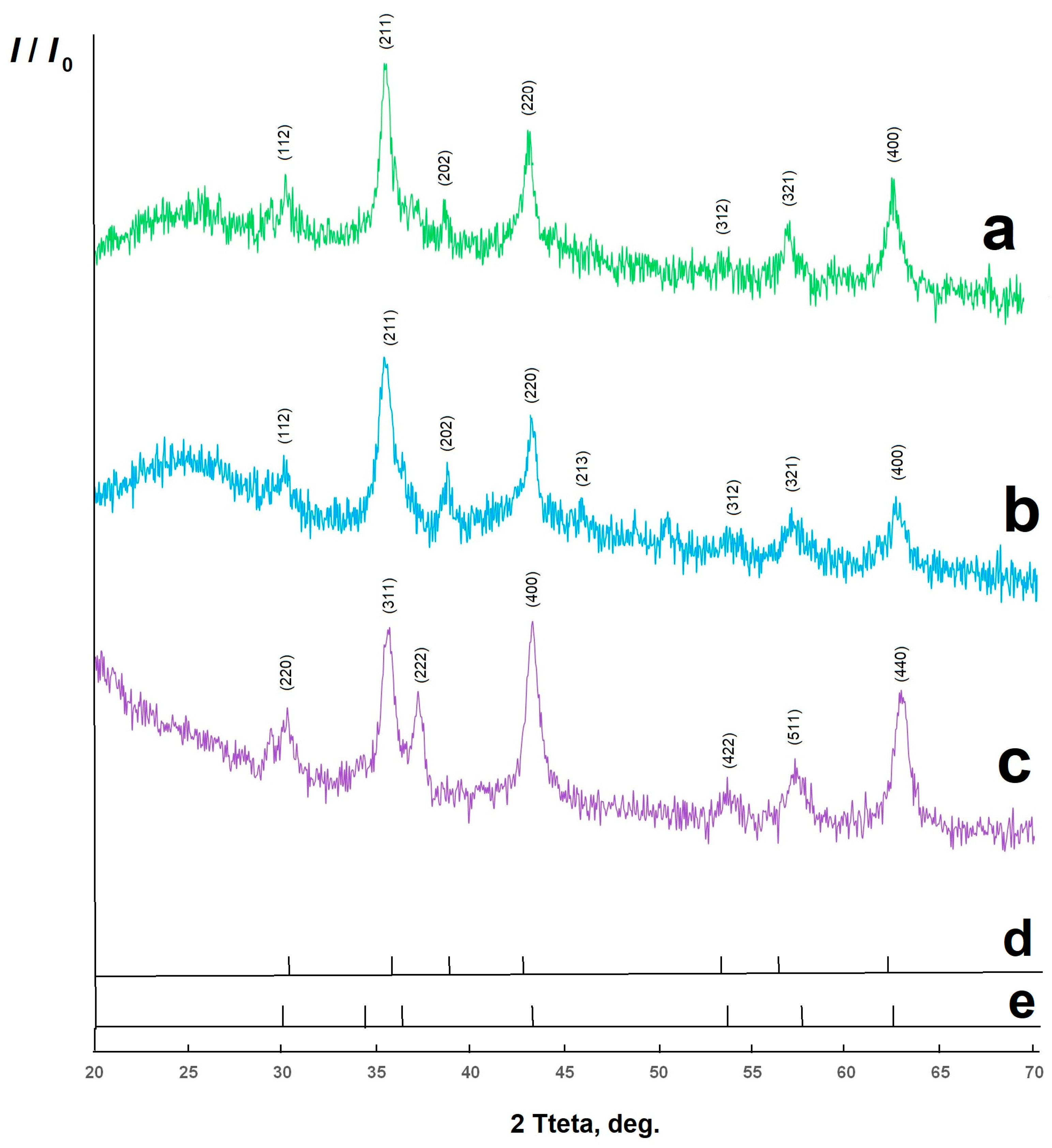
Figure 1. X-ray diffraction pattern of CuFe2O4/biochar composite materials (a), Cu0.5Ni0.5Fe2O4/biochar (b), NiFe2O4/biochar (c), reference peaks for NiFe2O4 (d) and CuFe2O4 (e).
Molecules 30 03900 g001
Figure 2. Schematic representation of the Jahn–Teller effect manifestation during the formation of CuFe2O4 tetragonal phase.
Molecules 30 03900 g002
Figure 3. FTIR spectra of synthesized composite materials: CuFe2O4/biochar (a), Cu0.5Ni0.5Fe2O4/biochar (b), NiFe2O4/biochar (c).
Molecules 30 03900 g003
Figure 4. SEM images of the synthesized materials: CuFe2O4/biochar (a), Cu0.5Ni0.5Fe2O4/biochar (b), NiFe2O4/biochar (c), biochar from sunflower husk (d).
Molecules 30 03900 g004
Figure 5. Destruction time of organic dye in aqueous solution in the presence of catalyst: CuFe2O4/biochar (a), Cu0.5Ni0.5Fe2O4/biochar (b), NiFe2O4/biochar (c).
Molecules 30 03900 g005
Figure 6. Forms of methyl orange indicator in aqueous solution.
Molecules 30 03900 g006
Table 1. Catalytic activity of oxide materials for removal of methyl orange.
No.The CatalystConcentration of the PollutantConditions of Catalysis, Degree of PurificationReference
11% Sn-ZnO-graphene5 mmol/LUV, 6 h, 89%
Visible, 3 h, 98%
[28]
25% Sn-ZnO-graphene5 mmol/LUV, 6 h, 80.4%
Visible, 3 h, 99%
[28]
3ZnO-Zn2TiO410 mg/LUV, 3 h, 90%[29]
4ZnO(10%)/TiO10.3 mg/LVisible, 280 min, 78%[30]
5citrate-modified clay50 mg/LUV, 90 min, 100%[31]
6Fe2+, β- Cyclodextrin26 mkmol/LVisible, 30 min, 100%[32]
7NiFe2O4/CoMoS4 10 mg/LVisible, 60 min, 99%[33]
8Fe3O4/biochar made from corn cob25 mg/LVisible, 14 min, 99.7%[34]
9fibrous silica/Fe3O410 mg/LVisible, 180 min, 80%[35]
Table 2. Structural parameters of spinels.
Sampleacc/aamVDLALB
CF0.83710.85981.030.84460.6021090.36570.2986
CNF0.83400.87321.040.83510.5821300.36160.2953
NF0.8341- 0.83410.5801070.36120.2949
Table 3. Characteristics of spinel crystals.
SampleλFormulaρ, g/sm3δ·103ε·103
CF0.24Fe0.24Cu0.76[Cu0.24Fe1.76]O45.290.1388.47
CNF0.87Cu0.13Fe0.87[Cu0.37Ni0.5Fe1.13]O45.420.14410.42
NF0.95Fe0.95Ni0.05[Ni0.95Fe1.05]O45.380.1187.03
Disclaimer/Publisher’s Note: The statements, opinions and data contained in all publications are solely those of the individual author(s) and contributor(s) and not of MDPI and/or the editor(s). MDPI and/or the editor(s) disclaim responsibility for any injury to people or property resulting from any ideas, methods, instructions or products referred to in the content.