Review

The Role of Plectin Dysregulation in Cancer: Recent Advances

by
Wenbin Wang
1,2,
Chang Lyu
1,2,
Zhihui Wang
1,2,
Xu Zhang
1,2,
Qing Luo
1,2 and
Guanbin Song
1,2,*
1
College of Bioengineering, Chongqing University, Chongqing 400030, China
2
Key Laboratory of Biorheological Science & Technology, Ministry of Education, Chongqing University, Chongqing 400030, China
*
Author to whom correspondence should be addressed.
Molecules 2025, 30(18), 3675; https://doi.org/10.3390/molecules30183675
Submission received: 6 August 2025 / Revised: 4 September 2025 / Accepted: 8 September 2025 / Published: 10 September 2025

Abstract

Plectin is a key cytolinker protein that functions as an integrator of the cytoskeletal networks by crosslinking intermediate filaments with actin filaments and microtubules. Mutations or function deficiencies of plectin lead to tissue disorders, particularly affecting the skin, muscle, and nervous tissues. Interestingly, plectin dysregulation in cancer, characterized by aberrant expression and mislocalization, has been increasingly observed, suggesting distinct roles in tumorigenesis and progression. Here, we focus on recent advances regarding the roles of plectin dysregulation in promoting cell proliferation, suppressing cell apoptosis, sustaining the stemness of cancer stem cells, and driving invasion and metastasis. We also discuss its bidirectional interplay with the tumor microenvironment, including modulating immune and inflammatory responses, promoting angiogenesis, sensing and transmitting mechanical cues from the extracellular matrix, and contributing to matrix remodeling. Finally, we highlight emerging therapeutic strategies that target plectin dysregulation with anticancer activity. By summarizing these advances, we aim to enhance the understanding of plectin dysregulation in cancer and illuminate its potential as a therapeutic target.

Graphical Abstract

1. Introduction

Plectin is a 500 kDa cytolinker protein encoded by the PLEC gene, also known as plectin-1 [1,2] or hemidesmosome protein 1 [3,4]. Structurally, it comprises an N-terminal actin-binding domain (ABD), a plakin domain, a central rod domain, and a C-terminal plectin repeat domain [5]. Through these domains, plectin associates with various components of the cytoskeleton, including actin filaments (AFs), intermediate filaments (IFs), and microtubules [6]. In addition, plectin participates in the assembly of cell junctions such as desmosomes, hemidesmosomes (HDs), and focal adhesions (FAs) [7,8,9,10,11]. The human PLEC gene coding sequence consists of 32 exons and spans approximately 32 kb, located in the telomeric region of chromosome 8 (8q24) [12]. Notably, the PLEC gene generates multiple isoforms through the alternative splicing of the 5′-terminal first exons, among which plectin 1, 1a, 1b, 1c, 1d, 1f, and 1k are the most extensively characterized [5,13,14]. Plectin 1 localizes to the perinuclear region [15]; plectin 1a anchors to hemidesmosomes [7]; plectin 1b regulates mitochondrial positioning and morphology [16]; plectin 1c associates with microtubules [7]; plectin 1d links the desmin intermediate filament network to the Z-disks of muscle fibers [17]; plectin 1f is enriched at focal adhesions [11]; and plectin 1k is involved in F-actin organization and podosome-like adhesions formation [18]. These isoforms primarily influence the binding properties of the ABD, resulting in structural specificity and targeting distinct cellular structures [14,19].
Under normal physiological conditions, plectin is predominantly localized in the cytoplasm, where it anchors cytoskeletal components and maintains the cell’s structural integrity [20]. Mutations in the PLEC gene or plectin deficiencies have been linked to a spectrum of human diseases, characterized by muscle weakness and atrophy, skin fragility and blistering, as well as signs of neuropathy [21,22]. Interestingly, emerging evidence in recent years has revealed the dysregulation of plectin in cancer. According to the ground-breaking discovery in 2008, plectin was first identified as a cell surface biomarker for pancreatic ductal adenocarcinoma (PDAC), despite its conventional intracellular localization [23]. Since then, accumulating evidence has demonstrated that both the expression levels and subcellular localization of plectin are frequently dysregulated in cancer [24,25,26,27,28]. Its mislocalization has been shown to confer unexpected functions in cancer progression, including the promotion of tumor growth and metastasis, as well as the maintenance of cancer stem cells (CSCs) stemness. Notably, the form of plectin that is aberrantly localized on the surface of tumor cells and exhibits pro-tumorigenic functions is referred to as cancer-specific plectin (CSP) [23,25]. The discovery of CSP underscores the pathological mislocalization of plectin and provides a promising avenue for tumor detection and targeted therapy.
Plectin dysregulation has emerged as a critical driver of tumor progression. It promotes tumor growth by sustaining cell proliferation [29], inhibiting apoptosis [30], and supporting the stemness of CSCs [27]. In addition, plectin functions as a metastasis-promoting factor by modulating signaling pathways involved in cell migration and invasion [31]. Recent evidence reveals that the dysregulated expression of plectin modulates the cellular and non-cellular architecture of the tumor microenvironment (TME), thereby facilitating a supportive niche for tumor invasion and dissemination. In cancer cells, plectin enhances resistance to mechanical stress [32] and is associated with hypoxic conditions [33]. Moreover, plectin contributes to immune evasion [34], extracellular matrix degradation [26], activation of inflammatory signaling [35], and angiogenesis [36], collectively promoting the establishment of a permissive pre-metastatic niche. In this review, we summarize the oncogenic roles of plectin dysregulation in tumor growth, metastasis, and its bidirectional interplay with the TME. We also discuss the novel therapeutic strategies targeting plectin dysregulation to promote precision cancer treatment, including small molecules, mRNA therapies, and antibody therapies.

2. Dysregulation of Plectin in Cancer

Although plectin deficiency is known to cause tissue disorders, a clearer understanding of plectin dysregulation and its implications in cancer is still necessary. In malignancies, the PLEC gene frequently undergoes copy number variations, leading to aberrant plectin expression, which is increasingly recognized as a potential hallmark of malignancy. More importantly, plectin in cancer cells often exhibits alterations in subcellular localization, shifting from its typical cytoplasmic distribution to the cell membrane. Here, we summarize the dysregulation of plectin, including its upregulation, downregulation, and mislocalization in different types of cancers, as shown in Table 1.

2.1. Upregulation of Plectin in Cancer

Plectin is primarily upregulated in cancer and contributes to tumorigenesis and tumor development. It is notably elevated in nearly all gastrointestinal cancers, even in precancerous lesions. For example, plectin expression is upregulated in pancreatic intraepithelial neoplasms (PanINs) and intraductal papillary mucinous neoplasms compared to normal pancreatic tissue [23,60]. This suggests that the upregulation of plectin may play a crucial role in the early stages of tumorigenesis. Similarly, abnormal overexpression of plectin has been reported in reproductive cancers, such as prostate cancer, testicular cancer, and ovarian cancer. In prostate cancer, relative to benign tissues, plectin expression is increased in both primary tumors and lymph node metastases [30], indicating a potential role in metastatic progression. Elevated plectin levels have also been documented in head and neck cancers [50]. Plectin has been proposed as a potential biomarker for early lesion detection and disease progression in oral squamous cell carcinoma (OSCC) [28]. Beyond these cancer types, aberrant plectin expression is observed in other malignancies. For example, an abnormal enrichment of plectin-positive spindle stromal cells has been observed in the bone marrow of acute myeloid leukemia (AML) patients [37]; glioblastoma multiforme (GBM) cell lines exhibit higher levels of surface plectin microdomains compared to normal astrocytes [48]; and plectin is upregulated in human primary melanoma samples relative to normal melanocytes [29].
Upregulation of the PLEC gene expression has been associated with cancer patient prognosis. In OSCC, lower PLEC gene expression is linked to a favorable prognosis, particularly among patients without lymph node metastasis and those with T1-stage tumors [28]. In GBM, both PLEC gene expression and mutation status have been proposed as predictive biomarkers for the efficacy of ADP-ribose polymerase inhibitors and patient outcomes [68]. In PTEN-negative prostate cancer, elevated PLEC gene expression serves as an independent prognostic factor, correlating with more aggressive disease and poorer clinical outcomes [61]. Similarly, in the TCGA bladder cancer cohort, high PLEC gene levels correlate with decreased overall and disease-free survival in patients with bladder cancer [69]. However, the prognostic significance of the PLEC gene appears to be specific to cancer type. For instance, in ovarian cancer, no significant correlation has been observed between PLEC gene expression and overall or progression-free survival [56], highlighting the heterogeneous role of plectin in tumor biology.

2.2. Downregulation of Plectin in Cancer

Although plectin is upregulated in most tumor types, its downregulation has also been observed in certain malignancies, indicating a dual expression pattern during cancer progression. For example, in basal and squamous cell carcinomas, plectin is expressed at lower levels in tumor tissues compared to normal skin tissue as a component of HDs [64]. Interestingly, even within the same tumor type, contradictory reports have emerged. In esophageal squamous cell carcinoma (ESCC), plectin is frequently overexpressed. However, its knockdown has been shown to promote carcinogenesis by disrupting the homeostasis of the stratified squamous epithelium (SSE), indicating that plectin downregulation may also contribute to tumor progression [45].
In hepatocellular carcinoma (HCC), the most robust evidence comes from Outla et al. (2025), who reported elevated plectin expression based on 17 independent patient datasets and quantitative immunofluorescence analysis of 19 paired tissue sections [26]. Functional studies demonstrated that inhibition of plectin significantly suppressed HCC proliferation, migration, invasion, cytoskeletal remodeling, and tumor growth via CRISPR/Cas9-mediated knockout, deletion of the intermediate filament-binding domain, or pharmacologic inhibition with plecstatin-1 (PST) in Huh7 and SNU-475 cells [26]. Xu et al. (2022) similarly found that shRNA-mediated plectin knockdown in MHCC97H and MHCC97L reduced extracellular signal-regulated kinase 1/2 (ERK1/2) phosphorylation, inhibiting cell migration and the epithelial–mesenchymal transition (EMT) process [31]. Furthermore, Wang et al. (2025) demonstrated that depletion of either plectin or integrin β1, or disruption of actin polymerization with latrunculin A, effectively blocked mechanical stiffness-induced F-actin polymerization and HCC cell migration [32].
Conversely, earlier studies have reported weaker plectin expression in HCC. Cheng et al. (2008) [70] observed strong plectin staining in normal liver tissue but weaker staining in tumor regions. This pattern was further supported by immunohistochemical analysis of five HCC cases, which showed markedly reduced plectin expression in tumor regions [70]. Similarly, Liu et al. (2011) observed weaker plectin staining in HCC sections compared with adjacent non-tumorous tissue across ten cases, consistent with Western blot results [71]. Additionally, Cheng (2015) noted heterogeneous morphology and relatively weak plectin staining in tumor regions of HCC patient tissues [65].
Notably, plectin expression in ovarian cancer appears to follow a dynamic pattern: it is upregulated during early tumorigenesis but subsequently downregulated as the disease progresses to high-grade or advanced stages [56]. Collectively, these findings highlight the complex and dual roles of plectin in cancer, indicating that its expression and function may vary among tumor types, stages, and histological subtypes.

2.3. Mislocalization of Plectin in Cancer

Plectin was initially characterized as a cytoplasmic protein ubiquitously expressed across mammalian tissues [13,24]. However, accumulating evidence suggests that plectin frequently translocates to the plasma membrane in cancer cells. It was first identified as a membrane-associated biomarker in PDAC, with cell surface expression progressively increasing from PanIN III to invasive PDAC [23]. Subsequently, an unbiased peptide-binding screen revealed plectin as a unique membrane marker for ALDH+ lung cancer stem cells [27]. Recent studies further demonstrated that in HCC, both clinical samples and chemically induced mouse models exhibit significant perimembranous enrichment of plectin [26]. In colorectal cancer (CRC), Ras-related protein Rap-2B (Rap2B) was found to colocalize with plectin in both the cytoplasm and plasma membrane. Rap2B translocates to the membrane via its C-terminal CAAX motif (C180), where it directly binds to the ABD of plectin, thereby promoting their membrane co-localization [72]. In ovarian cancer, a novel monoclonal antibody (1H11) was developed to specifically recognize CSP on the surface of both human and mouse tumor cells, confirming its membrane localization [25].
Plectin isoforms regulate the subtype-specific distribution of cytoskeletal connections and organelle localization through their variable N-terminal sequences. Consequently, the aberrant subcellular localization of plectin in cancer cells may result from the dysregulation of specific isoforms. In GBM, plectin 1c is the predominant isoform and shows notable co-localization with plasma membrane-associated aquaporin 4 [48]. In addition, plectin 1c is involved in HD composition, thus exhibiting peripheral localization [5]. In CRC, plectin 1 and plectin 1k are upregulated and specifically enriched at podosome-like adhesions in highly invasive cells, supporting their localization at membrane-associated adhesions [18]. In PDAC, the plectin isoforms 1a and 1f are abnormally localized on the cell surface via an exosome-mediated mechanism [24]. These findings highlight the crucial role of isoform-specific expression in regulating plectin localization and function during cancer progression.

3. The Role of Plectin Dysregulation in Cancer

Beyond its structural role in normal tissues, the dysexpression and mislocalization of plectin have been widely acknowledged for their functional relevance in cancer. Studies have shown that plectin dysregulation contributes to tumor progression through multiple mechanisms, such as promoting tumor growth, enhancing metastatic potential, and contributing to therapy resistance. In addition, plectin has increasingly been implicated as a key mediator of tumor–stroma interactions, contributing to the resistance and remodeling of the TME and fostering a pro-tumorigenic niche (As shown in Figure 1).

3.1. Sustaining Tumor Growth

Tumor growth is governed by a complex interplay of cellular processes, including survival, apoptosis, proliferation, and dormancy [73]. Among these hallmarks, sustained proliferation is a key feature of cancer progression, in which plectin has emerged as an essential regulator. Downregulation of plectin reduces tumor mass and attenuates cell proliferation by inhibiting the activity of the Src oncogene in melanoma cells [29]. In breast cancer, the oncogenic protein FAM83A interacts with plectin, modulating cytoskeletal architecture and activating downstream signaling pathways that promote cell survival and proliferation [74]. Additionally, plectin directly influences the expression of cell cycle regulators—for instance, silencing of plectin results in reduced expression of Cyclin D1 [29]. In the esophageal SSE, plectin connects HDs, desmosomes, and cytoskeletal elements to Notch1 and the PLEC gene regulator p63, playing a crucial role in maintaining cell anchorage, proliferation-differentiation equilibrium, and stratification. Thus, its dysregulation and mislocalization of plectin disrupt SSE homeostasis and contribute to ESCC carcinogenesis [45]. Collectively, these findings highlight the crucial role of plectin dysregulation in sustaining proliferative signaling essential for tumor growth.
Secondly, plectin contributes to resistance to apoptosis in cancer. It serves as a key target for caspase-mediated cytoskeletal disassembly during programmed cell death, with its cleavage facilitating apoptosis by disrupting the cytoskeletal framework. Plectin has been identified as a substrate of caspase-8 in both CD95- and tumor necrosis factor receptor-mediated apoptotic pathways, leading to the destabilization of the actin cytoskeleton [41]. Subsequent studies have demonstrated that plectin is also cleaved by caspase-3 and caspase-7, and this cleavage is blocked by the pan-caspase inhibitor zVAD-fmk, confirming the caspase-dependent nature of this process [75]. Clusterin, a glycoprotein that functions as a small heat shock protein, has been shown to induce apoptosis when translocated to the nucleus [76]. Preliminary findings suggest that plectin knockdown is associated with cell cycle arrest mediated by nuclear translocation of clusterin [30]. These findings indicate that plectin upregulation in cancer cells may help preserve cytoskeletal integrity and suppress apoptosis, thereby facilitating tumor cell survival.
Thirdly, the mislocalization of plectin has been identified as a crucial factor in promoting tumor growth. In PDAC, plectin is recruited into exosomes through an integrin β4-dependent pathway, resulting in its aberrant redistribution to the cell surface. These plectin-positive exosomes can enhance tumor growth even in recipient cells that lack plectin expression on their surface. Notably, the inhibition of exosome secretion via the knockout of Ras-related protein Rab-27A significantly impairs tumor growth, while supplementation with plectin-positive exosomes reverses this effect [24]. These findings underscore the tumor-promoting role of plectin-positive exosomes. In CRC, the membrane localization of plectin is modulated by Rap-2B, which in turn regulates F-actin dynamics and supports cellular proliferation [72]. Collectively, these observations demonstrate the functional significance of plectin mislocalization in promoting tumor progression.

3.2. Promoting Invasion and Metastasis

Cancer metastasis is the most advanced and lethal stage of cancer progression [77]. During EMT, the cytoskeleton undergoes extensive reorganization, which is closely associated with the invasive and metastatic potential of cancer cells. Plectin is frequently upregulated in metastatic tumors [78], while its downregulation inhibits cancer cell migration through various mechanisms. Structurally, plectin anchors intermediate filaments to cell–cell and cell–matrix junctions, such as vimentin, which can be recruited to focal adhesions via plectin isoform 1f, facilitating direct interactions with integrins [79]. Loss of plectin disrupts cytoskeletal integrity [26], shortens invadopodia [44], diminishes adhesion stability [45], and disrupts the integrin β4–plectin complex, thereby reducing metastatic potential [49].
Through this cytoskeletal anchoring, plectin maintains basal cell–ECM attachment and regulates focal adhesion kinase (FAK) activity, Src, ERK1/2, and p38 signaling, as well as RhoA-dependent actomyosin contractility [11]. Disruption of this linkage perturbs intracellular tension and adhesion dynamics, potentially compromising the mechanical feedback required for coordinated cell migration. Moreover, plectin promotes metastasis through Rap2B-dependent inhibition of F-actin polymerization, thereby altering cytoskeletal dynamics [72]. These findings highlight the role of plectin as an integrator of the mechanical network, influencing cancer cell motility and potentially regulating EMT-associated signaling proteins.
Beyond its structural roles, plectin integrates these structural and mechanical cues with signaling pathways to drive tumor growth and metastasis. In HCC, plectin is upregulated and promotes tumor cell motility through the activation of EMT and ERK1/2 signaling [31], whereas silencing plectin in HNSCC or HCC significantly inhibits migration and invasion [26,50]. In breast cancer, siRNA-mediated depletion of plectin suppresses migration, invasion, and adhesion, attenuating tumor growth and metastasis via the NF-κB1/CXCL9 axis [38]. Collectively, these studies highlight plectin as a central integrator of cytoskeletal remodeling, mechanotransduction, and the EMT process, coordinating intracellular tension, adhesion dynamics, and mesenchymal plasticity to drive metastatic dissemination across diverse tumor types.
Nevertheless, in the absence of plectin, human hepatocytes demonstrate higher motility and increased FAK activity, comparable to the invasive characteristics seen in infiltrative HCC [65]. This downregulation may weaken cell adhesion by impairing the formation and stability of HDs, thereby facilitating invasion and metastasis [80,81]. Within HDs, plectin forms a stable adhesion complex with type XVII collagen (Bullous pemphigoid antigen 180) and the integrin α6β4, which is essential for maintaining the balance between cell adhesion and motility in tumor cells [82,83]. Activation of the receptor disrupts the plectin–integrin β4 interaction through a PI3K-dependent mechanism, promoting pancreatic cancer cell migration [84]. In PTEN-deficient cells, disruption of α6β4 integrin-mediated HDs leads to plectin re-localization from the IFs network to integrin-rich focal adhesions, where it co-localizes with focal adhesion proteins such as paxillin and activates FAK/Src and EGFR/PI3K/Akt signaling pathways [61,65]. Moreover, in epithelial ovarian cancer, high plectin expression has been shown to suppress EMT-associated migration by interfering with membrane receptor-mediated signaling [56].
The aberrant localization of plectin on the cell surface is closely associated with enhanced invasive and metastatic potential in cancer cells. The plectin 1k isoform, which localizes to podosome-like structures, promotes cell migration and invasion by regulating AFs organization, integrin-mediated signaling, and cytoskeletal coordination. Notably, exogenous overexpression of plectin 1k effectively rescues the migratory deficits caused by endogenous plectin depletion [18]. Plectin can also be packaged into exosomes via integrin β4-dependent recruitment and transferred to plectin-negative recipient cells, where it localizes to the membrane and markedly enhances their motility and invasiveness [24]. Membrane-associated isoforms plectin 1a and plectin 1f, similarly released through exosome transport, have been shown to drive pancreatic tumor progression toward a more invasive phenotype [24]. Moreover, SNRPA1-mediated alternative splicing of exon 31 produces gliadin-like plectin isoforms with pro-metastatic properties, promoting lung colonization in breast cancer by modulating focal adhesions [42]. These findings indicate that plectin isoforms normally localized at cell–cell junctions may contribute to surface translocation in cancer cells, thereby facilitating migration, invasion, and metastatic dissemination.

3.3. Sustaining Tumor Stemness

CSCs are a rare subpopulation within the tumor tissues with the capacity of self-renewal and differentiation [85]. They are considered major contributors to metastasis and therapy resistance, posing a critical challenge to effective cancer treatment [86]. Emerging evidence suggests that plectin functions as a potential regulator of CSC properties [87]. Specifically, plectin has been identified as a novel surface marker for ALDH+ lung CSCs, where its knockdown reduces the expression of stemness-related markers and significantly impairs both colony formation and migratory capacity [27]. The Wnt/β-catenin pathway may contribute to the stemness of cancer cells [88]. Plectin activates the canonical Wnt/β-catenin signaling pathway by interacting with dishevelled-2, forming a stabilizing complex that protects dishevelled-2 from ubiquitin-mediated degradation [89]. Additionally, plectin knockdown has been shown to impair colony-forming potential in SL-13R-induced hematopoietic progenitor cells, suggesting a broader role in stem cell maintenance [90]. However, further investigation is needed to fully elucidate plectin’s role in CSC biology.
The contribution of plectin to drug resistance is also closely linked to its role in sustaining CSCs’ properties. Recent evidence has identified plectin as a key gene linked to the hypoxic TME in CRC, which is strongly correlated with therapeutic resistance [33]. In GBM, plectin was found to be upregulated in cisplatin-resistant cell lines, suggesting its role as a mediator of chemoresistance [91]. Plectin has also been implicated in resistance to histone deacetylase inhibitors (vorinostat, panobinostat) [43], topoisomerase II inhibitors (irinotecan), and microtubule-targeting agents (paclitaxel and vincristine) [92]. These findings collectively underscore the potential of plectin as a therapeutic target for overcoming drug resistance in multiple cancer types.

3.4. Bidirectional Regulation Between Plectin and TME

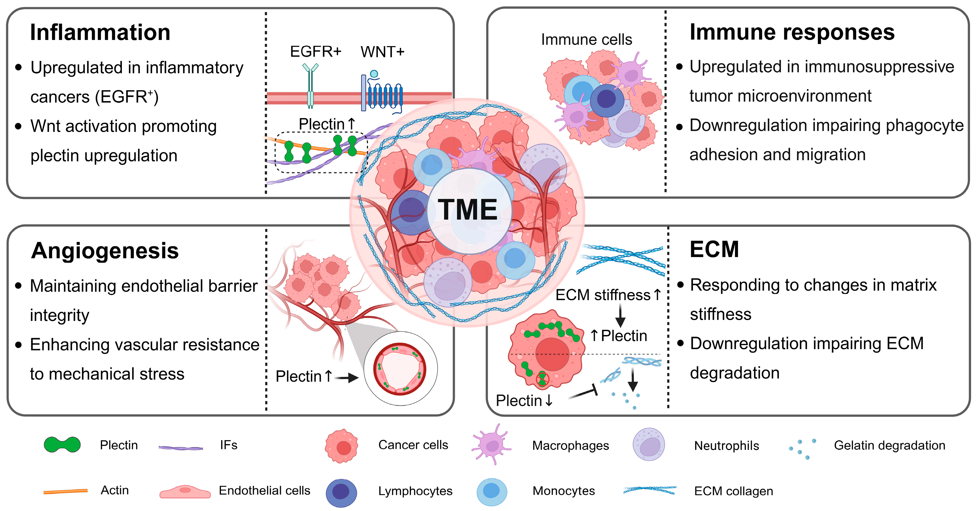
The TME comprises diverse cellular and acellular components that critically influence tumor growth, invasion, metastasis, and cancer therapy. Emerging evidence indicates that dysregulation of plectin plays a role in both the regulation and response to the TME. Specifically, plectin is upregulated in pro-inflammatory tumors and immunosuppressive microenvironments. Its upregulation enhances angiogenesis and vascular integrity, and mediates ECM mechanotransduction and gelatin degradation. These findings highlight the bidirectional role of plectin in the TME, where it drives remodeling of the TME and is dysregulated due to TME alterations (As shown in Figure 2).
Inflammation is a hallmark of cancer, profoundly affecting tumor progression, malignant transformation, and therapeutic efficacy. Epidermal growth factor receptor (EGFR) serves as a key regulator in orchestrating the balance between immunosuppressive and immunostimulatory responses within the TME, particularly in inflammatory breast cancer [93]. Notably, plectin is significantly upregulated in inflammatory breast cancer cells exhibiting EGFR+ expression, where it modulates cell migration through the integrin/EGFR signaling axis [35]. The Wnt signaling pathway is another well-established mediator of inflammatory signaling [94], which also influences plectin expression. In human induced pluripotent stem cell (hiPSC)-derived organoids treated with CHIR99021—a Wnt pathway agonist—plectin levels were elevated relative to controls [95]. When exposed to Aβ42 oligomers, these organoids exhibited a heightened inflammatory response, as indicated by upregulated TNFα, IL-6, and MMP2/9 expression. Insights from hiPSC provide a potential framework for understanding tumor inflammation, although tumor-specific microenvironmental and genetic factors must also be considered.
Plectin dysregulation also contributes to the modulation of the tumor immune microenvironment. Machine learning-based analyses have identified plectin as a biomarker and a potential therapeutic target in immunologically cold pancreatic adenocarcinomas [34]. Higher PLEC gene expression is generally associated with an immunosuppressive tumor microenvironment enriched in M2 macrophages, while in “cold-high” tumors, PLEC gene expression is negatively correlated with M2 abundance, suggesting that it may indirectly inhibit M2 recruitment or function that remains to be elucidated [34]. In Glutathione-s-transferases theta two wild-type mice, plectin expression correlates with the enrichment of M2-type macrophages, suggesting a role in promoting an immunosuppressive microenvironment. Moreover, following bacillus calmette guerin treatment, increased plectin levels were associated with elevated PD-L1 expression in an immune-activated tumor context, further supporting its role in immune modulation [69,96]. Mechanistically, the Integrin β4/plectin signaling axis is regulated by transmembrane protein 268, and its disruption has been shown to impair phagocyte adhesion and migration, thereby influencing immune cell dynamics within the TME [49]. However, whether plectin downregulation directly affects macrophage infiltration remains to be experimentally validated.
Tumor angiogenesis is essential for sustaining tumor growth by providing oxygen and nutrients while facilitating the removal of metabolic waste [97]. The integrin β4/plectin/vimentin complex forms critical transmembrane linkages between vascular endothelial cells and the basement membrane, thereby contributing to endothelial barrier integrity [98]. Studies have shown that endothelial cell layers deficient in plectin exhibit increased permeability and diminished resistance to shear stress and mechanical strain, underscoring its role in vascular stability [99]. Furthermore, the knockdown of Notch2 in vascular smooth muscle cells results in reduced plectin expression during vascular remodeling, further highlighting its importance in maintaining vascular wall architecture [100]. Plectin is also vital for the formation and mechanical adaptation of vascular endothelial cells, particularly within the mechanically dynamic TME. Notably, plectin expression is upregulated in endothelial cells exposed to cyclic mechanical stretch, where it acts as a key mechanosensitive regulator [36]. This upregulation is accompanied by increased expression of vascular development-related genes, including ANGPTL4, ANGPTL5, and PDE1A, suggesting that plectin contributes to cytoskeletal remodeling and mechanotransduction in angiogenic endothelial cells.
The mechanical properties of the ECM critically influence tumor growth and metastasis by shaping the behavior of cancer cells within the TME [101]. Emerging evidence implicates plectin as a pivotal mediator of mechanotransduction—the process by which mechanical signals from the ECM are converted into intracellular responses. Plectin expression is modulated by ECM stiffness and correlates significantly with clinical prognosis in ovarian cancer patients [59]. Recent findings have shown that increased ECM stiffness activates the integrin β1–plectin–F-actin signaling axis, thereby enhancing the migratory capacity of HCC cells [32]. Conversely, silencing plectin impairs matrix-degrading capacity and reduces cytoskeletal remodeling in HCC cells [26]. These observations underscore the essential role of plectin in enabling cancer cells to sense and respond to biomechanical cues from the ECM, ultimately facilitating tumor progression through cytoskeletal stabilization and enhanced invasiveness.

4. Targeting Plectin Dysregulation in Cancer Therapy

Therapeutic strategies aimed at disrupting plectin dysregulation in cancer have demonstrated potential in suppressing tumorigenesis and progression, including gene knockout, RNA interference-mediated knockdown, and functional inhibition.
One particularly promising compound, PST—a ruthenium (arene) pyridinecarbothioamide complex—selectively targets plectin and inhibits its cytoskeletal crosslinking function. CRISPR/Cas9-based gene knockout of the PLEC gene reorganizes keratin networks by disrupting circumferential rims and radial spokes, increasing actin stress fibers, and enhancing actomyosin contractility, leading to elevated tension at adherens junctions and desmosomes [102]. These findings highlight plectin’s essential role in maintaining cytoskeletal integrity and tension homeostasis. Similarly, treatment with 16 μM PST for 4 h in MDCK cells, cholangiocytes, and MCF-7 cells mimics the plectin gene knockout phenotype, inducing keratin bundling, loss of the circumferential keratin rim, and desmosome widening and tortuosity. PST also increases cellular contractility and line tension at adherens junctions and desmosomes, as measured by traction force microscopy and FRET-based tension sensors. Inhibition of actomyosin activity with blebbistatin or Rho kinase inhibitors reverses these structural changes, confirming that PST-induced cytoskeletal remodeling is actomyosin-dependent.
Further studies additionally support PST as a potential novel anticancer agent [103,104,105]. PST inhibits plectin expression and disrupts the formation of the plectin–MT1-MMP complex, thereby inhibiting the formation of invasive pseudopodia and suppressing matrix degradation and cellular invasion [106]. Preclinical studies indicate that PST is generally well tolerated and effectively suppresses HCC progression in both cellular and murine models, with treatment typically applied at 8 μM (corresponding to 25% of the IC50 in Huh7 cells) and incubation times adjusted according to specific experimental assays [26]. These results provide mechanistic evidence that targeted inhibition of plectin disrupts cytoskeletal networks and contractility pathways, suppressing tumorigenic phenotypes and highlighting its therapeutic potential.
In addition to targeting plectin at the protein level, therapeutic strategies directed at its mRNA also represent a promising avenue. mRNA-based interventions have demonstrated efficacy in modulating plectin expression. For instance, miR-124-3p directly targets the 3′-UTR of PLEC mRNA, leading to its downregulation and thereby reversing drug resistance-associated EMT and invasive phenotypes in cancer cells [91]. Likewise, the virus-encoded artificial microRNA amiR-4 reduces plectin expression and enhances the cytotoxicity of cancer cells, highlighting the potential of RNA-based therapeutics in oncology [107].
However, most cytoskeleton-targeting chemotherapeutic agents act on actin or microtubules, leading to significant side effects due to the ubiquitous presence of these structural proteins in normal cells. In contrast, CSP is characterized by aberrant overexpression and mislocalization to the cell surface, providing a more selective and tumor-restricted therapeutic target. Monoclonal antibodies developed against CSP have demonstrated potent anticancer activity in preclinical studies [25]. Among these, ZB131 (developed by ZielBio) is currently undergoing phase 1–2 clinical trials and has shown good tolerability in adult patients with PDAC, cholangiocarcinoma, and ovarian cancer [108]. In addition, surface-targeting peptides that bind to plectin, such as a peptide derived from Cyclin D2 (Pep5) [40] and a highly CSC-specific hit peptoid (PSC2) [52], exhibit high binding specificity and have been shown to inhibit tumor growth or induce cancer cell death. Although growing evidence supports plectin as a promising therapeutic target, these inhibitors still lack sufficient clinical trial validation to confirm their efficacy and safety in clinical applications.

5. Conclusions and Perspectives

Plectin dysregulation is observed in many cancer types and is primarily characterized by abnormal expression and mislocalization of plectin. Its dysregulation can promote tumor growth and metastasis and contribute to the establishment of a tumor-supportive microenvironment. Therefore, inhibitors targeting plectin dysregulation have potential in cancer therapy. Although we have valuable clues that plectin dysfunction can promote cancer development, many areas still need attention regarding its dysregulation and effects.
Plectin dysregulation exhibits tumor heterogeneity, with both upregulation and downregulation observed even within the same cancer type, reflecting its dual roles in tumor progression. Its upregulation promotes proliferation, invasion, and metastasis by stabilizing cytoskeletal architecture and activating oncogenic signaling pathways. Meanwhile, downregulation of plectin can enhance motility and dissemination by weakening intercellular adhesion and cytoskeletal integrity, as observed in HCC and high-grade ovarian tumors [66]. Plectin expression is also modulated by TME-derived cues, which in turn remodel the TME and foster tumor niches. Although plectin upregulation has been validated in pancreatic, ovarian, and liver cancers with ample clinical samples, clinical evidence in other tumor types remains limited, and the distinct implications of up- versus downregulation are not yet fully understood. Recognizing plectin dysregulation is critical for clinical practice, as it may refine prognostic stratification and guide the development of personalized therapeutic strategies targeting plectin-associated pathways.
This heterogeneity may in part result from isoform-specific functions of plectin, which have not yet been fully elucidated. Alternative splicing of the PLEC gene generates isoforms with distinct N-terminal sequences, conferring precise control over cytoskeletal architecture, organelle positioning, and tissue-specific functions [14]. In cancer cells, certain isoforms are aberrantly localized to the cell membrane and appear to drive tumor-promoting processes through isoform-specific mechanisms. These isoform-specific functions may partly explain the tumor heterogeneity observed in plectin dysregulation. However, the widespread use of pan-plectin antibodies has limited the ability to dissect these functional differences, resulting in the underappreciation of isoform-specific contributions in cancer research. The development and application of isoform-specific antibodies are therefore crucial for delineating the divergent roles of plectin isoforms in tumor initiation and progression.
In addition, more comprehensive and cost-efficient methods for detecting plectin dysregulation are needed to provide robust evidence. Current approaches for assessing plectin expression include protein- and mRNA-level assays, transcriptomic analyses, and immunofluorescence and immunohistochemical staining of clinical samples. Its subcellular localization has also been investigated through methods such as immunohistochemistry, phage display, unbiased peptoid screening, and immunogold staining [23,27]. These strategies have already underscored the potential of plectin to be a possible biomarker in diverse cancers—for instance, its upregulation in melanoma metastasis, enrichment in the immune-cold microenvironment of pancreatic cancer [34], aberrant membrane localization as a marker of invasion [24], and involvement in stemness maintenance [27]. Its dysregulation has also been linked to poor prognosis in multiple cancers, underscoring its clinical relevance. Developing practical and clinically applicable detection platforms will facilitate broader screening and reinforce plectin’s promise as both a diagnostic and therapeutic target.
Notably, CSP exhibits aberrant localization that contributes to tumor stemness and metastasis. CSP has been identified as a surface marker for lung cancer stem cells, and its downregulation reduces the expression of stemness-associated genes, implying a potential role for plectin in maintaining stemness [27]. Plectin undergoes abnormal membrane localization through the exosome pathway in PDAC and affects tumor invasion and metastasis [24]. Moreover, CSP’s selective membrane expression in cancer cells offers a promising strategy for targeted drug delivery, enabling precise therapeutic delivery to tumor sites [23,109]. These findings underscore the protumorigenic functions of CSP that extend beyond its utility as a diagnostic biomarker. Building on advances in understanding plectin/CSP biology, the development of plectin/CSP-targeting therapies offers a promising avenue for improving cancer diagnosis, treatment, and prognosis.

Author Contributions

W.W. conducted the literature review and drafted the manuscript. C.L., Z.W., X.Z. and Q.L. assisted with literature collection and review. G.S. contributed to the manuscript design and revision. All authors have read and agreed to the published version of the manuscript.

Funding

This work was supported by the Key Project of the National Natural Science Foundation of China (No. 11832008).

Institutional Review Board Statement

Not applicable.

Informed Consent Statement

Not applicable.

Data Availability Statement

No new data were created or analyzed in this study.

Conflicts of Interest

The authors declare no conflicts of interest.

Abbreviations

The abbreviations listed below are used in this manuscript:
AFsActin filaments
ABDActin-binding domain
AMLAcute myeloid leukemia
ALDHAldehyde dehydrogenase
BCCBasal cell carcinomas
CSCsCancer stem cells
CSPCancer-specific plectin
CRCColorectal cancer
ECMExtracellular matrix
EGFREpidermal growth factor receptor
EMTEpithelial–mesenchymal transition
Erk 1/2Extracellular signal-regulated kinase 1/2
ESCCEsophageal squamous cell carcinoma
FAsFocal adhesions
FAKFocal adhesion kinase
GBMGlioblastoma multiforme
HNSCCHead and neck squamous cell carcinoma
HDsHemidesmosomes
HCCHepatocellular carcinoma
IFsIntermediate filaments
OSCCOral squamous cell carcinoma
PDACPancreatic ductal adenocarcinoma
PanINsPancreatic intraepithelial neoplasms
PSTPlecstatin-1
SCCSquamous cell carcinomas
SrcRous sarcoma
SSEStratified squamous epithelium
Rac1Ras-related C3 botulinum toxin substrate 1
TMETumor microenvironment

References

  1. Bausch, D.; Thomas, S.; Mino-Kenudson, M.; Fernández-del, C.C.; Bauer, T.W.; Williams, M.; Warshaw, A.L.; Thayer, S.P.; Kelly, K.A. Plectin-1 as a Novel Biomarker for Pancreatic Cancer. Clin. Cancer Res. 2011, 17, 302–309. [Google Scholar] [CrossRef]
  2. Zhu, Q.; Zeng, S.; Yang, J.; Zhuo, J.; Wang, P.; Wen, S.; Fang, C. Plectin-1-Targeted Recognition for Enhancing Comprehensive Therapy in Pancreatic Ductal Adenocarcinoma. Nanoscale 2024, 16, 18584–18596. [Google Scholar] [CrossRef] [PubMed]
  3. Proby, C.; Fujii, Y.; Owaribe, K.; Nishikawa, T.; Amagai, M. Human Autoantibodies against HD1/Plectin in Paraneoplastic Pemphigus. J. Investig. Dermatol. 1999, 112, 153–156. [Google Scholar] [CrossRef]
  4. Schaapveld, R.Q.J.; Borradori, L.; Geerts, D.; van Leusden, M.R.; Kuikman, I.; Nievers, M.G.; Niessen, C.M.; Steenbergen, R.D.M.; Snijders, P.J.F.; Sonnenberg, A. Hemidesmosome Formation Is Initiated by the B4 Integrin Subunit, Requires Complex Formation of B4 and HD1/Plectin, and Involves a Direct Interaction between B4 and the Bullous Pemphigoid Antigen 180. J. Cell Biol. 1998, 142, 271–284. [Google Scholar] [CrossRef]
  5. Outla, Z.; Prechova, M.; Korelova, K.; Gemperle, J.; Gregor, M. Mechanics of Cell Sheets: Plectin as an Integrator of Cytoskeletal Networks. Open Biol. 2025, 15, 240208. [Google Scholar] [CrossRef]
  6. Wiche, G.; Winter, L. Plectin Isoforms as Organizers of Intermediate Filament Cytoarchitecture. Bioarchitecture 2011, 1, 14–20. [Google Scholar] [CrossRef] [PubMed]
  7. Andrä, K.; Kornacker, I.; Jörgl, A.; Zörer, M.; Spazierer, D.; Fuchs, P.; Fischer, I.; Wiche, G. Plectin-Isoform-Specific Rescue of Hemidesmosomal Defects in Plectin (–/–) Keratinocytes. J. Investig. Dermatol. 2003, 120, 189–197. [Google Scholar] [CrossRef]
  8. Chaudhari, P.R.; Charles, S.E.; D’Souza, Z.C.; Vaidya, M.M. Hemidesmosomal Linker Proteins Regulate Cell Motility, Invasion and Tumorigenicity in Oral Squamous Cell Carcinoma Derived Cells. Exp. Cell Res. 2017, 360, 125–137. [Google Scholar] [CrossRef]
  9. Eger, A.; Stockinger, A.; Wiche, G.; Foisner, R. Polarisation-Dependent Association of Plectin with Desmoplakin and the Lateral Submembrane Skeleton in MDCK Cells. J. Cell Sci. 1997, 110, 1307–1316. [Google Scholar] [CrossRef]
  10. Pieperhoff, S.; Rickelt, S.; Heid, H.; Claycomb, W.C.; Zimbelmann, R.; Kuhn, C.; Winter-Simanowski, S.; Kuhn, C.; Frey, N.; Franke, W.W. The Plaque Protein Myozap Identified as a Novel Major Component of Adhering Junctions in Endothelia of the Blood and the Lymph Vascular Systems. J. Cell. Mol. Med. 2012, 16, 1709–1719. [Google Scholar] [CrossRef] [PubMed]
  11. Gregor, M.; Osmanagic-Myers, S.; Burgstaller, G.; Wolfram, M.; Fischer, I.; Walko, G.; Resch, G.P.; Jörgl, A.; Herrmann, H.; Wiche, G. Mechanosensing through Focal Adhesion-anchored Intermediate Filaments. FASEB J. 2014, 28, 715–729. [Google Scholar] [CrossRef]
  12. Liu, C.G.; Maercker, C.; Castañon, M.J.; Hauptmann, R.; Wiche, G. Human Plectin: Organization of the Gene, Sequence Analysis, and Chromosome Localization (8q24). Proc. Natl. Acad. Sci. USA 1996, 93, 4278–4283. [Google Scholar] [CrossRef]
  13. Perez, S.M.; Brinton, L.T.; Kelly, K.A. Plectin in Cancer: From Biomarker to Therapeutic Target. Cells 2021, 10, 2246. [Google Scholar] [CrossRef]
  14. Rezniczek, G.A.; Abrahamsberg, C.; Fuchs, P.; Spazierer, D.; Wiche, G. Plectin 5’-Transcript Diversity: Short Alternative Sequences Determine Stability of Gene Products, Initiation of Translation and Subcellular Localization of Isoforms. Hum. Mol. Genet. 2003, 12, 3181–3194. [Google Scholar] [CrossRef]
  15. Staszewska, I.; Fischer, I.; Wiche, G. Plectin Isoform 1-Dependent Nuclear Docking of Desmin Networks Affects Myonuclear Architecture and Expression of Mechanotransducers. Hum. Mol. Genet. 2015, 24, 7373–7389. [Google Scholar] [CrossRef]
  16. Winter, L.; Kuznetsov, A.V.; Grimm, M.; Zeöld, A.; Fischer, I.; Wiche, G. Plectin Isoform P1b and P1d Deficiencies Differentially Affect Mitochondrial Morphology and Function in Skeletal Muscle. Hum. Mol. Genet. 2015, 24, 4530–4544. [Google Scholar] [CrossRef]
  17. Konieczny, P.; Fuchs, P.; Reipert, S.; Kunz, W.S.; Zeöld, A.; Fischer, I.; Paulin, D.; Schröder, R.; Wiche, G. Myofiber Integrity Depends on Desmin Network Targeting to Z-Disks and Costameres via Distinct Plectin Isoforms. J. Cell Biol. 2008, 181, 667–681. [Google Scholar] [CrossRef]
  18. McInroy, L.; Määttä, A. Plectin Regulates Invasiveness of SW480 Colon Carcinoma Cells and Is Targeted to Podosome-like Adhesions in an Isoform-Specific Manner. Exp. Cell Res. 2011, 317, 2468–2478. [Google Scholar] [CrossRef] [PubMed]
  19. Castañón, M.J.; Walko, G.; Winter, L.; Wiche, G. Plectin–Intermediate Filament Partnership in Skin, Skeletal Muscle, and Peripheral Nerve. Histochem. Cell Biol. 2013, 140, 33–53. [Google Scholar] [CrossRef] [PubMed]
  20. Wiche, G. Role of Plectin in Cytoskeleton Organization and Dynamics. J. Cell Sci. 1998, 111, 2477–2486. [Google Scholar] [CrossRef] [PubMed]
  21. Winter, L.; Wiche, G. The Many Faces of Plectin and Plectinopathies: Pathology and Mechanisms. Acta Neuropathol. 2013, 125, 77–93. [Google Scholar] [CrossRef]
  22. Natsuga, K. Plectin-Related Skin Diseases. J. Dermatol. Sci. 2015, 77, 139–145. [Google Scholar] [CrossRef]
  23. Kelly, K.A.; Bardeesy, N.; Anbazhagan, R.; Gurumurthy, S.; Berger, J.; Alencar, H.; DePinho, R.A.; Mahmood, U.; Weissleder, R. Targeted Nanoparticles for Imaging Incipient Pancreatic Ductal Adenocarcinoma. PLoS Med. 2008, 5, e85. [Google Scholar] [CrossRef] [PubMed]
  24. Shin, S.J.; Smith, J.A.; Rezniczek, G.A.; Pan, S.; Chen, R.; Brentnall, T.A.; Wiche, G.; Kelly, K.A. Unexpected Gain of Function for the Scaffolding Protein Plectin Due to Mislocalization in Pancreatic Cancer. Proc. Natl. Acad. Sci. USA 2013, 110, 19414–19419. [Google Scholar] [CrossRef] [PubMed]
  25. Perez, S.M.; Dimastromatteo, J.; Landen, C.N.; Kelly, K.A. A Novel Monoclonal Antibody Targeting Cancer-Specific Plectin Has Potent Antitumor Activity in Ovarian Cancer. Cells 2021, 10, 2218. [Google Scholar] [CrossRef]
  26. Outla, Z.; Oyman-Eyrilmez, G.; Korelova, K.; Prechova, M.; Frick, L.; Sarnova, L.; Bisht, P.; Novotna, P.; Kosla, J.; Bortel, P.; et al. Plectin-Mediated Cytoskeletal Crosstalk as a Target for Inhibition of Hepatocellular Carcinoma Growth and Metastasis. eLife 2025, 13, RP102205. [Google Scholar] [CrossRef]
  27. Raymond, A.C.; Gao, B.; Girard, L.; Minna, J.D.; Gomika Udugamasooriya, D. Unbiased Peptoid Combinatorial Cell Screen Identifies Plectin Protein as a Potential Biomarker for Lung Cancer Stem Cells. Sci. Rep. 2019, 9, 14954. [Google Scholar] [CrossRef]
  28. Rikardsen, O.G.; Magnussen, S.N.; Svineng, G.; Hadler-Olsen, E.; Uhlin-Hansen, L.; Steigen, S.E. Plectin as a Prognostic Marker in Non-Metastatic Oral Squamous Cell Carcinoma. BMC Oral Health 2015, 15, 98. [Google Scholar] [CrossRef] [PubMed]
  29. Mizuta, K.; Matsubara, T.; Goto, A.; Addison, W.N.; Nakatomi, M.; Matsuo, K.; Tada-Shigeyama, Y.; Yaginuma, T.; Honda, H.; Yoshioka, I.; et al. Plectin Promotes Tumor Formation by B16 Mouse Melanoma Cells via Regulation of Rous Sarcoma Oncogene Activity. BMC Cancer 2022, 22, 936. [Google Scholar] [CrossRef]
  30. Buckup, M.; Rice, M.A.; Hsu, E.-C.; Garcia-Marques, F.; Liu, S.; Aslan, M.; Bermudez, A.; Huang, J.; Pitteri, S.J.; Stoyanova, T. Plectin Is a Regulator of Prostate Cancer Growth and Metastasis. Oncogene 2021, 40, 663–676. [Google Scholar] [CrossRef]
  31. Xu, R.; He, S.; Ma, D.; Liang, R.; Luo, Q.; Song, G. Plectin Downregulation Inhibits Migration and Suppresses Epithelial Mesenchymal Transformation of Hepatocellular Carcinoma Cells via ERK1/2 Signaling. Int. J. Mol. Sci. 2022, 24, 73. [Google Scholar] [CrossRef]
  32. Wang, Z.; Wang, W.; Luo, Q.; Song, G. High Matrix Stiffness Accelerates Migration of Hepatocellular Carcinoma Cells through the Integrin Β1-Plectin-F-Actin Axis. BMC Biol. 2025, 23, 8. [Google Scholar] [CrossRef]
  33. Yuan, Y.; Tan, L.; Wang, L.; Zou, D.; Liu, J.; Lu, X.; Fu, D.; Wang, G.; Wang, L.; Wang, Z. The Expression Pattern of Hypoxia-Related Genes Predicts the Prognosis and Mediates Drug Resistance in Colorectal Cancer. Front. Cell Dev. Biol. 2022, 10, 814621. [Google Scholar] [CrossRef]
  34. Ge, J.; Ge, J.; Tang, G.; Xiong, D.; Zhu, D.; Ding, X.; Zhou, X.; Sang, M. Machine Learning-Based Identification of Biomarkers and Drugs in Immunologically Cold and Hot Pancreatic Adenocarcinomas. J. Transl. Med. 2024, 22, 775. [Google Scholar] [CrossRef] [PubMed]
  35. Zhang, E.Y.; Cristofanilli, M.; Robertson, F.; Reuben, J.M.; Mu, Z.; Beavis, R.C.; Im, H.; Snyder, M.; Hofree, M.; Ideker, T.; et al. Genome Wide Proteomics of ERBB2 and EGFR and Other Oncogenic Pathways in Inflammatory Breast Cancer. J. Proteome Res. 2013, 12, 2805–2817. [Google Scholar] [CrossRef]
  36. Ferrari, D.; Sengupta, A.; Heo, L.; Pethö, L.; Michler, J.; Geiser, T.; De Jesus Perez, V.A.; Kuebler, W.M.; Zeinali, S.; Guenat, O.T. Effects of Biomechanical and Biochemical Stimuli on Angio- and Vasculogenesis in a Complex Microvasculature-on-Chip. iScience 2023, 26, 106198. [Google Scholar] [CrossRef]
  37. Von Der Heide, E.K.; Neumann, M.; Vosberg, S.; James, A.R.; Schroeder, M.P.; Ortiz-Tanchez, J.; Isaakidis, K.; Schlee, C.; Luther, M.; Jöhrens, K.; et al. Molecular Alterations in Bone Marrow Mesenchymal Stromal Cells Derived from Acute Myeloid Leukemia Patients. Leukemia 2017, 31, 1069–1078. [Google Scholar] [CrossRef]
  38. Ni, K.-D.; Fu, X.; Luo, Y.; He, X.; Yin, H.-H.; Mo, D.-P.; Wu, J.-X.; Wu, M.-J.; Zheng, X.; Liu, Y.-N.; et al. Epoxy Metabolites of Linoleic Acid Promote the Development of Breast Cancer via Orchestrating PLEC/NFκB1/CXCL9-Mediated Tumor Growth and Metastasis. Cell Death Dis. 2024, 15, 901. [Google Scholar] [CrossRef] [PubMed]
  39. Rao, M.M.V.; Likith, M.; Kavya, R.; Hariprasad, T.P.N. Plectin as a Putative Novel Biomarker for Breast Cancer: An in Silico Study. Netw. Model. Anal. Health Inform. Bioinforma. 2022, 11, 49. [Google Scholar] [CrossRef]
  40. Russo, L.C.; Araujo, C.B.; Iwai, L.K.; Ferro, E.S.; Forti, F.L. A Cyclin D2-Derived Peptide Acts on Specific Cell Cycle Phases by Activating ERK1/2 to Cause the Death of Breast Cancer Cells. J. Proteom. 2017, 151, 24–32. [Google Scholar] [CrossRef]
  41. Stegh, A.H.; Herrmann, H.; Lampel, S.; Weisenberger, D.; Andrä, K.; Seper, M.; Wiche, G.; Krammer, P.H.; Peter, M.E. Identification of the Cytolinker Plectin as a Major Early In Vivo Substrate for Caspase 8 during CD95- and Tumor Necrosis Factor Receptor-Mediated Apoptosis. Mol. Cell. Biol. 2000, 20, 5665–5679. [Google Scholar] [CrossRef]
  42. Fish, L.; Khoroshkin, M.; Navickas, A.; Garcia, K.; Culbertson, B.; Hänisch, B.; Zhang, S.; Nguyen, H.C.B.; Soto, L.M.; Dermit, M.; et al. A Prometastatic Splicing Program Regulated by SNRPA1 Interactions with Structured RNA Elements. Science 2021, 372, eabc7531. [Google Scholar] [CrossRef]
  43. Harryman, W.L.; Pond, E.; Singh, P.; Little, A.S.; Eschbacher, J.M.; Cress, A.E. Laminin-Binding Integrin Gene Copy Number Alterations in Distinct Epithelial-Type Cancers. Am. J. Transl. Res. 2016, 8, 940–954. [Google Scholar] [PubMed]
  44. Sutoh Yoneyama, M.; Hatakeyama, S.; Habuchi, T.; Inoue, T.; Nakamura, T.; Funyu, T.; Wiche, G.; Ohyama, C.; Tsuboi, S. Vimentin Intermediate Filament and Plectin Provide a Scaffold for Invadopodia, Facilitating Cancer Cell Invasion and Extravasation for Metastasis. Eur. J. Cell Biol. 2014, 93, 157–169. [Google Scholar] [CrossRef]
  45. Qiao, L.; Hu, Y.; Xu, L.; Deng, G.; Yu, X.; Zheng, W.; Yang, Y.; Yuan, H.; Jiang, W.; Yu, X. Unraveling the Oncogenic Characteristics of the Cytolinker, Plectin, in Esophageal Squamous Cell Carcinoma. Cell. Mol. Gastroenterol. Hepatol. 2025, 19, 101549. [Google Scholar] [CrossRef] [PubMed]
  46. Hu, L.; Wu, Y.; Guan, X.; Liang, Y.; Yao, X.; Tan, D.; Bai, Y.; Xiong, G.; Yang, K. Germline Copy Number Loss of UGT2B28 and Gain of PLEC Contribute to Increased Human Esophageal Squamous Cell Carcinoma Risk in Southwest China. Am. J. Cancer Res. 2015, 5, 3056–3071. [Google Scholar] [PubMed]
  47. Pawar, H.; Kashyap, M.K.; Sahasrabuddhe, N.A.; Renuse, S.; Harsha, H.C.; Kumar, P.; Sharma, J.; Kandasamy, K.; Marimuthu, A.; Nair, B.; et al. Quantitative Tissue Proteomics of Esophageal Squamous Cell Carcinoma for Novel Biomarker Discovery. Cancer Biol. Ther. 2011, 12, 510–522. [Google Scholar] [CrossRef]
  48. Žugec, M.; Furlani, B.; Castañon, M.J.; Rituper, B.; Fischer, I.; Broggi, G.; Caltabiano, R.; Barbagallo, G.M.V.; Di Rosa, M.; Tibullo, D.; et al. Plectin Plays a Role in the Migration and Volume Regulation of Astrocytes: A Potential Biomarker of Glioblastoma. J. Biomed. Sci. 2024, 31, 14. [Google Scholar] [CrossRef]
  49. Hong, D.; Zhang, X.; Li, R.; Yu, J.; Lou, Y.; He, Q.; Li, X.; Xu, D.; Lv, P.; Lin, J.; et al. Deletion of TMEM268 Inhibits Growth of Gastric Cancer Cells by Downregulating the ITGB4 Signaling Pathway. Cell Death Differ. 2019, 26, 1453–1466. [Google Scholar] [CrossRef] [PubMed]
  50. Katada, K.; Tomonaga, T.; Satoh, M.; Matsushita, K.; Tonoike, Y.; Kodera, Y.; Hanazawa, T.; Nomura, F.; Okamoto, Y. Plectin Promotes Migration and Invasion of Cancer Cells and Is a Novel Prognostic Marker for Head and Neck Squamous Cell Carcinoma. J. Proteom. 2012, 75, 1803–1815. [Google Scholar] [CrossRef] [PubMed]
  51. Wang, C.-I.; Wang, C.-L.; Wu, Y.-C.; Feng, H.-P.; Liu, P.-J.; Chang, Y.-S.; Yu, J.-S.; Yu, C.-J. Quantitative Proteomics Reveals a Novel Role of Karyopherin Alpha 2 in Cell Migration through the Regulation of Vimentin–pErk Protein Complex Levels in Lung Cancer. J. Proteome Res. 2015, 14, 1739–1751. [Google Scholar] [CrossRef]
  52. Shukla, S.P.; Raymond, A.; Rustagi, V.; Kedika, S.R.; Tran, O.; Wang, L.; Guo, B.; Udugamasooriya, D.G. A Novel Peptidomimetic Therapeutic for Selective Suppression of Lung Cancer Stem Cells over Non-Stem Cancer Cells. Bioorg. Chem. 2021, 116, 105340. [Google Scholar] [CrossRef] [PubMed]
  53. Koroknai, V.; Ecsedi, S.; Vízkeleti, L.; Kiss, T.; Szász, I.; Lukács, A.; Papp, O.; Ádány, R.; Balázs, M. Genomic Profiling of Invasive Melanoma Cell Lines by Array Comparative Genomic Hybridization. Melanoma Res. 2016, 26, 100–107. [Google Scholar] [CrossRef] [PubMed]
  54. Hood, B.L.; Grahovac, J.; Flint, M.S.; Sun, M.; Charro, N.; Becker, D.; Wells, A.; Conrads, T.P. Proteomic Analysis of Laser Microdissected Melanoma Cells from Skin Organ Cultures. J. Proteome Res. 2010, 9, 3656–3663. [Google Scholar] [CrossRef] [PubMed]
  55. Militaru, I.V.; Rus, A.A.; Munteanu, C.V.A.; Manica, G.; Petrescu, S.M. New Panel of Biomarkers to Discriminate between Amelanotic and Melanotic Metastatic Melanoma. Front. Oncol. 2023, 12, 1061832. [Google Scholar] [CrossRef]
  56. Wesley, T.; Escalona, R.M.; Kannourakis, G.; Ahmed, N. Plakin Expression in Serous Epithelial Ovarian Cancer Has the Potential to Impede Metastatic Spread and Epithelial–Mesenchymal Transition: A Comparative Expression Analysis of Immunohistochemical and In Silico Datasets. Cancers 2024, 16, 4087. [Google Scholar] [CrossRef] [PubMed]
  57. Bai, L.; Qian, X.; Zhang, H.; Yuan, Y.; Cui, X.; Cheng, M.; Han, Y. Plectin, a Novel Regulator in Migration, Invasion and Adhesion of Ovarian Cancer. Cell Biosci. 2025, 15, 15. [Google Scholar] [CrossRef]
  58. Wang, Z.; Meng, F.; Zhong, Z. Emerging Targeted Drug Delivery Strategies toward Ovarian Cancer. Adv. Drug Deliv. Rev. 2021, 178, 113969. [Google Scholar] [CrossRef]
  59. Yang, X.; Wang, G.; Huang, X.; Cheng, M.; Han, Y. RNA-Seq Reveals the Diverse Effects of Substrate Stiffness on Epidermal Ovarian Cancer Cells. Aging 2020, 12, 20493–20511. [Google Scholar] [CrossRef]
  60. Bausch, D.; Mino-Kenudson, M.; Castillo, C.F.; Warshaw, A.L.; Kelly, K.A.; Thayer, S.P. Plectin-1 Is a Biomarker of Malignant Pancreatic Intraductal Papillary Mucinous Neoplasms. J. Gastrointest. Surg. Off. J. Soc. Surg. Aliment. Tract. 2009, 13, 1948–1954. [Google Scholar] [CrossRef]
  61. Wenta, T.; Schmidt, A.; Zhang, Q.; Devarajan, R.; Singh, P.; Yang, X.; Ahtikoski, A.; Vaarala, M.; Wei, G.-H.; Manninen, A. Disassembly of α6β4-Mediated Hemidesmosomal Adhesions Promotes Tumorigenesis in PTEN-Negative Prostate Cancer by Targeting Plectin to Focal Adhesions. Oncogene 2022, 41, 3804–3820. [Google Scholar] [CrossRef] [PubMed]
  62. Xiang, C.; Li, Y.; Wang, W.; Tao, H.; Liang, N.; Wu, S.; Yu, T.; Cui, X.; Xie, Y.; Zuo, H.; et al. Joint Analysis of WES and RNA-SEQ Identify Signature Genes Related to Metastasis in Prostate Cancer. J. Cell. Mol. Med. 2023, 27, 1947–1958. [Google Scholar] [CrossRef]
  63. Paumard-Hernández, B.; Calvete, O.; Inglada Pérez, L.; Tejero, H.; Al-Shahrour, F.; Pita, G.; Barroso, A.; Carlos Triviño, J.; Urioste, M.; Valverde, C.; et al. Whole Exome Sequencing Identifies PLEC, EXO5 and DNAH7 as Novel Susceptibility Genes in Testicular Cancer. Int. J. Cancer 2018, 143, 1954–1962. [Google Scholar] [CrossRef]
  64. Dumas, V.; Kanitakis, J.; Charvat, S.; Euvrard, S.; Faure, M.; Claudy, A. Expression of Basement Membrane Antigens and Matrix Metalloproteinases 2 and 9 in Cutaneous Basal and Squamous Cell Carcinomas. Anticancer Res. 1999, 19, 2929–2938. [Google Scholar]
  65. Cheng, C.-C.; Lai, Y.-C.C.; Lai, Y.-S.; Hsu, Y.-H.; Chao, W.-T.; Sia, K.-C.; Tseng, Y.-H.; Liu, Y.-H. Transient Knockdown-Mediated Deficiency in Plectin Alters Hepatocellular Motility in Association with Activated FAK and Rac1-GTPase. Cancer Cell Int. 2015, 15, 29. [Google Scholar] [CrossRef]
  66. Lai, Y.-S.; Cheng, C.-C.; Lee, M.-T.; Chao, W.-T.; Lai, Y.-C.C.; Hsu, Y.-H.; Liu, Y.-H. The Prognostic Value of Cytokeratin and Sal-Like Protein 4 Expression in Hepatocellular Carcinoma and Intra-Hepatic Cholangiocarcinoma in Taiwan. Int. J. Med. Sci. 2018, 15, 1746–1756. [Google Scholar] [CrossRef] [PubMed]
  67. Cheng, C.-C.; Chao, W.-T.; Liao, C.-C.; Tseng, Y.-H.; Lai, Y.-C.C.; Lai, Y.-S.; Hsu, Y.-H.; Liu, Y.-H. Plectin Deficiency in Liver Cancer Cells Promotes Cell Migration and Sensitivity to Sorafenib Treatment. Cell Adhes. Migr. 2018, 12, 19–27. [Google Scholar] [CrossRef]
  68. Dong, Q.; Liu, M.; Chen, B.; Zhao, Z.; Chen, T.; Wang, C.; Zhuang, S.; Li, Y.; Wang, Y.; Ai, L.; et al. Revealing Biomarkers Associated with PARP Inhibitors Based on Genetic Interactions in Cancer Genome. Comput. Struct. Biotechnol. J. 2021, 19, 4435–4446. [Google Scholar] [CrossRef]
  69. Patwardhan, M.V.; Kane, T.Q.; Chiong, E.; Rahmat, J.N.; Mahendran, R. Loss of Glutathione-S-Transferase Theta 2 (GSTT2) Modulates the Tumor Microenvironment and Response to BCG Immunotherapy in a Murine Orthotopic Model of Bladder Cancer. Int. J. Mol. Sci. 2024, 25, 13296. [Google Scholar] [CrossRef]
  70. Cheng, C.-C.; Liu, Y.-H.; Ho, C.-C.; Chao, W.-T.; Pei, R.-J.; Hsu, Y.-H.; Yeh, K.-T.; Ho, L.-C.; Tsai, M.-C.; Lai, Y.-S. The Influence of Plectin Deficiency on Stability of Cytokeratin18 in Hepatocellular Carcinoma. J. Mol. Histol. 2008, 39, 209–216. [Google Scholar] [CrossRef] [PubMed]
  71. Liu, Y.-H.; Ho, C.-C.; Cheng, C.-C.; Chao, W.-T.; Pei, R.-J.; Hsu, Y.-H.; Lai, Y.-S. Cytokeratin 18-Mediated Disorganization of Intermediate Filaments Is Induced by Degradation of Plectin in Human Liver Cells. Biochem. Biophys. Res. Commun. 2011, 407, 575–580. [Google Scholar] [CrossRef]
  72. Di, J.; Zhao, Z.; Xia, M.; Gao, K.; Chai, K.; Zhu, B.; Sun, W.; Zhang, Y.; Zheng, J.; Liu, Y. Rap2B Drives Tumorigenesis and Progression of Colorectal Cancer through Intestinal Cytoskeleton Remodeling. Cell Death Dis. 2025, 16, 290. [Google Scholar] [CrossRef]
  73. Chen, Y.; Gibson, S.B. Three Dimensions of Autophagy in Regulating Tumor Growth: Cell Survival/Death, Cell Proliferation, and Tumor Dormancy. Biochim. Biophys. Acta Mol. Basis Dis. 2021, 1867, 166265. [Google Scholar] [CrossRef]
  74. Marino, N.; German, R.; Podicheti, R.; Rockey, P.; Sandusky, G.E.; Temm, C.J.; Nakshatri, H.; Addison, R.J.; Selman, B.; Althouse, S.K.; et al. FAM83A Is a Potential Biomarker for Breast Cancer Initiation. Biomark. Res. 2022, 10, 8. [Google Scholar] [CrossRef] [PubMed]
  75. Aho, S. Plakin Proteins Are Coordinately Cleaved during Apoptosis but Preferentially through the Action of Different Caspases. Exp. Dermatol. 2004, 13, 700–707. [Google Scholar] [CrossRef]
  76. Scaltriti, M.; Santamaria, A.; Paciucci, R.; Bettuzzi, S. Intracellular Clusterin Induces G2-M Phase Arrest and Cell Death in prostate cancer-3 Prostate Cancer Cells1. Cancer Res. 2004, 64, 6174–6182. [Google Scholar] [CrossRef]
  77. Gerstberger, S.; Jiang, Q.; Ganesh, K. Metastasis. Cell 2023, 186, 1564–1579. [Google Scholar] [CrossRef]
  78. Wang, Y.; Cheng, T.; Lu, M.; Mu, Y.; Li, B.; Li, X.; Zhan, X. TMT-Based Quantitative Proteomics Revealed Follicle-Stimulating Hormone (FSH)-Related Molecular Characterizations for Potentially Prognostic Assessment and Personalized Treatment of FSH-Positive Non-Functional Pituitary Adenomas. EPMA J. 2019, 10, 395–414. [Google Scholar] [CrossRef] [PubMed]
  79. Strouhalova, K.; Přechová, M.; Gandalovičová, A.; Brábek, J.; Gregor, M.; Rosel, D. Vimentin Intermediate Filaments as Potential Target for Cancer Treatment. Cancers 2020, 12, 184. [Google Scholar] [CrossRef] [PubMed]
  80. Borradori, L.; Sonnenberg, A. Structure and Function of Hemidesmosomes: More Than Simple Adhesion Complexes. J. Investig. Dermatol. 1999, 112, 411–418. [Google Scholar] [CrossRef]
  81. Koivusalo, S.; Schmidt, A.; Manninen, A.; Wenta, T. Regulation of Kinase Signaling Pathways by α6β4-Integrins and Plectin in Prostate Cancer. Cancers 2023, 15, 149. [Google Scholar] [CrossRef] [PubMed]
  82. Litjens, S.H.M.; de Pereda, J.M.; Sonnenberg, A. Current Insights into the Formation and Breakdown of Hemidesmosomes. Trends Cell Biol. 2006, 16, 376–383. [Google Scholar] [CrossRef] [PubMed]
  83. Dmello, C.; Sawant, S.; Alam, H.; Gangadaran, P.; Tiwari, R.; Dongre, H.; Rana, N.; Barve, S.; Costea, D.E.; Chaukar, D.; et al. Vimentin-Mediated Regulation of Cell Motility through Modulation of Beta4 Integrin Protein Levels in Oral Tumor Derived Cells. Int. J. Biochem. Cell Biol. 2016, 70, 161–172. [Google Scholar] [CrossRef]
  84. Yu, P.T.; Babicky, M.; Jaquish, D.; French, R.; Marayuma, K.; Mose, E.; Niessen, S.; Hoover, H.; Shields, D.; Cheresh, D.; et al. The RON-Receptor Regulates Pancreatic Cancer Cell Migration through Phosphorylation-Dependent Breakdown of the Hemidesmosome. Int. J. Cancer. 2012, 131, 1744–1754. [Google Scholar] [CrossRef]
  85. Loh, J.-J.; Ma, S. Hallmarks of Cancer Stemness. Cell Stem Cell 2024, 31, 617–639. [Google Scholar] [CrossRef]
  86. Ma, D.; Liang, R.; Luo, Q.; Song, G. Pressure Loading Regulates the Stemness of Liver Cancer Stem Cells via YAP/BMF Signaling Axis. J. Cell. Physiol. 2025, 240, e31451. [Google Scholar] [CrossRef]
  87. Samardzija, C.; Greening, D.W.; Escalona, R.; Chen, M.; Bilandzic, M.; Luwor, R.; Kannourakis, G.; Findlay, J.K.; Ahmed, N. Knockdown of Stem Cell Regulator Oct4A in Ovarian Cancer Reveals Cellular Reprogramming Associated with Key Regulators of Cytoskeleton-Extracellular Matrix Remodelling. Sci. Rep. 2017, 7, 46312. [Google Scholar] [CrossRef]
  88. Wu, X.; Que, H.; Li, Q.; Wei, X. Wnt/β-Catenin Mediated Signaling Pathways in Cancer: Recent Advances, and Applications in Cancer Therapy. Mol. Cancer 2025, 24, 171. [Google Scholar] [CrossRef]
  89. Yin, H.; Han, S.; Cui, C.; Wang, Y.; Li, D.; Zhu, Q. Plectin Regulates Wnt Signaling Mediated-Skeletal Muscle Development by Interacting with Dishevelled-2 and Antagonizing Autophagy. Gene 2021, 783, 145562. [Google Scholar] [CrossRef]
  90. Nii, T.; Konno, K.; Matsumoto, M.; Bhukhai, K.; Borwornpinyo, S.; Sakai, K.; Hongeng, S.; Sugiyama, D. The Bioactive Peptide SL-13R Expands Human Umbilical Cord Blood Hematopoietic Stem and Progenitor Cells In Vitro. Molecules 2021, 26, 1995. [Google Scholar] [CrossRef] [PubMed]
  91. Nolan, J.C.; Salvucci, M.; Carberry, S.; Barat, A.; Segura, M.F.; Fenn, J.; Prehn, J.H.M.; Stallings, R.L.; Piskareva, O. A Context-Dependent Role for MiR-124-3p on Cell Phenotype, Viability and Chemosensitivity in Neuroblastoma in Vitro. Front. Cell Dev. Biol. 2020, 8, 559553. [Google Scholar] [CrossRef]
  92. Paradžik, M.; Humphries, J.D.; Stojanović, N.; Nestić, D.; Majhen, D.; Dekanić, A.; Samaržija, I.; Sedda, D.; Weber, I.; Humphries, M.J.; et al. KANK2 Links αVβ5 Focal Adhesions to Microtubules and Regulates Sensitivity to Microtubule Poisons and Cell Migration. Front. Cell Dev. Biol. 2020, 8, 125. [Google Scholar] [CrossRef]
  93. Wang, X.; Semba, T.; Manyam, G.C.; Wang, J.; Shao, S.; Bertucci, F.; Finetti, P.; Krishnamurthy, S.; Phi, L.T.H.; Pearson, T.; et al. EGFR Is a Master Switch between Immunosuppressive and Immunoactive Tumor Microenvironment in Inflammatory Breast Cancer. Sci. Adv. 2022, 8, eabn7983. [Google Scholar] [CrossRef]
  94. Kong, P.; Cui, Z.-Y.; Huang, X.-F.; Zhang, D.-D.; Guo, R.-J.; Han, M. Inflammation and Atherosclerosis: Signaling Pathways and Therapeutic Intervention. Signal Transduct. Target. Ther. 2022, 7, 131. [Google Scholar] [CrossRef]
  95. Muok, L.; Liu, C.; Chen, X.; Esmonde, C.; Arthur, P.; Wang, X.; Singh, M.; Driscoll, T.; Li, Y. Inflammatory Response and Exosome Biogenesis of Choroid Plexus Organoids Derived from Human Pluripotent Stem Cells. Int. J. Mol. Sci. 2023, 24, 7660. [Google Scholar] [CrossRef] [PubMed]
  96. Zhou, J.-G.; Liang, R.; Wang, H.-T.; Jin, S.-H.; Hu, W.; Frey, B.; Fietkau, R.; Hecht, M.; Ma, H.; Gaipl, U.S. Identification and Characterization of Circular RNAs as Novel Putative Biomarkers to Predict Anti-PD-1 Monotherapy Response in Metastatic Melanoma Patients—Knowledge from Two Independent International Studies. Neoplasia 2023, 37, 100877. [Google Scholar] [CrossRef]
  97. Hanahan, D.; Weinberg, R.A. Hallmarks of Cancer: The Next Generation. Cell 2011, 144, 646–674. [Google Scholar] [CrossRef]
  98. Homan, S.M.; Martinez, R.; Benware, A.; LaFlamme, S.E. Regulation of the Association of α6β4 with Vimentin Intermediate Filaments in Endothelial Cells. Exp. Cell Res. 2002, 281, 107–114. [Google Scholar] [CrossRef] [PubMed]
  99. Osmanagic-Myers, S.; Rus, S.; Wolfram, M.; Brunner, D.; Goldmann, W.H.; Bonakdar, N.; Fischer, I.; Reipert, S.; Zuzuarregui, A.; Walko, G.; et al. Plectin Reinforces Vascular Integrity by Mediating Crosstalk between the Vimentin and the Actin Networks. J. Cell Sci. 2015, 128, 4138–4150. [Google Scholar] [CrossRef]
  100. Peterson, S.M.; Turner, J.E.; Harrington, A.; Davis-Knowlton, J.; Lindner, V.; Gridley, T.; Vary, C.P.H.; Liaw, L. Notch2 and Proteomic Signatures in Mouse Neointimal Lesion Formation. Arterioscler. Thromb. Vasc. Biol. 2018, 38, 1576–1593. [Google Scholar] [CrossRef] [PubMed]
  101. Liang, R.; Song, G. Matrix Stiffness-Driven Cancer Progression and the Targeted Therapeutic Strategy. Mechanobiol. Med. 2023, 1, 100013. [Google Scholar] [CrossRef]
  102. Prechova, M.; Adamova, Z.; Schweizer, A.-L.; Maninova, M.; Bauer, A.; Kah, D.; Meier-Menches, S.M.; Wiche, G.; Fabry, B.; Gregor, M. Plectin-Mediated Cytoskeletal Crosstalk Controls Cell Tension and Cohesion in Epithelial Sheets. J. Cell Biol. 2022, 221, e202105146. [Google Scholar] [CrossRef] [PubMed]
  103. Borutzki, Y.; Skos, L.; Gerner, C.; Meier-Menches, S.M. Exploring the Potential of Metal-Based Candidate Drugs as Modulators of the Cytoskeleton. ChemBioChem 2023, 24, e202300178. [Google Scholar] [CrossRef]
  104. Tremlett, W.D.J.; Goodman, D.M.; Steel, T.R.; Kumar, S.; Wieczorek-Błauż, A.; Walsh, F.P.; Sullivan, M.P.; Hanif, M.; Hartinger, C.G. Design Concepts of Half-Sandwich Organoruthenium Anticancer Agents Based on Bidentate Bioactive Ligands. Coord. Chem. Rev. 2021, 445, 213950. [Google Scholar] [CrossRef]
  105. Meier, S.M.; Kreutz, D.; Winter, L.; Klose, M.H.M.; Cseh, K.; Weiss, T.; Bileck, A.; Alte, B.; Mader, J.C.; Jana, S.; et al. An Organoruthenium Anticancer Agent Shows Unexpected Target Selectivity For Plectin. Angew. Chem. Int. Ed. 2017, 56, 8267–8271. [Google Scholar] [CrossRef] [PubMed]
  106. Grafinger, O.R.; Hayward, J.J.; Meng, Y.; Geddes-McAlister, J.; Li, Y.; Mar, S.; Sheng, M.; Su, B.; Thillainadesan, G.; Lipsman, N.; et al. Cancer Cell Extravasation Requires Iplectin-Mediated Delivery of MT1-MMP at Invadopodia. Br. J. Cancer 2024, 131, 931–943. [Google Scholar] [CrossRef]
  107. Wedge, M.-E.; Jennings, V.A.; Crupi, M.J.F.; Poutou, J.; Jamieson, T.; Pelin, A.; Pugliese, G.; De Souza, C.T.; Petryk, J.; Laight, B.J.; et al. Virally Programmed Extracellular Vesicles Sensitize Cancer Cells to Oncolytic Virus and Small Molecule Therapy. Nat. Commun. 2022, 13, 1898. [Google Scholar] [CrossRef]
  108. Sommerhalder, D.; Piha-Paul, S.A.; Pelster, M.; Borad, M.J.; Vandross, A.L.; Spira, A.I.; Perez, S.; Brinton, L.; Kelly, K.; Ramanathan, R.K.; et al. A Phase 1/2, First-in-Human Trial of ZB131, a Novel Antibody Targeting Cancer-Specific Plectin (CSP) in Advanced Solid Tumors. J. Clin. Oncol. 2023, 41, 3083. [Google Scholar] [CrossRef]
  109. Dimastromatteo, J.; He, J.; Adams, R.B.; Kelly, K.A. Imaging Cell Surface Plectin in PDAC Patients—A First-In-Human Phase 0 Study Report. Mol. Imaging Biol. 2025, 27, 389–399. [Google Scholar] [CrossRef]
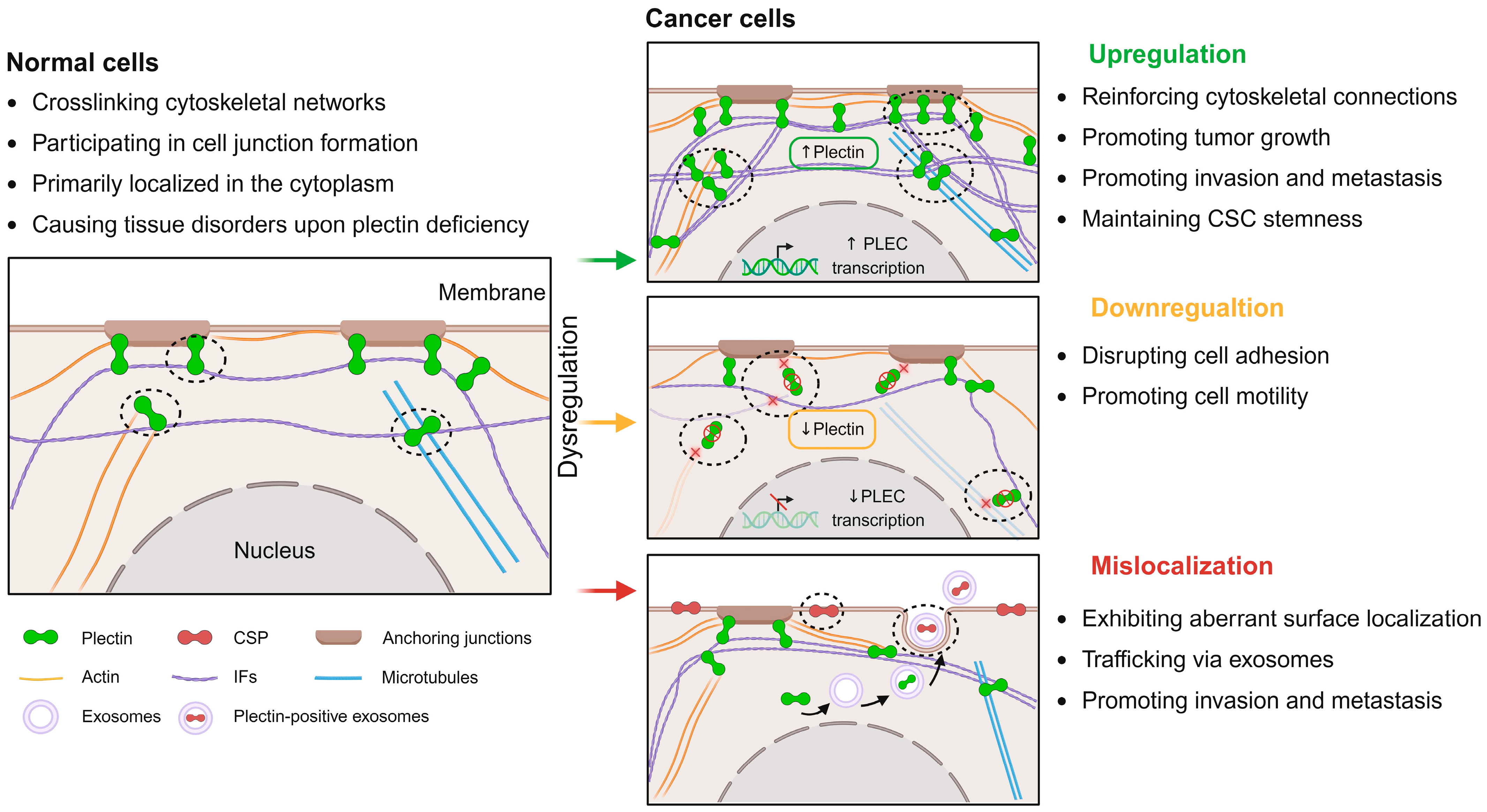
Figure 1. The impact of plectin dysregulation in cancer. In normal cells, plectin acts as a vital cytoskeletal linker protein and part of anchoring junctions, collectively maintaining cytoskeletal organization. However, plectin is dysregulated, exerting multifaceted effects on tumor progression. Its upregulation (indicated by upward arrows) facilitates cytoskeletal network crosslinking and activates signaling pathways that drive cancer cell growth, invasion, metastasis, and maintenance of cancer stemness. Conversely, decreased plectin expression (indicated by downward arrows) disrupts cell junctions and cytoskeletal architecture (indicated by cross signs), increasing cancer cell motility and morphological heterogeneity. Additionally, plectin mislocalizes to the cell membrane in cancer cells and may be trafficked via exosome formation. This process further regulates tumor growth and migration. Image created with BioRender.com.
Molecules 30 03675 g001
Figure 2. Bidirectional interactions between plectin and the TME. Plectin dysregulation contributes to TME remodeling, while TME-derived cues reciprocally induce plectin dysregulation. Specifically, plectin expression is upregulated (indicated by upward arrows) in EGFR+ inflammatory breast cancer and during Wnt-activated inflammatory signaling. Moreover, its upregulation is also observed in immunosuppressive tumor microenvironments. The loss of transmembrane protein 268, an upstream regulator of plectin, has been shown to influence phagocyte adhesion and migration. In addition, elevated plectin (indicated by upward arrows) further promotes the maintenance of vascular barriers, enhancing endothelial resistance to diverse mechanical stimuli and supporting angiogenesis within the TME. Finally, plectin upregulation (indicated by upward arrow preceding plectin) responds to increased ECM stiffness (indicated by upward arrow following ECM stiffness) to facilitate cancer cell invasion, whereas loss of plectin function (indicated by downward arrow following plectin) impairs gelatin degradation. Image created with BioRender.com.
Molecules 30 03675 g002
Table 1. Dysregulation of plectin in cancer.
DysregulationCancer TypesOutcomesReferences
UpregulationAMLPLEC gene mutation[37]
Breast cancerPromoting growth and metastasis; substrate for caspases; orchestrating PLEC/NFκB1/CXCL9 axis; SNRPA1-mediated PLEC alternative splicing[38,39,40,41,42]
Bladder cancerPromoting invasion and metastasis; promoting invadopodia formation[43,44]
CRCPromoting invasion; targeting podosome-like adhesions; contributing to drug resistance[18,33]
ESCCMaintaining anchorage and proliferation; increasing ESCC risk[45,46,47]
GBMEnhancing migration; regulating morphological changes[48]
Gastric cancerPromoting growth; integrin β4–plectin complex forming[49]
HCCPromoting migration and invasion; promoting EMT; involving the Integrin β1/Plectin/F-actin axis; upregulating in high matrix stiffness[26,31,32]
HNSCCPromoting migration and invasion; upregulating Erk 1/2 kinase[50]
Lung cancerEnhancing invasion and migration; maintaining stemness[27,51,52]
MelanomaMetastasis biomarker; promoting growth; activating Src signaling[29,53,54,55]
OSCCPrognostic marker; enhancing cell motility, invasion, and tumorigenicity[8,28]
Ovarian cancerPromoting migration, invasion, and adhesion; upregulating in high substrate stiffness[56,57,58,59]
Pancreatic cancerMalignant biomarker; promoting proliferation, migration, and invasion; participating in integrin β4-dependent exosomal transport[23,24,60]
Prostate cancerPromoting growth and metastasis; targeting focal adhesions[30,61,62]
Testicular cancerNovel susceptibility genes[63]
DownregulationBCC and SCCPromoting invasion[64]
ESCCDisrupting stratified squamous epithelium homeostasis[45]
HCCPromoting cell motility; activating FAK and Rac1-GTPase[65,66,67]
Ovarian cancerDownregulating during tumor progression; promoting EMT[56]
MislocalizationCRCPlectin 1k targeting podosome-like adhesions[18]
ESCCLocalizing in the cell membrane[47]
GBMCo-localization with membrane-related-aquaporin 4 aggregates[48]
HCCPerimembranous enrichment[26]
HNSCCUniform in the cytoplasm and the cell membrane[50]
Lung cancerBiomarker of ALDH+ lung cancer stem cells[27]
OSCCMainly at the plasma membrane[28]
Ovarian cancerMonoclonal antibodies targeting CSP; a target for drug delivery[56,57,58]
Pancreatic cancerParticipating in exosome formation and enhancing tumor growth[23,24]
ALDH: Aldehyde dehydrogenase; AML: Acute myeloid leukemia; BCC and SCC: Basal and squamous cell carcinomas; CRC: Colorectal cancer; CSP: cancer-specific plectin; EMT: Epithelial–mesenchymal transition; Erk 1/2: Extracellular signal-regulated kinase 1/2; ESCC: Esophageal squamous cell carcinoma; FAK: focal adhesion kinase; GBM: Glioblastoma multiforme; HCC: Hepatocellular carcinoma; HNSCC: Head and neck squamous cell carcinoma; OSCC: Oral squamous cell carcinoma; Rac1: Ras-related C3 botulinum toxin substrate 1; Src: Rous sarcoma oncogene.
Disclaimer/Publisher’s Note: The statements, opinions and data contained in all publications are solely those of the individual author(s) and contributor(s) and not of MDPI and/or the editor(s). MDPI and/or the editor(s) disclaim responsibility for any injury to people or property resulting from any ideas, methods, instructions or products referred to in the content.