Review

Metabolism, a Blossoming Target for Small-Molecule Anticancer Drugs

Laboratory Affiliated to Istituto Pasteur Italia-Fondazione Cenci Bolognetti, Department of Drug Chemistry and Technologies, Sapienza University of Rome, Piazzale Aldo Moro 5, 00185 Rome, Italy
*
Author to whom correspondence should be addressed.
Molecules 2025, 30(17), 3457; https://doi.org/10.3390/molecules30173457
Submission received: 10 July 2025 / Revised: 8 August 2025 / Accepted: 19 August 2025 / Published: 22 August 2025
(This article belongs to the Special Issue Small-Molecule Drug Design and Discovery)

Abstract

Reprogramming is recognized as a promising target in cancer therapy. It is well known that the altered metabolism in cancer cells, in particular malignancies, are characterized by increased aerobic glycolysis (Warburg effect) which promotes rapid proliferation. The effort to design compounds able to modulate these hallmarks of cancer are gaining increasing attention in drug discovery. In this context, the present review explores recent progress in the development of small molecule inhibitors of key metabolic pathways, such as glycolysis, glutamine metabolism and fatty acid synthesis. In particular, different mechanisms of action of these compounds are analyzed, which can target distinct enzymes, including LDH, HK2, PKM2, GLS and FASN. The findings underscore the relevance of metabolism-based strategies in developing next-generation anticancer agents with potential for improved efficacy and reduced systemic toxicity.

Graphical Abstract

1. Introduction

The cancer metabolism was first suggested by the Nobel Prize Otto Warburg who observed that cancer cells produce additional energy in vitro by converting a larger amount of glucose into lactate compared to healthy cells [1]. This event, known as the Warburg effect, is referred to as aerobic glycolysis. The abnormal aerobic glycolysis promotes cell differentiation and cancer cell proliferation [2]. Under normal conditions, differentiated cells metabolize glucose to yield two molecules of pyruvate, two molecules of adenosine triphosphate (ATP) and two molecules of reduced nicotinamide adenine dinucleotide (NADH) (Figure 1). The two pyruvates are then converted into acetyl CoA, which enters the mitochondria and combines with oxaloacetate in the tricarboxylic acid (TCA) cycle to form citric acid [3]. Normally, one molecule of glucose produces about 30–34 molecules of ATP in the presence of oxygen [4]. In low-oxygen conditions or in the absence of oxygen, glucose is metabolized through anaerobic respiration. However, even in the presence of oxygen, cancer cells tend to convert pyruvate into lactate by lactate dehydrogenase (LDH) after glycolysis, producing four molecules of ATP [5,6]. Although this conversion is less efficient in terms of ATP yield compared to mitochondrial oxidation, it is faster [7] and continuously provides the cancer cell with amino acid, lipid, and nucleotide building blocks required for tumor proliferation [8,9,10]. As a consequence, a smaller proportion of pyruvate is converted into acetyl CoA prior to entering the TCA cycle, resulting in reduced production of NADH, FADH2 and ATP (Figure 1) [11].

2. Developing Therapeutic Agents Based on Metabolism

Targeting metabolism in medicinal chemistry holds significant potential for developing effective agents with highly specific mechanisms of action at catalytic and allosteric sites within hydrophobic pockets of metabolic enzymes. The success achieved by metabolism-targeting chemotherapy has demonstrated the effectiveness of this strategy for tumor treatment. The hallmarks of cancer include acquired capabilities, such as sustaining proliferative signaling, evading growth suppressors, enabling replicative immortality, inducing angiogenesis, and activating invasion and metastasis [12]. Deregulated cellular metabolism can be considered a core hallmark of cancer [13]. In addition to the Warburg effect, relevant reprogramming events that promote cancer cell survival and growth include de novo biosynthesis of proteins, nucleic acids and lipids. The anabolic metabolism of cancer cells includes increased lipid synthesis during proliferation to face the elevated demand for energy and for the formation of biological membranes [14,15]. The growing interest in compounds that interfere with cancer metabolism has been well documented in recent excellent reviews, such as that by Montesdeoca on lipogenic enzymes inhibitors [16], Stine on precision oncology [17] and Masci on colorectal cancer therapy [18]. Herein, we present the most recent advances in small molecules targeting metabolism, focusing on mechanism of action and their potential as antitumor drugs.

2.1. Aerobic Glycolysis

2.1.1. Lactate Dehydrogenase (LDH)

The release of the LDH in the tumor microenvironment promotes cancer cell proliferation and increases the likelihood of metastasis [19,20]. Inhibition of LDH by decreasing lactate release in the tumor microenvironment produces an useful anticancer effect [21]. Consequently, the search for LDH inhibitors as potential anticancer agents has become a topic of considerable interest [22,23,24]. LDH exists in five major isoforms, resulting from combinations of M and H monomers forming homo- and hetero-tetramers. The 4M and 4H tetramers correspond to LDHA and LDHB, respectively. LDHA catalyzes the conversion of pyruvate to lactate, whereas LDHB drives the reverse conversion (Figure 2) [25,26].
LDHA is induced by hypoxia or MYC activation, enabling glycolysis to become the primary source of ATP production in response to oxygen deprivation and the tumor’s ability to recruit new blood vessels [27]. As such, several research projects have focused on the discovery of natural [28] and small-molecule [29] LDH inhibitors.
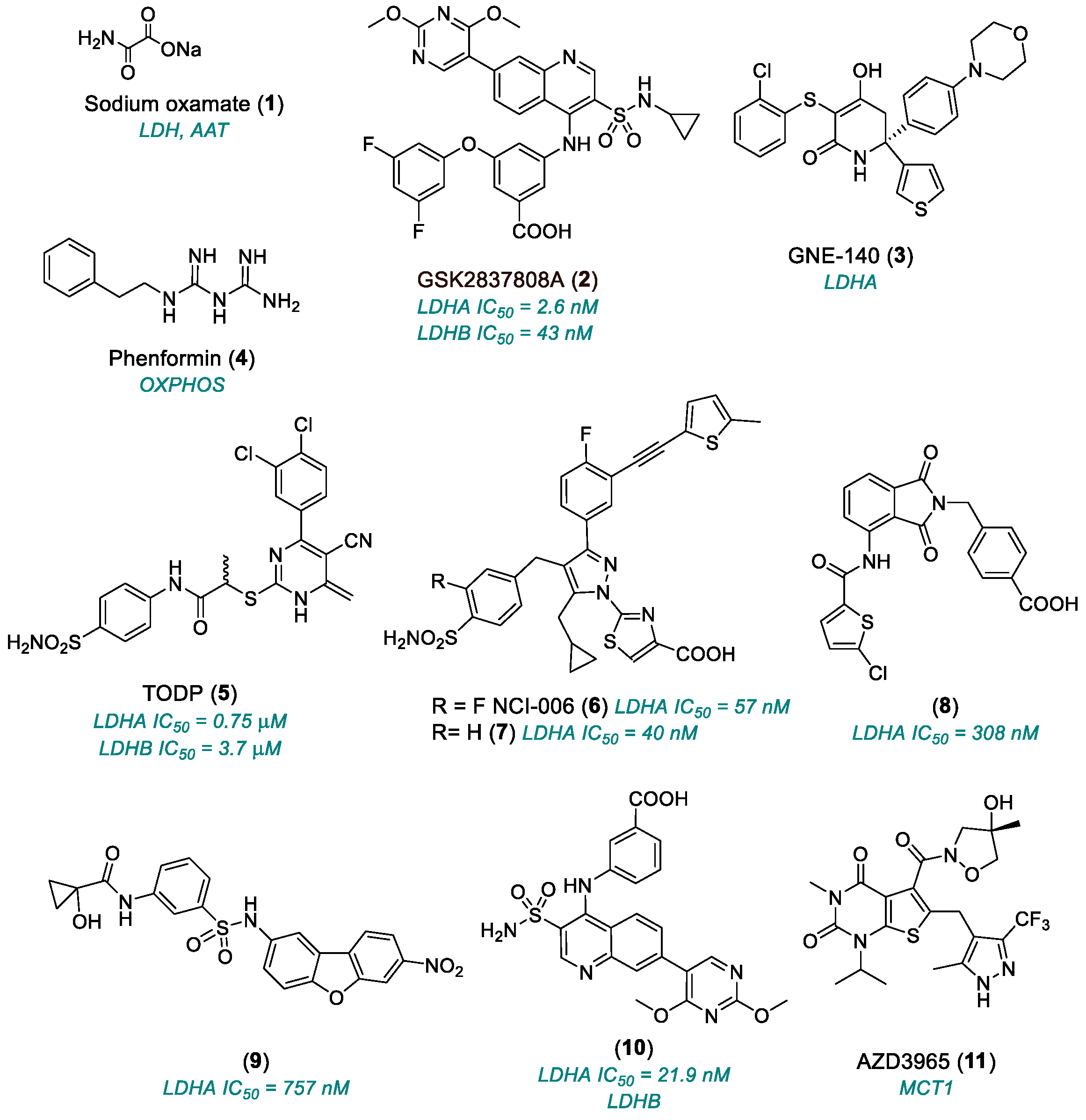
Sodium oxamate (1, Chart 1, Table 1) inhibits LDH and aspartate aminotransferase (AAT) and competes in vitro with pyruvate for the human LDHA. Oxamate and the AAT inhibitor amino oxyacetate suppressed proliferation of MDA-MB-231 cells and decreased tumor growth in MDA-MB-231 breast cancer xenografts in mice lacking the normal thymus gland [30]. Tumor suppressor p53 expression was strongly upregulated by (1), but only in glucose-treated cells, suggesting a connection with glycolysis inhibition [31]. While useful as a pharmacological tool, compound 1 shows limited enzymatic selectivity, weak potency and poor cellular uptake restricting its clinical utilization [32].
A series of quinoline 3-sulfonamides was found to inhibit potently and selectively LDHA and to rapidly reduce lactate production. GSK2837808A (2) inhibited LDHA at low nanomolar concentrations (IC50 = 2.6 ± 1.9 nM) and showed 80-fold selectivity over LDHB (IC50 = 43 ± 14 nM) in hepatocellular carcinoma cell lines [33]. LDHA inhibition by (2) suppressed glycolytic activity in temporomandibular joint osteoarthritis (TMJOA) synovial fluid (SF) containing >1000 kDa hyaluronic acid (HA). In the TMJOA microenvironment, vascularization and extracellular matrix (ECM) degradation correlate with increased nutrient consumption and active synovial proliferation [34]. As a result of LDHA inhibition by (2), HA production increased, and inflammation was reduced along with suppression of AMPK phosphorylation [35]. In Snu398 cells, which exhibit elevated glycolysis and pentose phosphate pathway activity, compound 2 reduced proliferation and induced strong changes in cell metabolism [33]. Pyruvate kinase (PK) activity increased in Snu398 but not in HepG2 cells, where the mitochondrial metabolism is significantly higher. Compound 2 also induced, in a dose-dependent manner, the inactive monomer of pyruvate kinase (PKM2) to form the M2 isoform, which catalyzes the final and rate-limiting reaction in the glycolytic pathway [36].
The 1,3-dihydropyrimidine GNE-140 (3) LDHA inhibitor rapidly affected the metabolism in MIA PaCa-2 human pancreatic cells, although cell death occurred only after two days of treatment [37]. Pancreatic cell lines relying primarily on oxidative phosphorylation (OXPHOS) for ATP production [38] were resistant to 3, due to activation of the AMPK-mTOR-S6K signaling pathway, which promoted OXPHOS; the OXPHOS inhibitor phenformin (4) could resensitize these cells to 3. Histone lysine lactylation (HKla) is a novel post-translational modification that connects cellular metabolism with epigenetic regulation. Although its role in the development of cardiac hypertrophy is still unclear, LDH inhibitor 3, as well as 1, has been shown to reduce HKla levels and mitigate cardiac hypertrophy development [39]. Compound 3 has also been shown to suppress osteogenic differentiation, proliferation, and migration while promoting apoptosis in alveolar bone marrow mesenchymal cells (ABMMCs). Mechanistically, these regulatory effects of lactate were thought to be mediated by HKla [40]. Compound 3 blocked dimethylsulfoxide-induced expression of Arg1, an M2 macrophage polarization marker, by lactate-H3K18la pathway in a methicillin-resistant Staphylococcus aureus (MRSA) disease model [41].
A screening of 2-thio-6-oxo-1,6-dihydropyrimidine derivatives led to the identification of a moderately potent LDHA inhibitor with an IC50 of 8.8 μM. Surface plasmon resonance (SPS) experiments indicated that simultaneous association with the NADH co-factor was required for an optimal binding to the LDHA. Based on these findings, medicinal chemistry work was conducted that aimed to improve the compound’s LDHA inhibition properties. As a result, compound TODP (5) inhibited LDHA with IC50 = 0.75 μM and LDHB with IC50 = 3.7 μM. A crystal structure of compound 5 in complex with LDHA at 1.90 Å resolution showed binding to Arg168, His192 and Asp165 residues of the LDH catalytic site and only the R-Me enantiomer was observed in the co-crystal structure [42].
Pyrazole-based LDH inhibitors were identified by quantitative high-throughput screening (qHTS) and structure-based design [42,43]. A lead compound effectively inhibited highly glycolytic MIA PaCa-2 (human pancreatic cancer) and A673 (human Ewing’s sarcoma) cell lines and showed favorable in vitro ADME properties although its PK profile was not suitable for in vivo use [44]. Optimization of the pyrazole-based chemotype yielded compounds 6 (NCI-006 NCATS-SM1440) and 7 (NCATS-SM1441), which stabilized LDHA in cells (IC50 57 nM and 40 nM, respectively). Increasing the concentrations of compounds 6 and 7 resulted in NAD+ depletion and full glycolytic pathway inhibition, as evidenced by decreased extracellular acidification rate (ECAR). Both compounds 6 and 7 showed excellent metabolic profiles in hepatocytes and high plasma protein binding in mouse and human plasma. Structure–activity relationship (SAR) studies highlighted that the 5-methylthienyl group in the alkyne region is required for robust in vivo LDH inhibition [44,45]. Compound 6 and its analog NCI-737 (structure unavailable) were evaluated in in vitro and in vivo preclinical models of Ewing sarcoma (EWS), a highly metastatic bone cancer, constituting 10% to 15% of all bone sarcomas [46], predominantly afflicting adolescents [47]. Both compounds suppressed the glycolytic flux by impairing the conversion of pyruvate to lactate, disrupted the NAD+/NADH ratio and decreased the extracellular acidification rate (ECAR) in multiple EWS cell lines. Intravenous administration led to intratumoral drug accumulation, induced cell death and reduced tumor growth [48].
New phthalimide- and dibenzofuran-based selective LDHA allosteric inhibitors with sub-micromolar in vitro activity were identified. Co-crystal structures demonstrated that compounds 8 and 9, with IC50 values of 308 nm and 757 nm, respectively, bound to a novel binding pocket distant from the polar, extended orthosteric substrate/cofactor site, while compound 10 (IC50 = 21.9 nm) was shown to hinder cofactor binding in an NADH competitive mode. Since few LDHA crystal structures show an allosteric pocket in an open conformation, the authors speculated that enzyme inhibition by (8) and (9) may depend on ligand-dependent conformational changes. Compound 10 also inhibited the LDHB, due to the high similarity among LDH isoforms active sites [49]. Allosteric inhibitors may take advantage by avoiding competition with endogenous substrates [50]. The architecture of the LDH protein plays a key role in enzymatic catalysis by tuning vibrational modes that promote chemical reactions [51]. Therefore, LDHA inhibition may result from conformational changes and/or dynamic processes crucial for enzymatic turnover [49].
AZD3965 (11) is a potent and selective inhibitor of monocarboxylate transporter 1 (MCT1), encoded by SLC16A1 [52], which is involved in lactate efflux following MYC-induced glycolysis [53]. Compound 11 potently inhibited MCT1 and MCT2, while sparing MCT3 and MCT4. It showed GI50 <100 nM (MTS growth assay) in diffuse large B-cell lymphoma (DLBCL) and non-Hodgkin lymphomas (NHL) cell lines. Inhibition of MCT1 led to decreased lactate efflux by (11). In in vivo studies, compound 11 was combined with doxorubicin or rituximab in DLBCL and Burkitt’s lymphoma models. Finally, when co-administered with a glutaminase (GLS) inhibitor, compound 11 enhanced cell death and growth inhibition compared to monotherapy treatment [53]. In a xenograft model of Raji lymphoblast-like cell in immunodeficient mice, compound 11 inhibited tumor growth, downregulated choline kinase alpha (which converts choline + ATP into phosphocholine + ADP + H+), and altered mRNA expression. These effects correlated with intracellular lactate accumulation induced by (11) [54]. In a multicenter phase I clinical trial, compound 11 was well tolerated at doses effective against MCT1-expressing cancers. Dose-limiting toxicities were mostly reversible, asymptomatic ocular changes. A dose of 10 mg twice daily was recommended for phase II trials [55].

2.1.2. Glucose Transporter 1 (GLUT1)

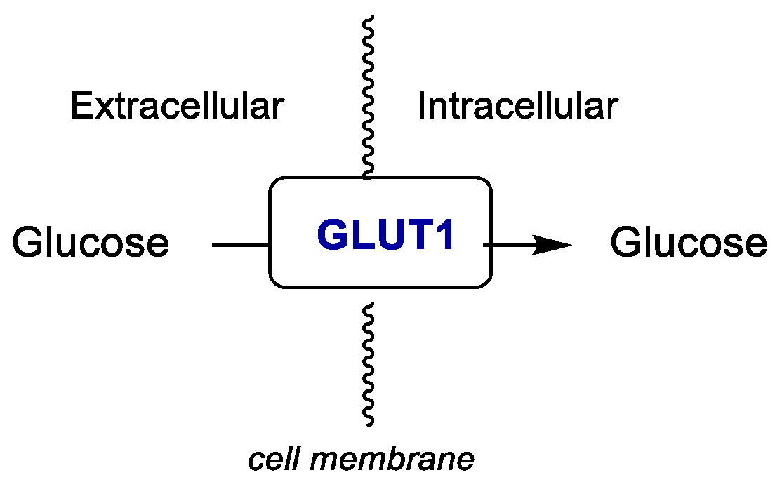
Glucose transporter type 1 (GLUT1) is the main transporter responsible for glucose uptake in many tissues (Figure 3). It is expressed in most tissues, but is highly abundant in brain endothelial cells, glial cells, erythrocytes and placenta [56]. GLUT1 is a glycoprotein belonging to the GLUT family of carriers and is responsible for the basal glucose uptake across the blood–tissues barriers, including the blood–brain barrier (BBB) [57]. Alterations in GLUT1 impair the glucose supply to glial cells and neurons. A reduction in brain glucose transport, primarily caused by mutations in the SLC2A1 gene encoding GLUT1 [58], is the etiological basis of the glucose transporter type 1 deficiency syndrome (GLUT1DS), an autosomal dominant disorder [59,60]. The classical form of GLUT1DS presents as early-onset encephalopathy (during the first year of life), characterized by severe epilepsy, a complex movement disorder and developmental delay, including microcephaly, due to immature tight junctions in the BBB that allow paracellular glucose transport [61].
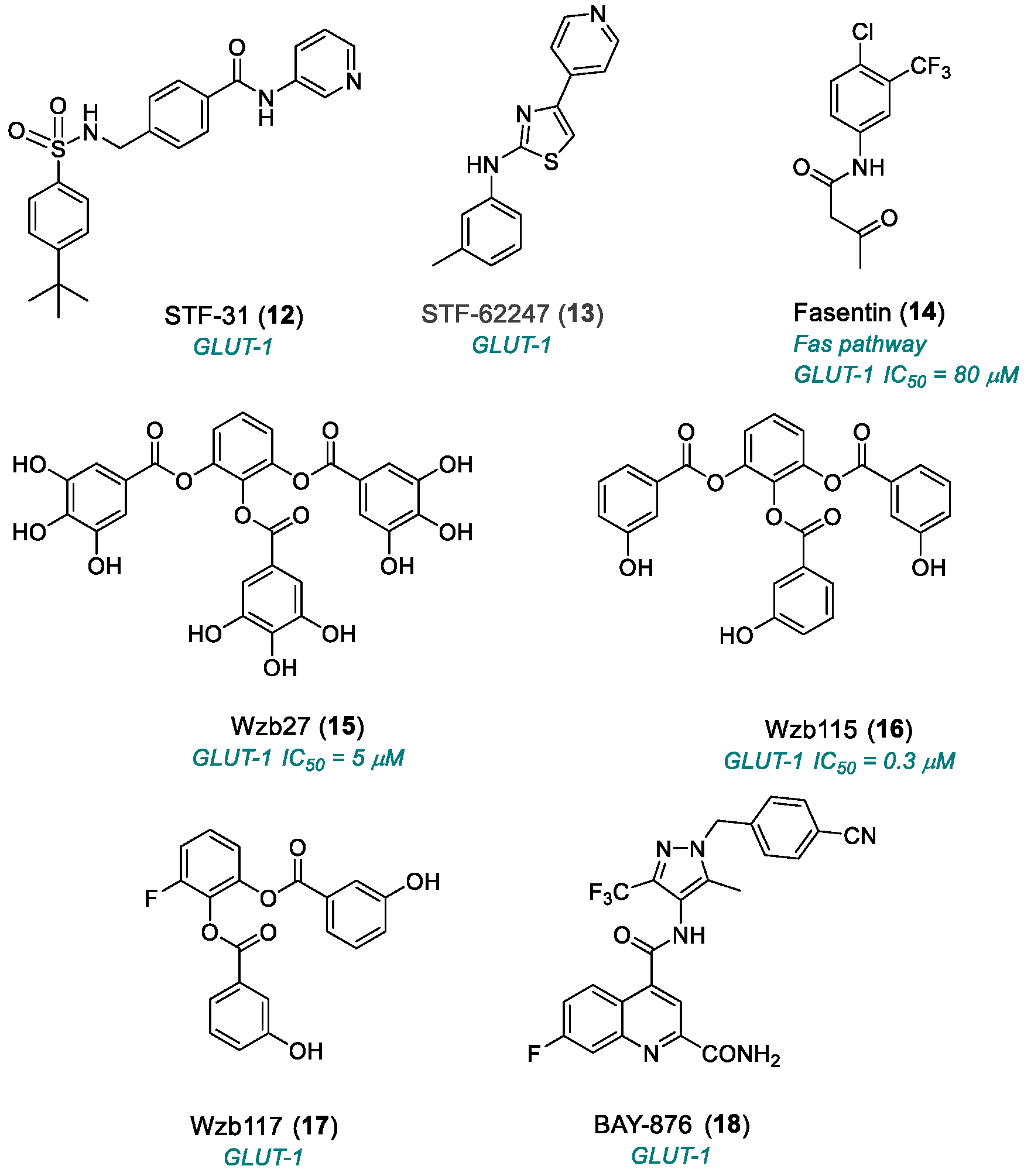
Approximately 80% of renal cell carcinomas (RCCs), similar to many other cancers, are associated with the loss of the von Hippel–Lindau (VHL) tumor suppressor gene, which is correlated with the Warburg effect and partially results from GLUT1 upregulation. Compound STF-31 (12) (Chart 2) was identified through a high-throughput screening of ~64,000 small molecules, aimed at discovering agents with selective lethality in VHL-deficient RCC cells. Two classes of compounds emerged from this screening: sulfonamides and pyridyl anilino thiazoles, including compound 12 and STF-62247 (13), respectively. RCC4 cells lacking VHL showed reduced viability in the presence of (12) in a dose-dependent manner compared to their wild-type cells RCC4/VHL. The clonogenic assay confirmed that compound 12 selectively targeted RCC4 cells. It did not induce any morphological or biochemical features of autophagy, nor did it deregulate HIF expression in VHL-deficient cells. Importantly, lactate production and extracellular acidification were significantly inhibited. Compound 12 decreased glycolysis by inhibiting glucose transport rather than targeting a particular glycolytic enzyme. Moreover, it was toxic to RCCs expressing GLUT1 but low or undetectable levels of GLUT2, indicating that STF-31 acts as a high-affinity GLUT1 inhibitor [62].
STF-62247 (13) was identified by screening for small molecules targeting selectively VHL-deficient RCC cells. Compound 13 induced cytotoxicity and reduced tumor growth in VHL-deficient RCC cells compared to VHL wild-type (WT) cells. The cytotoxicity of (13) was mediated through autophagy in a HIF-independent mechanism [63]. SAR studies identified several key features necessary for selective cytotoxicity against VHL-negative RCC cells: the aniline-NH and the 4-pyridyl-N groups likely participate in H-bonding, while an unsubstituted thiazole is required for VHL selectivity. Small lipophilic substituents at the 3- and 4-positions of the aniline ring suggested the presence of a lipophilic pocket within the target protein [64].
Fasentin (14) is a small molecule that sensitizes the Fas-signaling pathway (central to programmed cell death) by effecting glucose uptake. At 80 μM, (14) dramatically reduced glucose uptake in leukemia and prostate cancer cells [65]. Compound 14 was also used to shed light on the role played by GLUT1 in corticotropin-releasing hormone (CRH)-mediated glucose uptake in adenomatous corticotropes [66]. These findings have prompted the development of anticancer strategies based on glucose metabolism. Compound 14 inhibited endothelial cells growth without compromising their survival and reduced the number of enterochromaffin (EC) cells, tumor cells and, to a lesser extent, fibroblasts. At 50 nM, it impaired angiogenesis in the chick chorioallantoic membrane (CAM) assay. Notably, this anti-angiogenic activity was independent of glucose metabolism modulation. In human dermal microvascular endothelial cells, (14) reduced the uptake of glucose. It is worthy to note that 13 failed to inhibit tube formation in these cells [67].
Compounds Wzb27 (15) and Wzb115 (16) (IC50 = 5 μM and 0.3 μM, respectively) were serendipitously identified as inhibitors of basal glucose transport [68] during a screening for molecules that stimulate insulin receptor-mediated glucose uptake [69]. These compounds proved more potent than other GLUT1-inhibitory agents, such as (14) or apigenin, a natural product with anti-inflammatory and antitumor activity, in inhibiting basal glucose transport and cancer cell proliferation. Both (15) and (16) induced G1/S cell cycle arrest and apoptosis in lung and breast cancer cells without significantly affecting their normal cell counterparts. Apoptosis was mediated by caspase-3 activation, independent of p53, consistent with a glucose deprivation mechanism [68]. The analog compound WZB117 (17) inhibited cancer cell proliferation both in vitro and in vivo in a nude mouse model. It proved to inhibit glucose transport in human RBCs having GLUT1 as the only transporter. Compound 17 reduced GLUT1 protein levels, intracellular ATP, and glycolytic enzymes, while increasing AMP-activated protein kinase (AMPK). ATP depletion and induction of senescence were observed in GLUT1 inhibitor-treated cancer cells [70]. Compound 17 reversibly and competitively inhibited 3-O methylglucose (3MG) uptake in human erythrocytes but acted as a non-competitive inhibitor of glucose efflux. In HEK293 cells, (17) inhibited Glut4 more potently than GLUT1 and GLUT3 [71].
A high-throughput screening campaign was led to identify N-(1H-pyrazol-4-yl)quinoline-4-carboxamides as a promising scaffold for the development of GLUT1-selective small-molecule inhibitors. SAR studies led to single-digit nanomolar inhibitors with selectivity over GLUT2, GLUT3 and GLUT4. The most promising compound, BAY-876 (18), showed high GLUT1 potency and selectivity thanks to the presence of an unsubstituted amide at position 2 and a fluorine atom at position 7, respectively, of the quinoline ring. Compound 18 exhibited good in vitro metabolic stability and high oral bioavailability in vivo, with low clearance in rats and dogs [72]. Inhibition of GLUT1 by (18) halted glucose uptake in cancer cells, induced apoptosis, and suppressed the TNF-α-induced interleukin-8 (IL-8) production in head and neck squamous cell carcinoma (HNSCC) cells. BAY-876 reduced the viability of SCC47, RPMI2650, and FaDu cells after 24 h and induced apoptosis at higher concentrations in some cancers. These results highlight GLUT1 as a promising target in HNSCC of the upper aerodigestive tract [73].

2.1.3. Hexokinase (HK)

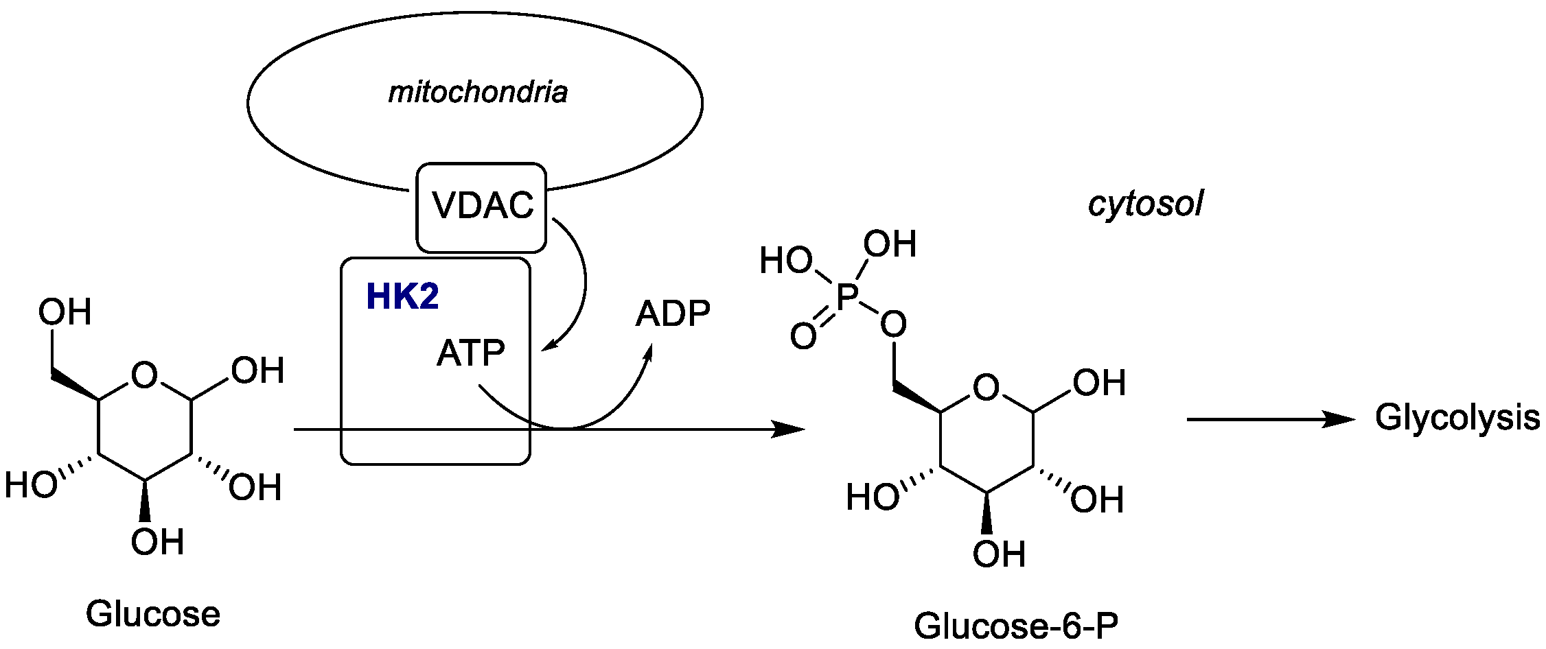
HK catalyzes the conversion of glucose to glucose-6-phosphate (G6P), the first irreversible step of glycolysis, and is involved in pathways associated with cancer cell growth, such as nucleotide and lipid synthesis, tricarboxylic acid cycle and pentose phosphate [74]. In humans, five HK isoenzymes are recognized: HK1, HK2, HK3, HK4 and the HK domain-containing protein 1 (HKDC1) [75]. Among them, HK2 isoenzyme was found to be overexpressed in various cancer types [76,77], while HK1 is the predominant isoform in normal cells. Due to its key role in reprogrammed glucose metabolism, HK2 has emerged as an attractive target for anticancer drug discovery [78]. The transport of mitochondrial metabolites across the outer mitochondrial membrane (OMM) is mediated by voltage-dependent anion channels (VDACs) [79] (Figure 4), with VDAC1 being the most prominent isoform, frequently overexpressed in several types of cancer [80]. HK2 was found to be associated with VDAC1 at the OMM, allowing ATP transport from mitochondria to the cytosol as ADP to convert glucose into glucose-6-phosphate. Moreover, HK2 contributes to apoptosis evasion in cancer cells by binding to VDAC1 and competing with Bax [81]. Thus, inhibition of the HK2-VDAC1 interaction turned out to be a promising strategy for cancer therapy. The crystal structure of HK2 (PDB ID: 2NZT, resolution: 2.45 Å) was used to design HK2 inhibitors. Additional inhibitors were developed to target the G6P-binding site by mimicking the G6P interaction.
Due to the high sequence similarity between rat HK1 (PDB ID: 1BG3, resolution: 2.80 Å) and human HK2, a homology model was constructed to study how VDAC1 phosphorylation may disrupt HK2 binding. For docking studies to HK2, the structure of VDAC1 (PDB ID: 2JK4, resolution: 4.10 Å) was used [78].
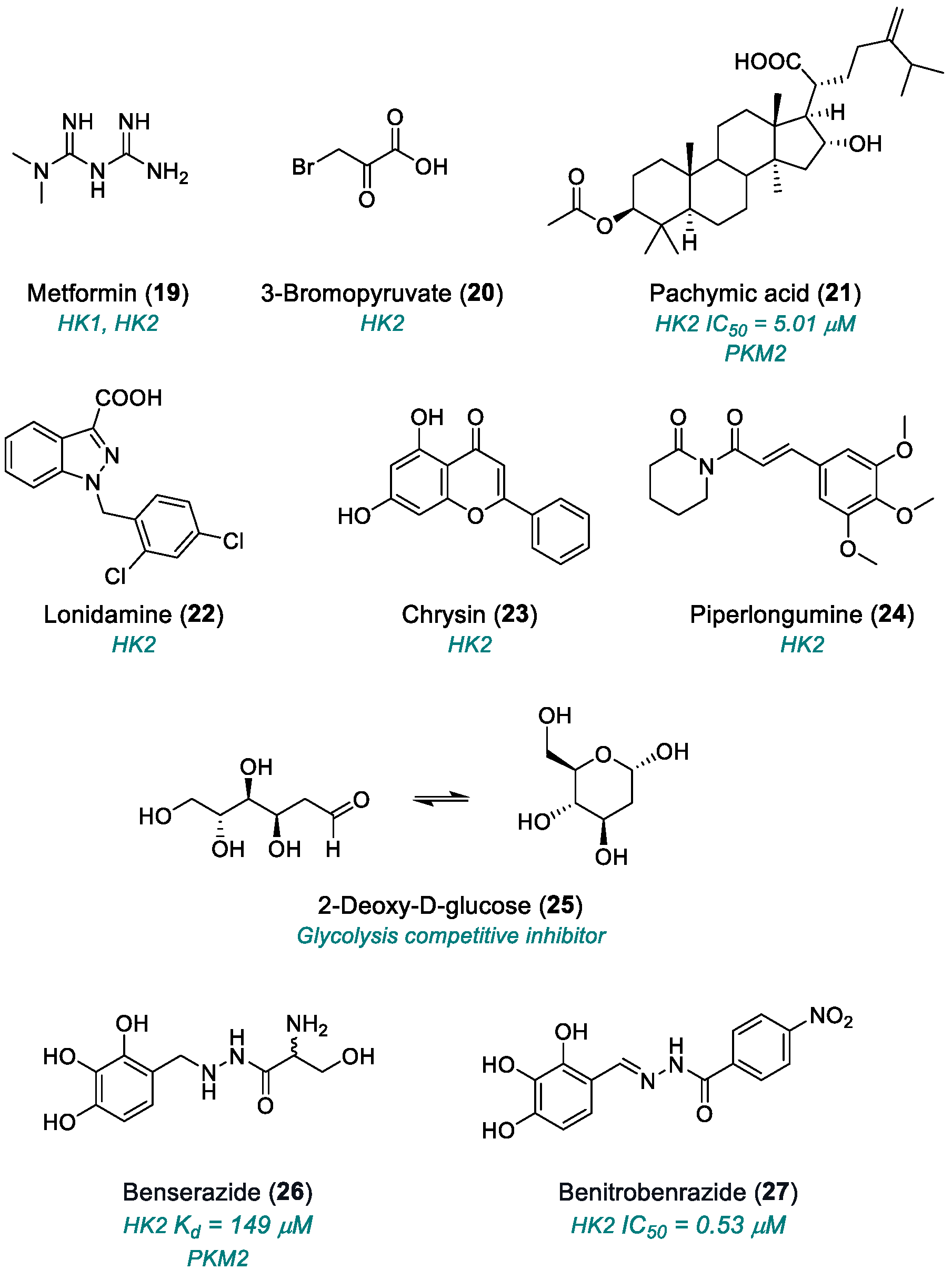
HK2-VDAC1 inhibitors. Metformin (19,Chart 3) was shown to impair energy balance in cancer cells in vitro by the selective enzymatic inhibition of HK1 and HK2 isoforms [82]. This activity was demonstrated in MDA-MB-231 triple-negative breast cancer cells in xenograft models [83]. Both isoforms were localized to the mitochondrial outer membrane; HK2′s membrane association was found to be partially dependent on G6P [84]. HK2 inhibition restricts cancer cell growth by limiting ATP access for glucose phosphorylation [85]. Notably, prolonged treatment with compound 19 caused a marked reduction in tumor growth without any visible necrosis, whereas acute drug administration resulted in profound cytotoxic effects [83].
3-Bromopyruvate (3-BrPA, 20) covalently modifies the HK2 protein, leading to the release of the apoptosis-inducing factor (AIF) from the mitochondria to cytosol as a consequence of the interaction of HK2 with AIF. The dissociation of HK2 from mitochondria alone was sufficient to cause apoptosis, particularly in the mitochondria-deficient ρ0 cells with high HK2 expression, without affecting mitochondrial membrane potential, ROS generation, or oxidative phosphorylation [86].
Pachymic acid (21) is a triterpenoid compound obtained from Poria cocos (Polyporaceae), a saprophytic fungus that grows on various Pinus species [87]. Compound 21 has demonstrated anti-inflammatory, anticancer, anti-aging, and insulin-like properties, which directly inhibited the HK2 with an IC50 of 5.01 μM, inducing mitochondrial dysfunction, ATP depletion, and ROS generation. Compound 21 acts as a competitive activator of PKM2 by mimicking fructose-1,6-bisphosphate, the natural activator. It was suggested that this compound could be a G6P mimic [88].
Lonidamine (22) is an anticancer agent approved as a monotherapy for advanced breast, prostate, lung and brain cancers. Its mechanism of action involves selective inhibition of aerobic glycolysis in tumor cells [89] through HK inhibition [90] and disruption of the mitochondrial membrane [91]. Compound 22 was administered alone or in combination with other chemotherapeutic agents (i.e., paclitaxel, docetaxel, 5-fluorouracil and 2-desoxy-2-glucose), surgery or radiation therapy in a sustained-release formulation to treat cancer, inflammation and autoimmune disease [92].
Chrysin (23) is a flavonoid compound extracted from natural sources, such as blue passion flower, propolis and honey, commonly used in traditional Chinese medicine. It showed antitumor activity in several human cancer cell lines and also possesses antioxidant, anti-inflammatory and antibacterial properties. Its antitumor activity involves activation of the extrinsic apoptosis pathway [93], alteration of cyclin-dependent kinases (CDKs) [94] and interference with key signaling pathways, including Ras-Raf-MAPKs, PI3K-Akt, STAT, NF-κB, Wnt-β-catenin and Notch [95,96]. Compound 23 inhibited glycolysis and induced apoptosis in hepatocellular carcinoma (HCC) cells both in vitro and in vivo, through inhibition of HK-2 (which was found to be overexpressed in the majority of HCC tissue) and tumor glycolysis and activation of mitochondria-associated apoptosis [97].
Piperlongumine (24), a natural compound isolated from long pepper piper longum L, showed anticancer activity against several human tumor types, including lung, liver, prostate, breast and colorectal cancers. Compound 24 inhibited the non-small-cell lung cancer (NSCLC) both in vitro and in vivo by downregulating glycolysis, decreasing HK2 expression and inducing mitochondrial apoptosis. The glycolytic suppression induced by (24) was mediated by the Akt signaling pathway [98].
Glucose-binding site inhibitors. 2-Deoxy-D-glucose (2-DG) (25) is a glucose analog bearing a hydrogen atom replacing the hydroxyl group at position 2, functioning as a competitive inhibitor of glycolysis [99,100]. It inhibits several glycolytic enzymes, leading to cell death. Compound 25 fulfills Lipinski’s rule of five and shows activity against hypoxic tumor cells, while also reducing oxygen dependency, including in the context of COVID-19 [101]. Due to its structural similarity to D-mannose, (25) interferes with glycosylation processes and induces endoplasmic stress [102,103]. Compound 25 has shown potential as a cancer and antiviral agent and enhanced the efficacy of paclitaxel when co-administered or co-encapsulated in nanoparticles for a lung cancer model [104]. Compound 25 was also shown to suppress the inflammation in innate immune cells [105] and the expression of herpes simplex virus type-1 receptor [106] by modulating anti-inflammatory mediators and the polarization of macrophages [107]. Applications of (25) as an anticancer agent have been reported in combination with the Bcl-2 antagonist ABT-263 and ABT-737 (ABT) [108], or with other therapeutic agents or radiotherapy to overcome its limited therapeutic effect [109]. 2-Halogenated D-glucose analogs were found to be more potent than (25) in killing hypoxic tumor cells and may be more clinically effective when combined with standard chemotherapeutic protocols [110]. The combination of low-dose (25) and (19) synergically inhibited cyst formation and human polycystic kidney cell proliferation [111] and proved effective across a broad spectrum of preclinical cancer models [112].
Benserazide (26) was identified as an HK2 inhibitor (Kd of 149 ± 4.95 μM) by structure-based virtual screening studies on FDA-approved drugs and nutraceuticals from the ZINC Drug Database [113]. Due to its inhibitory effect on peripheral dopa decarboxylase, compound 26 is used as a levodopa coadjuvant in standard Parkinson’s disease treatment. It showed approximately 8-fold selectivity for HK2, versus HK4, and about 5-fold selectivity against HK2 over HK1. (26) inhibited SW480 cancer cells proliferation and CRC xenograft growth by targeting HK2, inducing apoptotic cell death through energetic stress and activation of AMPKα, p53 and p27, along with loss of mitochondrial membrane potential [114].
Benitrobenrazide (27) is a nanomolar inhibitor of HK2 (IC50 of 0.53 ± 0.13 μM), induces apoptosis and inhibits proliferation of HK2-overexpressed cancer cells. It significantly inhibited glycolysis and cancer cell proliferation both in vitro and in vivo with low toxicity by directly targeting HK2. Upon oral administration, compound 27 effectively suppressed tumor growth in SW1990 and SW480 xenograft models [115].
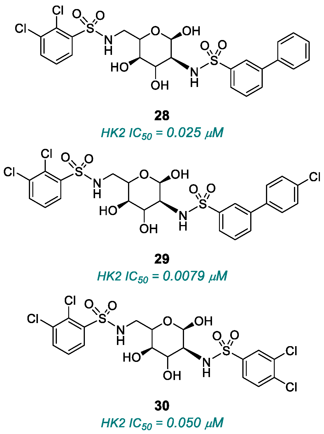
Glucose-6P-mimicking binding site inhibitors. Metformin (19) has also been reported to inhibit HK2 at μM concentrations similar to those of G6P (85). Glucose-6P binds to an allosteric site near, but distinct from, the glucose-binding site in the HK1 [116]. Co-crystallography studies revealed that the first structure of HK2 bound a ligand and G6P at 2.76 Å [117]. The co-crystal structure of HK2 highlighted the presence of a highly flexible binding site. SAR studies were conducted to enhance HK2 selectivity over HK1, given their high sequence similarity and conserved glucose-binding sites [118]. A 6-sulfonamide linker turned out to optimally mimic the phosphate group of G6P. Compounds 2830 (Chart 4) were identified as potent and selective HK2 inhibitors, with IC50 values of 0.025, 0.0079 and 0.050 μM, respectively [117].

2.1.4. Pyruvate Kinase (PK)

Pyruvate kinase (PK) is a rate-limiting glycolytic enzyme that catalyzes the final step of glycolysis, converting phosphoenolpyruvate (PEP) into pyruvate [119] (Figure 5). Among the four reported PK isoforms, PKM1 and PKM2 are expressed in several cell types, including neutrophils, while PKL is mainly found in the liver and PKR in erythrocytes [120]. The enzymatic activity of PKM2 is tightly regulated by endogenous allosteric effectors and intracellular signaling pathways, which influence both its enzymatic activity and oligomeric state. PKM2 can assume a dimeric or monomeric form, leading to the accumulation of glycolytic intermediates. The dimeric form of PKM2 can translocate to the cell nucleus, where it co-activates transcription factors, regulating genes involved in cell proliferation and glycolysis. Upon allosteric activation, PKM2 adopts a tetrameric conformation with high glycolytic activity [121]. ROS production in neutrophils is dependent on glycolytic flux. PKM2 regulates the dihydroxyacetone phosphate (DHAP) and diacylglycerol (DAG) levels, which activate protein kinase C (PKC), and, in turn, the NADPH oxidase complex, thereby promoting ROS production and neutrophil cytotoxic activity [122]. PKM2 was identified as a promising target for cancer therapy, with subcellular localization of PKM2 proposed as a potential biomarker for therapeutic response in non-small-cell lung cancer (NSCLC) cell lines [123].
The previously cited compound 26 was also identified as a novel inhibitor targeting PKM2 for melanoma treatment [124]. It also showed cystathionine-β-synthase (CBS) inhibitory activity among 8871 clinically used drugs, with relative selectivity versus cystathionine-γ lyase (CSE) and 3-mercaptopyruvate sulfurtransferase (3-MST). Benserazide (26) inhibited colon cancer cell proliferation in HCT116 and HT29 cell lines, both highly expressing CBS, impaired cellular bioenergetics in HCT116 cells and suppressed colon cancer tumor growth in mice [125].
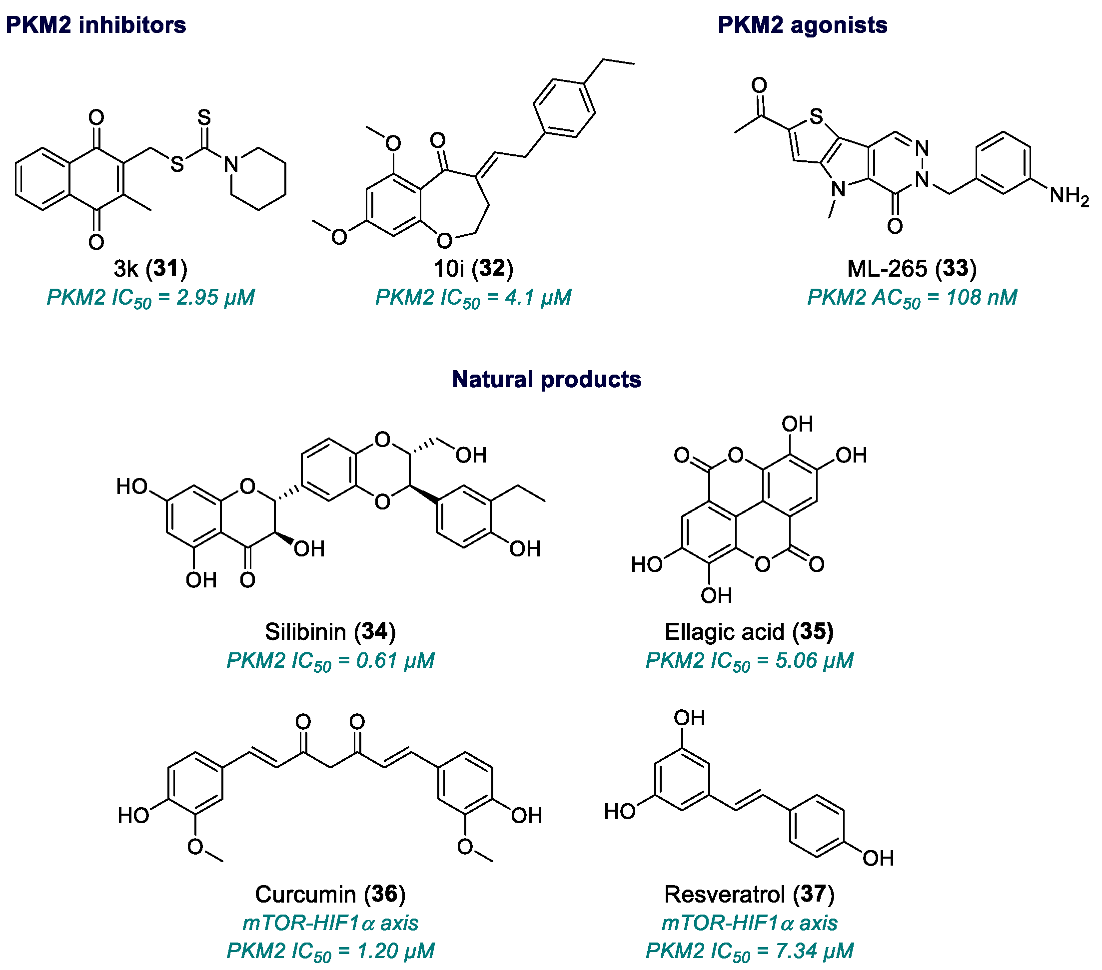
SAR studies on naphthoquinone derivatives led to the identification of thiocarbamate 3k (31, Chart 5) as a selective PKM2 inhibitor (IC50 = 2.95 ± 0.53 μM) with antiproliferative activity in PKM2-overexpressing HCT116, HeLa and H1299 cells, superior to the known PKM2 inhibitor shikonin [126]. Two novel 2,3-didithiocarbamate analogs of compound 31 exhibited even stronger inhibition of PKM2; their activities were superior to (31), showing dose-dependent cytotoxicity with IC50 values in nanomolar concentrations against HCT116, MCF7, HeLa, H1299 and B16 cells [127].
A series of benzoxepane derivatives, inspired by marine-derived natural products, was synthesized as novel anti-inflammatory agents. PKM2 was identified as the molecular target of compound 10i (32) (IC50 = 4.1 μm) [128]. Compound 32 exhibited anti-inflammatory activity in lipopolysaccharides (LPS)-stimulated RAW264.7 macrophages by inhibiting TNF-a protein release. In mouse primary microglia 10 μM of (32) showed potent anti-neuroinflammatory effects, with an inhibition rate of 87.9% and low toxicity. It inhibited PKM2 kinase activity without affecting PKM2 protein expression in vitro in a cell-free assay. After incubation in both RAW264.7 and mouse primary microglia, compound 32 caused a significant reduction in LDHA and NOD-like receptor protein 3 (NLRP3) inflammasome activation, consistent with the role of PKM2 in promoting glycolysis through transcriptional regulation of glycolysis-related genes [129,130].
ML-265 (33) was identified to study the neuroprotective effects of pharmacologically reprogramming photoreceptor metabolism by altering PKM2 function (AC50 = 108 ± 20 nM) during outer retinal apoptotic stress [131]. Compound 33 increased pyruvate kinase activity in 661W cells, a retinal ganglion precursor-like cell line in which glaucoma-associated optineurin mutants selectively induce cell death [132], both in vitro and in vivo following intravitreal injection in rat eyes, without affecting the expression of glucose metabolism-related genes [131]. After showing good microsomal stability, aqueous solubility and CaCO2 permeability, compound 33 was progressed to in vivo PK, where it showed good oral bioavailability, sustained exposure, long half-life and low clearance. In a 7-week mouse xenograft model, ML265 significantly reduced tumor size and occurrence without showing signs of acute toxicity [133].
Natural products. Silibinin (34) and ellagic acid (35) showed competitive inhibition of PKM2, with Ki values of 0.61 μM and 5.06 μM, respectively, while curcumin (36) and resveratrol (37) acted as non-competitive inhibitors of PKM2, with Ki values of 1.20 μM and 7.34 μM, respectively [134].
Silibinin (34) is the most bioactive component of silymarin, an extract obtained from milk thistle (Silybum marianum L. Gaertn.) [135]. Silymarin has demonstrated anticancer effects against several tumors, including lung, prostate, colon, breast, bladder and hepatocellular carcinoma [136]. Treatment with 75–100 μM of (34) for 24–48 inhibited histone deacetylases (HDAC) and DNA methyltransferase (DNMT) in H1299 human H1299 NSCLC cell lines [137]. At 300 μM, silibinin significantly inhibited DNMT activity in SW480 and SW620 colon cancer cells, with only weak HDAC inhibition [138]. Combined treatment with trichostatin A (TSA) and (34) demonstrated a synergistic effect on cancer cell death via DNMT inhibition [138]. Furthermore, compound 34 was shown to modulate non-coding RNAs (ncRNAs), which play a critical role in the epigenetic regulation of gene expression. In triple-negative breast cancer (TNBC) cells, (34) disrupted the metabolic reprogramming via modulation of the EGFR-MYC-TXNIP signaling pathway. Drug combination of compound 34 with paclitaxel demonstrated synergistic antiproliferative activity in TNBC models [139].
Ellagic acid (35) is a polyphenolic compound found in various plant-derived foods that was approved by the Japanese Ministry of Health, Labor and Welfare as an antioxidant supplement [140]. Beyond its free radical scavenging activity, (35) has gained interest as a tumor chemopreventive agent [141], also showing protective effects against chronic alcohol-induced liver damage in rats [142]. Its antitumor activity has been mostly correlated to direct inhibition of cell proliferation and induction of apoptosis. In small clinical studies, it has been evaluated in combination with standard chemotherapy in colorectal and prostate cancer patients [143]. Compound 35 showed high binding affinity to Cyclin-Dependent Kinase 6 (CDK6), downregulated its protein expression at the translational level [144] and inhibited cell growth in different human cancer cells lines [145]. In A549 cells, ellagic acid induced apoptosis via phosphoinositide 3-kinase/protein kinase B pathway in a CDK6-dependent manner [144].
Curcumin (36) is a plant-derived polyphenolic compound, found in turmeric (Curcuma longa), widely used in both traditional and modern medicine. It exhibits antioxidant, anti-inflammatory, antiviral, anti-angiogenic, anti-HIV-1 and anticancer activity due to its ability to adopt different conformations that facilitate interaction with different targets [146]. Compound 36 inhibited glucose uptake and lactate production in several cancer cell lines by downregulating PKM2 expression through the inhibition of the mTOR-HIF1α axis [147].
Resveratrol (37) (3,4′,5-trihydroxystilbene) is a phytoalexin found in the skin of red grapes and other fruits [148]. It exerts anticancer activity in all stages of tumor development: initiation, promotion and progression [149]. Compound 37 exerts a PKM2-mediated effect on cancer metabolism, decreasing PKM2 mRNA and protein expression via mTOR inhibition in HeLa, HepG2 and MCF-7 cells [150]. Since the mTOR pathway regulates PKM2 expression [151], PKM2 overexpression can reverse the effects of resveratrol. Several signaling pathways, involved in carcinogenesis and host defense response, are also modulated by resveratrol, accounting for its pleiotropic anticancer activity. In combination with 5-f or cisplatin, (37) displayed a synergistic effect and increased cancer cell sensitivity [152].
Table 1. Compounds targeting aerobic glycolysis.
NameTargetYearStageLimitationReferences
Sodium oxamate (1)LDHA, AAT1965Preclinical studiesNot suitable for clinical utilization[30,31,32]
GSK2837808A (2)LDHA2013Preclinical studiesPoor oral bioavailability; very rapid clearance in vivo; very low plasma concentrations[33,34,35,36]
GNE-140 (3)LDHA2015Preclinical studiesUnfavorable pharmacokinetics; rapid clearance[37,38,39,40,41]
TODP (5)LDHA2013Preclinical studiesUnfavorable pharmacokinetics[42]
NCI-006 (6)LDHA2020Advanced preclinical candidateLow cell permeability; high plasma binding; potential target saturation[45]
(8), (9)LDHA2020Preclinical
Studies
Only enzymatic assay[49]
(10)Allosteric site LDH2020Preclinical studiesOnly enzymatic assay; not selective for LDHA (also active on LDHB)[49]
AZD3965 (11)MCT1/22017Phase I (2020)Reduced efficacy in glycolytic/resistant tumors; potential ocular and cardiac toxicities; limited to lymphomas; limited systemic pharmacology and long-term data[52,53,54,55]
STF-31 (12)GLUT12011Preclinical studiesNo PK data available; necrotic cell death; complex mechanism[62]
STF-62247 (13)GLUT12011Preclinical studiesUndefined pharmacokinetics[62,63]
Fasentin (14)Fas pathway
GLUT1
2006Preclinical studiesUndefined pharmacokinetics[66]
WZB27 (15)GLUT12010Preclinical studiesModerate potency; no PK studies[68,69]
WZB115 (16)GLUT12010Preclinical studiesModerate potency; no PK studies[68,69]
WZB117 (17)GLUT12012Preclinical studiesLimited selectivity; no PK studies; in vivo instability[70,71]
BAY-876 (18)GLUT12016Preclinical studiesLimited cellular activity; effects on normal cells; tumor metabolic adaptation[72]
Metformin (19)HK1, HK22013Preclinical studiesCytotoxic effect[82,83]
]3-BrPA (20)HK22009Preclinical studiesSystemic toxicity; non-selective reactivity; acidic pH dependence; metabolic adaptation;[86]
]Pachymic acid (21)HK2, PKM22015Preclinical studiesPoor in vivo bioavailability; narrow therapeutic window; moderate potency; multiple and partly selective mechanisms; lack of systematic toxicity data[87,88]
Lonidamine (22)HK2, MCT1981Fase II–III (only in combination)Mitochondrial toxicity; low oral bioavailability[89,90,91,92]
Chrysin (23)HK2, PI3K/Akt, NF-κB2010Preclinical studiesPoor oral bioavailability[93,94,95,96,97]
]Piperlongumine (24)PI3K/Akt/mTOR e HIF-1α2011Preclinical studiesLow bioavailability; poor metabolic stability[98]
2-DG (25)Glycolysis competitive inhibitor2019Phase II completedSystemic toxicity; metabolic compensation[99,100,101,102,103,104]
Benserazide (26)HK2, PKM22017Preclinical studiesOff-label repositioning; neurological effects[113]
Benitrobenrazide (27)HK22021Advanced preclinical studiesAggregation tendency; poor ADME/Tox characterization; activity on minor HK isoenzymes[115]
(28), (29), (30)HK22015Preclinical studiesMicromolar potency; incomplete selectivity[117]
3k (31)PKM22017Preclinical studiesLow potency; relative selectivity; no full pharmacological development[126]
10i (32)PKM22019Preclinical studiesNeuroinflammatory/ischemic application only; no published antitumor validation[128]
ML-265 (33)PKM22012Advanced Preclinical studiesVariable cell-based potency; poor solubility; need for optimized formulation; incomplete toxicological data[131,132,133]
Silibinin (34)PKM22003Phase IIEffective only at micromolar range[134,135,136,137,138,139]
Ellagic acid (35)PKM21996Small phase I/II studies (in combination)Effective only at micromolar range[140,141,142,143,144,145]
Curcumin (36)mTOR-HIF1α axis1990Phase I/II completed, few phase III studiesLow bioavailability; high dosing; standardization issues[146,147]
Resveratrol (37)PI3K/Akt/mTOR, HIF1α1997Phase I/IIVery low bioavailability; rapid metabolism; high doses needed[148,149,150,151,152]

2.2. Glutamine Metabolisms

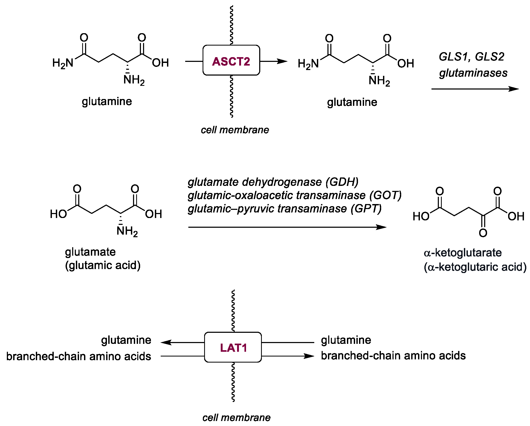
Mitochondrial glutaminases GLS1 and GLS2 catalyze the deamination of glutamine to glutamate. Glutamate is then converted into α-ketoglutarate by glutamate dehydrogenase, glutamic-oxaloacetic transaminase (GOT) or glutamic–pyruvic transaminase (GPT), entering the TCA cycle (Figure 6).
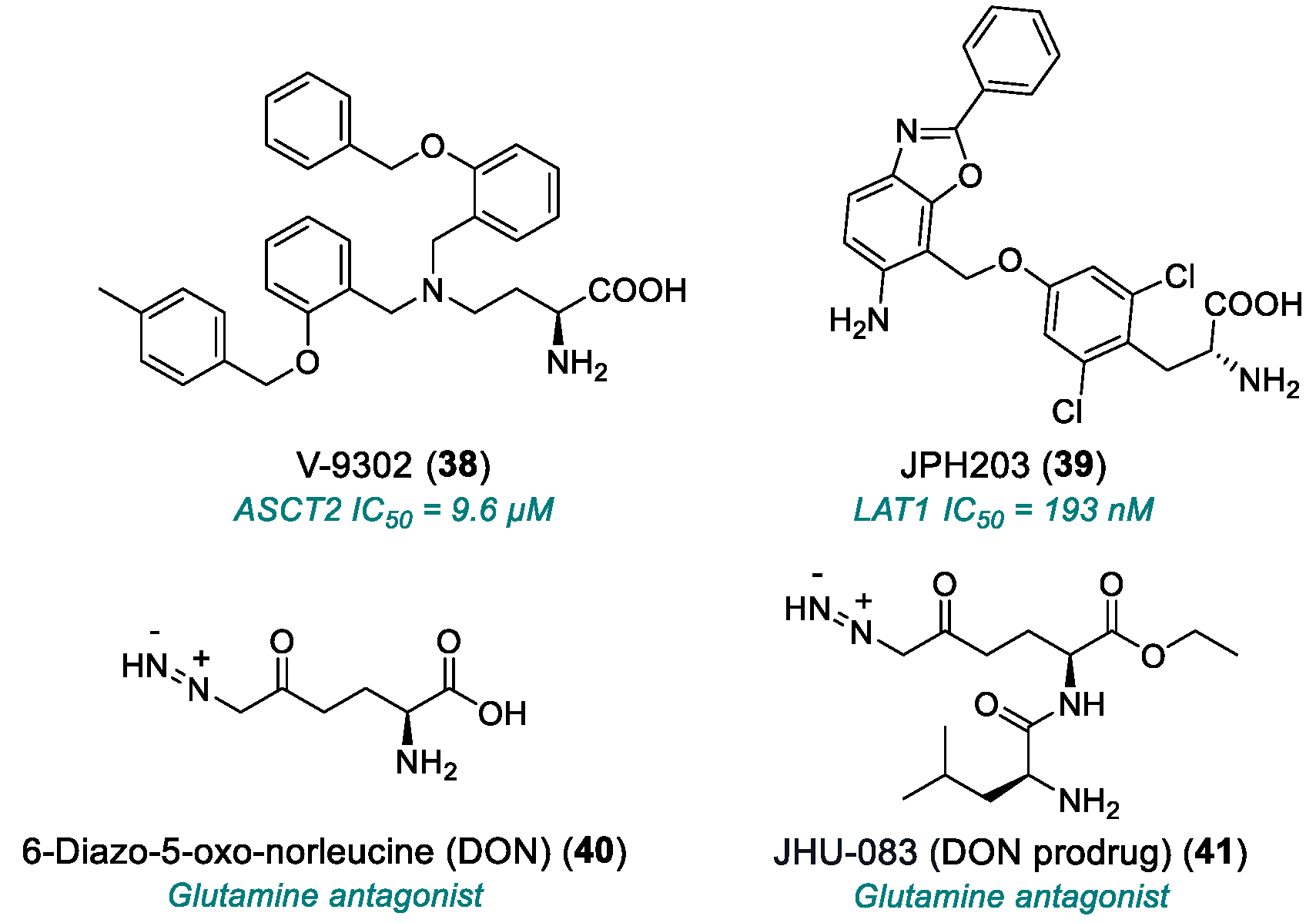
ASCT2 (also known as SLC1A5) is a major sodium-dependent transporter responsible for glutamine uptake [153], associated with the expression of oncogenic MYC and KRAS signaling and playing a role in many clinically relevant tumors [154,155]. Benzyloxy compound V-9302 (38, Chart 6, Table 2) significantly inhibited glutamine uptake mediated by the human ASCT2 isoform in HEK293 cells (IC50 = 9.6 μM) [153]. However, ASCT2 deletion in two human cancer cell lines did not abolish the cell growth inhibition by compound 38 or other ASCT2 inhibitors belonging to the same 2-amino-4-bis(aryloxybenzyl) aminobutanoic acid (AABA) class. AABA compounds inhibited glutamine transport even in ASCT2-deficient cells, but not in parental cells. Deletion of ASCT2 prompted the upregulation of SNAT2, another glutamine transporter, which was also inhibited by AABA compounds. Additionally, AABAs inhibited isoleucine uptake via LAT1, a transporter co-upregulated with ASCT2 in cancer cells [156], and essential for tumorigenesis in a KRAS-mutant model of CRC [157].
JPH203 (39) is anticancer drug targeting L-type amino acid transporter 1 (LAT1, also known as SLC7A5), which plays a primary role in the uptake of essential amino acids in tumor cells. It inhibits LAT1, with an IC50 value of 193 ± 50 nM. Compound 39 inhibits the uptake of branched-chain amino acids, including glutamine. Treatment of LAT1-positive HT-29 colon cancer cells with (39) resulted in a time-dependent reduction in leucine uptake activity. Its inhibitory effect on LAT1 was enhanced by preincubation, suggesting a synergistic mechanism when combined with co-incubation [158].
6-Diazo-5-oxo-norleucine (DON, 40) is a glutamine antagonist originally isolated from the fermentation broth of a Streptomyces in the 1950s [159]. It inhibits several glutamine-utilizing enzymes by a two-step, mechanism-based inhibition [160,161]. Despite its broad enzymatic inhibition, compound 40 showed weak efficacy in vitro and poor tolerability in vivo [162]. JHU-083 (41) is a dual DON prodrug developed to overcome its limitations. Simple alkyl ester-based prodrugs of (40) failed due to the formation of diazoimine byproducts. A successful strategy involved masking both the amine and carboxylate groups. The resulting prodrugs exhibited rapid metabolism in mouse plasma, with several analogs showing excellent stability in monkey and human plasma. These prodrugs achieved superior tumor cell-to-plasma ratio compared to DON [163,164]. The prodrug (41) inhibited cell growth and induced apoptosis in human MYC-expressing medulloblastoma cell lines. It also sensitized C57BL/6 mouse cerebellar stem and progenitor cells in a MYC-driven medulloblastoma mouse model. In immune-competent animals bearing orthotopic tumors, treatment with compound 41 increased survival [165].
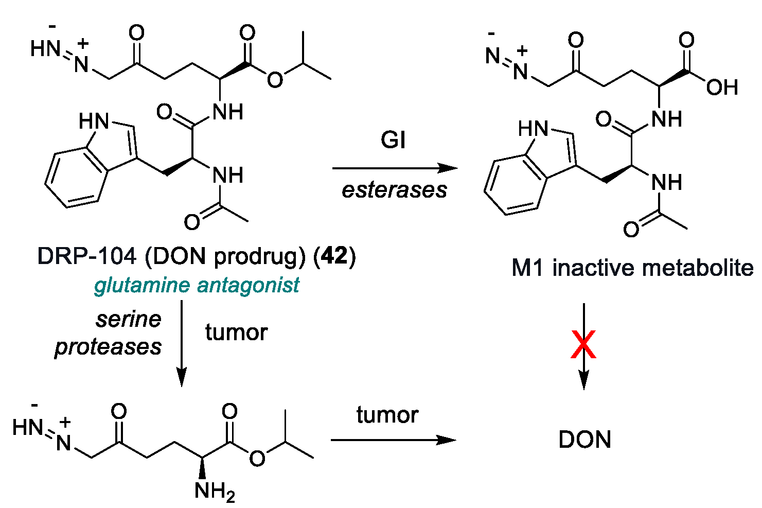
DRP-104, also known as Sirpiglenastat (42, Chart 7), is another DON prodrug that is selectively bioactivated to DON within tumor by serine proteases, while it is inactivated to the M1 metabolite in gastrointestinal tissue by carboxylesterases. This strategy improved drug delivery and reduced systemic toxicity. Compound 42 exhibited antitumor activity similar to (40), but with significantly lower toxicity. It reduced glutamine flux into the TCA cycle and affected multiple metabolic pathways in tumor, including those related to amino acids, nucleotides and carbohydrates/TCA cycle intermediates. The combination of compound 42 with anti-PD-1 immunotherapy showed superior efficacy compared to monotherapies. The antitumor activity of (42) depended on CD8+ T cells, resulting in sustained immunologic memory [166]. Additionally, compound 42 was shown to suppress KEAP1-mutant tumors frequently occurring in lung cancer by inhibiting glutamine-dependent nucleotide synthesis and promoting antitumor T cell responses [167].
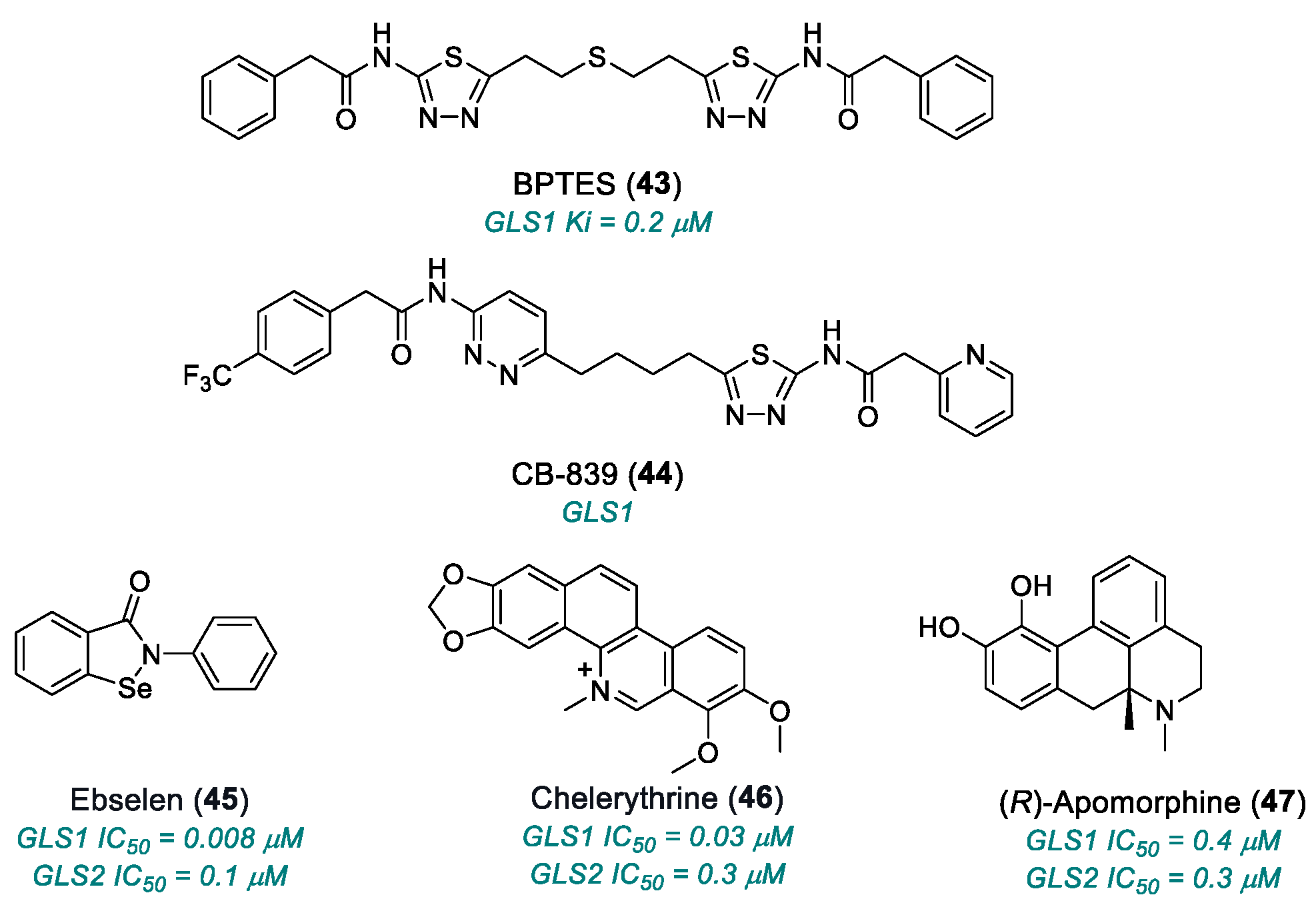
BPTES (bis-2-(5-phenylacetamido-1,3,4-thiadiazol-2-yl)ethyl sulfide) (43, Chart 8) was shown to inhibit selectively the growth of glioma cells [168]. Kinetic analysis indicated that BPTES is a potent uncompetitive inhibitor of GLS1, with a Ki of 0.2 μM. Its structure differs significantly from that of glutamine. The kinetics of BPTES were characterized using human kidney-type glutaminase lacking the C-terminal sequence present in the KGA isoform, which is expressed in the kidney, brain, intestine and cells of the immune system, or in the GAC isoform, expressed in the heart, pancreas, placenta, lung and in many transformed cells [169]. Compound 43 was more potent than DON but exhibited poor solubility and low bioavailability. Attempts to improve these properties through structural analogs were unsuccessful [170,171]. Drug resistance to GLS1-specific inhibitors, such as compound 43 or its analogue, CB-839 (44), also known as Teleglenstat, may be due to the compensatory activity of GLS2, suggesting the need for dual GLS1/GLS2 inhibitors.
Screening of 1280 compounds from the LOPAC1280 library of pharmacologically active compounds led to the identification of ebselen (45), chelerythrine chloride (46) and (R)-apomorphine hydrochloride (47) as potent glutaminase inhibitors. All compounds showed time- and concentration-dependent inhibition of glutaminase activity. Compounds 45 and 46 were 5- to 10-fold, respectively, more potent against GLS1 than GLS2, with IC50 values of 0.008 μM and 0.1 μM for 45 (GLS1 vs GLS2), and IC50 = 0.03 μM and 0.3 for 46 (GLS1 vs GLS2), while compound 47 showed similar activity against both isoforms (IC50 = 0.4 μM and 0.3 μM). Mechanistic studies indicated non-competitive inhibition for (45) (possibly through covalent adduct formation) and competitive inhibition for (46) and (47) [162]. The uncompetitive inhibition by compound 45 aligns with previous results [172], suggesting the formation of a selenenylsulfide (–Se–S–) bond [172] with cysteine residues in the active site of glutaminase [173]. Both (45) and (46) may stabilize the enzyme in an inactive tetrameric configuration or disrupt tetramerization [162].
Table 2. Compounds targeting glutamine metabolism.
NameTargetYearStageLimitationReferences
V-9302 (38)ASCT22016Preclinical studiesEffective only at micromolar range[153,154,155,156,157]
JPH203 (39)LAT12018Phase I completedHepatotoxicity; limited PK data[158]
DON (40)Glutamine antagonist1979Phase I/II completedWeak in vitro efficacy; poo3,172–172r in vivo tolerability; severe GI toxicity[159,160,161,162]
JHU-083 (41)Glutamine antagonist (DON prodrug)2016Phase I/IIToxicity at high doses[163,164,165]
DRP-104 (42)Glutamine antagonist (DON prodrug)2022Phase I/IIResidual toxicity (GI and systemic); narrow therapeutic window; poor PK data[166,167]
BPTES (43)GLS12010Preclinical studiesLimited solubility and stability; micromolar activity[168,169,170,171]
CB-839 (telaglenastat) (44)GLS12014Phase I completedNon-universal efficacy; moderate toxicity[170,171]
ebselen (45)GLS12015Preclinical studiesMulti-target, redox reactive; off-target risk and mitochondrial toxicity[162,172,173,174]
chelerythrine chloride (46)GLS12015Preclinical studiesSignificant inhibition but with off-target cytotoxic effects[162,172,173,174]
(R)-apomorphine hydrochloride (47)GLS1, GLS22015Preclinical studiesLow selectivity[162,172,173,174]

2.3. Fatty Acid Synthesis

Lipid metabolism has attracted significant interest in cancer therapy because lipids are involved in many biochemical processes occurring in cancer initiation and progression [15]. In fact, lipids which comprise a variety of biomolecules made up of fatty acids, play key roles in cancer cell growth, homeostasis, energy metabolism and as structural components of cell membrane [174]. Moreover, they are involved in signal transduction cascades and can be converted into mediators of cancer development [175]. During cancer cell proliferation, the rapid formation of biological membranes demands increased energy and a high rate of lipid synthesis [14].
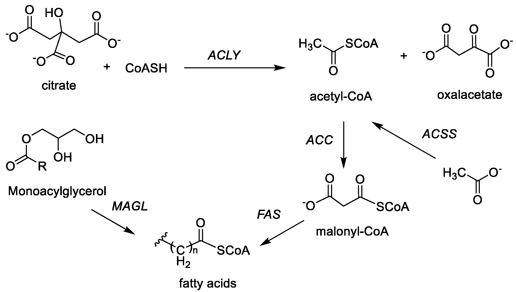
MAGL. Monoacylglycerol lipase (MAGL) oversees the conversion of monoacylglycerols into free fatty acids during lipogenesis in cancer cells, often due to mutation in metabolic pathways. The MAGL overactivation in tumor calls correlates with increased aggressiveness [176]. MAGL inhibition was shown to exert antitumor effects in several cancer cell lines [16,177]; however, the inhibition of MAGL may also cause neurodegenerative, inflammatory and metabolic side effects [178] (Figure 7).
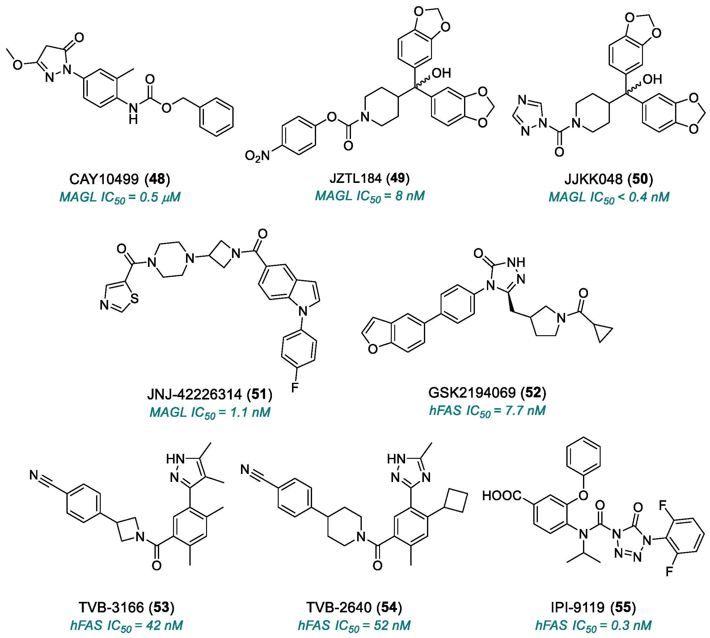
CAY10499 (48, Chart 9, Table 3) inhibited MAGL-mediated 4-nitrophenylacetate (4-NPA) hydrolysis, with IC50 values of 0.5 ± 0.03 μM and of 0.4 ± 0.04 μM using 2-oleoylglycerol (2-OG) as a substrate. Preincubation with compound 48 strongly increased the inhibition to nanomolar IC50 values, suggesting that it binds irreversibly to MAGL, even in the absence of a substrate [179]. Compound 48 was tested against several lipases, including adipose triglyceride lipase (ATGL), hormone-sensitive lipase (HSL), sn-1-diacylglycerol lipase (DAGL), monoacylglycerol lipase, a/b-hydrolase domain 6 and carboxylesterase 1 (CES1), using recombinant human and mouse enzymes, either in cell extracts or using purified enzymes. While (48) inhibited both mouse ATGL and HSL, it showed weaker inhibition of human HSL [180].
JZL184 (49), identified through activity-based protein profiling (ABPP) methods [181], is a potent and selective MAGL inhibitor (IC50 = 8 nM) that provides rapid and sustained MAGL blockade. In the mouse brain proteome, compound 49 potently and selectively inactivated MAGL and increased the 2-arachidonoylglycerol level (2-AG) in male C57Bl/6 mice [182]. It has been proposed for use in treating bone diseases associated with primary bone cancer and bone metastasis; however, activation of the skeletal endocannabinoid system may limit its osteoprotective potential [183]. In osteoblasts, treatment with (49) in the presence of multiple myeloma (MM) cell-derived factors reduced osteoblast proliferation, though not their maturation or bone nodule formation. In an in vivo assay, compound 49 caused moderate but significant bone loss in the long bones of immunocompetent mice inoculated with 5TGM1 MM cells [184].
JJKK-048 (50) was discovered by combining the bulky benzhydryl group of (49), required for selectivity, with a triazole leaving group, leading to a potent human and rodent MAGL inhibitor, with IC50 < 0.4 nM. It likely targets the catalytic serine residue (S122) of MAGL, forming an irreversible covalent bond. In HEK293 cells transiently overexpressing human MAGL (hMAGL), compound 50 inhibited 2-AG hydrolysis in a dose-dependent manner at nanomolar concentrations [185]. MAGL could promote hepatocellular carcinoma (HCC) progression via epithelial–mesenchymal transition (EMT), suggesting its role as a biomarker and potential therapeutic target [186]. Hypoxia-induced resistance to the multi-kinase inhibitor regorafenib, mediated by ABCG2 overexpression, was reduced by MAGL inhibition. Compound 50 lowered ABCG2 overexpression in MDA-MB-231 cells, enhanced regorafenib accumulation and improved its therapeutic response [187].
JNJ-42226314 (51) is a reversible and highly selective MAGL inhibitor (IC50 = 1.1 nM) via a competitive mechanism with respect to the 2-AG substrate. In in vivo studies, it produced antinociceptive effects in both inflammatory and neuropathic pain models. At 30 mg/kg, it induced hippocampal synaptic depression, altered sleep onset and decreased electroencephalogram gamma power. However, at 3 mg/kg, it still achieved approximately 80% enzyme occupancy, increased 2-AG and norepinephrine levels, and maintained antinociceptive activity without cognitive impairment [188].
FAS. Fatty acid synthase (FAS, FASN), encoded in humans by the FASN gene, is a key enzyme in the endogenous lipogenesis. It catalyzes the synthesis of palmitate from acetyl-CoA and malonyl-CoA using NADPH as a reducing agent [189].
GSK2194069 (52), developed at GSK, is a potent hFAS inhibitor, with an IC50 of 7.7 ± 4.1 nM (CoA-release assay) and 29 ± 3.2 nM (acetoacetyl-CoA assay) [190]. It showed little or no inhibition with b-hydroxybutyryl-CoA and crotonyl-CoA and did not inhibit the b-ketoacyl reductase (KR) domain. In gastric and non-small-cell lung cancer cell lines, compound 52 reduced de novo fatty acid synthesis. In the A549 non-small-cell lung cancer cell line, (52) also decreased phosphatidylcholine levels, with an EC50 of 15.5 ± 9 nM, correlating with reduced palmitate production. A crystal structure of compound 52 in the KR domain has been reported [191].
TVB-3166 (53) was reported as a FASN inhibitor (IC50 = 42 nM) with antitumor effects and potential clinical relevance. It showed inhibition in oral squamous cell carcinoma (OSCC) SCC-9 ZsG and metastatic LN-1A cells. Compound 53 significantly reduced cell viability and proliferation, promoted cell cycle arrest and apoptosis, increased adhesion to myogel in both OSCC cell lines and reduced cell migration [192].
TVB-2640 (54) is a selective, potent, reversible FASN inhibitor (IC50 = 52 nM) evaluated for non-alcoholic fatty liver disease (NAFLD) and non-alcoholic steatohepatitis (NASH) correlated with elevated hepatic de novo lipogenesis [193]. In the randomized clinical trial n. NCT03938246, ninety-nine patients received placebo or 25 mg or 50 mg of the drug (orally, once-daily for 12 weeks); compound 54 significantly reduced liver fat and improved biochemical, inflammatory and fibrotic biomarkers in a dose-dependent manner in patients with nonalcoholic steatohepatitis [194].
IPI-9119 (55) is an irreversible FASN inhibitor (IC50 of 0.3 nM) that acylates the thiesterase catalytic domain serine. It selectively inhibited the human purified FASN with negligible off-target effects and suppressed castration-resistant prostate cancer (CRPC) growth by reducing protein expression and transcriptional activity of both the full-length androgen receptor (AR) and the constitutively active AR variant V7 (AR-V7). In vivo, compound 55 inhibited AR-V7-driven CRPC xenografts and human metastatic CRPC-derived organoids and enhanced the efficacy of the androgen receptor inhibitor enzalutamide in CRPC cells [195].
ACLY. Human ATP citrate lyase (ACLY) is a cytosolic enzyme catalyzing the Mg-ATP-dependent conversion of citrate and CoA into acetyl-CoA and oxaloacetate [196]. In mammalian cells, it is mainly localized in the cytosol bound to endoplasmic reticulum, but was also found in the nucleus of human glioblastoma and colon carcinoma cells [197]. Given its central role in lipogenesis, ACLY is a promising target for the treatment of hyperlipidemia and hypercholesterolemia [198,199]. In cancer cells, ACLY plays a role between the glucose and lipid metabolism, two main metabolic alterations typical of tumors, being at the same time at the end of the glycolytic cascade and at the starting point of lipid synthesis. ACLY was shown to be a potential anticancer therapeutic target, since its inhibition interferes with both the glucose metabolism and lipid synthesis [198].
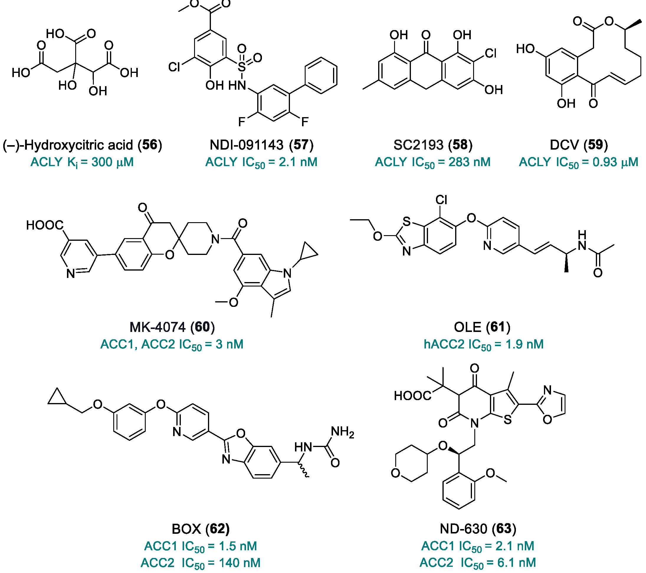
(–)-Hydroxycitric acid (56, Chart 10) is a naturally competitive ACLY inhibitor, with a Ki value of 300 μM. In mice implanted with syngeneic cancer cells, LL/2 Lewis lung carcinoma or MBT-2 murine bladder cancer cells, the triple combination (56) + lipoic acid + cisplatin or methotrexate showed enhanced antitumor efficacy [200].
NDI-091143 (57) was identified as part of a research project aimed at developing a class of 2-hydroxy-N-arylbenzensulfonamides as ACLY inhibitors. Compound 57 showed nanomolar inhibition of human ACLY, with IC50 values in the range of 2.1–4.8 nM, depending on the enzymatic assay, and proved to be competitive with the substrate, citrate [201,202,203]. Cryo-electron microscopy revealed an unexpected mechanism of inhibition: compound 57 binds to an allosteric hydrophobic pocket adjacent to the citrate-binding site, inducing extensive conformational changes in the enzyme that indirectly disrupt citrate binding [201].
SC2193 (58), 2-chloro-1, 3, 8-trihydroxy-6-methyl-9-anthrone, is another naturally occurring anthrone derivative extracted from a soil fungus. The compound showed an IC50 of 283 nM in the primary assay and, more specifically, a Ki of <100 nM against Mg citrate. It demonstrated specific and competitive inhibition of ACL with respect to the substrate Mg citrate and mixed non-competitive inhibition with respect to the essential cofactor Mg-ATP and CoA [204].
10,11-Dehydrocurvularin (DCV, 59) is a fungal-derived natural macrolide lactone of dihydroxyphenylacetic acid, identified via chemical proteomics in living Jurkat cells (IC50 = 0.93 µM). It has demonstrated potential anti-infective, anticancer and immune-modulatory activities [205]. Compound 59 acts as an irreversible inhibitor of ACLY through its α,β-unsaturated carbonyl group, which serves as a Michael acceptor for the alkylation of cysteine-thiol groups in the protein. DCV showed strong antineoplastic activity by inhibiting acute T-lymphocyte leukemia Jurkat cells, as well as HCT116 colorectal carcinoma cells and HeLa cervical adenocarcinoma cells [206].
ACC. Acetyl-CoA carboxylase (ACC) is a biotin-dependent multidomain enzyme that catalyzes the conversion of acetyl-CoA to malonyl-CoA, a critical precursor for fatty acid synthesis via FAS [207]. Two main ACC isoforms have been reported in mammals: ACC1, which is primarily found in the cytosol of liver, adipose tissue and lactating mammary gland, and ACC2, which is commonly located in the outer mitochondrial membrane of oxidative tissues such as skeletal muscle, the heart and the metabolically active liver [14]. ACC1 was found to be upregulated in several types of cancer [208], while ACC2 functions as a key regulator of fatty acid β-oxidation [209].
MK-4074 (60) is a liver-specific, strong dual inhibitor of ACC1 and ACC2, with IC50 values around 3 nM. It effectively suppressed de novo lipogenesis (DNL) and enhanced hepatic fatty acid oxidation (FAO) in cultured hepatocytes, preclinical animal models and clinical studies. Its hepatoselectivity is attributed to its function as a substrate for organic anion transporting polypeptide (OATP) transporters, which are expressed exclusively in hepatocytes. In Phase 1 human studies, conducted in healthy individuals, multiple-dose administration of (60) (140 mg, 7 days) led to maximal DNL inhibition. In a separate short-term study evaluating hepatic steatosis, a 4-week treatment with compound 60 resulted in a 36% reduction in hepatic fat content, compared to an 18% reduction observed with pioglitazone [210].
OLE (61) was developed by replacing the alkyne unit with an olefin linker and modifying the aryl group at the central and left portions of Abbot compound A-908292. Compound 61 exhibited high hACC2 selectivity, with an IC50 of 1.9 nM, compared to an IC50 of 1950 nM for hACC1. These results were confirmed by in vivo studies, in which (61) was effective in C57BL/6 mice and showed no adverse cardiac effects or clinical signs in rats treated orally at 50 mg/kg/day for 4 days [211].
BOX (62) was discovered in a medicinal chemistry campaign aimed at reducing the undesirable body weight effects associated with a previous hit compound. Substitution of the acetamide group with an ureido moiety led to compound 62, which exhibited improved selectivity for mouse ACC1 over ACC2, with IC50 values of 1.5 nM and 140 nM, respectively, and favorable PK in mice. Oral administration significantly reduced the malonyl-CoA concentration in HCT-116 xenograft tumors at doses exceeding 30 mg/kg. Furthermore, compound 62 showed significant antitumor efficacy in 786-O xenograft mice at an oral dose of 30 mg/kg [212].
ND-630 (63) is a potent allosteric ACC inhibitor discovered through structure-based drug design. It binds within the phosphopeptide acceptor and dimerization domain of ACC, thereby preventing dimerization and inhibiting the enzymatic activity of both ACC1 and ACC2 (IC50 = 2.1 nM and 6.1 nM). Compound 63 reduced fatty acid synthesis and promoted fatty acid oxidation in both cell cultures and animal models, displaying favorable drug-like properties. Chronic administration to diet-induced obese rats reduced hepatic steatosis, improved insulin sensitivity, decreased weight gain without affecting food intake and favorably affected dyslipidemia. In chronically treated Zucker diabetic fatty rats, (63) also reduced hepatic steatosis, enhanced glucose-stimulated insulin secretion and lowered hemoglobin A1c by 0.9% [213].
Table 3. Compounds targeting fatty acid synthesis.
NameTargetYearStageLimitationReferences
CAY10499 (48)MAGL2008Preclinical studiesPotential toxicity from normal lipid metabolism (liver, heart); poorly characterized bioavailability/toxicity; endocannabinoid accumulation[180,181]
JZL184 (49)MAGL2009Preclinical studiesOff-target effects at high doses; poorly characterized bioavailability/toxicity; endocannabinoid accumulation[182,183,184]
JJKK-048 (50)MAGL2013Preclinical studiesOff-target effects at high doses; poorly characterized bioavailability/toxicity; endocannabinoid accumulation[186,187,188]
JNJ-42226314 (51)MAGL2020Preclinical studiesEndocannabinoid accumulation[189]
GSK2194069 (52)FASN2011Preclinical studiesLimited oral bioavailability; metabolic and lipid side effects[191,192]
TVB-3166 (53)FASN2015Advanced preclinical studiesMetabolic and lipid side effects[193]
TVB-2640 (54)FASN2017Phase IIRisk of long-term resistance[194,195]
IPI-9119 (55)FASN2020Preclinical studiesIrreversible inhibitor[196]
(–)-Hydroxycitric acid (56)ACLY2012Preclinical studiesLow bioavailability[201]
NDI-091143 (57)ACLY2019Preclinical studiesNo PK studies; limited toxicity data[202,203,204]
SC2193 (58)ACLY2017Preclinical studiesNo PK studies; limited toxicity data[205]
10,11-Dehydrocurvularin, DCV (59)ACLY2015Preclinical studiesPoorly defined toxicity; instability; multi-target activity[206,207]
MK-4074 (60)ACC1, ACC22017Preclinical studiesNo PK studies; limited toxicity data[211]
OLE (61)hACC22018Preclinical studiesNo PK studies; limited toxicity data[212]
BOX (62)ACC12019Preclinical studiesNo PK studies; limited toxicity data[213]
ND-630 (63)ACC1, ACC22016Preclinical studiesNo PK studies; limited toxicity data[214]

2.4. Structural Considerations of Metabolic Targets

Recent structural studies of key metabolic enzymes targeted by small-molecule inhibitors have provided crucial insights into the binding and mechanisms of these potential antitumor compounds. These enzymes, often featuring conserved catalytic cores or allosteric regulatory domains, present well-defined pockets suitable for selective targeting.
For example, LDHA contains a highly conserved catalytic site, where residues such as Arg168, His192 and Asp165 play a critical role in pyruvate binding and NADH-mediated reduction to lactate. Co-crystal structures of LDHA-inhibitor complexes, such as TODP (5), highlight direct interactions with these residues, explaining the observed potency and selectivity [42].
Similarly, in HK2, both the glucose and ATP-binding pockets are critical for enzymatic function. The interaction with VDAC1 at the mitochondrial membrane allows coupling with ATP synthesis. Structural models (e.g., PDB ID: 2NZT) demonstrate how small molecules and G6P-mimicking inhibitors, such as compounds 28–30, exploit the enzyme’s flexible domain architecture [78]. This structural flexibility, particularly in the G6P allosteric site, enables the rational design of compounds that either compete with endogenous ligands or prevent conformational transitions required for activity.
GLUT1, a transmembrane transporter, has a central channel composed of helical bundles that accommodate glucose transport. Inhibitors such as STF-31 (12) and BAY-876 (18) bind within this channel, occluding glucose passage. Docking studies of GLUT1 analogs have highlighted roles for residues such as Phe 379 and Trp 388 in ligand binding [214].
Moreover, PKM2 represents another well-studied target, featuring both an active site and a regulatory allosteric pocket. The glycolytic flux can be modulated by allosteric activators (ML-265, 33) or inhibitors such as (31), (32) or natural compounds (silibinin 34 and curcumin 36), which can stabilize either the active tetrameric or inactive dimeric forms. Residues such as Arg399 have been implicated in conformational switching, particularly in the transition between inactive and active states, and are frequently involved in ligand binding [215].
Regarding FASN, crystal structures have resolved several domains, including the β-ketoacyl synthase and thioesterase domains, which are responsible for palmitate synthesis. Compounds such as TVB-2640 (54) bind selectively to the β-ketoacyl reductase domain, interacting with key catalytic residues, thereby disrupting lipid biosynthesis [216].
These structural insights guide the rational design of potent, selective inhibitors by targeting specific enzyme conformations or domains to overcome drug resistance and improve therapy. They also explain variations in compound efficacy across isoforms and tumor types, while the distinction between orthosteric and allosteric inhibition highlights the versatility of these targets and the opportunities to fine-tune therapeutic responses through domain-specific modulation.

3. Perspectives and Emerging Strategies in Tumor Metabolism

3.1. Metabolic Crosstalk and Compensatory Mechanisms

Numerous recent studies have highlighted how altered metabolic pathways in cancer do not operate in isolation but are highly interconnected and subject to functional compensation. This dynamic network, commonly referred to as metabolic crosstalk, presents a significant challenge to the efficacy of targeted metabolic therapies, yet also opens new avenues for more rational combinatorial strategies.
In particular, the inhibition of a single metabolic pathway often induces the compensatory activation of alternative routes. For example, glutaminase (GLS) inhibition has been shown to result in increased utilization of glucose and fatty acids, which can be metabolized through glycolysis or β-oxidation to support ATP production and the synthesis of biosynthetic intermediates. Furthermore, in certain cancer contexts, blockade of glutaminolysis has been associated with the reactivation of mitochondrial oxidative phosphorilation (OXPHOS), contributing to therapeutic resistance [217].
A promising strategy to overcome such compensations is the use of combination therapies. One notable example is AZD3965 (11), an inhibitor of the lactate transporter MCT1, which has been shown to enhance the activity of glutaminase inhibitors. This synergistic effect stems from the simultaneous inhibition of lactate efflux and glutamine metabolism, leading to redox collapse and suppression of tumor proliferation [218].
Similarly, inhibition of LDH-A, a key glycolysis enzyme, can be circumvented by cancer cells via enhancement of OXPHOS. In preclinical models of pancreatic cancer, treatment with GNE-140 (3), a potent LDH-A inhibitor, resulted in mitochondrial compensation, mediated by the activation of the AMPK-mTOR-S6K pathway. The combination of GNE-140 with phenformin (4), an OXPHOS inhibitor, successfully overcame metabolic resistance and restored treatment sensitivity [219].
The accumulation of metabolic intermediates from compensatory pathways can also contribute to the epigenetic modification of the tumor genome, with direct effects on gene expression and cancer progression. For instance, intracellular lactate levels can influence histone lactylation (HKla), an epigenetic mark recently associated with the regulation of pro-inflammatory and pro-tumor genes [220]. Thus, modulating metabolic crosstalk may also have significant epigenetic implications.
Overall, the expanding knowledge of tumor metabolic networks requires a paradigm shift: from single-target interventions to multi-target strategies capable of simultaneously inhibiting critical metabolic nodes and adaptive pathways. The identification and characterization of these adaptive circuits are increasingly facilitated by integrated metabolomics techniques, CRISPR-based screening systems and network modeling.

3.2. Emerging Technologies: PROTACs and Targeted Degradation of Metabolic Proteins

An area of growing interest in anticancer therapy is targeted protein degradation through technologies such as PROTACs (PROteolysis Targeting Chimeras). These bifunctional molecules promote the selective degradation of target proteins by recruiting specific E3 ligases, leading to polyubiquitination and subsequent proteasomal degradation.
Compared to traditional inhibitors, PROTACs offer several advantages: they act catalytically, require lower concentrations and can target previously considered “undruggable” proteins. Furthermore, by eliminating the entire protein rather than merely inhibiting its active site, PROTACs reduce the likelihood of resistance driven by binding site mutations [221].
Recent studies have reported promising preclinical results for PROTACs targeting metabolic enzymes such as LDH-A, PKM2 and GLS1. Some of these agents have been designed as tumor-selective pro-drugs, activated under specific enzymatic or acidic conditions present in the tumor microenvironment, thus improving selectivity for neoplastic cells [222].
Other emerging technologies include AUTACs (Autophagy-Targeting Chimeras) and ATTECs (Autophagosome-Tethering Compounds), which exploit the autophagic pathway to degrade not only proteins but also organelles and metabolic aggregates. These approaches offer novel therapeutic possibilities, particularly in tumors reliant on organelles such as mitochondria or lipid droplets [223].

3.3. Precision Metabolic Oncology: New Therapeutic Frontiers

The marked metabolic heterogeneity both between patients and within individual tumors presents a challenge, but also a unique opportunity, for personalized therapy. The integration of high-resolution metabolomics, multi-omics profiling and technologies such as single-cell RNA sequencing enables the identification of tumor-specific metabolic signatures and the prediction of drug responses.
A prominent example is DRP-104 (42), a prodrug derived from DON (40), which is selectively activated in the tumor microenvironment. Compound 42 has demonstrated not only direct antitumor effects but also the ability to enhance antitumor immunity, acting synergistically with anti-PD-1 antibodies in mouse models [224].
These findings suggest that metabolic modulation not only affects the intrinsic characteristics of tumor cells but can also reshape the tumor microenvironment, providing valuable support for immunotherapeutic strategies.

3.4. Concluding Remark

The identification of redundant metabolic pathways and crosstalk circuits, coupled with the advent of emerging technologies, such as PROTACs and targeted degradation platforms, is revolutionizing the landscape of metabolic cancer therapy. The future of the field lies in the integration of rational combinatorial approaches, grounded in a robust understanding of adaptive mechanisms and systems biology. In this context, precision metabolic medicine is no longer a distant goal, but a tangible and achievable reality.

4. Summary and Conclusions

Over the past decades, cancer metabolism has emerged as a promising and rapidly expanding field for the development of novel anticancer strategies. The reprogramming of metabolic pathways, long recognized as a hallmark of cancer, provides malignant cells with the necessary biochemical resources to support uncontrolled proliferation, resistance to apoptosis, immune evasion and metastasis. This review has highlighted recent advances in the identification and characterization of small-molecule inhibitors targeting key metabolic enzymes involved in glycolysis, glutaminolysis and fatty acid synthesis—processes that are consistently upregulated in various tumor types.
By targeting enzymes such as LDHA, GLUT1, HK2, PKM2, GLS1/2, FASN, ACLY and ACC, researchers have demonstrated the feasibility of disrupting tumor bioenergetics and biosynthetic machinery. Notably, several of these compounds, including BAY-876 (18), ML-265 (33), TVB-2640 (54) and DRP-104 (42), have progressed to preclinical or clinical stages, underscoring the translational potential of metabolism-targeting therapies. Additionally, the structural and mechanistic diversity of these inhibitors, including orthosteric, allosteric, covalent and substrate-mimicking strategies, offers new avenues for specificity and reduced off-target effects.
Moreover, the therapeutic relevance of metabolic modulation extends beyond tumor cell-intrinsic effects. Compounds such as (42) and MAGL inhibitors have demonstrated the ability to reshape the tumor microenvironment and potentiate antitumor immune responses. This aligns with a broader shift toward combinatorial approaches that integrate metabolic inhibitors with immunotherapies, chemotherapeutic agents, or targeted drugs.
Nevertheless, several challenges remain. Tumor heterogeneity, metabolic plasticity, and compensatory pathways can undermine the efficacy of single-agent treatments. The development of reliable metabolic biomarkers, patient stratification strategies and predictive models of response will be crucial to guide clinical application. Furthermore, a deeper understanding of the systemic effects and potential toxicities associated with long-term metabolic inhibition is needed.
Recent insights into metabolic crosstalk and compensatory mechanisms have further highlighted the need for multi-targeted and context-specific approaches. The emergence of targeted protein degradation technologies, such as PROTACs, and advances in metabolomic profiling are paving the way for precision oncology strategies that exploit tumor-specific metabolic vulnerabilities.
In conclusion, targeting tumor metabolism represents a compelling and versatile approach in the anticancer drug discovery landscape. Continued interdisciplinary efforts combining medicinal chemistry, structural biology, cancer biology and translational research will be key to unlocking the full therapeutic potential of metabolic interventions and delivering innovative treatments to patients with high unmet medical needs.

Author Contributions

Investigation, M.P.; writing—review and editing, M.P.; writing—original draft preparation, R.S. and G.L.R.; supervision, R.S. and G.L.R. All authors have read and agreed to the published version of the manuscript.

Funding

The authors wish to acknowledge AIRC IG 2020 n. 24703 granted to R.S.; MIUR PRIN 2022 2022TPPNTK (European Union-Next Generation EU) awarded to G.L.R.; Sapienza University of Rome RG11816428A9B4D5 and RM120172A7EAD07C granted to R.S.; RM1241904EB3828D and RG123188B4D193AE awarded to G.L.R; and AR224190786E7FA3 granted to M.P.

Conflicts of Interest

The authors declare no conflicts of interest.

Abbreviations

The following abbreviations are used in this manuscript:
AATaspartate aminotransferase
ACCacetyl-coa carboxylase
ACLYhuman atp citrate lyase
AIFapoptosis-inducing factor
ASCT2alanine, serine, cysteine transporter 2
ATPadenosine triphosphate
BBBblood–brain barrier
CDKscyclin-dependent kinases
CRPCcastration-resistant prostate cancer
DLBCLdiffuse large b cell lymphoma
DNMTDNA methyltransferase
ECMextracellular matrix
FADHflavin adenine dinucleotide
FASfatty acid synthase
FASNfatty acid synthase
G6Pglucose-6-phosphate
GLS1glutaminase 1
GLUTglucose transporter
GLUT1glucose transporter type 1
GLUT1DSglucose transporter type 1 deficiency syndrome
GOTglutamic-oxaloacetic transaminase
GPTglutamic–pyruvic transaminase
HCChepatocellular carcinoma
HKhexokinase
HKlahistone lysine lactylation
HNSCChead and neck squamous cell carcinoma
LAT1l-type amino acid transporter 1
LDHlactate dehydrogenase
LDHAlactate dehydrogenase type a
LDHBlactate dehydrogenase type b
MAGLmonoacylglycerol lipase
MCT1monocarboxylate transporter 1
NADHnicotinamide adenine dinucleotide
NHLnon-Hodgkin lymphomas
NSCLCnon-small-cell lung cancer
OMMouter mitochondrial membrane
OXPHOSoxidative phosphorylation
PKpyruvate kinase
PKM1pyruvate kinase m1
PKM2pyruvate kinase m2
RCCsrenal cell carcinomas
ROSreactive oxygen species
SARstructure-activity relationship
TCAtricarboxylic acid
TMJOAtemporomandibular joint osteoarthritis
TNBCin triple-negative breast cancer
VDACvoltage-dependent anion channels
VHLvon Hippel–Lindau

References

  1. Warburg, O. On the Origin of Cancer Cells. Science 1956, 123, 309–314. [Google Scholar] [CrossRef]
  2. Martínez-Reyes, I.; Chandel, N.S. Cancer Metabolism: Looking Forward. Nat. Rev. Cancer 2021, 21, 669–680. [Google Scholar] [CrossRef] [PubMed]
  3. Ngo, D.C.; Ververis, K.; Tortorella, S.M.; Karagiannis, T.C. Introduction to the Molecular Basis of Cancer Metabolism and the Warburg Effect. Mol. Biol. Rep. 2015, 42, 819–823. [Google Scholar] [CrossRef]
  4. Lunt, S.Y.; Vander Heiden, M.G. Aerobic Glycolysis: Meeting the Metabolic Requirements of Cell Proliferation. Annu. Rev. Cell Dev. Biol. 2011, 27, 441–464. [Google Scholar] [CrossRef]
  5. Kim, J.W.; Dang, C.V. Cancer’s Molecular Sweet Tooth and the Warburg Effect. Cancer Res. 2006, 66, 8927–8930. [Google Scholar] [CrossRef]
  6. DeBerardinis, R.J.; Lum, J.J.; Hatzivassiliou, G.; Thompson, C.B. The Biology of Cancer: Metabolic Reprogramming Fuels Cell Growth and Proliferation. Cell Metab. 2008, 7, 11–20. [Google Scholar] [CrossRef] [PubMed]
  7. Liberti, V.M.; Locasale, J.W. The Warburg effect: How does it benefit cancer cells? Trends Biochem. Sci. 2016, 41, 211–218. [Google Scholar] [CrossRef] [PubMed]
  8. Schwartz, L.; Supuran, T.C.; Alfarouk, O.K. The Warburg effect and the hallmarks of cancer. Anti-Cancer Agents Med. Chem. 2017, 17, 164–170. [Google Scholar] [CrossRef]
  9. Vander Heiden, M.G.; Cantley, L.C.; Thompson, C.B. Understanding the Warburg effect: The metabolic requirements of cell proliferation. Science 2009, 324, 1029–1033. [Google Scholar] [CrossRef]
  10. Shaw, R.J. Glucose metabolism and cancer. Curr. Opin. Cell Biol. 2006, 18, 598–608. [Google Scholar] [CrossRef]
  11. Gillies, R.J.; Robey, I.; Gatenby, R.A. Causes and consequences of increased glucose metabolism of cancers. J. Nucl. Med. 2008, 49, 24S–42S. [Google Scholar] [CrossRef]
  12. Hanahan, D.; Weinberg, R.A. Hallmarks of cancer: The next generation. Cell 2011, 144, 646–674. [Google Scholar] [CrossRef] [PubMed]
  13. Hanahan, D. Hallmarks of cancer: New dimensions. Cancer Discov. 2022, 12, 31–46. [Google Scholar] [CrossRef] [PubMed]
  14. Currie, E.; Schulze, A.; Zechner, R.; Walther, T.C.; Farese, R. V Jr. Cellular fatty acid metabolism and cancer. Cell Metab. 2013, 18, 153–161. [Google Scholar] [CrossRef]
  15. Beloribi-Djefaflia, S.; Vasseur, S.; Guillaumond, F. Lipid metabolic reprogramming in cancer cells. Oncogenesis 2016, 5, e189. [Google Scholar] [CrossRef]
  16. Montesdeoca, N.; López, M.; Ariza, X.; Herrero, L.; Makowski, K. Inhibitors of lipogenic enzymes as a potential therapy against cancer. FASEB J. 2020, 34, 11355–11381. [Google Scholar] [CrossRef]
  17. Stine, Z.E.; Schug, Z.T.; Salvino, J.M.; Dang, C.V. Targeting cancer metabolism in the era of precision oncology. Nat. Rev. Drug Discov. 2022, 21, 141–162. [Google Scholar] [CrossRef]
  18. Masci, D.; Puxeddu, M.; Silvestri, R.; La Regina, G. Metabolic rewiring in cancer: Small molecule inhibitors in colorectal cancer therapy. Molecules 2024, 29, 2110. [Google Scholar] [CrossRef] [PubMed]
  19. Rattigan, Y.; Patel, B.; Ackerstaff, E.; Sukenick, G.; Koutcher, J.; Glod, J.; Banerjee, D. Lactate is a mediator of metabolic cooperation between stromal carcinoma associated fibroblasts and, glycolytic tumor cells in the tumor microenvironment. Exp. Cell Res. 2012, 318, 326–335. [Google Scholar] [CrossRef]
  20. Ristow, M. Oxidative metabolism in cancer growth. Curr. Opin. Clin. Nutr. Metab. Care 2006, 9, 339–345. [Google Scholar] [CrossRef]
  21. Fantin, V.R.; St-Pierre, J.; Philip, L. Attenuation of LDH-A expression uncovers a link between glycolysis, mitochondrial physiology, and tumor maintenance. Cancer Cell 2006, 9, 425–434. [Google Scholar] [CrossRef]
  22. Di Magno, L.; Coluccia, A.; Bufano, M.; Ripa, S.; La Regina, G.; Nalli, M.; Di Pastena, F.; Canettieri, G.; Silvestri, R.; Frati, L. Discovery of novel human lactate dehydrogenase inhibitors: Structure-based virtual screening studies and biological assessment. Eur. J. Med. Chem. 2022, 240, 114605. [Google Scholar] [CrossRef]
  23. Zhou, Y.; Tao, P.; Wang, M.; Xu, P.; Lu, W.; Lei, P.; You, Q. Development of novel human lactate dehydrogenase A inhibitors: High-throughput screening, synthesis, and biological evaluations. Eur. J. Med. Chem. 2019, 177, 105–115. [Google Scholar] [CrossRef] [PubMed]
  24. Bononi, G.; Di Bussolo, V.; Tuccinardi, T.; Minutolo, F.; Granchi, C. A patent review of lactate dehydrogenase inhibitors (2014–present). Expert Opin. Ther. Pat. 2024, 34, 1121–1135. [Google Scholar] [CrossRef] [PubMed]
  25. Zhang, S.L.; He, Y.; Tam, K.Y. Targeting cancer metabolism to develop human lactate dehydrogenase (hLDH)5 inhibitors. Drug Discov. Today 2018, 23, 1407–1415. [Google Scholar] [CrossRef] [PubMed]
  26. Farhana, A.; Lappin, S.L. Biochemistry, Lactate Dehydrogenase; StatPearls Publishing: Treasure Island, FL, USA, 2023. [Google Scholar]
  27. Dang, C.V.; Semenza, G.L. Oncogenic alterations of metabolism. Trends Biochem. Sci. 1999, 24, 68–72. [Google Scholar] [CrossRef]
  28. Han, J.H.; Lee, E.J.; Park, W.; Ha, K.T.; Chung, H.S. Natural compounds as lactate dehydrogenase inhibitors: Potential therapeutics for lactate dehydrogenase inhibitors-related diseases. Front. Pharmacol. 2023, 14, 1275000. [Google Scholar] [CrossRef]
  29. Granchi, C.; Bertini, S.; Macchia, M.; Minutolo, F. Inhibitors of lactate dehydrogenase isoforms and their therapeutic potentials. Curr. Med. Chem. 2010, 17, 672–697. [Google Scholar] [CrossRef]
  30. Thornburg, J.M.; Nelson, K.K.; Clem, B.F.; Lane, A.N.; Arumugam, S.; Simmons, A.; Eaton, J.W.; Telang, S.; Chesney, S.J. Targeting aspartate aminotransferase in breast cancer. Breast Cancer Res. 2008, 10, R84. [Google Scholar] [CrossRef]
  31. Fiume, L.; Manerba, M.; Vettraino, M.; Di Stefano, G. Impairment of aerobic glycolysis by inhibitors of lactic dehydrogenase hinders the growth of human hepatocellular carcinoma cell lines. Pharmacology 2010, 86, 157–162. [Google Scholar] [CrossRef]
  32. Goldberg, E.; Nitowsky, H.; Colowick, S. The role of glycolysis in the growth of tumor cells The basis of glucose toxicity in oxamate-treated cultured cells. J. Biol. Chem. 1965, 240, 2791–2796. [Google Scholar] [CrossRef]
  33. Billiard, J.; Dennison, J.B.; Briand, J.; Annan, R.S.; Chai, D.; Colon, M.; Dodson, C.S.; Gilbert, S.A.; Greshock, J.; Jing, J.; et al. Quinoline 3-sulfonamides inhibit lactate dehydrogenase A and reverse aerobic glycolysis in cancer cells. Cancer Metab. 2013, 1, 19. [Google Scholar] [CrossRef]
  34. Cisewski, S.E.; Zhang, L.; Kuo, J.; Wright, G.J.; Wu, Y.; Kern, M.J.; Yao, H. Thee ffects of oxygen level and glucose concentration on the metabolism of porcine TMJ disc cells. Osteoarthr. Cartil. 2015, 23, 1790–1796. [Google Scholar] [CrossRef]
  35. Li, H.M.; Guo, H.L.; Xu, C.; Liu, L.; Hu, S.Y.; Hu, Z.H.; Jiang, H.H.; He, Y.M.; Li, Y.J.; Ke, J.; et al. Inhibition of glycolysis by targeting lactate dehydrogenase A facilitates hyaluronan synthase 2 synthesis in synovial fibroblasts of temporomandibular joint osteoarthritis. Bone 2020, 141, 115584. [Google Scholar] [CrossRef]
  36. Wong, N.; De Melo, J.; Tang, D. PKM2, a central point of regulation in cancer metabolism. Int. J. Cell Biol. 2013, 2013, 242513. [Google Scholar] [CrossRef] [PubMed]
  37. Boudreau, A.; Purkey, H.E.; Hitz, A.; Robarge, K.; Peterson, D.; Labadie, S.; Kwong, M.; Hong, R.; Gao, M.; Del Nagro, C.; et al. Metabolic plasticity underpins innate and acquired resistance to LDHA inhibition. Nat. Chem. Biol. 2016, 12, 779–786. [Google Scholar] [CrossRef] [PubMed]
  38. Abcam. Oxidative Phosphorylation Pathway. Available online: https://www.abcam.com/en-us/technical-resources/pathways/oxidative-phosphorylation-pathway (accessed on 1 November 2024).
  39. Zhao, S.S.; Liu, J.; Wu, Q.C.; Zhou, X.L. Lactate regulates pathological cardiac hypertrophy via histone lactylation modification. J. Cell Mol. Med. 2024, 28, e70022. [Google Scholar] [CrossRef] [PubMed]
  40. Zhai, M.; Cui, S.; Li, L.; Cheng, C.; Zhang, Z.; Liu, J.; Wei, F. Mechanical force modulates alveolar bone marrow mesenchymal cells characteristics for bone remodeling during orthodontic tooth movement through lactate production. Cells 2022, 11, 3724. [Google Scholar] [CrossRef]
  41. Ma, W.; Ao, S.; Zhou, J.; Li, J.; Liang, X.; Yang, X.; Zhang, H.; Liu, B.; Tang, W.; Liu, H.; et al. Methylsulfonylmethane protects against lethal dose MRSA-induced sepsis through promoting M2 macrophage polarization. Mol. Immunol. 2022, 146, 69–77. [Google Scholar] [CrossRef]
  42. Dragovich, P.S.; Fauber, B.P.; Corson, L.B.; Ding, C.Z.; Eigenbrot, C.; Ge, H.; Giannetti, A.M.; Hunsaker, T.; Labadie, S.; Liu, Y.; et al. Identification of substituted 2-thio-6-oxo-1,6-dihydropyrimidines as inhibitors of human lactate dehydrogenase. Bioorg. Med. Chem. Lett. 2013, 23, 3186–3194. [Google Scholar] [CrossRef]
  43. Fauber, B.P.; Dragovich, P.S.; Chen, J.; Corson, L.B.; Ding, C.Z.; Eigenbrot, C.; Giannetti, A.M.; Hunsaker, T.; Labadie, S.; Liu, Y.; et al. Identification of 2-amino-5-aryl-pyrazines as inhibitors of human lactate dehydrogenase. Bioorg. Med. Chem. Lett. 2013, 23, 5533–5539. [Google Scholar] [CrossRef]
  44. Rai, G.; Brimacombe, K.R.; Mott, B.T.; Urban, D.J.; Hu, X.; Yang, S.M.; Lee, T.D.; Cheff, D.M.; Kouznetsova, J.; Benavides, G.A.; et al. Discovery and Optimization of Potent, Cell-Active Pyrazole-Based Inhibitors of Lactate Dehydrogenase (LDH). J. Med. Chem. 2017, 60, 9184–9204. [Google Scholar] [CrossRef]
  45. Rai, G.; Urban, D.J.; Mott, B.T.; Hu, X.; Yang, S.M.; Benavides, G.A.; Johnson, M.S.; Squadrito, G.L.; Brimacombe, K.R.; Lee, T.D.; et al. Pyrazole-Based Lactate Dehydrogenase Inhibitors with Optimized Cell Activity and Pharmacokinetic Properties. J. Med. Chem. 2020, 63, 10984–11011. [Google Scholar] [CrossRef]
  46. Ludwig, J.A. Ewing sarcoma: Historical perspectives, current state-of-the-art, and opportunities for targeted therapy in the future. Curr. Opin. Oncol. 2008, 20, 412–418. [Google Scholar] [CrossRef]
  47. Durer, S.; Gasalberti, D.P.; Shaikh, H. Ewing Sarcoma. In StatPearls [Internet]; NIH, National Library of Medicine: Bethesda, MD, USA, 2024. Available online: https://www.ncbi.nlm.nih.gov/books/NBK559183/ (accessed on 15 November 2024).
  48. Yeung, C.; Gibson, A.E.; Issaq, S.H.; Oshima, N.; Baumgart, J.T.; Edessa, L.D.; Rai, G.; Urban, D.J.; Johnson, M.S.; Benavides, G.A.; et al. Targeting Glycolysis through inhibition of lactate dehydrogenase impairs tumor growth in preclinical models of Ewing sarcoma. Cancer Res. 2019, 79, 5060–5073. [Google Scholar] [CrossRef]
  49. Friberg, A.; Rehwinkel, H.; Nguyen, D.; Pütter, V.; Quanz, M.; Weiske, J.; Eberspächer, U.; Heisler, I.; Langer, G. Structural Evidence for Isoform-Selective Allosteric Inhibition of Lactate Dehydrogenase A. ACS Omega 2020, 5, 13034–13041. [Google Scholar] [CrossRef] [PubMed]
  50. Saito, M. Subunit cooperativity in the action of lactate dehydrogenase. Biochim. Biophys. Acta 1972, 258, 17–26. [Google Scholar] [CrossRef] [PubMed]
  51. Masterson, J.E.; Schwartz, S.D. Changes in protein architecture and subpicosecond protein dynamics impact the reaction catalyzed by lactate dehydrogenase. J. Phys. Chem. A 2013, 17, 7107–7113. [Google Scholar] [CrossRef]
  52. Curtis, N.J.; Mooney, L.; Hopcroft, L.; Michopoulos, F.; Whalley, N.; Zhong, H.; Murray, C.; Logie, A.; Revill, M.; Byth, K.F.; et al. Pre-clinical pharmacology of AZD3965, a selective inhibitor of MCT1: DLBCL, NHL and Burkitt’s lymphoma anti-tumor activity. Oncotarget 2017, 8, 69219–69236. [Google Scholar] [CrossRef]
  53. Doherty, J.R.; Yang, C.; Scott, K.E.; Cameron, M.D.; Fallahi, M.; Li, W.; Hall, M.A.; Amelio, A.L.; Mishra, J.K.; Li, F.; et al. Blocking lactate export by inhibiting the Myc target MCT1 Disables glycolysis and glutathione synthesis. Cancer Res. 2014, 74, 908–920. [Google Scholar] [CrossRef] [PubMed]
  54. Beloueche-Babari, M.; Casals Galobart, T.; Delgado-Goni, T.; Wantuch, S.; Parkes, H.G.; Tandy, D.; Harker, J.A.; Leach, M.O. Monocarboxylate transporter 1 blockade with AZD3965 inhibits lipid biosynthesis and increases tumour immune cell infiltration. Br. J. Cancer 2020, 122, 895–903. [Google Scholar] [CrossRef]
  55. Halford, S.; Veal, G.J.; Wedge, S.R.; Payne, G.S.; Bacon, C.M.; Sloan, P.; Dragoni, I.; Heinzmann, K.; Potter, S.; Salisbury, B.M.; et al. A phase I dose-escalation dtudy of AZD3965, an oral Monocarboxylate Transporter 1 inhibitor, in patients with advanced cancer. Clin. Cancer Res. 2023, 29, 1429–1439. [Google Scholar] [CrossRef] [PubMed]
  56. Landowski, C.P.; Suzuki, Y.; Hediger, M.A. The Mammalian Transporter Families. In Seldin and Giebisch’s The Kidney: Physiology and Pathophysiology, 4th ed.; Alpern, R.J., Hebert, S.C., Eds.; Elsevier: Oxford, UK, 2008; pp. 91–146. [Google Scholar]
  57. Mueckler, M.; Caruso, C.; Baldwin, S.A.; Panico, M.; Blench, I.; Morris, H.R.; Allard, W.J.; Lienhard, G.E.; Lodish, H.F. Sequence and structure of a human glucose transporter. Science 1985, 229, 941–945. [Google Scholar] [CrossRef]
  58. Tang, M.; Park, S.H.; De Vivo, D.C.; Monani, U.R. Therapeutic strategies for glucose transporter 1 deficiency syndrome. Ann. Clin. Transl. Neurol. 2019, 6, 1923–1932. [Google Scholar] [CrossRef]
  59. De Vivo, D.C.; Trifiletti, R.R.; Jacobson, R.I.; Ronen, G.M.; Behmand, R.A.; Harik, S.I. Defective glucose transport across the blood-brain barrier as a cause of persistent hypoglycorrhachia, seizures, and developmental delay. N. Engl. J. Med. 1991, 325, 703–709. [Google Scholar] [CrossRef]
  60. Vulturar, R.; Chiș, A.; Pintilie, S.; Farcaș, I.M.; Botezatu, A.; Login, C.C.; Sitar-Taut, A.V.; Orasan, O.H.; Stan, A.; Lazea, C.L.; et al. One molecule for mental nourishment and more: Glucose transporter type 1-biology and deficiency syndrome. Biomedicines 2022, 10, 1249. [Google Scholar] [CrossRef] [PubMed]
  61. Santer, R.; Klepper, J. Disorders of Glucose Transport in Inherited Metabolic Diseases, Diagnostic and Treatment, 6th ed.; Saudubray, J.M., Baumgartner, M., Waler, J., Eds.; Springer: Berlin/Heidelberg, Germany, 2016; pp. 175–184. [Google Scholar]
  62. Chan, D.A.; Sutphin, P.D.; Nguyen, P.; Turcotte, S.; Lai, E.W.; Banh, A.; Reynolds, G.E.; Chi, J.T.; Wu, J.; Solow-Cordero, D.E.; et al. Targeting GLUT1 and the Warburg effect in renal cell carcinoma by chemical synthetic lethality. Sci. Transl. Med. 2011, 3, 94ra70. [Google Scholar] [CrossRef]
  63. Turcotte, S.; Chan, D.A.; Sutphin, P.D.; Hay, M.P.; Denny, W.A.; Giaccia, A.J. A molecule targeting VHL-deficient renal cell carcinoma that induces autophagy. Cancer Cell 2008, 14, 90–102. [Google Scholar] [CrossRef]
  64. Hay, M.P.; Turcotte, S.; Flanagan, J.U.; Bonnet, M.; Chan, D.A.; Sutphin, P.D.; Nguyen, P.; Giaccia, A.J.; Denny, W.A. 4-Pyridylanilinothiazoles that selectively target von Hippel–Lindau deficient renal cell carcinoma cells by inducing autophagic cell death. J. Med. Chem. 2010, 53, 787–797. [Google Scholar] [CrossRef]
  65. Wood, T.E.; Dalili, S.; Simpson, C.D.; Hurren, R.; Mao, X.; Saiz, F.S.; Gronda, M.; Eberhard, Y.; Minden, M.D.; Bilan, P.J.; et al. A novel inhibitor of glucose uptake sensitizes cells to FAS-induced cell death. Mol. Cancer Ther. 2008, 7, 3546–3555. [Google Scholar] [CrossRef] [PubMed]
  66. Lu, J.; Montgomery, B.K.; Chatain, G.P.; Bugarini, A.; Zhang, Q.; Wang, X.; Edwards, N.A.; Ray-Chaudhury, A.; Merrill, M.J.; Lonser, R.R.; et al. Corticotropin releasing hormone can selectively stimulate glucose uptake in corticotropinoma via glucose transporter 1. Mol. Cell Endocrinol. 2018, 470, 105–114. [Google Scholar] [CrossRef] [PubMed]
  67. Ocaña, M.C.; Martínez-Poveda, B.; Marí-Beffa, M.; Quesada, A.R.; Medina, M.Á. Fasentin diminishes endothelial cell proliferation, differentiation and invasion in a glucose metabolism-independent manner. Sci. Rep. 2020, 10, 6132. [Google Scholar] [CrossRef] [PubMed]
  68. Zhang, W.; Liu, Y.; Chen, X.; Bergmeier, S.C. Novel inhibitors of basal glucose transport as potential anticancer agents. Bioorg. Med. Chem. Lett. 2010, 20, 2191–2194. [Google Scholar] [CrossRef] [PubMed]
  69. Liu, Y.; Zhang, W.; Cao, Y.; Bergmeier, S.; Chen, X. Small compound inhibitors of basal glucose transport inhibit cell proliferation and induce apoptosis in cancer cells via glucose-deprivation-like mechanisms. Cancer Lett. 2010, 298, 176–185. [Google Scholar] [CrossRef]
  70. Liu, Y.; Cao, Y.; Zhang, W.; Bergmeier, S.; Qian, Y.; Akbar, H.; Colvin, R.; Ding, J.; Tong, L.; Wu, S.; et al. A small-molecule inhibitor of glucose transporter 1 downregulates glycolysis, induces cell-cycle arrest, and inhibits cancer cell growth in vitro and in vivo. Mol. Cancer Ther. 2012, 11, 1672–1682. [Google Scholar] [CrossRef]
  71. Ojelabi, O.A.; Lloyd, K.P.; Simon, A.H.; De Zutter, J.K.; Carruthers, A. WZB117 (2-Fluoro-6-(m-hydroxybenzoyloxy) Phenyl m-Hydroxybenzoate) Inhibits GLUT1-mediated Sugar Transport by Binding Reversibly at the Exofacial Sugar Binding Site. J. Biol. Chem. 2016, 291, 26762–26772. [Google Scholar] [CrossRef]
  72. Siebeneicher, H.; Cleve, A.; Rehwinkel, H.; Neuhaus, R.; Heisler, I.; Müller, T.; Bauser, M.; Buchmann, B. Identification and Optimization of the First Highly Selective GLUT1 Inhibitor BAY-876. ChemMedChem 2016, 11, 2261–2271. [Google Scholar] [CrossRef]
  73. Miller, Z.A.; Muthuswami, S.; Mueller, A.; Ma, R.Z.; Sywanycz, S.M.; Naik, A.; Huang, L.; Brody, R.M.; Diab, A.; Carey, R.M.; et al. GLUT1 inhibitor BAY-876 induces apoptosis and enhances anti-cancer effects of bitter receptor agonists in head and neck squamous carcinoma cells. Cell Death Discov. 2024, 10, 339. [Google Scholar] [CrossRef]
  74. Pedersen, P.L. Warburg, me and Hexokinase 2: Multiple discoveries of key molecular events underlying one of cancers’ most common phenotypes, the “Warburg Effect”, i.e., elevated glycolysis in the presence of oxygen. J. Bioenerg. Biomembr. 2007, 39, 211–222. [Google Scholar] [CrossRef]
  75. Irwin, D.M.; Tan, H. Molecular evolution of the vertebrate hexokinase gene family: Identification of a conserved fifth vertebrate hexokinase gene. Comp. Biochem. Physiol. Part D Genom. Proteom. 2008, 3, 96–107. [Google Scholar] [CrossRef]
  76. Patra, K.C.; Wang, Q.; Bhaskar, P.T.; Miller, L.; Wang, Z.; Wheaton, W.; Chandel, N.; Laakso, M.; Muller, W.J.; Allen, E.L.; et al. Hexokinase 2 is required for tumor initiation and maintenance and its systemic deletion is therapeutic in mouse models of cancer. Cancer Cell 2013, 24, 213–228. [Google Scholar] [CrossRef]
  77. Anderson, M.; Marayati, R.; Moffitt, R.; Yeh, J.J. Hexokinase 2 promotes tumor growth and metastasis by regulating lactate production in pancreatic cancer. Oncotarget 2016, 8, 56081–56094. [Google Scholar] [CrossRef] [PubMed]
  78. Shan, W.; Zhou, Y.; Tam, K.Y. The development of small-molecule inhibitors targeting hexokinase 2. Drug Discov. Today 2022, 27, 2574–2585. [Google Scholar] [CrossRef]
  79. Benz, R. Permeation of hydrophilic solutes through mitochondrial outer membranes-review on mitochondrial porins. Biophys. Acta 1994, 1197, 167–196. [Google Scholar] [CrossRef] [PubMed]
  80. De Pinto, V.; Guarino, F.; Guarnera, A.; Messina, A.; Reina, S.; Tomasello, F.M.; Palermo, V.; Mazzoni, C. Characterization of human VDAC isoforms: A peculiar function for VDAC3? Biochim. Biophys. Acta 2010, 1797, 1268–1275. [Google Scholar] [CrossRef] [PubMed]
  81. Pastorino, J.G.; Hoek, J.B. Regulation of hexokinase binding to VDAC. J. Bioenerg. Biomembr. 2008, 40, 171–182. [Google Scholar] [CrossRef]
  82. Salani, B.; Marini, C.; Rio, A.D.; Ravera, S.; Massollo, M.; Orengo, A.M.; Amaro, A.; Passalacqua, M.; Maffioli, S.; Pfeffer, U.; et al. Metformin impairs glucose con- sumption and survival in Calu-1 cells by direct inhibition of hexokinase-II. Sci. Rep. 2013, 3, 2070. [Google Scholar] [CrossRef]
  83. Marini, C.; Salani, B.; Massollo, M.; Amaro, A.; Esposito, A.I.; Orengo, A.M.; Capitanio, S.; Emionite, L.; Riondato, M.; Bottoni, G.; et al. Direct inhibition of hexokinase activity by metformin at least partially impairs glucose metabolism and tumor growth in experimental breast cancer. Cell Cycle 2013, 12, 3490–3499. [Google Scholar] [CrossRef]
  84. John, S.; Weiss, J.N.; Ribalet, B. Subcellular localization of hexokinases I and II directs the metabolic fate of glucose. PLoS ONE 2011, 6, e17674. [Google Scholar] [CrossRef]
  85. Bustamante, E.; Pedersen, P.L. High aerobic glycolysis of rat hepatoma cells in culture: Role of mitochondrial hexokinase. Proc. Natl. Acad. Sci. USA 1977, 74, 3735–3739. [Google Scholar] [CrossRef]
  86. Chen, Z.; Zhang, H.; Lu, W.; Huang, P. Role of mitochondria-associated hexokinase II in cancer cell death induced by 3-bromopyruvate. Biochim. Biophys. Acta 2009, 1787, 553–560. [Google Scholar] [CrossRef]
  87. Ríos, J.L. Chemical constituents and pharmacological properties of Poria cocos. Planta Med. 2011, 77, 681–691. [Google Scholar] [CrossRef] [PubMed]
  88. Miao, G.; Han, J.; Zhang, J.; Wu, Y.; Tong, G. Targeting Pyruvate Kinase M2 and Hexokinase II, Pachymic acid impairs glucose metabolism and induces mitochondrial apoptosis. Biol. Pharm. Bull. 2019, 42, 123–129. [Google Scholar] [CrossRef]
  89. Floridi, A.; Paggi, M.G.; Marcante, M.L.; Silvestrini, B.; Caputo, A.; De Martino, C. Lonidamine, a selective inhibitor of aerobic glycolysis of murine tumor cells. J. Natl. Cancer Inst. 1981, 66, 497–499. [Google Scholar]
  90. Ben-Yoseph, O.; Lyons, J.C.; Song, C.W.; Ross, B.D. Mechanism of action of lonidamine in the 9L brain tumor model involves inhibition of lactate efflux and intracellular acidification. J. Neurooncol. 1998, 36, 149–157. [Google Scholar] [CrossRef]
  91. Ravagnan, L.; Marzo, I.; Costantini, P.; Susin, S.A.; Zamzami, N.; Petit, P.X.; Hirsch, F.; Goulbern, M.; Poupon, M.F.; Miccoli, L.; et al. Lonidamine triggers apoptosis via a direct, Bcl-2-inhibited effect on the mitochondrial permeability transition pore. Oncogene 1999, 18, 2537–2546. [Google Scholar] [CrossRef]
  92. Tidmarsh, G. Therapies for the Treatment of Cancer. U.S. Patent US20060276527A1, 7 December 2006. [Google Scholar]
  93. Tang, Q.; Ji, F.; Guo, J.; Wang, J.; Li, Y.; Bao, Y. Directional modification of chrysin for exerting apoptosis and enhancing significantly anti-cancer effects of 10- hydroxy camptothecin. Biomed. Pharmacother. 2016, 82, 693–703. [Google Scholar] [CrossRef]
  94. Pichichero, E.; Cicconi, R.; Mattei, M.; Muzi, M.G.; Canini, A. Acacia honey and chrysin reduce proliferation of melanoma cells through alterations in cell cycle progression. Int. J. Oncol. 2010, 37, 973–981. [Google Scholar] [CrossRef] [PubMed]
  95. Xia, Y.; Lian, S.; Khoi, P.N.; Yoon, H.J.; Han, J.Y.; Chay, K.O.; Kim, K.K.; Jung, Y.D. Chrysin inhibits cell invasion by inhibition of Recepteur d’origine Nantais via suppressing early growth response-1 and NF-kappaB transcription factor activities in gastric cancer cells. Int. J. Oncol. 2015, 46, 1835–1843. [Google Scholar] [CrossRef] [PubMed]
  96. Gao, A.M.; Ke, Z.P.; Shi, F.; Sun, G.C.; Chen, H. Chrysin enhances sensitivity of BEL- 7402/ADM cells to doxorubicin by suppressing PI3K/Akt/Nrf2 and ERK/Nrf2 pathway. Chem. Biol. Interact. 2013, 206, 100–108. [Google Scholar] [CrossRef]
  97. Xu, D.; Jin, J.; Yu, H.; Zhao, Z.; Ma, D.; Zhang, C.; Jiang, H. Chrysin inhibited tumor glycolysis and induced apoptosis in hepatocellular carcinoma by targeting hexokinase-2. J. Exp. Clin. Cancer Res. 2017, 36, 44. [Google Scholar] [CrossRef] [PubMed]
  98. Zhou, L.; Li, M.; Yu, X.; Gao, F.; Li, W. Repression of hexokinases II-mediated glycolysis contributes to piperlongumine-induced tumor suppression in non- small cell lung cancer cells. Int. J. Biol. Sci. 2019, 15, 826–837. [Google Scholar] [CrossRef]
  99. Pelicano, H.; Martin, D.S.; Xu, R.A.; Huang, P. Glycolysis inhibition for anticancer treatment. Oncogene 2006, 25, 4633–4646. [Google Scholar] [CrossRef]
  100. Zhu, G.; Guo, N.; Yong, Y.; Xiong, Y.; Tong, Q. Effect of 2-deoxy-D-glucose on gellan gum biosynthesis by Sphingomonas paucimobilis. Bioprocess. Biosyst. Eng. 2019, 42, 897–900. [Google Scholar] [CrossRef]
  101. Singh, R.; Gupta, V.; Kumar, A.; Singh, K. 2-Deoxy-D-glucose: A novel pharmacological agent for killing hypoxic tumor cells, oxygen dependence-lowering in COVID-19, and other pharmacological activities. Adv. Pharmacol. Pharm. Sci. 2023, 2023, 9993386. [Google Scholar] [CrossRef]
  102. IFokt, S.; Skora, C.; Conrad, T.; Madden, M.; Emmett, W. Priebe- D-glucose and D-mannose-based metabolic probes. Part 3: Synthesis of specifcally deuterated D-glucose, -mannose, and 2-deoxy-D-glucose. Carbohydr. Res. 2013, 368, 111–119. [Google Scholar]
  103. Xi, H.; Kurtoglu, M.; Lampidis, T.J. The wonders of 2- deoxy-D-glucose. IUBMB Life 2014, 66, 110–121. [Google Scholar] [CrossRef]
  104. Cunha, A.; Rocha, A.C.; Barbosa, F.; Baião, A.; Silva, P.; Sarmento, B.; Queirós, O. Glycolytic inhibitors potentiated the activity of paclitaxel and their nanoencapsulation increased their delivery in a lung cancer model. Pharmaceutics 2022, 14, 2021. [Google Scholar] [CrossRef] [PubMed]
  105. Francis, R.; Singh, P.K.; Singh, S.; Giri, S.; Kumar, A. Glycolytic inhibitor 2-deoxyglucose suppresses inflammatory response in innate immune cells and experimental staphylococcal endophthalmitis. Exp. Eye Res. 2020, 97, 108079. [Google Scholar] [CrossRef] [PubMed]
  106. Mohanty, J.G.; Rosenthal, K.S. 2-deoxy-D-glucose inhibition of herpes simplex virus type-1 receptor expression. Antivir. Res. 1986, 6, 137–149. [Google Scholar] [CrossRef]
  107. Dey, S.; Murmu, N.; Mondal, T.; Saha, I.; Chatterjee, S.; Manna, R.; Haldar, S.; Dash, S.K.; Sarkar, T.R.; Giri, B. Multifaceted entrancing role of glucose and its analogue, 2-deoxy-D-glucose in cancer cell proliferation, inflammation, and virus infection. Biomed. Pharmacother. 2022, 156, 113801. [Google Scholar] [CrossRef]
  108. Yamaguchi, R.; Janssen, E.; Perkins, G.; Ellisman, M.; Kitada, S.; Reed, J.C. Efficient elimination of cancer cells by deoxyglucose-ABT-263/737 combination therapy. PLoS ONE 2011, 6, e24102. [Google Scholar] [CrossRef]
  109. Zhang, D.; Li, J.; Wang, F.; Hu, J.; Wang, S.; Sun, Y. 2-Deoxy-D-glucose targeting of glucose metabolism in cancer cells as a potential therapy. Cancer Lett. 2014, 355, 176–183. [Google Scholar] [CrossRef] [PubMed]
  110. Lampidis, T.J.; Kurtoglu, M.; Maher, J.C.; Liu, H.; Krishan, A.; Sheft, V.; Szymanski, S.; Fokt, I.; Rudnicki, W.R.; Ginalski, K.; et al. Efficacy of 2-halogen substituted D-glucose analogs in blocking glycolysis and killing “hypoxic tumor cells”. Cancer Chemother. Pharmacol. 2006, 58, 725–734. [Google Scholar] [CrossRef] [PubMed]
  111. Zhao, J.; Ma, Y.; Zhang, Y.; Fu, B.; Wu, X.; Li, Q.; Cai, G.; Chen, X.; Bai, X.Y. Low-dose 2-deoxyglucose and metformin synergically inhibit proliferation of human polycystic kidney cells by modulating glucose metabolism. Cell Death Discov. 2019, 5, 76. [Google Scholar] [CrossRef]
  112. Cheong, J.H.; Park, E.S.; Liang, J.; Dennison, J.B.; Tsavachidou, D.; Nguyen-Charles, C.; Wa Cheng, K.; Hall, H.; Zhang, D.; Lu, Y.; et al. Dual inhibition of tumor energy pathway by 2-deoxyglucose and metformin is effective against a broad spectrum of preclinical cancer models. Mol. Cancer Ther. 2011, 10, 2350–2362. [Google Scholar] [CrossRef] [PubMed]
  113. Li, W.; Zheng, M.; Wu, S.; Gao, S.; Yang, M.; Li, Z.; Min, Q.; Sun, W.; Chen, L.; Xiang, G.; et al. Benserazide, a dopadecarboxylase inhibitor, suppresses tumor growth by targeting hexokinase 2. J. Exp. Clin. Cancer Res. 2017, 36, 58. [Google Scholar] [CrossRef]
  114. Chiara, F.; Castellaro, D.; Marin, O.; Petronilli, V.; Brusilow, W.S.; Juhaszova, M.; Sollott, S.J.; Forte, M.; Bernardi, P.; Rasola, A. Hexokinase II detachment from mitochondria triggers apoptosis through the permeability transition pore independent of voltage-dependent anion channels. PLoS ONE 2008, 3, e1852. [Google Scholar] [CrossRef]
  115. Zheng, M.; Wu, C.; Yang, K.; Yang, Y.; Liu, Y.; Gao, S.; Wang, Q.; Li, C.; Chen, L.; Li, H. Novel selective hexokinase 2 inhibitor Benitrobenrazide blocks cancer cells growth by targeting glycolysis. Pharmacol. Res. 2021, 164, 105367. [Google Scholar] [CrossRef] [PubMed]
  116. Mulichak, A.M.; Wilson, J.E.; Padmanabhan, K.; Garavito, R.M. The mammalian hexokinase-1. Nat. Struct. Biol. 1998, 5, 555–560. [Google Scholar] [CrossRef]
  117. Lin, H.; Zeng, J.; Xie, R.; Schulz, M.J.; Tedesco, R.; Qu, J.; Erhard, K.F.; Mack, J.F.; Raha, K.; Rendina, A.R.; et al. Discovery of a novel 2,6-disubstituted glucosamine series of potent and selective Hexokinase 2 inhibitors. ACS Med. Chem. Lett. 2015, 7, 217–222. [Google Scholar] [CrossRef]
  118. Deeb, S.S.; Malkki, M.; Laakso, M. human hexokinase II: Sequence and homology to other hexokinases. Biochem. Biophys. Res. Commun. 1993, 197, 68–74. [Google Scholar] [CrossRef]
  119. Yang, W.; Lu, Z. Pyruvate kinase M2 at a glance. J. Cell Sci. 2015, 128, 1655–1660. [Google Scholar] [CrossRef]
  120. Heng, T.S.; Painter, M.W.; The Immunological Genome Project Consortium; Elpek, K.; Lukacs-Kornek, V.; Mauermann, N.; Turley, S.J.; Koller, D.; Kim, F.S.; Wagers, A.J.; et al. The Immunological Genome Project: Networks of gene expression in immune cells. Nat. Immunol. 2008, 9, 1091–1094. [Google Scholar] [CrossRef]
  121. Alves-Filho, J.C.; Pålsson-McDermott, E.M. Pyruvate kinase M2: A potential target for regulating inflammation. Front. Immunol. 2016, 7, 145. [Google Scholar] [CrossRef]
  122. Toller-Kawahisa, J.E.; Hiroki, C.H.; de Souza Silva, C.M.; Nascimento, D.C.; Pùblio, G.A.; Varela Martins, T.; Alves Damascenco, L.E.; Protàsio Veras, F.; Ramos Viacava, P.; Yuji Sukesada, F.; et al. The metabolic function of pyruvate kinase M2 regulates reactive oxygen species production and microbial killing by neutrophils. Nat. Commun. 2023, 14, 4280. [Google Scholar] [CrossRef] [PubMed]
  123. Suzuki, A.; Puri, S.; Leland, P.; Puri, A.; Moudgil, T.; Fox, B.A.; Puri, R.K.; Joshi, B.H. Subcellular compartmentalization of PKM2 identifies anti-PKM2 therapy response in vitro and in vivo mouse model of human non-small-cell lung cancer. PLoS ONE 2019, 14, e0217131. [Google Scholar] [CrossRef] [PubMed]
  124. Zhou, Y.; Huang, Z.; Su, J.; Li, J.; Zhao, S.; Wu, L.; Zhang, J.; He, Y.; Zhang, G.; Tao, J.; et al. Benserazide is a novel inhibitor targeting PKM2 for melanoma treatment. Int. J. Cancer 2020, 147, 139–151. [Google Scholar] [CrossRef] [PubMed]
  125. Druzhyna, N.; Szczesny, B.; Olah, G.; Módis, K.; Asimakopoulou, A.; Pavlidou, A.; Szoleczky, P.; Gerö, D.; Yanagi, K.; Törö, G.; et al. Screening of a composite library of clinically used drugs and well-characterized pharmacological compounds for cystathionine β-synthase inhibition identifies benserazide as a drug potentially suitable for repurposing for the experimental therapy of colon cancer. Pharmacol. Res. 2016, 113, 18–37. [Google Scholar]
  126. Ning, X.; Qi, H.; Li, R.; Li, Y.; Jin, Y.; McNutt, M.A.; Liu, J.; Yin, Y. Discovery of novel naphthoquinone derivatives as inhibitors of the tumor cell specific M2 isoform of pyruvate kinase. Eur. J. Med. Chem. 2017, 138, 343–352. [Google Scholar] [CrossRef]
  127. Ning, X.; Qi, H.; Li, R.; Jin, Y.; McNutt, M.A.; Yin, Y. Synthesis and antitumor activity of novel 2, 3-didithiocarbamate substituted naphthoquinones as inhibitors of pyruvate kinase M2 isoform. J. Enzym. Inhib. Med. Chem. 2018, 33, 126–129. [Google Scholar] [CrossRef]
  128. Gao, C.L.; Hou, G.G.; Liu, J.; Ru, T.; Xu, Y.Z.; Zhao, S.Y.; Ye, H.; Zhang, L.Y.; Chen, K.X.; Guo, Y.W.; et al. Synthesis and target identification of benzoxepane derivatives as potential anti-neuroinflammatory agents for ischemic stroke. Angew. Chem. Int. Ed. Engl. 2020, 59, 2429–2439. [Google Scholar] [CrossRef]
  129. Zhong, W.J.; Yang, H.H.; Guan, X.X.; Xiong, J.B.; Sun, C.C.; Zhang, C.Y.; Luo, X.Q.; Zhang, Y.F.; Zhang, J.; Duan, J.X.; et al. Inhibition of glycolysis alleviates lipopolysaccharide-induced acute lung injury in a mouse model. J. Cell Physiol. 2019, 234, 4641–4654. [Google Scholar] [CrossRef]
  130. Xie, M.; Yu, Y.; Kang, R.; Zhu, S.; Yang, L.; Zeng, L.; Sun, X.; Yang, M.; Billiar, T.R.; Wang, H.; et al. PKM2-dependent glycolysis promotes NLRP3 and AIM2 inflammasome activation. Nat. Commun. 2016, 7, 13280. [Google Scholar] [CrossRef]
  131. Wubben, T.J.; Pawar, M.; Weh, E.; Smith, A.; Sajjakulnukit, P.; Zhang, L.; Dai, L.; Hager, H.; Pai, M.P.; Lyssiotis, C.A.; et al. Small molecule activation of metabolic enzyme pyruvate kinase muscle isozyme 2, PKM2, circumvents photoreceptor apoptosis. Sci. Rep. 2020, 10, 2990. [Google Scholar] [CrossRef]
  132. Sayyad, Z.; Sirohi, K.; Radha, V.; Swarp, G. 661W is a retinal ganglion precursor-like cell line in which glaucoma-associated optineurin mutants induce cell death selectively. Sci. Rep. 2017, 7, 16855. [Google Scholar] [CrossRef] [PubMed]
  133. Jiang, J.K.; Walsh, M.J.; Brimacombe, K.R.; Anastasiou, D.; Yu, Y.; Israelsen, W.J.; Hong, B.S.; Tempel, W.; Dimov, S.; Veith, H.; et al. ML265: A potent PKM2 activator induces tetramerization and reduces tumor formation and size in a mouse xenograft model. In Probe Reports from the NIH Molecular Libraries Program [Internet]; National Center for Biotechnology Information: Bethesda, MD, USA, 2012. Available online: https://www.ncbi.nlm.nih.gov/books/NBK153222/ (accessed on 9 July 2025).
  134. Sarfraz, I.; Rasul, A.; Jabeen, F.; Sultana, T.; Adem, Ş. Identification of natural compounds as inhibitors of pyruvate kinase M2 for cancer treatment. Molecules 2022, 27, 7113. [Google Scholar] [CrossRef]
  135. Tuli, H.S.; Mittal, S.; Aggarwal, D.; Parashar, G.; Parashar, N.C.; Upadhyay, S.K.; Barwal, T.S.; Jain, A.; Kaur, G.; Savla, R.; et al. Path of Silibinin from diet to medicine: A dietary polyphenolic flavonoid having potential anti-cancer therapeutic significance. Semin. Cancer Biol. 2021, 73, 196–218. [Google Scholar] [CrossRef] [PubMed]
  136. Zhu, X.X.; Ding, Y.H.; Wu, Y.; Qian, L.Y.; Zou, H.; He, Q. Silibinin: A potential old drug for cancer therapy. Expert. Rev. Clin. Pharmacol. 2016, 9, 1323–1330. [Google Scholar] [CrossRef] [PubMed]
  137. Mateen, S.; Raina, K.; Agarwal, C.; Chan, D.; Agarwal, R. Silibinin synergizes with histone deacetylase and DNA methyltransferase inhibitors in upregulating E-cadherin expression together with inhibition of migration and invasion of human non-small cell lung cancer cells. J. Pharmacol. Exp. Ther. 2013, 345, 206–214. [Google Scholar] [CrossRef]
  138. Kauntz, H.; Bousserouel, S.; Gossé, F.; Raul, F. Epigenetic effects of the natural flavonolignan silibinin on colon adenocarcinoma cells and their derived metastatic cells. Oncol. Lett. 2013, 5, 1273–1277. [Google Scholar] [CrossRef]
  139. Iqbal, M.A.; Chattopadhyay, S.; Siddiqui, F.A.; Ur Rehman, A.; Siddiqui, S.; Prakasam, G.; Khan, A.; Sultana, S.; Bamezai, R.N. Silibinin induces metabolic crisis in triple-negative breast cancer cells by modulating EGFR-MYC-TXNIP axis: Potential therapeutic implications. FEBS J. 2021, 288, 471–485. [Google Scholar] [CrossRef]
  140. Existing Food Additives, Ministry of Health, Labor and Welfare of Japan, Notification n.120. 1996. Available online: https://www.ffcr.or.jp/en/tenka/list-of-existing-food-additives/list-of-existing-food-additives.html (accessed on 26 February 2020).
  141. Tasaki, M.; Umemura, T.; Maeda, M.; Ishii, Y.; Okamura, T.; Inoue, T.; Kuroiwa, Y.; Hirose, M.; Nishikawa, A. Safety assessment of ellagic acid, a food additive, in a subchronic toxicity study using F344 rats. Food Chem. Toxicol. 2008, 46, 1119–1124. [Google Scholar] [CrossRef]
  142. Devipriya, N.; Srinivasan, M.; Sudheer, A.R.; Menon, V.P. Effect of ellagic acid, a natural polyphenol, on alcohol-induced prooxidant and antioxidant imbalance: A drug dose dependent study. Singap. Med. J. 2007, 48, 311–318. [Google Scholar]
  143. Ceci, C.; Lacal, P.M.; Tentori, L.; De Martino, M.G.; Miano, R.; Graziani, G. Experimental evidence of the antitumor, antimetastatic and antiangiogenic activity of ellagic acid. Nutrients 2018, 10, 1756. [Google Scholar] [CrossRef]
  144. Yousuf, M.; Shamsi, A.; Khan, P.; Shahbaaz, M.; AlAjmi, M.F.; Hussain, A.; Hassan, G.M.; Islam, A.; Rizwanul Haque, Q.M.; Hassan, M.I. Ellagic acid controls cell proliferation and induces apoptosis in breast cancer cells via inhibition of cyclin-dependent kinase 6. Int. J. Mol. Sci. 2020, 21, 3526. [Google Scholar] [CrossRef] [PubMed]
  145. Gupta, P.; Mohammad, T.; Khan, P.; Alajmi, M.F.; Hussain, A.; Rehman, M.T.; Hassan, M.I. Evaluation of ellagic acid as an inhibitor of sphingosine kinase 1: A targeted approach towards anticancer therapy. Biomed. Pharmacother. 2019, 118, 109245. [Google Scholar] [CrossRef]
  146. Stanić, Z. Curcumin, a compound from natural sources, a true scientific challenge—A review. Plant Foods Hum. Nutr. 2017, 72, 1–12. [Google Scholar] [CrossRef]
  147. Siddiqui, F.A.; Prakasam, G.; Chattopadhyay, S.; Rehman, A.U.; Padder, R.A.; Ansari, M.A.; Irshad, R.; Mangalhara, K.; Bamezai, R.N.K.; Husain, M.; et al. Curcumin decreases Warburg effect in cancer cells by down-regulating pyruvate kinase M2 via mTOR-HIF1α inhibition. Sci. Rep. 2018, 8, 8323. [Google Scholar] [CrossRef] [PubMed]
  148. Baur, J.A.; Sinclair, D.A. Therapeutic potential of resveratrol: The in vivo evidence. Nat. Rev. Drug Discov. 2006, 5, 493–506. [Google Scholar] [CrossRef]
  149. Jang, M.; Cai, L.; Udeani, G.O.; Slowing, K.V.; Thomas, C.F.; Beecher, C.W.; Fong, H.H.; Farnsworth, N.R.; Kinghorn, A.D.; Mehta, R.G.; et al. Cancer chemopreventive activity of resveratrol, a natural product derived from grapes. Science 1997, 275, 218–220. [Google Scholar] [CrossRef]
  150. Iqbal, M.A.; Bamezai, R.N. Resveratrol inhibits cancer cell metabolism by down regulating pyruvate kinase M2 via inhibition of mammalian target of rapamycin. PLoS ONE 2012, 7, e36764. [Google Scholar] [CrossRef]
  151. Sun, Q.; Chen, X.; Ma, J.; Peng, H.; Wang, F.; Zha, X.; Wang, Y.; Jing, Y.; Yang, H.; Chen, R.; et al. Mammalian target of rapamycin up-regulation of pyruvate kinase isoenzyme type M2 is critical for aerobic glycolysis and tumor growth. Proc. Natl. Acad. Sci. USA 2011, 108, 4129–4134. [Google Scholar] [CrossRef]
  152. Varoni, E.M.; Lo Faro, A.F.; Sharifi-Rad, J.; Iriti, M. Anticancer molecular mechanisms of resveratrol. Front. Nutr. 2016, 3, 8. [Google Scholar] [CrossRef]
  153. Schulte, M.L.; Khodadadi, A.B.; Cuthbertson, M.L.; Smith, J.A.; Manning, H.C. 2-Amino-4-bis(aryloxybenzyl)aminobutanoic acids: A novel scaffold for inhibition of ASCT2-mediated glutamine transport. Bioorg. Med. Chem. Lett. 2016, 26, 1044–1047. [Google Scholar] [CrossRef]
  154. Shimizu, K.; Kaira, K.; Tomizawa, Y.; Sunaga, N.; Kawashima, O.; Oriuchi, N.; Tominaga, H.; Nagamori, S.; Kanai, Y.; Yamada, M.; et al. ASC amino-acid transporter 2 (ASCT2) as a novel prognostic marker in non-small cell lung cancer. Br. J. Cancer 2014, 110, 2030. [Google Scholar] [CrossRef]
  155. Kaira, K.; Sunose, Y.; Arakawa, K.; Sunaga, N.; Shimizu, K.; Tominaga, H.; Oriuchi, N.; Nagamori, S.; Kanai, Y.; Oyama, T.; et al. Clinicopathological significance of ASC amino acid transporter-2 expression in pancreatic ductal carcinoma. Histopathology. 2015, 66, 234–243. [Google Scholar] [CrossRef]
  156. Bröer, A.; Fairweather, S.; Bröer, S. Disruption of amino acid homeostasis by novel ASCT2 inhibitors involves multiple targets. Front. Pharmacol. 2018, 9, 785. [Google Scholar] [CrossRef]
  157. Najumudeen, A.K.; Ceteci, F.; Fey, S.K.; Hamm, G.; Steven, R.T.; Hall, H.; Nikula, C.J.; Dexter, A.; Murta, T.; Race, A.M.; et al. The amino acid transporter SLC7A5 is required for efficient growth of KRAS-mutant colorectal cancer. Nat. Genet. 2021, 53, 16–26. [Google Scholar] [CrossRef]
  158. Okunushi, K.; Furihata, T.; Morio, H.; Muto, Y.; Higuchi, K.; Kaneko, M.; Otsuka, Y.; Ohno, Y.; Watanabe, Y.; Reien, Y.; et al. JPH203, a newly developed anti-cancer drug, shows a preincubation inhibitory effect on L-type amino acid transporter 1 function. J. Pharmacol. Sci. 2020, 144, 16–22. [Google Scholar] [CrossRef]
  159. Investigational Drug Branch CTTP. Clinical Brochure: DON—NSC 7365; Investigational Drug Branch CTTP: Bethesda, MD, USA, 1979; pp. 1–53. [Google Scholar]
  160. Lemberg, K.M.; Vornov, J.J.; Rais, R.; Slusher, B.S. We’re Not “DON” Yet: Optimal Dosing and Prodrug Delivery of 6-Diazo-5-oxo-L-norleucine. Mol. Cancer Ther. 2018, 17, 1824–1832. [Google Scholar] [CrossRef]
  161. Pinkus, L.M. Glutamine binding sites. Methods Enzymol. 1977, 46, 414–427. [Google Scholar]
  162. Thomas, A.G.; Rojas, C.; Tanega, C.; Shen, M.; Simeonov, A.; Boxer, M.B.; Auld, D.S.; Ferraris, D.V.; Tsukamoto, T.; Slusher, B.S. Kinetic characterization of ebselen, chelerythrine and apomorphine as glutaminase inhibitors. Biochem. Biophys. Res. Commun. 2013, 438, 243–248. [Google Scholar] [CrossRef]
  163. Rais, R.; Jančařík, A.; Tenora, L.; Nedelcovych, M.; Alt, J.; Englert, J.; Rojas, C.; Le, A.; Elgogary, A.; Tan, J.; et al. Discovery of 6-diazo-5-oxo-l-norleucine (DON) prodrugs with enhanced csf delivery in monkeys: A potential treatment for glioblastoma. J. Med. Chem. 2016, 59, 8621–8633. [Google Scholar] [CrossRef]
  164. Tenora, L.; Alt, J.; Dash, R.P.; Gadiano, A.J.; Novotná, K.; Veeravalli, V.; Lam, J.; Kirkpatrick, Q.R.; Lemberg, K.M.; Majer, P.; et al. Tumor-Targeted Delivery of 6-Diazo-5-oxo-l-norleucine (DON) Using Substituted Acetylated Lysine Prodrugs. J. Med. Chem. 2019, 62, 3524–3538. [Google Scholar] [CrossRef]
  165. Hanaford, A.R.; Alt, J.; Rais, R.; Wang, S.Z.; Kaur, H.; Thorek, D.L.J.; Eberhart, C.G.; Slusher, B.S.; Martin, A.M.; Raabe, E.H. Orally bioavailable glutamine antagonist prodrug JHU-083 penetrates mouse brain and suppresses the growth of MYC-driven medulloblastoma. Transl. Oncol. 2019, 12, 1314–1322. [Google Scholar] [CrossRef]
  166. Rais, R.; Lemberg, K.M.; Tenora, L.; Arwood, M.L.; Pal, A.; Alt, J.; Wu, Y.; Lam, J.; Aguilar, J.M.H.; Zhao, L.; et al. Discovery of DRP-104, a tumor-targeted metabolic inhibitor prodrug. Sci. Adv. 2022, 8, eabq5925. [Google Scholar] [CrossRef]
  167. Pillai, R.; LeBoeuf, S.E.; Hao, Y.; New, C.; Blum, J.L.E.; Rashidfarrokhi, A.; Huang, S.M.; Bahamon, C.; Wu, W.L.; Karadal-Ferrena, B.; et al. Glutamine antagonist DRP-104 suppresses tumor growth and enhances response to checkpoint blockade in KEAP1 mutant lung cancer. Sci. Adv. 2024, 10, eadm9859. [Google Scholar] [CrossRef]
  168. Seltzer, M.J.; Bennett, B.D.; Joshi, A.D.; Gao, P.; Thomas, A.G.; Ferraris, D.V.; Tsukamoto, T.; Rojas, C.J.; Slusher, B.S.; Rabinowitz, J.D.; et al. Inhibition of glutaminase preferentially slows growth of glioma cells with mutant IDH1. Cancer Res. 2010, 70, 8981–8987. [Google Scholar] [CrossRef]
  169. Elgadi, K.M.; Meguid, R.A.; Qian, M.; Souba, W.W.; Abcouwer, S.F. Cloning and analysis of unique human glutaminase isoforms generated by tissue-specific alternative splicing. Physiol. Genom. 1999, 1, 51–62. [Google Scholar] [CrossRef]
  170. Hartwick, E.W.; Curthoys, N.P. BPTES inhibition of hGA(124–551), a truncated form of human kidney-type glutaminase. J. Enzym. Inhib. Med. Chem. 2012, 27, 861–867. [Google Scholar] [CrossRef]
  171. Shukla, K.; Ferraris, D.V.; Thomas, A.G.; Stathis, M.; Duvall, B.; Delahanty, G.; Alt, J.; Rais, R.; Rojas, C.; Gao, P.; et al. Design, synthesis, and pharmacological evaluation of bis-2-(5-phenylacetamido-1,2,4-thiadiazol-2-yl)ethyl sulfide 3 (BPTES) analogs as glutaminase inhibitors. J. Med. Chem. 2012, 5, 10551–10563. [Google Scholar] [CrossRef]
  172. Sakurai, T.; Kanayama, M.; Shibata, T.; Itoh, K.; Kobayashi, A.; Yamamoto, M.; Uchida, K. Ebselen, a seleno-organic antioxidant, as an electrophile. Chem. Res. Toxicol. 2006, 19, 1196–1204. [Google Scholar] [CrossRef]
  173. De La Barre, B.; Gross, S.; Fang, C.; Gao, Y.; Jha, A.; Jiang, F.; Song, J.J.; Wei, W.; Hurov, J.B. Full-length human glutaminase in complex with an allosteric inhibitor. Biochemistry 2011, 50, 10764–10770. [Google Scholar] [CrossRef]
  174. Santos, C.R.; Schulze, A. Lipid metabolism in cancer. FEBS J. 2012, 279, 2610–2623. [Google Scholar] [CrossRef]
  175. Baenke, F.; Peck, B.; Miess, H.; Schulze, A. Hooked on fat: The role of lipid synthesis in cancer metabolism and tumour development. Dis. Models Mech. 2013, 6, 1353–1363. [Google Scholar] [CrossRef]
  176. Nomura, D.K.; Long, J.Z.; Niessen, S.; Hoover, H.S.; Ng, S.W.; Cravatt, B.F. Monoacylglycerol lipase regulates a fatty acid network that promotes cancer pathogenesis. Cell 2010, 140, 49–61. [Google Scholar] [CrossRef]
  177. Zhang, J.; Liu, Z.; Lian, Z.; Liao, R.; Chen, Y.; Qin, Y.; Wang, J.; Jiang, Q.; Wang, X.; Gong, J. Monoacylglycerol Lipase: A Novel Potential Therapeutic Target and Prognostic Indicator for Hepatocellular Carcinoma. Sci. Rep. 2016, 6, 35784. [Google Scholar] [CrossRef]
  178. Deng, H.; Li, W. Monoacylglycerol lipase inhibitors: Modulators for lipid metabolism in cancer malignancy, neurological and met- abolic disorders. Acta Pharm. Sin. B 2020, 10, 582–602. [Google Scholar] [CrossRef]
  179. Muccioli, G.G.; Labar, G.; Lambert, D.M. CAY10499, a novel monoglyceride lipase inhibitor evidenced by an expeditious MGL assay. ChemBioChem 2008, 9, 2704–2710. [Google Scholar] [CrossRef]
  180. Iglesias, J.; Lamontagne, J.; Erb, H.; Gezzar, S.; Zhao, S.; Joly, E.; Truong, V.L.; Skorey, K.; Crane, S.; Madiraju, S.R.; et al. Simplified assays of lipolysis enzymes for drug discovery and specificity assessment of known inhibitors. J. Lipid Res. 2016, 57, 131–141. [Google Scholar] [CrossRef]
  181. Cravatt, B.F.; Wright, A.T.; Kozarich, J.W. Activity-based protein profiling: From enzyme chemistry to proteomic chemistry. Annu. Rev. Biochem. 2008, 77, 383–414. [Google Scholar] [CrossRef]
  182. Long, J.Z.; Li, W.; Booker, L.; Burston, J.J.; Kinsey, S.G.; Schlosburg, J.E.; Pavón, F.J.; Serrano, A.M.; Selley, D.E.; Parsons, L.H.; et al. Selective blockade of -arachidonoylglycerol hydrolysis produces cannabinoid behavioral effects. Nat. Chem. Biol. 2009, 5, 37–44. [Google Scholar] [CrossRef]
  183. Marino, S.; de Ridder, D.; Bishop, R.T.; Renema, N.; Ponzetti, M.; Sophocleous, A.; Capulli, M.; Aljeffery, A.; Carrasco, G.; Gens, M.D.; et al. Paradoxical effects of JZL184, an inhibitor of monoacylglycerol lipase, on bone remodelling in healthy and cancer-bearing mice. EBioMedicine 2019, 44, 452–466. [Google Scholar] [CrossRef]
  184. Marino, S.; Carrasco, G.; Li, B.; Shah, K.M.; Lath, D.L.; Sophocleous, A.; Lawson, M.A.; Idris, A.I. JZL184, A Monoacylglycerol Lipase Inhibitor, Induces Bone Loss in a Multiple Myeloma Model of Immunocompetent Mice. Calcif. Tissue Int. 2020, 107, 72–85. [Google Scholar] [CrossRef]
  185. Aaltonen, N.; Savinainen, J.R.; Ribas, C.R.; Rönkkö, J.; Kuusisto, A.; Korhonen, J.; Navia-Paldanius, D.; Häyrinen, J.; Takabe, P.; Käsnänen, H.; et al. Piperazine and piperidine triazole ureas as ultrapotent and highly selective inhibitors of monoacylglycerol lipase. Chem. Biol. 2013, 20, 379–390. [Google Scholar] [CrossRef]
  186. Zhu, W.; Zhao, Y.; Zhou, J.; Wang, X.; Pan, Q.; Zhang, N.; Wang, L.; Wang, M.; Zhan, D.; Liu, Z.; et al. Monoacylglycerol lipase promotes progression of hepatocellular carcinoma via NF-κB-mediated epithelial-mesenchymal transition. J. Hematol. Oncol. 2016, 9, 127. [Google Scholar] [CrossRef]
  187. Puris, E.; Petralla, S.; Auriola, S.; Kidron, H.; Fricker, G.; Gynther, M. Monoacylglycerol Lipase Inhibitor JJKK048 Ameliorates ABCG2 Transporter-Mediated Regorafenib Resistance Induced by Hypoxia in Triple Negative Breast Cancer Cells. J. Pharm. Sci. 2023, 112, 2581–2590. [Google Scholar] [CrossRef]
  188. Wyatt, R.M.; Fraser, I.; Welty, N.; Lord, B.; Wennerholm, M.; Sutton, S.; Ameriks, M.K.; Dugovic, C.; Yun, S.; White, A.; et al. Pharmacologic Characterization of JNJ-42226314, [1-(4-fluorophenyl)indol-5-yl]-[3-[4-(thiazole-2-carbonyl)piperazin-1-yl]azetidin-1-yl]methanone, a reversible, selective, and potent monoacylglycerol lipase inhibitor. J. Pharmacol. Exp. Ther. 2020, 372, 339–353. [Google Scholar] [CrossRef]
  189. Jones, S.F.; Infante, J.R. Molecular pathways: Fatty acid synthase. Clin. Cancer Res. 2015, 21, 5434–5438. [Google Scholar] [CrossRef]
  190. Adams, N.A.; Aquino, C.J.; Chaudhari, A.M.; Ghergurovich, J.M.; Kiesow, T.J.; Parrish, C.A.; Reif, A.J.; Wiggall, K. Triazolones as Fatty Acid Synthase inhibitors. World Intellectual Property. Organization. Patent No. WO2011103546, 25 August 2011. [Google Scholar]
  191. Hardwicke, M.A.; Rendina, A.R.; Williams, S.P.; Moore, M.L.; Wang, L.; Krueger, J.A.; Plant, R.N.; Totoritis, R.D.; Zhang, G.; Briand, J.; et al. A human fatty acid synthase inhibitor binds β-ketoacyl reductase in the keto-substrate site. Nat. Chem. Biol. 2014, 10, 774–779. [Google Scholar] [CrossRef]
  192. Aquino, I.G.; Bastos, D.C.; Cuadra-Zelaya, F.J.M.; Teixeira, I.F.; Salo, T.; Coletta, R.D.; Graner, E. Anticancer properties of the fatty acid synthase inhibitor TVB-3166 on oral squamous cell carcinoma cell lines. Arch. Oral Biol. 2020, 113, 104707. [Google Scholar] [CrossRef]
  193. Syed-Abdul, M.M.; Parks, E.J.; Gaballah, A.H.; Bingham, K.; Hammoud, G.M.; Kemble, G.; Buckley, D.; McCulloch, W.; Manrique-Acevedo, C. Fatty Acid Synthase Inhibitor TVB-2640 Reduces Hepatic de Novo Lipogenesis in Males With Metabolic Abnormalities. Hepatology 2020, 72, 103–118. [Google Scholar] [CrossRef]
  194. Loomba, R.; Mohseni, R.; Lucas, K.J.; Gutierrez, J.A.; Perry, R.G.; Trotter, J.F.; Rahimi, R.S.; Harrison, S.A.; Ajmera, V.; Wayne, J.D.; et al. TVB-2640 (FASN Inhibitor) for the Treatment of Nonalcoholic Steatohepatitis: FASCINATE-1, a Randomized, Placebo-Controlled Phase 2a Trial. Gastroenterology. 2021, 161, 1475–1486. [Google Scholar] [CrossRef]
  195. Zadra, G.; Ribeiro, C.F.; Chetta, P.; Ho, Y.; Cacciatore, S.; Gao, X.; Syamala, S.; Bango, C.; Photopoulos, C.; Huang, Y.; et al. Inhibition of de novo lipogenesis targets androgen receptor signaling in castration-resistant prostate cancer. Proc. Natl. Acad. Sci. USA 2020, 116, 631–640, Erratum in Proc. Natl. Acad. Sci. USA 2020, 117, 18893. [Google Scholar] [CrossRef]
  196. Chypre, M.; Zaidi, N.; Smans, K. ATP-citrate lyase: A mini-review. Biochem. Biophys. Res. Commun. 2012, 422, 1–4. [Google Scholar] [CrossRef]
  197. Granchi, C. ATP citrate lyase (ACLY) inhibitors: An anti-cancer strategy at the crossroads of glucose and lipid metabolism. Eur. J. Med. Chem. 2018, 157, 1276–1291. [Google Scholar] [CrossRef]
  198. Shi, L.; Tu, B.P. Acetyl-CoA and the regulation of metabolism: Mechanisms and consequences. Curr. Opin. Cell Biol. 2015, 33, 125–131. [Google Scholar] [CrossRef]
  199. Lemus, H.N.; Mendivil, C.O. Adenosine triphosphate citrate lyase: Emerging target in the treatment of dyslipidemia. J. Clin. Lipidol. 2015, 9, 384–389. [Google Scholar] [CrossRef]
  200. Guais, A.; Baronzio, G.; Sanders, E.; Campion, F.; Mainini, C.; Fiorentini, G.; Montagnani, F.; Behzadi, M.; Schwartz, L.; Abolhassani, M. Adding a combination of hydroxycitrate and lipoic acid (METABLOC™) to chemotherapy improves effectiveness against tumor development: Experimental results and case report. Investig. New Drugs 2012, 30, 200–211. [Google Scholar] [CrossRef]
  201. Wei, J.; Leit, S.; Kuai, J.; Therrien, E.; Rafi, S.; Harwood, H.J.; De La Barre, B.; Tong, L. An allosteric mechanism for potent inhibition of human ATP-citrate lyase. Nature 2019, 568, 566–570. [Google Scholar] [CrossRef]
  202. Granchi, C. Discovery of Allosteric Inhibition of Human ATP-Citrate Lyase. Trends Pharmacol. Sci. 2019, 40, 364–366. [Google Scholar] [CrossRef]
  203. Li, J.J.; Wang, H.; Tino, J.A.; Robl, J.A.; Herpin, T.F.; Lawrence, R.M.; Biller, S.; Jamil, H.; Ponticiello, R.; Chen, L.; et al. 2-hydroxy-N-arylbenzenesulfonamides as ATP-citrate lyase inhibitors. Bioorg. Med. Chem. Lett. 2007, 17, 3208–3211. [Google Scholar] [CrossRef]
  204. Oleynek, J.J.; Barrow, C.J.; Burns, M.P.; Sedlock, D.M.; Murphy, D.J.; Kaplita, P.V.; Sun, H.H.; Copper, R.; Gillum, M.G.; Chadwick, C.C. Anthrones, naturally occurring competitive inhibitors of adenosine-triphosphate-citrate lyase. Drug Dev. Res. 1995, 36, 35–42. [Google Scholar] [CrossRef]
  205. Denga, Z.; Denga, A.; Luoa, D.; Gongb, D.; Zoua, K.; Pengc, Y.; Guoa, Z. Biotransformation of (-)-(10E,15S)-10,11-Dehydrocurvularin. Nat. Prod. Commun. 2015, 10, 1277–1278. [Google Scholar] [CrossRef]
  206. Deng, Z.; Wong, N.K.; Guo, Z.; Zou, K.; Xiao, Y.; Zhou, Y. Dehydrocurvularin is a potent antineoplastic agent irreversibly blocking ATP-citrate lyase: Evidence from chemoproteomics. Chem. Commun. 2019, 55, 4194–4197. [Google Scholar] [CrossRef]
  207. Wang, C.; Ma, J.; Zhang, N.; Yang, Q.; Jin, Y.; Wang, Y. The acetyl-CoA carboxylase enzyme: A target for cancer therapy? Expert Rev. Anticancer Ther. 2015, 15, 667–676. [Google Scholar] [CrossRef]
  208. Yahagi, N.; Shimano, H.; Hasegawa, K.; Ohashi, K.; Matsuzaka, T.; Najima, Y.; Sekiya, M.; Tomita, S.; Okazaki, H.; Tamura, Y.; et al. Co-ordinate activation of lipogenic enzymes in hepatocellular carcinoma. Eur. J. Cancer 2005, 41, 1316–1322. [Google Scholar] [CrossRef]
  209. Brownsey, R.W.; Boone, A.N.; Elliott, J.E.; Kulpa, J.E.; Lee, W.M. Regulation of acetyl-CoA carboxylase. Biochem. Soc. Trans. 2006, 34, 223–227. [Google Scholar] [CrossRef]
  210. Kim, C.W.; Addy, C.; Kusunoki, J.; Anderson, N.N.; Deja, S.; Fu, X.; Burgess, S.C.; Li, C.; Ruddy, M.; Chakravarthy, M.; et al. Acetyl CoA Carboxylase Inhibition Reduces Hepatic Steatosis but Elevates Plasma Triglycerides in Mice and Humans: A Bedside to Bench Investigation. Cell Metab. 2017, 23, 94–406, Erratum in Cell Metab. 2017, 26, 576. [Google Scholar]
  211. Nishiura, Y.; Matsumura, A.; Kobayashi, N.; Shimazaki, A.; Sakamoto, S.; Kitade, N.; Tonomura, Y.; Ino, A.; Okuno, T. Discovery of a novel olefin derivative as a highly potent and selective acetyl-CoA carboxylase 2 inhibitor with in vivo efficacy. Bioorg. Med. Chem. Lett. 2018, 28, 2498–2503. [Google Scholar] [CrossRef] [PubMed]
  212. Mizojiri, R.; Asano, M.; Sasaki, M.; Satoh, Y.; Yamamoto, Y.; Sumi, H.; Maezaki, H. The identification and pharmacological evaluation of potent, selective and orally available ACC1 inhibitor. Bioorg. Med. Chem. Lett. 2019, 29, 126749. [Google Scholar] [CrossRef]
  213. Harriman, G.; Greenwood, J.; Bhat, S.; Huang, X.; Wang, R.; Paul, D.; Tong, L.; Saha, A.K.; Westlin, W.F.; Kapeller, R.; et al. Acetyl-CoA carboxylase inhibition by ND-630 reduces hepatic steatosis, improves insulin sensitivity, and modulates dyslipidemia in rats. Proc. Natl. Acad. Sci. USA 2016, 113, E1796–E1805. [Google Scholar] [CrossRef]
  214. Almahmoud, S.; Wang, X.; Vennerstrom, J.L.; Zhong, H.A. Conformational Studies of Glucose Transporter 1 (GLUT1) as an Anticancer Drug Target. Molecules 2019, 24, 2159. [Google Scholar] [CrossRef]
  215. Yang, W. Structural basis of PKM2 regulation. Protein Cell 2015, 6, 238–240. [Google Scholar] [CrossRef]
  216. Vander Steen, T.; Espinoza, I.; Duran, C.; Casadevall, G.; Serrano-Hervás, E.; Cuyàs, E.; Verdura, S.; Kemble, G.; Kaufmann, S.; McWilliams, R.; et al. Fatty acid synthase (FASN) inhibition cooperates with BH3 mimetic drugs to overcome resistance to mitochondrial apoptosis in pancreatic cancer. Neoplasia 2025, 62, 101143. [Google Scholar] [CrossRef]
  217. Leone, R.D.; Zhao, L.; Englert, J.M.; Sun, I.M.; Sun, I.H.; Wen, J.; Tam, A.J.; Blosser, R.L.; Siska, P.J.; Rathmell, J.C. Crosstalk between glutaminolysis and mitochondrial respiration in cancer resistance. Trends Cancer 2023, 9, 345–358. [Google Scholar]
  218. Faubert, B.; Li, L.; Cai, L.; Hensley, C.T.; Kim, J.; Zacharias, L.G.; Yang, C.; Do, Q.N.; Doucette, S.; Burguete, D.; et al. Targeting Lactate Transport Augments Glutaminase Inhibition in Lymphoma. Nat. Cancer 2023, 4, 1193–1205. [Google Scholar]
  219. Vàzquez, A.; Chandel, N.S.; DeBerardinis, R.J.; Mehta, M.M.; Venneti, S. Phenformin Enhances LDH Inhibitor Sensitivity via AMPK Reprogramming. Cell Metab. 2024, 36, 235–248. [Google Scholar]
  220. Zhang, D.; Tang, Z.; Huang, H.; Zhou, G.; Cui, C.; Weng, Y.; Liu, W.; Kim, S.; Lee, S.; Perez-Neut, M.; et al. Lactylation-Dependent Epigenetic Regulation Links Metabolism to Inflammation. Nat. Rev. Mol. Cell Biol. 2022, 23, 587–604. [Google Scholar]
  221. Vetma, V.; Yang, X.; Liu, X.; Novakova, M.; Xiang, W.; Garza, C.; Hirsch, M.; Cao, Y. Development of PROTAC Degrader Drugs for Cancer. Annu. Rev. Cancer Biol. 2025, 9, 119–140. [Google Scholar] [CrossRef]
  222. Wang, Y.; Zhao, Y.; Zhang, H.; Liu, X.; Zhang, Y.; Sun, C.; Wang, C.; Zhang, W. Development of Tumor-Specific PROTAC Prodrugs for LDHA Degradation. Mol. Cancer 2024, 23, 75. [Google Scholar]
  223. Li, W.; Zhang, H.; Ma, Y.; Tian, W.; Wang, C.; Lin, W.; Han, Y.; Wang, Y. Emerging Targeted Protein Degradation Platforms: AUTACs, ATTECs and LYTACs. Signal Transduct. Target. Ther. 2023, 8, 111. [Google Scholar]
  224. Rajagopalan, K.; Liu, Y.; Wu, H.; Zhang, J.; Tan, H.; Sun, J.; Liu, J.; Zhao, Y.; Peterson, T.E.; Kaufman, D.R.; et al. DRP-104 Induces Durable Immune-Mediated Tumor Regression. Cancer Immunol. Res. 2024, 12, 420–434. [Google Scholar]
Figure 1. Aerobic glycolysis in normal and cancer cells.
Molecules 30 03457 g001
Figure 2. The general reaction catalyzed by LDH.
Molecules 30 03457 g002
Chart 1. Chemical structures of LDH inhibitors 111.
Molecules 30 03457 ch001
Figure 3. GLUT1 function.
Molecules 30 03457 g003
Chart 2. Chemical structures of GLUT1 inhibitors 1218.
Molecules 30 03457 ch002
Figure 4. Hexokinase function in glucose metabolism.
Molecules 30 03457 g004
Chart 3. Chemical structures of HK2-VDAC1 inhibitors 1924 and glucose binding site inhibitors 2527.
Molecules 30 03457 ch003
Chart 4. Chemical structures of glucose-6P-mimicking binding site 2830.
Molecules 30 03457 ch004
Figure 5. PK role in glycolysis.
Molecules 30 03457 g005
Chart 5. Chemical structures of PKM1 inhibitors and agonists 3137.
Molecules 30 03457 ch005
Figure 6. Glutamine metabolism.
Molecules 30 03457 g006
Chart 6. Chemical structures of glutamine uptake inhibitors 3841.
Molecules 30 03457 ch006
Chart 7. Chemical structures and bioactivation of 42.
Molecules 30 03457 ch007
Chart 8. Chemical structures of glutamine metabolism inhibitors 4347.
Molecules 30 03457 ch008
Figure 7. Fatty acid metabolism. ACC: acetyl-CoA carboxylase; ACLY: acetyl-CoA lyase; ACSS: acetyl-CoA synthetase short chain family member; FAS: fatty acid synthase; MAGL: monoacylglycerol lipase.
Molecules 30 03457 g007
Chart 9. Chemical structures of MAGL inhibitors (4852) and FAS inhibitors (5355).
Molecules 30 03457 ch009
Chart 10. Chemical structures of ACLY (5659) and ACC inhibitors (6063).
Molecules 30 03457 ch010
Disclaimer/Publisher’s Note: The statements, opinions and data contained in all publications are solely those of the individual author(s) and contributor(s) and not of MDPI and/or the editor(s). MDPI and/or the editor(s) disclaim responsibility for any injury to people or property resulting from any ideas, methods, instructions or products referred to in the content.