
Natural Bioactive Compound-Integrated Nanomaterials for Diabetic Wound Healing: Synergistic Effects, Multifunctional Designs, and Challenges
 , , , and
Abstract

1. Introduction
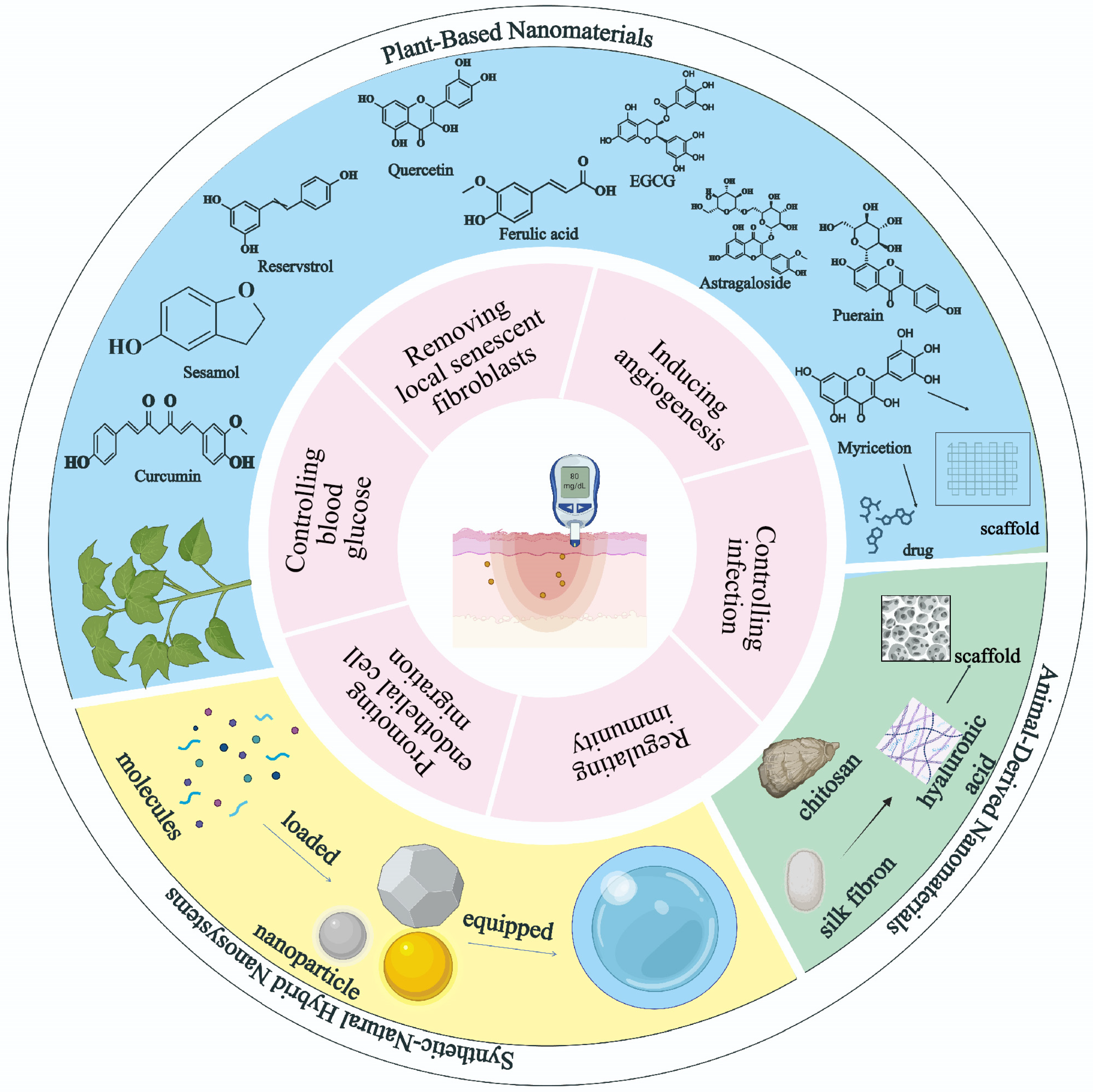
2. Natural Product-Derived Nanomaterial
2.1. Plant-Based Nanomaterials
2.1.1. Plant-Extracted Molecule Compound Loaded in Nanoplatforms
Curcumin
Sesamol
Quercetin
Ferulic Acid
Epigallocatechin Gallate (EGCG)
Astragaloside
Resveratrol (RES)
Puerarin (PUE)
Myricetin

| Small Molecule Compound | Vehicle | Components | Model | Mechanisms | Ref. |
|---|---|---|---|---|---|
| Curcumin | CSNPs | CS; Aqueous acetic acid; Sodium; Tripolyphosphate; Anhydrous ethanol; Curcumin. | Diabetic rat excision skin wound model | Regulating immunity Inducing angiogenesis | [31] |
| Sesamol | Cellulose acetate (CA)–zein composite nanofiber membrane | Acetic acid; Zein; Sesamol. | Diabetic mice excision skin wound model | Regulating immunity Enhancing keratinocyte proliferation and migration. | [38] |
| Quercetin | ADM-GO-PEG hybrid scaffold | Graphene oxide(GO) sheet; 6-armed PEG; Quercetin; ADM. | Diabetic mouse excision skin wound model | Inducing angiogenesis | [46] |
| Ferulic acid | Silk-sericin-based CA and PCL hybrid nanofibers | CA; PCL; Acetone; Ferulic acid; Silk sericin protein. | Diabetic rat full-thickness skin wound model | Regulating immunity Promoting endothelial cell migration. | [49] |
| Epigallocatechin gallate | Ascorbic acid, gelatin, and CSNPs | CS; Gelatin; Sodium EGCG; Ascorbic acid. | Diabetic mouse full-thickness skin wound model | Regulating immunity Inducing angiogenesis | [51] |
| Astragaloside | Matrix metalloproteinase-2-responsive HA end-conjugated polyamidoamine(HA-pep-PAMAM) dendrimers | HA; PAMAM; Astragaloside IV. | Diabetic mouse excision skin wound model | Regulating immunity | [56] |
| Resveratrol | GelMA/SFMA/PDEVs composite hydrogel | Gelatin; Methacrylic anhydride SF; Glycidyl Methacrylate; Amino-functionalized MSN NPs; Resveratrol; PDEVS; LiBr; Phenyl-2,4,6trimethylbenzoylphosphinate. | Full-thickness skin defect model of diabetic mice | Regulating immunity Inducing angiogenesis | [62] |
| Puerarin | CS nanofiber hydrogel | CS; Puerarin. | Type I diabetic mouse model of full-thickness skin wound | Regulating immunity Inducing angiogenesis | [67] |
| Myricetin | PEG–acryloyl chloride/phenylboronic acid–HA hybrid hydrogel | PEG; Acryloyl chloride; HA, myricetin. | Type I diabetic rat model of full-thickness wound | Regulating immunity Inducing angiogenesis | [70] |
2.1.2. Plant Polysaccharide-Based Nanoscaffolds
Alginate
Cellulose
| Natural Bioactive Compounds | Functions | Perspectives | Limitations | Priority | Ref. |
|---|---|---|---|---|---|
| Curcumin | Strong antioxidant and anti-inflammatory effects. | Nanoformulations show potentials for targeted delivery and enhanced stability. | Poor water solubility. Low intrinsic bioavailability. | High | [28,29,31] |
| Sesamol | Powerful antioxidant and anti-inflammatory effects. Promoting IL-6-mediated keratinocyte proliferation. | Nanomaterials extend local retention time. | Limited clinical validation. Short biological half-life in free forms. | High | [32,33,35,36,37,38] |
| Quercetin | Anti-inflammatory effect via Wnt/β-catenin pathway and NF-κB pathway. Stimulating collagen deposition. | Optimized nanocrystal and hydrogel systems increase efficacy. | Poor solubility and bioavailability. Carrier-dependent efficacy. | Medium | [40,41,42,43,44,46] |
| Ferulic acid | Antidiabetic, antioxidant, and antimicrobial effects. | Nanoparticle encapsulation show enhanced stability. Emerging potential for controlling macrophage polarization. | Limited solubility and bioavailability. | Medium | [47,48,49,50] |
| Epigallocatechin gallate | Strong antioxidant activity. Stimulating collagen deposition. | Potential for combinatorial regimens. | Poor stability without encapsulation. | High | [51,52,53] |
| Astragaloside | Antioxidant and anti-inflammatory effects. Promoting angiogenesis and collagen deposition. Activating the JNK/Nrf2 signaling pathway to reduce oxidative damage. | Potential to combine with other materials for targeted delivery. | Bioavailability challenges. | Medium | [56,58,59] |
| Resveratrol | Strong antioxidant, anti-inflammatory effects. Reducing insulin resistance Upregulating SIRT1 to enhance angiogenesis. | Novel hydrogel/nanoparticle systems improve retention and efficacy. | Rapid metabolism. Limited stability in vivo. | Medium | [60,61,62] |
| Puerarin | Enhancing microcirculation and angiogenesis. | Advanced nanoplatforms. Potential therapeutic candidate for ischemic wounds. | Poor water solubility and bioavailability. | Low | [64,65,66] |
| Myricetin | Highly efficient ROS scavenging. Inhibiting the digestion and absorption of carbohydrate. | Promising, smart, glucose-responsive delivery. Emerging candidate for diabetic wounds. | Insufficient comparative studies. Limited long-term safety data. | Low | [68,69,70] |
| Alginate | Excellent hydrophilicity suitable for wet wound environments. Blending with collagen to enhance mechanical strength. | Strong potential for multifunctional polymeric micelle formulations. Smart dressings integrating antimicrobial peptides. | Clinical translation requires precise optimization of formulation variables to maintain functional integrity. | High | [72,74,75] |
| Cellulose | Providing structural support and exudate management via controllable nanofiber networks. Sustained therapeutic release (e.g., prolonged curcumin half-life) via thermosensitive hydrogels. | Promising potential in thermoresponsive hydrogels and advanced nanofiber scaffolds. Effective integration with functional NPs for enhanced multifunctional wound therapy. | Need strict optimization. Performance is highly dependent on the type of NPs. | High | [76,77,78] |
2.2. Animal-Derived Nanomaterials
2.2.1. Collagen-Based Nanostructures
2.2.2. Chitosan-Based Nanostructures
2.2.3. Hyaluronic Acid-Based Nanostructures
2.2.4. Silk Fibroin-Based Nanostructures
| Material | Therapeutic Advantages | Limitations | Ref. |
|---|---|---|---|
| Collagen-Based Nanostructures | Excellent biocompatibility and biodegradability. Supporting cell migration and vascularization. Adaptable to diverse dosage forms. | Suboptimal efficacy when used alone. | [81,82,85] |
| Chitosan-Based Nanostructures | Strong antibacterial activity against pathogens. High drug-loading capacity. | Suboptimal efficacy when used alone. Limited long-term biocompatibility data. | [79,86,90] |
| Hyaluronic Acid-Based Nanostructures | Regulating inflammation, angiogenesis, antibacterial activity and tissue regeneration. | Limited stimulation of endothelial cells and angiogenesis. Potential instability in complex formulations. | [93,94,95,96,97] |
| Silk Fibroin-Based Nanostructures | Superior biocompatibility and non-toxic degradation byproducts. Enabling precise drug release profiles for therapeutic delivery. Genetic modifiability for antimicrobial features. | Suboptimal structural properties when used alone. High costs for genetically engineered variants. | [98,99,100,101,102,103,104,105] |
3. Synthetic–Natural Hybrid Nanosystems
3.1. Metallic NPs
3.1.1. AgNPs
3.1.2. AuNPs
3.1.3. CuNPs
3.1.4. ZnONPs
3.2. Non-Metallic NPs
3.2.1. SiO2 NPs
3.2.2. Carbon NPs
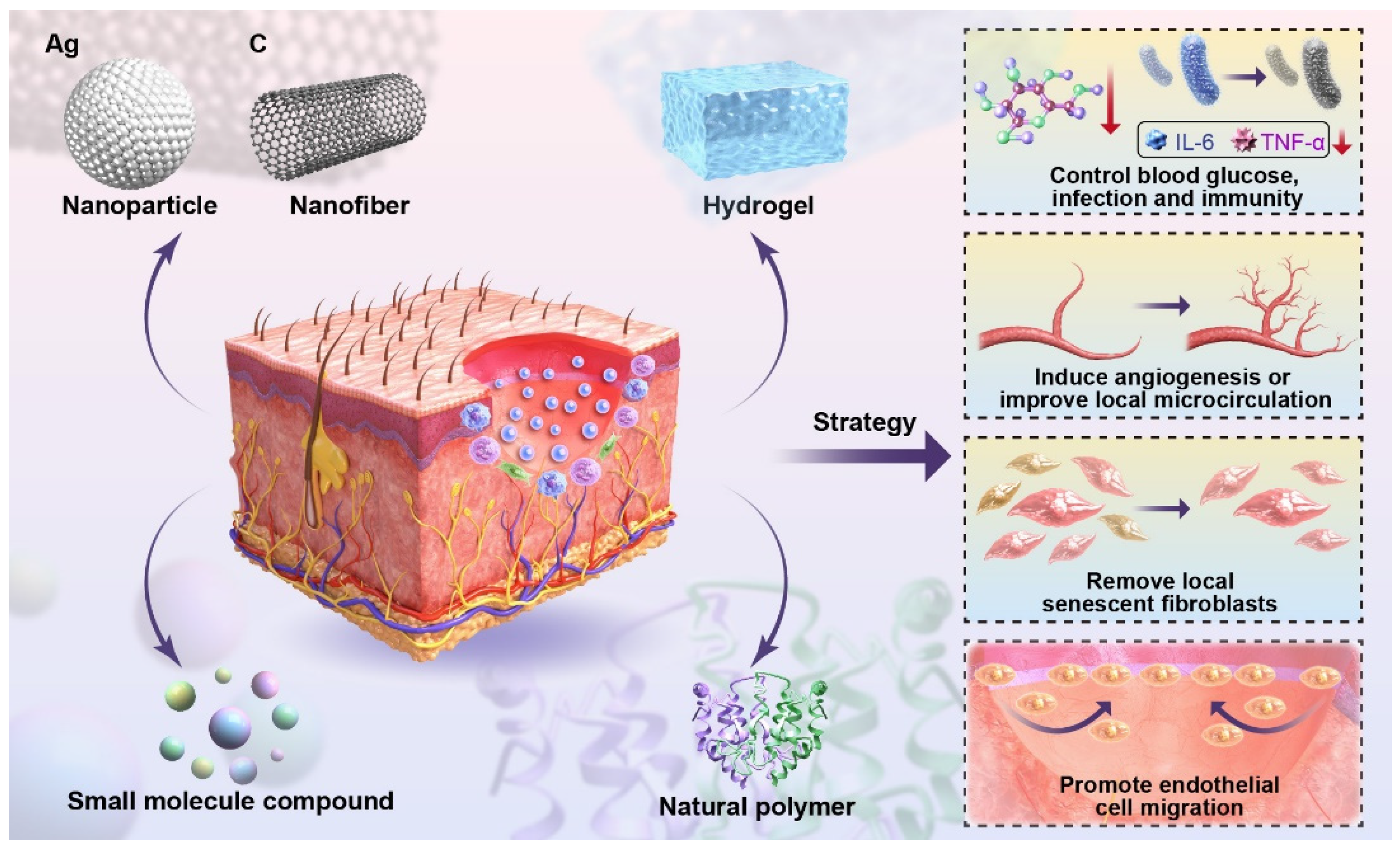
4. The Molecular Mechanism of NPs with Natural Products on the Healing of Diabetic Wounds
4.1. Blood Glucose Control
4.2. Infection Control
4.3. Regulation of Immunity
4.4. Inducing Angiogenesis or Improving Local Microcirculation
4.5. Removing Local Senescent Fibroblasts
4.6. Promotion of Endothelial Cell Migration
5. Advantages of Nanomedicine in the Healing of Diabetic Wounds
5.1. Safety of Medication Use
5.2. Multiple Functions Simultaneously
5.3. Advantageous Drug Carrier
6. Challenges for Nanomedicines
Author Contributions
Funding
Institutional Review Board Statement
Informed Consent Statement
Data Availability Statement
Acknowledgments
Conflicts of Interest
References
- NCD Risk Factor Collaboration. Worldwide trends in diabetes prevalence and treatment from 1990 to 2022: A pooled analysis of 1108 population-representative studies with 141 million participants. Lancet 2024, 404, 2077–2093. [Google Scholar] [CrossRef] [PubMed]
- Burgess, J.L.; Wyant, W.A.; Abdo, A.B.; Kirsner, R.S.; Jozic, I. Diabetic wound-healing science. Med. Lith. 2021, 57, 1072. [Google Scholar] [CrossRef]
- Wang, T.; Dong, D.; Chen, T.; Zhu, J.; Wang, S.; Wen, W.; Zhang, X.; Tang, H.; Liang, J.; Wang, S.; et al. Acidity-responsive cascade nanoreactor based on metal-nanozyme and glucose oxidase combination for starving and photothermal-enhanced chemodynamic antibacterial therapy. Chem. Eng. J. 2022, 446, 137172. [Google Scholar] [CrossRef]
- Yang, Z.; Chen, H.; Yang, P.; Shen, X.; Hu, Y.; Cheng, Y.; Yao, H.; Zhang, Z. Nano-oxygenated hydrogels for locally and permeably hypoxia relieving to heal chronic wounds. Biomaterials 2022, 282, 121401. [Google Scholar] [CrossRef] [PubMed]
- Bai, L.; Zhang, X.; Li, X.; Wang, S.; Zhang, Y.; Xu, G. Impact of a novel hydrogel with injectable platelet-rich fibrin in diabetic wound healing. J. Diabetes Res. 2023, 2023, 7532637. [Google Scholar] [CrossRef]
- Oprita, E.I.; Iosageanu, A.; Craciunescu, O. Natural polymeric hydrogels encapsulating small molecules for diabetic wound healing. Gels 2023, 9, 867. [Google Scholar] [CrossRef]
- Bai, Q.; Han, K.; Dong, K.; Zheng, C.; Zhang, Y.; Long, Q.; Lu, T. Potential applications of nanomaterials and technology for diabetic wound healing. Int. J. Nanomed. 2020, 15, 9717–9743. [Google Scholar] [CrossRef]
- Gao, S.; He, X.; Liu, H.; Liu, Y.; Wang, H.; Zhou, Z.; Chen, L.; Ji, X.; Yang, R.; Xie, J. Multifunctional bioactive nanozyme systems for enhanced diabetic wound healing. Adv. Healthc. Mater. 2025, 14, e2401580. [Google Scholar] [CrossRef]
- Ding, J.; Sun, L.; Zhu, Z.; Wu, X.; Xu, X.; Xiang, Y. Nano drug delivery systems: A promising approach to scar prevention and treatment. J. Nanobiotechnol. 2023, 21, 268. [Google Scholar] [CrossRef]
- Yusuf Aliyu, A.; Adeleke, O.A. Nanofibrous scaffolds for diabetic wound healing. Pharmaceutics 2023, 15, 986. [Google Scholar] [CrossRef]
- Vijayavenkataraman, S. Nerve guide conduits for peripheral nerve injury repair: A review on design, materials and fabrication methods. Acta Biomater. 2020, 106, 54–69. [Google Scholar] [CrossRef] [PubMed]
- Reddy, M.S.B.; Ponnamma, D.; Choudhary, R.; Sadasivuni, K.K. A comparative review of natural and synthetic biopolymer composite scaffolds. Polymers 2021, 13, 1105. [Google Scholar] [CrossRef]
- Miguel, S.P.; Sequeira, R.S.; Moreira, A.F.; Cabral, C.S.D.; Mendonça, A.G.; Ferreira, P.; Correia, I.J. An overview of electrospun membranes loaded with bioactive molecules for improving the wound healing process. Eur. J. Pharm. Biopharm. Off. J. Arbeitsgemeinschaft Fur Pharm. Verfahrenstechnik e.V 2019, 139, 1–22. [Google Scholar] [CrossRef]
- Adamu, B.F.; Gao, J.; Jhatial, A.K.; Kumelachew, D.M. A review of medicinal plant-based bioactive electrospun nano fibrous wound dressings. Mater. Des. 2021, 209, 109942. [Google Scholar] [CrossRef]
- Anand, S.; Rajinikanth, P.S.; Arya, D.K.; Pandey, P.; Gupta, R.K.; Sankhwar, R.; Chidambaram, K. Multifunctional biomimetic nanofibrous scaffold loaded with asiaticoside for rapid diabetic wound healing. Pharmaceutics 2022, 14, 273. [Google Scholar] [CrossRef]
- Guleken, Z.; Depciuch, J.; Ege, H.; İlbay, G.; Kalkandelen, C.; Ozbeyli, D.; Bulut, H.; Sener, G.; Tarhan, N.; Kuruca, S.E. Spectrochemical and biochemical assay comparison study of the healing effect of the aloe vera and hypericum perforatum loaded nanofiber dressings on diabetic wound. Spectrochim. Acta Part A Mol. Biomol. Spectrosc. 2021, 254, 119639. [Google Scholar] [CrossRef]
- Qian, Y.; Zheng, Y.; Jin, J.; Wu, X.; Xu, K.; Dai, M.; Niu, Q.; Zheng, H.; He, X.; Shen, J. Immunoregulation in diabetic wound repair with a photoenhanced glycyrrhizic acid hydrogel scaffold. Adv. Mater. 2022, 34, e2200521. [Google Scholar] [CrossRef]
- Maity, B.; Alam, S.; Samanta, S.; Prakash, R.G.; Govindaraju, T. Antioxidant silk fibroin composite hydrogel for rapid healing of diabetic wound. Macromol. Biosci. 2022, 22, e2200097. [Google Scholar] [CrossRef]
- Ahmed, R.; Tariq, M.; Ali, I.; Asghar, R.; Noorunnisa Khanam, P.; Augustine, R.; Hasan, A. Novel electrospun chitosan/polyvinyl alcohol/zinc oxide nanofibrous mats with antibacterial and antioxidant properties for diabetic wound healing. Int. J. Biol. Macromol. 2018, 120, 385–393. [Google Scholar] [CrossRef]
- Yadav, S.; Arya, D.K.; Pandey, P.; Anand, S.; Gautam, A.K.; Ranjan, S.; Saraf, S.A.; Mahalingam Rajamanickam, V.; Singh, S.; Chidambaram, K.; et al. ECM mimicking biodegradable nanofibrous scaffold enriched with curcumin/ZnO to accelerate diabetic wound healing via multifunctional bioactivity. Int. J. Nanomed. 2022, 17, 6843–6859. [Google Scholar] [CrossRef]
- Natarajan, J.; Sanapalli, B.K.R.; Bano, M.; Singh, S.K.; Gulati, M.; Karri, V.V.S.R. Nanostructured lipid carriers of pioglitazone loaded collagen/chitosan composite scaffold for diabetic wound healing. Adv. Wound Care 2019, 8, 499–513. [Google Scholar] [CrossRef] [PubMed]
- Han, Y.; Jiang, Y.; Li, Y.; Wang, M.; Fan, T.; Liu, M.; Ke, Q.; Xu, H.; Yi, Z. An aligned porous electrospun fibrous scaffold with embedded asiatic acid for accelerating diabetic wound healing. J. Mater. Chem. B 2019, 7, 6125–6138. [Google Scholar] [CrossRef] [PubMed]
- Monirul Islam, M.; Hemmanahalli Ramesh, V.; Durga Bhavani, P.; Goudanavar, P.S.; Naveen, N.R.; Ramesh, B.; Fattepur, S.; Narayanappa Shiroorkar, P.; Habeebuddin, M.; Meravanige, G.; et al. Optimization of process parameters for fabrication of electrospun nanofibers containing neomycin sulfate and malva sylvestris extract for a better diabetic wound healing. Drug Deliv. 2022, 29, 3370–3383. [Google Scholar] [CrossRef] [PubMed]
- Derakhshan, M.A.; Nazeri, N.; Khoshnevisan, K.; Heshmat, R.; Omidfar, K. Three-layered PCL-collagen nanofibers containing melilotus officinalis extract for diabetic ulcer healing in a rat model. J. Diabetes Metab. Disord. 2022, 21, 313–321. [Google Scholar] [CrossRef]
- Yang, B.; Hu, C.; Huang, W.; Ho, C.; Yao, C.; Huang, C. Effects of bilayer nanofibrous scaffolds containing curcumin/lithospermi radix extract on wound healing in streptozotocin-induced diabetic rats. Polymers 2019, 11, 1745. [Google Scholar] [CrossRef]
- Xu, X.; Wang, X.; Qin, C.; Khan, A.U.R.; Zhang, W.; Mo, X. Silk fibroin/poly-(l-lactide-co-caprolactone) nanofiber scaffolds loaded with huangbai liniment to accelerate diabetic wound healing. Colloids Surf. B Biointerfaces 2021, 199, 111557. [Google Scholar] [CrossRef]
- Li, Y.; Song, W.; Kong, L.; He, Y.; Li, H. Injectable and microporous microgel-fiber granular hydrogel loaded with bioglass and siRNA for promoting diabetic wound healing. Small 2024, 20, e2309599. [Google Scholar] [CrossRef]
- Ali, S.M.A.; Khan, J.; Shahid, R.; Shabbir, S.; Ayoob, M.F.; Imran, M. Chitosan-carrageenan microbeads containing nano-encapsulated curcumin: Nano-in-micro hydrogels as alternative-therapeutics for resistant pathogens associated with chronic wounds. Int. J. Biol. Macromol. 2024, 278, 134841. [Google Scholar] [CrossRef]
- Sideek, S.A.; El-Nassan, H.B.; Fares, A.R.; Elmeshad, A.N.; Elkasabgy, N.A. Different curcumin-loaded delivery systems for wound healing applications: A comprehensive review. Pharmaceutics 2022, 15, 38. [Google Scholar] [CrossRef]
- Karri, V.V.; Kuppusamy, G.; Talluri, S.V.; Mannemala, S.S.; Kollipara, R.; Wadhwani, A.D.; Mulukutla, S.; Raju, K.R.; Malayandi, R. Curcumin loaded chitosan nanoparticles impregnated into collagen-alginate scaffolds for diabetic wound healing. Int. J. Biol. Macromol. 2016, 93, 1519–1529. [Google Scholar] [CrossRef]
- Li, F.; Shi, Y.; Liang, J.; Zhao, L. Curcumin-loaded chitosan nanoparticles promote diabetic wound healing via attenuating inflammation in a diabetic rat model. J. Biomater. Appl. 2019, 34, 476–486. [Google Scholar] [CrossRef] [PubMed]
- Zhou, S.; Zou, H.; Huang, G.; Chen, G. Preparations and antioxidant activities of sesamol and it’s derivatives. Bioorg. Med. Chem. Lett. 2021, 31, 127716. [Google Scholar] [CrossRef] [PubMed]
- Nair, A.B.; Dalal, P.; Kadian, V.; Kumar, S.; Garg, M.; Rao, R.; Almuqbil, R.M.; Alnaim, A.S.; Aldhubiab, B.; Alqattan, F. Formulation strategies for enhancing pharmaceutical and nutraceutical potential of sesamol: A natural phenolic bioactive. Plants 2023, 12, 1168. [Google Scholar] [CrossRef] [PubMed]
- Bosebabu, B.; Cheruku, S.P.; Chamallamudi, M.R.; Nampoothiri, M.; Shenoy, R.R.; Nandakumar, K.; Parihar, V.K.; Kumar, N. An appraisal of current pharmacological perspectives of sesamol: A review. Mini Rev. Med. Chem. 2020, 20, 988–1000. [Google Scholar] [CrossRef]
- Jan, K.; Ho, C.; Hwang, L.S. Elimination and metabolism of sesamol, a bioactive compound in sesame oil, in rats. Mol. Nutr. Food Res. 2009, 53 (Suppl. 1), S36–S43. [Google Scholar] [CrossRef]
- Jan, K.; Ho, C.; Hwang, L.S. Bioavailability and tissue distribution of sesamol in rat. J. Agric. Food. Chem. 2008, 56, 7032–7037. [Google Scholar] [CrossRef]
- Gourishetti, K.; Keni, R.; Nayak, P.G.; Jitta, S.R.; Bhaskaran, N.A.; Kumar, L.; Kumar, N.; Krishnadas, N.; Shenoy, R.R. Sesamol-loaded PLGA nanosuspension for accelerating wound healing in diabetic foot ulcer in rats. Int. J. Nanomed. 2020, 15, 9265–9282. [Google Scholar] [CrossRef]
- Liu, F.; Li, X.; Wang, L.; Yan, X.; Ma, D.; Liu, Z.; Liu, X. Sesamol incorporated cellulose acetate-zein composite nanofiber membrane: An efficient strategy to accelerate diabetic wound healing. Int. J. Biol. Macromol. 2020, 149, 627–638. [Google Scholar] [CrossRef]
- Deol, P.K.; Kaur, I.P.; Dhiman, R.; Kaur, H.; Sharma, G.; Rishi, P.; Ghosh, D. Investigating wound healing potential of sesamol loaded solid lipid nanoparticles: Ex-vivo, in vitro and in-vivo proof of concept. Int. J. Pharm. 2024, 654, 123974. [Google Scholar] [CrossRef]
- Mi, Y.; Zhong, L.; Lu, S.; Hu, P.; Pan, Y.; Ma, X.; Yan, B.; Wei, Z.; Yang, G. Quercetin promotes cutaneous wound healing in mice through wnt/beta-catenin signaling pathway. J. Ethnopharmacol. 2022, 290, 115066. [Google Scholar] [CrossRef]
- Panthi, V.K.; Imran, M.; Chaudhary, A.; Paudel, K.R.; Mohammed, Y. The significance of quercetin-loaded advanced nanoformulations for the management of diabetic wounds. Nanomedicine 2023, 18, 391–411. [Google Scholar] [CrossRef] [PubMed]
- Liu, D.; Hu, H.; Lin, Z.; Chen, D.; Zhu, Y.; Hou, S.; Shi, X. Quercetin deformable liposome: Preparation and efficacy against ultraviolet b induced skin damages in vitro and in vivo. J. Photochem. Photobiol. B Biol. 2013, 127, 8–17. [Google Scholar] [CrossRef] [PubMed]
- Tapfumaneyi, P.; Imran, M.; Alavi, S.E.; Mohammed, Y. Science of, and insights into, thermodynamic principles for dermal formulations. Drug Discov. Today 2023, 28, 103521. [Google Scholar] [CrossRef]
- Wang, L.; Dong, J.; Zhao, Z.; Li, D.; Dong, W.; Lu, Y.; Jin, B.; Li, H.; Liu, Q.; Deng, B. Quarternized chitosan/quercetin/polyacrylamide semi-interpenetrating network hydrogel with recoverability, toughness and antibacterial properties for wound healing. Int. J. Biol. Macromol. 2023, 228, 48–58. [Google Scholar] [CrossRef]
- Li, X.; Yang, X.; Wang, Z.; Liu, Y.; Guo, J.; Zhu, Y.; Shao, J.; Li, J.; Wang, L.; Wang, K. Antibacterial, antioxidant and biocompatible nanosized quercetin-PVA xerogel films for wound dressing. Colloid. Surf. B-Biointerfaces 2022, 209, 112175. [Google Scholar] [CrossRef]
- Chu, J.; Shi, P.; Yan, W.; Fu, J.; Yang, Z.; He, C.; Deng, X.; Liu, H. PEGylated graphene oxide-mediated quercetin-modified collagen hybrid scaffold for enhancement of MSCs differentiation potential and diabetic wound healing. Nanoscale 2018, 10, 9547–9560. [Google Scholar] [CrossRef]
- Locarno, S.; Eleta-Lopez, A.; Lupo, M.G.; Gelmi, M.L.; Clerici, F.; Bittner, A.M. Electrospinning of pyrazole-isothiazole derivatives: Nanofibers from small molecules. RSC Adv. 2019, 9, 20565–20572. [Google Scholar] [CrossRef]
- Bairagi, U.; Mittal, P.; Singh, J.; Mishra, B. Preparation, characterization, and in vivo evaluation of nano formulations of ferulic acid in diabetic wound healing. Drug Dev. Ind. Pharm. 2018, 44, 1783–1796. [Google Scholar] [CrossRef]
- Anand, S.; Pandey, P.; Begum, M.Y.; Chidambaram, K.; Arya, D.K.; Gupta, R.K.; Sankhwar, R.; Jaiswal, S.; Thakur, S.; Rajinikanth, P.S. Electrospun biomimetic multifunctional nanofibers loaded with ferulic acid for enhanced antimicrobial and wound-healing activities in STZ-induced diabetic rats. Pharmaceuticals 2022, 15, 302. [Google Scholar] [CrossRef]
- Anuradha, U.; Bhavana, V.; Chary, P.S.; Kalia, N.P.; Mehra, N.K. Exploration of the topical nanoemulgel bearing with ferulic acid and essential oil for diabetic wound healing. Pathophysiol. Off. J. Int. Soc. Pathophysiol. 2024, 31, 680–698. [Google Scholar] [CrossRef]
- Sun, M.; Xie, Q.; Cai, X.; Liu, Z.; Wang, Y.; Dong, X.; Xu, Y. Preparation and characterization of epigallocatechin gallate, ascorbic acid, gelatin, chitosan nanoparticles and their beneficial effect on wound healing of diabetic mice. Int. J. Biol. Macromol. 2020, 148, 777–784. [Google Scholar] [CrossRef] [PubMed]
- Bulboaca, A.E.; Boarescu, P.; Porfire, A.S.; Dogaru, G.; Barbalata, C.; Valeanu, M.; Munteanu, C.; Râjnoveanu, R.M.; Nicula, C.A.; Stanescu, I.C. The effect of nano-epigallocatechin-gallate on oxidative stress and matrix metalloproteinases in experimental diabetes mellitus. Antioxidants 2020, 9, 172. [Google Scholar] [CrossRef] [PubMed]
- Chen, G.; He, L.; Zhang, P.; Zhang, J.; Mei, X.; Wang, D.; Zhang, Y.; Ren, X.; Chen, Z. Encapsulation of green tea polyphenol nanospheres in PVA/alginate hydrogel for promoting wound healing of diabetic rats by regulating PI3k/AKT pathway. Mater. Sci. Eng. C 2020, 110, 110686. [Google Scholar] [CrossRef]
- Zhang, J.; Li, W.; Tao, Z.; Zhou, X.; Chen, X.; Zhou, J.; Sun, H.; Fang, Y.; Liu, Y. Endogenous glucose-driven cascade reaction of nano-drug delivery for boosting multidrug-resistant bacteria-infected diabetic wound healing. J. Colloid. Interface. Sci. 2024, 672, 63–74. [Google Scholar] [CrossRef]
- Yang, Y.; Fan, L.; Jiang, J.; Sun, J.; Xue, L.; Ma, X.; Kuai, L.; Li, B.; Li, Y. M2 macrophage-polarized anti-inflammatory microneedle patch for accelerating biofilm-infected diabetic wound healing via modulating the insulin pathway. J. Nanobiotechnol. 2024, 22, 489. [Google Scholar] [CrossRef]
- Zhang, D.; Huang, Q. Encapsulation of astragaloside with matrix metalloproteinase-2-responsive hyaluronic acid end-conjugated polyamidoamine dendrimers improves wound healing in diabetes. J. Biomed. Nanotechnol. 2020, 16, 1229–1240. [Google Scholar] [CrossRef]
- Wang, B.S.; Ma, X.F.; Zhang, C.Y.; Li, Y.X.; Liu, X.Z.; Hu, C.Q. Astragaloside IV improves angiogenesis and promotes wound healing in diabetic rats via the activation of the SUMOylation pathway. Biomed. Environ. Sci. 2021, 34, 124–129. [Google Scholar]
- Deng, L. Astragaloside IV as potential antioxidant against diabetic ketoacidosis in juvenile mice through activating JNK/nrf2 signaling pathway. Arch. Med. Res. 2020, 51, 654–663. [Google Scholar] [CrossRef]
- Wang, E.; Wang, L.; Ding, R.; Zhai, M.; Ge, R.; Zhou, P.; Wang, T.; Fang, H.; Wang, J.; Huang, J. Astragaloside IV acts through multi-scale mechanisms to effectively reduce diabetic nephropathy. Pharmacol. Res. 2020, 157, 104831. [Google Scholar] [CrossRef]
- Huang, X.; Sun, J.; Chen, G.; Niu, C.; Wang, Y.; Zhao, C.; Sun, J.; Huang, H.; Huang, S.; Liang, Y.; et al. Resveratrol promotes diabetic wound healing via SIRT1-FOXO1-c-myc signaling pathway-mediated angiogenesis. Front. Pharmacol. 2019, 10, 421. [Google Scholar] [CrossRef]
- Bejenaru, L.E.; Biţă, A.; Belu, I.; Segneanu, A.; Radu, A.; Dumitru, A.; Ciocîlteu, M.V.; Mogoşanu, G.D.; Bejenaru, C. Resveratrol: A review on the biological activity and applications. Appl. Sci. 2024, 14, 4534. [Google Scholar] [CrossRef]
- Zhu, W.; Dong, Y.; Xu, P.; Pan, Q.; Jia, K.; Jin, P.; Zhou, M.; Xu, Y.; Guo, R.; Cheng, B. A composite hydrogel containing resveratrol-laden nanoparticles and platelet-derived extracellular vesicles promotes wound healing in diabetic mice. Acta Biomater. 2022, 154, 212–230. [Google Scholar] [CrossRef] [PubMed]
- Meng, F.; Guo, B.; Ma, Y.; Li, K.; Niu, F. Puerarin: A review of its mechanisms of action and clinical studies in ophthalmology. Phytomed. Int. J. Phytother. Phytopharm. 2022, 107, 154465. [Google Scholar] [CrossRef] [PubMed]
- Cao, H.; Li, L.; Li, L.; Meng, X.; Liu, Y.; Cheng, W.; Zhang, P.; Gao, Y.; Qin, L.; Wang, X. New use for old drug: Local delivery of puerarin facilitates critical-size defect repair in rats by promoting angiogenesis and osteogenesis. J. Orthop. Transl. 2022, 36, 52–63. [Google Scholar] [CrossRef]
- Zhang, L.; Duan, C.; Feng, S.; Zhao, B.; Li, H.; Zhang, X.; Zhou, Y.; Qin, Z. Preparation and evaluation of puerarin-loaded PLGA nanoparticles for improving oral bioavailability in SD rats. Biomed. Pharmacother. 2024, 181, 117670. [Google Scholar] [CrossRef]
- Su, X.; Geng, X.; Li, F.; Song, M.; Lv, R.; Zhang, Y.; Yuan, J.; Dong, J.; Shi, Y.; Zhao, L. Microneedles loaded with l-arginine-modified puerarin-derived carbon nanoparticles improved treatment of diabetic wound via photothermal and nitric oxide-based gas therapy. J. Colloid. Interface. Sci. 2025, 691, 137353. [Google Scholar] [CrossRef]
- Zeng, X.; Chen, B.; Wang, L.; Sun, Y.; Jin, Z.; Liu, X.; Ouyang, L.; Liao, Y. Chitosan@puerarin hydrogel for accelerated wound healing in diabetic subjects by mir-29ab1 mediated inflammatory axis suppression. Bioact. Mater. 2023, 19, 653–665. [Google Scholar] [CrossRef]
- Zhao, Z.; Chen, Y.; Li, X.; Zhu, L.; Wang, X.; Li, L.; Sun, H.; Han, X.; Li, J. Myricetin relieves the symptoms of type 2 diabetes mice and regulates intestinal microflora. Biomed. Pharmacother. Biomed. Pharmacother. 2022, 153, 113530. [Google Scholar] [CrossRef]
- Lalitha, N.; Sadashivaiah, B.; Ramaprasad, T.R.; Singh, S.A. Anti-hyperglycemic activity of myricetin, through inhibition of DPP-4 and enhanced GLP-1 levels, is attenuated by co-ingestion with lectin-rich protein. PLoS ONE 2020, 15, e0231543. [Google Scholar] [CrossRef]
- Xu, Z.; Liu, G.; Huang, J.; Wu, J. Novel glucose-responsive antioxidant hybrid hydrogel for enhanced diabetic wound repair. ACS Appl. Mater. Interfaces 2022, 14, 7680–7689. [Google Scholar] [CrossRef]
- Varaprasad, K.; Jayaramudu, T.; Kanikireddy, V.; Toro, C.; Sadiku, E.R. Alginate-based composite materials for wound dressing application:a mini review. Carbohydr. Polym. 2020, 236, 116025. [Google Scholar] [CrossRef] [PubMed]
- Severino, P.; Da Silva, C.F.; Andrade, L.N.; de Lima Oliveira, D.; Campos, J.; Souto, E.B. Alginate nanoparticles for drug delivery and targeting. Curr. Pharm. Des. 2019, 25, 1312–1334. [Google Scholar] [CrossRef] [PubMed]
- Akbar, M.U.; Zia, K.M.; Akash, M.S.H.; Nazir, A.; Zuber, M.; Ibrahim, M. In-vivo anti-diabetic and wound healing potential of chitosan/alginate/maltodextrin/pluronic-based mixed polymeric micelles: Curcumin therapeutic potential. Int. J. Biol. Macromol. 2018, 120, 2418–2430. [Google Scholar] [CrossRef] [PubMed]
- Oliveira, D.; Rezende, P.S.; Barbosa, T.C.; Andrade, L.N.; Bani, C.; Tavares, D.S.; Da, S.C.; Chaud, M.V.; Padilha, F.; Cano, A.; et al. Double membrane based on lidocaine-coated polymyxin-alginate nanoparticles for wound healing: In vitro characterization and in vivo tissue repair. Int. J. Pharm. 2020, 591, 120001. [Google Scholar] [CrossRef]
- Choudhary, M.; Chhabra, P.; Tyagi, A.; Singh, H. Scar free healing of full thickness diabetic wounds: A unique combination of silver nanoparticles as antimicrobial agent, calcium alginate nanoparticles as hemostatic agent, fresh blood as nutrient/growth factor supplier and chitosan as base matrix. Int. J. Biol. Macromol. 2021, 178, 41–52. [Google Scholar] [CrossRef]
- Antunes, B.D.F.; Santana, L.R.; Oliveira, R.M.; Valério Filho, A.; Carreno, N.L.V.; Wolke, S.I.; Da Silva, R.; Fajardo, A.R.; Dias, A.R.G.; Zavareze, E.D.R. Cellulose, cellulose nanofibers, and cellulose acetate from butia fruits (butia odorata): Chemical, morphological, structural, and thermal properties. Int. J. Biol. Macromol. 2024, 281, 136151. [Google Scholar] [CrossRef]
- Shah, S.A.; Sohail, M.; Karperien, M.; Johnbosco, C.; Mahmood, A.; Kousar, M. Chitosan and carboxymethyl cellulose-based 3d multifunctional bioactive hydrogels loaded with nano-curcumin for synergistic diabetic wound repair. Int. J. Biol. Macromol. 2023, 227, 1203–1220. [Google Scholar] [CrossRef]
- Kamalipooya, S.; Fahimirad, S.; Abtahi, H.; Golmohammadi, M.; Satari, M.; Dadashpour, M.; Nasrabadi, D. Diabetic wound healing function of PCL/cellulose acetate nanofiber engineered with chitosan/cerium oxide nanoparticles. Int. J. Pharm. 2024, 653, 123880. [Google Scholar] [CrossRef]
- Elgadir, M.A.; Uddin, M.S.; Ferdosh, S.; Adam, A.; Chowdhury, A.; Sarker, M. Impact of chitosan composites and chitosan nanoparticle composites on various drug delivery systems: A review. J. Food Drug Anal. 2015, 23, 619–629. [Google Scholar] [CrossRef]
- Sahana, T.G.; Rekha, P.D. Biopolymers: Applications in wound healing and skin tissue engineering. Mol. Biol. Rep. 2018, 45, 2857–2867. [Google Scholar] [CrossRef]
- Wosicka-Frąckowiak, H.; Poniedziałek, K.; Woźny, S.; Kuprianowicz, M.; Nyga, M.; Jadach, B.; Milanowski, B. Collagen and its derivatives serving biomedical purposes: A review. Polymers 2024, 16, 2668. [Google Scholar] [CrossRef] [PubMed]
- Lim, Y.; Ok, Y.; Hwang, S.; Kwak, J.; Yoon, S. Marine collagen as a promising biomaterial for biomedical applications. Mar. Drugs 2019, 17, 467. [Google Scholar] [CrossRef] [PubMed]
- Tan, Q.; Chen, B.; Yan, X.; Lin, Y.; Xiao, Z.; Hou, X.; Dai, J. Promotion of diabetic wound healing by collagen scaffold with collagen-binding vascular endothelial growth factor in a diabetic rat model. J. Tissue Eng. Regen. Med. 2014, 8, 195–201. [Google Scholar] [CrossRef]
- Ragothaman, M.; Kannan Villalan, A.; Dhanasekaran, A.; Palanisamy, T. Bio-hybrid hydrogel comprising collagen-capped silver nanoparticles and melatonin for accelerated tissue regeneration in skin defects. Mater. Sci. Eng. C Mater. Biol. Appl. 2021, 128, 112328. [Google Scholar] [CrossRef]
- Fu, C.; Fan, Y.; Liu, G.; Li, W.; Ma, J.; Xiao, J. One-step fabrication of an injectable antibacterial collagen hydrogel with in situ synthesized silver nanoparticles for accelerated diabetic wound healing. Chem. Eng. J. 2024, 480, 148288. [Google Scholar] [CrossRef]
- Luo, X.; Yan, C.; Feng, Y. Nanomedicine for the treatment of diabetes-associated cardiovascular diseases and fibrosis. Adv. Drug Deliv. Rev. 2021, 172, 234–248. [Google Scholar] [CrossRef]
- Chen, S.; Tian, H.; Mao, J.; Ma, F.; Zhang, M.; Chen, F.; Yang, P. Preparation and application of chitosan-based medical electrospun nanofibers. Int. J. Biol. Macromol. 2023, 226, 410–422. [Google Scholar] [CrossRef]
- Wang, T.; Zheng, Y.; Shen, Y.; Shi, Y.; Li, F.; Su, C.; Zhao, L. Chitosan nanoparticles loaded hydrogels promote skin wound healing through the modulation of reactive oxygen species. Artif. Cell. Nanomed. Biotechnol. 2018, 46, 138–149. [Google Scholar] [CrossRef]
- Sheir, M.M.; Nasra, M.; Abdallah, O.Y. Chitosan alginate nanoparticles as a platform for the treatment of diabetic and non-diabetic pressure ulcers: Formulation and in vitro/in vivo evaluation. Int. J. Pharm. 2021, 607, 120963. [Google Scholar] [CrossRef]
- Chhabra, P.; Chauhan, G.; Kumar, A. Augmented healing of full thickness chronic excision wound by rosmarinic acid loaded chitosan encapsulated graphene nanopockets. Drug Dev. Ind. Pharm. 2020, 46, 878–888. [Google Scholar] [CrossRef]
- Zhang, C.; Zhang, X.; Li, F.; Li, B.; Zhang, M.; Li, W.; Zhuge, P.; Yao, J.; Zhang, Y.; Chen, S.; et al. Thermosensitive hydrogel integrated with bimetallic nano-enzymes for modulating the microenvironment in diabetic wound beds. Adv. Sci. 2025, 12, e2411575. [Google Scholar] [CrossRef] [PubMed]
- Shen, T.; Dai, K.; Yu, Y.; Wang, J.; Liu, C. Sulfated chitosan rescues dysfunctional macrophages and accelerates wound healing in diabetic mice. Acta Biomater. 2020, 117, 192–203. [Google Scholar] [CrossRef] [PubMed]
- Graça, M.F.P.; Miguel, S.P.; Cabral, C.S.D.; Correia, I.J. Hyaluronic acid—Based wound dressings: A review. Carbohydr. Polym. 2020, 241, 116364. [Google Scholar] [CrossRef]
- Pereira, G.G.; Detoni, C.B.; Balducci, A.G.; Rondelli, V.; Colombo, P.; Guterres, S.S.; Sonvico, F. Hyaluronate nanoparticles included in polymer films for the prolonged release of vitamin e for the management of skin wounds. Eur. J. Pharm. Sci. 2016, 83, 203–211. [Google Scholar] [CrossRef]
- Xu, Z.; Liu, G.; Liu, P.; Hu, Y.; Chen, Y.; Fang, Y.; Sun, G.; Huang, H.; Wu, J. Hyaluronic acid-based glucose-responsive antioxidant hydrogel platform for enhanced diabetic wound repair. Acta Biomater. 2022, 147, 147–157. [Google Scholar] [CrossRef]
- Yang, H.; Song, L.; Sun, B.; Chu, D.; Yang, L.; Li, M.; Li, H.; Dai, Y.; Yu, Z.; Guo, J. Modulation of macrophages by a paeoniflorin-loaded hyaluronic acid-based hydrogel promotes diabetic wound healing. Mater. Today Bio 2021, 12, 100139. [Google Scholar] [CrossRef]
- Liu, Y.; Zhang, X.; Yang, S.; Guo, Q.; Zhang, Y.; Wang, Z.; Xu, S.; Qiao, D.; Ma, M.; Zheng, P.; et al. Targeting starvation therapy for diabetic bacterial infections with endogenous enzyme-triggered hyaluronan-modified nanozymes in the infection microenvironment. Int. J. Biol. Macromol. 2024, 270, 132277. [Google Scholar] [CrossRef]
- Kundu, B.; Rajkhowa, R.; Kundu, S.C.; Wang, X. Silk fibroin biomaterials for tissue regenerations. Adv. Drug Deliv. Rev. 2013, 65, 457–470. [Google Scholar] [CrossRef]
- Zheng, H.; Zuo, B. Functional silk fibroin hydrogels: Preparation, properties and applications. J. Mater. Chem. B 2021, 9, 1238–1258. [Google Scholar] [CrossRef]
- Li, Q.; Hu, W.; Huang, Q.; Yang, J.; Li, B.; Ma, K.; Wei, Q.; Wang, Y.; Su, J.; Sun, M.; et al. MiR146a-loaded engineered exosomes released from silk fibroin patch promote diabetic wound healing by targeting IRAK1. Signal Transduct. Target. Ther. 2023, 8, 62. [Google Scholar] [CrossRef]
- Mei, J.; Zhou, J.; Kong, L.; Dai, Y.; Zhang, X.; Song, W.; Zhu, C. An injectable photo-cross-linking silk hydrogel system augments diabetic wound healing in orthopaedic surgery through spatiotemporal immunomodulation. J. Nanobiotechnol. 2022, 20, 232. [Google Scholar] [CrossRef] [PubMed]
- Guo, Z.; Yan, L.; Zhou, B.; Zhao, P.; Wang, W.; Dong, S.; Cheng, B.; Yang, J.; Wang, X.; Li, B. In situ photo-crosslinking silk fibroin based hydrogel accelerates diabetic wound healing through antibacterial and antioxidant. Int. J. Biol. Macromol. 2023, 242, 125028. [Google Scholar] [CrossRef] [PubMed]
- Zha, S.; Utomo, Y.K.S.; Yang, L.; Liang, G.; Liu, W. Mechanic-driven biodegradable polyglycolic acid/silk fibroin nanofibrous scaffolds containing deferoxamine accelerate diabetic wound healing. Pharmaceutics 2022, 14, 601. [Google Scholar] [CrossRef] [PubMed]
- Zhang, Y.; Sheng, R.; Chen, J.; Wang, H.; Zhu, Y.; Cao, Z.; Zhao, X.; Wang, Z.; Liu, C.; Chen, Z.; et al. Silk fibroin and sericin differentially potentiate the paracrine and regenerative functions of stem cells through multiomics analysis. Adv. Mater. 2023, 35, e2210517. [Google Scholar] [CrossRef]
- Wang, F.; Lei, H.; Tian, C.; Ji, Y.; Wang, F.; Deng, H.; Zhou, H.; Chen, S.; Zhou, Y.; Meng, Z.; et al. An efficient biosynthetic system for developing functional silk fibroin-based biomaterials. Adv. Mater. 2025, 37, e2414878. [Google Scholar] [CrossRef]
- Hakimov, S.; Kylychbekov, S.; Harness, B.; Neupane, S.; Hurley, J.; Brooks, A.; Banga, S.; Er, A.O. Evaluation of silver nanoparticles attached to methylene blue as an antimicrobial agent and its cytotoxicity. Photodiagnosis Photodyn. Ther. 2022, 39, 102904. [Google Scholar] [CrossRef]
- Hamdan, S.; Pastar, I.; Drakulich, S.; Dikici, E.; Tomic-Canic, M.; Deo, S.; Daunert, S. Nanotechnology-driven therapeutic interventions in wound healing: Potential uses and applications. ACS Cent. Sci. 2017, 3, 163–175. [Google Scholar] [CrossRef]
- Prasath, S.; Palaniappan, K. Is using nanosilver mattresses/pillows safe? A review of potential health implications of silver nanoparticles on human health. Environ. Geochem. Health 2019, 41, 2295–2313. [Google Scholar] [CrossRef]
- Mahalingam, S.; Govindaraji, P.K.; Solomon, V.G.; Kesavan, H.; Neelan, Y.D.; Bakthavatchalam, S.; Kim, J.; Bakthavatchalam, P. Biogenic synthesis and characterization of silver nanoparticles: Evaluation of their larvicidal, antibacterial, and cytotoxic activities. ACS Omega 2023, 8, 11923–11930. [Google Scholar] [CrossRef]
- Permyakova, E.S.; Konopatsky, A.S.; Ershov, K.I.; Bakhareva, K.I.; Sitnikova, N.A.; Shtansky, D.V.; Solovieva, A.O.; Manakhov, A.M. Ag-contained superabsorbent curdlan-chitosan foams for healing wounds in a type-2 diabetic mice model. Pharmaceutics 2022, 14, 724. [Google Scholar] [CrossRef]
- Liu, H.; Ai, R.; Liu, B.; He, L. Tea polyphenol nano-crosslinked dynamical hyaluronic acid-based hydrogel for diabetic wound healing. Int. J. Biol. Macromol. 2024, 282, 136856. [Google Scholar] [CrossRef] [PubMed]
- Wróblewska, A.M.; Milewska, A.; Drozd, M.; Matczuk, M. Targeted delivery of cisplatin by gold nanoparticles: The influence of nanocarrier surface modification type on the efficiency of drug binding examined by CE-ICP-MS/MS. Int. J. Mol. Sci. 2022, 23, 2324. [Google Scholar] [CrossRef] [PubMed]
- Mendoza, G.; Regiel-Futyra, A.; Andreu, V.; Sebastian, V.; Kyziol, A.; Stochel, G.; Arruebo, M. Bactericidal effect of gold-chitosan nanocomposites in coculture models of pathogenic bacteria and human macrophages. ACS Appl. Mater. Interfaces 2017, 9, 17693–17701. [Google Scholar] [CrossRef] [PubMed]
- Di Carlo, G.; Curulli, A.; Toro, R.G.; Bianchini, C.; De Caro, T.; Padeletti, G.; Zane, D.; Ingo, G.M. Green synthesis of gold-chitosan nanocomposites for caffeic acid sensing. Langmuir 2012, 28, 5471–5479. [Google Scholar] [CrossRef]
- Rizwan, H.; Satapathy, S.S.; Si, S.; Kumar, S.; Kumari, G.; Pal, A. Effect of au@SiO(2) core shell nanoparticles on HG-induced oxidative stress triggered apoptosis in keratinocytes. Life Sci. 2023, 328, 121893. [Google Scholar] [CrossRef]
- Zhang, B.; Lv, Y.; Yu, C.; Zhang, W.; Song, S.; Li, Y.; Chong, Y.; Huang, J.; Zhang, Z. Au-pt nanozyme-based multifunctional hydrogel dressing for diabetic wound healing. Biomater. Adv. 2022, 137, 212869. [Google Scholar] [CrossRef]
- Ermini, M.L.; Voliani, V. Antimicrobial nano-agents: The copper age. ACS Nano 2021, 15, 6008–6029. [Google Scholar] [CrossRef]
- Chompunut, L.; Wanaporn, T.; Anupong, W.; Narayanan, M.; Alshiekheid, M.; Sabour, A.; Karuppusamy, I.; Lan Chi, N.T.; Shanmuganathan, R. Synthesis of copper nanoparticles from the aqueous extract of cynodon dactylon and evaluation of its antimicrobial and photocatalytic properties. Food Chem. Toxicol. Int. J. Publ. Br. Ind. Biol. Res. Assoc. 2022, 166, 113245. [Google Scholar] [CrossRef]
- Krishnaraj, C.; Young, G.M.; Yun, S.I. In vitro embryotoxicity and mode of antibacterial mechanistic study of gold and copper nanoparticles synthesized from angelica keiskei (miq.) Koidz. Leaves extract. Saudi J. Biol. Sci. 2022, 29, 2552–2563. [Google Scholar] [CrossRef]
- Qi, L.; Huang, Y.; Sun, D.; Liu, Z.; Jiang, Y.; Liu, J.; Wang, J.; Liu, L.; Feng, G.; Li, Y.; et al. Guiding the path to healing: CuO(2)-laden nanocomposite membrane for diabetic wound treatment. Small 2024, 20, e2305100. [Google Scholar] [CrossRef]
- Hostynek, J.J.; Dreher, F.; Maibach, H.I. Human skin penetration of a copper tripeptide in vitro as a function of skin layer. Inflamm Res. 2011, 60, 79–86. [Google Scholar] [CrossRef] [PubMed]
- Li, S.; Wang, X.; Chen, J.; Guo, J.; Yuan, M.; Wan, G.; Yan, C.; Li, W.; Machens, H.; Rinkevich, Y.; et al. Calcium ion cross-linked sodium alginate hydrogels containing deferoxamine and copper nanoparticles for diabetic wound healing. Int. J. Biol. Macromol. 2022, 202, 657–670. [Google Scholar] [CrossRef]
- Chai, G.; Wang, N.; Xu, M.; Ma, L.; Liu, X.; Ding, Q.; Zhang, S.; Li, A.; Xia, G.; Zhao, Y.; et al. Poly (vinyl alcohol)/sodium alginate/carboxymethyl chitosan multifunctional hydrogel loading HKUST-1 nanoenzymes for diabetic wound healing. Int. J. Biol. Macromol. 2024, 268, 131670. [Google Scholar] [CrossRef]
- Mariadoss, A.V.A.; Sivakumar, A.S.; Lee, C.; Kim, S.J. Diabetes mellitus and diabetic foot ulcer: Etiology, biochemical and molecular based treatment strategies via gene and nanotherapy. Biomed. Pharmacother. Biomed. Pharmacother. 2022, 151, 113134. [Google Scholar] [CrossRef]
- Sirelkhatim, A.; Mahmud, S.; Seeni, A.; Kaus, N.H.M.; Ann, L.C.; Bakhori, S.K.M.; Hasan, H.; Mohamad, D. Review on zinc oxide nanoparticles: Antibacterial activity and toxicity mechanism. Nano-Micro Lett. 2015, 7, 219–242. [Google Scholar] [CrossRef]
- Singh, T.A.; Sharma, A.; Tejwan, N.; Ghosh, N.; Das, J.; Sil, P.C. A state of the art review on the synthesis, antibacterial, antioxidant, antidiabetic and tissue regeneration activities of zinc oxide nanoparticles. Adv. Colloid. Interface. Sci. 2021, 295, 102495. [Google Scholar] [CrossRef]
- Rayyif, S.M.I.; Mohammed, H.B.; Curuțiu, C.; Bîrcă, A.C.; Grumezescu, A.M.; Vasile, B.; Dițu, L.M.; Lazăr, V.; Chifiriuc, M.C.; Mihăescu, G.; et al. ZnO nanoparticles-modified dressings to inhibit wound pathogens. Materials 2021, 14, 3084. [Google Scholar] [CrossRef]
- Elsawy, H.; Sedky, A.; Abou Taleb, M.F.; El-Newehy, M.H. Antidiabetic wound dressing materials based on cellulosic fabrics loaded with zinc oxide nanoparticles synthesized by solid-state method. Polymers 2022, 14, 2168. [Google Scholar] [CrossRef]
- Zhai, X.; Hu, H.; Hu, M.; Ji, S.; Lei, T.; Wang, X.; Zhu, Z.; Dong, W.; Teng, C.; Wei, W. A nano-composite hyaluronic acid-based hydrogel efficiently antibacterial and scavenges ROS for promoting infected diabetic wound healing. Carbohydr. Polym. 2024, 334, 122064. [Google Scholar] [CrossRef]
- Mirzahosseinipour, M.; Khorsandi, K.; Hosseinzadeh, R.; Ghazaeian, M.; Shahidi, F.K. Antimicrobial photodynamic and wound healing activity of curcumin encapsulated in silica nanoparticles. Photodiagnosis Photodyn. Ther. 2020, 29, 101639. [Google Scholar] [CrossRef]
- Alvarez, G.S.; Helary, C.; Mebert, A.M.; Wang, X.; Coradin, T.; Desimone, M.F. Antibiotic-loaded silica nanoparticle-collagen composite hydrogels with prolonged antimicrobial activity for wound infection prevention. J. Mat. Chem. B 2014, 2, 4660–4670. [Google Scholar] [CrossRef] [PubMed]
- Sune, P.R.; Jumde, K.S.; Hatwar, P.R.; Bakal, R.L.; More, S.D.; Korde, A.V. Nanoparticles: Classification, types and applications: A comprehensive review. GSC Biol. Pharm. Sci. 2024, 29, 190–197. [Google Scholar] [CrossRef]
- Rahman, M.A.; Abul Barkat, H.; Harwansh, R.K.; Deshmukh, R. Carbon-based nanomaterials: Carbon nanotubes, graphene, and fullerenes for the control of burn infections and wound healing. Curr. Pharm. Biotechnol. 2022, 23, 1483–1496. [Google Scholar] [CrossRef] [PubMed]
- Díez-Pascual, A.M. State of the art in the antibacterial and antiviral applications of carbon-based polymeric nanocomposites. Int. J. Mol. Sci. 2021, 22, 10511. [Google Scholar] [CrossRef]
- Dai, S.; Yao, L.; Liu, L.; Cui, J.; Su, Z.; Zhao, A.; Yang, P. Carbon dots-supported zn single atom nanozymes for the catalytic therapy of diabetic wounds. Acta Biomater. 2024, 186, 454–469. [Google Scholar] [CrossRef]
- Li, D.; Chen, T.; Zhang, Y.; Xu, Y.; Niu, H. Synergistical starvation and chemo-dynamic therapy for combating multidrug-resistant bacteria and accelerating diabetic wound healing. Adv. Healthc. Mater. 2021, 10, e2100716. [Google Scholar] [CrossRef]
- Fu, L.; Qi, C.; Hu, Y.; Lin, J.; Huang, P. Glucose oxidase-instructed multimodal synergistic cancer therapy. Adv. Mater. 2019, 31, e1808325. [Google Scholar] [CrossRef]
- Yang, J.; Zeng, W.; Xu, P.; Fu, X.; Yu, X.; Chen, L.; Leng, F.; Yu, C.; Yang, Z. Glucose-responsive multifunctional metal-organic drug-loaded hydrogel for diabetic wound healing. Acta Biomater. 2022, 140, 206–218. [Google Scholar] [CrossRef]
- Liao, Y.; Zhang, Z.; Zhao, Y.; Zhang, S.; Zha, K.; Ouyang, L.; Hu, W.; Zhou, W.; Sun, Y.; Liu, G. Glucose oxidase: An emerging multidimensional treatment option for diabetic wound healing. Bioact. Mater. 2025, 44, 131–151. [Google Scholar] [CrossRef]
- Yadav, J.P.; Verma, A.; Pathak, P.; Dwivedi, A.R.; Singh, A.K.; Kumar, P.; Khalilullah, H.; Jaremko, M.; Emwas, A.; Patel, D.K. Phytoconstituents as modulators of NF-κb signalling: Investigating therapeutic potential for diabetic wound healing. Biomed. Pharmacother. 2024, 177, 117058. [Google Scholar] [CrossRef]
- Hu, H.; Zhong, D.; Li, W.; Lin, X.; He, J.; Sun, Y.; Wu, Y.; Shi, M.; Chen, X.; Xu, F.; et al. Microalgae-based bioactive hydrogel loaded with quorum sensing inhibitor promotes infected wound healing. Nano Today 2022, 42, 101368. [Google Scholar] [CrossRef]
- Li, Y.; Wei, S.; Chu, H.; Jian, H.; Anand, A.; Nain, A.; Huang, Y.; Chang, H.; Huang, C.; Lai, J. Poly-quercetin-based nanovelcro as a multifunctional wound dressing for effective treatment of chronic wound infections. Chem. Eng. J. 2022, 437, 135315. [Google Scholar] [CrossRef]
- Louiselle, A.E.; Niemiec, S.M.; Zgheib, C.; Liechty, K.W. Macrophage polarization and diabetic wound healing. Transl. Res. J. Lab. Clin. Med. 2021, 236, 109–116. [Google Scholar] [CrossRef] [PubMed]
- Fortelny, N.; Farlik, M.; Fife, V.; Gorki, A.; Lassnig, C.; Maurer, B.; Meissl, K.; Dolezal, M.; Boccuni, L.; Ravi Sundar Jose Geetha, A.; et al. JAK-STAT signaling maintains homeostasis in t cells and macrophages. Nat. Immunol. 2024, 25, 847–859. [Google Scholar] [CrossRef]
- Guo, G.; Gong, T.; Shen, H.; Wang, Q.; Jiang, F.; Tang, J.; Jiang, X.; Wang, J.; Zhang, X.; Bu, W. Self-amplification immunomodulatory strategy for tissue regeneration in diabetes based on cytokine-ZIFs system. Adv. Funct. Mater. 2021, 31, 2100795. [Google Scholar] [CrossRef]
- Lou, D.; Luo, Y.; Pang, Q.; Tan, W.; Ma, L. Gene-activated dermal equivalents to accelerate healing of diabetic chronic wounds by regulating inflammation and promoting angiogenesis. Bioact. Mater. 2020, 5, 667–679. [Google Scholar] [CrossRef]
- Das, A.; Ash, D.; Fouda, A.Y.; Sudhahar, V.; Kim, Y.; Hou, Y.; Hudson, F.Z.; Stansfield, B.K.; Caldwell, R.B.; Mcmenamin, M.; et al. Cysteine oxidation of copper transporter CTR1 drives VEGFR2 signalling and angiogenesis. Nat. Cell Biol. 2022, 24, 35–50. [Google Scholar] [CrossRef]
- Xiao, J.; Chen, S.; Yi, J.; Zhang, H.F.; Ameer, G.A. A cooperative copper metal-organic framework-hydrogel system improves wound healing in diabetes. Adv. Funct. Mater. 2017, 27, 1604872. [Google Scholar] [CrossRef]
- Lu, Y.; Li, H.; Wang, J.; Yao, M.; Peng, Y.; Liu, T.; Li, Z.; Luo, G.; Deng, J. Engineering bacteria-activated multifunctionalized hydrogel for promoting diabetic wound healing. Adv. Funct. Mater. 2021, 31, 2105749. [Google Scholar] [CrossRef]
- Massoud, D.; Fouda, M.M.A.; Sarhan, M.; Salama, S.G.; Khalifa, H.S. Topical application of aloe gel and/or olive oil combination promotes the wound healing properties of streptozotocin-induced diabetic rats. Environ. Sci. Pollut. Res. 2022, 29, 59727–59735. [Google Scholar] [CrossRef]
- Zhang, G.; Samarawickrama, P.N.; Gui, L.; Ma, Y.; Cao, M.; Zhu, H.; Li, W.; Yang, H.; Li, K.; Yang, Y.; et al. Revolutionizing diabetic foot ulcer care: The senotherapeutic approach. Aging Dis. 2025, 16, 946–970. [Google Scholar] [CrossRef] [PubMed]
- López-Otín, C.; Blasco, M.A.; Partridge, L.; Serrano, M.; Kroemer, G. Hallmarks of aging: An expanding universe. Cell 2023, 186, 243–278. [Google Scholar] [CrossRef] [PubMed]
- Samarawickrama, P.N.; Zhang, G.; Zhu, E.; Dong, X.; Nisar, A.; Zhu, H.; Ma, Y.; Zhou, Z.; Yang, H.; Gui, L.; et al. Clearance of senescent cells enhances skin wound healing in type 2 diabetic mice. Theranostics 2024, 14, 5429–5442. [Google Scholar] [CrossRef] [PubMed]
- Zhao, R.; Jin, X.; Li, A.; Xu, B.; Shen, Y.; Wang, W.; Huang, J.; Zhang, Y.; Li, X. Precise diabetic wound therapy: PLS nanospheres eliminate senescent cells via DPP4 targeting and PARP1 activation. Adv. Sci. 2022, 9, 2104128. [Google Scholar] [CrossRef]
- Wang, X.; Li, M.; Fang, Q.; Zhao, W.; Lou, D.; Hu, Y.; Chen, J.; Wang, X.; Tan, W. Flexible electrical stimulation device with chitosan-vaseline® dressing accelerates wound healing in diabetes. Bioact. Mater. 2021, 6, 230–243. [Google Scholar] [CrossRef]
- Chakraborty, R.; Borah, P.; Dutta, P.P.; Sen, S. Evolving spectrum of diabetic wound: Mechanistic insights and therapeutic targets. World J. Diabetes 2022, 13, 696–716. [Google Scholar] [CrossRef]
- Wang, J.; Chen, X.; Zhao, Y.; Yang, Y.; Wang, W.; Wu, C.; Yang, B.; Zhang, Z.; Zhang, L.; Liu, Y.; et al. Ph-switchable antimicrobial nanofiber networks of hydrogel eradicate biofilm and rescue stalled healing in chronic wounds. ACS Nano 2019, 13, 11686–11697. [Google Scholar] [CrossRef]
- Wang, Y.; Ying, T.; Li, J.; Xu, Y.; Wang, R.; Ke, Q.; Shen, S.G.F.; Xu, H.; Lin, K. Hierarchical micro/nanofibrous scaffolds incorporated with curcumin and zinc ion eutectic metal organic frameworks for enhanced diabetic wound healing via anti-oxidant and anti-inflammatory activities. Chem. Eng. J. 2020, 402, 126273. [Google Scholar] [CrossRef]
- Yuan, Y.; Fan, D.; Shen, S.; Ma, X. An m2 macrophage-polarized anti-inflammatory hydrogel combined with mild heat stimulation for regulating chronic inflammation and impaired angiogenesis of diabetic wounds. Chem. Eng. J. 2022, 433, 133859. [Google Scholar] [CrossRef]
- Choudhury, H.; Pandey, M.; Lim, Y.Q.; Low, C.Y.; Lee, C.T.; Marilyn, T.C.L.; Loh, H.S.; Lim, Y.P.; Lee, C.F.; Bhattamishra, S.K.; et al. Silver nanoparticles: Advanced and promising technology in diabetic wound therapy. Mater. Sci. Eng. C Mater. Biol. Appl. 2020, 112, 110925. [Google Scholar] [CrossRef]
- Zahoor, S.; Tahir, H.M.; Ali, S.; Ali, A.; Muzamil, A.; Murtaza, Z.; Zahoor, N. Diabetic wound healing potential of silk sericin protein based hydrogels enriched with plant extracts. Int. J. Biol. Macromol. 2023, 242, 125184. [Google Scholar] [CrossRef] [PubMed]
- Huang, F.; Lu, X.; Yang, Y.; Yang, Y.; Li, Y.; Kuai, L.; Li, B.; Dong, H.; Shi, J. Microenvironment-based diabetic foot ulcer nanomedicine. Adv. Sci. 2023, 10, e2203308. [Google Scholar] [CrossRef] [PubMed]
- Zhang, J.; Hu, K.; Di, L.; Wang, P.; Liu, Z.; Zhang, J.; Yue, P.; Song, W.; Zhang, J.; Chen, T.; et al. Traditional herbal medicine and nanomedicine: Converging disciplines to improve therapeutic efficacy and human health. Adv. Drug Deliv. Rev. 2021, 178, 113964. [Google Scholar] [CrossRef]
- Ren, J.; Yang, L.; Qiu, S.; Zhang, A.; Wang, X. Efficacy evaluation, active ingredients, and multitarget exploration of herbal medicine. Trends Endocrinol. Metab. 2023, 34, 146–157. [Google Scholar] [CrossRef]
- Ho, D.; Wang, P.; Kee, T. Artificial intelligence in nanomedicine. Nanoscale Horiz. 2019, 4, 365–377. [Google Scholar] [CrossRef]
- Nagy, A.; Robbins, N.L. The hurdles of nanotoxicity in transplant nanomedicine. Nanomedicine 2019, 14, 2749–2762. [Google Scholar] [CrossRef]
- Yan, C.; Feng, K.; Bao, B.; Chen, J.; Xu, X.; Jiang, G.; Wang, Y.; Guo, J.; Jiang, T.; Kang, Y.; et al. Biohybrid nanorobots carrying glycoengineered extr acellular vesicles promote diabetic wound repair through dual-enhanced cell and tissue penetration. Adv. Sci. 2024, 11, e2404456. [Google Scholar] [CrossRef]
| Nanomaterial | Biomolecule or Drug | Mechanisms | Ref. |
|---|---|---|---|
| Polyvinyl alcohol (PVA)–sodium alginate (SA)–SF-based multifunctional nanofibrous scaffold | Asiaticoside | Enhanced keratinocyte proliferation and migration. Controlling infection. | [15] |
| Aloe vera or Hypericum perforatum oil-loaded nanofiber | Hypericum perforatum oil | Regulating immunity | [16] |
| Aloe vera | Regulating immunity | ||
| SF/glycyrrhizic acid/Zn hybrid hydrogel | Glycyrrhizic acid | Regulating immunity | [17] |
| SF–melanin-berberine composite hydrogel | Berberine and melanin | Regulating immunity | [18] |
| Chitosan (CS)/PVA/Zinc oxide (ZnO) nanofibrous membranes | - | Controlling infection | [19] |
| CS-PVA-ZnO–Curcumin electrospun nanofibers | ZnO and curcumin | Regulating immunity. Promoting endothelial cell migration. | [20] |
| Collagen-CS scaffold | Pioglitazone | Regulating immunity | [21] |
| Asiatic acid-embedded aligned porous poly (l-lactic acid) (PLLA) electrospun fibrous scaffold | Asiatic acid | Regulating immunity Inducing Angiogenesis | [22] |
| Malva sylvestris-neomycin sulfate nanofibers | Malva sylvestris extract and neomycin sulfate | Controlling infection | [23] |
| Three-layered Polycaprolactone (PCL)-collagen nanofibers | Melilotus officinalis extract | Regulating immunity Inducing angiogenesis | [24] |
| Gelatin/CS bilayer nanofibrous scaffolds | Curcumin and lithospermi radix extract | Regulating immunity Inducing angiogenesis | [25] |
| SF/poly-(l-lactide-co-caprolactone) (PLCL) nanofiber scaffolds | Huangbai Liniment | Regulating immunity | [26] |
| Injectable and microporous microgel–fiber granular hydrogel loaded with bioglass and siRNA | small interfering RNA (siRNA) and bioglass | Regulating immunity Inducing angiogenesis | [27] |
| Material | Properties | Limitations | Ref. |
|---|---|---|---|
| AgNPs | Broad-spectrum antimicrobial activity. Anti-inflammatory via modulating cytokine production. | Potential cytotoxicity at high concentrations. Risks of silver accumulation in tissues. | [106,110,111] |
| AuNPs | Antioxidant and antimicrobial properties. Promoting angiogenesis and tissue regeneration. | Potential biocompatibility issues. | [112,113,114,115,116] |
| CuNPs | Antimicrobial effect. | Dose-dependent cytotoxicity. | [117,118,119,120,121,122] |
| ZnONPs | Effective antimicrobial activity. | Potential ROS-induced cytotoxicity. Formulation-dependent efficacy. | [124,125,126,127,128] |
Disclaimer/Publisher’s Note: The statements, opinions and data contained in all publications are solely those of the individual author(s) and contributor(s) and not of MDPI and/or the editor(s). MDPI and/or the editor(s) disclaim responsibility for any injury to people or property resulting from any ideas, methods, instructions or products referred to in the content. |
© 2025 by the authors. Licensee MDPI, Basel, Switzerland. This article is an open access article distributed under the terms and conditions of the Creative Commons Attribution (CC BY) license (https://creativecommons.org/licenses/by/4.0/).
Share and Cite
Lu, T.; Zhou, X.; Jiang, S.-Y.; Zhao, Q.-A.; Liu, Z.-Y.; Ding, D.-F. Natural Bioactive Compound-Integrated Nanomaterials for Diabetic Wound Healing: Synergistic Effects, Multifunctional Designs, and Challenges. Molecules 2025, 30, 2562. https://doi.org/10.3390/molecules30122562
Lu T, Zhou X, Jiang S-Y, Zhao Q-A, Liu Z-Y, Ding D-F. Natural Bioactive Compound-Integrated Nanomaterials for Diabetic Wound Healing: Synergistic Effects, Multifunctional Designs, and Challenges. Molecules. 2025; 30(12):2562. https://doi.org/10.3390/molecules30122562
Chicago/Turabian StyleLu, Tao, Xuan Zhou, Shuai-Yu Jiang, Qing-Ao Zhao, Zi-Yi Liu, and Dao-Fang Ding. 2025. "Natural Bioactive Compound-Integrated Nanomaterials for Diabetic Wound Healing: Synergistic Effects, Multifunctional Designs, and Challenges" Molecules 30, no. 12: 2562. https://doi.org/10.3390/molecules30122562
APA StyleLu, T., Zhou, X., Jiang, S.-Y., Zhao, Q.-A., Liu, Z.-Y., & Ding, D.-F. (2025). Natural Bioactive Compound-Integrated Nanomaterials for Diabetic Wound Healing: Synergistic Effects, Multifunctional Designs, and Challenges. Molecules, 30(12), 2562. https://doi.org/10.3390/molecules30122562