Article

De Novo Terpenes Emitted from Juvenile Leaves of Eucalyptus globulus Labill. subsp. globulus

by
Anthony J. Winters
1,†,
Charles H. Hocart
2,3,4,*,
Jörg-Peter Schnitzler
5,
Ina Zimmer
5,
Mark A. Adams
6,7,
Heinz Rennenberg
8,9,
Jürgen Kreuzwieser
8 and
Claudia Keitel
6
1
Plant Breeding Institute, The University of Sydney, Cobbitty, NSW 2570, Australia
2
Research School of Biology, Australian National University, Canberra, ACT 2600, Australia
3
College of Climate and Environment, Jinan University, Panyu District, Guangzhou 511443, China
4
Innovation Base for Air Quality Science and Management for Guangdong, Hong Kong and Macao Great Bay Area, Guangzhou 511443, China
5
Research Unit Environmental Simulation, Helmholtz Zentrum München, D-85764 Neuherberg, Germany
6
Centre for Carbon Water and Food, School of Life and Environmental Sciences, The University of Sydney, Brownlow Hill, NSW 2570, Australia
7
School of Science, Computing and Emerging Technologies, University of Swinbourne, Hawthorn, VIC 3122, Australia
8
Institut für Forstbotanik und Baumphysiologie, University of Freiburg, D-79110 Freiburg, Germany
9
Center of Molecular Ecophysiology (CMEP), College of Resources and Environment, Southwest University, No. 2, Beibei District, Chongqing 400715, China
*
Author to whom correspondence should be addressed.
Deceased author.
Molecules 2025, 30(10), 2234; https://doi.org/10.3390/molecules30102234
Submission received: 31 March 2025 / Revised: 30 April 2025 / Accepted: 15 May 2025 / Published: 21 May 2025
(This article belongs to the Section Chemical Biology)

Abstract

:
The contributions of de novo synthesis to terpene emissions from Eucalyptus globulus subsp. globulus were determined by fumigating branchlets with 13CO2 in a gas exchange system. Of more than thirty-four terpenes emitted by this species, only four, i.e., isoprene, iso-valeraldehyde, cis-ocimene, and trans-caryophyllene, incorporated 13C into the terpene carbon skeleton during the ~5–6 h experiment. 13C incorporation into isoprene and iso-valeraldehyde reached a maximum of ca. 82% of the carbon skeleton, similar to cis-ocimene, with a maximum of 77% 13C incorporation after ~2.5 h exposure to 13CO2. Only ca. 20% of carbon was labelled in trans-caryophyllene after 5–6 h. the incorporation of 13C was observed only in compounds emitted from leaves, and was not detected in either individual oil glands or in bulk leaf tissue. The results suggest the de novo synthesis of some terpenes (isoprene, cis-ocimene, trans-caryophyllene, and iso-valeraldehyde) and their emission is independent of emissions of terpenes stored in oil glands.

Graphical Abstract

1. Introduction

Terrestrial vegetation is the dominant source of biogenic volatile organic compounds (BVOCs) in the atmosphere [1]. BVOCs play fundamental roles in atmospheric chemistry and physics, including in the formation of secondary organic aerosols, surface layer ozone, and atmospheric radicals [2,3,4]. Accurate estimates of emissions of these compounds are particularly important for constraining models of atmospheric chemistry and transport. Terpenes are the most important quantitative class of compounds emitted by plants, with isoprene (c. 500 Tg C y−1) and monoterpenes comprising the largest sources in this group (c. 160 Tg C y−1), followed by sesquiterpenes (about 30 Tg C y−1) [1,5].
Our understanding of the chemical and biochemical regulation of these emissions remains modest. Isoprene (C5H8) and monoterpenes (C10H15) are synthesised by enzymes of the methylerithritol phosphate (MEP) pathway in the chloroplast, whereas sesquiterpenes (C15H24) are formed in the mevalonate (MVA) pathway in the cytosol [6]. In addition to their importance for tropospheric chemistry, terpenes influence global warming via their effects on atmospheric methane (CH4). The unsaturated double bonds of terpenes are highly reactive, leading to significantly depleted hydroxyl concentrations in the lower troposphere. These, in turn, reduce rates of CH4 decomposition [7,8].
Short-term emissions of terpenes at the leaf-level are known to be regulated by environmental factors, such as light and temperature, that directly influence their production and storage in oil glands within leaves [9]. For terpenes, two sources of emission have been identified: direct emission as volatile products of recent biosynthesis (“de novo” biosynthesis), or emission from a pool of previously synthesised volatile oils that are stored in specific storage structures [10]. One or both terpene sources may operate at the leaf level. For example, the monoterpene α-pinene is emitted but not stored by Quercus ilex [11], while α-pinene emitted from Picea abies needles originates from both stored and recently synthesised sources [12].
Many eucalypt species develop specialised leaf oil glands [13], a trait common to many members of the Myrtaceae [14]. The scant evidence to date suggests that eucalypt oil glands are the major, possibly sole, sources of terpene emissions. Eucalypts were used as a model to develop a temperature-dependent emission algorithm for α-pinene and eucalyptol (1,8-cineole) [15,16]—a model that has been used extensively in leaf-level BVOC emission studies. However, there is no direct evidence that storage organs are the sole sources of terpene emissions. Large areas of the Australian continent are dominated by eucalypts and several eucalypt species are planted worldwide for fibre and timber production, particularly E. globulus, E. camaldulensis and E. grandis [17]. A more detailed understanding of the rates and regulation of BVOC emissions from eucalypts will help elucidate the responsible chemical and biochemical processes, as well as better inform physical models of atmospheric pollution [18].
Gas exchange studies that manipulate light and temperature generally provide the first indications of de novo synthesis, particularly in species that possess storage organs [19]. While straightforward in principle, such studies can provide ambiguous results, largely because physicochemical properties such as solubility or vapour pressure can mask the temperature-dependent nature of emissions. Niinemets et al. [20] pointed out that even plants without specialised storage structures still partition BVOC to different sites within the leaf tissue (which then act as a slow-release terpene pool). A fast-release terpene pool can be defined as one consisting of molecules that have not been partitioned in this way, and that are emitted directly into the atmosphere after synthesis. Emissions from storage structures depend mainly on temperature, although they are dependent on the saturated vapour pressure of individual terpenes or classes thereof [21]. For example, monoterpene emissions from coniferous trees are mainly calculated using temperature-dependent algorithms [15,22].
The fumigation of leaves with 13C-labelled CO2 provides unambiguous evidence for the incorporation of recently assimilated carbon into the emitted BVOC. This method has been widely applied over the last 20 years to a number of plant species [10,12,23,24,25]. Experiments on plants lacking specific storage organs have shown that 100% incorporation of the labelled carbon is never achieved [26,27,28,29,30] while other, non-labelled, carbon sources also feed in intermediates to terpene synthesis [23,25,31,32,33]. While efforts to partition emissions from stored and non-stored sources may thus be confounded [34], identification of the phenomenon remains an important first step in constraining emission estimates.
To determine whether the emitted BVOC in eucalypts originated from de novo synthesis and/or storage glands, leaves of E. globulus were fumigated with 13CO2 and the emitted BVOC were analysed for 13C incorporation. In addition, we identified the relationship between monoterpene synthase activity and the emission of monoterpenes that incorporated labelled carbon.

2. Results

2.1. Origin of 13C-Labelled Terpenes

Following six hours of 13CO2 fumigation under constant light (500 μmol m2 s1) and temperature (28 °C), only four terpenes detected by PTR-MS and GC-MS incorporated 13C. These terpenes were isoprene (C5H8), iso-valeraldehyde (C5H10O), cis-ocimene (C10H16, 1,8-cineole), and trans-caryophyllene (C15H24). All were collected from the gas phase. There was no evidence of 13C incorporation into any of the 34 terpenes isolated from either oil glands or whole-leaf samples (Figure 1, Table 1).

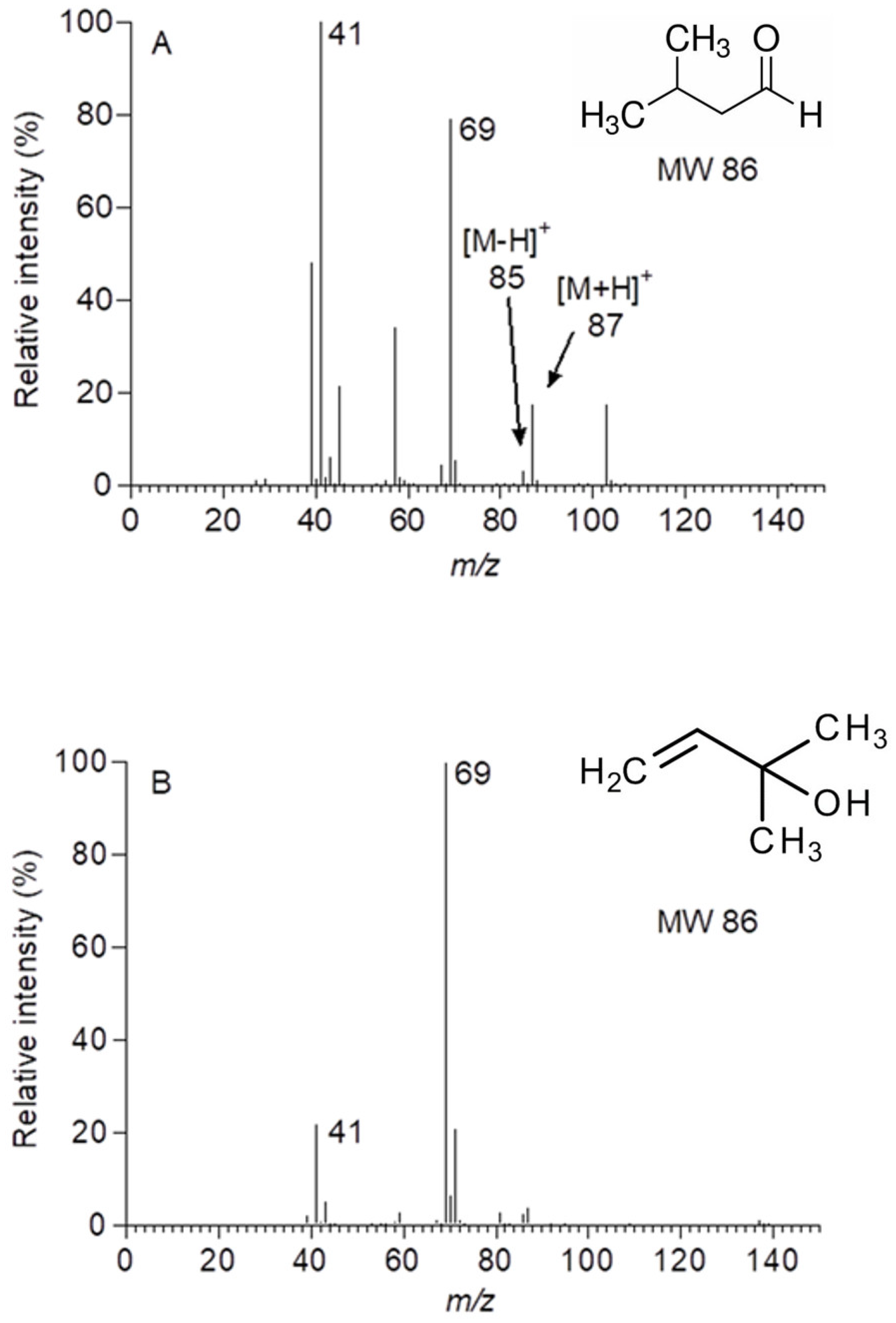
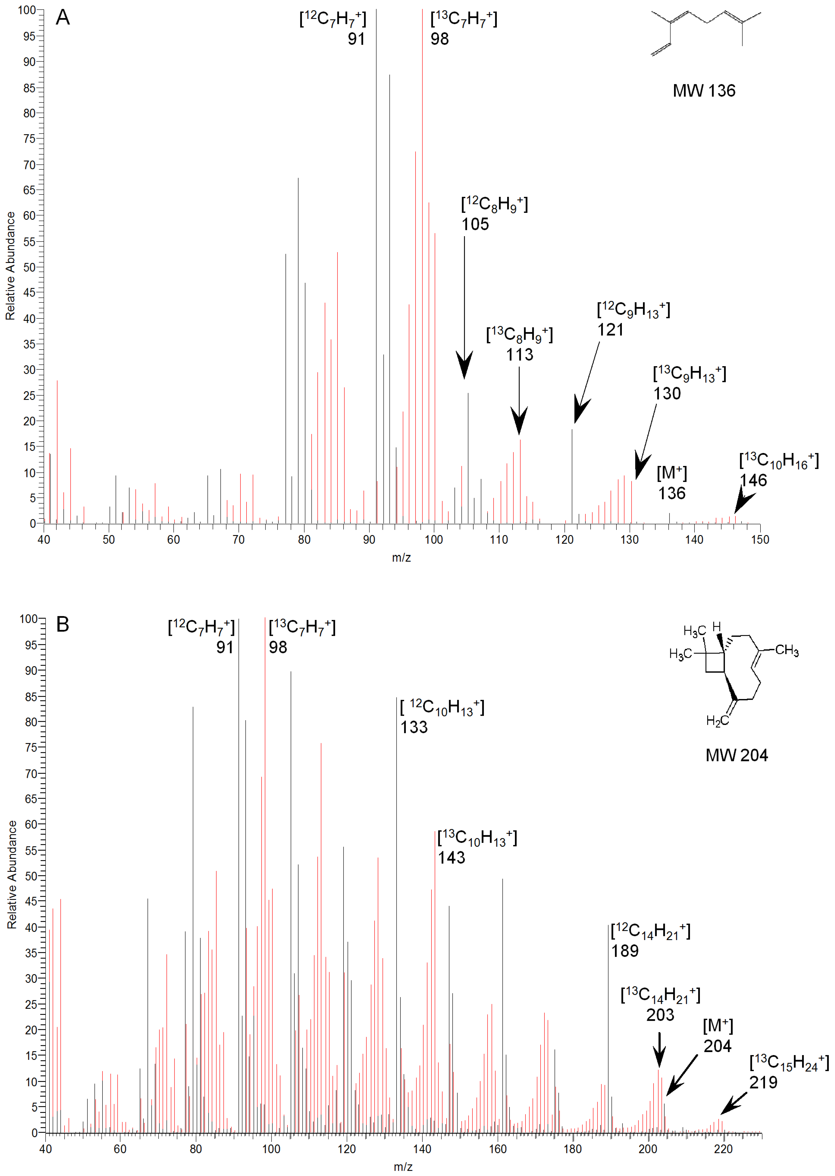
2.2. Identification and Kinetics of 13C-Labelled Compounds

13C-labelled compounds were identified with a combination of shifts in the isotopomer-signals of the PTR-MS and GC-MS analysis following desorption from charcoal cartridges. The monitoring of molecular and fragment ions of mono- or sesquiterpenes with PTR-MS could not be used to quantify the incorporation of 13C owing to their low abundance and the background emission of stored terpenes.
Isoprene was not detected by GC-MS (boiling point 34 °C) as the option of cryogenic focussing at the top of the column was not available. However, PTR-MS data showed 13C being incorporated into isoprene approximately 10 min after 13CO2 fumigation started (Figure 2), reaching a maximum incorporation of 82%, as shown by the intensity of m/z 74 ([MH]+ 13C5 isotopomer) after 2.5 h exposure to 13CO2. After reverting to CO2 at natural isotopic abundance, 13C incorporation declined, mirroring its incorporation after introduction.
A second compound observed in the PTR-MS trace demonstrated similar 13C incorporation kinetics. 13C incorporation began about 15 min after fumigation started (Figure 3), reaching a maximum of 82% after 5 h (i.e., m/z 92, [MH]+. 13C5 isotopomer). It should be noted here that m/z 90 ions are a mixture of the 13C5 [M-H]+ and 13C312C2 [MH]+. isotopomers. The rates of 13C incorporation were slightly faster into isoprene (m/z 69, τ1/2 = 4.47 min) compared to the second compound (m/z 85, τ1/2 = 5.11 min; m/z 87, τ1/2 = 5.94 min). This finding was confirmed by monitoring the corresponding decreases in the intensity of both the [MH]+ ions (m/z 87) and the [M-H]+. ions (m/z 85) representing the 12C5 isotopomer (Figure 4). Incrementally greater masses showed transient peaks following the sequential replacement of 12C with 13C atoms, akin to the labelling pattern of isoprene (Figure 2). Hence, the second molecule seems to be a C5 compound of molecular mass 86, with ionisation in the PTR-MS inducing the addition (m/z 87) and removal (m/z 85) of a single proton.
The molecular mass of 86 corresponds to iso-valeraldehyde and several methylbutenol isomers, all with the structural formulae C5H10O. Mass scans in the range m/z 81–95 ruled out monoterpenes (C10H16) as potential contributors, while isoprene could also be excluded given its lack of ion fragments at these masses. p-Cymene produces ion fragments at m/z 93 [35] (Winters, unpublished data) but showed no response to 13CO2 fumigation. Iso-valeraldehyde, detected previously in the analysis of leaf oils from a number of eucalypt species [36] and two alcohols—3-methyl-3-buten-1-ol, emitted by Querus ilex [27] and 2-methyl-3-buten-2-ol, emitted by a number of Pinus species [37,38,39]—were considered candidates given their common molecular mass and elemental composition. Qualitative gas standards, produced by the dilution of neat liquids into N2, were used to examine the fragmentation spectrums of each compound ionised by the PTR-MS. Iso-valeraldehyde produced fragments at m/z 85 and m/z 87 in similar proportions to those seen in the labelling study (Figure 4A), while 2-methyl-3-buten-2-ol did not (Figure 4B). Subsequent analysis by GC-MS after desorption from charcoal cartridges confirmed iso-valeraldehyde as the likely compound.
GC-MS (following desorption from charcoal cartridges) provided clear evidence for the incorporation of 13C into two further terpenes, the monoterpene (C10) cis-ocimene and the sesquiterpene (C15) trans-caryophyllene (Figure 5). An isomeric form of ocimene, tentatively identified as trans-ocimene through a comparison of the retention times and fragmentation patterns with an authentic standard, also incorporated 13C, but the compound was not consistently present in samples and was excluded from further analysis. The degree of the incorporation of labelled carbon into these C10 and C15 terpenes was determined through a comparison of the respective mass spectra over the course of the labelling experiment, with ions selected according to the abundance in the spectrum or their relationship to the molecular ion. Despite the low relative abundance (<2%) of the cis-ocimene molecular ion (M+., m/z 136), a corresponding ion at m/z 146, representing the incorporation of ten 13C atoms, was clearly discernible (Figure 5A). This incorporation was confirmed through analysis of an [M-15]+. ion (m/z 121; 18%) and the incorporation of nine 13C atoms could be clearly observed in the corresponding labelled ion at m/z 130. The incorporation of 13C into the [M-15]+. ion reached a maximum of approximately 77% after 2.5 h (Figure 6A), similar to that reported for isoprene. The trans-caryophyllene molecular ion (M+., m/z 204) had a 6% abundance and the incorporation of fifteen 13C atoms could be clearly observed in the corresponding ion at m/z 219 (Figure 6B). The incorporation of 13C into trans-caryophyllene appeared to stabilise at 15–20% after 5–6 h.

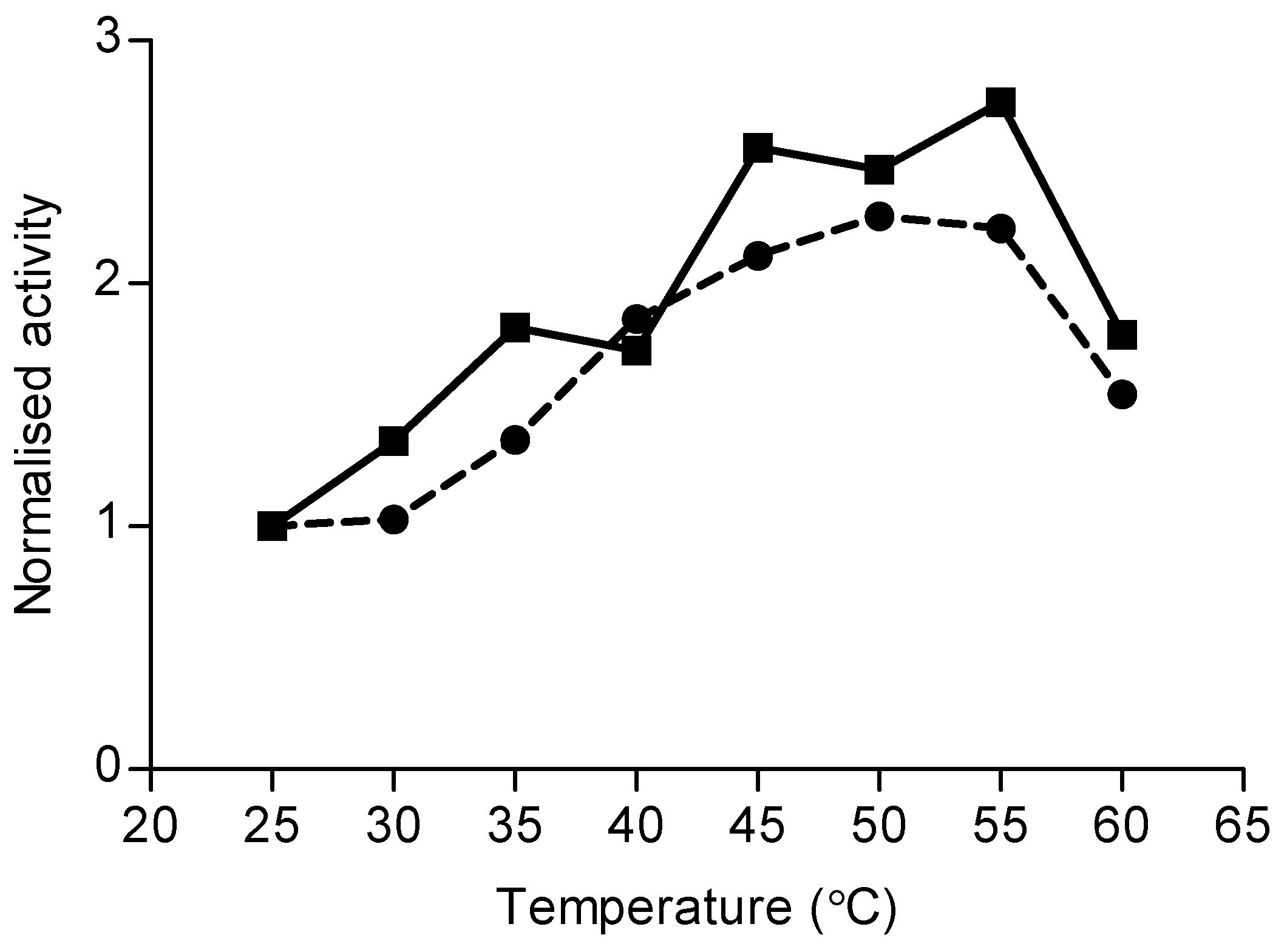
2.3. Monoterpene Synthase Activity

Mono-terpene synthase (TPS) activities were determined by headspace analysis following the incubation of protein extracts from eucalyptus leaves with geranyl diphosphate (GDP) [40]. Assays were problematic owing to stored oils being carried through the protein extraction process, as a headspace analysis of denatured (boiled) extracts produced similar results. Variations in the protocol, including salting out extracts and radioactive phosphatase assays using [1-3H]-GDP [40], failed to resolve the issue. However, incremental increases in incubation temperature resulted in increased production of cis-ocimene. This phenomenon was observed in leaf extracts of both E. globulus and E. viminalis (Figure 7). In both species, the maximum enzymatic production of cis-ocimene was recorded between 50 °C and 55 °C. Above this temperature range, activity declined (likely due to enzyme denaturation). Other monoterpenes, such as α-pinene, showed no response to temperature.

3. Discussion

The exposure of E. globulus leaves to 13CO2 suggests that isoprene and iso-valeraldehyde preferentially incorporated recently fixed carbon. In addition, the monoterpene cis-ocimene and the sesquiterpene trans-caryophyllene also incorporated recently synthesised carbon and appeared subject to regulation by light as well as temperature.

3.1. The Origin of Labelled Terpenes

The terpenes emitted by E. globulus likely originate from both oil glands and mesophyll tissue. The emissions of stored and de novo synthesised compounds are also compartmentalised at these two sites, as the absence of labelled carbon in compounds stored in oil glands argues against the possibility of synthesis in gland epithelial cells with the products deposited into the glands themselves. It seems unlikely that we lacked the detection power for labelled compounds in oil glands given the number of glands sampled and the sensitivity of our analysis. Additionally, labelled compounds were absent in whole-leaf tissue, i.e., they were absent from tissue outside oil glands. However, clearly labelled compounds were detected in leaf emissions, and given the similarity in labelling patterns among the three terpenes and iso-valeraldehyde, it seems likely that the chloroplasts of mesophyll cells are both a route for carbon fixation and terpene synthesis. Detailed biochemical analysis is needed to verify this circumstantial evidence. Nonetheless, an almost identical pattern to that reported here was found in Norway spruce and Scots pine following 13CO2 fumigation [10]. In contrast, 14C labelling in Maritime pine detected monoterpene synthesis in the epithelial cells of growing parts of needles, while older parts were significantly depleted in labelled carbon, and labelled sesquiterpenes were detected only in whole-leaf tissue surrounding resin ducts [41].
The 13C label appeared rapidly in isoprene following exposure to 13CO2, but the temporal resolution required the use of a charcoal adsorbent. Hence, the appearance of 13C in cis-ocimene and trans-caryophyllene could not be quantified as accurately as in isoprene. Nonetheless, both the time taken for 13C enrichment to saturate, and the final proportion of molecules enriched with 13C, were remarkably similar between isoprene and cis-ocimene, with both reaching around 80% enrichment after 2.5 h exposure to 13CO2. This pattern appears to be a consequence of the rapid incorporation of labelled carbon into the C5 and C10 terpene substrates dimethylallyl diphosphate (DMADP) and geranyl diphosphate (GDP), respectively. The rates of incorporation observed in emissions in E. globulus are comparable to the rates reported for conifer species [10], but much slower than those reported for monoterpene labelling in Q. ilex [10,27], which saturated at around 80% after 20–30 min. Differences in leaf architecture, such as bulk leaf density, may explain this effect. The labelling of trans-caryophyllene was slower and less complete than that of C5 isoprene and C10 cis-ocimene, which fits with the export of isopentenyl diphosphate (IPP) from plastids into the cytosol, where carbon from the mevalonate pathway contributes to the synthesis of sesquiterpenes [25,42]. The link between the two compartments was demonstrated previously in E. globulus, where deuterated deoxyxylulose was incorporated into both ocimene and caryophyllene at 83% [43].

3.2. Identification of 13C Labelling

The labelling experiments reported here unequivocally confirm the incorporation of recently fixed carbon into cis-ocimene and trans-caryophyllene, but the extent of this incorporation can be obscured by emissions from stored sources. This is of particular significance when analysing samples by GC-MS, and when the molecular ion is absent or present only at low concentrations. To determine whether smaller ion fragments could be reliably used to examine labelling kinetics, Loreto et al. [27] tested a statistical null model that assumed a random distribution of C atoms in the terpene molecule against the observed labelling. The conclusion of the authors that all carbons were probably randomly labelled is now known not to be true, since carbon atoms are drawn from specific substrates [23,31,44,45]. However, in the absence of a suitably abundant molecular ion, the [M-CH3]+. ion could be used to determine incorporation kinetics, providing the number of carbons in the molecule is sufficiently high. Terpene emissions from stored sources could pose a greater problem for interpretation if they are large relative to the de novo synthesised fraction, as the incorporation of the 13C label would need to be determined against an intense endogenous background.
The light response observed for cis-ocimene in a related experiment (Winters et al., unpublished data) found that cis-ocimene emissions were six times greater when exposed to 500 μmol m−2 s−1 PAR (15 nmol m−2 min−1) compared to darkness (2.5 nmol m−2 min−1), where dark emissions represent those from the stored pool. Thus, the light–dark emission ratio is estimated to be around 6:1, but as the temperature was 3 °C warmer in this study (with the same light level), this ratio might be slightly smaller, as the warmer temperatures may have volatilised more stored oils; therefore, a conservative ratio of 5:1 is assumed. Thus, 20% of the 12C signal (at saturated enrichment of the de novo fraction) could result from stored oils. Correcting for this origin of the 12C signal, the calculated 13C incorporation increased from 77% to 79%, which is within the range of error for these estimates. Using the same spectral data, but changing the emissions from stored pools to 70% of de novo synthesis, the saturated enrichment would be corrected to 93%, representing a large change. As an alternative, Ghirardo et al. [10] used PTR-MS data, assuming the labelling seen in the monoterpenes reflects that seen in isoprene, given their proximity in the biosynthetic pathway.
The presence of iso-valeraldehyde was confirmed in the present study by GC-MS. This compound is a reasonable candidate for the ions observed at m/z 85 and 87, particularly given previous reports of iso-valeraldehyde in the oil extracts of a number of eucalypt species, including E. globulus [36]. However, caution is warranted as the rate of labelling seems fast relative to its putative source within the leaf. The few reports on iso-valeraldehyde in the literature [46,47] suggest it is emitted from pyruvate decarboxylase activity on pyruvate during the catabolism of L-leucine [47,48]. If this process is indeed the source of iso-valeraldehyde emitted from eucalypt leaves, it seems at odds with the rapid incorporation of 13C into the molecule. Conversely, 2-methyl-3-buten-2-ol and various structural isomers have been reported in the literature, mostly in pines [37,38,39,49], but also in Quercus ilex [27]. Retention time-matching by gas chromatography was inconclusive (early eluting broad peak due to lack of cryogenic focussing), but the major ion fragments generated from an iso-valeraldehyde standard using PTR-MS (Figure 4A) matched those ions seen during fumigation with 13CO2 (Figure 2). Although more ion fragments were seen in the standard, it provided a better fit than methyl butanol (Figure 4B), which has not been reported to be emitted from eucalypts.

3.3. The Putative Importance of De Novo Synthesis

Monoterpene synthase assays were initially of limited value due to the passage of terpene oils from the ground leaves into the protein extract. Using an incremental rise in temperature, we were able to discriminate contaminant oils from those synthesised during the incubation period, revealing that only cis-ocimene was produced enzymatically. The optimum temperature at 50 °C was around 10 °C higher than that found in Q. ilex [40], but in a similar range to the optimum isoprene synthase temperature in leaves of E. globulus (Winters, Schnitzler, and Zimmer, unpublished data). These results confirm that a fraction of the cis-ocimene emitted from E. globulus leaves originates indeed from a de novo source. Although no other monoterpenes were found to be synthesised during the assay, the presence of low levels of de novo-synthesised monoterpenes would have been very difficult to detect against the background of the oils carried-over through protein extraction. The absence of 13C labelling in any other monoterpene, either emitted or stored, suggests monoterpene synthesis may be subject to temporal regulation within E. globulus leaves. The seasonal regulation of monoterpene synthesis has been demonstrated previously in Quercus ilex [40], which does not store terpenes, and in Pinus sylvestris [50], which both stores and synthesises terpenes. In peppermint (Mentha x piperita L.), monoterpene synthases are active during leaf development, but inactive in mature leaves [51,52]. The present results suggest that once oil glands have been filled during leaf expansion, the epithelial cells become either dormant or degenerate and the oil produced until that point will be the only oil present during the leaf’s two- to four-year life span. However, the biosynthesis of non-stored terpenes persists within the mesophyll for the entire life of the leaf. A similar developmental pattern is observed in Maritime pine [41]. Preliminary evidence for this pattern also comes from an unpublished comparison of the emissions of young and old juvenile leaves of E. globulus, where young leaves emitted eucalyptol at a magnitude two to three orders of greater than that of cis-ocimene, while in the aged leaf, emissions dropped to 1:1 (Winters, unpublished data).

4. Materials and Methods

4.1. Plant Material

Seedlings of E. globulus Labill. subsp. globulus were cultivated in 20 L pots containing a sterilised mix of vermiculite and loam in a ratio of 1:2 (v/v). Plants were supplied with a low phosphorous, slow-release fertiliser (Osmocote Plus™ ICL, Sydney, Australia) and maintained under ambient light with periodic shading provided on hot days. Average diurnal temperatures ranged between 21 and 32 °C in the month before the experiments and plants were watered daily. Plants were about 18 months old and 1.5–2 m high at the time of the experiment.

4.2. Gas Exchange System

Gas exchange measurements were made on the distal 2–3 pairs of opposite leaves on branchlets of E. globulus. Healthy and visually similar leaves were placed in a 0.75 L cuvette consisting of two chambers: a lower chamber in which the leaf was housed and an upper chamber though which water from a Julabo water bath (John Morris Scientific, Sydney, Australia) was circulated to control leaf temperature. Prior to placing the leaves in the cuvette, the leaf stem was wrapped in polytetrafluoroethylene (PTFE) tape to limit abrasion of the stem. Leaves were illuminated with a 250 W metal-halide lamp and held constant at 500 μmol m2 s1 photosynthetically active radiation (PAR) for the duration of the experiment. Leaf temperature was held at 27.9 ± 1.3 °C for all experiments. Zero-grade air (BOC Gases, Sydney, Australia, 20.5% oxygen in nitrogen) was supplied to the cuvette at 2.3 L min1. The air was humidified to approximately 28% relative humidity via passage through a bubbler held at 4 °C. CO2 (BOC Gases, Sydney, Australia) of natural isotopic abundance was added to the humidified air stream to a concentration of 400 + 9 ppm in the cuvette. The exchange of H2O and CO2 was monitored with a CIRAS II infra-red gas analyser (IRGA, PP Systems, Hertfordshire, UK) sampling air before and after the cuvette. IRGAs are less sensitive to 13CO2 (Cambridge Isotope Laboratories, Inc., Tewksbury, MA, USA) than natural abundance CO2, but the rates of gas exchange were used primarily to monitor plant function throughout the experiment. Leaves were positioned in the cuvette on the evening before experiments commenced, with the cuvette lid open and exposed to laboratory air. The following morning, the cuvette was sealed, and light and temperature control, and air supply commenced. The plant was allowed at least 2 h were to stabilise before the beginning of the measurements. Natural carbon abundance CO2 was initially supplied to the leaves for the first hour of measurements, before switching to 13CO2 for 6–8 h, then reverting to natural abundance CO2 for a further 1–2 h.

4.3. BVOC Sampling and Analysis

A high-sensitivity proton transfer reaction-mass spectrometer (PTR-MS, Ionicon Analytic GmbH, Innsbruck, Austria) was used to monitor changes in the isotopic signal of terpenes. As the quadrupole mass analyser in the PTR-MS is of unit mass accuracy and resolution, it has a limited ability to resolve and identify individual BVOC components. The airflow (80 mL min1) from the cuvette was split between the PTR-MS and charcoal-filled adsorbent cartridges, which could trap BVOC for later identification using gas chromatography–mass spectrometry (GC-MS). Samples were also collected each morning and evening from the upstream side of the cuvette in order to check for potential background interferences (always negligible). Protonated parent and fragment ions of isoprene (m/z 69 to 74) and monoterpenes (m/z 81 to 95; 135 to 145; 155 to 165) were monitored using PTR-MS in selected ion mode (SIM), with the drift tube operated at 126 Td and held at 40 °C. The instrument was calibrated daily with an eight-component standard gas mix (Apel Reimer Environmental, Denver, CO, USA) and transmission was determined weekly using liquid standards diluted in Tedlar bags.
Custom-made adsorbent charcoal cartridges collected gas-phase monoterpenes for the subsequent analysis of isotopic incorporation using GC-MS. Borosilicate glass tubes (7 cm × 3.2 mm id) were packed with 5 mg of 20–60 mesh activated charcoal (Sigma Aldrich, Sydney, Australia) and plugged at either end with silanised glass wool. The charcoal was pre-treated by heating to 450 °C while passing helium through the bed at 80 mL min1 for 4 h. During sampling, air was drawn through the cartridge at 0.08 L min1 for 60 min. Trapped terpenes were solvent-extracted by transferring the charcoal bed to a 2 mL glass vial containing a 300 μL insert. Carbon disulphide (50 μL) was added and allowed to stand for 30 min at room temperature when the vial was centrifuged and the supernatant was transferred to a second vial for storage at 4 °C before analysis by GC-MS. A pilot study indicated that extraction times greater than 30 min did not improve analyte recovery and analytes were found to be stable for up to eight weeks.

4.4. Extraction of Terpenes from Individual Oil Glands and Leaf Tissue

PTR-MS revealed that the incorporation of 13C into emitted terpenes reached a maximum after ca. 6 h. To determine if 13C was taken up by terpenes remaining within the leaf tissue, a further four studies were carried out where leaves were removed from the cuvette immediately, after 6–8 h of fumigation with 13CO2. Of the four leaves, two were stored in liquid N2 for the later extraction of terpene oils from homogenised leaf tissue. Leaves were ground under liquid N2, and 100 mg (fresh weight) was placed in a vial containing 2 mL of chilled pentane spiked with 100 μg mL1 1,2,4-trimethylbenzene as an internal standard. The extracts were allowed to stand at room temperature for four days before the liquid was drawn off for subsequent analysis by GC-MS.
Individual oil glands were sampled on the remaining two leaves using a glass capillary previously drawn to a fine point with a custom-made capillary puller. A bottom-lit dissecting microscope was used to locate the subdermal oil glands and position the capillary tip. A Petri dish containing chilled water was used as a stage to allow the light to pass up into the leaf while limiting the transfer of heat to the leaf. A syringe connected via flexible tubing to the capillary was used to draw oil from the pierced gland into the tip of the capillary. Once 30–40 glands across the leaf had been sampled, the tip was rinsed in a 2 mL vial containing a 300 µL insert filled with 40 µL of chilled pentane. A pilot study demonstrated no variation in oil composition between glands within a given leaf.

4.5. GC-MS Analysis of Individual Terpenes

Aliquots of 1 µL were injected via an autosampler into a Polaris Q GC-MS (Thermo Fisher Scientific, Sydney, Australia) (injection port 220 °C; interface 230 °C; source 200 °C). Chromatographic separation was achieved using a SolGel Wax fused–silica capillary column (SGE Analytical Science, Melbourne, Australia; 30m × 0.25 mm id, 0.25 µm phase thickness) with He as the carrier gas (inlet pressure 96.5 kPa). The temperature programme began at 30 °C and was held for 0.5 min; increased to 150 °C at 5 °C min1; and increased to 240 °C at 15 °C min1 and was held for 2 min. The mass spectrometer was operated in the electron ionisation (EI) mode with an ionisation energy of 70 eV. Mass spectra were acquired with full scans from 50 to 250 u in 0.33 s. Peaks were identified through comparing retention time and fragmentation spectra (MS similarity > 80% with authentic standards), and where these were not available, through comparison with the NIST-Wiley Mass Spectra Library (1997) (MS similarity > 80%).

4.6. Monoterpene Synthase Assay

Leaves from the trees used in the gas exchange study, in addition to similarly aged leaves from specimens of E. viminalis, grown under identical conditions in Australia, were assayed for monoterpene synthase (mono-TPS) activity according to the protocol described in [40,53]. Healthy juvenile leaves were instantly frozen in liquid N2 and packed in solid CO2 for transport. Upon arrival, they were stored at −80 °C until being assayed, approximately four weeks later. Proteins were isolated using the modified protocols previously developed for Quercus ilex [40]. The terpene synthase activity protocols developed for Q. ilex suffered from interference from the terpene oils, which had been carried into the protein extract, and various attempts to separate the oil from the extract proved unsuccessful. The enzyme activity was therefore assayed using temperature response to identify mono-TP synthase activity above the background level.
For the temperature response assay, leaves were ground under liquid N2 and 100 mg (fresh weight) of the powder was suspended in 5 mL of chilled extraction buffer (0.7 M MOPS, pH 7.3, 1.5% PEG, 20 mM MgCl2, 1% PVP-30 (w/v), 8.3% (w/v) Dowex 1 × 2, 0.5 M Na-ascorbate, 0.5 M Na2S2O5, 0.5 M DTT). The remaining steps in the protocol were conducted at 4 °C. After stirring for 15 min, the homogenate was centrifuged at 18,000× g for 20 min and 2.5 mL of the supernatant was desalted on PD-10 columns (Amersham-Pharmacia, Freiburg, Germany) with elution buffer (50 mM KPi, pH 7.3, 10% glycerol (v/v) and antioxidants (200 mM Na-ascorbate and 50 mM β–mercaptoethanol). From the resulting 3.5 mL of protein extract, 93 μL was transferred into 2 mL gas-tight crimp vials, followed by 2 μL of 1 M MgCl2. The enzyme reaction was started by adding 5 μL of 5 mM geranyl diphosphate (GDP) (final concentration: 250 μM) and vials were incubated for 60 min at one of eight 5 °C temperature increments between 25 °C and 60 °C. The reaction was stopped by removing the reaction mixture with a syringe and rinsing the vial with 150 μL of distilled water. Monoterpenes generated through the reaction of mono-TPS with the GDP substrate were sampled from the headspace and analysed via gas chromatography (GC).
A CP-9000 gas chromatograph (Chrompack, Frankfurt/M., Germany) equipped with a headspace-volume-autosampler (Gerstel, Mülheim, Germany) and temperature-programmed injection system (KAS 3, Gerstel, Mülheim, Germany) was used for analysis (40). Headspace injections of 1 mL were concentrated on a precolumn trap containing Tenax TA (60/80 mesh) at 27 °C. Trapped compounds were desorped at 240 °C onto a capillary column (DB-1701, 30 m × 0.25 mm I.D., 1 μm film thickness; J&W Scientific, Folsom, CA, USA). The column was then temperature-programmed from 35 °C (held for 0.5 min) to 78 °C at 30 °C min1 (held for 4 min), then increased to 160 °C at 9 °C min1, and then to 250 °C at 35 °C min1. Products were detected with an FID operating at 270 °C. Compounds were identified through a retention time comparison with authentic standards. Protein extracts incubated without the GDP substrate were used for background correction of the terpene signals.

4.7. Data Analysis and Error Determination

Error estimates incorporating calibration and sampling errors were calculated through the propagation of errors [54]. The half-life time of the decay of the unlabelled signal of compounds detected by PTR-MS was determined from the time evolution of this signal according to the equations N(t) = N0 e−λτ and τ(1/2) = ln(2)/λ, where N(t) is the emission rate at time t, N0 is the calibrated instrument signal at t = 0, and λ is an empirically determined decay constant. Constants within each function were determined by fitting the function to each set of normalised data points and constraining it until an acceptable fit was achieved (R2 > 90%).” Half-life time calculations were made using GraphPad Prism 5 (GraphPad Software, San Diego, CA, USA).

5. Conclusions

Apart from isoprene, previous studies of eucalypts suggest that oil glands dominate terpene emissions that are mainly regulated by temperature. Given the general predominance of oil glands in eucalypts, these are logical conclusions. Here, we show that while stored pools account for most terpenes emitted from juvenile leaves of E. globulus, the monoterpene cis-ocimene and the sesquiterpene trans-caryophyllene originate from de novo synthesis. In addition to isoprene, an oxygenated C5 compound identified as iso-valeraldehyde was also emitted directly from de novo synthesis. Future studies need to identify how these emissions respond to light and temperature, given the preponderance of high-light, high-temperature eucalypt habitats. Similarly, further attention is required for the identification and quantification of iso-valeraldehyde, as well as to determine its response to environmental variables. This need is underscored by the large fragments produced by iso-valeraldehyde at m/z 69, which may result in serious errors in the estimation of isoprene fluxes.

Author Contributions

Conceptualization, A.J.W., H.R., J.K. and M.A.A.; Methodology, A.J.W.; C.H.H., J.-P.S., I.Z. and C.K.; Software, A.J.W.; Validation, A.J.W.; Formal Analysis, A.J.W. and C.H.H.; Investigation, A.J.W., C.H.H. and I.Z.; Resources, J.-P.S.; Data Curation, A.J.W.; Writing—Original Draft Preparation, A.J.W.; Writing—Review and Editing, C.H.H., C.K., J.-P.S., I.Z., H.R., M.A.A. and J.K.; Visualisation, A.J.W.; Supervision, M.A.A., J.-P.S., H.R. and J.K.; Funding Acquisition, M.A.A. and J.-P.S. All authors have read and agreed to the published version of the manuscript.

Funding

This work was funded by an International Science Linkage grant from the Australian Department of Education, Science and Training and by the German Government (BMBF) under contract number AUS-02/004.

Institutional Review Board Statement

Not applicable.

Informed Consent Statement

Not applicable.

Data Availability Statement

Raw data are available from the authors on request.

Acknowledgments

Sincere thanks to Marilyn Ball for generously providing the laboratory and office space for AJW within the Functional Ecology Lab at RSBS. We are also grateful to the RSBS Workshop team—Brian McNamara, Istvan Zaveczky, Bill Speed, and Shane Green—and to the RSBS Plant Services staff—Steve Dempsey and Sue Lyons—for their invaluable advice and assistance, and the fabrication of essential components. Additional thanks go to the members of Environmental Biology, including Chin Wong, Hilary Stuart-Williams, and Peter Groeneveld; David Kelly of the Research School of Physics and Engineering (ANU); and Stephen Roxburgh of CSIRO Plant Industry for their support and contributions. Tragically, Tony Winters was killed in a paragliding accident on 28 December 2023. Tony left an indelible mark as a highly skilled scientist, networker, and instrument builder, and demonstrated remarkable ability to bring people and resources together to conduct his research into BVOC emissions from eucalypts. Tony developed an automated BVOC sampling system (based on an SRI portable GC), with an online water trap and Peltier cooling. He fabricated the essential components and adapted the software for the operating system. All showcased Tony’s remarkable technical prowess and innovation. The latter qualities stood him in good stead in his later career as a Field Engineer for Agilent Technologies. He is greatly missed by his colleagues, friends, family, and his loving wife and daughter.

Conflicts of Interest

The authors declare no conflicts of interest.

Abbreviations

The following abbreviations are used in this manuscript:
BVOCBiogenic volatile organic compounds
DMADPDimethylally diphosphate
GC-MSGas chromatography–mass spectrometry
GDPGeranyl diphosphate
MVAMevalonic acid
MEPMethylerithritol phosphate
PARPhotosynthetically active radiation
PTR-MSProton transfer reaction–mass spectrometer
RTRetention time
SIMSelected ion monitoring
TPSTerpene synthase

References

  1. Guenther, A.; Hewitt, C.N.; Erickson, D.; Fall, R.; Geron, C.; Graedel, T.; Harley, P.; Klinger, L.; Lerdau, M.; McKay, W.A.; et al. A global model of natural volatile organic compound emissions. J. Geophys. Res. Atmos. 1995, 100, 8873–8892. [Google Scholar] [CrossRef]
  2. Atkinson, R. Atmospheric chemistry of VOCs and NOx. Atmos. Environ. 2000, 34, 2063–2101. [Google Scholar] [CrossRef]
  3. Fuentes, J.D.; Gu, L.; Lerdau, M.; Atkinson, R.; Baldocchi, D.; Bottenheim, J.W.; Ciccioli, P.; Lamb, B.; Geron, C.; Guenther, A.; et al. Biogenic hydrocarbons in the atmospheric boundary layer: A review. Bull. Am. Meteorol. Soc. 2000, 81, 1537–1575. [Google Scholar] [CrossRef]
  4. Claeys, M.; Graham, B.; Vas, G.; Wang, W.; Vermeylen, R.; Pashynska, V.; Cafmeyer, J.; Guyon, P.; Andreae, M.O.; Artaxo, P.; et al. Formation of secondary organic aerosols through photooxidation of isoprene. Science 2004, 303, 1173–1176. [Google Scholar] [CrossRef]
  5. Guenther, A.; Karl, T.; Harley, P.; Wiedinmyer, C.; Palmer, P.I.; Geron, C. Estimates of global terrestrial isoprene emissions using MEGAN (Model of Emissions of Gases and Aerosols from Nature). Atmos. Chem. Phys. 2006, 6, 3181–3210, Corrected in Atmos. Chem. Phys. 2006, 6, 3181–3210. [Google Scholar] [CrossRef]
  6. Külheim, C.; Yeoh, S.H.; Wallis, I.R.; Laffan, S.; Moran, G.F.; Foley, W.J. The molecular basis of quantitative variation in foliar secondary metabolites in Eucalyptus globulus. New Phytol. 2011, 191, 1041–1053. [Google Scholar] [CrossRef]
  7. Poisson, N.; Kanakidou, M.; Crutzen, P.J. Impact of non-methane hydrocarbons on tropospheric chemistry and the oxidizing power of the global troposphere: 3-dimensional modelling results. J. Atmos. Chem. 2000, 36, 157–230. [Google Scholar] [CrossRef]
  8. Bell, N.; Heard, D.E.; Pilling, M.J.; Tomlin, A.S. Atmospheric lifetime as a probe of radical chemistry in the boundary layer. Atmos. Environ. 2003, 37, 2193–2205. [Google Scholar] [CrossRef]
  9. Loreto, F.; Schnitzler, J.-P. Abiotic stresses and induced BVOCs. Trends Plant Sci. 2010, 15, 154–166. [Google Scholar] [CrossRef]
  10. Ghirardo, A.; Koch, K.; Taipale, R.; Zimmer, I.; Schnitzler, J.; Rinne, J. Determination of de novo and pool emissions of terpenes from four common boreal/alpine trees by 13CO2 labelling and PTR-MS analysis. Plant Cell Environ. 2010, 33, 781–792. [Google Scholar] [CrossRef]
  11. Loreto, F.; Ciccioli, P.; Cecinato, A.; Brancaleoni, E.; Frattoni, M.; Tricoli, D. Influence of environmental factors and air composition on the emission of [α]-pinene from Quercus ilex leaves. Plant Physiol. 1996, 110, 267–275. [Google Scholar] [CrossRef] [PubMed]
  12. Ziegler, H.; Kotzias, D.; Steinbrecher, R.; Schürmann, W.; Schönwitz, R. Emission of biosynthesized monoterpenes from needles of Norway spruce. Sci. Nat. 1993, 80, 276–278. [Google Scholar] [CrossRef]
  13. Brooker, M.I.H.; Kleinig, D.A. Field Guide to Eucalypts. Volume 1, South-Eastern Australia; Bloomings Books: Victoria, Australia, 1999. [Google Scholar]
  14. Wilson, P.G.; O’Brien, M.M.; Gadek, P.A.; Quinn, C.J. Myrtaceae revisited: A reassessment of infrafamilial groups. Am. J. Bot. 2001, 88, 2013–2025. [Google Scholar] [CrossRef]
  15. Guenther, A.B.; Monson, R.K.; Fall, R. Isoprene and monoterpene emission rate variability: Observations with eucalyptus and emission rate algorithm development. J. Geophys. Res. Atmos. 1991, 96, 10799–10808. [Google Scholar] [CrossRef]
  16. Guenther, A.B.; Zimmerman, P.R.; Harley, P.C.; Monson, R.K.; Fall, R. Isoprene and monoterpene emission rate variability: Model evaluations and sensitivity analyses. J. Geophys. Res. Atmos. 1993, 98, 12609–12617. [Google Scholar] [CrossRef]
  17. Coppen, J.J.W. (Ed.) Estimates of eucalypt plantations worldwide. In Eucalyptus: The Genus Eucalyptus; Taylor and Francis: New York, NY, USA, 2002; pp. 408–411. [Google Scholar]
  18. Wiedinmyer, C.; Tie, X.; Guenther, A.; Neilson, R.; Granier, C. Future Changes in Biogenic Isoprene Emissions: How Might They Affect Regional and Global Atmospheric Chemistry? Earth Interact. 2006, 10, 1–19. [Google Scholar] [CrossRef]
  19. Owen, S.; Harley, P.; Guenther, A.; Hewitt, C. Light dependency of VOC emissions from selected Mediterranean plant species. Atmos. Environ. 2002, 36, 3147–3159. [Google Scholar] [CrossRef]
  20. Niinemets, Ü.; Reichstein, M.; Staudt, M.; Seufert, G.; Tenhunen, J.D. Stomatal constraints may affect emission of oxygenated monoterpenoids from the foliage of Pinus pinea. Plant Physiol. 2002, 130, 1371–1385. [Google Scholar] [CrossRef]
  21. Grote, R.; Niinemets, Ü. Modeling volatile isoprenoid emissions—A story with split ends. Plant Biol. 2008, 10, 8–28. [Google Scholar] [CrossRef]
  22. Tingey, D.T.; Manning, M.; Grothaus, L.C.; Burns, W.F. Influence of light and temperature on monoterpene emission rates from slash pine. Plant Physiol. 1980, 65, 797–801. [Google Scholar] [CrossRef]
  23. Werner, C.; Fasbender, L.; Romek, K.M.; Yáñez-Serrano, A.M.; Kreuzwieser, J. Heat waves change plant carbon allocation among primary and secondary metabolism altering CO2 assimilation, respiration, and VOC emissions. Front. Plant Sci. 2020, 11, 1242. [Google Scholar] [CrossRef] [PubMed]
  24. Byron, J.; Kreuzwieser, J.; Purser, G.; van Haren, J.; Ladd, S.N.; Meredith, L.K.; Werner, C.; Williams, J. Chiral monoterpenes reveal forest emission mechanisms and drought responses. Nature 2022, 609, 307–312. [Google Scholar] [CrossRef]
  25. Kreuzwieser, J.; Meischner, M.; Grün, M.; Yáñez-Serrano, A.M.; Fasbender, L.; Werner, C. Drought affects carbon partitioning into volatile organic compound biosynthesis in Scots pine needles. New Phytol. 2021, 232, 1930–1943. [Google Scholar] [CrossRef]
  26. Delwiche, C.F.; Sharkey, T.D. Rapid appearance of 13C in biogenic isoprene when 13CO2 is fed to intact leaves. Plant Cell Environ. 1993, 16, 587–591. [Google Scholar] [CrossRef]
  27. Loreto, F.; Ciccioli, P.; Brancaleoni, E.; Cecinato, A.; Frattoni, M.; Sharkey, T.D. Different sources of reduced carbon contribute to form three classes of terpenoid emitted by Quercus ilex L. leaves. Proc. Natl. Acad. Sci. USA 1996, 93, 9966–9969. [Google Scholar] [CrossRef]
  28. Loreto, F.; Ciccioli, P.; Brancaleoni, E.; Frattoni, M.; Delfine, S. Incomplete 13C labelling of α-pinene content of Quercus ilex leaves and appearance of unlabelled C in α-pinene emission in the dark. Plant Cell Environ. 2000, 23, 229–234. [Google Scholar] [CrossRef]
  29. Fasbender, L.; Yáñez-Serrano, A.M.; Kreuzwieser, J.; Dubbert, D.; Werner, C. Real-time carbon allocation into biogenic volatile organic compounds (BVOCs) and respiratory carbon dioxide (CO2) traced by PTR-TOF-MS, 13CO2 laser spectroscopy and 13C-pyruvate labelling. PLoS ONE 2018, 13, e0204398. [Google Scholar] [CrossRef]
  30. Schnitzler, J.-P.; Graus, M.; Kreuzwieser, J.; Heizmann, U.; Rennenberg, H.; Wisthaler, A.; Hansel, A. Contribution of different carbon sources to isoprene biosynthesis in poplar leaves. Plant Physiol. 2004, 135, 152–160. [Google Scholar] [CrossRef]
  31. Kreuzwieser, J.; Graus, M.; Wisthaler, A.; Hansel, A.; Rennenberg, H.; Schnitzler, J. Xylem-transported glucose as an additional carbon source for leaf isoprene formation in Quercus robur. New Phytol. 2002, 156, 171–178. [Google Scholar] [CrossRef]
  32. Schnitzler, J.; Steinbrecher, R.; Zimmer, I.; Steigner, D.; Fladung, M. Hybridization of European oaks (Quercus ilex × Q. robur) results in a mixed isoprenoid emitter type. Plant Cell Environ. 2004, 27, 585–593. [Google Scholar] [CrossRef]
  33. Yáñez-Serrano, A.M.; Mahlau, L.; Fasbender, L.; Byron, J.; Williams, J.; Kreuzwieser, J.; Werner, C. Heat stress increases the use of cytosolic pyruvate for isoprene biosynthesis. J. Exp. Bot. 2019, 70, 5827–5838. [Google Scholar] [CrossRef] [PubMed]
  34. Shao, M.; Czapiewski, K.V.; Heiden, A.C.; Kobel, K.; Komenda, M.; Koppmann, R.; Wildt, J. Volatile organic compound emissions from Scots pine: Mechanisms and description by algorithms. J. Geophys. Res. Atmos. 2001, 106, 20483–20491. [Google Scholar] [CrossRef]
  35. Tani, A.; Hayward, S.; Hansel, A.; Hewitt, C.N. Effect of water vapour pressure on monoterpene measurements using proton transfer reaction-mass spectrometry (PTR-MS). Int. J. Mass Spectrom. 2004, 239, 161–169. [Google Scholar] [CrossRef]
  36. Brophy, J.J.; House, A.P.N.; Boland, D.J.; Lassak, E.V. Digests of the essential oils of 111 species from northern and eastern Australia. In Eucalyptus Leaf Oils: Use, Chemistry, Distillation and Marketing; Boland, D.J., Brophy, J.J., Eds.; Inkarta Press: Melbourne, Australia, 1991; pp. 29–156. [Google Scholar]
  37. Harley, P.; Fridd-Stroud, V.; Greenberg, J.; Guenther, A.; Vasconcellos, P. Emission of 2-methyl-3-buten-2-ol by pines: A potentially large natural source of reactive carbon to the atmosphere. J. Geophys. Res. Atmos. 1998, 103, 25479–25486. [Google Scholar] [CrossRef]
  38. Baker, B.; Guenther, A.; Greenberg, J.; Goldstein, A.; Fall, R. Canopy fluxes of 2-methyl-3-buten-2-ol over a ponderosa pine forest by relaxed eddy accumulation: Field data and model comparison. J. Geophys. Res. Atmos. 1999, 104, 26107–26114. [Google Scholar] [CrossRef]
  39. Gray, D.W.; Goldstein, A.H.; Lerdau, M.T. The influence of light environment on photosynthesis and basal methylbutenol emission from Pinus ponderosa. Plant Cell Environ. 2005, 28, 1463–1474. [Google Scholar] [CrossRef]
  40. Fischbach, R.J.; Zimmer, I.; Steinbrecher, R.; Pfichner, A.; Schnitzler, J.-P. Monoterpene synthase activities in leaves of Picea abies (L.) Karst. and Quercus ilex L. Phytochemistry 2000, 54, 257–265. [Google Scholar] [CrossRef]
  41. Bernard-Dagan, C.; Carde, J.P.; Gleizes, M. Etude des composés terpéniques au cours de la croissance des aiguilles du Pin maritime: Comparaison de données biochimiques et ultrastructurales. Can. J. Bot. 1979, 57, 255–263. [Google Scholar] [CrossRef]
  42. Laule, O.; Fürholz, A.; Chang, H.-S.; Lange, M. Crosstalk between cytosolic and plastidial pathways of isoprenoid biosyn-thesis in Arabidopsis thaliana. Proc. Natl. Acad. Sci. USA 2003, 100, 6866–6871. [Google Scholar] [CrossRef]
  43. Piel, J.; Atzorn, R.; Gäbler, R.; Kühnemann, F.; Boland, W. Cellulysin from the plant parasitic fungus Trichoderma viride elicits volatile biosynthesis in higher plants via the octadecanoid signalling cascade. FEBS Lett. 1997, 416, 143–148. [Google Scholar] [CrossRef]
  44. Karl, T.; Fall, R.; Rosenstiel, T.; Prazeller, P.; Larsen, B.; Seufert, G.; Lindinger, W. On-line analysis of the 13 CO 2 labeling of leaf isoprene suggests multiple subcellular origins of isoprene precursors. Planta 2002, 215, 894–905. [Google Scholar] [CrossRef] [PubMed]
  45. Trowbridge, A.M.; Asensio, D.; Eller, A.S.D.; Way, D.A.; Wilkinson, M.J.; Schnitzler, J.-P.; Jackson, R.B.; Monson, R.K. Contribution of various carbon sources toward isoprene biosynthesis in poplar leaves mediated by altered atmospheric CO2 concentrations. PLoS ONE 2012, 7, e32387. [Google Scholar] [CrossRef]
  46. Dalal, K.; Salunkhe, D.; Olson, L.; Do, J.; Yu, M.H. Volatile components of developing tomato fruit grown under field and greenhouse conditions. Plant Cell Physiol. 1968, 9, 389–400. [Google Scholar] [CrossRef]
  47. Nimitkeatkai, H.; Ueda, Y.; Furukawa, H.; Inamoto, K.; Doi, M. Emission of methylbutyric acid from Gypsophila paniculata L. during bud opening: Changes in amino acid catabolism. Sci. Hortic. 2005, 106, 370–380. [Google Scholar] [CrossRef]
  48. Yoshimoto, H.; Fukushige, T.; Yonezawa, T.; Sakai, Y.; Okawa, K.; Iwamatsu, A.; Sone, H.; Tamai, Y. Pyruvate decarboxylase encoded by the PDC1 gene contributes, at least partially, to the decarboxylation of α-ketoisocaproate for isoamyl alcohol formation in Saccharomyces cerevisiae. J. Biosci. Bioeng. 2001, 92, 83–85. [Google Scholar] [CrossRef]
  49. Gray, D.W.; Lerdau, M.T.; Goldstein, A.H. Influences of temperature history, water stress and needle age on methyl butenol emissions. Ecology 2003, 84, 765–776. [Google Scholar] [CrossRef]
  50. Hakola, H.; Tarvainen, V.; Bäck, J.; Ranta, H.; Bonn, B.; Rinne, J.; Kulmala, M. Seasonal variation of mono- and sesquiterpene emission rates of Scots pine. Biogeosciences 2006, 3, 93–101, Discussions 2, 1697–1717. [Google Scholar] [CrossRef]
  51. Turner, G.; Gershenzon, J.; Nielson, E.E.; Froehlich, J.E.; Croteau, R. Limonene synthase, the enzyme responsible for monoterpene biosynthesis in peppermint, is localized to leucoplasts of oil gland secretory cells. Plant Physiol. 1999, 120, 879–886. [Google Scholar] [CrossRef]
  52. McConkey, M.E.; Gershenzon, J.; Croteau, R.B. Developmental regulation of monoterpene biosynthesis in the glandular trichomes of peppermint. Plant Physiol. 2000, 122, 215–224. [Google Scholar] [CrossRef]
  53. Schnitzler, J.-P.; Zimmer, I.; Bachl, A.; Arend, M.; Fromm, J.; Fischbach, R.J. Biochemical properties of isoprene synthase in poplar (Populus × canescens). Planta 2005, 222, 777–786. [Google Scholar] [CrossRef]
  54. Dörffel, R.A. Statistik in der Analytischen Chemie. 3 Auflage; Verlag Chemie: Weinheim, Germany, 1984; pp. 51–72. [Google Scholar]
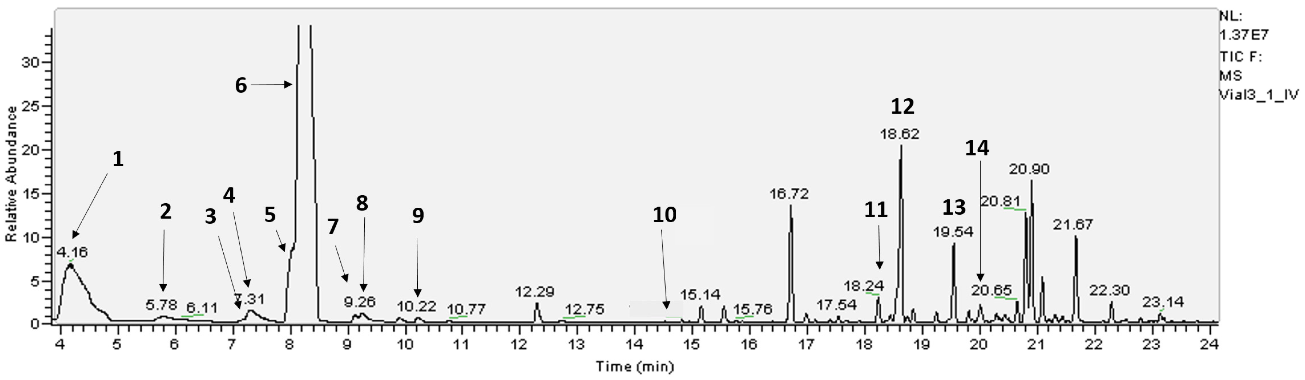
Figure 1. Typical GC-MS chromatogram of leaf oils of E. globulus. See Table 1 for the identity of the compounds, indicated in bold numbers, eluting under the respective peak.
Molecules 30 02234 g001
Figure 2. PTR-MS (SIM) monitoring of the time course of the ion intensities of 13C incorporation into isoprene emitted from the leaves of E. globulus. Ions were identified as follows: m/z 69 = 12C isoprene; m/z 70 = isoprene with one 13C atom; m/z 71 = isoprene with two 13C atoms, etc. The vertical dotted lines indicate switching between 1.1% and 99.5% 13CO2. Leaves were exposed at 28 °C under 500 μmol m2 s1 PAR for the duration of the experiment. The data represent a typical isotope profile representative of four replicates.
Molecules 30 02234 g002
Figure 3. PTR-MS (SIM) monitoring the time course of the ion intensities of 13C incorporation for the m/z range 85–92. Ions at m/z 87 and m/z 85 declined in response to fumigation with 13CO2. Transient increases and decreases in the abundances of m/z 86, 88, and 89 are evident, as are sustained increases in m/z 91and m/z 92, indicating the incorporation of 13C. Evidence suggests that 13C is incorporated into a five-carbon BVOC, later identified as iso-valeraldehyde (C5H10O). Vertical dotted lines indicate switching between 1.1% and 99.5% 13CO2. Experimental conditions were identical to those in Figure 1. The data represent a typical isotope profile representative of four replicates.
Molecules 30 02234 g003
Figure 4. PTR-MS mass spectra of (A) iso-valeraldehyde and (B) 2-methyl-3-buten-2-ol (1 μL), diluted into individual 1 L Tedlar bags and filled with ultrapure N2. Spectra represent the background-subtracted average of 10–15 cycles of the PTR-MS in scan mode.
Molecules 30 02234 g004
Figure 5. EI mass spectra from GC-MS analysis of (A) cis-ocimene and (B) trans-caryophyllene, emitted from leaves of E. globulus, showing a shift in fragmentation resulting from the incorporation of 13C. The 13C-labelled spectra (red) were captured 6 h after the onset of labelling and are compared with the unlabelled spectra (black) showing the natural isotopic abundance.
Molecules 30 02234 g005
Figure 6. Time course of 13C incorporation into (A) cis-ocimene and (B) trans-caryophyllene emitted from leaves of E. globulus. The relative incorporation of 13C was calculated as [m/z (labelled)/(m/z unlabelled + m/z labelled)] × 100 using ions from EI mass spectra averaged across the total ion chromatogram for a given compound at each time point. The duration of 13CO2 fumigation (0.5 to 6.5 h) is indicated on each graph by the vertical dotted lines. Data are from a single tree but are typical of the pattern observed in four trees.
Molecules 30 02234 g006
Figure 7. In vitro synthesis of cis-ocimene in response to incrementally higher incubation temperatures during the monoterpene synthase assay for leaves of E. globulus (squares) and E. viminalis (circles). Each symbol is the mean of two assays at the temperature indicated on the x-axis.
Molecules 30 02234 g007
Table 1. The relative chromatographic abundance of 34 terpene compounds in the oil glands and whole leaf of juvenile E. globulus under light and dark conditions (n = 4). Numbers in front of the compound name indicate compounds positively identified against authentic standards. Compounds 7 and 11 undergo de novo synthesis during emission. See Figure 1 for GC-MS chromatogram. Unnumbered compounds identified by chemical formula had mass spectra indicative of terpenes. * Note that 1,8-cineole is also known as eucalyptol.
Oil GlandsWhole Leaf
CompoundRTLightDarkLightDark
(min)MeanSEMeanSEMeanSEMeanSE
1. α-pinene4.168.43±0.459.62±0.599.44±0.348.82±0.36
2. β-pinene5.780.49±0.020.52±0.010.59±0.020.52±0.02
3. R-(-)-α-phellandrene7.170.06±0.010.06±0.010.21±0.150.07±0.01
4. myrcene7.310.93±0.060.97±0.041.13±0.081.01±0.04
5. limonene8.042.96±0.093.09±0.092.72±0.093.12±0.20
6. 1,8-cineole *8.2259.1±0.9058.55±1.2156.43±0.7958.31±3.18
7. cis-ocimene9.100.23±0.040.14±0.040.28±0.050.32±0.02
8. p-cymene9.260.32±0.030.34±0.030.31±0.020.35±0.03
9. trans-ocimene10.20.04±0.010.04±0.010.05±0.010.04±0.01
C10H1610.740.07±0.010.08±0.020.07±0.010.06±0.01
10. p-α-dimethylstyrene14.50.02±0.010.02±0.010.03±0.010.02±0.01
C15H2414.930.03±0.010.03±0.010.05±0.010.04±0.01
C15H2415.1340.60±0.030.57±0.040.60±0.030.59±0.06
C15H2415.530.37±0.060.35±0.070.56±0.070.45±0.11
C15H2415.850.02±0.010.03±0.010.03±0.010.03±0.01
C15H2416.360.01±0.010.01±0.010.01±0.010.01±0.01
C10H16O16.724.43±0.254.19±0.364.70±0.224.55±0.45
C15H2416.970.23±0.010.22±0.020.26±0.010.24±0.03
C15H2417.210.04±0.010.05±0.010.08±0.010.05±0.01
C15H2417.400.05±0.010.04±0.010.05±0.010.04±0.01
C15H2417.540.02±0.010.02±0.010.04±0.010.03±0.01
C15H2417.650.06±0.010.06±0.010.07±0.010.06±0.01
C15H2417.880.01±0.010.01±0.010.02±0.010.02±0.01
11. trans-caryophyllene18.240.65±0.030.63±0.050.73±0.030.71±0.01
C15H2418.430.12±0.010.12±0.010.13±0.010.13±0.01
12. aromadendrene18.6125.37±0.285.10±0.284.90±0.385.47±0.02
C15H2418.720.12±0.010.11±0.010.10±0.010.12±0.63
C15H2418.830.24±0.010.22±0.020.24±0.020.24±0.02
C15H2419.230.29±0.010.30±0.020.31±0.010.30±0.03
13. alloaromadendrene19.541.61±0.361.78±0.331.78±0.232.42±0.04
C15H2419.80.69±0.360.72±0.390.75±0.400.54±0.05
C15H2419.990.19±0.020.15±0.020.17±0.030.22±0.30
C15H26O20.110.06±0.030.05±0.020.06±0.020.05±0.01
14. a-terpineol20.020.01±0.010.02±0.010.02±0.010.02±0.02
Disclaimer/Publisher’s Note: The statements, opinions and data contained in all publications are solely those of the individual author(s) and contributor(s) and not of MDPI and/or the editor(s). MDPI and/or the editor(s) disclaim responsibility for any injury to people or property resulting from any ideas, methods, instructions or products referred to in the content.

