
Structure-Based Design of Novel Thiazolone[3,2-a]pyrimidine Derivatives as Potent RNase H Inhibitors for HIV Therapy


 ,
,  , , ,
, , ,  ,
, 
Abstract
1. Introduction
2. Results and Discussion
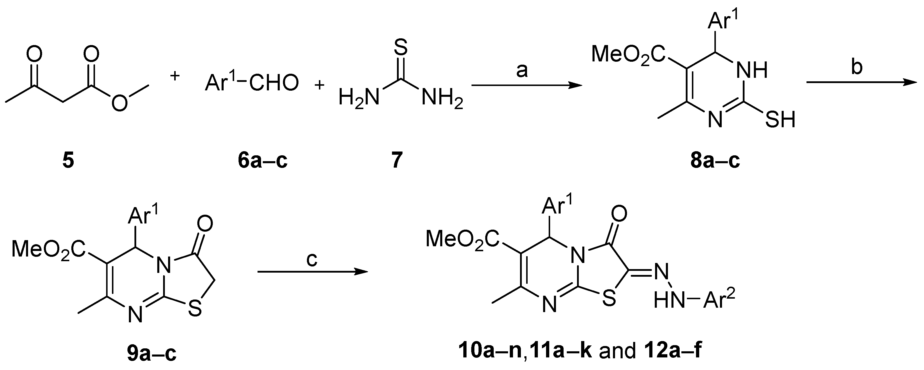
2.1. Chemistry
2.2. Anti-HIV-1 Activity of Compounds 10a–n, 11a–k, and 12a–f
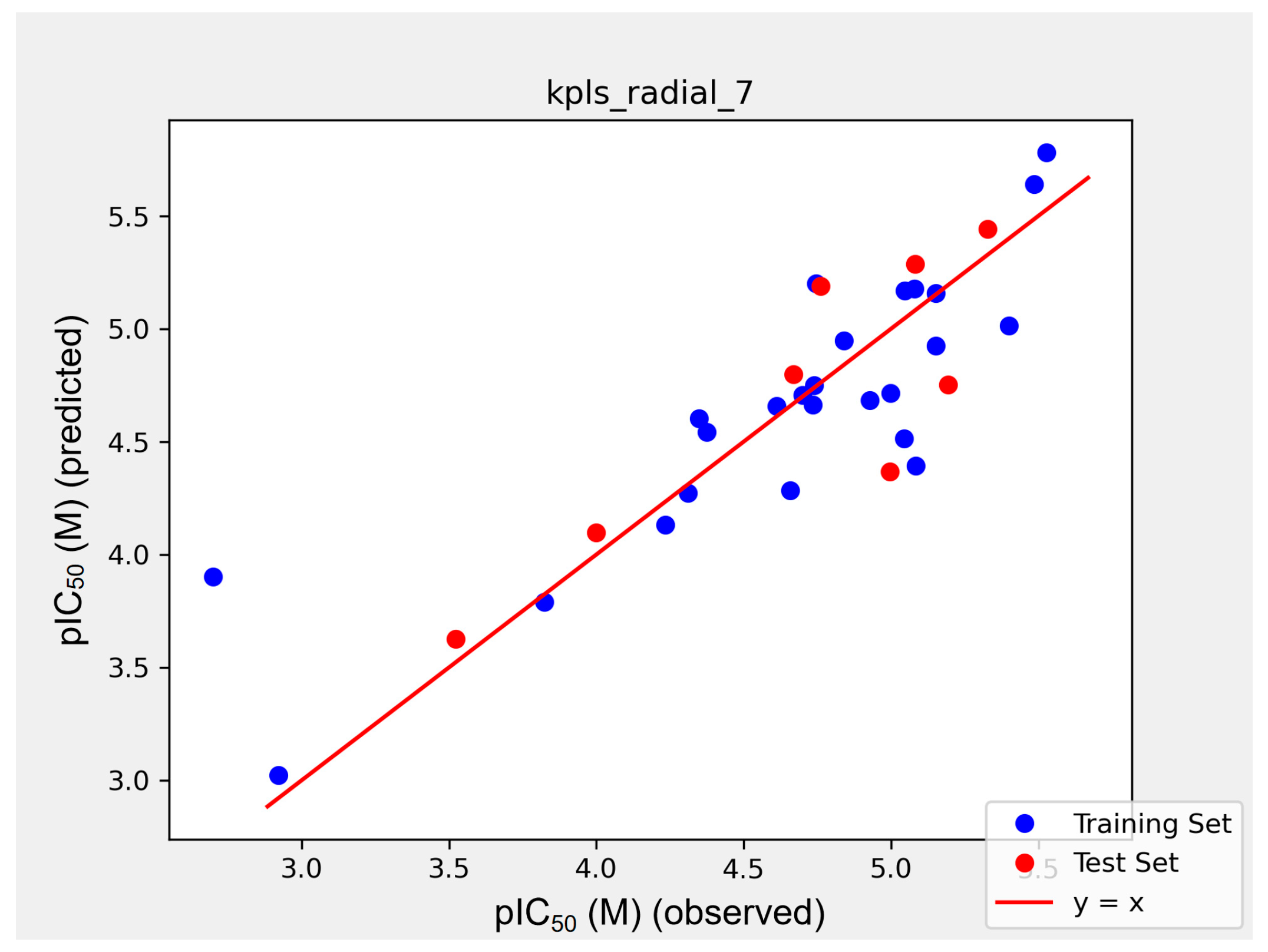
2.3. QSAR Model
2.4. Prediction of Physicochemical Properties
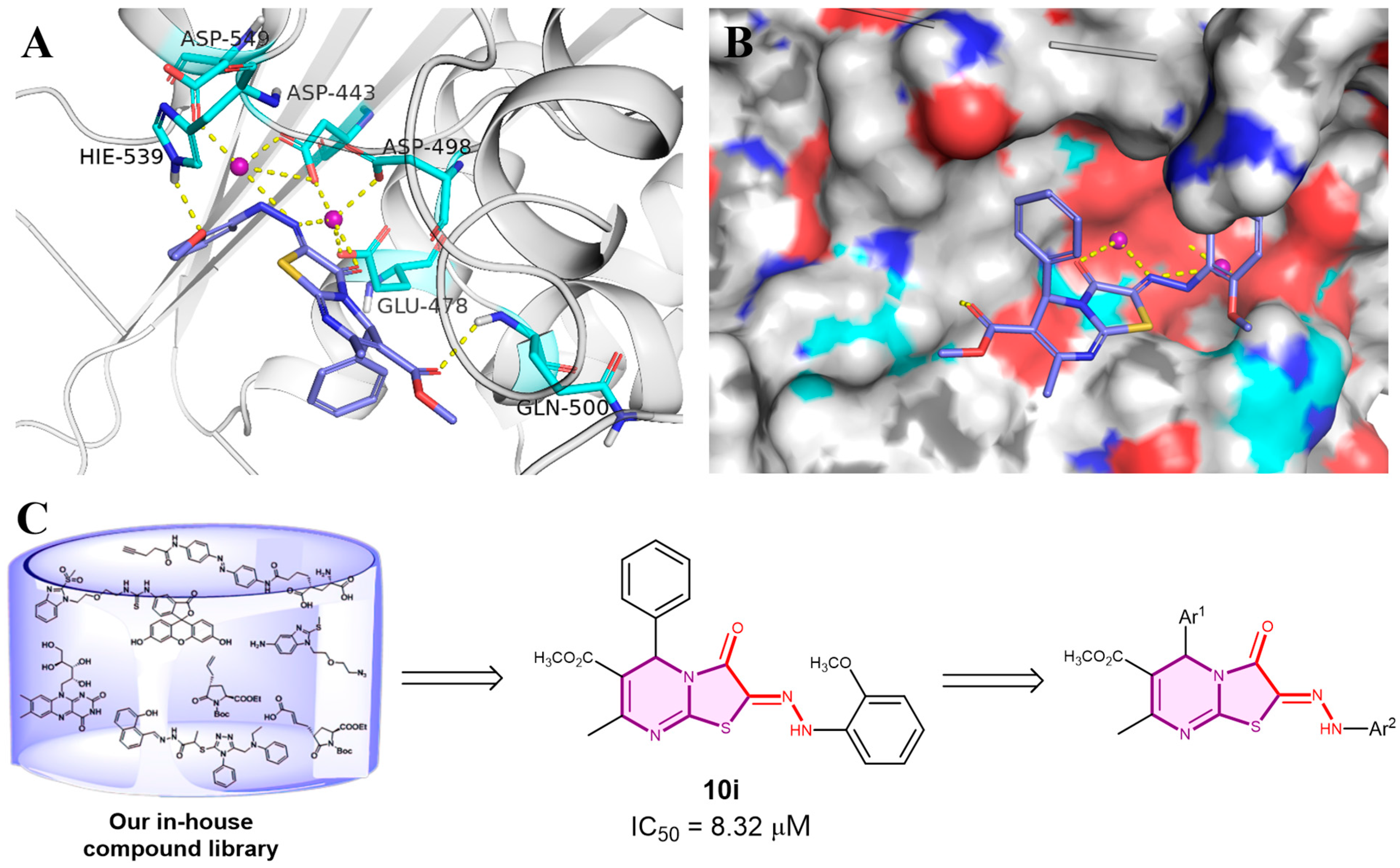
2.5. Molecular Docking
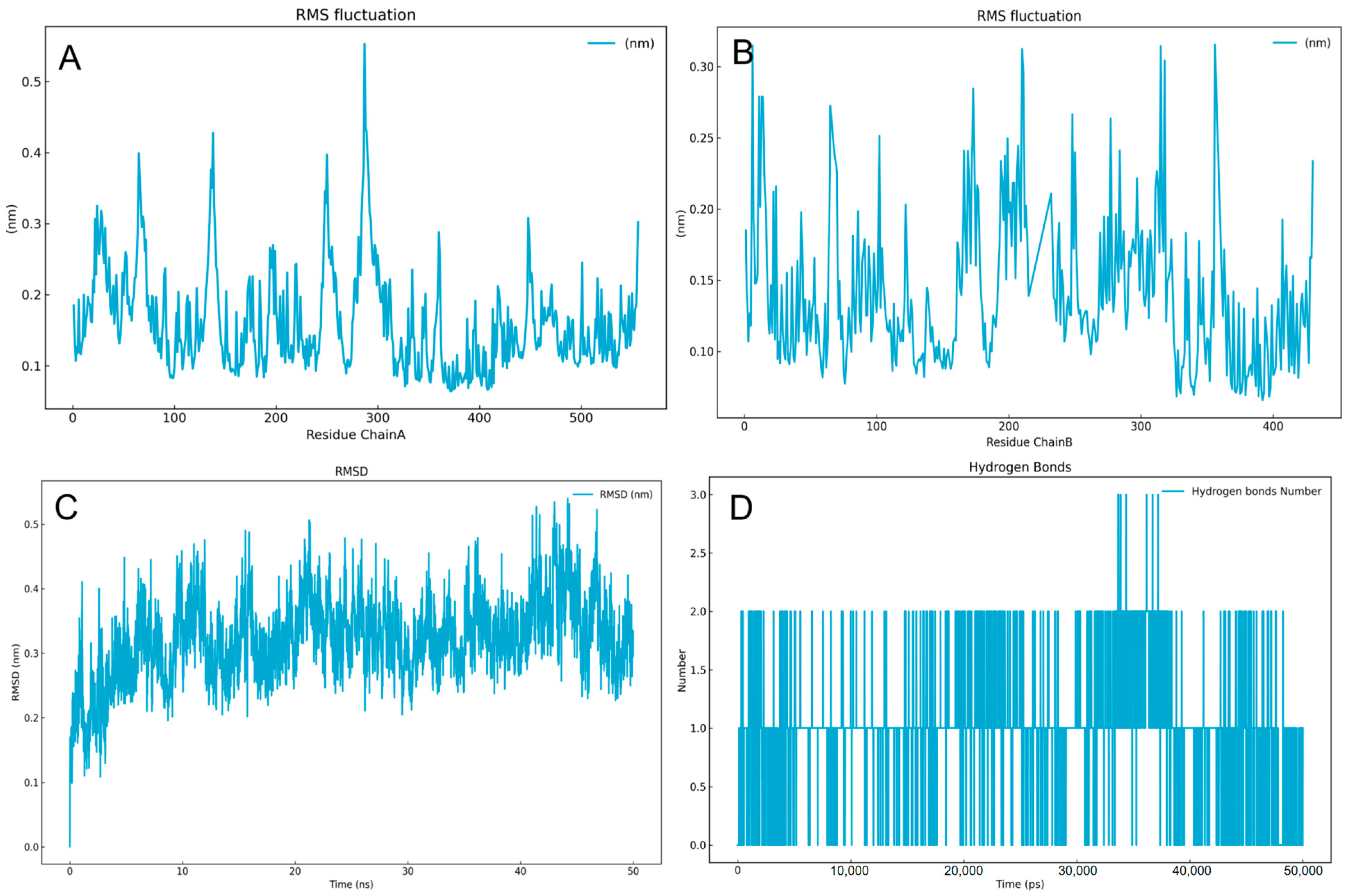
2.6. Dynamic Simulation
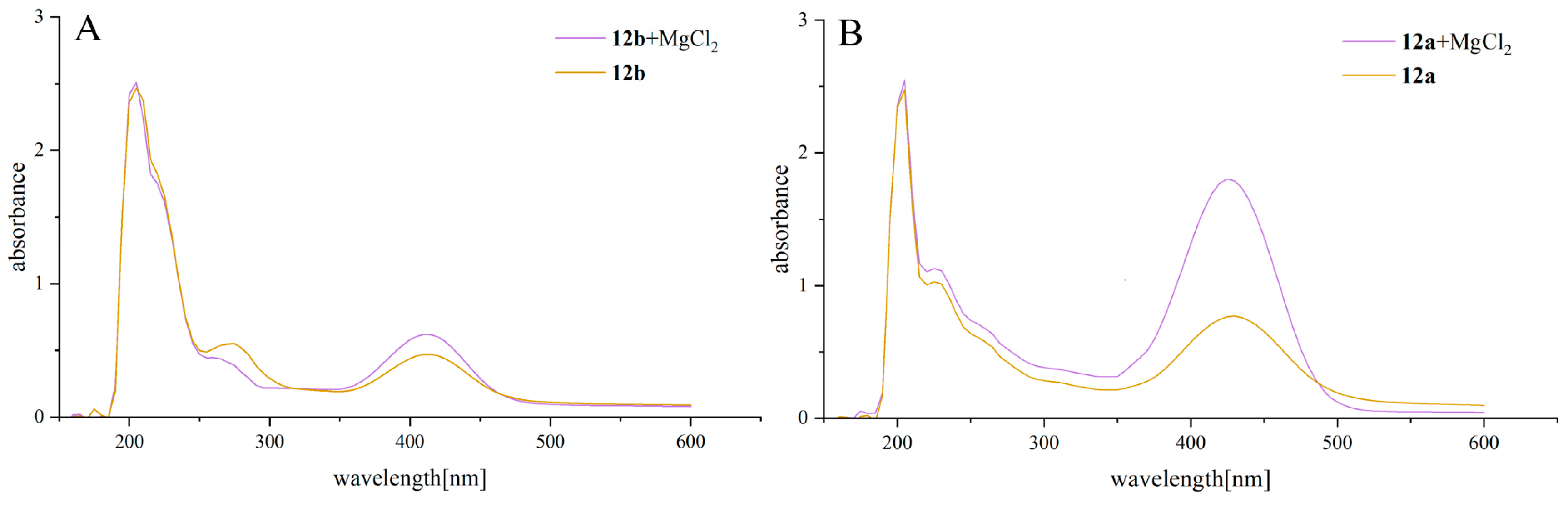
2.7. Magnesium Complexation
3. Conclusions
4. Experimental
4.1. Chemistry
4.1.1. General Procedure for the Synthesis of Compounds 8a–c
4.1.2. General Procedure for the Synthesis of Compounds 9a and 9c
- Methyl 7-methyl-3-oxo-5-phenyl-2,3-dihydro-5H-thiazolo[3,2-a]pyrimidine-6-carboxylate (9a). Yellow solid; 1H NMR (CDCl3, 400 MHz): δ [ppm] 7.23–6.98 (m, 5H), 5.84 (s, 1H), 3.63 (d, J = 17.5 Hz, 1H), 3.51 (d, J = 17.5 Hz, 1H), 3.42 (s, 3H), 2.26 (s, 3H).
- Methyl 5-(4-methoxyphenyl)-7-methyl-3-oxo-2,3-dihydro-5H-thiazolo[3,2-a]pyrimidine-6-carboxylate (9c). Yellow solid; 1H NMR (CDCl3, 400 MHz): δ 7.29 (d, J = 8.6 Hz, 2H), 6.83 (d, J = 8.7 Hz, 2H), 6.02 (s, 1H), 3.84 (d, J = 17.4 Hz, 1H), 3.78 (s, 3H), 3.73 (d, J = 17.5 Hz, 1H), 3.64 (s, 3H), 2.48 (s, 3H).
4.1.3. General Procedure for the Synthesis of Compound 9b
- Methyl 7-methyl-3-oxo-5-(2,3,4-trimethoxyphenyl)-2,3-dihydro-5H-thiazolo[3,2-a]pyrimidine-6-carboxylate (9b). Yellow solid; 1H NMR (CDCl3, 400 MHz): δ [ppm] 7.03 (d, J = 8.6 Hz, 1H), 6.58 (d, J = 8.7 Hz, 1H), 6.09 (s, 1H), 3.88 (s, 3H), 3.83 (s, 3H), 3.81 (s, 3H), 3.64 (s, 3H), 2.39 (s, 3H).
4.1.4. General Procedure for the Synthesis of Compounds 10a–n and 12a–f
- Methyl 2-(2-(2-hydroxyphenyl)hydrazineylidene)-7-methyl-3-oxo-5-phenyl-2,3-dihydro-5H-thiazolo[3,2-a]pyrimidine-6-carboxylate (10a). Yellow solid, yield: 75%, mp: 227–229 °C; 1H NMR (400 MHz, CDCl3) δ 7.43 (d, J = 7.0 Hz, 2H), 7.34 (t, J = 8.1 Hz, 4H), 6.90 (dd, J = 17.3, 7.9 Hz, 3H), 6.25 (s, 1H), 3.69 (s, 3H), 2.56 (s, 3H). 13C NMR (101 MHz, CDCl3 + CD3OD) δ 169.99, 165.12, 158.27, 155.99, 148.44, 143.42, 132.78, 132.67, 131.74, 131.64, 127.66, 124.38, 119.38, 119.03, 117.17, 113.45, 59.07, 55.51, 26.23. HRMS (ESI) m/z: calcd for C21H18N4O4S, [M − H]−: 421.0969; found: 421.0972.
- (2-(6-(Methoxycarbonyl)-7-methyl-3-oxo-5-phenyl-5H-thiazolo[3,2-a]pyrimidin-2(3H)-ylidene)hydrazineyl)-3-methylbenzoic acid (10b). Yellow solid, yield: 88%, mp: 261–263 °C; 1H NMR (400 MHz, DMSO-d6) δ 11.60 (s, 1H), 7.77 (d, J = 6.4 Hz, 1H), 7.45 (d, J = 7.4 Hz, 1H), 7.40–7.27 (m, 5H), 7.10 (t, J = 7.7 Hz, 1H), 6.02 (s, 1H), 3.60 (s, 3H), 2.40 (s, 3H), 2.39 (s, 3H). 13C NMR (101 MHz, DMSO-d6) δ 170.42, 166.07, 160.50, 155.59, 152.61, 145.89, 141.48, 135.24, 129.77, 129.22, 128.87, 127.63, 126.41, 123.47, 121.49, 118.56, 108.42, 54.54, 51.86, 23.08, 22.26. HRMS (ESI) m/z: calcd for C23H20N4O5S, [M + H]+: 465.1255; found: 465.1262.
- Methyl 7-methyl-3-oxo-5-phenyl-2-(2-(3,4,5-trimethoxyphenyl)hydrazineylidene)-2,3-dihydro-5H-thiazolo[3,2-a]pyrimidine-6-carboxylate (10c). Yellow solid, yield: 85%, mp: 211–213 °C; 1H NMR (400 MHz, DMSO-d6) δ 10.90 (s, 1H), 7.38–7.26 (m, 5H), 6.52 (s, 2H), 6.01 (s, 1H), 3.74 (s, 6H), 3.59 (d, J = 2.7 Hz, 6H), 2.38 (s, 3H). 13C NMR (101 MHz, CDCl3 + CD3OD) δ 169.98, 165.53, 159.24, 157.73, 155.80, 143.54, 143.16, 137.62, 132.77, 132.65, 131.74, 131.68, 123.04, 113.28, 96.06, 64.82, 59.83, 59.05, 55.52, 26.23. HRMS (ESI) m/z: calcd for C24H24N4O6S, [M + H]+: 497.1551; found: 497.1567.
- Methyl 2-((2-(methoxycarbonyl)phenyl)diazenyl)-7-methyl-3-oxo-5-phenyl-2,3-dihydro-5H-thiazolo[3,2-a]pyrimidine-6-carboxylate (10d). Yellow solid, yield: 65%, mp: 184–186 °C; 1H NMR (400 MHz, CDCl3) δ 11.35 (s, 1H), 7.95 (d, J = 6.5 Hz, 1H), 7.76 (d, J = 8.6 Hz, 1H), 7.50 (t, J = 7.1 Hz, 1H), 7.44–7.38 (m, 2H), 7.33–7.27 (m, 3H), 7.00 (t, J = 7.9 Hz, 1H), 6.24 (s, 1H), 3.94 (s, 3H), 3.66 (s, 3H), 2.53 (s, 3H). 13C NMR (101 MHz, CDCl3) δ 168.77, 165.79, 160.71, 152.63, 152.34, 145.05, 139.43, 134.95, 130.80, 128.86, 128.79, 127.99, 125.98, 121.59, 115.00, 112.12, 109.92, 55.27, 52.51, 51.65, 22.75. HRMS (ESI) m/z: calcd for C23H20N4O5S, [M + H]+: 465.1254; found: 465.1274.
- 5-Methoxy-2-(2-(6-(methoxycarbonyl)-7-methyl-3-oxo-5-phenyl-5H-thiazolo[3,2-a]pyrimidin-2(3H)-ylidene)hydrazineyl)benzenesulfonic acid (10e). Yellow solid, yield: 86%, mp: 247–249 °C; 1H NMR (400 MHz, DMSO-d6) δ 10.89 (s, 1H), 7.38–7.25 (m, 6H), 6.91 (d, J = 8.8 Hz, 1H), 6.86 (d, J = 2.5 Hz, 1H), 6.01 (s, 1H), 3.70 (s, 3H), 3.60 (s, 3H), 2.38 (s, 3H). 13C NMR (101 MHz, DMSO-d6) δ 165.88, 160.45, 154.77, 153.42, 151.61, 140.83, 133.90, 132.26, 129.29, 129.04, 127.65, 122.12, 117.29, 115.59, 112.36, 109.68, 55.90, 55.05, 52.01, 22.87. HRMS (ESI) m/z: calcd for C22H20N4O7S2, [M + H]+: 517.0854; found: 517.0848.
- (6-(Methoxycarbonyl)-7-methyl-3-oxo-5-phenyl-5H-thiazolo[3,2-a]pyrimidin-2(3H)-ylidene)hydrazineyl)naphthalene-1-sulfonic acid (10f). Yellow solid, yield: 80%, mp: 289–293 °C; 1H NMR (400 MHz, DMSO-d6) δ 12.84 (s, 1H), 8.83 (d, J = 9.1 Hz, 1H), 7.91 (d, J = 9.2 Hz, 1H), 7.80 (d, J = 7.6 Hz, 1H), 7.71 (d, J = 9.1 Hz, 1H), 7.51–7.45 (m, 1H), 7.41–7.28 (m, 6H), 6.07 (s, 1H), 3.62 (s, 3H), 2.40 (s, 3H). 13C NMR (101 MHz, DMSO-d6) δ 165.87, 160.55, 153.48, 151.54, 146.57, 142.85, 140.77, 136.52, 131.87, 130.90, 130.18, 129.30, 129.06, 128.44, 127.71, 127.60, 127.11, 124.75, 124.56, 123.61, 114.57, 109.77, 55.11, 52.03, 22.87. HRMS (ESI) m/z: calcd for C25H20N4O6S2, [M − H]−: 535.0744; found: 535.0749.
- Methyl 2-(2-(2-acetylphenyl)hydrazineylidene)-7-methyl-3-oxo-5-phenyl-2,3-dihydro-5H-thiazolo[3,2-a]pyrimidine-6-carboxylate (10g). Yellow solid, yield: 78%, mp: 199–202 °C; 1H NMR (400 MHz, CDCl3) δ 12.07 (s, 1H), 7.73 (t, J = 7.4 Hz, 2H), 7.43 (t, J = 7.1 Hz, 1H), 7.34 (d, J = 6.5 Hz, 2H), 7.26–7.17 (m, 3H), 6.94 (t, J = 7.6 Hz, 1H), 6.15 (s, 1H), 3.59 (s, 3H), 2.58 (s, 3H), 2.46 (s, 3H). 13C NMR (101 MHz, CDCl3) δ 202.76, 165.80, 160.71, 152.67, 152.32, 145.11, 139.38, 135.46, 131.71, 128.89, 128.81, 128.01, 126.48, 121.37, 118.97, 115.25, 109.95, 55.28, 51.70, 27.99, 22.74. HRMS (ESI) m/z: calcd for C23H20N4O4S, [M − H]−: 447.1125; found: 447.1126.
- Methyl 2-(2-(3,4-dimethoxyphenyl)hydrazineylidene)-7-methyl-3-oxo-5-phenyl-2,3-dihydro-5H-thiazolo[3,2-a]pyrimidine-6-carboxylate (10h). Yellow solid, yield: 73%, mp: 204–206 °C; 1H NMR (400 MHz, CDCl3) δ 8.44 (s, 1H), 7.32 (d, J = 6.6 Hz, 2H), 7.26–7.17 (m, 3H), 6.79 (d, J = 2.5 Hz, 1H), 6.65 (d, J = 8.6 Hz, 1H), 6.55 (dd, J = 8.6, 2.5 Hz, 1H), 6.13 (s, 1H), 3.74 (s, 3H), 3.70 (s, 3H), 3.58 (s, 3H), 2.44 (s, 3H). 13C NMR (101 MHz, CDCl3) δ 166.04, 159.82, 150.05, 149.95, 145.88, 145.78, 139.95, 136.04, 128.87, 128.84, 127.99, 127.82, 121.04, 111.74, 111.65, 109.44, 108.11, 106.59, 105.80, 99.27, 98.11, 55.99, 55.37, 54.87, 51.79, 22.91. HRMS (ESI) m/z: calcd for C23H22N4O5S, [M + H]+: 467.1430; found: 467.1438.
- Methyl 2-(2-(2-methoxyphenyl)hydrazineylidene)-7-methyl-3-oxo-5-phenyl-2,3-dihydro-5H-thiazolo[3,2-a]pyrimidine-6-carboxylate (10i). Yellow solid, yield: 55%, mp: 189–192 °C; 1H NMR (400 MHz, DMSO-d6) δ 10.35 (s, 1H), 7.39–7.24 (m, 6H), 7.03 (q, J = 7.9 Hz, 2H), 6.93 (t, J = 6.9 Hz, 1H), 6.02 (s, 1H), 3.86 (s, 3H), 3.60 (s, 3H), 2.38 (s, 3H). 13C NMR (101 MHz, CDCl3) δ 165.73, 160.61, 153.08, 152.02, 146.28, 139.44, 130.96, 128.88, 128.85, 128.79, 127.99, 127.79, 123.35, 121.62, 114.51, 110.40, 109.74, 55.76, 55.23, 51.66, 22.53. HRMS (ESI) m/z: calcd for C22H20N4O4S, [M + H]+: 437.1321; found: 437.1332.
- Methyl 7-methyl-3-oxo-5-phenyl-2-(2-phenylhydrazineylidene)-2,3-dihydro-5H-thiazolo[3,2-a]pyrimidine-6-carboxylate (10j). Yellow solid, yield: 49%, mp: 124–127 °C; 1H NMR (400 MHz, DMSO-d6) δ 10.99 (s, 1H), 7.38–7.29 (m, 7H), 7.23 (d, J = 7.4 Hz, 2H), 6.99 (t, J = 7.3 Hz, 1H), 6.04 (s, 1H), 3.61 (s, 3H), 2.39 (s, 3H). 13C NMR (101 MHz, CDCl3) δ 165.80, 160.84, 152.95, 152.17, 141.94, 139.43, 129.44, 128.89, 128.86, 128.83, 127.97, 123.56, 122.08, 114.62, 109.82, 55.27, 51.70, 22.67. HRMS (ESI) m/z: calcd for C21H18N4O3S, [M − H]−: 405.1020; found: 405.1022.
- (2-(6-(Methoxycarbonyl)-7-methyl-3-oxo-5-phenyl-5H-thiazolo[3,2-a]pyrimidin-2(3H)-ylidene)hydrazineyl)benzenesulfonic acid (10k). Yellow solid, yield: 84%, mp: 262–266 °C; 1H NMR (400 MHz, DMSO-d6) δ 11.24 (s, 1H), 7.99–7.82 (m, 2H), 7.57 (d, J = 7.3 Hz, 1H), 7.37–7.29 (m, 6H), 6.05 (s, 1H), 3.62 (s, 3H), 2.39 (s, 3H). 13C NMR (101 MHz, DMSO-d6) δ 165.85, 160.46, 153.24, 151.42, 140.71, 138.56, 132.72, 131.07, 129.30, 129.07, 128.00, 127.69, 123.86, 122.28, 113.90, 109.90, 55.14, 52.04, 22.85. HRMS (ESI) m/z: calcd for C21H18N4O6S2, [M − H]−: 485.0588; found: 485.0593.
- Methyl 7-methyl-2-(2-(3-nitrophenyl)hydrazineylidene)-3-oxo-5-phenyl-2,3-dihydro-5H-thiazolo[3,2-a]pyrimidine-6-carboxylate (10l). Yellow solid, yield: 71%, mp: 250–252 °C; 1H NMR (400 MHz, CDCl3) δ 8.00 (s, 1H), 7.88 (d, J = 8.0 Hz, 1H), 7.67 (s, 1H), 7.57 (d, J = 9.5 Hz, 1H), 7.47 (t, J = 8.2 Hz, 1H), 7.41 (d, J = 6.4 Hz, 2H), 7.31 (q, J = 8.5, 7.5 Hz, 3H), 6.24 (s, 1H), 3.67 (s, 3H), 2.54 (s, 3H). 13C NMR (101 MHz, CDCl3 + CD3OD) δ 165.78, 161.05, 154.73, 148.99, 144.07, 139.16, 130.11, 128.94, 128.77, 127.84, 120.16, 117.17, 109.71, 109.31, 55.16, 51.69, 22.22. HRMS (ESI) m/z: calcd for C21H17N5O5S, [M − H]−: 450.0870; found: 450.0879.
- Methyl 2-(2-(3-acetylphenyl)hydrazineylidene)-7-methyl-3-oxo-5-phenyl-2,3-dihydro-5H-thiazolo[3,2-a]pyrimidine-6-carboxylate (10m). Yellow solid, yield: 45%, mp: 213–216 °C; 1H NMR (400 MHz, DMSO-d6) δ 11.12 (s, 1H), 7.76 (s, 1H), 7.59 (d, J = 3.1 Hz, 1H), 7.51–7.41 (m, 2H), 7.40–7.25 (m, 5H), 6.01 (s, 1H), 3.61 (s, 3H), 2.56 (s, 3H), 2.38 (s, 3H). 13C NMR (101 MHz, CDCl3) δ 203.14, 169.95, 165.43, 158.98, 155.78, 147.26, 143.38, 141.80, 133.57, 132.81, 132.77, 132.68, 131.85, 131.77, 126.77, 124.72, 123.21, 118.11, 113.44, 59.05, 55.56, 30.51, 26.25. HRMS (ESI) m/z: calcd for C23H20N4O4S, [M + H]+: 449.1295; found: 449.1286.
- Methyl 2-(2-(3-(methoxycarbonyl)phenyl)hydrazineylidene)-7-methyl-3-oxo-5-phenyl-2,3-dihydro-5H-thiazolo[3,2-a]pyrimidine-6-carboxylate (10n). Yellow solid, yield: 42%, mp: 214–216 °C; 1H NMR (400 MHz, DMSO-d6) δ 11.14 (s, 1H), 7.92–7.17 (m, 9H), 6.03 (s, 1H), 3.86 (s, 3H), 3.61 (s, 3H), 2.39 (s, 3H). 13C NMR (101 MHz, CDCl3 + CD3OD) δ 167.07, 165.87, 161.21, 154.60, 151.85, 142.92, 139.45, 130.97, 129.46, 128.86, 128.76, 127.88, 123.97, 121.17, 118.97, 118.92, 115.50, 115.45, 55.11, 52.30, 51.68, 22.45. HRMS (ESI) m/z: calcd for C23H20N4O5S, [M + H]+: 465.1254; found: 465.1252.
- Methyl 2-(2-(2-acetylphenyl)hydrazineylidene)-5-(4-methoxyphenyl)-7-methyl-3-oxo-2,3-dihydro-5H-thiazolo[3,2-a]pyrimidine-6-carboxylate (12a). Yellow solid, yield: 64%, mp: 199–201 °C; 1H NMR (400 MHz, CDCl3) δ 12.07 (s, 1H), 7.74 (t, J = 8.5 Hz, 2H), 7.43 (t, J = 8.6 Hz, 1H), 7.26 (d, J = 8.8 Hz, 2H), 7.02–6.89 (m, 1H), 6.75 (d, J = 8.7 Hz, 2H), 6.11 (s, 1H), 3.68 (s, 3H), 3.59 (s, 3H), 2.59 (s, 3H), 2.46 (s, 3H). 13C NMR (101 MHz, CDCl3) δ 202.71, 165.88, 160.81, 159.88, 152.48, 152.03, 145.15, 135.44, 131.70, 131.66, 129.41, 126.67, 121.33, 118.98, 115.28, 114.08, 110.10, 55.27, 54.74, 51.69, 27.98, 22.69. HRMS (ESI) m/z: calcd for C24H22N4O5S, [M − H]−: 477.1231; found: 477.1236.
- Methyl 2-(2-(2-(methoxycarbonyl)phenyl)hydrazineylidene)-5-(4-methoxyphenyl)-7-methyl-3-oxo-2,3-dihydro-5H-thiazolo[3,2-a]pyrimidine-6-carboxylate (12b). Yellow solid, yield: 75%, mp: 219–221 °C; 1H NMR (400 MHz, CDCl3) δ 11.32 (s, 1H), 7.94 (d, J = 6.4 Hz, 1H), 7.76 (d, J = 7.3 Hz, 1H), 7.49 (t, J = 7.4 Hz, 1H), 7.34 (d, J = 8.8 Hz, 2H), 6.99 (t, J = 8.1 Hz, 1H), 6.82 (d, J = 8.7 Hz, 2H), 6.19 (s, 1H), 3.94 (s, 3H), 3.76 (s, 3H), 3.66 (s, 3H), 2.52 (s, 3H). 13C NMR (101 MHz, CDCl3) δ 168.79, 165.90, 160.87, 159.86, 152.47, 152.10, 145.07, 134.97, 131.68, 130.81, 129.41, 126.12, 121.58, 115.02, 114.08, 112.12, 110.05, 55.28, 54.72, 52.53, 51.69, 22.72. HRMS (ESI) m/z: calcd for C24H22N4O6S, [M + H]+: 495.1348; found: 495.1339.
- Methyl 2-(2-(3-(methoxycarbonyl)phenyl)hydrazineylidene)-5-(4-methoxyphenyl)-7-methyl-3-oxo-2,3-dihydro-5H-thiazolo[3,2-a]pyrimidine-6-carboxylate (12c). Yellow solid, yield: 80%, mp: 209–211 °C; 1H NMR (400 MHz, CDCl3) δ 7.78 (s, 1H), 7.59 (d, J = 7.7 Hz, 1H), 7.48 (d, J = 7.7 Hz, 1H), 7.30 (dt, J = 10.7, 3.3 Hz, 2H), 7.24 (s, 1H), 6.77 (d, J = 8.7 Hz, 2H), 6.09 (s, 1H), 3.84 (s, 3H), 3.70 (s, 3H), 3.61 (s, 3H), 2.44 (s, 3H). 13C NMR (101 MHz, CDCl3 + CD3OD) δ 167.17, 165.99, 161.34, 159.78, 154.75, 151.54, 143.05, 131.74, 130.94, 129.42, 129.24, 123.90, 120.95, 118.95, 115.46, 114.00, 109.59, 55.16, 54.50, 52.21, 51.62, 22.29. HRMS (ESI) m/z: calcd for C24H22N4O6S, [M + H]+: 495.1348; found: 495.1341.
- (2-(6-(Methoxycarbonyl)-5-(4-methoxyphenyl)-7-methyl-3-oxo-5H-thiazolo[3,2-a]pyrimidin-2(3H)-ylidene)hydrazineyl)-3-methylbenzoic acid (12d). Yellow solid, yield: 77%, mp: 265–267 °C; 1H NMR (400 MHz, DMSO-d6) δ 7.82 (d, J = 7.7 Hz, 1H), 7.22 (d, J = 8.7 Hz, 2H), 7.17 (d, J = 7.4 Hz, 1H), 6.90 (d, J = 8.8 Hz, 2H), 6.85 (d, J = 7.5 Hz, 1H), 5.98 (s, 1H), 3.72 (s, 3H), 3.60 (s, 3H), 2.43 (s, 3H), 2.38 (s, 3H). 13C NMR (101 MHz, DMSO-d6) δ 166.07, 160.56, 159.66, 154.97, 152.20, 145.25, 135.40, 133.40, 129.80, 129.10, 126.60, 123.18, 121.74, 119.44, 114.49, 108.80, 55.55, 54.04, 51.90, 23.00, 22.11. HRMS (ESI) m/z: calcd for C24H22N4O6S, [M + H]+: 495.1348; found: 495.1344.
- Methyl 5-(4-methoxyphenyl)-7-methyl-3-oxo-2-(2-(3,4,5-trimethoxyphenyl)hydrazineylidene)-2,3-dihydro-5H-thiazolo[3,2-a]pyrimidine-6-carboxylate (12e). Yellow solid, yield: 65%, mp: 204–206 °C; 1H NMR (400 MHz, DMSO-d6) δ 10.88 (s, 1H), 7.22 (d, J = 8.7 Hz, 2H), 6.90 (d, J = 8.8 Hz, 2H), 6.54 (s, 2H), 5.97 (s, 1H), 3.76 (s, 6H), 3.72 (s, 3H), 3.61 (s, 3H), 3.60 (s, 3H), 2.39 (s, 3H). 13C NMR (101 MHz, DMSO-d6) δ 165.93, 160.85, 159.75, 154.21, 154.01, 151.69, 139.69, 133.50, 132.94, 129.21, 120.34, 114.50, 109.39, 92.19, 60.61, 56.13, 55.57, 55.36, 54.36, 51.95, 22.86. HRMS (ESI) m/z: calcd for C25H26N4O7S, [M + H]+: 527.1619; found: 527.1632.
- (6-(Methoxycarbonyl)-5-(4-methoxyphenyl)-7-methyl-3-oxo-5H-thiazolo[3,2-a]pyrimidin-2(3H)-ylidene)hydrazineyl)naphthalene-1-sulfonic acid (12f). Yellow solid, yield: 90%, mp: 271–273 °C; 1H NMR (400 MHz, DMSO-d6) δ 12.81 (s, 1H), 8.84 (d, J = 7.7 Hz, 1H), 7.92 (d, J = 9.1 Hz, 1H), 7.80 (d, J = 6.7 Hz, 1H), 7.73 (d, J = 9.1 Hz, 1H), 7.48 (t, J = 7.0 Hz, 1H), 7.37 (t, J = 6.8 Hz, 1H), 7.24 (d, J = 8.7 Hz, 2H), 6.91 (d, J = 8.8 Hz, 2H), 6.02 (s, 1H), 3.72 (s, 3H), 3.62 (s, 3H), 2.41 (s, 3H). 13C NMR (101 MHz, DMSO-d6) δ 165.93, 160.60, 159.78, 153.19, 151.35, 136.56, 132.82, 131.90, 130.90, 130.17, 129.16, 128.45, 127.57, 127.13, 124.56, 123.76, 114.58, 109.92, 55.58, 54.53, 52.03, 22.83. HRMS (ESI) m/z: calcd for C26H22N4O7S2, [M + H]+: 567.1016; found: 567.1003.
4.1.5. General Procedure for the Synthesis of Compounds 11a–k
- Methyl 2-(2-(2-methoxyphenyl)hydrazineylidene)-7-methyl-3-oxo-5-(2,3,4-trimethoxyphenyl)-2,3-dihydro-5H-thiazolo[3,2-a]pyrimidine-6-carboxylate (11a). Yellow solid, yield: 61%, mp: 178–180 °C; 1H NMR (400 MHz, CDCl3) δ 7.89 (s, 1H), 7.49 (dd, J = 7.7, 1.9 Hz, 1H), 7.08 (d, J = 8.7 Hz, 1H), 7.00–6.89 (m, 2H), 6.85 (dd, J = 7.8, 1.6 Hz, 1H), 6.58 (d, J = 8.7 Hz, 1H), 6.22 (s, 1H), 3.90 (s, 6H), 3.81 (s, 3H), 3.78 (s, 3H), 3.67 (s, 3H), 2.43 (s, 3H). 13C NMR (101 MHz, CDCl3) δ 166.24, 160.68, 154.31, 152.64, 146.17, 142.02, 131.14, 125.57, 124.93, 123.04, 121.58, 114.36, 110.32, 109.46, 106.41, 60.68, 60.56, 55.91, 55.75, 53.54, 51.45, 22.79. HRMS (ESI) m/z: calcd for C25H26N4O7S, [M + H]+: 527.1619; found: 527.1634.
- Methyl 7-methyl-3-oxo-2-(2-phenylhydrazineylidene)-5-(2,3,4-trimethoxyphenyl)-2,3-dihydro-5H-thiazolo [3,2-a]pyrimidine-6-carboxylate (11b). Yellow solid, yield: 46%, mp: 189–192 °C; 1H NMR (400 MHz, CDCl3) δ 7.49 (s, 1H), 7.33 (t, J = 7.8 Hz, 2H), 7.22 (d, J = 7.6 Hz, 2H), 7.11 (d, J = 8.6 Hz, 1H), 7.05 (t, J = 7.3 Hz, 1H), 6.61 (d, J = 8.7 Hz, 1H), 6.25 (s, 1H), 3.92 (s, 3H), 3.85 (s, 3H), 3.82 (s, 3H), 3.70 (s, 3H), 2.46 (s, 3H). 13C NMR (101 MHz, CDCl3 + CD3OD) δ 166.53, 161.55, 155.53, 154.35, 152.60, 150.25, 142.88, 141.98, 129.27, 125.73, 125.00, 123.04, 119.80, 114.63, 109.05, 106.56, 60.61, 60.55, 55.90, 53.47, 51.48, 22.45. HRMS (ESI) m/z: calcd for C24H24N4O6S, [M + H]+: 497.1509; found: 497.1519.
- (2-(6-(Methoxycarbonyl)-7-methyl-3-oxo-5-(2,3,4-trimethoxyphenyl)-5H-thiazolo[3,2-a]pyrimidin-2(3H)-ylidene)hydrazineyl)-3-methylbenzoic acid (11c). Yellow solid, yield: 30%, mp: 209–213 °C; 1H NMR (400 MHz, DMSO-d6) δ 7.81 (s, 1H), 7.15 (d, J = 7.3 Hz, 1H), 6.96 (d, J = 8.6 Hz, 1H), 6.84 (t, J = 7.4 Hz, 1H), 6.75 (d, J = 8.7 Hz, 1H), 6.06 (s, 1H), 3.78 (s, 6H), 3.71 (s, 3H), 3.58 (s, 3H), 2.41 (s, 3H), 2.30 (s, 3H). 13C NMR (101 MHz, DMSO-d6) δ 166.36, 160.48, 154.96, 154.12, 152.26, 150.82, 145.44, 141.92, 135.32, 129.72, 126.58, 126.26, 125.23, 123.13, 121.53, 119.69, 108.38, 107.56, 61.00, 60.62, 56.24, 52.70, 51.62, 23.04, 22.06. HRMS (ESI) m/z: calcd for C26H26N4O8S, [M + H]+: 554.1570; found: 555.1573.
- Methyl 7-methyl-2-(2-(3-nitrophenyl)hydrazineylidene)-3-oxo-5-(2,3,4-trimethoxyphenyl)-2,3-dihydro-5H-thiazolo[3,2-a]pyrimidine-6-carboxylate (11d). Yellow solid, yield: 48%, mp: 236–238 °C; 1H NMR (400 MHz, CDCl3) δ 8.20 (s, 1H), 7.97 (t, J = 2.2 Hz, 1H), 7.82 (d, J = 6.8 Hz, 1H), 7.50 (d, J = 9.4 Hz, 1H), 7.41 (t, J = 8.1 Hz, 1H), 7.08 (d, J = 8.6 Hz, 1H), 6.58 (d, J = 8.7 Hz, 1H), 6.21 (s, 1H), 3.88 (s, 3H), 3.82 (s, 3H), 3.79 (s, 3H), 3.68 (s, 3H), 2.43 (s, 3H). 13C NMR (101 MHz, CDCl3) δ 166.05, 160.48, 154.50, 152.60, 149.70, 149.01, 143.49, 142.01, 130.29, 125.72, 124.27, 120.09, 117.56, 109.84, 109.35, 106.46, 60.72, 60.60, 55.91, 54.00, 51.62, 22.59. HRMS (ESI) m/z: calcd for C24H23N5O8S, [M + H]+: 541.1357; found: 542.1361.
- 2-(2-(6-(Methoxycarbonyl)-7-methyl-3-oxo-5-(2,3,4-trimethoxyphenyl)-5H-thiazolo[3,2-a]pyrimidin-2(3H)-ylidene)hydrazineyl)benzenesulfonic acid (11e). Yellow solid, yield: 74%, mp: 238–240 °C; 1H NMR (400 MHz, DMSO-d6) δ 11.15 (s, 1H), 7.59 (d, J = 7.7 Hz, 1H), 7.35 (d, J = 7.7 Hz, 2H), 6.99 (d, J = 8.6 Hz, 2H), 6.76 (d, J = 8.7 Hz, 1H), 6.07 (s, 1H), 3.78 (s, 6H), 3.70 (s, 3H), 3.60 (s, 3H), 2.31 (s, 3H). 13C NMR (101 MHz, DMSO-d6) δ 166.17, 160.32, 154.31, 152.88, 152.37, 149.94, 141.89, 138.67, 132.55, 131.04, 127.98, 125.50, 125.35, 124.20, 122.06, 113.76, 109.42, 107.44, 61.02, 60.64, 56.24, 53.38, 51.82, 22.86. HRMS (ESI) m/z: calcd for C24H24N4O9S2, [M + H]+: 577.1067; found: 577.1062.
- 5-Methoxy-2-(2-(6-(methoxycarbonyl)-7-methyl-3-oxo-5-(2,3,4-trimethoxyphenyl)-5H-thiazolo[3,2-a]pyrimidin-2(3H)-ylidene)hydrazineyl)benzenesulfonic acid (11f). Yellow solid, yield: 76%, mp: 244–246 °C; 1H NMR (400 MHz, DMSO-d6) δ 10.93 (s, 1H), 7.31 (d, J = 8.9 Hz, 1H), 7.13 (s, 1H), 6.98 (d, J = 8.6 Hz, 2H), 6.75 (d, J = 8.6 Hz, 1H), 6.06 (s, 1H), 3.77 (s, 6H), 3.73 (s, 3H), 3.70 (s, 3H), 3.59 (s, 3H), 2.31 (s, 3H). 13C NMR (101 MHz, DMSO-d6) δ 166.19, 160.34, 154.58, 154.28, 153.06, 152.36, 150.10, 141.87, 133.57, 132.38, 125.47, 122.52, 117.31, 115.45, 112.32, 109.23, 107.43, 61.02, 60.63, 56.22, 55.88, 53.30, 51.79, 22.87. HRMS (ESI) m/z: calcd for C25H26N4O10S2, [M − H]−: 605.1010; found: 605.1006.
- (2-(6-(Methoxycarbonyl)-7-methyl-3-oxo-5-(2,3,4-trimethoxyphenyl)-5H-thiazolo[3,2-a]pyrimidin-2(3H)-ylidene)hydrazineyl)naphthalene-1-sulfonic acid (11g). Yellow solid, yield: 76%, mp: 241–244 °C; 1H NMR (400 MHz, DMSO-d6) δ 12.74 (s, 1H), 8.85 (d, J = 8.9 Hz, 1H), 7.90 (d, J = 9.0 Hz, 1H), 7.79 (d, J = 8.1 Hz, 1H), 7.71 (d, J = 9.0 Hz, 1H), 7.48 (t, J = 7.8 Hz, 1H), 7.36 (t, J = 7.5 Hz, 1H), 7.00 (d, J = 8.7 Hz, 1H), 6.77 (d, J = 8.6 Hz, 1H), 6.09 (s, 1H), 3.78 (d, J = 4.4 Hz, 6H), 3.70 (s, 3H), 3.60 (s, 3H), 2.32 (s, 3H). 13C NMR (101 MHz, DMSO-d6) δ 166.20, 160.41, 154.32, 153.09, 152.36, 150.03, 141.95, 136.69, 131.82, 130.95, 130.10, 128.42, 127.57, 127.08, 125.49, 125.43, 124.55, 124.46, 123.96, 114.59, 109.41, 107.54, 61.04, 60.64, 56.28, 53.29, 51.77, 22.86. HRMS (ESI) m/z: calcd for C28H26N4O9S2, [M − H]−: 625.1061; found: 625.1056.
- Methyl 2-(2-(2-(methoxycarbonyl)phenyl)hydrazineylidene)-7-methyl-3-oxo-5-(2,3,4-trimethoxyphenyl)-2,3-dihydro-5H-thiazolo[3,2-a]pyrimidine-6-carboxylate (11h). Yellow solid, yield: 55%, mp: 200–202 °C; 1H NMR (400 MHz, CDCl3) δ 11.29 (s, 1H), 7.95 (d, J = 6.4 Hz, 1H), 7.74 (d, J = 8.4 Hz, 1H), 7.48 (t, J = 7.1 Hz, 1H), 7.08 (d, J = 8.6 Hz, 1H), 7.01–6.94 (m, 1H), 6.58 (d, J = 8.6 Hz, 1H), 6.23 (s, 1H), 3.95 (s, 3H), 3.90 (s, 3H), 3.82 (s, 3H), 3.79 (s, 3H), 3.67 (s, 3H), 2.43 (s, 3H). 13C NMR (101 MHz, CDCl3) δ 168.42, 165.89, 160.36, 153.97, 152.29, 144.83, 141.67, 134.57, 130.44, 126.06, 125.20, 124.48, 121.03, 114.60, 111.66, 109.27, 106.03, 60.31, 60.21, 55.56, 53.22, 52.14, 51.10, 22.49. HRMS (ESI) m/z: calcd for C26H26N4O8S, [M + H]+: 554.1570; found: 555.1574.
- Methyl 2-(2-(2-acetylphenyl)hydrazineylidene)-7-methyl-3-oxo-5-(2,3,4-trimethoxyphenyl)-2,3-dihydro-5H-thiazolo[3,2-a]pyrimidine-6-carboxylate (11i). Yellow solid, yield: 52%, mp: 205–208 °C; 1H NMR (400 MHz, CDCl3) δ 12.12 (s, 1H), 7.81 (dd, J = 15.9, 8.3 Hz, 2H), 7.49 (t, J = 7.2 Hz, 1H), 7.08 (d, J = 8.6 Hz, 1H), 7.01 (t, J = 7.0 Hz, 1H), 6.58 (d, J = 8.7 Hz, 1H), 6.23 (s, 1H), 3.90 (s, 3H), 3.82 (s, 3H), 3.78 (s, 3H), 3.67 (s, 3H), 2.67 (s, 3H), 2.44 (s, 3H). 13C NMR (101 MHz, CDCl3) δ 202.65, 166.25, 160.69, 154.31, 152.63, 152.54, 150.55, 145.27, 142.02, 135.40, 131.69, 127.02, 125.55, 124.83, 121.12, 118.88, 115.20, 109.68, 106.39, 60.68, 60.56, 55.91, 53.55, 51.46, 27.98, 22.88. HRMS (ESI) m/z: calcd for C26H26N4O7S, [M + H]+: 539.1609; found: 539.1605.
- Methyl 2-(2-(3-(methoxycarbonyl)phenyl)hydrazineylidene)-7-methyl-3-oxo-5-(2, 3, 4-trimethoxyphenyl)-2, 3-dihydro-5H-thiazolo[3,2-a]pyrimidine-6-carboxylate (11j). Yellow solid, yield: 67%, mp: 213–215 °C; 1H NMR (400 MHz, CDCl3) δ 7.94 (s, 1H), 7.72 (d, J = 7.6 Hz, 1H), 7.60 (d, J = 8.3 Hz, 1H), 7.44 (d, J = 8.4 Hz, 1H), 7.16 (d, J = 8.7 Hz, 1H), 6.69 (d, J = 8.7 Hz, 1H), 6.27 (s, 1H), 3.97 (d, J = 5.4 Hz, 6H), 3.91 (s, 6H), 3.76 (s, 3H), 2.50 (s, 3H). 13C NMR (101 MHz, CDCl3 + CD3OD) δ 167.36, 166.49, 161.39, 155.14, 154.43, 152.66, 150.18, 143.33, 142.05, 131.10, 129.54, 125.80, 124.89, 123.91, 121.30, 119.05, 115.54, 109.27, 60.67, 60.62, 55.97, 53.65, 52.34, 51.56, 22.55. HRMS (ESI) m/z: calcd for C26H26N4O8S, [M + H]+: 661.2733; found: 555.1577.
- Methyl 2-(2-(3-acetylphenyl)hydrazineylidene)-7-methyl-3-oxo-5-(2,3,4-trimethoxyphenyl)-2,3-dihydro-5H-thiazolo[3,2-a]pyrimidine-6-carboxylate (11k). Yellow solid, yield: 40%, mp: 216–219 °C; 1H NMR (400 MHz, DMSO-d6) δ 11.00 (s, 1H), 7.75 (s, 1H), 7.59 (dt, J = 6.4, 2.1 Hz, 1H), 7.51–7.40 (m, 2H), 6.97 (d, J = 8.7 Hz, 1H), 6.75 (d, J = 8.8 Hz, 1H), 6.04 (s, 1H), 3.78 (s, 6H), 3.70 (s, 3H), 3.59 (s, 3H), 2.56 (s, 3H), 2.31 (s, 3H). 13C NMR (101 MHz, DMSO-d6) δ 198.09, 166.19, 160.67, 154.25, 152.31, 141.86, 138.23, 130.31, 125.45, 122.89, 119.04, 109.04, 107.46, 61.05, 60.62, 56.24, 53.08, 51.78, 27.25, 22.90. HRMS (ESI) m/z: calcd for C26H26N4O7S, [M + H]+: 539.1609; found: 539.1604.
4.2. Other Protocols
Supplementary Materials
Author Contributions
Funding
Institutional Review Board Statement
Informed Consent Statement
Data Availability Statement
Conflicts of Interest
References
- Cohen, M.S.; Chen, Y.Q.; McCauley, M.; Gamble, T.; Hosseinipour, M.C.; Kumarasamy, N.; Hakim, J.G.; Kumwenda, J.; Grinsztejn, B.; Pilotto, J.H.S.; et al. Antiretroviral Therapy for the Prevention of HIV-1 Transmission. N. Engl. J. Med. 2016, 375, 830–839. [Google Scholar] [CrossRef]
- De Clercq, E. Antiviral drugs in current clinical use. J. Clin. Virol. 2004, 30, 115–133. [Google Scholar] [CrossRef]
- Singh, A.K.; Das, K. Insights into HIV-1 Reverse Transcriptase (RT) Inhibition and Drug Resistance from Thirty Years of Structural Studies. Viruses 2022, 14, 1027. [Google Scholar] [CrossRef]
- Arts, E.J.; Hazuda, D.J. HIV-1 antiretroviral drug therapy. Cold Spring Harb. Perspect. Med. 2012, 2, a007161. [Google Scholar] [CrossRef]
- Chun, T.W.; Stuyver, L.; Mizell, S.B.; Ehler, L.A.; Mican, J.A.; Baseler, M.; Lloyd, A.L.; Nowak, M.A.; Fauci, A.S. Presence of an inducible HIV-1 latent reservoir during highly active antiretroviral therapy. Proc. Natl. Acad. Sci. USA 1997, 94, 13193–13197. [Google Scholar] [CrossRef]
- Bagasra, O. A unified concept of HIV latency. Expert Opin. Biol. Ther. 2006, 6, 1135–1149. [Google Scholar] [CrossRef]
- Menéndez-Arias, L. Molecular basis of human immunodeficiency virus type 1 drug resistance: Overview and recent developments. Antivir. Res. 2013, 98, 93–120. [Google Scholar] [CrossRef]
- Zhan, P.; Pannecouque, C.; De Clercq, E.; Liu, X. Anti-HIV Drug Discovery and Development: Current Innovations and Future Trends. J. Med. Chem. 2016, 59, 2849–2878. [Google Scholar] [CrossRef]
- Zhang, L.; Wei, F.; Zhang, J.; Liu, C.; López-Carrobles, N.; Liu, X.; Menéndez-Arias, L.; Zhan, P. Current medicinal chemistry strategies in the discovery of novel HIV-1 ribonuclease H inhibitors. Eur. J. Med. Chem. 2022, 243, 114760. [Google Scholar] [CrossRef]
- Kang, J.X.; Zhao, G.K.; Yang, X.M.; Huang, M.X.; Hui, W.Q.; Zeng, R.; Ouyang, Q. Recent advances on dual inhibitors targeting HIV reverse transcriptase associated polymerase and ribonuclease H. Eur. J. Med. Chem. 2023, 250, 115196. [Google Scholar] [CrossRef]
- Lapkouski, M.; Tian, L.; Miller, J.T.; Le Grice, S.F.J.; Yang, W. Complexes of HIV-1 RT, NNRTI and RNA/DNA hybrid reveal a structure compatible with RNA degradation. Nat. Struct. Mol. Biol. 2013, 20, 230–236. [Google Scholar] [CrossRef]
- Di Santo, R. Inhibiting the HIV Integration Process: Past, Present, and the Future. J. Med. Chem. 2014, 57, 539–566. [Google Scholar] [CrossRef]
- Moianos, D.; Prifti, G.-M.; Makri, M.; Zoidis, G. Targeting Metalloenzymes: The “Achilles’ Heel” of Viruses and Parasites. Pharmaceuticals 2023, 16, 901. [Google Scholar] [CrossRef]
- Kitagawa, Y.; Liao, Z.; Morikawa, K.; Oda, M. Metal-binding and folding thermodynamics of Escherichia coli ribonuclease HI related to its catalytic function. Biophys. Chem. 2023, 295, 106961. [Google Scholar] [CrossRef]
- Kankanala, J.; Kirby, K.A.; Liu, F.; Miller, L.; Nagy, E.; Wilson, D.J.; Parniak, M.A.; Sarafianos, S.G.; Wang, Z. Design, Synthesis, and Biological Evaluations of Hydroxypyridonecarboxylic Acids as Inhibitors of HIV Reverse Transcriptase Associated RNase H. J. Med. Chem. 2016, 59, 5051–5062. [Google Scholar] [CrossRef]
- Borkow, G.; Fletcher, R.S.; Barnard, J.; Arion, D.; Motakis, D.; Dmitrienko, G.I.; Parniak, M.A. Inhibition of the ribonuclease H and DNA polymerase activities of HIV-1 reverse transcriptase by N-(4-tert-butylbenzoyl)-2-hydroxy-1-naphthaldehyde hydrazone. Biochemistry 1997, 36, 3179–3185. [Google Scholar] [CrossRef]
- Messore, A.; Corona, A.; Madia, V.N.; Saccoliti, F.; Tudino, V.; De Leo, A.; Scipione, L.; De Vita, D.; Amendola, G.; Di Maro, S.; et al. Pyrrolyl Pyrazoles as Non-Diketo Acid Inhibitors of the HIV-1 Ribonuclease H Function of Reverse Transcriptase. ACS Med. Chem. Lett. 2020, 11, 798–805. [Google Scholar] [CrossRef]
- Wang, L.; Tang, J.; Huber, A.D.; Casey, M.C.; Kirby, K.A.; Wilson, D.J.; Kankanala, J.; Xie, J.; Parniak, M.A.; Sarafianos, S.G.; et al. 6-Arylthio-3-hydroxypyrimidine-2,4-diones potently inhibited HIV reverse transcriptase-associated RNase H with antiviral activity. Eur. J. Med. Chem. 2018, 156, 652–665. [Google Scholar] [CrossRef]
- Wang, X.; Gao, P.; Menendez-Arias, L.; Liu, X.; Zhan, P. Update on Recent Developments in Small Molecular HIV-1 RNase H Inhibitors (2013–2016): Opportunities and Challenges. Curr. Med. Chem. 2018, 25, 1682–1702. [Google Scholar] [CrossRef]
- Tramontano, E.; Corona, A.; Menéndez-Arias, L. Ribonuclease H, an unexploited target for antiviral intervention against HIV and hepatitis B virus. Antivir. Res. 2019, 171, 104613. [Google Scholar] [CrossRef]
- Cihlar, T.; Ray, A.S. Nucleoside and nucleotide HIV reverse transcriptase inhibitors: 25 years after zidovudine. Antivir. Res. 2010, 85, 39–58. [Google Scholar] [CrossRef] [PubMed]
- de Béthune, M.P. Non-nucleoside reverse transcriptase inhibitors (NNRTIs), their discovery, development, and use in the treatment of HIV-1 infection: A review of the last 20 years (1989–2009). Antivir. Res. 2010, 85, 75–90. [Google Scholar] [CrossRef] [PubMed]
- Beilhartz, G.L.; Götte, M. HIV-1 Ribonuclease H: Structure, Catalytic Mechanism and Inhibitors. Viruses 2010, 2, 900–926. [Google Scholar] [CrossRef] [PubMed]
- Corona, A.; Di Leva, F.S.; Thierry, S.; Pescatori, L.; Cuzzucoli Crucitti, G.; Subra, F.; Delelis, O.; Esposito, F.; Rigogliuso, G.; Costi, R.; et al. Identification of highly conserved residues involved in inhibition of HIV-1 RNase H function by Diketo acid derivatives. Antimicrob. Agents Chemother. 2014, 58, 6101–6110. [Google Scholar] [CrossRef] [PubMed]
- Lansdon, E.B.; Liu, Q.; Leavitt, S.A.; Balakrishnan, M.; Perry, J.K.; Lancaster-Moyer, C.; Kutty, N.; Liu, X.; Squires, N.H.; Watkins, W.J.; et al. Structural and binding analysis of pyrimidinol carboxylic acid and N-hydroxy quinazolinedione HIV-1 RNase H inhibitors. Antimicrob. Agents Chemother. 2011, 55, 2905–2915. [Google Scholar] [CrossRef] [PubMed]
- Costa, G.; Rocca, R.; Corona, A.; Grandi, N.; Moraca, F.; Romeo, I.; Talarico, C.; Gagliardi, M.G.; Ambrosio, F.A.; Ortuso, F.; et al. Novel natural non-nucleoside inhibitors of HIV-1 reverse transcriptase identified by shape- and structure-based virtual screening techniques. Eur. J. Med. Chem. 2019, 1, 1–10. [Google Scholar] [CrossRef] [PubMed]
- Corona, A.; Meleddu, R.; Esposito, F.; Distinto, S.; Bianco, G.; Masaoka, T.; Maccioni, E.; Menéndez-Arias, L.; Alcaro, S.; Le Grice, S.F.; et al. Ribonuclease H/DNA Polymerase HIV-1 Reverse Transcriptase Dual Inhibitor: Mechanistic Studies on the Allosteric Mode of Action of Isatin-Based Compound RMNC6. PLoS ONE 2016, 11, e0147225. [Google Scholar] [CrossRef]
- Corona, A.; Schneider, A.; Schweimer, K.; Rösch, P.; Wöhrl, B.M.; Tramontano, E. Inhibition of foamy virus reverse transcriptase by human immunodeficiency virus type 1 RNase H inhibitors. Antimicrob. Agents Chemother. 2014, 58, 4086–4093. [Google Scholar] [CrossRef]


 | |||
|---|---|---|---|
| Comp. | Ar1 | Ar2 | IC50 (μM) a |
| 10a | Ph | 2-OH-Ph | 18.23 ± 0.85 |
| 10b | Ph | 2-Me-5-CO2H-Ph | 44.86 ± 3.53 |
| 10c | Ph | 3,4,5-triOMe-Ph | 24.38 ± 1.25 |
| 10d | Ph | 2-CO2CH3-Ph | 8.27 ± 0.39 |
| 10e | Ph | 2-SO3H-4-OMe-Ph | 58.37 ± 1.71 |
| 10f | Ph | 2-SO3H-Naph | 7.04 ± 0.57 |
| 10g | Ph | 2-COCH3-Ph | 9.01 ± 2.17 |
| 10h | Ph | 3,4-bis-OMe-Ph | 14.42 ± 1.23 |
| 10i | Ph | 2-OMe-Ph | 8.32 ± 0.06 |
| 10j | Ph | Ph | 21.45 ± 0.10 |
| 10k | Ph | 2-SO3H-Ph | >100 (85%) b |
| 10l | Ph | 3-NO2-Ph | 6.41 ± 0.24 |
| 10m | Ph | 3-COCH3-Ph | 18.39 ± 0.79 |
| 10n | Ph | 3-CO2CH3-Ph | 7.03 ± 0.17 |
| 11a | 2,3,4-3OMe-Ph | 2-OMe-Ph | 11.81 ± 0.91 |
| 11b | 2,3,4-3OMe-Ph | Ph | 48.95 ± 4.73 |
| 11c | 2,3,4-3OMe-Ph | 2-Me-5-CO2H-Ph | 8.23 ± 2.44 |
| 11d | 2,3,4-3OMe-Ph | 3-NO2 | 10.10 ± 1.64 |
| 11e | 2,3,4-3OMe-Ph | 2-SO3H-Ph | >100 (97%) |
| 11f | 2,3,4-3OMe-Ph | 2-SO3H-4-OMe-Ph | >100 (61%) |
| 11g | 2,3,4-3OMe-Ph | 2-SO3H-Naph | 19.95 ± 3.84 |
| 11h | 2,3,4-3OMe-Ph | 2-CO2CH3 | 10.05 ± 0.95 |
| 11i | 2,3,4-3OMe-Ph | 2-COCH3 | >100 (100%) |
| 11j | 2,3,4-3OMe-Ph | 3-CO2CH3-Ph | 42.15 ± 9.78 |
| 11k | 2,3,4-3OMe-Ph | 3-COCH3-Ph | 21.96 ± 0.36 |
| 12a | 4-OMe-Ph | 2-COCH3-Ph | 3.98 ± 0.30 |
| 12b | 4-OMe-Ph | 2-CO2CH3-Ph | 2.97 ± 0.88 |
| 12c | 4-OMe-Ph | 3-CO2CH3-Ph | 4.71 ± 0.86 |
| 12d | 4-OMe-Ph | 2-Me-5-CO2H | 17.97 ± 1.87 |
| 12e | 4-OMe-Ph | 3,4,5-tri-OMe-Ph | 8.99 ± 0.27 |
| 12f | 4-OMe-Ph | 2-SO3H-Naph | 3.28 ± 0.92 |
| RDS1643 [24] | 8.6 ± 1.30 | ||
| Compd. | a GI Absorption | b BBB Permeant | c P-gp Substrate | CYP1A2 Inhibitor | CYP2D6 Inhibitor | d Lipinski | e PAINS |
|---|---|---|---|---|---|---|---|
| 10d | High | No | No | No | No | Yes; 0 violation | 0 alert |
| 10f | Low | No | No | No | No | Yes; 1 violation | 0 alert |
| 12a | High | No | No | No | No | Yes; 0 violation | 0 alert |
| 12b | Low | No | No | No | No | Yes; 0 violation | 0 alert |
| 12e | Low | No | No | No | No | No; 2 violations | 0 alert |
| 12f | Low | No | No | No | No | No; 2 violations | 0 alert |
Disclaimer/Publisher’s Note: The statements, opinions and data contained in all publications are solely those of the individual author(s) and contributor(s) and not of MDPI and/or the editor(s). MDPI and/or the editor(s) disclaim responsibility for any injury to people or property resulting from any ideas, methods, instructions or products referred to in the content. |
© 2024 by the authors. Licensee MDPI, Basel, Switzerland. This article is an open access article distributed under the terms and conditions of the Creative Commons Attribution (CC BY) license (https://creativecommons.org/licenses/by/4.0/).
Share and Cite
Zhu, X.-D.; Corona, A.; Maloccu, S.; Tramontano, E.; Wang, S.; Pannecouque, C.; De Clercq, E.; Meng, G.; Chen, F.-E. Structure-Based Design of Novel Thiazolone[3,2-a]pyrimidine Derivatives as Potent RNase H Inhibitors for HIV Therapy. Molecules 2024, 29, 2120. https://doi.org/10.3390/molecules29092120
Zhu X-D, Corona A, Maloccu S, Tramontano E, Wang S, Pannecouque C, De Clercq E, Meng G, Chen F-E. Structure-Based Design of Novel Thiazolone[3,2-a]pyrimidine Derivatives as Potent RNase H Inhibitors for HIV Therapy. Molecules. 2024; 29(9):2120. https://doi.org/10.3390/molecules29092120
Chicago/Turabian StyleZhu, Xuan-De, Angela Corona, Stefania Maloccu, Enzo Tramontano, Shuai Wang, Christophe Pannecouque, Erik De Clercq, Ge Meng, and Fen-Er Chen. 2024. "Structure-Based Design of Novel Thiazolone[3,2-a]pyrimidine Derivatives as Potent RNase H Inhibitors for HIV Therapy" Molecules 29, no. 9: 2120. https://doi.org/10.3390/molecules29092120
APA StyleZhu, X.-D., Corona, A., Maloccu, S., Tramontano, E., Wang, S., Pannecouque, C., De Clercq, E., Meng, G., & Chen, F.-E. (2024). Structure-Based Design of Novel Thiazolone[3,2-a]pyrimidine Derivatives as Potent RNase H Inhibitors for HIV Therapy. Molecules, 29(9), 2120. https://doi.org/10.3390/molecules29092120