Article

Splenic Elemental Composition of Breast Cancer-Suffering Rats Supplemented with Pomegranate Seed Oil and Bitter Melon Extract

1
The Kielanowski Institute of Animal Physiology and Nutrition, Polish Academy of Sciences, Instytucka 3, 05-110 Jabłonna, Poland
2
The Biological Threats Identification and Countermeasure Center of the General Karol Kaczkowski Military Institute of Hygiene and Epidemiology, Lubelska 4 St., 24-100 Puławy, Poland
3
Biological and Chemical Research Centre, Faculty of Chemistry, University of Warsaw, Żwirki i Wigury 101, 02-089 Warsaw, Poland
4
School of Health and Medical Sciences, University of Economics and Human Sciences in Warsaw, Okopowa 59, 01-043 Warsaw, Poland
*
Author to whom correspondence should be addressed.
Molecules 2024, 29(9), 1942; https://doi.org/10.3390/molecules29091942
Submission received: 14 March 2024 / Revised: 19 April 2024 / Accepted: 22 April 2024 / Published: 24 April 2024
(This article belongs to the Special Issue Plants Extractions in Health Care)

Abstract

:
The aim of this study was to investigate how dietary modifications with pomegranate seed oil (PSO) and bitter melon aqueous extract (BME) affect mineral content in the spleen of rats both under normal physiological conditions and with coexisting mammary tumorigenesis. The diet of Sprague-Dawley female rats was supplemented either with PSO or with BME, or with a combination for 21 weeks. A chemical carcinogen (7,12-dimethylbenz[a]anthracene) was applied intragastrically to induce mammary tumors. In the spleen of rats, the selected elements were determined with a quadrupole mass spectrometer with inductively coupled plasma ionization (ICP-MS). ANOVA was used to evaluate differences in elemental composition among experimental groups. Multivariate statistical methods were used to discover whether some subtle dependencies exist between experimental factors and thus influence the element content. Experimental factors affected the splenic levels of macroelements, except for potassium. Both diet modification and the cancerogenic process resulted in significant changes in the content of Fe, Se, Co, Cr, Ni, Al, Sr, Pb, Cd, B, and Tl in rat spleen. Chemometric analysis revealed the greatest impact of the ongoing carcinogenic process on the mineral composition of the spleen. The obtained results may contribute to a better understanding of peripheral immune organ functioning, especially during the neoplastic process, and thus may help develop anticancer prevention and treatment strategies.

1. Introduction

The spleen is a dark red to blue-black organ and the largest lymphatic internal organ of the body, constituting an important part of the reticuloendothelial system (RES), also known as the mononuclear phagocyte system. It is located in the abdomen, directly beneath the diaphragm, and is connected to the stomach. Its functions include phagocytosis of damaged or old cells and particulate matter, iron recycling, involvement in immunologic response, interception of blood elements, and erythropoiesis [1,2]. Its unique structure corresponds to its functions. The white pulp acts as an immune organ and is a site of production and maturation of B cells and T cells. It regulates inflammation and response to infections. The red pulp, which acts as a phagocytic organ, filters the blood; removes old, damaged, or defective red blood cells (RBCs) and antibody-coated blood cells and bacteria; and serves as a reservoir for blood elements, especially white blood cells (WBCs) and platelets. It also harvests the iron from the old RBCs for recycling into new ones, and it has the ability to produce new RBCs if the bone marrow does not work properly. Splenic macrophages together with hepatic macrophages provide storage for the iron obtained from RBC degradation and are able to release it in response to stimuli from the bone marrow. The spleen is also a place of organization of antibody production against numerous antigens due to the cooperation of T lymphocytes, antigen-presenting cells, and B lymphocytes [1,3,4,5].
Impaired spleen functions are associated with the appearance of different diseases, including sickle cell anemia and malaria. Splenectomy (removal of the spleen) is sometimes required to treat cancers involving the spleen. Metastasis to the spleen is very rare and occurs at the terminal stage of cancer, accompanying multiorgan metastasis and dissemination. Breast, lung, ovary, colorectal, kidney, and skin are the most frequent primary cancers that metastasize to the spleen [6]. Splenomegaly is considered to be connected with poor prognosis in different types of leukemia [7]. Hypersplenism developing after abdominal radiotherapy in gastric carcinoma resulted later in numerous complications [8]. It was also observed that splenectomies correlated directly to a significant increase in malignant tumor induction, as splenectomized rats and mice showed a significant increase in malignant tumor induction [9]. Epidemiological studies by Kristinsson et al. revealed an increased risk of numerous solid tumors (lung, buccal, esophagus, pancreas, liver, colon, and prostate) as well as hematologic malignancies in splenectomized patients [10]. Also, Noma et al. postulated the involvement of the spleen in immunosuppression in advanced gastric cancer patients, which may favor splenectomy in them [11].
On the other hand, tumor-associated macrophages and tumor-associated neutrophils, which infiltrate most solid cancers in humans and can promote cancer cell proliferation, metastatic spread, and angiogenesis via different mechanisms, may partially originate from the spleen and splenectomy before or after tumor initiation, and may effectively delay tumor growth [12]. Also, after splenectomy, Stoth et al. observed a reduced number of hematogenous breast cancer lung metastases, which may have resulted from changes in the immune composition of the pre-metastatic niche in the lungs of breast cancer-bearing mice. They concluded that spleen removal was responsible for the changes in the immune microenvironment not only of primary tumors but also of pre-metastatic and metastatic sites [13].
Ongoing tumors are known for metabolic reprogramming, which is associated with cellular proliferation, energy storage, and the generation of signaling molecules. We have revealed previously that ongoing neoplastic processes indicate great dysregulation of lipid metabolism, which concerns all classes of lipids and most pathways of their transformation, with a special emphasis on lipid peroxidation and LOX-mediated metabolism [14]. Demand for micronutrients (vitamins and minerals) that are required for physiological cellular processes may also be changed and reprogrammed by the ongoing neoplastic process. The involvement of minerals in oncogenesis has been confirmed, as they are essential to the energetic balance of the cell and its oxidative state. They have a role in DNA stability, epigenetic regulation, and immunological response, and they may also be used as biomarkers. For example, iron from ferritin can react with water to form hydroxyl radical, which is one of the reactive oxygen species (ROS) interacting with lipid bilayers, causing lipid peroxidation; damage of the DNA, proteins, and DNA-repair enzymes; and activation of p53 and NF-κB, resulting in the cell cycle altering. Iron is also present in the first three mitochondrial complexes. Copper is a component of mitochondrial complex IV. Iodide is involved in ROS inactivation. Zinc acting via the zinc protein ERp72 can modulate DNA repair processes and regulate NF-κB. Zinc-containing protein ATOX1 can also modulate expression of cyclin D1 and SOD3 genes, which are involved in the regulation of the oxidative environment. Moreover, iron and zinc, being the components of the DNA replication complex, are involved in DNA replication [15,16]. The cellular levels of elements are regulated, i.e., by dietary intake, physiological or pathological state, dietary modifications (especially vegetable and fruit consumption as rich dietary sources of vitamins and minerals), and dietary supplements. According to Velicer et al., many cancer patients (14–32%) decided to initiate dietary supplementation after diagnosis; the highest number of dietary supplement usage was noted among breast cancer patients [17]. However, a systematic review of Huang et al. did not confirm the presence or absence of benefits resulting from vitamins and mineral dietary supplements in preventing cancer occurrence [18]. Moreover, the fact that most cancer patients who decide to use dietary supplements neither consult with their physicians, pharmacists, or dietitians about this decision nor inform their caregivers is alarming [17]. As we indicated previously, the effect of dietary supplements (pomegranate seed oil (PSO) and bitter melon aqueous extract (BME)) depends on the state of an organism and can be modified or even reversed in pathological conditions [14,19,20,21,22].
Pomegranate seeds contain from 7.6 to 16.2% of PSO and are an abundant source of phospholipids, phytosterols, tocopherols, tocotrienols, squalene, and triterpene. Also, PSO is known as a rich source of cis9trans11cis13C18:3 fatty acid (punicic acid; the C18:3 isomer). Bitter melon fruits are sources of cucurbitane-type triterpenoids and glycosides, phenolic acids, flavonoids, carotenoids, essential oils, fatty acids, sterols, saponins, some proteins and amino acids, micro- and macro-elements, and vitamins [23].
Therefore, the aim of the present study was to investigate the influence of diet modifications with PSO and BME on the content of selected elements in the spleen of rats in the physiological state and with co-existing mammary tumorigenesis.

2. Results

2.1. Element Content in Fodder and Dietary Supplements

The content of macro- and microelements in fodder, which was consumed by the animals daily ad libitum, as well as in the applied dietary supplements, is presented in Table 1. In Labofeed H, the most abundant macroelements are K, Ca, Mg, and Na, which make them predominant in the diets of experimental animals. In dietary supplements, the levels of elements were lower compared to the laboratory fodder. In PSO, K and Fe were detected in the highest amounts, whereas the contents of Na, Cu, Co, Al, Sr, Pb, Cd, B, and Tl were below the LOD. In the dry matter of bitter melon fruit, all elements were determined, and in the BME given to animals daily as drinking fluid, only Se and Cd were absent. Taking into account the content of the examined elements in applied dietary supplements and the amounts of consumed supplements, it seems that diet modification with PSO or/and BME did not influence the mean dietary intake of the investigated elements in all experimental groups, as the part of the daily dose of elements obtained from PSO or/and BME was negligibly small.

2.2. Element Content in Spleen Samples

The performed ICP-MS analyses enabled the determination of 4 macroelements (K, Mg, Na, and Ca) and 14 microelements in spleen samples (Table 2). In the case of sodium, the highest levels were determined for Gplus and CONplus, which differed significantly from the levels determined in the M group. Among macroelements, no significant differences among experimental groups were observed, except for potassium levels.
Among all microelements, Fe was the most abundant. Its highest contents were determined in CON, CONplus, and Mplus, whereas the lowest amounts were detected in Gplus and GMplus, which may indicate the influence of PSO supplementation on iron levels and metabolism in the spleen. For Zn, Cu, and Mn, no significant differences were observed among experimental groups. In the case of Se, Co, Cr, and Ni, the levels of these elements were higher in groups of animals that were not subjected to DMBA treatment than in DMBA-treated groups. Carcinogen application also seemed to influence Al, Sr, B, and Tl levels in the spleen, as the contents of these elements were higher in DMBA-treated groups than in animals not subjected to carcinogen treatment. The highest levels of Pb were determined in the CON group, and they were significantly higher than in the Mplus and GMplus groups. The CON group was also characterized by the highest levels of Cd, which significantly exceeded the amounts of this element detected in the spleen of the Mplus, Gplus, and GMplus groups. Also, the G group was characterized by high levels of Cd, which significantly differed from amounts of this element detected in Gplus and GMplus.

2.3. Cluster Analysis

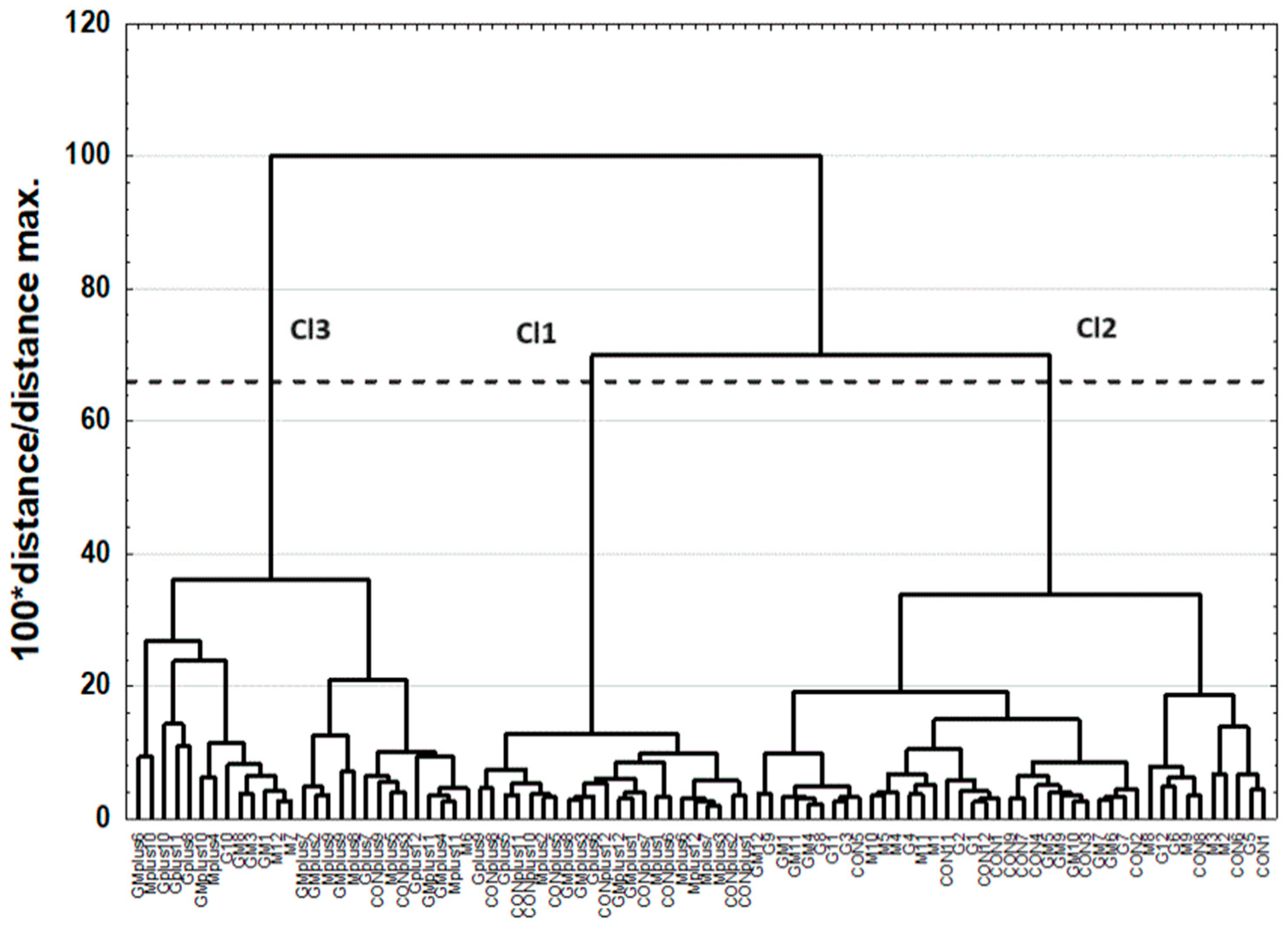
The results of CA are presented as dendrograms in Figure 1 and Figure 2. The application of the less restrictive Sneath’s criterion (66%) to the dendrogram analysis allowed for three clusters to be distinguished that differentiated the examined elements (Figure 1). The first cluster (Cl1) included three elements: Mg, Zn, and K; the second cluster (Cl2) included ten elements: Ca, Fe, Cu, Mn, Se, Co, Cr, Ni Pb, and Cd; and the third cluster (Cl3) included five elements: Na, Al, B, Sr, and Tl. The application of the less restrictive Sneath’s criterion (66%) to the second dendrogram analysis allowed for three clusters to be distinguished that differentiated the examined spleen samples (Figure 2).
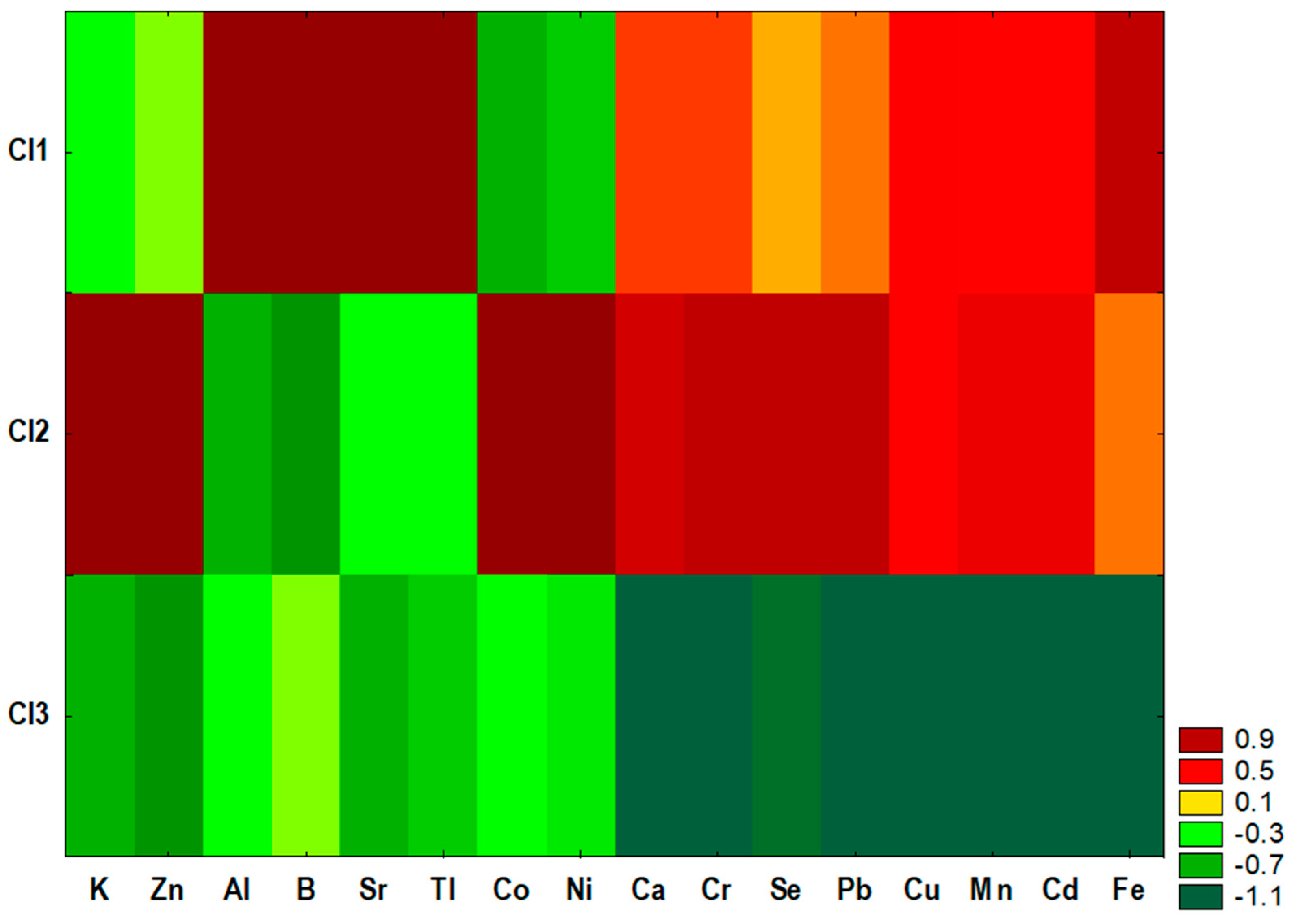
The first cluster (Cl1) included most of the spleen samples obtained from animals treated with DMBA, the second cluster (Cl2) included most of the spleen samples obtained from animals not treated with the carcinogen, and the third cluster (Cl3) was a mixture and included three spleen samples from the M group, one sample from the G group, three samples from the GM group, two samples from the CONplus group, six samples from the Mplus group, five samples from the Gplus group, and seven samples from the GMplus group. Similarity analysis was performed by grouping features and objects to prepare a heat map (Figure 3).
Statistical analysis revealed significant differences in the content of most examined elements among existing clusters of spleen samples (Table 3). Only Mg and Na content did not differ among clusters. Cluster Cl1 was characterized by the highest contents of Al, Sr, B, and Tl, which significantly exceeded the content of these elements in clusters Cl2 and Cl3. Cluster Cl2 was characterized by the highest content of Zn, which was significantly higher than its content in Cl3, and by the highest contents of Se, Co, Ni, and Pb, which were significantly higher than in clusters Cl1 and Cl3. For cluster Cl3, the lowest contents of K, Ca, Cu, Mn, Se, Cr, Pb, and Cd were observed, which were significantly lower than in Cl2 (for K) or in Cl1 and Cl2 (for the rest of the elements) (Table 3).

2.4. Principal Component Analysis (PCA)

The contents of all assayed elements were subjected to PCA. As shown by the screen test, three principal components (PCs) were enough to explain 64.7% of the total variance (Table 4).
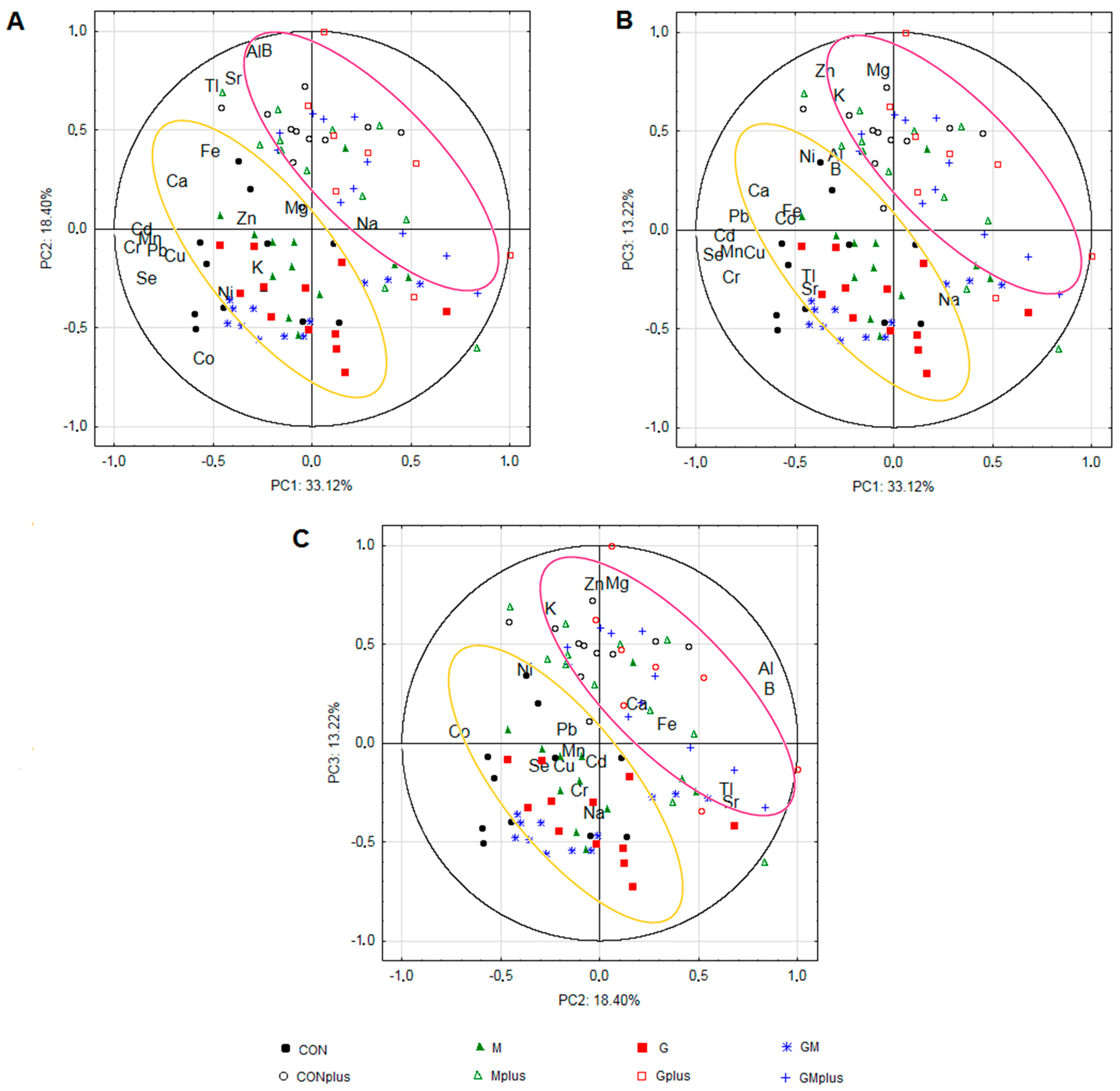
The highest contribution (>70%) to the first PC exhibited the content of Cd, Se, Cr, Mn, Pb, Cu. The levels of B, Al, Co, Sr, and Tl demonstrated variance in the second PC. The third PC gained variance (>60%) from the content of Mg, Zn, and K. The PC1/PC2, PC1/PC3, and PC2/PC3 biplots enabled the samples from the DMBA-treated and untreated animals to be divided; however, no clear separation among groups receiving various supplementation was observed (Figure 4). Strong positive correlations were observed between contents of B and Al, and Sr and Tl, as well as among Cu, Cd, Mn, Cr, and Pb, while a negative correlation was observed between Na, Mg, and Ca levels.

2.5. Linear Discriminant Analysis (LDA)

LDA was used to obtain appropriate classification rules for the examined samples of spleen. Relevant discriminant functions were calculated in a stepwise progressive method. In the performed analysis, 16 variables were included in the final model, from which 10 were statistically significant. The applied canonical analysis allowed for seven discriminant functions to be distinguished, from which four discriminant functions (DF1–DF4) were statistically significant. DF1 was the most significant function, as it explained over 93.63% of the discriminatory power. DF2, DF3, and DF4 explained only 2.40%, 1.76%, and 1.16% of the discriminatory power, respectively (Table 5).
Analysis of the canonical mean variables indicated that DF1 had the greatest impact on the distinction of groups of animals treated with DMBA (“plus” groups) compared to groups that were not subjected to carcinogen treatment. Overall, a good separation of experimental groups regarding the presence of chemical carcinogens was obtained.
The calculated classification matrix indicated that the average classification efficiency based on the calculated functions was 84.44% (Table 6). For individual groups, these coefficients were as follows: 100% for M, 91.67% for Mplus, 90.91% for CONplus and GMplus, 83.33% for CON and GM, and 75.00% for Gplus. The lowest coefficient (58.33%) was revealed for the G group.

3. Discussion

The immune system is one of the most complex systems in the body [24], as it consists of internal organs and tissues, cellular elements, and humoral products.
The spleen works together with the liver and bone marrow to activate the defense response through innate and adaptive immunity [25]. Breast cancer has been considered one with the least immune response [24]. Progression through the carcinogenic pathway from normal breast tissue to breast cancer is accompanied by quantitative and qualitative changes in the nature and location of the immune cell population [26,27]. These cells generate an anti-cancer immune response, which is, however, inhibited by the action of myeloid-derived suppressor cells, which becoming functionally active and extramedullary in organs such as the spleen [27]. The coordination of immunity across organs is fundamental to cancer development and progression. However, whether metabolic alterations in secondary lymphoid tissues (e.g., a spleen) may affect anti-tumor immunity is still under consideration [28].
As discussed previously, there were no spontaneous tumors of any kind in animals not treated with DMBA, whereas mammary cancer incidence in “plus” groups was 42% in CONplus, 67% in Mplus, and 100% in Gplus and GMplus groups, respectively [22]. This may be since DMBA-treated animals have decreased immune response, in terms of, e.g., antibody production, cell-mediated cytolysis, and number of T cells [29,30]. One hundred percent morbidity in groups receiving PSO together with the fact that the first mammary tumors appeared much earlier in Gplus than in other supplemented groups indicated PSO’s ability to accelerate the neoplastic process in this model.
The effect of dietary fatty acids (FAs) on the immune system was suggested by early epidemiological studies of the incidence of multiple sclerosis. It was also confirmed both in vitro and ex vivo that polyunsaturated fatty acids (PUFAs) may influence such essential lymphocyte functions as proliferation, activity of cytotoxic T-lymphocyte and NK cells, production of interleukin 2 (IL-2) and prostaglandin E2 (PGE2), and presentation of antigens. It should be emphasized that this influence depends on the FA type, e.g., dietary sources of n-3 PUFAs exhibit immunosuppressive properties, while oils rich in n-6 PUFAs are less suppressive, and oleic acid—a monounsaturated fatty acid (MUFA) representative—acts as an immunomodulator [31,32,33]. Also, two main isomers of conjugated linoleic acid (c9t11 CLA and t10c12 CLA) enhance IgA, IgG, and IgM production in spleen lymphocytes and mesenteric lymph nodes of rats [34]. Our previous results confirmed that dietary sources of isomers of conjugated linolenic acid (CLnA) also affect the FA profile of the spleen in both physiological and pathological states [35]. Thus, as a continuation and an extension, we also decided to evaluate the effect of such experimental conditions on the elemental composition of a rat’s spleen.
One of the basic indicators of immune system functioning may be considered the relative weight of lymphoid organs, with the following correlation: The greater the weight of these organs the stronger their immune activity [36]. In the present study, neither applied supplementation nor carcinogen treatment influenced the absolute spleen weight. However, in the case of relative spleen weight, a significant increase was observed in all DMBA-treated groups, despite the applied supplementation [22]. This may be explained by the fact that spleen volume increased as a function of tumor growth [30]. There are many types of cells located in the enlarged spleen of tumor-bearing organisms, and they might have various effects on tumor progression via different mechanisms [37].
Nowadays, tumor and non-tumoral lymphoid tissues (like bone marrow and spleen) and non-invasive measurements of glucose metabolism are used as tumor aggressiveness and cell proliferation indicators as well as to predict the recurrence and onset of the metastasis of a broad spectrum of tumor types [38]. The glucose metabolism seems to be of utmost importance, as the spleens of mice with breast cancer are metabolically reprogrammed to the glycolytic state. Magnesium, by influencing the activity of insulin receptor, tyrosine kinase, and glucose transport protein 4 (GLUT4), directly participates in the regulation of glucose translocation into the cell. Intracellular transformation of glucose into adenosine-5′-triphosphate (ATP) also affects the equilibrium between K+ and Ca2+ ion flow through the membrane [28]. Although in the current study the potassium levels in the spleen of rats were unaffected by any experimental factor, the contents of Mg and Ca significantly differed among groups (Table 2). It may be observed that in terms of the neoplastic process, BME strongly affected the Mg levels in the spleen, as when applied separately, both supplements showed an antagonistic effect, whereas when supplemented jointly, the effect of PSO seemed to be less pronounced/neutralized by BME. Where Ca is concerned, a different effect of the experimental factors was observed. In DMBA-receiving rats, both supplements acted synergistically and decreasingly, which was confirmed by the lowest levels of Ca in the spleen of animals from the GMplus group. The lowering effect of BME and PSO on splenic levels of Ca was observed in DMBA-deprived rats, but there was a lack of interaction between supplements.
Microelements play different important roles in many metabolic processes in all biological systems in both physiological and pathological conditions [39]. Immune cells and their effector activities are sensitive to marginal and moderate deficiencies of trace elements, e.g., zinc deficiency in mice decreased levels of IgA both in serum and in mucosa [40]. An adequate supply of trace elements is required for the structure and function of metalloproteins that participate in energy production and protection against oxidative stress. An element strongly involved in antioxidant processes is selenium. Se exerts its biological functions through selenoproteins [41], among which more than half play a key role in neutralizing free radicals. Levels of Se were significantly affected by both experimental factors. The decrease in the splenic selenium content of rats exposed to cancerogenic agent seems to confirm that the neoplastic process intensifies free radical activity.
Trace element status also affects the synthesis and secretion of cytokines and chemokines that modulate the activities of immune cells and other cells [42]. Indeed, the spleen, as an internal organ with high metabolic activity, contains a reservoir of myeloid progenitors and monocytes, constituting an important source of inflammatory cells [43]. CLnA isomers, punicic acid (c9t11c13C18:3, PA), and α-eleostearic acid (c9t11t13C18:3, ESA) found in dietary supplements applied to laboratory animals in the present study exhibit anti-inflammatory properties, as claimed by Saha et al. [44].
Currently, the spleen is considered a prominent site of extramedullary hematopoiesis in cancers, during which not only myeloid cells are produced, e.g., in late-stage cancers, the spleen generates unique erythrocytic cell populations [45]. Tumor growth could play a pivotal role in reprogramming the host immune system by regulating hematopoiesis, which might be associated with the expansion of bone marrow- and/or spleen-derived immunosuppressive cells [38]. Tumors interfere with splenic erythropoiesis, resulting in the accumulation of late erythroid precursors [37]. Erythroid cells form rose junctions with invasive pathogens (e.g., tumor cells) and facilitate their elimination as macrophages pass through the liver and spleen. Thus, the immunity mediated by erythrocytes may prevent cancer cells from spreading through the bloodstream [46]. RBCs are hydrolyzed in the phagolysosome of the splenic macrophages. Proteolytic degradation of hemoglobin takes place, and hem is released and further catabolized into biliverdin, carbon monoxide, and ferrous ion (Fe2+) [2].
Dietary supplementation of healthy animals caused a decrease in the splenic content of Fe to the greatest extent in the groups receiving only PSO. In rats treated with DMBA, the lowest level of this element was observed in the spleen of animals receiving joint supplementation with BME and PSO, whereas the highest was in the Mplus group. Another trace element important for the hematopoietic process is Co, which, as a part of cobalamin (vitamin B12), takes part in the multiplication and division of RBCs. Co is also essential for the activation of enzymes and plays an important role in the functioning of abzymes with DNase and RNase activities. Abzymes are monoclonal antibodies with catalytic properties, which may be found in organisms suffering from autoimmune diseases. Ions of Co2+-activated mammalian DNase I, however, the relative activity of metal-dependent enzymes hydrolyzing DNA depends also on the type and timing of the disease as well as on the individual variability [39]. Levels of Co were significantly lower in the spleen of DMBA-treated rats in comparison to healthy animals and remained unchanged, regardless of the applied supplementation. The same dependence was confirmed by the results obtained for the revealed clusters of animals (Table 3). A similar tendency in the fluctuation depending on the experimental factors may be noticed in the content of Ni. Nickel is a transition metal, present in active sites of eight enzymes, e.g., hydrogenase and urease, with similar properties to Co. Levels of this trace element were higher in the spleen of healthy rats in comparison to DMBA-treated animals. When Ni accumulates to a certain level in the body, it can be toxic to living organisms [41]. The International Agency for Research on Cancer (IARC) classifies Ni, Cd, and hexavalent Cr as human carcinogen group 1. BME supplemented alone decreased the content of both Cd and Cr to the greatest extent in the spleens of healthy rats. On the other hand, in DMBA-treated animals, the strongest properties for lowering Cd content exhibited PSO, supplemented alone or jointly with BME, whereas in the case of Cr, it was BME that exhibited such a strong decreasing effect, either alone or in combination with PSO.
Although significant differences were observed between experimental groups in the content of most of the examined macro- and microelements, all chemometric methods clearly confirmed that the ongoing carcinogenic process had the greatest impact regarding the mineral composition of the spleen. The visible division into DMBA-treated and DMBA-untreated groups was observed in CA, PCA, and LDA. CA and PCA also revealed great similarities in the observed trends. Cl1, containing only animals treated with DMBA, was characterized by the highest amounts of Al, Sr, B, and Tl (Table 3), which was confirmed by the fact that strong positive correlations were observed between the contents of these elements in PCA (Figure 4). There is much evidence that these elements are involved in carcinogenic processes in different organs and tissues (elevated levels in the organisms, increased intake, and the potential to stimulate oxidative stress or weaken the antioxidant defense of the organisms were confirmed) [47,48,49,50,51]. Our results also confirmed that ongoing mammary tumorigenesis was associated with elevated levels of these elements in the spleen. On the other hand, it should be noticed that Cl2, including only DMBA-deprived animals, was characterized by the highest contents of Zn and Se, which are known for their anti-carcinogenic properties [52,53]. This also confirms that the elemental composition of tissues and organs corresponds to the physiological and pathological conditions of the organism and may influence the risk of cancer development.

4. Materials and Methods

4.1. Dietary Ingredients

4.1.1. Bitter Melon Aqueous Extract (BME)

Bitter melon dried fruits were purchased from the local market in Warsaw, Poland, as the main ingredient of tea for brewing (Tra Kho Qua, Hung Phat Corp, Ho Chi Minh City, Vietnam). Daily fresh extract of 1% (w/v) was prepared according to the manufacturer’s description. An exact portion of the dried material was weighed, and a matching measured quantity of hot water (80 °C) was added to obtain 1% extract (w/v). After 10 min, the fresh extract was filtered, brought to room temperature, and administered to the animals ad libitum.

4.1.2. Pomegranate Seed Oil (PSO)

Pomegranate seed oil from the seeds of pomegranate fruits was purchased from the retail market in Warsaw, Poland. It was cold pressed, unrefined, 100% seed oil (ECOSPA), originating from Great Britain. PSO was stored unopened at 8 °C in the manufacturer’s original dark glass package. Before administration to the animals, it was brought to room temperature. PSO was administered to the animals via gavage daily in the amount of 0.15 mL per animal.

4.2. Animal Experiment

The II Local Ethical Committee on Animal Experiments in Warsaw approved the whole experiment as well as the guiding principles for the use and care of laboratory animals (decisions no 56/2013 and 54/2015). The animals were purchased from the Central Laboratory of Experimental Animals, Medical University of Warsaw, Poland. Maiden Sprague-Dawley rats (n = 96, age 30 days) were kept in the animal room in plastic colony cages (3 individuals per cage) at a constant temperature (21 ± 1 °C), with a relative humidity of 50–60% and a 12 h light–dark cycle during the entire experiment. Moreover, during the experiment, they were provided ad libitum access to a standard laboratory fodder Labofeed H (Feed and Concentrates Production Plant, A. Morawski, Żurawia 19, Kcynia, Poland) and drinking liquid. The detailed composition of Labofeed H fodder was published previously [22]. After 1 week of adaptation, the animals were randomly divided into 8 groups of 12 individuals each. Detailed characteristics of the experimental groups are presented below:
-
CON and CONplus—control groups without diet supplementation, fed a standard diet and tap water ad libitum;
-
M and Mplus—animals fed a standard diet supplemented with 1% BME ad libitum;
-
G and Gplus—animals fed a standard diet and water ad libitum and given 0.15 mL/day of PSO via gavage,
-
GM and GMplus—animals fed the standard diet and supplemented with both 0.15 mL/day of PSO via gavage and 1% BME ad libitum.
Rats of four groups marked with “plus” obtained on the 50th day of life 7,12-dimethylbenz[a]anthracene (DMBA) in the dose of 80 mg/kg body weight for the induction of mammary tumors. The chemical composition of the applied supplements was described previously [23]. Throughout the experiment, the rats were monitored daily for signs of welfare disorders (e.g., appetite loss, ruffling, sluggishness, apathy, hiding, curling up) by a qualified veterinarian. They were also checked for any specific signs of health deterioration and weighed each week. The carcinogenic agent was dissolved either in PSO (for the Gplus and GMplus groups) or rapeseed oil (for the CONplus and Mplus groups) and was administered as a solution via gavage intragastrically. The experimental period lasted 21 weeks, and afterwards, the rats were decapitated and exsanguinated, and their spleens were removed, weighed, and stored at −80 °C for further chemical analysis.

4.3. ICP-MS Element Determination

Analytical-grade reagents, solvents, and standards were obtained from Merck (Darmstadt, Germany), J.T. Baker (Philipsburg, NJ, USA), and PerkinElmer (Waltham, MA, USA). Deionized water obtained from the Milli-Q system (18.2 MΩ; Merck, Darmstadt, Germany) was used for samples and standard dilution. A standard reference material, 1577c bovine liver (NIST, Gaithersburg, MD, USA), was used for verification of the analytical procedure of the determination of selected elements (Fe, K, Mg, Mn, Na, Ca, Cd, C, Cr, Cu, Ni, Pb, Se, Sr, Zn) in animal tissue. To investigate the intricate relationships between dietary supplementation and elemental composition, inductively coupled plasma mass spectrometry (ICP-MS) was employed. Certified reference materials with certified values for the selected elements in surface water (SPS-SW1 and SPS-SW2, Spectrapure Standards, Oslo, Norway) were used to check the accuracy of the measurements by ICP-MS.
The content of elements in thawed spleens (approximately 50 mg) was determined in tissues subjected to mineral digestion in 65% nitric acid in glass tubes of a microwave ultraWave closed system with Single Reaction Chamber technology (Milestone, Shelton, CT, USA) for 15 min at up to 200 °C and 10 min at 200 °C. After cooling down to room temperature, digests were diluted in deionized water according to the level of the measured element. Due to the limited amount of material, the analytical procedure was conducted for a single weight of test samples.
In this study, a PerkinElmer Sciex quadrupole spectrometer (NexION 300D, Waltham, MA, USA) was used for isotope-specific detection of selected elements (11B, 23Na, 24Mg, 25Mg, 27Al, 39K, 42Ca, 57Fe, 53Cr, 55Mn, 59Co, 60Ni, 63Cu, 66Zn, 88Sr, 111Cd, 205Tl, and 208Pb). The spectrometer was equipped with a conventional sample introduction system for liquid samples: a quartz cyclonic spray chamber and a Meinhard nebulizer. The working conditions of the spectrometer were as follows: radio frequency plasma power—1350 W, constant nebulizer gas (Ar) flow—0.9 dm3/min. Transient signals of the selected isotopes were monitored (1 reading/5 sweeps/3 replicates) with a dwell time of 100 ms/isotope. Quantitation was achieved by 5-point external calibration (concentration range: 1 μg/dm3–100 μg/dm3). Limits of detection (LODs) and limits of quantitation (LOQs) were calculated for each element by summing the mean of ten measurements of blank samples and three standard deviations for LOD, or ten standard deviations for LOQ, following the IUPAC recommendation [54]. These values are presented in Supplementary Table S1. Blank samples were prepared under the same conditions as the samples. The contents of elements in the fodder, PSO, and BME (both in dry matter and in fluid) are given in Table 1. Detailed chemical characteristics of the dietary supplements are presented in Table S2.

4.4. Statistical Analysis

All data were presented as mean values ± standard deviation. For variables with skew distribution, data were transformed into logarithms, retransformed after calculations, and presented as mean and confidence intervals. Statistica 13.5 software (StatSoft, Tulsa, OH, USA) was used for the statistical analysis. To eliminate the outliers, the obtained results were tested with the Q-Dixon test followed by a one-way ANOVA and a RIR Tukey post-hoc test to verify the differences among groups (marked * in Table 2). For variables that were not characterized by the normality of distribution, the obtained results were tested with the Kruskal-Wallis test, which is a non-parametric equivalent of the one-way ANOVA, with a post-hoc multiple-comparisons test. The acceptable level of significance was established as p ≤ 0.05.
To verify whether the diet modifications and applied experimental conditions significantly affected group diversity, chemometric analyses were performed. Element content in the spleen samples were used as descriptors to study possible discrimination of the spleen samples. Prior to the analyses, the original data were transformed into natural logarithms and then auto-scaled (standardized).
Cluster analysis (CA) was carried out using the agglomeration method. Euclidean distance was used as the distance determination method, and the Ward method was used as the agglomeration method. The application of the less restrictive Sneath’s criterion (66%) was used for dendrogram analysis and cluster distinguishing (Figure 1 and Figure 2). To determine the differences among existing clusters of spleen samples, a one-way ANOVA with a post hoc RIR Tukey test was performed. The accepted significance level was established as p ≤ 0.05. Similarity analysis was performed by grouping features and objects for variables differing significantly among existing clusters to prepare a heat map (Figure 3).
To evaluate whether elemental composition in spleens significantly differs among dietary groups and according to carcinogenic agent usage, principal component analysis (PCA) was performed. A matrix of 20 variables (the content of the detected elements) was used. The number of principal components (PCs) was chosen by the screen test criterion. Results were presented as case projections on the plane of factors (PC1/PC2, PC1/PC3, PC2/PC3) (Figure 4).
Next, in order to obtain appropriate classification rules for spleen samples in the experimental groups, a linear discriminant analysis (LDA) for the examined variables (element content) differing significantly among clusters was performed. Relevant discriminant functions were calculated in a stepwise progressive method, with the adopted tolerance value of 1 − R2 = 0.01 to optimize the LDA.

5. Conclusions

Understanding the immune system’s functioning, its components, and peripheral organs like the spleen during the neoplastic process is crucial for developing effective anticancer prevention and treatment strategies. This information is also important as a preliminary step to confirm/justify the use of PSO and BME as ingredients in functional foods or pharmaceuticals.

Supplementary Materials

The following supporting information can be downloaded at: https://www.mdpi.com/article/10.3390/molecules29091942/s1, Table S1: Values of limit of detection (LOD) and limit of quantitation (LOQ) of detected minerals; Table S2: Chemical characteristics of dietary supplements.

Author Contributions

M.B.: conceptualization, formal analysis, project administration, resources, software, writing—original draft, writing—review and editing; T.L.: investigation, resources, writing—review and editing; W.W.: investigation, writing—review and editing; A.R.: methodology, data curation, investigation, writing—review and editing; E.B.: methodology, resources; writing—review and editing; M.C.: conceptualization, funding acquisition, supervision, writing—review and editing; A.B.: conceptualization, formal analysis, investigation, methodology, resources, software, writing—original draft, writing—review and editing. All authors have read and agreed to the published version of the manuscript.

Funding

This research received no external funding.

Institutional Review Board Statement

The animal study protocol was approved by the II Local Ethical Committee on Animal Experiments in Warsaw (decision Nos. 56/2013 and 54/2015).

Informed Consent Statement

Not applicable.

Data Availability Statement

Dataset available on request from the authors.

Conflicts of Interest

The authors declare no conflicts of interest.

References

  1. Catalano, O.A.; Soricelli, A.; Salvatore, M. Spleen anatomy, function and development. In Abdominal Imaging; Springer: Berlin/Heidelberg, Germany, 2013; Volume 9783642133275, pp. 1479–1494. ISBN 9783642133275. [Google Scholar]
  2. Mebius, R.E.; Kraal, G. Structure and function of the spleen. Nat. Rev. Immunol. 2005, 5, 606–616. [Google Scholar] [CrossRef]
  3. Sajdikova, M.; Novakova, L. 5. Spleen—Functions of Cells and Human Body. Available online: https://fblt.cz/en/skripta/v-krev-a-organy-imunitniho-systemu/5-slezina/ (accessed on 22 November 2023).
  4. Cesta, M.F. Normal structure, function, and histology of the spleen. Toxicol. Pathol. 2006, 34, 455–465. [Google Scholar] [CrossRef]
  5. Nayak, B.N.; Buttar, H.S. Functions of spleen in health and disease. J. Pharm. Sci. Tech. MGMT 2015, 1, 1–7. [Google Scholar]
  6. Karakuchi, N.; Yanagawa, S.; Takei, D.; Kodama, S.; Takeshima, Y.; Sumimoto, K. A case of peritoneal dissemination and splenic metastasis after gastric cancer surgery that could be controlled with multidisciplinary treatment. Case Rep. Oncol. 2020, 13, 1164–1170. [Google Scholar] [CrossRef]
  7. Di Grande, A.; Peirs, S.; Donovan, P.D.; van Trimpont, M.; Morscio, J.; Lintermans, B.; Reunes, L.; Vandamme, N.; Goossens, S.; Nguyen, H.A.; et al. The spleen as a sanctuary site for residual leukemic cells following ABT-199 monotherapy in ETP-ALL. Blood Adv. 2021, 5, 1963–1976. [Google Scholar] [CrossRef]
  8. Velu, U.; Shetty, P.S.; Sharan, K.; Salins, S.; Singh, A. Spleen as an organ at risk in adjuvant chemoradiotherapy for gastric cancer: A retrospective dosimetric study. J. Radiother. Pract. 2023, 22, e57. [Google Scholar] [CrossRef]
  9. El Hadad, S.; Al Rowily, E.; Aldahlawi, A.; Alrahimi, J.; Hassoubah, S. The role of P53 and K-Ras in regulating spleen innate mediators in mice with colon cancer. Pharmacophore 2021, 12, 19–27. [Google Scholar] [CrossRef]
  10. Kristinsson, S.Y.; Gridley, G.; Hoover, R.N.; Check, D.; Landgren, O. Long-term risks after splenectomy among 8149 cancer-free american veterans: A cohort study with up to 27 years follow-up. Haematologica 2014, 99, 392–398. [Google Scholar] [CrossRef]
  11. Noma, K.; Yamaguchi, Y.; Okita, R.; Matsuura, K.; Toge, T. The spleen plays an immunosuppressive role in patients with gastric cancer: Involvement of CD62L+ cells and TGF-Beta. Anticancer Res. 2005, 25, 645–650. [Google Scholar]
  12. Cortez-Retamozo, V.; Etzrodt, M.; Newton, A.; Rauch, P.J.; Chudnovskiy, A.; Berger, C.; Ryan, R.J.H.; Iwamoto, Y.; Marinelli, B.; Gorbatov, R.; et al. Origins of tumor-associated macrophages and neutrophils. Proc. Natl. Acad. Sci. USA 2012, 109, 2491–2496. [Google Scholar] [CrossRef] [PubMed]
  13. Stöth, M.; Freire Valls, A.; Chen, M.; Hidding, S.; Knipper, K.; Shen, Y.; Klose, J.; Ulrich, A.; Ruiz de Almodovar, C.; Schneider, M.; et al. Splenectomy reduces lung metastases and tumoral and metastatic niche inflammation. Int. J. Cancer 2019, 145, 2509–2520. [Google Scholar] [CrossRef] [PubMed]
  14. Białek, A.; Jelińska, M.; Białek, M.; Lepionka, T.; Czerwonka, M.; Czauderna, M. The effect of diet supplementation with pomegranate and bitter melon on lipidomic profile of serum and cancerous tissues of rats with mammary tumours. Antioxidants 2020, 9, 243. [Google Scholar] [CrossRef] [PubMed]
  15. Venturelli, S.; Leischner, C.; Helling, T.; Renner, O.; Burkard, M.; Marongiu, L. Minerals and cancer: Overview of the possible diagnostic value. Cancers 2022, 14, 1256. [Google Scholar] [CrossRef]
  16. Ames, B.N.; Wakimoto, P. Are vitamin and mineral deficiencies a major cancer risk? Nat. Rev. Cancer 2002, 2, 694–704. [Google Scholar] [CrossRef] [PubMed]
  17. Velicer, C.M.; Ulrich, C.M. Vitamin and mineral supplement use among us adults after cancer diagnosis: A systematic review. J. Clin. Oncol. 2008, 26, 665–673. [Google Scholar] [CrossRef]
  18. Huang, H.-Y.; Caballero, B.; Chang, S.; Alberg, A.J.; Semba, R.D.; Schneyer, C.R.; Wilson, R.F.; Cheng, T.-Y.; Vassy, J.; Prokopowicz, G.; et al. The efficacy and safety of multivitamin and mineral supplement use to prevent cancer and chronic disease in adults: A systematic review for a national institutes of health state-of-the-science conference. Ann. Intern. Med. 2006, 145, 372–385. [Google Scholar] [CrossRef]
  19. Lepionka, T.; Białek, M.; Czauderna, M.; Białek, A. Pomegranate seed oil and bitter melon extract supplemented in diet influence the lipid profile and intensity of peroxidation in livers of SPRD rats exposed to a chemical carcinogen. Prostaglandins Other Lipid Mediat. 2021, 152, 106495. [Google Scholar] [CrossRef] [PubMed]
  20. Białek, A.; Białek, M.; Lepionka, T.; Pachniewicz, P.; Czauderna, M. Oxysterols and lipidomic profile of myocardium of rats supplemented with pomegranate seed oil and/or bitter melon aqueous extract—Cardio-oncological animal model research. Chem. Phys. Lipids 2021, 235, 105057. [Google Scholar] [CrossRef] [PubMed]
  21. Białek, A.; Białek, M.; Lepionka, T.; Ruszczyńska, A.; Bulska, E.; Czauderna, M. Cancer influences the elemental composition of the myocardium more strongly than conjugated linoleic acids-chemometric approach to cardio-oncological studies. Molecules 2021, 26, 7127. [Google Scholar] [CrossRef]
  22. Lepionka, T.; Białek, A.; Białek, M.; Czauderna, M.; Stawarska, A.; Wrzesień, R.; Bielecki, W.; Paśko, P.; Galanty, A.; Bobrowska-Korczak, B. Mammary cancer risk and serum lipid profile of rats supplemented with pomegranate seed oil and bitter melon extract. Prostaglandins Other Lipid Mediat. 2019, 142, 33–45. [Google Scholar] [CrossRef]
  23. Białek, M.; Białek, A.; Lepionka, T.; Paśko, P.; Galanty, A.; Tokarz, A.; Czauderna, M. Punica granatum (pomegranate) seed oil and Momordica charantia (bitter melon) extract affect the lipid’s profile and oxidative stability of femoral muscles of rats. Eur. J. Lipid Sci. Technol. 2019, 121, 1800420. [Google Scholar] [CrossRef]
  24. Amens, J.N.; Bahçecioglu, G.; Zorlutuna, P. Immune system effects on breast cancer. Cell. Mol. Bioeng. 2021, 14, 279–292. [Google Scholar] [CrossRef]
  25. Hao, S.; Fang, H.; Fang, S.; Zhang, T.; Zhang, L.; Yang, L. Changes in nuclear factor kappa b components expression in the ovine spleen during early pregnancy. J. Anim. Feed Sci. 2022, 31, 3–11. [Google Scholar] [CrossRef]
  26. Goff, S.L.; Danforth, D.N. The role of immune cells in breast tissue and immunotherapy for the treatment of breast cancer. Clin. Breast Cancer 2021, 21, e63–e73. [Google Scholar] [CrossRef]
  27. Salemme, V.; Centonze, G.; Cavallo, F.; Defilippi, P.; Conti, L. The crosstalk between tumor cells and the immune microenvironment in breast cancer: Implications for immunotherapy. Front. Oncol. 2021, 11, 610303. [Google Scholar] [CrossRef]
  28. Wang, Y.; Xu, M.; Sun, J.; Li, X.; Shi, H.; Wang, X.; Liu, B.; Zhang, T.; Jiang, X.; Lin, L.; et al. Glycolytic neutrophils accrued in the spleen compromise anti-tumour T cell immunity in breast cancer. Nat. Metab. 2023, 5, 1408–1422. [Google Scholar] [CrossRef]
  29. Okuda, S.; Kubo, C.; Taniguchi, K.; Nomoto, K. Different susceptibility of various immune reactions to suppressive effect in the tumor-bearing state. Cancer Immunol. Immunother 1980, 9, 87–92. [Google Scholar] [CrossRef]
  30. Merdrignac, G.; Haddada, H.; De Vaux Saint Cyr, C.; Genetet, B. Spleen cell populations in normal and tumor-bearing hamsters. Immunol. Lett. 1985, 10, 81–86. [Google Scholar] [CrossRef]
  31. Yaqoob, P.; Calder, P.C. The effects of fatty acids on lymphocyte functions. Int. J. Biochem. 1993, 25, 1705–1714. [Google Scholar] [CrossRef]
  32. Jeffery, N.M.; Newsholme, E.A.; Calder, P.C. Level of polyunsaturated fatty acids and the n-6 to n-3 polyunsaturated fatty acid ratio in the rat diet alter serum lipid levels and lymphocyte functions. Prostaglandins Leukot. Essent. Fat. Acids 1997, 57, 149–160. [Google Scholar] [CrossRef]
  33. Peterson, L.; Jeffery, N.; Thies, F.; Sanderson, P.; Newsholme, E.; Calder, P. Eicosapentaenoic and docosahexaenoic acids alter rat spleen leukocyte fatty acid composition and prostaglandin e2 production but have different effect on lymphocyte functions and cell-mediated immunity. Lipids 1998, 33, 171–180. [Google Scholar] [CrossRef]
  34. Yamasaki, M.; Chujo, H.; Hirao, A.; Koyanagi, N.; Okamoto, T.; Tojo, N.; Oishi, A.; Iwata, T.; Yamauchi-Sato, Y.; Yamamoto, T.; et al. Immunoglobulin and cytokine production from spleen lymphocytes is modulated in C57BL/6J mice by dietary Cis-9, Trans-11 and Trans-10, Cis-12 conjugated linoleic acid. J. Nutr. 2003, 133, 784–788. [Google Scholar] [CrossRef]
  35. Lepionka, T.; Białek, M.; Czauderna, M.; Wojtak, W.; Maculewicz, E.; Białek, A. Exploring the influence of the selected conjugated fatty acids isomers and cancerous process on the fatty acids profile of spleen. Cancers 2024, 16, 479. [Google Scholar] [CrossRef]
  36. Saied, A.M.; Attia, A.I.; El-Kholy, M.S.; Reda, F.M.; EL Nagar, A.G. Effect of cinnamon oil supplementation into broiler chicken diets on growth, carcass traits, haemato-biochemical parameters, immune function, antioxidant status and caecal microbial count. J. Anim. Feed Sci. 2022, 31, 21–33. [Google Scholar] [CrossRef]
  37. Han, Y.; Liu, Q.; Hou, J.; Gu, Y.; Zhang, Y.; Chen, Z.; Fan, J.; Zhou, W.; Qiu, S.; Zhang, Y.; et al. Tumor-induced generation of splenic erythroblast-like ter-cells promotes tumor progression. Cell 2018, 173, 634–648.e12. [Google Scholar] [CrossRef]
  38. Seban, R.D.; Rouzier, R.; Latouche, A.; Deleval, N.; Guinebretiere, J.M.; Buvat, I.; Bidard, F.C.; Champion, L. Total metabolic tumor volume and spleen metabolism on baseline [18F]-FDG PET/CT as independent prognostic biomarkers of recurrence in resected breast cancer. Eur. J. Nucl. Med. Mol. Imaging 2021, 48, 3560–3570. [Google Scholar] [CrossRef]
  39. Zaksas, N.P.; Nevinsky, G.A. Minor and trace elements in whole blood, tissues, proteins and immunoglobulins of mammals. In Trace Elements—Human Health and Environment; InTech: Houston, TX, USA, 2018. [Google Scholar]
  40. Wan, X.L.; Zheng, X.C.; Liang, J.R.; Xiao, X.; Yang, H.M.; Wang, Z.Y. Dietary vitamin a supplementation improves intestinal morphology and immune performance of goslings. J. Anim. Feed Sci. 2022, 31, 217–223. [Google Scholar] [CrossRef]
  41. Zhang, X.; Xu, L.; Liu, P.; Ma, W.; Liu, Y.; Qiao, S.; Liu, Q.; Cai, J.; Zhang, Z. Editorial: The mechanism of trace elements on regulating immunity in prevention and control of human and animal diseases. Front. Immunol. 2023, 14, 1159289. [Google Scholar] [CrossRef]
  42. Failla, M.L. Trace elements and host defense: Recent advances and continuing challenges. J. Nutr. 2003, 133, 1443S–1447S. [Google Scholar] [CrossRef]
  43. Fernández-García, V.; González-Ramos, S.; Martín-Sanz, P.; Castrillo, A.; Boscá, L. Contribution of Extramedullary Hematopoiesis to Atherosclerosis. The spleen as a neglected hub of inflammatory cells. Front. Immunol. 2020, 11, 586527. [Google Scholar] [CrossRef]
  44. Saha, S.S.; Ghosh, M. Antioxidant effect of vegetable oils containing conjugated linolenic acid isomers against induced tissue lipid peroxidation and inflammation in rat model. Chem. Biol. Interact. 2011, 190, 109–120. [Google Scholar] [CrossRef] [PubMed]
  45. Wu, C.; Hua, Q.; Zheng, L. Generation of myeloid cells in cancer: The spleen matters. Front. Immunol. 2020, 11, 1126. [Google Scholar] [CrossRef] [PubMed]
  46. Zhang, X.; Zhang, L.; Xia, K.; Dai, J.; Huang, J.; Wang, Y.; Zhu, G.; Hu, Z.; Zeng, Z.; Jia, Y. Effects of dietary selenium on immune function of spleen in mice. J. Funct. Foods 2022, 89, 104914. [Google Scholar] [CrossRef]
  47. Moussaron, A.; Alexandre, J.; Chenard, M.P.; Mathelin, C.; Reix, N. Correlation between daily life aluminium exposure and breast cancer risk: A systematic review. J. Trace Elem. Med. Biol. 2023, 79, 127247. [Google Scholar] [CrossRef] [PubMed]
  48. Van Gerwen, M.; Alerte, E.; Alsen, M.; Little, C.; Sinclair, C.; Genden, E. The role of heavy metals in thyroid cancer: A meta-analysis. J. Trace Elem. Med. Biol. 2022, 69, 126900. [Google Scholar] [CrossRef] [PubMed]
  49. Genchi, G.; Carocci, A.; Lauria, G.; Sinicropi, M.S.; Catalano, A. Thallium use, toxicity, and detoxification therapy: An overview. Appl. Sci. 2021, 11, 8322. [Google Scholar] [CrossRef]
  50. Chen, L.J.; Tang, L.Y.; He, J.R.; Su, Y.; Cen, Y.L.; Yu, D.D.; Wu, B.H.; Lin, Y.; Chen, W.Q.; Song, E.W.; et al. Urinary strontium and the risk of breast cancer: A case-control study in Guangzhou, China. Environ. Res. 2012, 112, 212–217. [Google Scholar] [CrossRef] [PubMed]
  51. Białek, M.; Czauderna, M.; Krajewska, K.A.; Przybylski, W. Selected physiological effects of boron compounds for animals and humans. a review. J. Anim. Feed Sci. 2019, 28, 307–320. [Google Scholar] [CrossRef]
  52. Rayman, M.P. Selenium in cancer prevention: A review of the evidence and mechanism of action. Proc. Nutr. Soc. 2005, 64, 527–542. [Google Scholar] [CrossRef]
  53. Skrajnowska, D.; Bobrowska-Korczak, B. Role of zinc in immune system and anti-cancer defense mechanisms. Nutrients 2019, 11, 2273. [Google Scholar] [CrossRef]
  54. Currie, L.A. Nomenclature in evaluation of analytical methods, including detect ion and quantification capabilities (IUPAC Recommendations 1995). Pure Appl. Chem. 1995, 67, 1699–1723. [Google Scholar] [CrossRef]
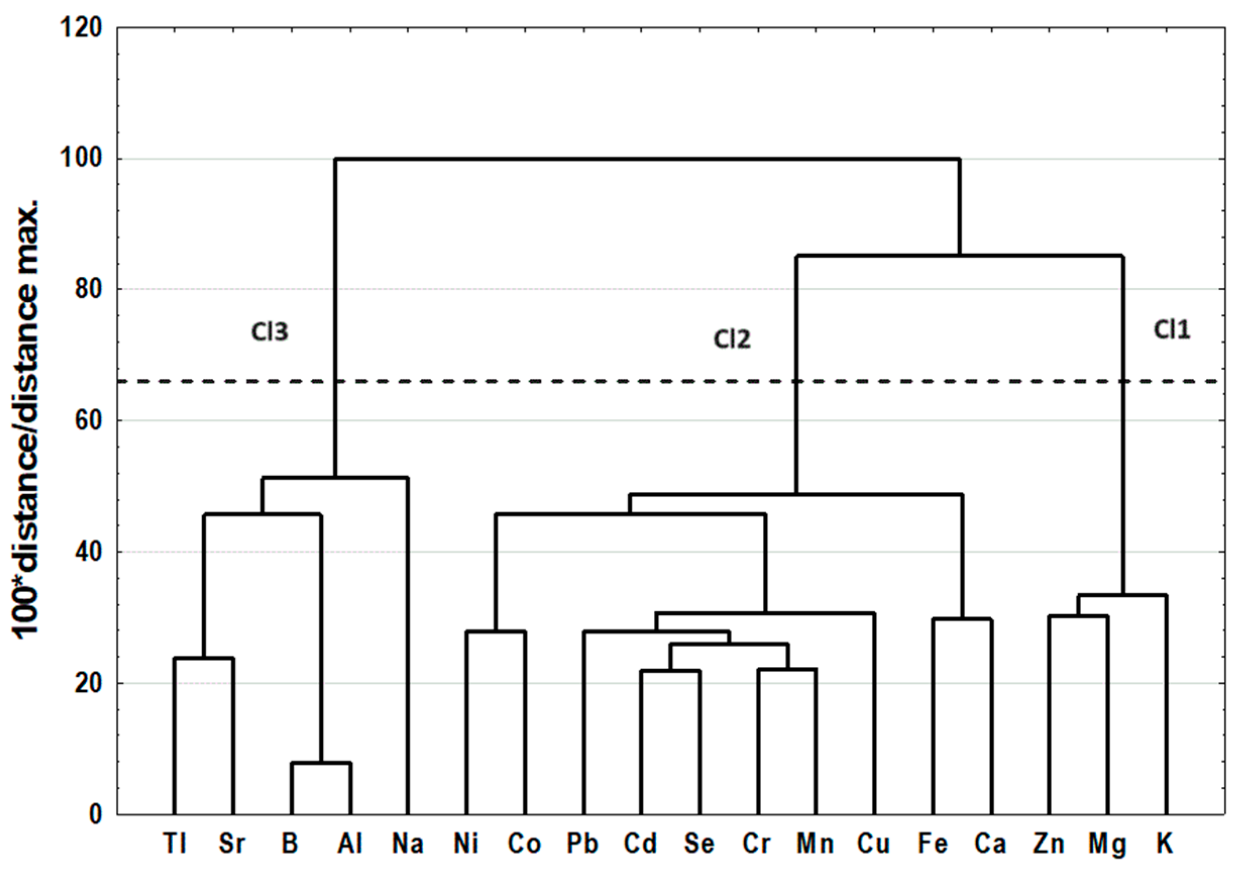
Figure 1. Dendrogram of similarities of examined features (variables).
Molecules 29 01942 g001
Figure 2. Dendrogram of similarities of examined objects (individuals).
Molecules 29 01942 g002
Figure 3. Similarity analysis performed by grouping features and objects to prepare a heat map.
Molecules 29 01942 g003
Figure 4. Biplots of mineral content in the spleen of rats from dietary supplementation groups: (A) PC1/PC2, (B) PC1/PC3, (C) PC2/PC3. CON—animals without dietary supplementation; CONplus—animals without dietary supplementation treated with DMBA; M—animals receiving BME; Mplus—animals receiving BME treated with DMBA; G—animals receiving PSO; Gplus—animals receiving PSO treated with DMBA; GM—animals receiving both PSO and BME; GMplus—animals receiving both PSO and BME treated with DMBA.
Molecules 29 01942 g004
Table 1. Elemental composition of dietary ingredients.
Labofeed HPSOBitter Melon (Dry Matter)BME (Fluid)
mg/kgµg/L
Macroelements
K 18,57013.6628,896524,761
Mg 2930<LOQ291338,942
Na2416<LOD18014,018
Ca 7686<LOD189626,924
Microelements
Fe225.94.13160.3263.4
Zn 34.28<LOD15.61399.3
Cu9.88<LOD6.7980.32
Mn43.35<LOD239.81741
Se0.160.050.11<LOD
Co0.23<LOD0.161.78
Cr 0.290.561.8122.18
Ni 1.02<LOD2.6147.25
Al149.6<LOD527.13427
Sr 10.74<LOD17.0120.8
Pb <LOD<LOD1.416.90
Cd <LOD<LOD0.06<LOD
B <LOD<LOD<LOQ212.8
Tl<LOQ<LOD<LOD<LOD
LOD—limit of detection, LOQ—limit of quantitation.
Table 2. Content of elements (per kg of wet matter) in spleens of rats from experimental groups.
CONMGGMCONplusMplusGplusGMplusp-Value
Macroelements
K [mg/kg]5206 ± 1685098 ± 2775102 ± 2745091 ± 1894908 ± 5875024 ± 5974269 ± 8705091 ± 2730.0815
Mg [mg/kg]255.2 ± 22.9244.7 ± 18.9240.2 ± 32.3232.1 ± 17.2244.3 ± 26.2 257.3 ± 29.8 223.1 ± 36.5 258.4 ± 20.0 0.0276
Na [mg/kg]19.53 ± 10.7411.49 ± 5.87 a,b16.98 ± 10.4817.65 ± 5.0526.07 ± 10.16 a17.43 ± 11.2951.60 (11.33–235.0) b13.85 ± 9.250.0048
Ca [mg/kg]188.2 ± 38.0 a,b166.9 ± 37.9 c169.3 ± 30.9 d176.9 ± 28.5 e191.3 ± 25.0 f,g149.9 ± 35.4128.9 ± 57.2 a,g115.6 ± 21.5 b,c,d,e,f<0.0001 *
Microelements
Fe [mg/kg]3939 ± 783 a,b3476 ± 4782885 ± 4963044 ± 1553665 ± 561 c3925 ± 1734 d,e2420 ± 818 a,d2130 ± 1152 b,c,e<0.0001
Zn [mg/kg]22.98 ± 2.7126.31 ± 7.8922.91 ± 4.2121.26 ± 2.0921.27 ± 2.2422.19 ± 1.6720.34 ± 3.7121.79 ± 0.910.2763
Cu [mg/kg]3.38 ± 0.762.82 ± 0.772.86 ± 0.642.96 ± 0.712.98 ± 0.662.85 ± 0.492.45 ± 0.592.84 ± 0.460.1581 *
Mn [mg/kg]0.59 ± 0.110.53 ± 0.110.52 ± 0.110.55 ± 0.150.50 ± 0.080.51 ± 0.140.42 ± 0.100.50 ± 0.180.1937
Se [mg/kg]0.73 ± 0.06 a,b,c,d0.63 ± 0.090.67 ± 0.12 e0.68 ± 0.11 f0.58 ± 0.10 a0.57 ± 0.14 b0.48 ± 0.09 c,d,f0.54 ± 0.07 d<0.0001
Co [mg/kg]0.054 ± 0.010 a,b,c,d0.046 ± 0.010 e,f0.059 ± 0.016 g,h,i,j0.064 ± 0.016 k,l,m,n0.010 ± 0.001 a,e,g,k0.010 ± 0.003 b,h,l0.009 ± 0.002 c,f,i,m0.010 ± 0.004 d,j,n<0.0001
Cr [mg/kg]0.51 ± 0.15 a,b0.38 ± 0.080.46 ± 0.100.50 ± 0.100.40 ± 0.070.36 ± 0.10 a0.39 ± 0.090.36 ± 0.11 b0.0019*
Ni [mg/kg]0.16 ± 0.06 a,b,c0.17 ± 0.14 d,e0.14 ± 0.12 f,g0.11 ± 0.080.03 ± 0.01 a,d,f0.05 ± 0.02 b0.09 ± 0.060.03 ± 0.01 c,e,g<0.0001
Al [mg/kg]49.33 ± 9.1952.13 ± 10.3342.02 ± 7.53 a41.29 ± 3.22 b,c57.80 ± 6.41 a,b51.87 ± 11.7155.44 ± 16.0653.82 ± 10.04 c0.0003
Sr [mg/kg]0.20 ± 0.040.17 ± 0.02 a,b0.18 ± 0.06 c0.17 ± 0.04 d0.28 ± 0.07 a,c,d0.24 ± 0.070.25 ± 0.06 b0.21 ± 0.06<0.0001
Pb [mg/kg]0.18 ± 0.05 a,b0.16 ± 0.050.14 ± 0.030.16 ± 0.060.14 ± 0.020.12 ± 0.03 a0.13 ± 0.030.11 ± 0.03 b0.0024
Cd [μg/kg]3.76 ± 0.69 a,b,c2.77 ± 0.712.98 ± 0.743.27 ± 0.68 d,e3.02 ± 0.882.62 ± 1.11 a2.02 ± 0.53 b,d2.08 ± 0.74 c,e<0.0001 *
B [mg/kg]72.34 ± 13.3977.61 ± 15.6364.27 ± 12.38 a63.74 ± 4.96 b87.17 ± 9.56 a,b76.22 ± 18.7982.65 ± 24.5783.47 ± 17.620.0018
Tl [μg/kg]9.59 ± 2.826.88 ± 1.53 a,b,c8.03 ± 3.14 d7.46 ± 2.25 e,f11.99 ± 2.59 a,d10.85 ± 3.40 b,e9.37 ± 3.0811.76 ± 3.99 c,f<0.0001 *
Data are shown as mean values ± standard deviation (SD). p-value ≤ 0.05—significant differences among groups in one-way ANOVA (*) or Kruskal–Wallis test. Values sharing a letter in one row are significantly different (p < 0.05) in the RIR Tukey test or multiple-comparisons test.
Table 3. Content of elements (per kg of wet matter) in spleens of rats from revealed clusters.
Cl1Cl2Cl3p-Value
Macroelements
K (mg/kg)4919 ± 4175152 ± 229 a4849 ± 724 a0.0255
Mg (mg/kg)246.2 ± 22.0245.9 ± 23.9243.4 ± 35.30.9160
Na (mg/kg)23.20 ± 10.8616.75 ± 9.2655.19 ± 142.960.1345
Ca (mg/kg)168.0 ± 32.1 a178.5 ± 35.5 b133.6 ± 43.2 a,b<0.0001
Microelements
Fe (mg/kg)3732 ± 1053 a3376 ± 691 b2586 ± 1263 a,b0.0002
Zn (mg/kg)21.75 ± 1.7223.91 ± 5.14 a20.93 ± 2.56 a0.0061
Cu (mg/kg)3.17 ± 0.34 a3.20 ± 0.60 b2.26 ± 0.48 a,b0.0000
Mn (mg/kg)0.56 ± 0.07 a0.58 ± 0.09 b0.39 ± 0.12 a,b<0.0001
Se (mg/kg)0.62 ± 0.07 a,b0.71 ± 0.07 a,c0.48 ± 0.08 b,c<0.0001
Co (mg/kg)0.011 ± 0.002 a0.059 ± 0.013 a,b0.016 ± 0.014 b<0.0001
Cr (mg/kg)0.44 ± 0.06 a0.48 ± 0.11 b0.31 ± 0.08 a,b<0.0001
Ni (mg/kg)0.04 ± 0.02 a0.16 ± 0.10 a,b0.05 ± 0.04 b<0.0001
Al (mg/kg)57.93 ± 7.00 a,b46.68 ± 8.92 a49.00 ± 12.93 b0.0002
Sr (mg/kg)0.29 ± 0.04 a,b0.19 ± 0.03 a0.18 ± 0.06 b<0.0001
Pb (mg/kg)0.15 ± 0.02 a,b0.17 ± 0.05 a,c0.10 ± 0.02 b,c<0.0001
Cd (μg/kg)3.12 ± 0.70 a3.36 ± 0.70 b1.87 ± 0.53 a,b<0.0001
B (mg/kg)87.99 ± 10.94 a,b69.55 ± 12.66 a74.11 ± 20.41 b0.0001
Tl (μg/kg)13.23 ± 2.37 a,b8.40 ± 2.52 a7.93 ± 2.82 b<0.0001
Data are shown as mean values ± standard deviation (SD). p-value ≤ 0.05—significant differences among groups in one-way ANOVA. Values sharing a letter in one row are significantly different (p < 0.05) in RIR Tukey test.
Table 4. Loadings, eigenvalues, and variances of the significant principal components.
VariablesPC1PC2PC3
K −0.278−0.2490.624
Mg−0.0800.0520.751
Na0.279−0.029−0.408
Ca−0.6820.1860.144
Fe−0.5170.3380.042
Zn−0.348−0.0280.751
Cu−0.702−0.178−0.170
Mn−0.818−0.103−0.162
Se−0.840−0.306−0.171
Co−0.550−0.7090.002
Cr−0.824−0.104−0.293
Ni−0.435−0.3790.315
Al−0.2930.8410.321
Sr−0.4290.660−0.353
Pb−0.785−0.1650.016
Cd−0.861−0.059−0.157
B−0.2920.8490.252
Tl−0.4330.636−0.288
Eigenvalue11.654
Variance (%)33.1218.4013.22
Cumulative (%)64.7
The most significant loadings are boldfaced.
Table 5. Coefficients and average values of canonical variables included in the final model.
Coefficients of Canonical Variables
DF1DF2DF3DF4DF5DF6DF7
Variables93.63%2.40%1.76%1.16%0.59%0.36%0.08%
Co6.963631.092790.1287440.215180.234430.1471560.88032
Sr−1.04424−0.296920.634247−0.28200−0.37835−0.7024811.35566
Mn−0.938290.55813−0.8904730.24603−0.527140.020905−1.34120
Ni0.408050.194310.391771−0.992950.69071−0.2944690.41979
Ca0.70833−1.38438−0.594031−0.14685−0.875120.2017520.05789
B−0.009172.01984−0.9405491.15620−1.144420.2695351.01068
Cr−0.16064−0.544111.6634460.625220.19558−0.226185−0.76844
Fe0.04448−0.33491−0.557203−0.844950.07122−0.695610−0.56046
Cu−0.122310.25923−0.959160−0.87929−0.100491.019662−0.23898
Na0.22432−0.590060.5336000.10718−0.008300.232868−0.08378
Mg0.21959−0.746600.6136140.372651.15938−0.0620230.00759
Tl−0.16485−0.25790−0.1421770.607500.79269−0.9046040.74983
K0.147400.16593−0.6069160.080940.09168−0.2315320.32261
Cd−0.00744−0.651290.1974020.675240.659590.7898250.55133
Zn−0.167660.00680−0.0842580.22020−0.88783−0.7395140.33817
Al−0.87280−0.096410.495011−1.723491.330401.441113−1.30752
Average Values of Canonical Variables
CON6.33793−0.75139−0.01459−0.538071.2509800.4124740.040574
M5.422231.80042−1.07040−1.20665−0.5171380.096160−0.017901
G7.95468−0.215250.469860.54690−0.207331−0.6345820.385911
GM8.61790−0.554390.444530.95220−0.4715480.216111−0.379717
CONplus−8.32973−1.72443−0.71386−0.03044−0.6585490.5934160.203602
Mplus−7.50768−0.87100−0.70078−0.403680.233689−0.844036−0.233464
Gplus−8.182770.662462.88315−0.90017−0.1625210.089866−0.024297
GMplus−8.437411.88802−0.432381.393430.4618530.1636340.037266
Table 6. Classification results of LDA presenting percentage of predicted group membership for actual groups.
Predicted Group Membership
Actual GroupCorrect ClassificationCONMGGMCONplusMplusGplusGMplus
CON83.33%100200000
M100.00%012000000
G58.33%10740000
GM83.33%002100000
CONplus90.91%000010100
Mplus91.67%000001101
Gplus75.00%00001160
GMplus90.91%000001010
Total84.44%111211141114611
Disclaimer/Publisher’s Note: The statements, opinions and data contained in all publications are solely those of the individual author(s) and contributor(s) and not of MDPI and/or the editor(s). MDPI and/or the editor(s) disclaim responsibility for any injury to people or property resulting from any ideas, methods, instructions or products referred to in the content.