Article

Isolation, Identification and Chemical Modification of Bufadienolides from Bufo melanostictus Schneider and Their Cytotoxic Activities against Prostate Cancer Cells

1
Key Laboratory of Tropical Medicinal Resource Chemistry of Ministry of Education, Key Laboratory of Tropical Medicinal Plant Chemistry of Hainan Province, College of Chemistry and Chemical Engineering, Hainan Normal University, Haikou 571158, China
2
Hainan General Hospital & Hainan Affiliated Hospital of Hainan Medical University, Haikou 570311, China
3
Hubei Province Key Laboratory of Traditional Chinese Medicine Resource and Chemistry, Department of Pharmacy, Hubei University of Chinese Medicine, Wuhan 430065, China
4
Institute of Traditional Chinese Medicine and Natural Products, College of Pharmacy, Jinan University, Guangzhou 510632, China
*
Authors to whom correspondence should be addressed.
These authors contributed equally to this work.
Molecules 2024, 29(7), 1571; https://doi.org/10.3390/molecules29071571
Submission received: 3 January 2024 / Revised: 10 March 2024 / Accepted: 29 March 2024 / Published: 31 March 2024

Abstract

:
The traditional Chinese medicine toad venom (Venenum bufonis) has been extensively used to treat various diseases, including cancers, in China and other Southeast Asian countries. The major constituents of toad venom, e.g., bufadienolides and alkaloids, exhibit broad-spectrum pharmacological effects in cancers. Herein, two new bufadienolides (1 and 2), along with eleven known compounds (313) were successfully isolated from Bufo melanostictus Schneider. Their structures were elucidated by extensive spectroscopic data and X-ray diffraction analysis. Furthermore, four lactam derivatives were synthesized through the transformation of bufadienolides lactones. The inhibitory effects of these compounds against human prostate cancer cell lines PC-3 and DU145 were evaluated. The outcomes indicated a notable trend, with a substantial subset displaying nanomolar range IC50 values against PC-3 and DU145 cells, underscoring their pronounced cytotoxicity. Moreover, a noteworthy distinction surfaces, wherein lactones consistently outperformed their lactam counterparts, further validating their heightened potency for the treatment of prostate cancer. This study contributes significant preclinical evidence substantiating the therapeutic viability of bufadienolides and toad venom as intervention strategies for prostate cancer.

1. Introduction

Toad venom, known as Venenum bufonis, has been utilized for therapeutic purposes for many centuries in modern China as well as other Asian nations including South Korea and Japan, following it being appropriately dried and formulated [1,2]. Toad venom plays a central role as the primary constituent in various therapeutic formulations or Chinese patent medicines [3]. Of note, the cinobufacini injection, as a sanctioned therapy, has been used in the management of cancer, including lung and liver malignancies, in China [4,5]. Research has elucidated that the pharmacologically active components within toad venom chiefly comprise alkaloids and bufadienolides, which are distinguished for their diverse bioactive activity for different diseases [6,7,8,9]. Previous studies have shown that bufadienolides demonstrate potent cytotoxic activities against diverse cancer cell lines in vitro and in vivo, with IC50 values in the nanomolar range [10]. Additionally, many studies have revealed some of the structure–activity relationship of bufadienolides in toad venom in cancer cells. One study showed that the 14β-hydroxy derivatives are more active than the 14,15-epoxy compounds, which is the fundamental bufadienolide skeleton structure that is critical for maintaining its cytotoxic activity, while the 3-hydroxyl epimerization leads to a significant decrease in terms of toxicity [11]. While a significant number of bufadienolides have been identified from toad venom derived from different origins, their chemical profiles remain incompletely elucidated, and thus more studies are needed to reveal all necessary information for wider clinical use [12,13,14]. Previous studies have shown that the dichloromethane layer of toad venom has very strong activity against PC-3 and DU145 cells with IC50s < 0.011 μM [15], which may suggest that the organic layers are the main bioactive components to be isolated and characterized. In the current study, two new bufadienolides (1 and 2), along with 11 analogues (313) were isolated from the B. melanostictus Schneider by the bioassay-guided phytochemical method (Figure 1). Notably, four lactam derivatives (1417) were semi-synthesized from the corresponding lactone structures (3, 5, 7 and 13) to augment the stability of these lactones. All compounds have been evaluated for their cytotoxic activity against human prostate cancer cell lines PC-3 and DU145. Herein, the isolation, structural elucidation, and cytotoxic activity of these compounds are described.

2. Results and Discussion

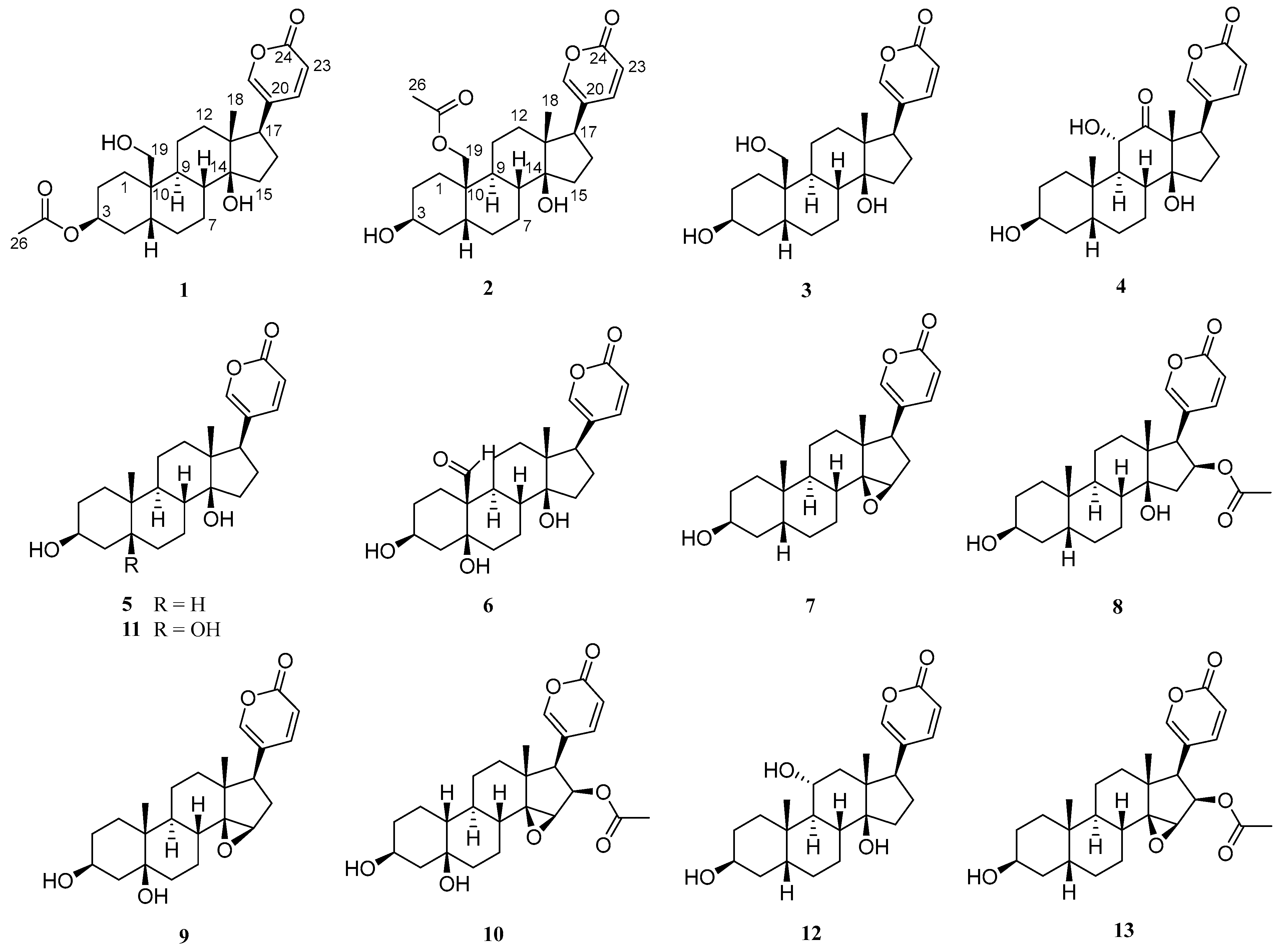
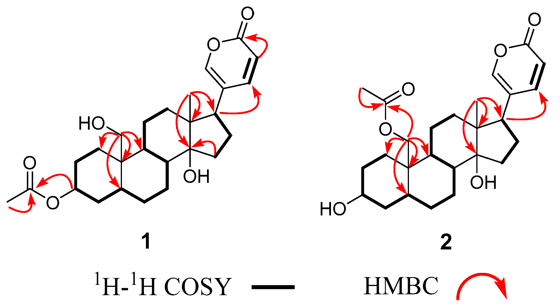
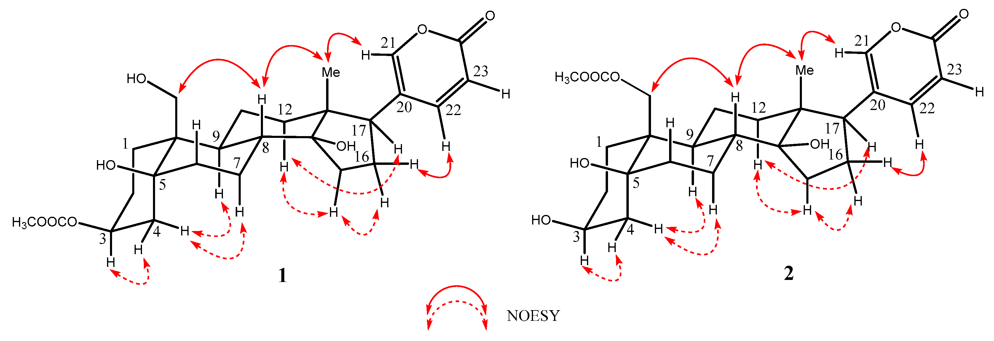
2.1. Structural Elucidation of the Isolated Compounds

Compound 1 was obtained as a white powder. The molecular formula C26H36O6 of 1 was determined by its HR-ESI-MS data [m/z 467.2319 [M + Na]+, calculated for 467.2410]. The 1H NMR data of 1 exhibited three olefinic protons at δH 7.99 (1H, dd, J = 12.0, 4.0 Hz), 7.43 (1H, d, J = 4.0 Hz), and 6.28 (1H, d, J = 12.0 Hz), one oxygenated proton signal at δH 4.06 (1H, s); one methylene group at δH 3.88 (1H, d, J = 12.0 Hz) and 3.37 (1H, d, J = 12.0 Hz)]; one methyl group at δH 0.71 (3H, s) (see details in Table 1). The 13C NMR and DEPT-135 data of 1 showed the presence of 26 carbon resonances, including one carbonyl carbon at δc 164.8, four olefinic carbons at δC 150.5, 149.4, 125.0, and 115.4, ten methylene carbons at δC 65.4, 42.2, 33.0, 31.4, 30.3, 27.2, 25.7, 24.9, 22.6 and 22.1, five methine carbons at δC 72.1, 52.3, 42.8, 36.6 and 30.3, and two methyl carbons at δC 21.3 and 17.3. The comparison of the 1H and 13C NMR of 1 with the known compound 19-hydroxybufalin [16] suggested that their structures are very similar, except for the presence of one carbonyl signal at δc 173.3 and methyl signal at δc 21.3 in 1.
In the 1H-1H COSY (Correlated Spectroscopy) spectrum of 1, three spin systems [H-1 (δH 1.82)↔ H-2 (δH 1.68) ↔ H-3 (δH 5.05) ↔ H-4 (δH 2.00) ↔ H-5 (δH 2.16) ↔ H-6 (δH 1.84) ↔ H-7 (δH 1.87) ↔ H-8 (δH 1.68)↔ H-9 (δH 1.80) ↔ H-11 (δH 1.22)↔ H-12 (δH 1.50), H-15 (δH 1.73) ↔ H-16 (δH 2.18) ↔ H-17 (δH 2.56) and H-22 (δH 7.99) ↔ H-23 (δH 6.28)] were observed. In the HMBC (Heteronuclear Multiple Bond Correlation) spectrum, the key correlations from H-26 (δH 2.04) to C-25 (δc 172.7), from H-3 (δH 5.05) to C-25 (δc 172.7) suggested that an acetyl group linked at the C-3 position of 1 (Figure 2). The relative configuration of 1 was established by the NOESY data. In the NOESY (Nuclear Overhauser Effect Spectroscopy) spectrum of 1, the correlations of H-19 (δH 3.89) and H-8 (δH 1.80), H-8 (δH 1.80) and H3-18 (δH 0.71), H3-18 (δH 0.71) and H-21 (δH 7.43) indicated that these H-18, H-19 and H-21 are placed at β configurations. The correaltions of H-3 (δH 5.05) and H-4 (δH 2.00), H-4 (δH 1.48) and H-9 (δH 1.80)/H-7 (δH 1.22), H-12α (δH 1.50) and H-15 (δH 2.16)/H-17 (δH 2.56) indicated that these positions are at α configurations (Figure 3). To support the configurations of 1, an X-ray crystal structure with a Flack parameter of 0.27(13) was obtained (see Figure 4). Hence, the structure of 1 was determined and named as 3-acetylbufalin.
Compound 2 was isolated also as a white powder, with the molecular formula of C26H36O6 based on the HR-ESI-MS spectrum (m/z 467.2319 [M + Na]+, calculated for C26H36O6Na: 467.2410). The 1H and 13C NMR spectra of 2 were similar to those of 1, except for the significant chemical shifts at C-3 and C-19, indicating the acetyl group of 2 was located at a different position. In the 1H-1H COSY spectrum of 2, three spin systems were observed and drawn in boldface as detailed in Figure 2. In the HMBC spectrum, the key correlations from H-26 (δH 2.07) to C-25 (δc 173.3), from H-19 (δH 4.38) to C-25 (δc 173.3) suggested that an acetyl group located at the C-19 in 2. In the NOESY spectrum of 2, the correlations of H-19 (δH 4.38) and H-8 (δH 1.68), H-8 (δH 1.68), and H-18 (δH 0.72) suggested they were located at β configurations. Furthermore, the correlations of H-3 (δH 4.06) and H-4 (δH 1.98), H-4 (δH 1.98) and H-9 (δH 1.87)/H-7 (δH 1.65), H-12α (δH 1.53) and H-14 (δH 2.16)/H-17 (δH 2.56) indicated that these positions are at α configurations. Thus, the structure of 2 was elucidated to be 19-acetoxybufalin.
By comparing the physical and spectroscopic data with literature values, 11 known compounds were identified as 19-hydroxybufalin (3) [16], arenobufagin (4) [17], bufalin (5) [18], hellebrigenin (6) [19], resibufogenin (7) [20], bufotalin (8), marinobufagin (9) [21], cinobufotalin (10) [17], telocinobufagin (11) [22], gamabufotalin (12) [23], and cinobufagin (13) [24] as shown in Figure 1.

2.2. Spectroscopic Data
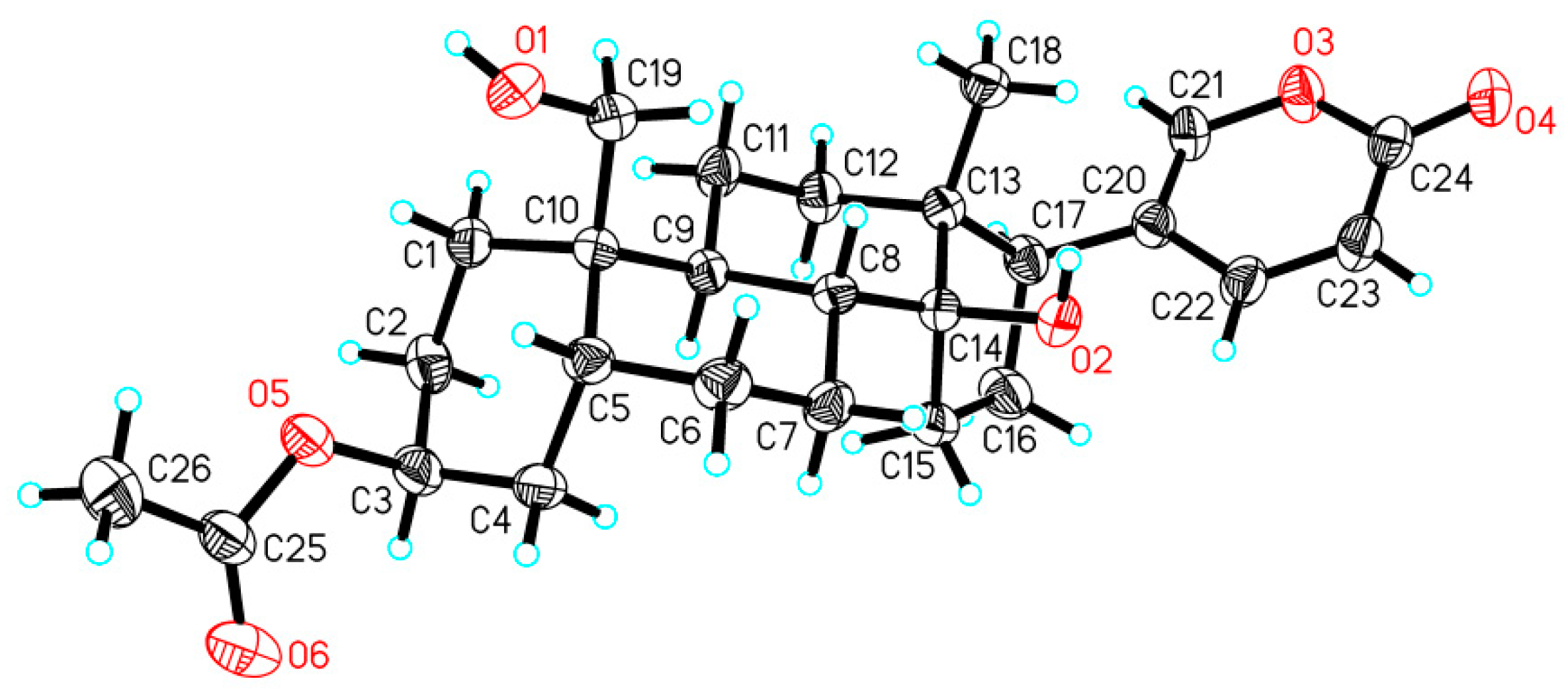
3-Acetylbufalin (1): [ α ] D 25 -0.01 (c 0.1, CH3OH); IR (KBr) νmax 3416 (OH), 1735 (C=O) cm−1; 1H NMR (400 MHz, MeOD) and 13C NMR (100 MHz, MeOD), see Table 1; HR-ESI-MS m/z 467.2391 [M + Na]+ (calcd for C26H36O6Na, 467.2410).
19-Acetoxybufalin (2): [ α ] D 25 -0.04 (c 0.1, CH3OH); IR (KBr) νmax 3420 (OH), 1735 (C=O) cm−1; 1H NMR (400 MHz, MeOD) and 13C NMR (100 MHz, MeOD), see Table 1; HR-ESI-MS m/z 467.2319 [M + Na]+ (calcd for C26H36O6Na, 467.2410).

2.3. X-ray Analysis

Crystal data for 1: C26H36O6, Mr = 444.55, triclinic, a = 8.5025(5) Å, b = 8.5651(5) Å, c = 8.7725(5) Å, α = 72.201(5)°, β = 77.341(5)°, γ = 77.229(5)°, V = 585.27(6) Å3, space group P 1, Z = 1, Dx = 1.261 mg/mm3, μ (Cu Kα) = 0.715 mm−1, and F(000) = 240. Independent reflections: 3917 (Rint = 0.0339). The final R1 values were 0.0358, wR2 = 0.0954 (I > 2σ(I)). Flack parameter = 0.27(13). CCDC number 2288954.

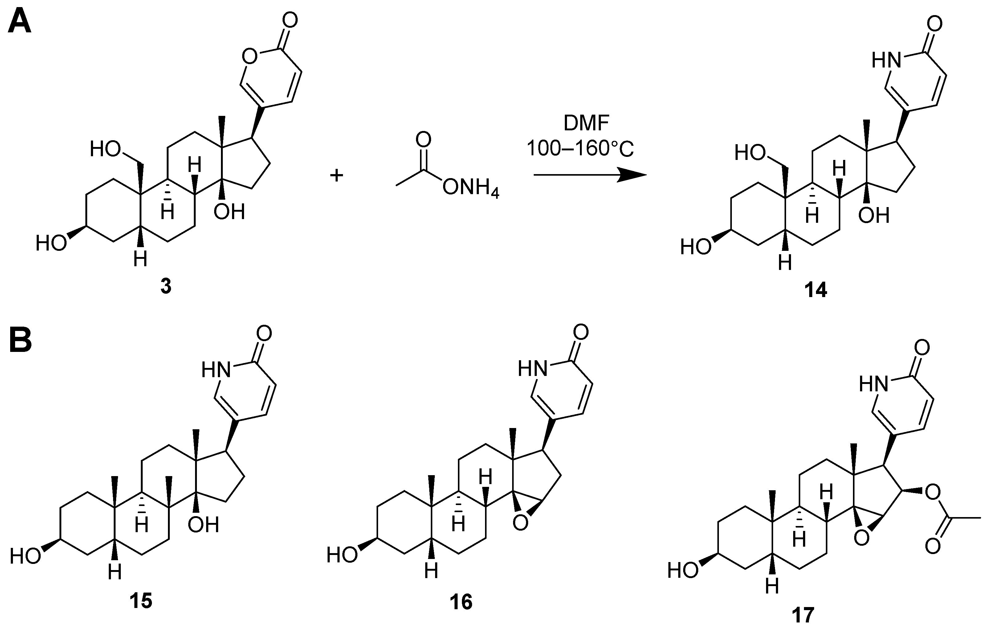
2.4. Synthesis of Lactam Derivatives of BDs

It is known that two prominent limitations warrant consideration in the realm of bufadienolides regarding their developability or drug-likeness: the inherent toxic effects and physicochemical stability concerns arising majorly from the lactone moiety which is easily and readily undergoing hydrolysis procedure in mild acidic or basic condition or in physiological condition. In light of these challenges, a strategic medicinal–chemistry approach was undertaken to address these issues. Notably, the transformation of bufadienolides with a lactone moiety into counterparts bearing a lactam structure was pursued.
The rationale underpinning this approach stems from the recognized superior stability and prevalence of the amide bond (CONH) in a lactam moiety in contrast to the ester bond (OCO) in lactone presented in bufadienolides. This transformation engenders the potential to ameliorate the inherent toxic effects and the stability quandary associated with the lactone motif.
To this end, four lactam derivatives, 19-hydroxybufalin-lactam (14), bufalin-lactam (15), resibufogenin-lactam (16), and cinobufagin-lactam (17) were successfully synthesized via a one-step chemical reaction (see Figure 5A for chemical condition and Figure 5B for the structures of synthesized lactams). Four lactam derivatives were evaluated for their cytotoxicity against human prostate cancer PC-3 and DU145 cell lines.

2.5. The Anti-Proliferative Activities of All Isolated Compounds 113 and Four Modified Compound in Prostate Cancer Cell Lines

The cytotoxic activity of the isolated compounds (113) and four modified compounds (1417) were evaluated for their cytotoxic activity against human prostate cancer cell lines PC-3 and DU145 using MTT assay. The results in Table 2 showed that nearly all isolated compounds had potent cytotoxic effects against PC-3 and DU145 cells with IC50 values < 0.5 μM. Among them, compounds 3, 5 and 12 showed the strongest activity against these two cancer cells with IC50 values < 0.02 μM. However, the two new compounds 1 and 2 showed lower cytotoxicity than Paclitaxel in both cell lines. Notably, lactone-bearing compounds demonstrated markedly heightened activity against prostate cancer cells in contrast to their lactam counterparts 1417 (see details in Table 2).
It is also worth noting that several bufadienolides demonstrated higher (3, 5, and 12, 2–4 folds) or comparable (10) potentials in PC-3 cells compared to the positive control Paclitaxel, but all four showed lower cytotoxicity in DU145 cells than Paclitaxel. Interestingly, both PC-3 and DU145 cells are androgen receptor-independent cell lines that can both be categorized as castration-resistant prostate cancer cells. It is thus unknown why these compounds showed different cytotoxic profiles toward these two similar cell lines. Further studies are needed to decipher the underlying mechanisms. These results may suggest that the cytotoxicity of bufadienolides is cell type-dependent and that compounds 3, 5, and 12 can be further evaluated in PC-3 cells xenograft mice models.
We have used the human nonmalignant prostate epithelial RWPE-1 cell line for parallel comparison. All compounds showed similar toxicity in RWPE-1 cells compared to the two prostate cancer cell lines. However, when we used the African green monkey kidney-derived Vero cell line, the compounds showed much lower toxicity than in prostate cancer cell lines. More work is needed to address the toxic issues.

2.6. The Pharmacophore Modeling of BDs against Prostate Cancer Cells

Subsequently, the structure–activity relationship of all compounds against two prostate cancer cells was discussed using molecular dynamics simulation by computer-aided drug design software. This analytical approach aimed to unravel the intricate interplay between the compounds’ structural attributes and their activity profiles which may provide essential information for further structural modification.
The outcomes of this endeavor disclosed that bufadienolides harbor two distinctive categories of features, namely hydrophobic groups and hydrogen bond donors as shown in Figure 6. Notably, the positioning of hydrogen bond receptors has emerged as a pivotal determinant in maintaining the cytotoxic efficacy of bufadienolides. Structurally, changes in the cyclopentane conformation affect the size of the molecule, thereby influencing the overlap with hydrophobic and hydrogen bond feature elements, a revelation corroborated by the insights gleaned from Figure 6. Compared to bufalin (5), bufotalin (8) (16β-acetoxyl) exhibited decreased activity, which contrasts with previous reports [14] and may be attributed to the variety of cancer cells used in the study. The transformation of the bufadienolide structure (5) into an internal amide (15) resulted in a significant decrease in activity. The disruption of the lactone structure led to a decrease in activity, highlighting the crucial role of the lactone structure in the activity of these compounds, and that it should be intact while undergoing further medicinal–chemistry study.

3. Methods and Materials

3.1. General Experimental Procedures

The optical rotations were measured on a JASCO P-1020 digital polarimeter (JASCO, Tokyo, Japan) using a thermostable optical glass cell (0.1 dm path length and c in g/100 mL). Melting points were determined on a WRX-4 micro melting point apparatus (Shanghai YiCe Apparatus and Equipment Co., Ltd., Shanghai, China) and were uncorrected prior to use. Ultraviolet (UV) spectra were recorded on a U-3900 UV-VIS spectrophotometer (Hitachi, Ltd., Tokyo, Japan). The infrared (IR) spectra were recorded on a Thermo Scientific Nicolet 6700 using the KBr disks spectrophotometer (Thermo Scientific, Madison, WI, USA). Single-crystal data were measured by an Agilent Gemini Ultra X-ray single-crystal diffractometer (Cu Kα radiation). 1D and 2D NMR spectra were recorded from a Bruker AV spectrometer (400 MHZ for 1H and 100 MHZ for 13C) and a JNM-ECZS spectrometer (600 HMZ for 1H and 150 MHZ for 13C). HR-ESI-MS spectra were obtained from a Q-TOF Ultima Global GAA076 LC mass spectrometer (Waters Corporation, Milford, MA, USA). Preparative high-performance liquid chromatography (HPLC) of Agilent 1100 prep-HPLC system with an Agilent C18 analytical HPLC column was used for purification (Agilent Technologies, Waldbronn, Germany). Sephadex LH-20 (Pharmacia Co. Ltd., Sandwich, UK) and silica gel (200−300 and 300−400 mesh, Qingdao Marine Chemical Factory, Qingdao, China) were used for column chromatography (CC). All chemical solvents (analytic grade) were purchased from Xilong Chemical Reagent Factory (Guangzhou, China).

3.2. Materials and Reagents

The toad venom of Bufo melanostictus Schneider was purchased from Guilin of Guangxi province, in February 2020. The employed materials and reagents encompassed 3-[4,5-dimethylthiazol-2-yl]-2,5 diphenyl tetrazolium bromide (MTT, Sigma, USA, Lot No. MKBL6647V), dimethyl sulfoxide (DMSO, Sigma, MO, USA, Lot No. WXBB3106V), Ham’s F-12 Basal Medium (Procell, Wuhan, China, Lot No. WH1022E131), MEM (inclusive of NEAA) basal medium (Procell, Wuhan, China, Lot No. WH0023X131), fetal bovine serum (Four Seasons, Hangzhou, China, Lot No. 22090701), Trypsin (Gibco, NY, USA, batch No. 2428760), sodium chloride (Sinopharm Chemical Reagent Co. Ltd., Shanghai, China, Lot No. 20161025), and disodium hydrogen phosphate anhydrous (Sinopharm Chemical Reagent Co., Ltd., Shanghai, China, Lot No. 20210301). All mentioned substances were procured from commercially established suppliers.

3.3. Extraction and Isolation

The dry toad venom (1.0 kg) was extracted by 95% ethanol under ultrasound conditions under room temperature. After filtration, the resulting product was further concentrated under reduced pressure to yield the crude extract (260 g). Additionally, the crude extract was dissolved in 10% MeOH–H2O and subsequently partitioned by CH2Cl2 to obtain the organic layer (90.6 g) according to a previous study [15]. The CH2Cl2 layer was subjected to a silica gel column with a gradient mixture of petroleum ether and ethyl acetate to finally yield 15 fractions. Fraction 4 was separated over an ODS column and further purified by the preparative HPLC with MeOH and water to yield compounds 8 (14.8 mg), 9 (16.0 mg), and 13 (41.0 mg). Similarly, compounds 4 (9.6 mg), 5 (40.5 mg), 7 (39.4 mg), 10 (17.7 mg), 11 (18.0 mg), and 12 (16.3 mg) were obtained by using a reverse C18 column and preparative HPLC with MeOH and water in fraction 6. Additionally, fraction 7 was separated by preparative HPLC to produce compounds 1 (3.2 mg) and 2 (4.1 mg). Fraction 10 was separated by preparative HPLC using MeOH and water to obtain compounds 3 (48.9 mg) and 6 (11.2 mg).

3.4. X-ray Crystallographic Analysis of Compound 1

Colorless crystals of 1 were obtained from the MeOH solution. Single-crystal X-ray diffraction data were collected on an Xcalibur, Atlas, Geminiultra diffractometer with Cu Kα radiation (λ = 1.54184 Å) at 293 (2) K. Crystallographic data of 1 has been deposited in the Cambridge Crystallographic Data Centre with the deposition number. Copies of the data can be obtained, free of charge, on application to the Director, CCDC, 12 Union Road, Cambridge CB2 1EZ, UK [Fax: +44-(0)1223-336033, or e-mail: [email protected]].

3.5. The Lactone to Lactam Conversion of the Bufadienolides

The conversion of four bufadienolides (3, 5, 7, and 13) into their respective lactams (1417) were accomplished by refluxing a mixture of bufadienolides and ammonium acetate (at a ratio of 3–8 equivalents) in N,N-dimethylformamide (DMF) at temperatures ranging from 100–160 °C for a duration of 0.5–3 h. Subsequent to the reaction, the target lactam was isolated by the preparative HPLC, using a mobile phase composed of MeOH and H2O. Comprehensive details regarding this process are documented in the Supplementary Materials.

3.6. Cell Culture

Human prostate cancer PC-3 and DU145 cells were purchased from Shanghai Zhongqiao Xinzhou Biotechnology Co. (Shanghai, China). The cultivation of human prostate cancer PC-3 cells entailed their placement within Ham’s F-12 medium, supplemented with 10% fetal bovine serum and 1% penicillin-streptomycin. Similarly, human prostate cancer DU145 cells were cultured in MEM medium, supplemented with 10% fetal bovine serum and 1% penicillin-streptomycin. Both cell types were cultured within a 5% CO2, 37 °C incubator, under precise conditions maintained by a Carbon Dioxide Incubator (Thermo, USA) [25]. Routine medium replacement occurred every 2 days. Upon achieving approximately 80% confluence, the cells underwent a 3 min trypsinization process, and their morphological changes were monitored microscopically. The trypsinization process was halted by the introduction of a culture medium containing 10% fetal bovine serum, followed by transfer to a centrifuge tube. Subsequent centrifugation at 1000 rpm for 3 min, and then they were transferred to a new culture flask, which completed the process. For experimental purposes, cells in the logarithmic growth phase were selected.

3.7. Cytotoxicity Assay

Cytotoxic activities of all compounds against PC-3 and DU145 cell lines were evaluated by the MTT method. In short, PC-3 and DU145 (3000–5000 cells/well) were seeded in the 96-well plates. After overnight attachment, fresh medium with gradient concentrations of Paclitaxel as positive control and all compounds were added to the wells and cocultured for another 72 h. The cells treated with 0.1% DMSO in medium were used as the negative control, consistent with the solvent used for dissolving the sample. Then 100 μL MTT solution (0.5 mg/mL) was added to each well. At the end of treatment, the medium in each well was carefully removed, 100 μL DMSO was added into each well, and the plates were shaken for 30 s for a complete dissolve. The absorbance was measured at 490 and 570 nm using a microplate reader (Bio-Tek Instruments Inc., Winooski, VT, USA). The cell survival rate was computed using the formula: Cell survival rate (%) = (Absorbance of Experimental group/Absorbance of Control group) × 100%. The half-maximal inhibitory concentration (IC50) of all bufadienolides and four lactam derivatives against PC-3 cells and DU145 cells was subsequently calculated and analyzed.

3.8. Pharmacophore Modeling

Pharmacophore modeling was conducted employing the Common Feature Pharmacophore application within Discovery Studio 3.0, following established protocols as detailed in prior literature [26,27]. The conversion of 2D chemical structures into 3D conformations was carried out using the Conformations application, and the subsequent generation of pharmacophores was accomplished via the HipHop module. The generated pharmacophore hypotheses were capped at a maximum of 10. Feature options were defined with a minimum of 4 and a maximum of 6. Principal values were assigned as 0 (IC50 > 1 μM), 1 (1 μM > IC50 > 0.3 μM), and 2 (IC50 > 0.3 μM), corresponding to their cytotoxic activity levels [26,27].

4. Conclusions

Thirteen bufadienolides, including two new compounds 12, were isolated from traditional Chinese medicine toad venom. In addition, four lactam derivatives of bufadienolides were obtained by chemical modification. Cytotoxicity of all compounds against prostate cancer cells PC-3 and DU145 were evaluated by the MTT method. Compounds 3, 5 and 12 displayed very strong inhibitory effect against PC-3 and DU145 with IC50S < 0.02 μM, which were lower than positive control Paclitaxel. The structure–activity relationship showed that the lactone ring of bufadienolides is important in terms of antitumor activity since the lactam derivatives of bufadienolides showed significantly decreased activities or lost activities. This study provided possible new potential anti-tumor molecules for clinical treatment for prostate cancers.

Supplementary Materials

The following supporting information can be downloaded at: https://www.mdpi.com/article/10.3390/molecules29071571/s1, Figure S1: 1D and 2D NMR, HRESIMS of new compounds 12. Figure S2: 1D NMR of four lactam of bufadienolides 1417. Table S1–S4: 1H and 13C NMR spectroscopic data of 1417.

Author Contributions

Q.Y. and C.Z. designed the study. Q.Y. conducted the isolation, purification, and characterization of these compounds. R.L. and J.L. performed the in vitro cytotoxicity assay. Q.Y. wrote the original article. Q.Y., Z.C. and C.Z. reviewed and revised this article. All authors have read and agreed to the published version of the manuscript.

Funding

This work was financially supported by the National Natural Science Foundation of China (Grant number: 82160735).

Institutional Review Board Statement

Not applicable.

Informed Consent Statement

Not applicable.

Data Availability Statement

The original contributions presented in the study are included in the article/Supplemental Materials. Further inquiries can be directed to the corresponding authors.

Conflicts of Interest

The authors declare no conflicts of interest.

References

  1. Ye, Q.; Zhou, X.; Han, F.; Zheng, C. Toad Venom-Derived Bufadienolides and Their Therapeutic Application In Prostate Cancers: Current Status and Future Directions. Front. Chem. 2023, 11, 1137547. [Google Scholar] [CrossRef]
  2. Ye, Q.; Zhou, X.; Ren, H.; Han, F.; Lin, R.; Li, J. An Overview of the Past Decade of Bufalin in the Treatment of Refractory and Drug-Resistant Cancers: Current Status, Challenges, and Future Perspectives. Front. Pharmacol. 2023, 14, 1274336. [Google Scholar] [CrossRef]
  3. Jia, J.; Li, J.; Zheng, Q.; Li, D. A Research Update on the Antitumor Effects of Active Components of Chinese Medicine Chansu. Front. Oncol. 2022, 12, 1014637. [Google Scholar] [CrossRef]
  4. Kong, L.; Liu, X.; Yu, B.; Yuan, Y.; Zhao, Q.; Chen, Y.; Qu, B.; Du, X.; Tian, X.; Shao, R.; et al. Cinobufacini Injection Inhibits the Proliferation of Triple-Negative Breast Cancer Through the Pin1-Taz Signaling Pathway. Front. Pharmacol. 2022, 13, 797873. [Google Scholar] [CrossRef]
  5. Li, Q.; Liang, R.L.; Yu, Q.R.; Tian, D.Q.; Zhao, L.N.; Wang, W.W.; Xiao, H.; Yong, X.J.; Peng, X.D. Efficacy and Safety of Cinobufacini Injection Combined with Vinorelbine and Cisplatin Regimen Chemotherapy for Stage Iii/Iv Non-Small Cell Lung Cancer: A Protocol for Systematic Review and Meta-Analysis of Randomized Controlled Trials. Medicine 2020, 99, E21539. [Google Scholar] [CrossRef]
  6. Zheng, Y.; Deng, L.; Cao, H.; Xu, N.; Zhang, D.; Tian, H.; Li, B.; Lu, Z.; Ye, W.; Yu, L.; et al. Screening of Bufadienolides from Toad Venom Identifies Gammabufotalin as a Potential Anti-Inflammatory Agent. Planta Med. 2022, 88, 43–52. [Google Scholar] [CrossRef]
  7. Dong, Q.; Liu, L.; Yuan, Y.; Turdu, G.; Mirzaakhmedov, S.; Aisa, H.A.; Yili, A. Two New Polyamine Alkaloids from the Bufo Viridis Toad Venom. Nat. Prod. Res. 2022, 37, 3538–3542. [Google Scholar] [CrossRef]
  8. Li, F.J.; Hu, J.H.; Ren, X.; Zhou, C.M.; Liu, Q.; Zhang, Y.Q. Toad Venom: A Comprehensive Review of Chemical Constituents, Anticancer Activities, and Mechanisms. Arch. Pharm. 2021, 354, E2100060. [Google Scholar] [CrossRef]
  9. Chen, Y.L.; Dai, Y.H.; Wang, A.D.; Zhou, Z.Y.; Lei, M.; Liu, J.; Lin, B.; Xia, M.Y.; Wang, D. Two New Indole Alkaloids from Toad Venom of Bufo bufo gargarizans. Molecules 2020, 25, 4511. [Google Scholar] [CrossRef]
  10. Zhou, S.-W.; Quan, J.-Y.; Li, Z.-W.; Ye, G.; Shang, Z.; Chen, Z.-P.; Wang, L.; Li, X.-Y.; Zhang, X.-Q.; Li, J.; et al. Bufadienolides from the eggs of the toad Bufo bufo gargarizans and their antimelanoma activities. J. Nat. Prod. 2021, 84, 1425–1433. [Google Scholar] [CrossRef]
  11. Liu, J.S.; Deng, L.J.; Tian, H.Y.; Ruan, Z.X.; Cao, H.H.; Ye, W.C.; Zhang, D.M.; Yu, Z.L. Anti-tumor effects and 3D-quantitative structure-activity relationship analysis of bufadienolides from toad venom. Fitoterapia 2019, 134, 362–371. [Google Scholar] [CrossRef]
  12. Wu, J.H.; Cao, Y.T.; Pan, H.Y.; Wang, L.H. Identification of Antitumor Constituents in Toad Venom by Spectrum-Effect Relationship Analysis and Investigation on Its Pharmacologic Mechanism. Molecules 2020, 25, 4269. [Google Scholar] [CrossRef]
  13. Dai, Y.H.; Wang, A.D.; Chen, Y.L.; Xia, M.Y.; Shao, X.Y.; Liu, D.C.; Wang, D. A New Indole Alkaloid from the Traditional Chinese Medicine Chansu. J. Asian Nat. Prod. Res. 2018, 20, 581–585. [Google Scholar] [CrossRef]
  14. Gao, F.; Wang, X.; Li, Z.; Zhou, A.; Tiffany-Castiglioni, E.; Xie, L.; Qian, Y. Identification of Anti-Tumor Components from Toad Venom. Oncol. Lett. 2017, 14, 15–22. [Google Scholar] [CrossRef]
  15. Meng, L.; Li, S.; Kong, Q.; Wang, M.; Zhang, X.; Zhu, X.; Yu, W.; Jiang, N.; Chun, Z.; Li, N.; et al. Two new 19-hydroxy bufadienolides with cytotoxic activity from the skins of Bufo melanosticus. Nat. Prod. Res. 2021, 35, 4894–4900. [Google Scholar] [CrossRef]
  16. Rodriguez, C.; Ibanez, R.; Mojica, L.; Ng, M.; Spadafora, C.; Durant-Archibold, A.A.; Gutierrez, M. Bufadienolides from the Skin Secretions of the Neotropical Toad Rhinella Alata (Anura: Bufonidae): Antiprotozoal Activity against Trypanosoma cruzi. Molecules 2021, 26, 4217. [Google Scholar] [CrossRef]
  17. Zhao, H.Y.; Wu, F.K.; Qiu, Y.K.; Wu, Z.; Jiang, Y.T.; Chen, J.Y. Studies On Cytotoxic Constituents from the Skin of the Toad Bufo bufo gargarizans. J. Asian Nat. Prod. Res. 2010, 12, 793–800. [Google Scholar] [CrossRef]
  18. Gao, H.; Popescu, R.; Kopp, B.; Wang, Z. Bufadienolides And Their Antitumor Activity. Nat. Prod. Rep. 2011, 28, 953–969. [Google Scholar] [CrossRef]
  19. Wang, D.L.; Qi, F.H.; Tang, W.; Wang, F.S. Chemical Constituents and Bioactivities of the Skin of Bufo bufo gargarizans Cantor. Chem. Biodivers. 2011, 8, 559–567. [Google Scholar] [CrossRef] [PubMed]
  20. Zhu, Z.T.; Deng, S.; Liu, D.; Zhang, B.J.; Sun, H.Z.; Tian, Y.; Wang, C.Y.; Wang, L.; Ma, X.C. Isolation and Identification of Phase I Metabolites of Resibufogenin in Rats. Xenobiotica 2013, 43, 479–485. [Google Scholar] [CrossRef] [PubMed]
  21. Kamano, Y.; Yamamoto, H.; Hatayama, K.; Tanaka, Y.; Shinohara, M.; Komatsu, M. The Isolation and Structure of New Bufadienolide, Resibufagin and the Isolation of Marinobufagin. Tetrahedron Lett. 1968, 5669–5672. [Google Scholar] [CrossRef]
  22. Zhang, X.; Ye, M.; Dong, Y.H.; Hu, H.B.; Tao, S.J.; Yin, J.; Guo, D.A. Biotransformation of Bufadienolides by Cell Suspension Cultures of Saussurea involucrata. Phytochemistry 2011, 72, 1779–1785. [Google Scholar] [CrossRef]
  23. Asrorov, A.M.; Kayumov, M.; Mukhamedov, N.; Yashinov, A.; Mirakhmetova, Z.; Huang, Y.; Yili, A.; Aisa, H.A.; Tashmukhamedov, M.; Salikhov, S.; et al. Toad Venom Bufadienolides and Bufotoxins: An Updated Review. Drug Develop. Res. 2023, 84, 815–838. [Google Scholar] [CrossRef]
  24. Qiao, L.; Zhou, Y.Z.; Chen, H.; Cao, J.Q.; Pei, Y.H. One New Bufadienolide Biotransformed from Cinobufagin by Cunninghamella elegans. Chin. Chem. Lett. 2008, 19, 299–301. [Google Scholar] [CrossRef]
  25. Peery, R.; Cui, Q.; Kyei-Baffour, K.; Josephraj, S.; Huang, C.; Dong, Z.; Dai, M.; Zhang, J.T.; Liu, J.Y. A novel survivin dimerization inhibitor without a labile hydrazone linker induces spontaneous apoptosis and synergizes with docetaxel in prostate cancer cells. Bioorgan. Med. Chem. 2022, 65, 116761. [Google Scholar] [CrossRef]
  26. Yu, W.; Mackerell, A.J. Computer-Aided Drug Design Methods. Methods Mol. Biol. 2017, 1520, 85–106. [Google Scholar] [CrossRef]
  27. Zhao, S.; Peng, W.; Li, X.; Wang, L.; Yin, W.; Wang, Y.D.; Hou, R.; Chen, W.D. Pharmacophore Modeling and Virtual Screening Studies for Discovery of Novel Farnesoid X Receptor (Fxr) Agonists. RSC Adv. 2021, 11, 2158–2166. [Google Scholar] [CrossRef]
Figure 1. The chemical structures of compounds 113. Compounds 12 were unreported bufadienolides, and compounds 311 were described bufadienolides.
Molecules 29 01571 g001
Figure 2. 1H–1H COSY and key HMBC correlations of compounds 1 and 2. Bold lines indicate the 1H-1H coupling, and arrows indicated 1H/13C long-range correlations.
Molecules 29 01571 g002
Figure 3. The key NOESY correlations of compounds 1 and 2. Solid arrows indicate correlations in the β-orientation, and dashed arrows show correlations in the α-orientation.
Molecules 29 01571 g003
Figure 4. X-ray structure of 1. Hydrogen atoms were shown as blue spheres of arbitrary size, and oxygen atoms were shown in red.
Molecules 29 01571 g004
Figure 5. (A) The synthetic method of transforming lactone (3) into lactam (14). (B) Other three synthesized lactam derivatives, bufalin-lactam (15), resibufogenin-lactam (16), and cinobufagin-lactam (17) from compounds 5, 7, and 13.
Molecules 29 01571 g005
Figure 6. Common feature pharmacophore models. The IC50 values and FitValues for Bufalin (5), bufotalin (8), and bufalin-lactam (15) are presented. The green represents the hydrogen bond acceptors, and the blue represents the hydrophobic centers.
Molecules 29 01571 g006
Table 1. 1H NMR (400 MHz) and 13C NMR (100 MHz) data for compounds 1 and 2 in CD3OD a.
Position1 2
δCδH (J in Hz)δCδH (J in Hz)
124.9, CH21.82 a, 1.42 a24.7, CH21.80 a, 1.46 a
225.7, CH21.68 a27.5, CH21.82 a, 1.26 a
372.1, CH5.05, br s67.1, CH4.06, (br. s)
431.4, CH22.00 a, 1.48 a34.0, CH21.98 a, 1.39 a
530.3, CH2.16 a30.7, CH2.16 a
622.1, CH21.84 a, 1.34 a22.1, CH21.85 a, 1.36 a
727.2, CH21.87 a, 1.24 a28.1, CH21.65 a, 1.58 a
842.8, CH1.68 a 43.0, CH1.68 a
936.6, CH1.80 a36.4, CH1.87 a
1040.4, C-39.6, C-
1122.6, CH21.46 a, 1.22 a22.8, CH21.53 a, 1.22 a
1242.2, CH21.50 a, 1.42 a42.0, CH21.53 a, 1.44 a
1349.8, C-49.7, C-
1486.2, C-86.1, C-
1533.0, CH22.16 a, 1.73 a33.0, CH22.14 a, 1.73 a
1629.8, CH22.18 a, 1.73 a29.8, CH22.18 a, 1.74 a
1752.3, CH2.56, m52.2, CH2.56, dd (9.4, 6.0)
1817.3, CH30.71, s17.3, CH30.72, s
1965.4, CH23.89 (d, 11.2); 3.38 (d, 11.2)68.9, CH24.38 (d, 11.2); 4.00 (d, 11.2)
20125.0, C-125.0, C-
21150.5, CH7.43 (dd, 2.6, 1.0)150.4, CH7.44 (dd, 2.6, 1.0)
22149.4, CH7.99 (dd, 9.7, 2.6)149.3, CH8.00 (dd, 9.7, 2.6)
23115.4, CH6.28 (dd, 9.7, 1.0)115.4, CH6.29 (dd, 2.6, 1.0)
24164.8, C-164.7, C-
25172.7, C-173.3, C-
2621.3, CH32.04, s20.9, CH32.07, s
a Overlapped signals are reported without designating multiplicity.
Table 2. The cytotoxic activity against prostate cancer cells of compounds 117 a.
CompoundCancer Cells (μM)
PC-3DU145
Paclitaxel0.031 ± 0.0300.001 ± 0.001
10.162 ± 0.1560.050 ± 0.015
20.214 ± 0.2860.211 ± 0.221
30.014 ± 0.0010.010 ± 0.007
40.091 ± 0.0620.032 ± 0.010
50.012 ± 0.0030.017 ± 0.014
60.040 ± 0.0460.020 ± 0.016
70.259 ± 0.0070.104 ± 0.054
86.740 ± 2.76314.947 ± 5.074
90.571 ± 0.1302.485 ± 0.772
100.035 ± 0.0220.137 ± 0.131
110.040 ± 0.0260.037 ± 0.003
120.007 ± 0.0020.017 ± 0.007
130.050 ± 0.0240.028 ± 0.006
146.261 ± 3.9016.667 ± 2.744
1511.233 ± 0.82113.263 ± 1.506
16>25>25
1756.297 ± 7.76368.563 ± 14.742
Data were presented as mean ± S.E. The experiments were performed at least three times. a Paclitaxel was used as positive control.
Disclaimer/Publisher’s Note: The statements, opinions and data contained in all publications are solely those of the individual author(s) and contributor(s) and not of MDPI and/or the editor(s). MDPI and/or the editor(s) disclaim responsibility for any injury to people or property resulting from any ideas, methods, instructions or products referred to in the content.

