Article

Discovery of Acyl-Surugamide A2 from Marine Streptomyces albidoflavus RKJM-0023—A New Cyclic Nonribosomal Peptide Containing an N-ε-acetyl-L-lysine Residue

1
Department of Biomedical Sciences, Atlantic Veterinary College, University of Prince Edward Island, Charlottetown, PE C1A 4P3, Canada
2
Nautilus Biosciences, Croda Canada Limited, Charlottetown, PE C1A 4P3, Canada
3
Department of Chemistry & Chemical Biology, Barnett Institute for Chemical and Biological Analysis, Northeastern University, Boston, MA 02115, USA
4
Bruker Biospin Corp., 15 Fortune Drive, Billerica, MA 01821, USA
5
Department of Pharmacology, Comprehensive Center for Pain & Addiction, College of Medicine, University of Arizona, Tucson, AZ 85724, USA
6
Department of Chemistry, University of Prince Edward Island, Charlottetown, PE C1A 4P3, Canada
*
Authors to whom correspondence should be addressed.
Molecules 2024, 29(7), 1482; https://doi.org/10.3390/molecules29071482
Submission received: 21 February 2024 / Revised: 21 March 2024 / Accepted: 21 March 2024 / Published: 27 March 2024

Abstract

:
We report the discovery of a novel cyclic nonribosomal peptide (NRP), acyl-surugamide A2, from a marine-derived Streptomyces albidoflavus RKJM-0023 (CP133227). The structure of acyl-surugamide A2 was elucidated using a combination of NMR spectroscopy, MS2 fragmentation analysis, and comparative analysis of the sur biosynthetic gene cluster. Acyl-surugamide A2 contains all eight core amino acids of surugamide A, with a modified N-ε-acetyl-L-lysine residue. Our study highlights the potential of marine Streptomyces strains to produce novel natural products with potential therapeutic applications. The structure of cyclic peptides can be solved using MS2 spectra and analysis of their biosynthetic gene clusters.

1. Introduction

Surugamides are a family of cyclic peptides that were first isolated from a marine-derived S. albidoflavus [1,2]. They are characterized by their eight amino acid cyclic peptide structure, which includes four D-amino acid residues. The surugamide family includes several core members, the surugamides A–E [1] and G–J [3], albucyclone A–F [3] and acyl-surugamide A [3]. Several other natural products can be included in the surugamide family when extending the family to other cyclic peptides that are cyclized by the same type of unique standalone β-lactamase superfamily cyclase enzyme [4,5]. These related natural products are the surugamide F–F2 [4], cyclosurugamide F [4], desotamides [6,7], wollamides [8], ulleungmycins [9], and noursamycins/curacomycins [10,11]. These cyclic peptides range in size between six and ten amino acids in length and share the characteristic presence of at least one tryptophan or phenylalanine and a C-terminal glycine or D-amino acid [12]. A biosynthetic gene cluster was proposed for the surugamides from Streptomyces sp. JAMM992 by using next-generation sequencing to sequence the genome, AntiSMASH [13] to identify the cluster, and gene knockouts to confirm associated genes [14]. The cluster has four non-ribosomal peptide synthetase (NRPS) genes, surABCD, two for the core surugamide A and two of which are responsible for the biosynthesis of the structurally unrelated surugamide F-F2 [14]. Surugamide F was discovered as a linear peptide; however, it was shown that, using recombinant technology, the same cyclization enzyme, surE, is capable of cyclizing surugamide F into cyclosurugamide F [4].
The discovery of new bioactive cyclic peptide natural products is highly desirable for their potential pharmacological applications. Cyclic peptides are highly desirable due to their conformational rigidity and increased metabolic stability, making them more resistant to enzymatic degradation compared to linear peptides [15]. Cyclic peptides possess the ability to be orally available, which is a significant advantage over linear peptides that are readily degraded in the gut [16,17]. Surugamides have been found to have promising biological activities, including antifungal [3], antimicrobial activity [18], and antiproliferative CDK inhibitors [19], as well as being a cathepsin B inhibitors [1]. Acyl-surugamide A is another derivative of surugamide A that was isolated from Streptomyces albus J1074 and has been shown to have good antifungal bioactivity (IC50 3.5 µM) against Saccharomyces cerevisiae [3]. There have been mentions of additional members of the family in several previous publications; however, their structures have yet to be elucidated [3,20].
Ultra-high pressure liquid chromatography paired with high-resolution mass spectrometry (UHPLC–HRMS/MS) working in tandem to acquire both MS1 and MS2 data have significantly enhanced the utility of untargeted metabolomic analysis approaches for the identification of related molecules in complex mixtures of NPs. The UHPLC–HRMS/MS data can be processed by Global Natural Products Social Molecular Networking (GNPS) to cluster group the related NPs together based on MS2 fragment patterns [21]. GNPS has emerged as an indispensable tool for small molecule dereplication by comparing annotated natural product fragmentation patterns [21,22]. Utilizing an untargeted metabolomic approach using GNPS, we were able to determine that marine S. albidoflavus RKJM-0023 produces trace amounts of several unidentified natural products structurally related to surugamide A [1]. Using molecular networks to identify known molecules is a quick way to find new analogues and expand the chemical space of natural products produced by an organism [23]. Herein, we describe the isolation and structural elucidation of acyl-surugamide A2, a new cyclic surugamide containing a rare N-ε-acetyl-L-lysine residue, that to the best of our knowledge has only been identified in a couple examples [3,24]. Utilizing 2D NMR data (HMBC, TOCSY, and HSQC) coupled with UHPLC–HR-ESI-MS/MS for structural fragment annotation, and biosynthetic gene cluster annotation, we were able to elucidate the structure of acyl-surugamide A2.

2. Results and Discussion

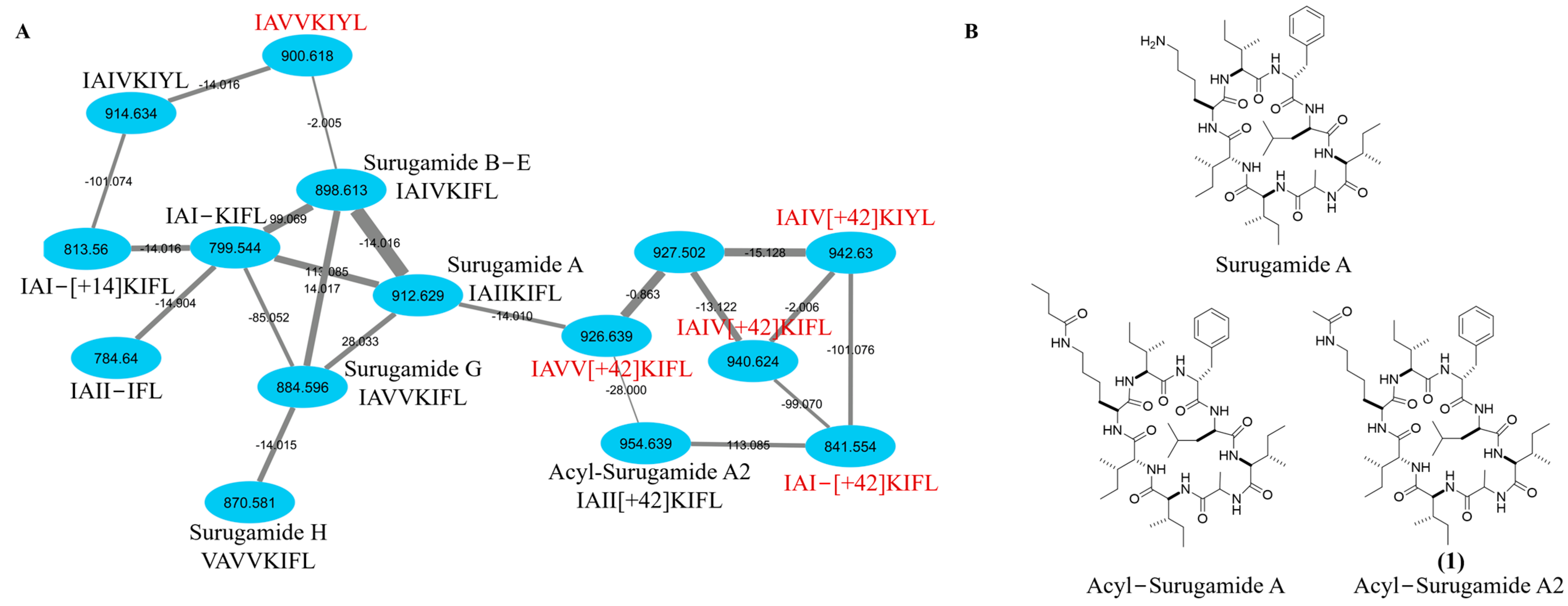
2.1. Targeted Isolation of Acyl-Surugamide A2 from S. albidoflavus RKJM-0023 Isolated from a Marine Tunicate

Strain RKJM-0023 was isolated from the tunicate Halocynthia papillosa (Red Sea Squirt), collected from the Mediterranean Sea on the coast of Turkey. Analysis of the nearly complete 16S rRNA gene sequence (1456 bp) using the EzBioCloud 16S identification tool [25] determined that RKJM0023 was most closely related to S. daghestanicus NRRL B-5418T and S. albidoflavus DSM 40455T (99.86% identity). Chemical screening of fermentations of RKJM-0023 identified the production of surugamides and putative novel surugamide analogs in ethyl acetate extracts of fermentations conducted in BFM15m medium. The fermentation extracts were characterized by UHPLC–ESI-HRMS/MS and organized into molecular clusters using GNPS [21] resulting in the identification of a cluster with GNPS database matches to surugamide A and D (Figure 1). Additionally, two ions with m/z matching literature values for surugamide G and H [3] were also present within the cluster (Figure 1). The BFM15m fermentation extract was chosen for further investigation due to the production of significant quantities of the putatively novel surugamide analogue with an m/z 954.64.
The surugamide molecular network cluster from S. albidoflavus RKJM0023 was manually annotated based on the literature of both isolated and predicted amino acid sequences (Figure 1). Previously isolated surugamides A, B–E, G, and H were detected, while several other surugamide analogues have been previously predicted by cyclic peptide sequence algorithm NPMiner, including 914.63 (IAIVKIYL), 813.56 (IAI-[+14]KIFL), 799.54 (IAI-KIFL), and 784.64 (IAII-IFL) [26], while the m/z 926.64 (IAII[+14]KIFL), 940.62 (IAII[+28]KIFL), and 954.64 (IAII[+42]KIFL) were previously predicted using the MultiTag algorithm where they were referred to as reginamides [27]. Additionally, previously unreported surugamide associated ions (m/z 900.62, 927.50, 942.63, and 841.55) were included in the cluster. The m/z 841.55 has a distinct difference of 113.09, matching an isoleucine/leucine residue; therefore, the predicted amino acid sequence of IAI-[+42]KIFL lacking isoleucine is suggested. For m/z 940.62 and m/z 926.64, the MS2 fragmentation pattern compared to 954.64 suggests a similar +42-Lys residue with one or two Ile substitutions for a Val, similar to surugamide A valine substituted equivalents, surugamides B–E and G. The annotated sequences for m/z 926.64 and 940.62 contradicts the previously predicted amino acid sequences [27]; however, this is a different organism and therefore may be making different surugamide analogues.
The putatively novel surugamide m/z 954.64 analog was found to be most abundant in fermentations conducted in BFM15m, thus fermentations were scaled up in this medium (10 × 1 L). The fermentations were extracted with ethyl acetate and a combination of flash chromatography and reversed-phase HPLC were used to purify m/z 954.6385, named acyl-surugamide A2 (0.6 mg) (Figure S1).

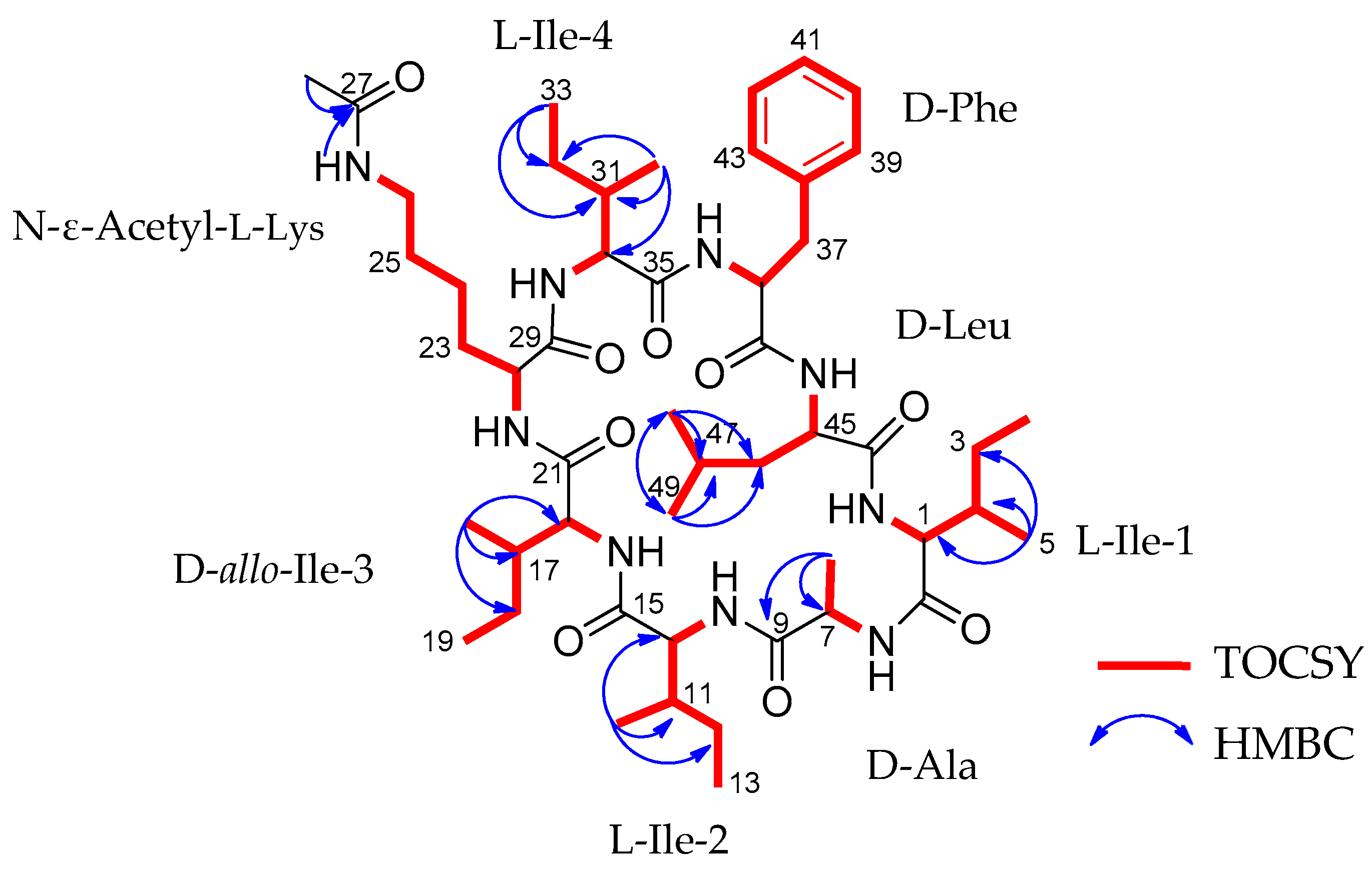
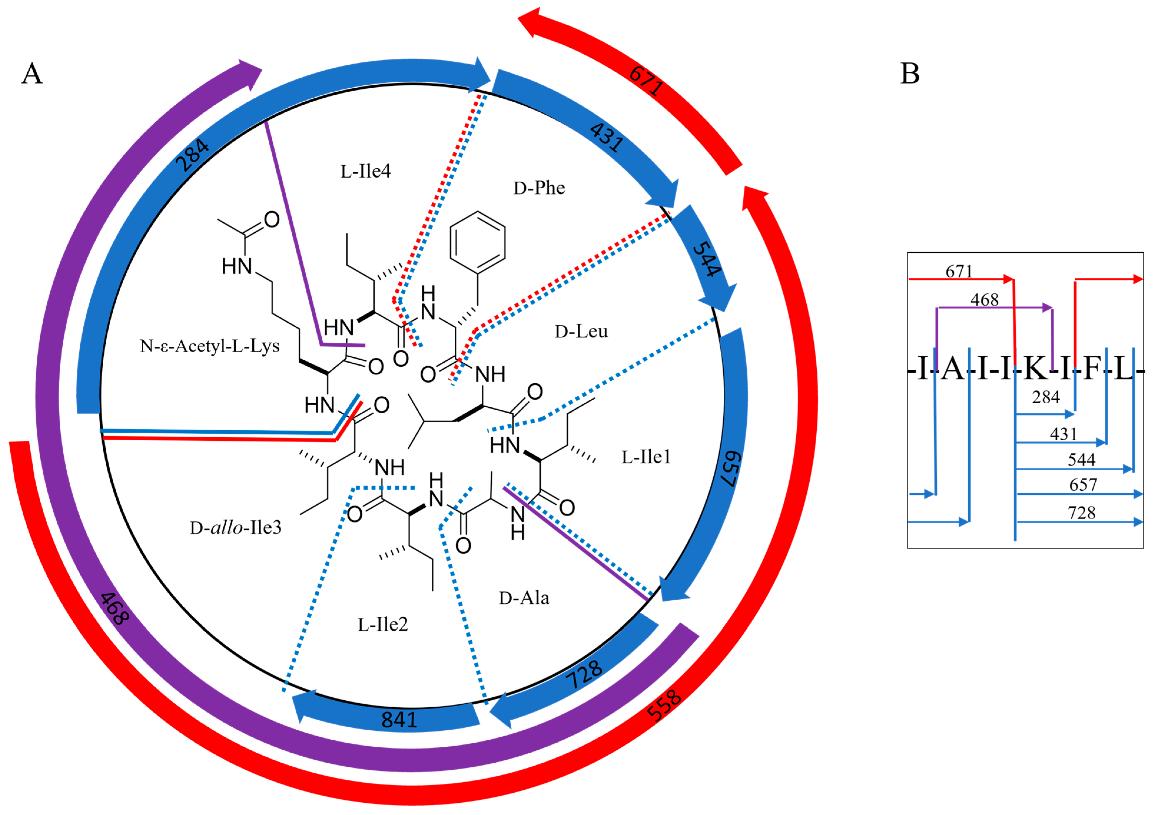
2.2. Structural Characterization of Acyl-Surugamide A2 via NMR and UPLC-HR-ESI-MS/MS Fragment Annoatation

Acyl-surugamide A2 (1) (Figure 1, Table 1) was obtained as a white powder and HR-ESI-MS supported a molecular formula of C50H83N9O9, requiring 13 degrees of unsaturation. The peptidic nature of the compound was determined by analysis of the 1H NMR spectrum (Figure S2) which revealed the presence of nine amide protons, 50-NH (δH 7.13), 6-NH (δH 7.82), 9-NH (δH 8.29), 15-NH (δH 7.95), 21-NH-(δH 7.61), 26-NH (δH 7.75), 29-NH (δH 7.80), 35-NH (δH 8.44), 44-NH (δH 7.73), of which eight have a pairing alpha protons consistent with the molecular network identification surugamide A backbone, plus one amide bond. Analysis of 2D NMR HMBC, HSQC, and TOCSY (Figures S3–S5) spectra confirmed the identity of the amino acid constituents and revealed the presence of Ala, Leu, four Ile, Phe, and a unique acetyl-Lys residue (Table 1, Figure 2). To account for the remaining degree of unsaturation, it was determined that this compound must be cyclic. It was determined that this molecule cyclized in a peptide bond between the N-terminal and C-terminal amino acids as there are no hydroxyl groups, no shifts suggesting an ester-bond found in depsipeptide cyclization [28], and no aldehyde protons in the 1H. This is consistent with a surugamide A [1] core and a modification on the side chain residue of Lys, similar to what was observed with acyl-surugamide A [3]. The acetyl group methyl C-28 (δH 1.77, δC 23.1) is a distinct singlet with no TOSCY correlations. Due to the low amount of material, the HMBC spectra only presented correlations from each methyl group on the molecules, as well as the CH3 of the acetyl group to the carbonyl C27 (δC 169.4) and 26-NH (δH 7.75). The Lys secondary amide 26-NH shares a TOCSY spin system with H-26/H-25/H-24/H-23/H-22, placing it as the Lys residue functional group. This corroborates the MS/MS interpretation of the addition of a functional group extending from the Lys residue. Through careful review of the acquired NMR and comparison to the previously published NMR shifts for surugamide A [1] and acyl-surugamide A [3], all protons could be assigned to acyl-surugamide A2 (Table 1). To determine the sequence of amino acids, a combination of UHPLC–HR-ESI-MS/MS fragment annotation and biosynthetic gene cluster (BGC) analysis were used.
The amino acid sequence of cyclic peptides, such as the surugamides, can be determined using various methods. One strategy involves rebuilding the sequence based on the HRMS/MS predictable amino acid fragmentation of acyl-surugamide A2 amino acids and comparing it directly to the reference fragments of surugamide A in the GNPS library [21]. A similar reconstruction of the MS2 fragments was used to determine the sequence of surugamide A [1] and acyl-surugamide A [3] (Figure S6). Utilizing the information obtained from the NMR confirms that the molecule contains all eight core surugamide A amino acids with a modified acetyl-lysine (Table 1).
To determine the amino acid sequence of acyl-surugamide A2 using the MS2 fragments, the location of the three none-Leu/Ile amino acids must be determined; Ala (A, fragment mass difference of 71.04 m/z), Phe (F, fragment mass difference of 147.07 m/z), and the modified acetyl-Lys (aK, fragment mass difference of 170.11 m/z). The substitution of K for aK produces a mass difference of 42 m/z, accounting for the mass difference between surugamide A and acyl-surugamide A2. MS2 amino acid fragmentation of acyl-surugamide A2 was annotated by comparing the acquired fragments to the MS2 spectrum of surugamide A (Figure 3, Figures S6 and S7, Table 2). The annotated fragments match a full assembly of cyclo-[(I/L)A(I/L)(I/L)aK(I/L)F(I/L)] for acyl-surugamide A2. All fragments predicted to contain aK have complementary MS2 peaks in the surugamide A spectrum with a difference of −42 m/z (Figures S6 and S7, Table 2). For each additional m/z in the surugamide A cluster from S. albidoflavus RKJM0023 (Figure 3), the predicted amino acid sequence and composition were previously algorithmically predicted [26,27] or manually annotated based on their shared fragments to surugamide A and acyl-surugamide A2 (Figure S7).

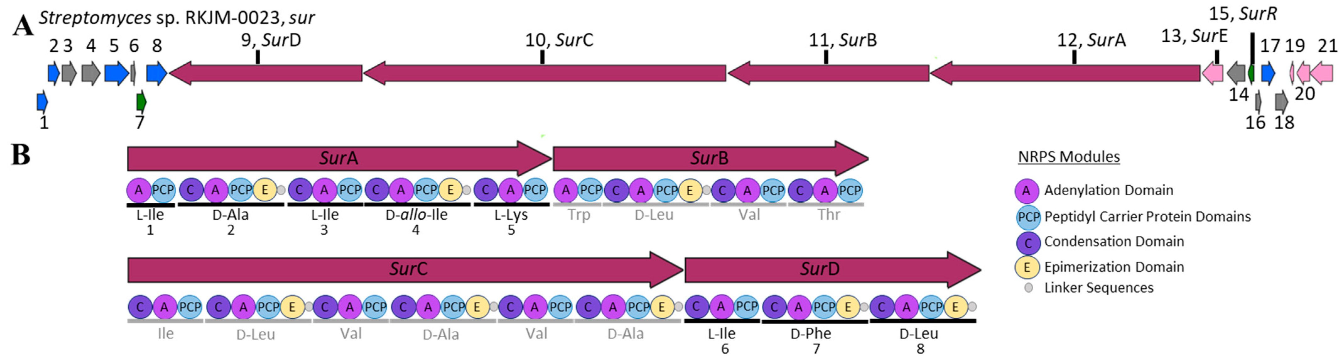
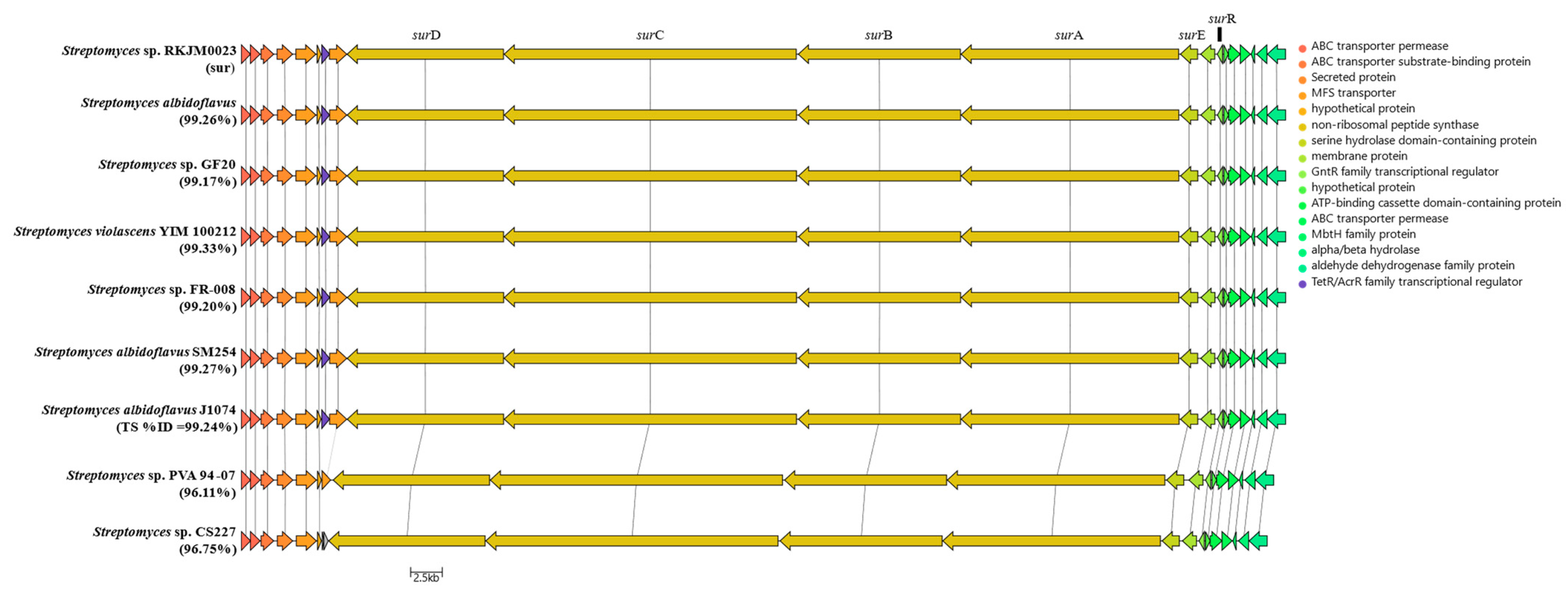
2.3. S. albidoflavus RKJM-0023 Surugamide Biosynthetic Gene Cluster Analysis (sur)

The biosynthetic gene cluster of a NRP can also be used to determine the amino acid order of cyclic-NRPs [1,5]. To date, all core surugamides follow the LDLDLLDD amino acid configuration sequence [1,2,3,5,29] as well as the core AA order cyclo-[IAIIKIFL] with possible substitutions of L-Ile-1, L-Ile-2, or L-Ile-4 for Val [1,3,14]. The genome of S. albidoflavus RKJM-0023 was sequenced using PacBio RSII sequencing [30] and 1.5 × 10⁶ resulting reads were assembled into two contigs representing a 7,031,575 bp genome (GenBank accession CP133227) and a 90,910 bp plasmid (GenBank accession CP133227) [31]. The assembled contigs had a mean coverage depth of 1542X, an N50 of 7,031,575, and a GC content of 73.35%. The ends of both contigs were screened for regions of internal overlap; however, none were found, suggesting that both the genome and plasmid are linear. Annotation of the genome using the GenBank PGAP pipeline identified 6126 CDSs and 67 tRNAs.
The draft genome sequence was mined for natural product biosynthetic gene clusters (BGCs) using AntiSMASH (7.0.0.0) [13]. Complete BGC identification was determined by a consensus rate of higher than 99% matching annotated BGCs in MIBiG [32]. Twenty-one BGC-containing areas were identified in the genome and none were detected on the plasmid. From the twenty-one BGC-containing regions, seven can be annotated due to high consensus to known BGC (≥99% identical) nucleotide sequences; these include SGR PTMs (BGC0001043), cyclofaulknamycin (BGC0002358), geosmin (BGC0001181), surugamide (BGC0001792), desferrioxamine B (BGC0000941), ectoine (BGC0000853), and antimycin (BGC0000958). One BGC was predicted to match the published surugamide cluster (sur) with a 100% consensus with the known cluster blast and MIBiG [32] reference sequence (Figure 4) (BGC0001792) [3,14,32,33].
The S. albidoflavus RKJM-0023 sur cluster contains twenty-one genes (Table 3) including the six sur genes has been previously established in the literature [3,14,34]. The core NRPS genes for the octapeptide surugamide A backbone are surA and surD [14]; combined, these two synthetases are the proposed core NRPS modules for the other surugamide analogues with modified Lys residues, acyl-surugamide A (butyryl functionalized Lys), and albucyclones A-F (albuquinone A functionalized Lys) [3]. The other two sur NRPS modules, surB and surC, are the synthetases for the biosynthesis of the separate decapeptide core of the surugamide F’s [14] (Figure 4). The additional biosynthetic gene surE, is the standalone cyclase domain with homology to a penicillin-binding protein-type thioesterase. SurE has been established to cyclase both the octapeptide and the decapeptide cores of the surugamides [5,34,35]. The Gnt-R transcriptional regulator, surR, has been shown to silence the sur gene cluster when expression is induced [3]. The function of the remaining 15 coding sequences of the sur BGC has not been formally established. The Pfam annotations for each sur protein coding region were annotated by BlastP [36] and are summarized in Table 3. The sur BGC from S. albidoflavus RKJM-0023 was compared directly to the sur BGC from S. albidoflavus J1074 (CP004370.1) and several other publicly available surugamide BGC sequences.
The sur nucleotide sequence obtained from RKJM-0023 was directly compared to the sur BGC sequence from S. albidoflavus J1074 (BGC0001792 (MIBiG), NCBI accession CP004370.1). The nucleotide sequences had a high percent identity of 99.24% and a similarity score of 1.488 × 10⁵. The RKJM-0023 sur BGC sequence exhibits a high degree of gene synteny with previously published sur BGC sequence structures [4,14,37] with no additional modules and no significant deletions (Figure 5). The sur BGC identified from S. albidoflavus RKJM-0023 is a complete sur gene cassette, complete with the four core NRPS genes surABCD, the trans-acting PBP-type TE gene surE, and the regulator surR [4,5,14]. AntiSMASH detected epimerization (E) domains in modules 2 and 4 of surA and modules 7 and 8 of surD (Figure 4B). Furthermore, antiSMASH also predicted that condensation (C) domains from modules 3, 5, and 8 would accept D-configured substrates. The collinearity of E domains and D-accepting C domains is consistent with previously reported surugamide stereochemistry [3,14]. To verify that each epimerization domain was functional, the E domain amino acid sequences from modules 2, 4, 7, and 8 were extracted from the sur BGC of S. albidoflavus RKJM-0023 and compared to the homologous domains from S. albidoflavus J1071 and JAMM992 (Figure S8), as the stereochemistry of the surugamides produced by these strains were previously determined by Marfey’s analysis [1,3,38]. The sur E domains were aligned to the reference domains from modules 2 and 4 of the gramicidin BGC (BGC000367) and the conserved E domain active sited motifs (E1–E5) were annotated (Figure S8) [39,40,41]. The sequences of the E domain active site motifs were identical between the three strains for each module compared (Figure S8). This suggested that none of the S. albidoflavus RKJM-0023 sur E domains had acquired a mutation that would render any of the E domains inactive. While we did not determine the amino acid stereochemistry of acyl surugamide A2, the alignment of the pattern of the epimerization domains in S. albidoflavus RKJM0023 sur cluster suggests that acyl-surugamide A2 is consistent with the amino acid configuration pattern of L-D-L-D-L-L-D-D as previously described for all surugamide A cores [14]. The order of the modules corroborates the observed amino acid sequence determined by the fragmentation patterns by MS2 (Figure 3, Table 2). Acyl-surugamide A2 has a sequence of cyclo-[L-Ile-D-Ala-L-Ile-D-allo-L-Lys-L-Ile-D-Phe-D-Leu], established by MS2 and sur BGC analysis.
The biosynthetic origins of the acetyl-Lys in acyl-surugamide A2 is unknown. No acetylase was observed in the BGC (Table 3). Other potential routes of biosynthesis include using acetyl-Lys as a building block incorporated by a promiscuous surA adenylation domain in the fifth module of the NRPS from surA, or added post-NRPS biosynthesis by an acetylase after the surugamide core is made. Another potential root is nonenzymatic via acetylation by acetyl phosphate, a known method of widespread protein acetylation in Streptomyces spp. [42]. Analysis of the sur BGC genes and surrounding area revealed no identifiable acetyltransferase in the RKJM-0023 sur BGC (Figure 5); however, NCBI Prokaryotic Genome Annotation Pipeline identified 69 GNAT family N-acetyltransferase domains scattered throughout the genome [43]. Lysine acetylation in living cells, including Streptomycetes, is a ubiquitous and conserved post-translational modification in primary metabolism [44]; however, to the best of our knowledge there are no examples of a post-translational modification occurring on a natural product lysine residues. The acetyl-Lys post-translational modification is typically reserved for proteins and serves a critical and unique role in histone interactions as a central epigenetic control of gene transcription [45].
A comparative analysis of the sur BGC to nine other sur BGC protein sequences shows the highly conserved sur BGC in many Streptomyces spp. (Figure 5). The sur clusters with the highest percent identity were both marine isolates, S. albidoflavus YIM 100212 and SM254, having 99.33% and 99.27%, respectively. The sur BGC is highly conserved among S. albidoflavus isolates, with BiGFAM identifying 85 submitted sequences of the sur BGC and showing that all sur BGCs have been identified exclusively in Streptomyces species to date [46].

3. Methods and Materials

3.1. General Experimental for MS Analysis

A Thermo Scientific (Waltham, MA, USA) Vanquish UHPLC chromatograph equipped with HRMS-CAD-UV detection, which included a Thermo Scientific ID-X Tribrid mass spectrometer fitted with a heated electrospray ionization (H-ESI) source, a Thermo Scientific charged aerosol detector VF-D20-A, and a Thermo Scientific diode array detector (DAD) VF-D11-A-01 scanning 190–600 nm, was used. The solvents A = 0.1% FA in water and B = 0.1% FA in acetonitrile were used at a 0.5 mL/min flow rate with a Kinetex 1.7 μm C18 100 Å (50 × 2.1 mm) with the following gradient: 0 min = 5% B, 0.2 min = 5% B (isocratic), 4.8 min = 98% B, 8 min = 98% B (isocratic), 8.5 min = 5% B, 9.8 min = 5% B (isocratic). The MS parameters include positive ion scans performed from 150–2000 amu at an ion transfer tube temperature of 300 °C and a vaporizer temperature of 275 °C. NMR spectra were obtained on a Bruker (Billerica, MA, USA) AvanceNeo NMR spectrometer (1H: 600 MHz, 13C: 150.9 MHz) equipped with a 5 mm TCI cryoprobe. All chemical shifts (δ) are referenced to the DMSO-d6 residual solvent peaks [1H (DMSO-d6): 2.50 ppm; 13C (DMSO-d6): 39.51 ppm]. Automated flash chromatography was performed on a Teledyne (Waterloo, ON, Canada) Combiflash Rf200 using C18 RediSep columns (24 g). HPLC purifications were carried out on a Waters Corporation (Milford, MA, USA) auto purification system coupled with an evaporative light-scattering detector and UV detector. All reagents were purchased from commercial sources and used without further purification unless otherwise stated.

3.2. Isolation of RKJM-0023

RKJM-0023 was isolated in April 2013 from a marine sample collected under a permit issued to Prof. Dr. Belma Konuklugil, Ankara University, Faculty of Pharmacy, Department of Pharmacognosy, 06100 Tandoğan Ankara (granted 02.01.2012 by the Ministry of Food, Agriculture, and Animal Husbandry, Directorate General on Agricultural Researches and Policies; Issue: B.12.0.TAG.0.04.03.730.10-2457). RKJM-0023 was isolated from the tissue of a tunicate Halocynthia papillosa collected in the Mediterranean Sea off the coast of Turkey (36.591415, 30.600488) at a depth of 18 m via SCUBA. The tunicate tissue was homogenized and serial dilutions were plated on raffinose histidine agar plates (raffinose 10 g/L, histidine 1 g/L, KH2PO4 1 g/L, FeSO4·7H2O 0.01 g/L, noble agar 12 g, 1 L ddH2O with the pH = 7.5) supplement with Instant Ocean® (Mystic, CT, USA) marine salts (18 g/L) [47], cycloheximide (50.0 µg/mL), and nalidixic acid (15.0 µg/mL) [48]. The plates were incubated at 22.5 ± 2.5 °C and strain RKJM-0023 was purified by serial subculturing. To identify the strain, the 16S rRNA gene was amplified and sequenced as described previously [49]. The 16S rRNA sequence (1456 bp) was analyzed using the EZBioCloud 16S rRNA classification tool (database ver. 2021.07.07) [25]. RKJM0023 was archived in a solution of 25% glycerol at −80 °C.

3.3. Fermentations and Extraction

A two-stage seed culture process was used to generate inoculum for fermentations. Approximately 50 µL of glycerol stock was used to inoculate 7 mL of BSM1m medium (dextrose 10 g/L, yeast extract 4 g/L, agar 0.4 g/L, soluble starch 15 g/L, calcium carbonate 1 g/L, NZ Amine A 4 g/L, Instant Ocean® 18 g/L, pH 7.3) in a 25 × 150 mm culture tube containing five 4 mm glass beads and incubated at 30 °C and 200 RPM. After 24 h, 1 mL of the first-stage seed was transferred to 50 mL of fresh BSM1m broth and incubated under the same conditions for 24 h. For the small-scale media screen, 200 µL of the second stage seed was transferred to test-tubes containing 7 mL of one of the following media: BFM15m (sucrose 20 g/L, Bacto peptone 2 g/L, cane molasses 5 g/L, FeSO4·7H2O 0.1 g/L, MgSO4·7H2O 0.2 g/L, potassium iodide 0.5 g/L, calcium carbonate 5 g/L, Instant Ocean® 18 g/L, in 1 L ddH2O) [50], BFM16m (glucose 40 g/L, dried yeast 5 g/L, K2HPO4 1 g/L, NaCl 1 g/L, (NH4)SO4 2 g/L, CaCO3 2 g/L, FeSO4-7H2O 0.001 g/L, MnCl2-4H2O 0.001 g/L, ZnSO4-7H2O 0.001 g/L, NaI 0.0005 g/L, in 1 L ddH2O) [50], BFM17m (corn starch 10 g/L, pharmamedia 5 g/L, CaCO3 1 g/L, NaI 0.0005 g/L, in 1 L ddH2O) [50], BFM18m (glucose 40 g/L, casamino acids 15 g/L, NaCl 5 g/L, CaCO3 2 g/L, K2HPO4 1 g/L, MgSO4 12.5 g/L, in 1 L ddH2O) [50], BFM19m (glycerol 30 g/L, corn syrup 15 g/L, pharmamedia 10 g/L, fish meal 10 g/L CaCO3 6 g/L, in 1 L ddH2O) [50], BFM20m (molasses 60 g/L, soluble starch 20 g/L, fish meal 20 g/L, CuSO4-5H2O 0.1 g/L, NaI 0.0005 g/L, CaCO3 2 g/L, in 1 L ddH2O) [50], BFM31m (modified PVA; maltose 20 g/L, Organotechnie Vegetal peptone ET1 10 g/L, V8 juice 100 mL/L, in 1 L ddH2O, and pH adjusted to 7.0 ± 0.2) [51], or ISP2m. Fermentations were extracted with 10 mL of EtOAc and concentrated for UHPLC–HRMS/MS analysis. For the large-scale fermentation, the second-stage seeds from multiple flasks were combined, and 10 mL of seed culture was used to inoculate each of 10 Fernbach flasks, each containing 1 L of BFM15m medium. After 5 days at 30 °C with shaking at 200 RPM, the cultures were extracted three times with equal volumes of EtOAc. The organic layers were combined and dried in vacuo.

3.4. Global Natural Product Social Networking (GNPS) Analysis of Family Members

The UHPLC–HR-ESI-MS/MS chromatograms obtained were converted from a .RAW file to an open-source MS file type .mzML using msConvert (ver. 3.0.18232), which is part of the ProteoWizard tool kit [52]. The .mzML file was then uploaded to the GNPS server using WinSCP (https://winscp.net/eng/download.php) (accessed on 1 April 2021). The classical molecular network was generated using Global Natural Products Social Molecular Networking (GNPS) [21]. Notable molecular network setting parameters included precursor ion mass tolerance of 2.0, fragment ion mass tolerance of 0.5, minimum pairs cos of 0.7, network TopK of 10, minimum matched peaks of 6, and minimum cluster size of 2. The molecular network was analyzed and visualized using Cytoscape (ver. 3.8.1) [52]. The surugamide molecular network cluster contained 11 unknowns, including GNPS fragmentation database matches to annotations of surugamide A and D [1], while also containing literature matching m/z values for surugamide G and H [3]. The analogues were then reanalyzed with Xcalibur, and their MS2 fragmentation patterns were compared to that of surugamide A, resulting in a total of 15 compounds within the Surugamide family produced by RKJM-0023. The acyl-surugamide A2 fragmentation pattern was manually annotated to determine the amino acid sequence.

3.5. Chromatographic Purification

The S. albidoflavus RKJM-0023 crude extract (350 mg) was prepared for solid load injection by adsorbing on C18 with initial fractionation performed using a 24 g C18 column (High-Performance GOLD RediSep Rf) using a mobile phase flow rate of 30 mL/min. The mobile phase consisted of a linear gradient from CH3OH:H2O (10%:90%) to 100% CH3OH over 30 min followed by 100% CH3OH for 5 min. Acyl-surugamide A2 was further purified via RP-HPLC using a Waters Corporation semi-preparative C18 column (SunFire C18 100 Å, 3.5 µm, 4.6 mm × 150 mm). Isocratic elution with 48% H2O containing 0.1% formic acid and 52% CH3OH containing 0.1% formic acid was used over 40 min. The eluent was monitored by ELSD and MS at m/z 954.6. Acyl-surugamide A2 eluted as a single peak at 19 min. Subsequent evaporation in vacuo resulted in 0.6 mg of pure acyl-surugamide A2.
Acyl-surugamide A2 (1): white solid; UV (ACN) λmax (log ε) 190, 210; 1H NMR (DMSO-d6, 600 MHz) and 13C NMR chemical shifts extrapolated from 2D HSQC data (DMSO-d6, 150.99 MHz) are described in Table 1; HRESIMS m/z 954.63851 [M + H]+ (calcd for C50H83N9O9, m/z 954.63865).

3.6. DNA Isolation, Genome Sequencing, and Biosynthetic Gene Cluster Analysis of RKJM-0023

Genomic DNA (gDNA) was isolated from S. albidoflavus RKJM-0023 using the DNeasy UltraCLean Microbial kit (Qiagen, Hilden, Germany) according to the manufacturer’s instructions. Biomass for DNA isolation was obtained by culturing RKJM-0023 in ISP2m medium for two days in ISP2m medium (yeast extract 4 g/L, malt extract 10 g/L, dextrose 4 g/L, supplemented with 18 g/L instant ocean, in 1 L of deionized water) at 30 °C and 200 RPM.
The gDNA was repurified with a DNeasy Power Clean Pro kit (Qiagen), followed by library preparation using the SMRTbell® prep kit 3.0 protocol. The gDNA library was sequenced on a Pacific Biosciences Sequel II instrument using the adaptive loading protocol, Sequel II Sequencing Kit 2.0, SMRT Cell 8M and 30 h movies with a 2h pre-extension time by McGill University and the Genome Quebec Innovation Centre (Genome Quebec). The assembly was carried out using the HGAP4 workflow developed by PacBio (pb_hgap4 from SMRT Link v 11.0.0). The assembled genome was returned as two contigs; ctg.1 with 7,031,575 bp and ctg.2 with 90,910 bp. The genome was deposited in NCBI (genome CP133227 and plasmid CP133228) and annotated by the NCBI Prokaryotic Genome Annotation Pipeline (PGAP) [43]. The number GNAT family N-acetyltransferase domains were counted using the NCBI Genome Workbench (version 3.8.2) [53] to review the PGAP annotations. For localization of potential BGCs the consensus assembly sequence was annotated using AntiSMASH 7.0.0.0 [33], and the S. albidoflavus RKJM-0023 sur biosynthetic gene cluster sequence was extracted for further analysis.
Comparative analysis of the gene synteny and sur coding genes of the S. albidoflavus RKJM-0023 sur cluster was performed by gathering eight sur BGC sequences by searching for annotated surugamide clusters in NCBI [23] and selecting ClusterBlast matches from AntiSMASH [13]. Comparative analysis of the GenBank sequences of sur BGC was done using the clinker tool [54] and compared using BlastN for percent identity to the RKJM-0023 sur nucleotide sequence [36]. For epimerization domain analysis between S. albidoflavus RKJM0023 (CP133227) and previously stereochemical elucidated surugamide producing strains S. sp. JAMM992 (surA AXN72677.1, surD AXN72680.1) and S. albidoflavus J1074 (BGC0001792, CP004370.1), the protein sequences of each epimerization domain from the core surugamide A NRPS modules were aligned to reference epimerization domains from gramicidin BGC (BGC000367, AP008955.1) using MUSCLE [55] using Geneious Prime® , https://www.geneious.com (accessed on 20 October 2023, ver. 2023.2.1).

4. Conclusions

Based on our findings, we have discovered a novel surugamide A analogue, acyl-surugamide A2, from a marine-derived S. albidoflavus RKJM-0023. The targeting of analogues is greatly simplified when using GNPS to group natural products based on their MS2 fragmentation patterns. Continued work will see acyl-surugamide A2 tested for biological activity. Our study highlights the potential of unique Streptomyces isolated from marine environments to produce novel natural products [56]. We also demonstrate that MS2 fragmentation patterns and analysis of biosynthetic gene clusters can be used to solve the structure of cyclic peptides, as previously shown in studies on surugamides [1,3,4]. It is also another demonstration of the utility of using GNPS-calculated molecular networks to highlight potential bioactive natural product analogues that can be streamlined for isolation [21,23]. Our study adds to the growing body of research on natural product biosynthesis and highlights the importance of exploring marine environments for the discovery of novel natural products with potential therapeutic applications. Future studies may choose to synthesize acyl-surugamide A2 using the established solid-phase peptide synthesis strategy [5,57] for further studies of the biosynthesis and bioactivity of acyl-surugamide A2 as natural abundance of the natural product is extremely low. Further, other surugamide analogues discoveries may lead to the development of new bio-actives.

Supplementary Materials

The following supporting information can be downloaded at: https://www.mdpi.com/article/10.3390/molecules29071482/s1, Figure S1: UHPLC-HR-ESIMS chromatogram for purified acyl-surugamide A2, including the UV plot, CAD detector, total ion chromatogram (TIC), extracted ion chromatogram (EIC) for acyl-surugamide A2 (m/z 954.6385 [M + H]+), and MS1 spectrum at 3.51 min; Figure S2: Proton (1H 600 MHz, 13C 150 MHz, DMSO-d6) for Acyl-Surugamide A2 (1); Figure S3: TOCSY (1H 600 MHz, 13C 150 MHz, DMSO-d6) for Acyl-Surugamide A2 (1); Figure S4: HSQC (1H 600 MHz, 13C 150 MHz, DMSO-d6) for Acyl-Surugamide A2 (1); Figure S5: HMBC (1H 600 MHz, 13C 150 MHz, DMSO-d6) for Acyl-Surugamide A2 (1); Figure S6: MS2 mirror plot comparing the MS2 spectrum of surugamide A (912) to acyl-surugamide A2 (954); Figure S7: MS2 fragment structures of key acyl-surugamide A2 fragments. The fragments are structurally grouped; in blue are the iterative fragment structures N-terminus acetyl-lysine, in purple are the identified fragments ending with C-terminus acetyl-lysine, and red are fragments with exact matches for surugamide A fragments used to confirm the sequence without acetyl-lysine; Figure S8: MUSCLE protein alignment of the epimerization domains for the sur BGCs from S. albidoflavus RKJM0023 (CP133227), J1074 (BGC0001792, CP004370.1) and JAMM993 (surA AXN72677.1, surD AXN72680.1), compared to the first two epimerization domains of the gramicidin BGC (BGC000367, AP008955.1). Blue annotations indicate the highly conserved active site motifs for a functional epimerization domain.

Author Contributions

Conceptualization, Z.A.M., B.H. and R.G.K.; methodology, Z.A.M. and C.C.; formal analysis, Z.A.M.; investigation, Z.A.M.; NMR data acquisition and analysis, Z.A.M., J.J.G. and D.M.B.; resources, R.G.K.; data curation, Z.A.M.; writing—original draft preparation, Z.A.M.; writing—review and editing, Z.A.M., C.C., J.J.G., B.H. and R.G.K.; visualization, Z.A.M.; supervision, R.G.K.; funding acquisition, R.G.K. All authors have read and agreed to the published version of the manuscript.

Funding

We gratefully acknowledge financial support from the Natural Sciences and Engineering Research Council of Canada (605010), the Canada Foundation for Innovation, and the Jean and Jean-Louis Lévesque Foundation.

Institutional Review Board Statement

Not applicable.

Informed Consent Statement

Not applicable.

Data Availability Statement

The genome sequences for S. albidoflavus RKJM-0023 are available in NCBI (genome CP133227 and plasmid CP133228). The NMR data for RKJM-0023 was deposited into MP-MRD (Xwaiting ID), while the molecular network generated by GNPS can be accessed following the URL (https://gnps.ucsd.edu/ProteoSAFe/result.jsp?view=network_displayer&componentindex=9&task=28b48abe40944e2e995f8d966d5708e4&show=true, accessed on 1 April 2021).

Acknowledgments

We would like to acknowledge Belma Konuklugil for facilitating research in Turkey, Bulent Gozcelioglu for his valuable service guiding the collection and identifying invertebrates, and Jillian MacAulay for isolating the bacterium.

Conflicts of Interest

Author Bradley A. Haltli and Russell G. Kerr were employed by the Nautilus Biosciences; Author Donna M. Baldisseri was employed by the Bruker Biospin Corp. The remaining authors declare that the research was conducted in the absence of any commercial or financial relationships that could be construed as a potential conflict of interest.

References

  1. Takada, K.; Ninomiya, A.; Naruse, M.; Sun, Y.; Miyazaki, M.; Nogi, Y.; Okada, S.; Matsunaga, S. Surugamides A–E, Cyclic Octapeptides with Four d-Amino Acid Residues, from a Marine Streptomyces sp.: LC–MS-Aided Inspection of Partial Hydrolysates for the Distinction of d- and l-Amino Acid Residues in the Sequence. J. Org. Chem. 2013, 78, 6746–6750. [Google Scholar] [CrossRef] [PubMed]
  2. Matsuda, K.; Kuranaga, T.; Sano, A.; Ninomiya, A.; Takada, K.; Wakimoto, T. The Revised Structure of the Cyclic Octapeptide Surugamide A. Chem. Pharm. Bull. 2019, 67, 476–480. [Google Scholar] [CrossRef] [PubMed]
  3. Xu, F.; Nazari, B.; Moon, K.; Bushin, L.B.; Seyedsayamdost, M.R. Discovery of a Cryptic Antifungal Compound from Streptomyces albus J1074 Using High-Throughput Elicitor Screens. J. Am. Chem. Soc. 2017, 139, 9203–9212. [Google Scholar] [CrossRef]
  4. Matsuda, K.; Kobayashi, M.; Kuranaga, T.; Takada, K.; Ikeda, H.; Matsunaga, S.; Wakimoto, T. SurE Is a Trans -Acting Thioesterase Cyclizing Two Distinct Non-Ribosomal Peptides. Org. Biomol. Chem. 2019, 17, 1058–1061. [Google Scholar] [CrossRef] [PubMed]
  5. Kuranaga, T.; Matsuda, K.; Sano, A.; Kobayashi, M.; Ninomiya, A.; Takada, K.; Matsunaga, S.; Wakimoto, T. Total Synthesis of the Nonribosomal Peptide Surugamide B and Identification of a New Offloading Cyclase Family. Angew. Chem. 2018, 130, 9591–9595. [Google Scholar] [CrossRef]
  6. Miao, S.; Anstee, M.R.; LaMarco, K.; Matthew, J.; Huang, L.H.T.; Brasseur, M.M. Inhibition of Bacterial RNA Polymerases. Peptide Metabolites from the Cultures of Streptomyces sp. J. Nat. Prod. 1997, 60, 858–861. [Google Scholar] [CrossRef]
  7. Song, Y.; Li, Q.; Liu, X.; Chen, Y.; Zhang, Y.; Sun, A.; Zhang, W.; Zhang, J.; Ju, J. Cyclic Hexapeptides from the Deep South China Sea-Derived Streptomyces scopuliridis SCSIO ZJ46 Active Against Pathogenic Gram-Positive Bacteria. J. Nat. Prod. 2014, 77, 1937–1941. [Google Scholar] [CrossRef] [PubMed]
  8. Khalil, Z.G.; Salim, A.A.; Lacey, E.; Blumenthal, A.; Capon, R.J. Wollamides: Antimycobacterial Cyclic Hexapeptides from an Australian Soil Streptomyces. Org. Lett. 2014, 16, 5120–5123. [Google Scholar] [CrossRef] [PubMed]
  9. Son, S.; Hong, Y.-S.; Jang, M.; Heo, K.T.; Lee, B.; Jang, J.-P.; Kim, J.-W.; Ryoo, I.-J.; Kim, W.-G.; Ko, S.-K.; et al. Genomics-Driven Discovery of Chlorinated Cyclic Hexapeptides Ulleungmycins A and B from a Streptomyces Species. J. Nat. Prod. 2017, 80, 3025–3031. [Google Scholar] [CrossRef]
  10. Mudalungu, C.M.; von Törne, W.J.; Voigt, K.; Rückert, C.; Schmitz, S.; Sekurova, O.N.; Zotchev, S.B.; Süssmuth, R.D. Noursamycins, Chlorinated Cyclohexapeptides Identified from Molecular Networking of Streptomyces noursei NTR-SR4. J. Nat. Prod. 2019, 82, 1478–1486. [Google Scholar] [CrossRef]
  11. Kaweewan, I.; Komaki, H.; Hemmi, H.; Kodani, S. Isolation and Structure Determination of New Antibacterial Peptide Curacomycin Based on Genome Mining. Asian J. Org. Chem. 2017, 6, 1838–1844. [Google Scholar] [CrossRef]
  12. Fazal, A.; Webb, M.E.; Seipke, R.F. The Desotamide Family of Antibiotics. Antibiotics 2020, 9, 452. [Google Scholar] [CrossRef] [PubMed]
  13. Blin, K.; Shaw, S.; Steinke, K.; Villebro, R.; Ziemert, N.; Lee, S.Y.; Medema, M.H.; Weber, T. antiSMASH 5.0: Updates to the Secondary Metabolite Genome Mining Pipeline. Nucleic Acids Res. 2019, 47, W81–W87. [Google Scholar] [CrossRef] [PubMed]
  14. Ninomiya, A.; Katsuyama, Y.; Kuranaga, T.; Miyazaki, M.; Nogi, Y.; Okada, S.; Wakimoto, T.; Ohnishi, Y.; Matsunaga, S.; Takada, K. Biosynthetic Gene Cluster for Surugamide A Encompasses an Unrelated Decapeptide, Surugamide F. ChemBioChem 2016, 17, 1709–1712. [Google Scholar] [CrossRef] [PubMed]
  15. Choi, J.-S.; Joo, S.H. Recent Trends in Cyclic Peptides as Therapeutic Agents and Biochemical Tools. Biomol. Ther. 2020, 28, 18–24. [Google Scholar] [CrossRef]
  16. Nielsen, D.S.; Shepherd, N.E.; Xu, W.; Lucke, A.J.; Stoermer, M.J.; Fairlie, D.P. Orally Absorbed Cyclic Peptides. Chem. Rev. 2017, 117, 8094–8128. [Google Scholar] [CrossRef] [PubMed]
  17. Wang, C.K.; Northfield, S.E.; Colless, B.; Chaousis, S.; Hamernig, I.; Lohman, R.-J.; Nielsen, D.S.; Schroeder, C.I.; Liras, S.; Price, D.A.; et al. Rational Design and Synthesis of an Orally Bioavailable Peptide Guided by NMR Amide Temperature Coefficients. Proc. Natl. Acad. Sci. USA 2014, 111, 17504–17509. [Google Scholar] [CrossRef] [PubMed]
  18. Wang, X.; Shaaban, K.A.; Elshahawi, S.I.; Ponomareva, L.V.; Sunkara, M.; Copley, G.C.; Hower, J.C.; Morris, A.J.; Kharel, M.K.; Thorson, J.S. Mullinamides A and B, New Cyclopeptides Produced by the Ruth Mullins Coal Mine Fire Isolate Streptomyces sp. RM-27-46. J. Antibiot. 2014, 67, 571–575. [Google Scholar] [CrossRef] [PubMed]
  19. Hight, S.K.; Clark, T.N.; Kurita, K.L.; McMillan, E.A.; Bray, W.; Shaikh, A.F.; Khadilkar, A.; Haeckl, F.P.J.; Carnevale-Neto, F.; La, S.; et al. High-Throughput Functional Annotation of Natural Products by Integrated Activity Profiling. Proc. Natl. Acad. Sci. USA 2022, 119, e2208458119. [Google Scholar] [CrossRef]
  20. Mohimani, H.; Yang, Y.-L.; Liu, W.-T.; Hsieh, P.-W.; Dorrestein, P.C.; Pevzner, P.A. Sequencing Cyclic Peptides by Multistage Mass Spectrometry. Proteomics 2011, 11, 3642–3650. [Google Scholar] [CrossRef]
  21. Wang, M.; Carver, J.J.; Phelan, V.V.; Sanchez, L.M.; Garg, N.; Peng, Y.; Nguyen, D.D.; Watrous, J.; Kapono, C.A.; Luzzatto-Knaan, T.; et al. Sharing and Community Curation of Mass Spectrometry Data with Global Natural Products Social Molecular Networking. Nat. Biotechnol. 2016, 34, 828–837. [Google Scholar] [CrossRef] [PubMed]
  22. Wang, M.; Jarmusch, A.K.; Vargas, F.; Aksenov, A.A.; Gauglitz, J.M.; Weldon, K.; Petras, D.; da Silva, R.; Quinn, R.; Melnik, A.V.; et al. Mass Spectrometry Searches Using MASST. Nat. Biotechnol. 2020, 38, 23–26. [Google Scholar] [CrossRef] [PubMed]
  23. LeClair, M.M.; Maw, Z.A.; Grunwald, A.L.; Kelly, J.R.; Haltli, B.A.; Kerr, R.G.; Cartmell, C. Discovery of Levesquamide B through Global Natural Product Social Molecular Networking. Molecules 2022, 27, 7794. [Google Scholar] [CrossRef] [PubMed]
  24. Allfrey, V.G.; Faulkner, R.; Mirsky, A.E. Acetylation and Methylation of Histones and Their Possible Role in the Regulation of Rna Synthesis. Proc. Natl. Acad. Sci. USA 1964, 51, 786–794. [Google Scholar] [CrossRef] [PubMed]
  25. Yoon, S.-H.; Ha, S.-M.; Kwon, S.; Lim, J.; Kim, Y.; Seo, H.; Chun, J. Introducing EzBioCloud: A Taxonomically United Database of 16S rRNA Gene Sequences and Whole-Genome Assemblies. Int. J. Syst. Evol. Microbiol. 2017, 67, 1613–1617. [Google Scholar] [CrossRef] [PubMed]
  26. Behsaz, B.; Bode, E.; Gurevich, A.; Shi, Y.-N.; Grundmann, F.; Acharya, D.; Caraballo-Rodríguez, A.M.; Bouslimani, A.; Panitchpakdi, M.; Linck, A.; et al. Integrating Genomics and Metabolomics for Scalable Non-Ribosomal Peptide Discovery. Nat. Commun. 2021, 12, 3225. [Google Scholar] [CrossRef] [PubMed]
  27. Mohimani, H.; Liu, W.-T.; Yang, Y.-L.; Gaudêncio, S.P.; Fenical, W.; Dorrestein, P.C.; Pevzner, P.A. Multiplex De Novo Sequencing of Peptide Antibiotics. J. Comput. Biol. 2011, 18, 1371–1381. [Google Scholar] [CrossRef] [PubMed]
  28. Kitani, S.; Yoshida, M.; Boonlucksanawong, O.; Panbangred, W.; Anuegoonpipat, A.; Kurosu, T.; Ikuta, K.; Igarashi, Y.; Nihira, T. Cystargamide B, a Cyclic Lipodepsipeptide with Protease Inhibitory Activity from Streptomyces sp. J. Antibiot. 2018, 71, 662–666. [Google Scholar] [CrossRef]
  29. Takeuchi, A.; Hirata, A.; Teshima, A.; Ueki, M.; Satoh, T.; Matsuda, K.; Wakimoto, T.; Arakawa, K.; Ishikawa, M.; Suzuki, T. Characterization of the Surugamide Biosynthetic Gene Cluster of TUA-NKU25, a Streptomyces diastaticus Strain Isolated from Kusaya, and Its Effects on Salt-Dependent Growth. Biosci. Biotechnol. Biochem. 2023, 87, 320–329. [Google Scholar] [CrossRef]
  30. Shin, S.C.; Ahn, D.H.; Kim, S.J.; Lee, H.; Oh, T.-J.; Lee, J.E.; Park, H. Advantages of Single-Molecule Real-Time Sequencing in High-GC Content Genomes. PLoS ONE 2013, 8, e68824. [Google Scholar] [CrossRef]
  31. Benson, D.A.; Cavanaugh, M.; Clark, K.; Karsch-Mizrachi, I.; Ostell, J.; Pruitt, K.D.; Sayers, E.W. GenBank. Nucleic Acids Res. 2018, 46, D41–D47. [Google Scholar] [CrossRef] [PubMed]
  32. Terlouw, B.R.; Blin, K.; Navarro-Muñoz, J.C.; Avalon, N.E.; Chevrette, M.G.; Egbert, S.; Lee, S.; Meijer, D.; Recchia, M.J.J.; Reitz, Z.L.; et al. MIBiG 3.0: A Community-Driven Effort to Annotate Experimentally Validated Biosynthetic Gene Clusters. Nucleic Acids Res. 2023, 51, D603–D610. [Google Scholar] [CrossRef] [PubMed]
  33. Blin, K.; Shaw, S.; Augustijn, H.E.; Reitz, Z.L.; Biermann, F.; Alanjary, M.; Fetter, A.; Terlouw, B.R.; Metcalf, W.W.; Helfrich, E.J.N.; et al. antiSMASH 7.0: New and Improved Predictions for Detection, Regulation, Chemical Structures and Visualisation. Nucleic Acids Res. 2023, 51, W46–W50. [Google Scholar] [CrossRef]
  34. Zhou, Y.; Lin, X.; Xu, C.; Shen, Y.; Wang, S.-P.; Liao, H.; Li, L.; Deng, H.; Lin, H.-W. Investigation of Penicillin Binding Protein (PBP)-like Peptide Cyclase and Hydrolase in Surugamide Non-Ribosomal Peptide Biosynthesis. Cell Chem. Biol. 2019, 26, 737–744.e4. [Google Scholar] [CrossRef]
  35. Matsuda, K.; Zhai, R.; Mori, T.; Kobayashi, M.; Sano, A.; Abe, I.; Wakimoto, T. Heterochiral Coupling in Non-Ribosomal Peptide Macrolactamization. Nat. Catal. 2020, 3, 507–515. [Google Scholar] [CrossRef]
  36. Johnson, M.; Zaretskaya, I.; Raytselis, Y.; Merezhuk, Y.; McGinnis, S.; Madden, T.L. NCBI BLAST: A Better Web Interface. Nucleic Acids Res. 2008, 36, W5–W9. [Google Scholar] [CrossRef]
  37. Almeida, E.L.; Kaur, N.; Jennings, L.K.; Carrillo Rincón, A.F.; Jackson, S.A.; Thomas, O.P.; Dobson, A.D.W. Genome Mining Coupled with OSMAC-Based Cultivation Reveal Differential Production of Surugamide A by the Marine Sponge Isolate Streptomyces sp. SM17 When Compared to Its Terrestrial Relative S. albidoflavus J1074. Microorganisms 2019, 7, 394. [Google Scholar] [CrossRef] [PubMed]
  38. Marfey, P. Determination ofD-Amino Acids. II. Use of a Bifunctional Reagent, 1,5-Difluoro-2,4-Dinitrobenzene. Carlsberg Res. Commun. 1984, 49, 591. [Google Scholar] [CrossRef]
  39. Stachelhaus, T.; Walsh, C.T. Mutational Analysis of the Epimerization Domain in the Initiation Module PheATE of Gramicidin S Synthetase. Biochemistry 2000, 39, 5775–5787. [Google Scholar] [CrossRef]
  40. Keating, T.A.; Marshall, C.G.; Walsh, C.T.; Keating, A.E. The Structure of VibH Represents Nonribosomal Peptide Synthetase Condensation, Cyclization and Epimerization Domains. Nat. Struct. Mol. Biol. 2002, 9, 522–526. [Google Scholar] [CrossRef]
  41. Chen, W.-H.; Li, K.; Guntaka, N.S.; Bruner, S.D. Interdomain and Intermodule Organization in Epimerization Domain Containing Nonribosomal Peptide Synthetases. ACS Chem. Biol. 2016, 11, 2293–2303. [Google Scholar] [CrossRef]
  42. Takahashi-Íñiguez, T.; Flores, M.E. Acetyl Phosphate Acetylates Proteins of Streptomyces coelicolor M-145. Appl. Biochem. Microbiol. 2023, 59, 450–455. [Google Scholar] [CrossRef]
  43. Tatusova, T.; DiCuccio, M.; Badretdin, A.; Chetvernin, V.; Nawrocki, E.P.; Zaslavsky, L.; Lomsadze, A.; Pruitt, K.D.; Borodovsky, M.; Ostell, J. NCBI Prokaryotic Genome Annotation Pipeline. Nucleic Acids Res. 2016, 44, 6614–6624. [Google Scholar] [CrossRef] [PubMed]
  44. Liao, G.; Xie, L.; Li, X.; Cheng, Z.; Xie, J. Unexpected Extensive Lysine Acetylation in the Trump-Card Antibiotic Producer Streptomyces roseosporus Revealed by Proteome-Wide Profiling. J. Proteom. 2014, 106, 260–269. [Google Scholar] [CrossRef] [PubMed]
  45. Mujtaba, S.; Zeng, L.; Zhou, M.-M. Structure and Acetyl-Lysine Recognition of the Bromodomain. Oncogene 2007, 26, 5521–5527. [Google Scholar] [CrossRef]
  46. Kautsar, S.A.; Blin, K.; Shaw, S.; Weber, T.; Medema, M.H. BiG-FAM: The Biosynthetic Gene Cluster Families Database. Nucleic Acids Res. 2021, 49, D490–D497. [Google Scholar] [CrossRef] [PubMed]
  47. Maldonado, L.A.; Fragoso-Yáñez, D.; Pérez-García, A.; Rosellón-Druker, J.; Quintana, E.T. Actinobacterial Diversity from Marine Sediments Collected in Mexico. Antonie Van Leeuwenhoek 2009, 95, 111–120. [Google Scholar] [CrossRef] [PubMed]
  48. Dalisay, D.S.; Williams, D.E.; Wang, X.L.; Centko, R.; Chen, J.; Andersen, R.J. Marine Sediment-Derived Streptomyces Bacteria from British Columbia, Canada Are a Promising Microbiota Resource for the Discovery of Antimicrobial Natural Products. PLoS ONE 2013, 8, e77078. [Google Scholar] [CrossRef]
  49. Duncan, K.R.; Haltli, B.; Gill, K.A.; Correa, H.; Berrué, F.; Kerr, R.G. Exploring the Diversity and Metabolic Potential of Actinomycetes from Temperate Marine Sediments from Newfoundland, Canada. J. Ind. Microbiol. Biotechnol. 2015, 42, 57–72. [Google Scholar] [CrossRef] [PubMed]
  50. Zazopoulos, E.; Huang, K.; Staffa, A.; Liu, W.; Bachmann, B.O.; Nonaka, K.; Ahlert, J.; Thorson, J.S.; Shen, B.; Farnet, C.M. A Genomics-Guided Approach for Discovering and Expressing Cryptic Metabolic Pathways. Nat. Biotechnol. 2003, 21, 187–190. [Google Scholar] [CrossRef]
  51. R.Tormo, J.; García, J.B.; DeAntonio, M.; Feliz, J.; Mira, A.; Díez, M.T.; Hernández, P.; Peláez, F. A Method for the Selection of Production Media for Actinomycete Strains Based on Their Metabolite HPLC Profiles. J. Ind. Microbiol. Biotechnol. 2003, 30, 582–588. [Google Scholar] [CrossRef] [PubMed]
  52. Shannon, P.; Markiel, A.; Ozier, O.; Baliga, N.S.; Wang, J.T.; Ramage, D.; Amin, N.; Schwikowski, B.; Ideker, T. Cytoscape: A Software Environment for Integrated Models of Biomolecular Interaction Networks. Genome Res. 2003, 13, 2498–2504. [Google Scholar] [CrossRef] [PubMed]
  53. Kuznetsov, A.; Bollin, C.J. NCBI Genome Workbench: Desktop Software for Comparative Genomics, Visualization, and GenBank Data Submission. In Multiple Sequence Alignment: Methods and Protocols; Katoh, K., Ed.; Methods in Molecular Biology; Springer: New York, NY, USA, 2021; pp. 261–295. ISBN 978-1-07-161036-7. [Google Scholar]
  54. Gilchrist, C.L.M.; Chooi, Y.-H. Clinker & Clustermap.Js: Automatic Generation of Gene Cluster Comparison Figures. Bioinformatics 2021, 37, 2473–2475. [Google Scholar] [CrossRef] [PubMed]
  55. Edgar, R.C. MUSCLE: Multiple Sequence Alignment with High Accuracy and High Throughput. Nucleic Acids Res. 2004, 32, 1792–1797. [Google Scholar] [CrossRef]
  56. Udwary, D.W.; Zeigler, L.; Asolkar, R.N.; Singan, V.; Lapidus, A.; Fenical, W.; Jensen, P.R.; Moore, B.S. Genome Sequencing Reveals Complex Secondary Metabolome in the Marine Actinomycete Salinispora Tropica. Proc. Natl. Acad. Sci. USA 2007, 104, 10376–10381. [Google Scholar] [CrossRef]
  57. Kopp, F.; Marahiel, M.A. Macrocyclization Strategies in Polyketide and Nonribosomal Peptide Biosynthesis. Nat. Prod. Rep. 2007, 24, 735–749. [Google Scholar] [CrossRef]
Figure 1. (A) Molecular network cluster generated by GNPS including the annotation of previously isolated surugamides A, B–E, G, and H. Nodes are labelled by their m/z, edges are labelled by the mass difference between neighbors, and edge thickness represents the cosine score between the MS1 ion (0.65-1). Each node is annotated with a previously predicted amino acid sequence (black) or our predicted amino acid sequence (red), where the + sign represents the addition of either [+14.02 Da], [+28.00 Da], or [+42.02 Da] to the following amino acid in the sequence. (B) Structure of surugamide A, acyl–surugamide A, and acyl–surugamide A2 (1).
Molecules 29 01482 g001
Figure 2. The TOCSY spin systems in red bold bonds and blue arrows indicate the measurable HMBC correlations for acyl-surugamide A2.
Molecules 29 01482 g002
Figure 3. (A) The structure of (1) with diagnostic MS2 amino acid fragments of acyl-surugamide A2 was used to determine the amino acid sequence. (B) Amino acid sequence of acyl-surugamide A2 with fragments masses. The iterative addition of each amino acid on the fragment starting with acetyl-lysine (K) is in blue, the diagnostic fragment for AIIK is in purple, and the large matching surugamide A fragments confirming that K is the only modified amino acid is in red.
Molecules 29 01482 g003
Figure 4. (A) Biosynthetic gene cluster organization of sur (surugamide cassette) from S. albidoflavus RKJM-0023, identified in contig 1 (region 4,039,078 to 4,121,529 bp). The coding proteins are sequentially numbered and color coded to indicate predicted function: core NRPS genes (red), regulatory (green), transport (blue), and additional biosynthetic genes (pink). Named sur genes are labeled; core NRPS surA–D, penicillin-binding protein-type thioesterase surE, and regulatory gene surR. (B) The domain composition of each module in the core NRPS (surA–D). The modules for the surugamide A core are in black and the surugamide F core in gray. Core NRPS module annotation from AntiSMASH 7.0.0.0 [33].
Molecules 29 01482 g004
Figure 5. Comparative analysis of the sur biosynthetic gene cluster from S. albidoflavus RKJM-0023 (CP133227). Genes are color coded by proposed functions from the top hit on NCBI BlastP function. The nucleotide sequence identity of each sur is listed compared to RKJM-0023 and each cluster is predicted to synthesize surugamide demonstrating that the BGC is highly conserved.
Molecules 29 01482 g005
Table 1. NMR spectroscopic data (1H 600 MHz, 13C 150.9 MHz, DMSO-d6), TOCSY, and HMBC for acyl-surugamide A2 (1).
Acyl-Surugamide A2 (1)
ResiduePositiona δC typeδH (J in Hz)TOCSYHMBC
L-Ile-150-NH 7.13, m1, 2, 4, 5
157.7, CH4.07, t (7.0)50-NH, 2, 3, 4, 5
236.0, CH1.77, m50-NH, 1, 3, 4, 5
324.5, CH21.26, 1.11, m1, 2, 4, 5
411.7, CH30.79, m50-NH, 2, 3
515.6, CH30.79, m50-NH, 2, 33, 2, 1
6ND, C
D-Ala6-NH 7.82, m7, 8
748.45, CH4.22, m6-NH, 8
819.3, CH31.21, d (6.7) 9, 7
9b 173.0, C
L-Ile-29-NH 8.29, brd (7.44)10, 11, 12, 13, 14
1058.0, CH4.16, m9-NH, 11, 12, 13, 14
1135.8, CH1.75, m9-NH, 10, 12, 13, 14
1224.9, CH21.46, 1.12, m9-NH, 10, 11, 13, 14
1311.1, CH30.82, m9-NH, 10, 11, 12, 14
1414.9, CH30.82, m9-NH, 10, 11, 12, 1312, 11, 10
15ND, C
D-allo-Ile-315-NH 7.95, m16, 17, 18, 19, 20
1656.9, CH4.18, m15-NH, 17, 18, 19, 20
1736.7, CH1.81, m15-NH, 16, 18, 19, 20
1826.2, CH21.30, 1.21, m15-NH, 16, 17, 19, 20
1911.9, CH30.81, m15-NH, 16, 17, 18, 20
2015.1, CH30.81, m15-NH, 16, 17, 18, 1918, 17, 16
21ND, C
N-ε-Acetyl-L-Lys21-NH 7.61, m22, 23, 24
2252.43, CH4.27, m21-NH, 23, 24, 25, 26
2332.1, CH21.54, 1.41, m21-NH, 22, 26, 26-NH
2422.7, CH21.20, 1.13, m21-NH, 22, 26, 26-NH
2528.8, CH21.27, m26, 22, 26-NH
2638.93, CH23.01, 2.87, m23, 24, 25, 26-NH
26-NH 7.75, m22, 23, 24, 25, 2627
27b 169.4, C
2823.1, CH31.77, s 27
29ND, C
L-Ile-429-NH 7.81, m30, 31, 32, 33, 34
3058.3, CH3.85, m29-NH, 31, 32, 33, 34
3136.2, CH21.43, m29-NH, 30, 32, 33, 34
3225.1, CH21.14, 0.81, m29-NH, 30, 32, 33, 34
3311.5, CH30.68, t (7.55)29-NH, 30, 31, 32, 3432, 31
3415.2, CH30.44, d (6.75)29-NH, 30, 31, 32, 3332, 31, 30
35ND, C
D-Phe35-NH 8.44, d (8.24)36, 37
3655.0, CH4.38, m35-NH, 37
3736.8, CH22.68, t (12.57), 3.24, m35-NH
38b 138.5, C
39, 43128.6, CH7.24, m37
40, 42129.6, CH7.22, m37
41126.7, CH7.17, m
44ND, C
D-Leu44-NH 7.73, m45, 46, 47, 48, 49
4552.6, CH4.23, m44-NH, 46, 47, 48, 49
4640.8, CH21.85, 1.47, m44-NH, 45, 47, 48, 49
4724.8, CH1.68, m44-NH, 45, 46, 48, 49
4823.7, CH30.92, d (6.7)44-NH,45, 46, 47, 4949, 47, 46
4921.9, CH30.85, d (6.6)44-NH, 45, 46, 47, 4848, 47, 46
50ND, C
a Carbon shifts inferred from HSQC experiment. b Select carbon inferred from HMBC correlations. Missing carbon shifts marked as not detected (ND).
Table 2. Major MS2 fragments of acyl-surugamide A2, their predicted fragment sequence, the equivalent fragment mass for surugamide A [1,21], and the mass difference of the fragments.
MS2 Fragments of Acyl-Surugamide A2, m/zFragment Amino Acid SequenceEquivalent MS2 Fragments Surugamide A, m/zMass Difference, m/z
841KIFLIAI-79942
728KIFLIA--68642
657KIFLI---61542
544KIFL----50242
431KIF-----ND
284KI------ND
397K-----II37342
671--FLIAII6710
581KI---AII53942
558--FLIAI-5580
468K----AII42642
374--FLI---3740
298-----AII2980
261--FL----2610
185-----IA-1850
Table 3. The annotated S. albidoflavus sur genes and their function category predicted (Pfam) functions based on identifiable sequences from BlastN NCBI [36], and the literature annotation of the sur homologs. Gene functiond grouped by color; transport genes blue, core biosynthesis red, additional biosynthetic genes orange, regulatory green, and other gray.
FunctionPredicted Functionsur Homolog
1transportABC transporter permease
2transportABC transporter permease
3otherABC transporter substrate-binding protein
4otherSecreted protein
5transportMFS transporter
6otherhypothetical protein
7regulatoryTetR/AcrR family transcriptional regulator
8transportMFS transporter
9biosyntheticnon-ribosomal peptide synthasesurA
10biosyntheticnon-ribosomal peptide synthasesurB
11biosyntheticnon-ribosomal peptide synthasesurC
12biosyntheticnon-ribosomal peptide synthasesurD
13biosynthetic-additionalserine hydrolase domain-containing proteinsurE
14othermembrane protein
15regulatoryGntR family transcriptional regulatorsurR
16otherhypothetical protein
17transportATP-binding cassette domain-containing protein
18otherABC transporter permease
19biosynthetic-additionalMbtH family protein
20biosynthetic-additionalalpha/beta hydrolase
21biosynthetic-additionalaldehyde dehydrogenase family protein
Disclaimer/Publisher’s Note: The statements, opinions and data contained in all publications are solely those of the individual author(s) and contributor(s) and not of MDPI and/or the editor(s). MDPI and/or the editor(s) disclaim responsibility for any injury to people or property resulting from any ideas, methods, instructions or products referred to in the content.

