Article

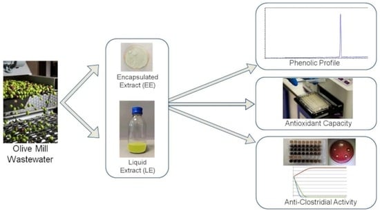
The Effects of Encapsulation on the In Vitro Anti-Clostridial Activity of Olive Mill Wastewater Polyphenolic Extracts: A Promising Strategy to Limit Microbial Growth in Food Systems

1
Department of Veterinary Medicine, University of Perugia, Via San Costanzo 4, 06126 Perugia, Italy
2
Istituto Zooprofilattico Sperimentale dell’Umbria e delle Marche “Togo Rosati”, Via Salvemini 1, 06126 Perugia, Italy
*
Authors to whom correspondence should be addressed.
These authors contributed equally to this work.
Molecules 2024, 29(7), 1441; https://doi.org/10.3390/molecules29071441
Submission received: 27 February 2024 / Revised: 18 March 2024 / Accepted: 20 March 2024 / Published: 23 March 2024

Abstract

:
Despite the technologies applied to food production, microbial contamination and chemical deterioration are still matters of great concern. In order to limit these phenomena, new natural approaches should be applied. In this context, the present study aimed to assess the antioxidant and anti-Clostridial effects of two different polyphenolic extracts derived from olive mill vegetation water, one liquid (LE) and one encapsulated (EE). The extracts have been preliminary characterized using Liquid Chromatography Quadrupole Time-Of Flight spectrometry. The Oxygen Radical Absorbance Capacity method was used to determine the antioxidant capacity, registering a higher value for EE compared to that for LE (3256 ± 85 and 2446 ± 13 µgTE/g, respectively). The antibacterial activity against C. perfringens, C. botulinum and C. difficile was studied by the agar well diffusion method, MIC and MBC determination and a time-kill test. The results confirm that EE and LE are able to limit microbial growth, albeit with minor effects when the phenolic compounds are encapsulated. Further studies are needed to evaluate the possible application of these extracts in food systems.

Graphical Abstract

1. Introduction

Food microbiological contamination is still a matter of great concern worldwide, being closely related to issues like food poisoning and consumers’ health, together with food waste, sustainability and food business cost-effectiveness [1]. Microbial pathogens are the cause of many foodborne diseases, representing a significant safety concern for public health and a pervasive food safety problem [2].
Clostridium botulinum and Clostridium perfringens are the two Clostridia most commonly associated with food borne illnesses [3]. In particular, C. perfringens can frequently cause food borne illnesses, and in 2021, C. perfringens toxins caused the highest number of cases and deaths among foodborne outbreaks involving toxigenic bacteria [4]. C. botulinum is a severe hazard in foodstuffs due to its ability to produce the most poisonous substances known, the botulinum neurotoxin, which is the causative agent of botulism [3]. On the other hand, Clostridioides difficile has changed, and some reviews describe an increase in community-associated infection not linked to the traditional risk factors (recent antibiotic therapy, older age, significant comorbidity or previous hospitalization) [5]. In consideration of this aspect, new possible routes of transmission of C. difficile have been considered, including the ingestion of contaminated food [5,6].
To counteract the undesirable effect of microbial growth, various food preservation approaches have been developed, namely thermal and chemical methods [7]. The thermal processing of foods can have, in certain conditions, depletive organoleptic and nutritional effects and may not be sufficient against spore-forming bacteria [8]. Synthetic food preservatives instead, like nitrates, benzoates, sulfites, sorbates and formaldehyde, are known for their health-threatening side effects, raising considerable concern among consumers, especially related to long-term exposure [9,10]. In this context, novel sustainable methodologies are required as an alternative to chemical and thermal preservations to inhibit the growth of pathogenic bacteria and prolong the shelf-life of food products, ensuring, therefore, food safety, while not affecting the food quality standards, and representing, at the same time, a sustainable approach [11,12]. These imperatives have led to ever-increasing scientific interest in defining novel biopreservation methods through the possible utilization of some plant extracts as effective natural food preservatives [13,14].
Several studies have shown that agro-industrial by-products are good sources of bioactive compounds with strong antimicrobial effects against food-related microorganisms [13,15,16]. Among the by-products currently under investigation, promising results have been shown by liquid wastes from olive oil production [17]. Olive mill wastewaters (OMWWs) are characterized by a high content of phenolic compounds with known antioxidant and antibacterial activities, such as hydroxytyrosol and tyrosol [17], although little information has been given regarding their efficacy towards specific food-borne microorganisms, especially those belonging to the Clostridia genus.
After appropriate purification and extraction, these by-products could represent a valuable source of these mentioned molecules with a great potential for application in food production as natural additives to increase the food quality and safety avoiding the use of chemical preservatives, but also, in some cases, as bioactive ingredients to protect consumers’ health [18]. However, these molecules are likely subjected to degradation processes promoted by environmental and processing conditions oxygen, water, light or other conditions, which limit their efficiency and reduce shelf life [19] For this reason, microencapsulation has been considered a powerful technology for the stabilization of such compounds overcoming the aforementioned issues and enabling their use in the food industry [20].
Freeze-drying encapsulation in amorphous carbohydrate microstructure matrices, such as maltodextrin, has been proved to be the most suitable method for drying delicate and thermosensitive biomaterials, minimizing the thermal degradation reactions, chemical changes such as oxidation, and protecting against undesirable physical phenomena (stickiness and collapse) [20,21]. To this end, microencapsulation can be considered a promising technology for overcoming the susceptibility of these compounds to adverse external effects or harmful food processing conditions in order to improve their miscibility and stability during shelf life [22]. A study in the literature proved the efficacy of encapsulated polyphenols in improving products’ shelf-life [22].
This type of process also enables the modification of physical properties, allowing for the transformation of liquid extracts, such as OMWW, into powders for easier handling and inclusion in certain types of food matrices [20,23]; additionally, it can be useful to mask unpleasant feelings during eating, such as a bitter taste and the astringency of polyphenols [24].
The aim of this work was to study the in vitro antioxidant and antimicrobial effects against C. perfringens, C. botulinum and C. difficile of two different polyphenolic extracts derived from OMWW, one of which was encapsulated, and the other was pure liquid. The assessment of specific antibacterial activity, as well as the evaluation of the encapsulation impact, will allow for the definition of threshold doses and most suitable formulation for the future application of food models.
The acquired results could provide the interesting possibility of the reuse of OMWW to be considered not just as a waste material, but instead as an important source of bioactive compounds with antioxidant and antibacterial effects, with a multitude of possible applications addressing the complex issue of food preservation.

2. Results

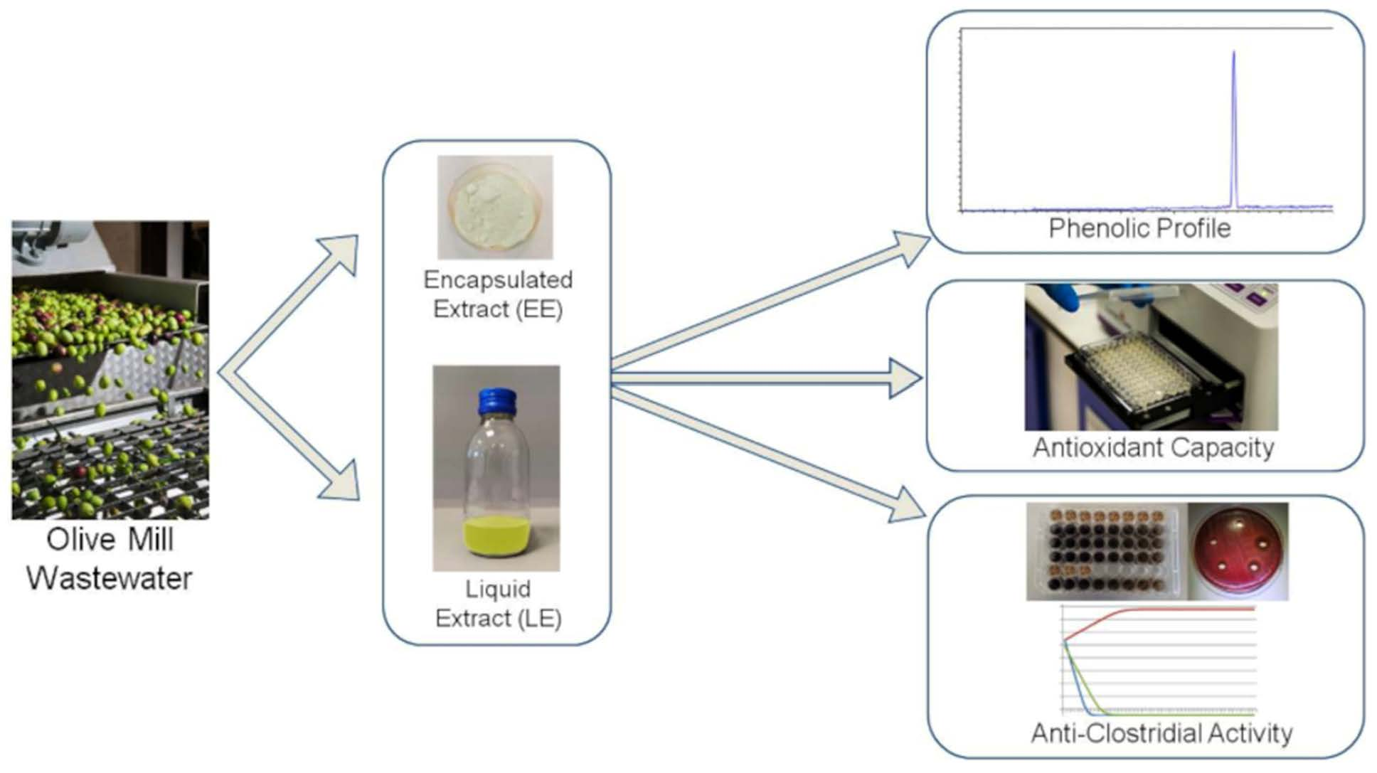
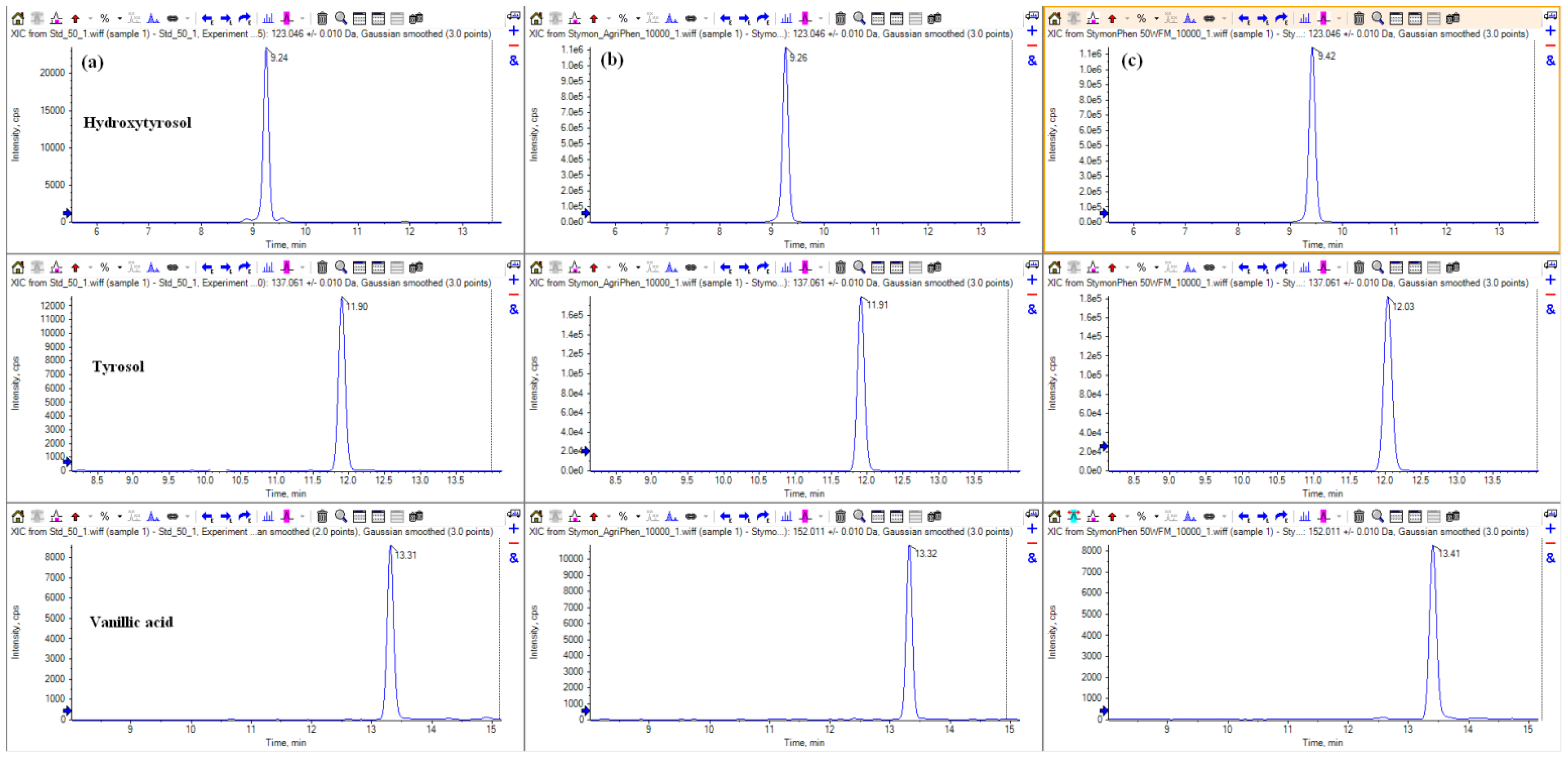
2.1. Determination of the Phenolic Profile

The specific content of major phenolic compounds in LE and EE, as well as their specific chromatograms, are reported in Table 1 and Figure 1.

2.2. Antioxidant Activity

The ORACFL results show a higher antioxidant activity level for EE compared to LE, as the first registered as 3256 ± 85 µgTE/g, and the second was 2446 ± 13 µgTE/g. The encapsulated extract has a higher antioxidant activity level than the liquid extract, most likely due to its higher phenol content.

2.3. In Vitro Evaluation of Antibacterial Activity—Agar Well Diffusion and Broth Microdilution

The in vitro antibacterial activity of EE and LE against the three selected Clostridia was preliminary assessed using an agar well diffusion method, defining the presence and diameter of inhibition zones (Table 2).
The results reported in Table 2 show that both the encapsulated and liquid polyphenolic extracts exert antibacterial activity against the Clostridia strains. In particular, the data suggest that LE has a higher antibacterial activity level at the tested concentrations, as its highest concentration tested (pure) is more powerful than the highest EE concentration tested (1 g/mL) for all the microorganisms (p < 0.05). For instance, EE 1 g/mL is comparable to LE 0.558 g/mL for C. difficile, slower than LE 0.558 g/mL for C. botulinum, and between the value for LE pure and 0.558 g/mL for C. perfringens.
Overall, the data reported in Table 2 suggests that the three Clostridia species have a slightly different sensitivity to the two extracts, with C. perfringens being the most sensitive, followed, in order, by C. difficile and C. botulinum.
Concerning the results for the broth microdilution method, EE obtained the results reported in Table 3, confirming what was previously reported for the agar well diffusion method in terms of species sensitivity. For these extracts, indeed, the three Clostridia tested show a decreasing susceptibility, in the order C. perfringens, followed by C. difficile and C. botulinum. Concerning LE, the assessment revealed the same values of MIC for all the strains, corresponding to 0.070 g/mL. Albeit, these two dilution scales are not directly comparable; 0.070 g/mL represents a smaller amount of extract with respect to 0.125 g/mL. Therefore, it can be inferred that LE has a larger antimicrobial effect on the targeted bacteria tested with the broth microdilution method. It is important to note that in all the cases, the MIC and MBC values are equivalent, suggesting that these extracts are characterized by a bactericidal nature towards Clostridia species.

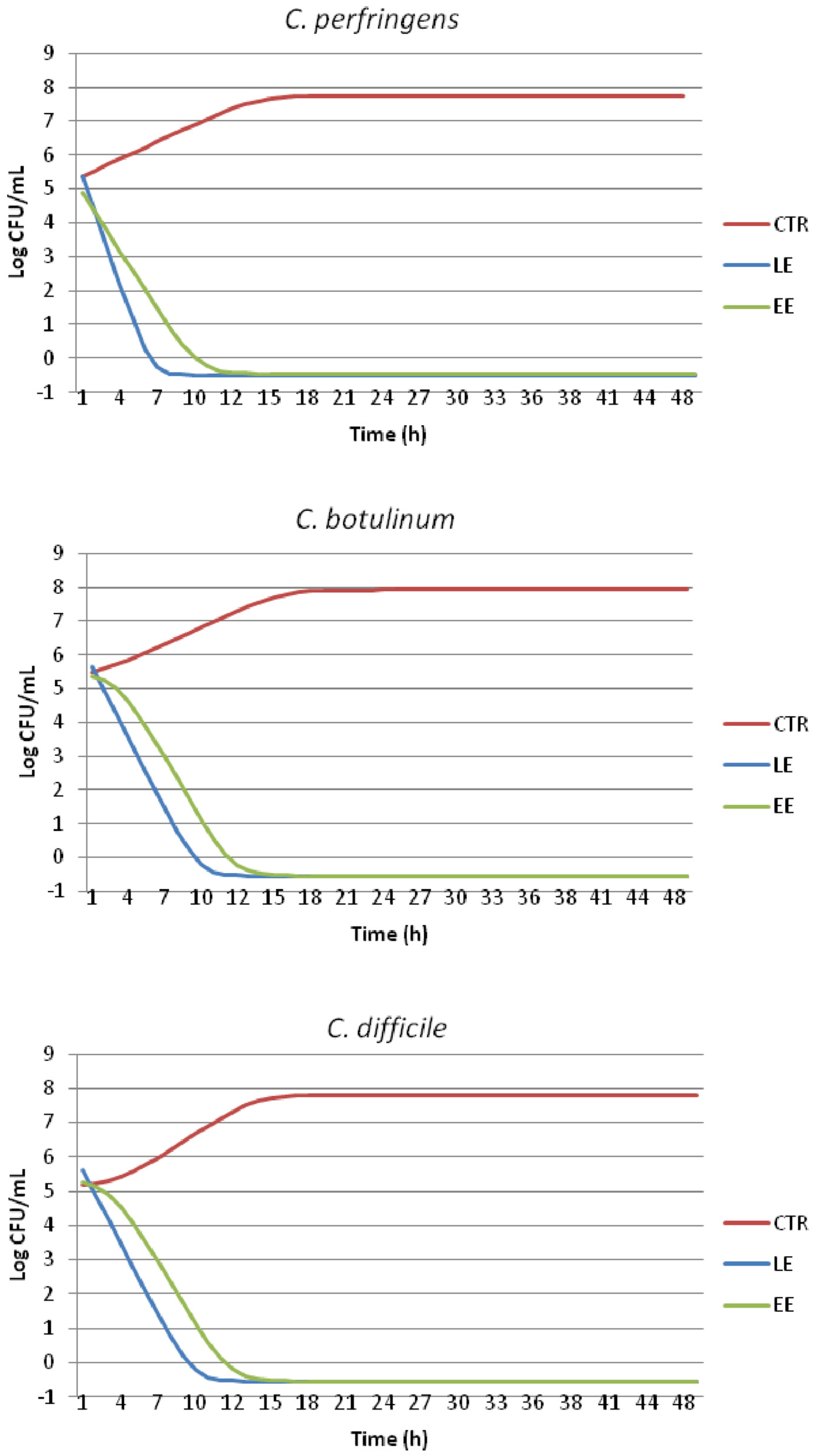
2.4. Time-Kill Test and Evaluation of Growth Dynamics

The microbial growth values (Log CFU/mL) registered at each point of the time-kill test for the targeted Clostridia under the effect of EE and LE compared to those of the CTRL are reported in Table 4. The results confirm that both the olive mill wastewater extracts (EE and LE) are able to decrease the microbial proliferation of all the bacteria starting as early as the fourth hour of testing (p < 0.05). Particularly, the liquid extract (LE) seems to possess a higher antibacterial activity level as, for all the three strains, the limit of quantification (LOQ = −0.50 Log CFU/mL) is reached at 8 h, while for the encapsulated extracts (EE), this is reached slightly later at 12 h.
Overall, from the beginning of the test (time 0) to the end (time 48), on average, the growth control (CTRL) has a ∆ Log CFU/g of 2.49, while for LE and EE, these are −5.85 and −5.84, respectively.
This outcome is also shown in Figure 2, representing the estimated growth curves of the tested bacteria cultured with the addition of EE and LE compared to that of the CTRL. The curves constructed using ComBase DMfit represent the best fitting of the raw data reported in Table 4, allowing for a modeled quantification of microbial responses through the definition of the growth kinetic parameters (Table 5).
Both Figure 2 and Table 5 confirm what was previously highlighted by the analytical data (Table 4) as for all the three bacteria tested, the growth curves of the polyphenolic liquid extract (LE) show no lag phase and drop quickly after T0, reaching the LOQ earlier compared to EE. Furthermore, the maximum growth rate is always lower for LE compared to that of EE.
The fitted curves of the CTRL show the rapid proliferation of microorganisms to a final value of almost 8 Log CFU/mL at around 20 h of testing, followed by a stationary phase.

3. Discussion

Consumers’ preference has recently changed towards minimally processed foodstuffs characterized by a low salt concentration, the absence of additives, a bland heat treatment and vacuum packaging [25]. As a consequence, a possible drawback is represented by an increased risk of food-borne diseases onset (e.g., botulism) and the quicker deterioration of food characteristics (e.g., lipid and protein oxidation), resulting in a shorter shelf-life [25,26]. In order to counteract this phenomenon, novel antimicrobial and antioxidant control measures should be applied [26].
Recently, great interest has been paid to natural antimicrobial and antioxidants in the food industry as they can be used as technological strategies to improve the quality of foodstuffs [26,27]. These molecules can potentially be applied directly to food products or by coating packaging materials to improve products’ oxidative and microbial stability, thus avoiding or reducing the use of chemical compounds [26,27]. This approach could also mitigate consumers’ perceptions of the risks related to the food chain, meeting their increasing preference towards more healthy and clean-labeled food products [9,10].
The antioxidant activity registered for the tested olive mill waste water extracts with the ORACFL method is in agreement with the previous results on similar extracts [26], confirming the powerful antioxidant activity of olive-derived phenolic compounds [26,28]. It has been observed that phenols act as hydrogen donors and that the o-dihydroxyl group possesses a high antioxidant activity level, counteracting the free radicals [29].
In the literature, the antioxidant properties of olive mill waste water extracts have also been tested when directly added into foodstuffs. A previous study shows that these types of polyphenolic extracts exert a higher antioxidant activity level, even when compared with a commercial antioxidant additive, such as ascorbic acid [26]. Particularly, the greater effect was observed for lipid oxidation, thus improving the product’s shelf life [26]. Oxidation has been demonstrated as the main non-microbial cause of food quality deterioration [30]. For instance, oxidative deterioration is capable to limit food acceptability and shortening its shelf-life by causing discoloration, the development of off-flavors and the formation of toxic compounds [28].
It has been hypothesized that this high antioxidant activity level of the olive mill waste water extracts is mainly attributable to hydroxytyrosol due to its chemical structure, which includes a phenol ring formed by a catechol group and three hydroxyl groups [29]. For instance, Martínez-Zamora et al. [31] observed the reduction in meat products’ oxidation when incorporating synthetic hydroxytyrosol to the recipe. However, the highest activity level is reached in synergy with other molecules contained in natural extracts [17,26], such as EE and LE.
Olive polyphenols, such as hydroxytyrosol, have been shown to have high levels of antioxidant activity, as have some plant extracts [32] and fruit by-products [33], making them useful in a variety of market segments, including the pharmaceutical sector and, in particular, the food industry, where they can be used to produce functional foods or as natural additives to prevent lipid oxidation and microbial alteration [34].
The in vitro anti-Clostridial effect of different phenolic compounds has already been reported in the literature. A previous study by Primavilla et al. [35] refers to the similar antibacterial activity of Crocus sativus L. extract (1 g/mL) assessed by agar well diffusion, albeit C. botulinum and C. perfringens seemed more sensitive than C. difficile. This diverse outcome is of no surprise as the different chemical composition and formulation of the extracts can lead to different results of microbial growth modulation.
Concerning the effects of similar natural compounds, Dağdelen [36] investigated the antimicrobial activities of the phenolic extracts of virgin olive oils and found that those extracts rich in hydroxytyrosol, tyrosol and vanillic acid were able to exert strong antibacterial activity against Clostridia, specifically C. perfringens.
Medina [37], who largely studied the antibacterial activity of olive-derived polyphenols, reported strong bactericidal activity against C. perfringens exerted by hydroxytyrosol and tyrosol, and both compounds are present in the two tested extracts.
The antimicrobial activity of vanillic acid has previously been reported elsewhere [38,39]; however, the data lack information on its efficacy against Clostridioides.
As mentioned above, the results concerning the broth microdilution method show the same concentration of OMWW-derived extracts for MIC and MBC in all the cases considered, suggesting, as reported elsewhere [40], that these extracts are characterized by a bactericidal nature towards some microorganisms, including Clostridia species. The MIC/MBC obtained in the present study are in agreement with some other studies testing the antimicrobial activity of olive-derived polyphenolic extracts, showing similar values, albeit for other bacteria [41]. In the literature, the anti-Clostridial activity of bioactive extracts of turmeric was assessed against C. difficile that was inhibited at concentrations ranging from 4 to 32 μg/mL [42], which are lower than those reported in the present study.
Concerning the time-kill test, in a previous study testing saffron natural extracts on the same bacterial strains, the LOQ was reached at around 24 h, reflecting different antibacterial actions most certainly due to the different chemical compositions of the extracts [35]. Manuka (Leptospermum scoparium) honey was reported as having in vitro bactericidal activity and a therapeutic role in the prevention of C. difficile infection [43], in addition to potent biofilm inhibition activity in vitro [44].
Albeit the mechanism of action of OMWW compounds has not been completely elucidated, a recent study aiming to investigate some natural products with bactericidal effects, such as cinnamon root powder and peppermint oil against C. difficile strains, revealed that cytoplasmic membrane damage was recognized as the mechanism of action mainly responsible for the activity of several natural products against bacteria, in addition to ATP (adenosine triphosphate) and protein leakage [45,46]. This finding is in line with the previous studies hypothesizing this mechanism of action of OMWW for other microorganisms [47].
The results of the present study indicate that this encapsulated extract has a slightly smaller antimicrobial effect against the Clostridia tested compared to that of the liquid one. This finding is in agreement with other studies reporting that pure extracts exert superior antimicrobial activity due to the direct release of active chemical compounds [48]. Contrarily, in some other experiments, the antibacterial activity level of natural compounds was increased after encapsulation [49].
In the literature, polyphenolic extracts were shown to have anti-Clostridial activities, preventing vegetative cell growth or spore germination [25]. This aspect has not been investigated in the present study, but the specific effect of polyphenolic compounds on spore survival and germination could be an interesting topic for future research.
The effects of encapsulation on extracts’ antimicrobial capacity, such as the decrease, increase or conservation of their bioactivity, depend on different aspects, including the interactions established between the functional groups of the encapsulated compound and the encapsulating material, as well as interaction with the matrix [1]. Therefore, albeit different polyphenolic extracts have already been experimentally added to foodstuffs [50,51], it is crucial to assess the applicability of food-grade OMWW polyphenolic extracts, both encapsulated and pure, in specific complex food systems, aiming to provide them with improved characteristics. Consumers’ attitudes, perceptions and behaviors towards these products have changed in recent years, and their preferences are shifting towards “natural” compounds for a healthier lifestyle. These attitudes emphasize the responsibility of industries and national authorities to provide a safer product, albeit, currently, there are no specific regulations or legal guidelines on the use of such products. Nevertheless, bioactive compounds must be assessed for any hazard to human health and meet legal requirements by force before entering the market [52].
It is also worth noting that recovering olive phenols from wastewater can make a significant contribution to environmental protection, as their dispersion in soil poses an environmental threat, while the water after phenolic extraction is suitable for irrigation. For this reason, the purification and extraction of biomolecules make it very appealing to use OMWW as a starting material for a large variety of applications, including food production [27].

4. Materials and Methods

4.1. Test Materials

OMWW extracts enriched in polyphenols were provided by Stymon Natural Products P.C., Patras, Greece (www.stymon.com, accessed on 20 November 2023). This product derives from OMWW (Olea europaea L., Koroneiki cultivar). The extracts tested were formulated in two different products, liquid (not encapsulated) and powder encapsulated extract (EE). EE was treated with hydrolytic enzymes, filtered by a membrane process, and encapsulated with a food-grade maltodextrin carrier (1:1 dw), followed by lyophilization (freeze-drying technique at −55 °C, 0.1 mbar) and a grinding process. The above two products were produced via a patented process (GR1010150, EP4049543A1) using green technologies. The extract was chosen by the authors because it is food-grade (certificate released by General Chemical State Laboratory of Greece n. 30/003/000/3810), and the treatment was carried out using only water without the addition of any chemicals.
The concentrations of both polyphenol extracts were measured by Liquid Chromatography Quadrupole Time-Of Flight spectrometry (LC-QTOF-LC-TripleTOF 6600, Sciex, Framingham, MA, USA). Acetonitrile (ACN) and methanol were LC-MS-grade and supplied by Carlo Erba Reagents (Milan, Italy). Dimethyl sulfoxide (DMSO) was provided by VWR International (Radnor, PA, USA). Butylated hydroxytoluene (BHT) was purchased from Alfa Aesar (Haverhill, MA, USA), and acetic acid (99%) was bought from Merck (Darmstadt, Germany). Deionized water was produced using a Milli-Q purification system (Millipore, Molsheim, France). Hydroxytyrosol and verbascoside were purchased from Extrasynthese (Genay, France). Oleuropein aglycone was supplied by TRC (Toronto, ON, Canada). Tyrosol, vanillic acid, vanillin, p-coumaric acid, oleuropein, pinoresinol, luteolin and apigenin were obtained from Merck. Individual standard stock solutions (1 mg mL−1) were prepared in methanol, except apigenin, which was prepared in methanol/DMSO 70/30 (v/v). Stock solutions were stored in the freezer (−20 °C) for six months. By mixing the appropriate amount of individual stock solutions, a working standard mixture at 10 µg mL−1 was obtained in methanol. From the latter, the calibration curve (25, 50, 100, 250 and 500 ng mL−1) was measured by diluting it with a mixture of acetic acid 0.025%/methanol (90/10, v/v).
The encapsulated polyphenolic extract (EE) was analyzed starting from 1 g (1.0 g ± 0.1 g) of sample, which was mixed with 5 mL of a methanol/water 80/20 (v/v) solution containing 20 mg/L of BHT. The extraction was repeated a second time, and after centrifugation, three aliquots of the extract were diluted 1000-, 2500- and 5000-fold, respectively, with a mixture of acetic acid 0.025%/methanol 90/10 (v/v). Then, each diluted aliquot was injected into the instrumental platform.
For liquid extract (LE) analysis, 0.1 g (1.0 g ± 0.1 g) of the sample was solubilized with 10 mL of methanol/water 80/20 (v/v) solution containing 20 mg/L of BHT. Three aliquots were then diluted 1000-, 2500- and 5000-fold, respectively, with a mixture of acetic acid 0.025%/methanol 90/10 (v/v) and injected.
Instrumental analysis was performed by Liquid Chromatography Quadrupole Time-Of Flight spectrometry (LC-QTOF), determining eleven polyphenols. The platform consisted of an Exion LC™ coupled to a 6600+ TripleTOF™ (AB Sciex, Foster, CA, USA) equipped with an electrospray ionization source operating in negative mode (ESI-). Chromatographic separation was carried out using an Acquity BEH C18 (150 mm × 2.1 mm, 1.7 µm, Waters, Milford, MA, USA). Water with 0.025% acetic acid (A) and methanol/ACN 90/10 v/v % (B) were the mobile phases. The gradient started with 0% of B (1 min); then, the percentage of B was increased to 20% after 10 min, followed by an increase to 50% B after 4 min and another one to 100% after 3 min. After 5 min, the percentage of B was restored to the initial conditions (0%) in 1 min. Finally, the system was re-equilibrated for 6 min (run time: 30 min). The column temperature was set at 40 °C, and the autosampler temperature was kept at 25 °C. Flow rate and injection volume were 0.25 mL/min and 10 µL, respectively. Compressed air was used as GS1 (55 psi) and GS2 (55 psi), whereas nitrogen was the curtain gas (40 psi). The spray voltage was set at −4.5 kV, and the interface source temperature was set at 450 °C. Single infusions of each analyte were carried out to optimize the declustering potential and collision energy. The precursor ([M − H+]) and fragment ions acquired in MRMHR (multiple reaction-monitoring high resolution) mode are listed in Table 6. Mass error was <5 ppm.

4.2. Antioxidant Capacity Determination

The antioxidant capacity of polyphenolic extracts was measured using the Oxygen Radical Absorbance Capacity method (ORACFL). This test is based on the decay rate of the fluorescence probe due to radical oxygen species (ROO) compared to the reference standard, Trolox (6-hydroxy-2,5,7,8-tetramethylchroman-2-carboxylic acid, Sigma-Aldrich, Steinheim, Germany). One mL of LE or 1 g of EE was mixed with a buffer, 75 mM, at pH 7.2, containing 13.19 g of K2HPO4 and 10.26 g of KH2PO4 in 900 mL of deionized water. The obtained mixture was homogenized for 1 min with an Ultra-Turrax homogenizer (Ultra Turrax T25 Basic, IKA Labortechnik Janke & Kunkel GmbH, Stavfen, Germany), and then vortexed for 2 min. After centrifugation at 6000 rpm for 20 min at +4 °C, the supernatant was used for antioxidant capacity determination. ORACFL assays were executed on an FLUO-star OPTIMA microplate fluorescence reader (BMGLABTECH, Offenburg, Germany) at excitation and emission wavelengths of 485 and 520 nm, respectively. The results of the test are expressed as µg of Trolox equivalents (TE) per g of sample.

4.3. In Vitro Evaluation of Antibacterial Activity—Agar Well Diffusion

The agar well diffusion method was used to screen the antibacterial activities of the extracts against Clostridium perfringens, Clostridium botulinum and Clostridioides difficile [53]. For this test, EE was suspended, and then diluted as needed in demineralized sterile water, while LE was used pure or diluted with demineralized sterile water when necessary.
C. perfringens and C. difficile strains were derived from the Istituto Zooprofilattico Sperimentale dell’Umbria e delle Marche “Togo Rosati” food isolates collection, while C. botulinum was provided by the Italian National Institute of Health (National Referral Center for Botulism).
As previously described [35], 100 µL of bacteria suspensions (0.5 McFarland in 0.9% sterile saline solution) was spread using a swab on Mueller Hinton agar 5% defibrinated sheep blood plates (MHAB—Oxoid Limited, Basingstoke, UK). Two-fold serial dilutions were prepared for each extract (EE: from 1 g/mL to 0.125 g/mL; LE: from pure to 0.140 g/mL), and 50 µL of each of them was inoculated in 7 mm holes previously made by scooping out the medium with a sterilized cork borer. Negative (sterile demineralized water) and positive controls (Penicillin G 10UI discs—Oxoid Limited, Basingstoke, UK) were set up. Plates were incubated as indicated in Table 7, and after incubation, the diameters of the inhibition halos were measured with a gauge and expressed in mm. The experiments were repeated thrice; mean values and standard deviations of the diameters were calculated.

4.4. MIC/MBC Assay

The evaluation of the minimal inhibitory concentration (MIC) and of the minimal bactericidal concentration (MBC) of the extracts was performed in triplicate using a standard broth microdilution technique, as described in the Clinical Laboratory Standards Institute (CLSI) guidelines [54]. Briefly, a decreasing concentration of each extract (ranging from 0.5 g/mL to 0.004 g/mL for EE and from 0.558 g/mL to 0.004 g/mL for LE) was added to an equal volume of bacterial suspensions of 105 colony forming unit/ mL (CFU/mL) in Mueller Hinton broth with 5% blood (Biolife Italiana s.r.l., Milan, Italy) in a 96-well microplate (Starlab International GmbH, Hamburg, Germany). In order to check the experiment, three different control were set up as follows: antibiotic control (bacterial suspension and decreasing concentration of benzylpenicillin sodium salt; Sigma-Aldrich, St. Louis, MO, USA), organism control (bacterial suspension) and negative control (extract solution at the same decreasing concentrations tested). The microplates were incubated for 48 h at 37 °C under anaerobic conditions, and the MIC was determined by evaluating the lowest concentration with no visible bacterial growth [54], while the MBC, was figured out by subculturing 10 μL of wells corresponding to the MIC and to higher MIC concentrations onto 5% Sheep Blood agar dishes (Microbiol s.r.l., Cagliari, Italy). The sheep blood plates were then incubated at 37 °C for 48 h [40]. The MIC and MBC values are expressed as means ± standard deviations.

4.5. Time-Kill Test and Evaluation of Growth Dynamics

As described in the Clinical & Laboratory Standards Institute (CLSI) guidelines (document M26-A) [54], three broth cultures were set up (105 CFU/mL in Mueller Hinton broth with 5% blood; Biolife Italiana s.r.l., Milan, Italy). The first two broth cultures were respectively added to the EE and LE extracts (1 × MIC), while the third one was used as a growth control (CTRL). The broth cultures were incubated at 37 °C under anaerobic conditions using anaerobic jars (2.5 L AnaeroJar, AG002, together with AnaeroGen 2.5 L, AN0025—Oxoid Limited, Basingstoke, UK). At regular time intervals (0, 4, 8, 12, 24 and 48 h), the count of viable cells was performed. The results are expressed as Log CFU/mL, and in case of no bacterial growth, at the 1.00 Log CFU/mL level (<10 CFU/mL), a value of −0.50 Log CFU/mL was assigned [40]. The experiments were repeated thrice, and standard deviations were determined.
C. perfringens, C. botulinum and C. difficile growth curves and their kinetic parameters (initial value, lag phase duration, maximum growth rate and final value) were defined using the DMFit tool of ComBase’s, free predictive microbiology software (https://www.combase.cc/index.php/en/DMFit—2021 version, accessed on 11 October 2023), by fitting the experimental data to Baranyi and Roberts’ model [55].

4.6. Statistical Analysis

ANOVA (analysis of variance) was performed with the data obtained from the agar well diffusion and the time kill-test using SAS’s generalized linear model (GLM) approach (SAS Institute Inc., Cary, NC, USA, 2001) [56]. For the first experiment, a mixed model with treatments (EE, LE, and CTRL) and microorganisms as fixed factors were utilized, and for the second test, the same model was employed, with treatments (EE, LE, and CTRL) and time (T0, T4, T8, T12, T24 and T48) as fixed factors. Duplicate effects were determined to be insignificant and were deleted from the model. Tukey’s post hoc analysis was used to explain significant mean differences (p < 0.05).
The effects of extracts’ formulation on the growth curve parameters were assessed using the one-way ANOVA model with treatment (EE, LE, and CTRL) as a fixed factor, and Tukey’s test (p < 0.05) was used to determine significant differences among the results.

5. Conclusions

To ensure microbiological safety and improve the quality of foodstuffs in accordance with consumers’ preferences for “clean label” products, food corporations are looking for new strategies to replace the traditional preservatives. Polyphenolic compounds are among the most well-liked candidates for natural food additives, and the application of encapsulation techniques could address the stability, solubility and bioavailability issues. The results demonstrate a promising antioxidant capacity and anti-Clostridial effect for both the tested polyphenolic extracts derived from olive mill vegetation water, especially for the liquid one. Further investigations are needed to evaluate and refine the possible applications of these extracts in complex food systems in order to provide consumers with safer, healthier and greener products. Additional studies should focus on the evaluation of the conformity and applicability of food-grade encapsulated polyphenolic extracts in the context of real food processes and products, as well as clarify their molecular mechanisms of action.

6. Patents

Stymon Natural Products P.C., Patras, Greece (www.stymon.com, accessed on 20 November 2023): patented process (GR1010150, EP4049543A1) and food-grade certificate (certificate released by General Chemical State Laboratory of Greece n. 30/003/000/3810).

Author Contributions

Conceptualization, R.R., S.P. and D.R.; methodology, R.R., S.P. and R.G.; validation, R.G. and A.V.; formal analysis, R.R. and F.P.; investigation, R.R., S.P. and F.P.; resources, R.B. and A.V.; data curation, R.R., S.P., D.R. and C.A.; writing (original draft preparation), R.R. and S.P.; writing (review and editing), R.R., S.P., D.R. and R.B.; visualization, S.P. and R.B.; supervision, R.B.; project administration, R.B. and A.V. All authors have read and agreed to the published version of the manuscript.

Funding

This research received no external funding.

Institutional Review Board Statement

Not applicable.

Informed Consent Statement

Not applicable.

Data Availability Statement

The data are contained within this article.

Acknowledgments

The authors thank Domenico Pettas and Dimitrios Alfredos Parthenopoulos for technical assistance and for providing the olive polyphenolic extracts (STYMON Natural Products IKE, Patras, Greece).

Conflicts of Interest

The authors declare no conflicts of interest.

References

  1. Pisoschi, A.M.; Pop, A.; Cimpeanu, C.; Turcuş, V.; Predoi, G.; Iordache, F. Nanoencapsulation techniques for compounds and products with antioxidant and antimicrobial activity-A critical view. Eur. J. Med. Chem. 2018, 157, 1326–1345. [Google Scholar] [CrossRef]
  2. Bintsis, T. Foodborne pathogens. AIMS Microbiol. 2017, 3, 529. [Google Scholar] [CrossRef]
  3. Stringer, S.C.; Peck, M.W. Foodborne clostridia and the safety of in-pack preserved foods. In Woodhead Publishing Series in Food Science, Technology and Nutrition, In-Pack Processed Foods; Richardson, P., Ed.; Woodhead Publishing: Shaston, UK, 2008; pp. 251–276. ISBN 9781845692469. [Google Scholar]
  4. European Food Safety Authority and European Centre for Disease Prevention and Control. The European Union One Health 2022 Zoonoses Report. EFSA J. 2023, 21, e8442. [Google Scholar]
  5. Candel-Pérez, C.; Ros-Berruezo, G.; Martínez-Graciá, C. A review of Clostridioides [Clostridium] difficile occurrence through the food chain. Food Microbiol. 2019, 77, 118–129. [Google Scholar] [CrossRef]
  6. Primavilla, S.; Farneti, S.; Petruzzelli, A.; Drigo, I.; Scuota, S. Contamination of hospital food with Clostridium difficile in Central Italy. Anaerobe 2019, 55, 8–10. [Google Scholar] [CrossRef]
  7. Singh, S.; Shalini, R. Effect of hurdle technology in food preservation: A review. Crit. Rev. Food Sci. Nutr. 2016, 56, 641–649. [Google Scholar] [CrossRef] [PubMed]
  8. Milani, E.; Silva, F.V. Comparing high pressure thermal processing and thermosonication with thermal processing for the inactivation of bacteria, moulds, and yeasts spores in foods. J. Food Eng. 2017, 214, 90–96. [Google Scholar]
  9. Etienne, J.; Chirico, S.; McEntaggart, K.; Papoutsis, S.; Millstone, E. EU Insights–Consumer perceptions of emerging risks in the food chain. EFSA Support. Publ. 2018, 15, 1394. [Google Scholar]
  10. Lucera, A.; Costa, C.; Conte, M.; Del Nobile, A. Food applications of natural antimicrobial compounds Front. Microbiol. 2012, 3, 287. [Google Scholar]
  11. Batiha, G.E.S.; Hussein, D.E.; Algammal, A.M.; George, T.T.; Jeandet, P.; Al-Snafi, A.E.; Tiwari, A.; Pagnossa, J.P.; Lima, C.M.; Thorat, N.D.; et al. Application of natural antimicrobials in food preservation: Recent views. Food Control 2021, 126, 108066. [Google Scholar] [CrossRef]
  12. Gonelimali, F.D.; Lin, J.; Miao, W.; Xuan, J.; Charles, F.; Chen, M.; Hatab, S. Antimicrobial properties and mechanism of action of some plant extracts against food pathogens and spoilage microorganisms. Front. Microbiol. 2018, 9, 1639. [Google Scholar] [CrossRef]
  13. Oulahal, N.; Degraeve, P. Phenolic-rich plant extracts with antimicrobial activity: An alternative to food preservatives and biocides? Front. Microbiol. 2022, 12, 753518. [Google Scholar] [CrossRef]
  14. Sanz-Puig, M.; Moreno, P.; Pina-Pérez, M.C.; Rodrigo, D.; Martínez, A. Combined effect of high hydrostatic pressure (HHP) and antimicrobial from agro-industrial by-products against S. Typhimurium. LWT 2017, 77, 126–133. [Google Scholar] [CrossRef]
  15. Mattos, G.N.; Tonon, R.V.; Furtado, A.A.; Cabral, L.M. Grape by-product extracts against microbial proliferation and lipid oxidation: A review. J. Sci. Food Agric. 2017, 97, 1055–1064. [Google Scholar] [CrossRef]
  16. Ibáñez-Peinado, D.; Pina-Pérez, C.; García-Carrión, G.; Martínez, A.; Rodrigo., D. In vivo antimicrobial activity assessment of a cauliflower by-product extract against Salmonella typhimurium. Front. Sustain. Food Syst. 2020, 4, 8. [Google Scholar] [CrossRef]
  17. Roila, R.; Valiani, A.; Ranucci, D.; Ortenzi, R.; Servili, M.; Veneziani, G.; Branciari, R. Antimicrobial efficacy of a polyphenolic extract from olive oil by-product against “Fior di latte” cheese spoilage bacteria. Int. J. Food Microbiol. 2019, 295, 49–53. [Google Scholar] [CrossRef]
  18. Robert, P.; Gorena, T.; Romero, N.; Sepulveda, E.; Chavez, J.; Saenz, C. Encapsulation of polyphenols and anthocyanins from pomegranate (Punica granatum) by spray drying. Int. J. Food Sci. Technol. 2010, 45, 1386–1394. [Google Scholar] [CrossRef]
  19. Peanparkdee, M.; Patrawart, J.; Iwamoto, S. Physicochemical stability and in vitro bioaccessibility of phenolic compounds and anthocyanins from Thai rice bran extracts. Food Chem. 2020, 329, 127157. [Google Scholar] [CrossRef] [PubMed]
  20. Fang, Z.; Bhandari, B. Encapsulation of polyphenols—A review. Trends Food Sci. Technol. 2010, 21, 510–523. [Google Scholar] [CrossRef]
  21. Sanchez, V.; Baeza, R.; Galmarini, M.V.; Zamora, M.C.; Chirife, J. Freeze-drying encapsulation of red wine polyphenols in an amorphous matrix of maltodextrin. Food Biopr. Technol. 2013, 6, 1350–1354. [Google Scholar] [CrossRef]
  22. Ianni, F.; Gagliardi, A.; Taticchi, A.; Servili, M.; Pinna, N.; Schoubben, A.; Sardella, R.; Bruscoli, S. Exploiting food-grade mesoporous silica to preserve the antioxidant properties of fresh olive mill wastewaters phenolic extracts. Antioxidants 2021, 10, 1361. [Google Scholar] [CrossRef] [PubMed]
  23. Gharsallaoui, A.; Roudaut, G.; Chambin, O.; Voilley, A.; Saurel, R. Applications of spray-drying in microencapsulation of food ingredients: An overview. Food Res. Int. 2007, 40, 1107–1121. [Google Scholar] [CrossRef]
  24. Nedovic, V.; Kalusevic, A.; Manojlovic, V.; Levic, S.; Bugarski, B. An overview of encapsulation technologies for food applications. Procedia Food Sci. 2011, 1, 1806–1815. [Google Scholar] [CrossRef]
  25. Cui, H.; Gabriel, A.A.; Nakano, H. Antimicrobial efficacies of plant extracts and sodium nitrite against Clostridium botulinum. Food Control 2010, 21, 1030–1036. [Google Scholar] [CrossRef]
  26. Roila, R.; Sordini, B.; Esposto, S.; Ranucci, D.; Primavilla, S.; Valiani, A.; Taticchi, A.; Branciari, R.; Servili, M. Effect of the Application of a Green Preservative Strategy on Minced Meat Products: Antimicrobial Efficacy of Olive Mill Wastewater Polyphenolic Extract in Improving Beef Burger Shelf-Life. Foods 2022, 11, 2447. [Google Scholar] [CrossRef]
  27. Lourenço, S.C.; Moldão-Martins, M.; Alves, V.D. Antioxidants of natural plant origins: From sources to food industry applications. Molecules 2019, 24, 4132. [Google Scholar] [CrossRef] [PubMed]
  28. Liu, T.; Liu, L.; Gong, X.; Chi, F.; Ma, Z. Fabrication and comparison of active films from chitosan incorporating different spice extracts for shelf life extension of refrigerated pork. LWT 2021, 135, 110181. [Google Scholar] [CrossRef]
  29. Servili, M.; Sordini, B.; Esposto, S.; Urbani, S.; Veneziani, G.; Maio, I.D.; Selvaggini, R.; Taticchi, A. Biological activities of phenolic compounds of extra virgin olive oil. Antioxidants 2013, 3, 1–23. [Google Scholar] [CrossRef]
  30. Insani, E.M.; Eyherabide, A.; Grigioni, G.; Sancho, A.M.; Pensel, N.A.; Descalzo, A.M. Oxidative stability and its relationship with natural antioxidants during refrigerated retail display of beef produced in Argentina. Meat Sci. 2008, 79, 444–452. [Google Scholar] [CrossRef]
  31. Martínez-Zamora, L.; Ros, G.; Nieto, G. Synthetic vs. Natural Hydroxytyrosol for Clean Label Lamb Burgers. Antioxidants 2020, 9, 851. [Google Scholar] [CrossRef]
  32. Dudonne, S.; Vitrac, X.; Coutiere, P.; Woillez, M.; Mérillon, J.M. Comparative study of antioxidant properties and total phenolic content of 30 plant extracts of industrial interest using DPPH, ABTS, FRAP, SOD, and ORAC assays. J. Agric. Food Chem. 2009, 57, 1768–1774. [Google Scholar] [CrossRef]
  33. Pinna, N.; Ianni, F.; Selvaggini, R.; Urbani, S.; Codini, M.; Grispoldi, L.; Cenci-Goga, B.T.; Cossignani, L.; Blasi, F. Valorization of Pumpkin Byproducts: Antioxidant Activity and Carotenoid Characterization of Extracts from Peel and Filaments. Foods 2023, 12, 4035. [Google Scholar] [CrossRef] [PubMed]
  34. Ciriminna, R.; Meneguzzo, F.; Fidalgo, A.; Ilharco, L.M.; Pagliaro, M. Extraction, benefits and valorization of olive polyphenols. Eur. J. Lipid Sci. Technol. 2016, 118, 503–511. [Google Scholar] [CrossRef]
  35. Primavilla, S.; Pagano, C.; Roila, R.; Branciari, R.; Ranucci, D.; Valiani, A.; Ricci, M.; Perioli, L. Antibacterial Activity of Crocus sativus L. Petals Extracts against Foodborne Pathogenic and Spoilage Microorganisms, with a Special Focus on Clostridia. Life 2022, 13, 60. [Google Scholar] [CrossRef] [PubMed]
  36. Dağdelen, A. Identifying antioxidant and antimicrobial activities of the phenolic extracts and mineral contents of virgin olive oils (Olea europaea L. cv. Edincik Su) from different regions in Turkey. J. Chem. 2016, 9589763. [Google Scholar]
  37. Medina, E.; de Castro, A.; Romero, C.; Ramírez, E.; Brenes, M. Effect of antimicrobial compounds from olive products on microorganisms related to health, food and agriculture. Microbial Pathogens and Strategies for Combating Them. Sci. Technol. Edu. 2013, 2, 1087–1094. [Google Scholar]
  38. Ibrahim, H.A.; Soliman, H.S.; Hamed, F.M.; Marrez, D.A.; Othman, S.M. Antibacterial activity of vanillic acid and catechol produced by microbial biotransformation of caffeic acid. J. Pharm. Sci. Res. 2020, 12, 740–743. [Google Scholar]
  39. Qian, W.; Yang, M.; Wang, T.; Sun, Z.; Liu, M.; Zhang, J.; Zeng, Q.; Li, Y. Antibacterial mechanism of vanillic acid on physiological, morphological, and biofilm properties of carbapenem-resistant Enterobacter hormaechei. J. Food Prot. 2020, 83, 576–583. [Google Scholar] [CrossRef]
  40. Roila, R.; Branciari, R.; Ranucci, D.; Ortenzi, R.; Urbani, S.; Servili, M.; Valiani, A. Antimicrobial activity of olive mill wastewater extract against Pseudomonas fluorescens isolated from mozzarella cheese. Ital. J. Food Saf. 2016, 5, 111–115. [Google Scholar] [CrossRef]
  41. Fasolato, L.; Cardazzo, B.; Balzan, S.; Carraro, L.; Taticchi, A.; Montemurro, F.; Novelli, E. Minimum bactericidal concentration of phenols extracted from oil vegetation water on spoilers, starters and food-borne bacteria. Ital. J. Food Saf. 2015, 4, 4519. [Google Scholar] [CrossRef]
  42. Mody, D.; Athamneh, A.I.; Seleem, M.N. Curcumin: A natural derivative with antibacterial activity against Clostridium difficile. J. G. Antimicrob. Resist. 2020, 21, 154–161. [Google Scholar] [CrossRef] [PubMed]
  43. Georgescu, M.; Ginghina, O.; Raita, S.; Tapaloaga, D.; Ilie, L.; Negrei, C.; Popa, D.E.; Varlas, V.; Multescu, R.; Rosca, A.C.; et al. Natural alternative remedies in the background of updated recommendations for the prophylactic and therapeutic approach of clostridium difficile infections. Farmacia 2018, 66, 563–572. [Google Scholar] [CrossRef]
  44. Piotrowski, M.; Karpiński, P.; Pituch, H.; Van Belkum, A.; Obuch-Woszczatyński, P. Antimicrobial effects of Manuka honey on in vitro biofilm formation by Clostridium difficile. Eur. J. Clin. Microbiol. Infect. Dis. 2017, 36, 1661–1664. [Google Scholar] [CrossRef] [PubMed]
  45. Roshan, N.; Riley, T.V.; Knight, D.R.; Steer, J.H.; Hammer, K.A. Natural products show diverse mechanisms of action against Clostridium difficile. J. Appl. Microbiol. 2019, 126, 468–479. [Google Scholar] [CrossRef] [PubMed]
  46. Balta, I.; Linton, M.; Pinkerton, L.; Kelly, C.; Stef, L.; Pet, I.; Stef, D.; Criste, A.; Gundogdu, O.; Corcionivoschi, N. The effect of natural antimicrobials against Campylobacter spp. and its similarities to Salmonella spp., Listeria spp., Escherichia coli, Vibrio spp., Clostridium spp. and Staphylococcus spp. Food Control 2021, 121, 107745. [Google Scholar] [CrossRef]
  47. Obied, H.K.; BedgoodJr, D.R.; Prenzler, P.D.; Robards, K. Bioscreening of Australian olive mill waste extracts: Biophenol content, antioxidant, antimicrobial and molluscicidal activities. Food Chem. Toxicol. 2007, 45, 1238–1248. [Google Scholar] [CrossRef] [PubMed]
  48. dos Santos Alves, M.J.; de Sousa, M.H.O.; de Moura, N.F.; Cesca, K.; Verruck, S.; Monteiro, A.R.; Valencia, G.A. Starch nanoparticles containing phenolic compounds from green propolis: Characterization and evaluation of antioxidant, antimicrobial and digestibility properties. Int. J. Biol. Macromol. 2023, 255, 128079. [Google Scholar] [CrossRef]
  49. Salehi, A.; Rezaei, A.; Damavandi, M.S.; Kharazmi, M.S.; Jafari, S.M. Almond gum-sodium caseinate complexes for loading propolis extract: Characterization, antibacterial activity, release, and in-vitro cytotoxicity. Food Chem. 2023, 405, 134801. [Google Scholar] [CrossRef]
  50. Rocchetti, G.; Gregorio, R.P.; Lorenzo, J.M.; Barba, F.J.; Oliveira, P.G.; Prieto, M.A.; Simal-Gandara, J.; Mosele, M.J.; Lucini, L. Functional implications of bound phenolic compounds and phenolics–food interaction: A review. Compr. Rev. Food Sci. Food Saf. 2022, 21, 811–842. [Google Scholar] [CrossRef]
  51. Elsebaie, E.M.; Essa, R.Y. Microencapsulation of red onion peel polyphenols fractions by freeze drying technicality and its application in cake. J. Food Process. Preserv. 2018, 42, e13654. [Google Scholar] [CrossRef]
  52. Vilas-Boas, A.A.; Pintado, M.; Oliveira, A.L. Natural bioactive compounds from food waste: Toxicity and safety concerns. Foods 2021, 10, 1564. [Google Scholar] [CrossRef] [PubMed]
  53. Balouiri, M.; Sadiki, M.; Ibnsouda, S.K. Methods for in vitro evaluating antimicrobial activity: A review. J. Pharm. Anal. 2016, 6, 71–79. [Google Scholar] [CrossRef] [PubMed]
  54. Clinical and Laboratory Standards Institute. Methods for Determining Bactericidal Activity of Antimicrobial Agents; Approved Guideline; CLSI Document M26-A; CLSI: Wayne, PA, USA, 1998. [Google Scholar]
  55. Baranyi, J.; Roberts, T.A. A dynamic approach to predicting bacterial growth in food. Int. J. Food Microbiol. 1994, 23, 277–294. [Google Scholar] [CrossRef] [PubMed]
  56. SAS. Statistical Software and User’s Guide, Version 8.2; SAS Inst. Inc.: Cary, NC, USA, 2001. [Google Scholar]
Figure 1. Extract ion chromatograms of the three most abundant polyphenols: (a) standard solution at 50 ng/mL; (b) liquid extract (10,000-fold dilution); (c) encapsulated extract (10,000-fold dilution).
Molecules 29 01441 g001
Figure 2. Modeled growth curves of C. perfringens, C. botulinum and C. difficile cultured with encapsulated polyphenolic extract (EE) and the liquid polyphenolic extract (LE) compared to that of CTRL samples.
Molecules 29 01441 g002
Table 1. Phenolic contents of the liquid and encapsulated extract (mg/g).
Phenolic CompoundsPolyphenol Sum
HydroxytyrosolTyrosolVanillic Acid
EE14.71 ± 0.273.95 ± 0.170.26 ± 0.0118.9
LE8.58 ± 0.192.13 ± 0.050.21 ± 0.1010.9
Table 2. Diameters of inhibition halos (mm) obtained when testing the encapsulated polyphenolic extract (EE), the polyphenolic liquid extract (LE) and the Penicillin G-positive control (PC) for the three targeted Clostridia. Values are expressed as means of three determination ± standard deviation.
EE (g/mL)LE (g/mL)PC
10.50.250.125Pure0.5580.2790.140
C. difficile10.94 ± 0.10 bB8.7 ± 0.45 aA--18.58 ± 0.38 bC11.38 ± 0.42 bB7.88 ± 0.12 aA-31.44 ± 1.12 aD
C. botulinum8.1 ± 0.20 aA---17.51 ± 0.27 aC10.66 ± 0.47 aB--32.24 ± 0.98 bD
C. perfringens17.01 ± 0.37 cD14.37 ± 0.49 bC12.19 ± 0.33 B-18.99 ± 0.06 bE13.85 ± 0.29 cC8.92 ± 0.39 bA-32.91 ± 0.70 bF
Different letters in the same row (A,B,C,D,E,F) indicate differences between mean values of halos diameter for the two extracts and the control group (p < 0.05); different letters in the same column (a,b,c) indicate differences between mean values of halos diameter for different micro-organisms within the same concentration of extract (p < 0.05); positive control = Penicillin G disc.
Table 3. Minimum inhibitory concentration (MIC) and minimum bactericidal concentration (MBC) of encapsulated polyphenolic extract (EE), liquid polyphenolic extract (LE) and of the Benzylpenicillin positive control (PC) against C. perfringens, C. botulinum and C. difficile.
EE (g/mL)LE (g/mL)PC (µg/mL)
MICMBCMICMBCMICMBC
C. difficile0.2500.2500.0700.0706 × 10−86 × 10−8
C. botulinum0.5000.5000.0700.0705 × 10−75 × 10−7
C. perfringens0.1250.1250.0700.0701 × 10−61 × 10−6
Table 4. Results of the time-kill test for the targeted microorganisms. Values are expressed as means of three determinations ± standard deviations (Log CFU/mL).
Time (h)TreatmentC. perfringensC. botulinumC. difficile
0CTRL5.43 ± 0.03 A5.52 ± 0.01 A5.21 ± 0.04 A
LE5.36 ± 0.02 C5.36 ± 0.04 C5.32 ± 0.04 C
EE5.36 ± 0.04 D5.38 ± 0.02 D5.28 ± 0.05 D
4CTRL5.41 ± 0.05 cA5.87 ± 0.03 cB5.46 ± 0.12 cB
LE1.00 ± 0.00 aB3.32 ± 0.00 aB3.30 ± 0.06 aB
EE1.49 ± 0.20 bC3.95 ± 0.17 bC3.92 ± 0.06 bC
8CTRL6.95 ± 0.01 cB6.95 ± 0.01 cC6.76 ± 0.03 cC
LE−0.50 ± 0.00 aA−0.50 ± 0.00 aA−0.50 ± 0.00 aA
EE0.94 ± 0.03 bB1.60 ± 0.56 bB1.65 ± 0.16 bB
12CTRL7.25 ± 0.05 bC7.25 ± 0.07 bD7.27 ± 0.08 bD
LE−0.50 ± 0.00 aA−0.50 ± 0.00 aA−0.50 ± 0.00 aA
EE−0.50 ± 0.00 aA−0.50 ± 0.00 aA−0.50 ± 0.00 aA
24CTRL7.71 ± 0.02 bD7.90 ± 0.03 bE7.79 ± 0.03 bE
LE−0.50 ± 0.00 aA−0.50 ± 0.00 aA−0.50 ± 0.00 aA
EE−0.50 ± 0.00 aA−0.50 ± 0.00 aA−0.50 ± 0.00 aA
48CTRL7.82 ± 0.03 bE7.96 ± 0.04 bE7.83 ± 0.03 bE
LE−0.50 ± 0.00 aA−0.50 ± 0.00 aA−0.50 ± 0.03 aA
EE−0.50 ± 0.00 aA−0.50 ± 0.00 aA−0.50 ± 0.03 aA
Considering each of the tested microorganisms, different letters within the sampling time for different treatments (a,b,c) and within each treatment during sampling times (A,B,C,D,E) denote statistical difference (p < 0.05). EE = encapsulated polyphenolic extract; LE = liquid polyphenolic extract; CTRL = growth control.
Table 5. Dynamic parameters (initial value—Log CFU/mL; lag phase—h; maximum growth rate—Log CFU/mL/h; final value—Log CFU/mL) and regression diagnostics of the modeled growth kinetics of C. perfringens-, C. botulinum- and C. difficile-treated encapsulated polyphenolic extract (EE) and the liquid polyphenolic extract (LE) compared to those of CTRL samples.
EELECTRL
C. perfringens
Initial value4.91 ± 0.075.36 ± 0.025.27 ± 0.16
Lag phase--1.76 ± 2.91
Max growth rate−0.61 ± 0.01 b−1.102 ± 0.01 aA0.31 ± 0.22 c
Final value−0.45 ± 0.03 aA−0.50 ± 0.01 a7.71 ± 0.09 bA
R2 value0.8910.9830.892
Standard Error of Fit0.7500.0200.357
C. botulinum
Initial value5.51 ± 0.135.64 ± 0.045.48 ± 0.03
Lag phase2.05 ± 0.07 b-1.31 ± 0.01 a
Max growth rate−0.61 ± 0.04 b−0.74 ± 0.01 aB0.16 ± 0.014 c
Final value−0.56 ± 0.01 aB−0.56 ± 0.00 a7.93 ± 0.03 bC
R2 value0.9890.9620.956
Standard Error of Fit0.2620.5072.215
C. difficile
Initial value5.26 ± 0.045.60 ± 0.035.17 ± 0.05
Lag phase2.13 ± 0.20 a-2.45 ± 0.33 b
Max growth rate−0.64 ± 0.02 a−0.74 ± 0.01 aB0.24 ± 0.02 b
Final value−0.55 ± 0.01 aB−0.56 ± 0.01 a7.79 ± 0.03 bB
R2 value0.9960.9600.961
Standard Error of Fit0.1610.5100.225
Different letters in the same row (a,b,c) indicate differences between mean values for the two extracts and the control group (p < 0.05); different letters in the same column (A,B,C) indicate differences between mean values for different microorganisms within the same treatment (EE, LE or CTRL) (p < 0.05).
Table 6. Retention times (RTs) and monitored ions acquired using LC-QTOF in multiple reaction-monitoring high-resolution modes.
AnalyteRT
(min)
Molecular
Formula
Precursor Ion (m/z)Fragment Ion (m/z)Declustering Potential (V)Collision Energy (V)
Hydroxytyrosol9.2C8H10O3153.0557123.0455−80−14
Tyrosol11.9C8H10O2137.0608119.0520−90−18
Vanillic acid13.3C8H8O4167.0350152.0111−70−15
Vanillin14.9C8H8O3151.0401136.0166−60−14
p-Coumaric acid15.4C9H8O3163.0401119.0500−60−14
Verbascoside16.4C29H36O15623.1981161.0251−90−38
Oleuropein17.3C25H32O13539.1770307.0824−100−27
Pinoresinol17.4C20H22O6357.1344151.0410−80−20
Luteolin17.5C15H10O6285.0405133.0293−110−36
Oleuropein aglycone17.6C19H22O8377.1242307.0824−80−14
Apigenin17.7C15H10O5269.0456117.0343−110−35
Table 7. Incubation times and temperatures and positive control used for the agar well diffusion test.
MicroorganismsGrowth ConditionsPositive Controls
Clostridium perfringens37 °C—24–48 h under anaerobic conditions * in MHABPenicillin G 10UI/disc
Clostridium botulinum
ISS CNRB CL 14NT
37 °C—24–48 h under anaerobic conditions * in MHABPenicillin G 10UI/disc
Clostridioides difficile37 °C—24–48 h under anaerobic conditions * in MHABPenicillin G 10UI/disc
* anaerobic jars (2.5 L AnaeroJar, AG0025 with AnaeroGen 2.5 L, AN0025—Oxoid Limited, Basingstoke, UK).
Disclaimer/Publisher’s Note: The statements, opinions and data contained in all publications are solely those of the individual author(s) and contributor(s) and not of MDPI and/or the editor(s). MDPI and/or the editor(s) disclaim responsibility for any injury to people or property resulting from any ideas, methods, instructions or products referred to in the content.

