Abstract
Ophiopogonis Radix (OR) is a traditional Chinese medicine. In recent years, in order to achieve the purpose of drying, bleaching, sterilizing and being antiseptic, improving appearance, and easy storage, people often use sulfur fumigation for its processing. However, changes in the chemical composition of medicinal herbs caused by sulfur fumigation can lead to the transformation and loss of potent substances. Therefore, the development of methods to rapidly reveal the chemical transformation of medicinal herbs induced by sulfur fumigation can guarantee the safe clinical use of medicines. In this study, a combined full scan-parent ions list-dynamic exclusion acquisition-diagnostic product ions analysis strategy based on UHPLC-LTQ-Orbitrap MS was proposed for the analysis of steroidal saponins and their transformed components in sulfur-fumigated Ophiopogonis Radix (SF-OR). Based on precise mass measurements, chromatographic behavior, neutral loss ions, and diagnostic product ions, 286 constituents were screened and identified from SF-OR, including 191 steroidal saponins and 95 sulfur-containing derivatives (sulfates or sulfites). The results indicated that the established strategy was a valuable and effective analytical tool for comprehensively characterizing the material basis of SF-OR, and also provided a basis for potential chemical changes in other sulfur-fumigated herbs.
1. Introduction
Traditional Chinese medicine (TCM) needs to be processed before it can be used to treat various diseases. This is one of the characteristics of the clinical use of Chinese medicine. As a unique pre-processing method, sulfur fumigation (SF) is a highly efficient and vital traditional post-harvest handling process for foods, agriculture products, and TCM [1,2]. The mechanism of SF is that sulfur burns at high temperatures to generate SO2, which prevents pest infestation, mold, and bacterial contamination and provides a favorable appearance [3]. In a humid environment, SO2 combines with water to produce reductive components, which play a role in reducing the coloring components to facilitate the drying of the material [4]. However, residual SO2 can induce respiratory symptoms such as cough, chest tightness, and throat irritation [5,6]. Therefore, SO2 content is proposed as an official standard requirement for the quality control of sulfur-fumigated medicinal herbs in China [7] and many other countries. In addition, SF can trigger chemical transformations of original bioactive components to generate characteristic sulfate and sulfite derivatives in fumigated herbs [8,9,10]. However, the evaluation of Chinese medicinal materials for SF remains at the level of sulfur dioxide residues (including sulfite derivatives) in the Pharmacopoeia of the People’s Republic of China. This does not truly reflect the transformations in the process of SF. Thus, it is essential to develop a rapid and sensitive approach to ascertain the SF state of a given medicinal herb for TCM quality control.
Traditionally, the detection of common constituents with predictable molecular weights is accomplished by acquiring full-scan LC/MS data followed by the generation of extracted ion chromatograms corresponding to their mass-to-charge (m/z) values. However, not all constituents, especially microconsitutents, can be detected in full-scan MS data because of their differing amounts and poor chromatographic separation. Their MS/MS acquisitions cannot be triggered when coeluted with the constituents of a relative higher content [11]. Therefore, a new strategy to enhance the constituent detection and identification capacities of LC-MS/MS was established. Since the multiple constituents contained in a specific traditional herb are derived from one or more certain biosynthetic pathways, these constituents could usually be structurally classified into several chemical families with the same carbon skeletons or substructures. So, it is easily understood that their formulas and molecular weights are predictable. Additionally, constituents with the same carbon skeletons will undergo similar fragmentation pathways in collision-induced dissociation (CID) mode, and thus generate similar diagnostic product-ions (DPIs) from their common carbon skeletons. In other words, a series of DPIs representing a specific parent nucleus or substitution groups can be used as the characteristic peaks to select out the corresponding chemical family [12,13,14].
Ophiopogonis Radix (Maidong in Chinese, OR), a widely used TCM published initially in the Herbal Canon of Shen Nong, originates from the dried roots of Ophiopogon japonicus (L. f.) Ker-Gawl. According to traditional Chinese medicine theory, OR nourishes the yin, promotes body fluid production, moistens the lung, eases the mind, and clears heart fire [7]. Phytochemical studies have revealed the presence of various biologically active compounds, including steroidal saponins, homoisoflavonoids, and polysaccharides. These compounds have therapeutic effects against acute and chronic inflammation, diabetes, cardiovascular diseases, and other disorders [15]. However, there are limited studies focused on SF-OR. Due to the complexity of the steroidal saponin composition, the full chemical transformation of SF-OR has not been obtained so far. Whether sulfur fumigation alters the chemical composition of OR and the identification of new sulfur-containing derivatives have become important issues for the effectiveness and safety control of OR. Therefore, there is an urgent need to develop a rapid, generally reliable, and accurate analytical method to fully characterize the chemical transformation of OR induced by sulfur fumigation.
In this study, we established a laboratory simulation method to obtain SF-OR samples. Then, a UHPLC-LTQ-Orbitrap MS combined with parent ions list-dynamic exclusion (PIL-DE) acquisition and a DPIs analysis was developed as a strategy for the comprehensive screening and identification of the steroidal saponin constituents of SF-OR. Conversely, a case study was used to validate the effectiveness and feasibility of the proposed strategy. It provides a basis for the identification of other sulfur-fumigated herbal components.
2. Results
2.1. Identification of Steroidal Saponins from SF-OR
2.1.1. The Establishment of an Analytical Strategy
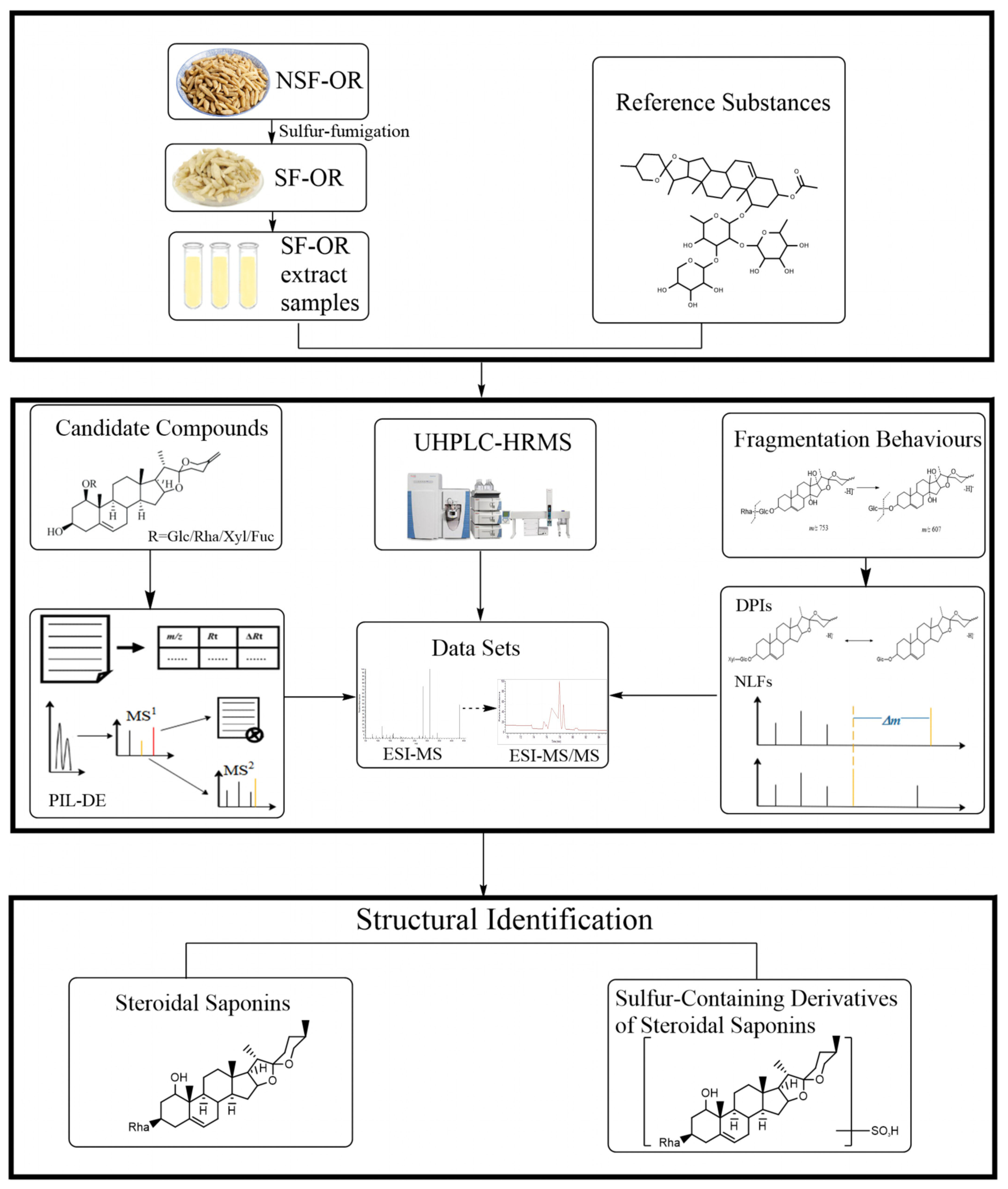
An efficient and integrated strategy was established for the target identification of steroidal saponins and their sulfur-containing derivatives in SF-OR using a UHPLC-LTQ-Orbitrap MS coupled with post-acquisition data-mining processing techniques (Figure 1).
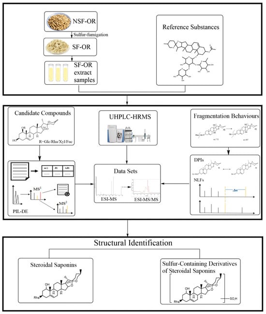
Figure 1.
Summary diagram of the developed strategy and methodology.
First, the samples were injected into the UHPLC-LTQ-Orbitrap MS to gain high-resolution mass spectrometry (HRMS) data with full MS scanning acquisition. Second, the reactions (hydroxylation, glucosylation, xylosation, and rhamnosylation) were predicted using information derived from structural characteristics, the literature, and compound databases. A preliminary screening of the candidate compounds was performed using the Thermo Xcalibur 2.1 software to obtain their retention times and accurate molecular weights. According to the PIL-DE scan mode, multi-stage mass spectrometry data were collected. Third, to guide the subsequent rapid analysis, these compounds’ DPI and specific neutral loss filter (NLF) were summarized based on the mass spectrometric cracking rules reported in the literature and the cracking information of reference substances.
2.1.2. Molecular Design of Steroidal Saponins from Ophiopogonis Radix
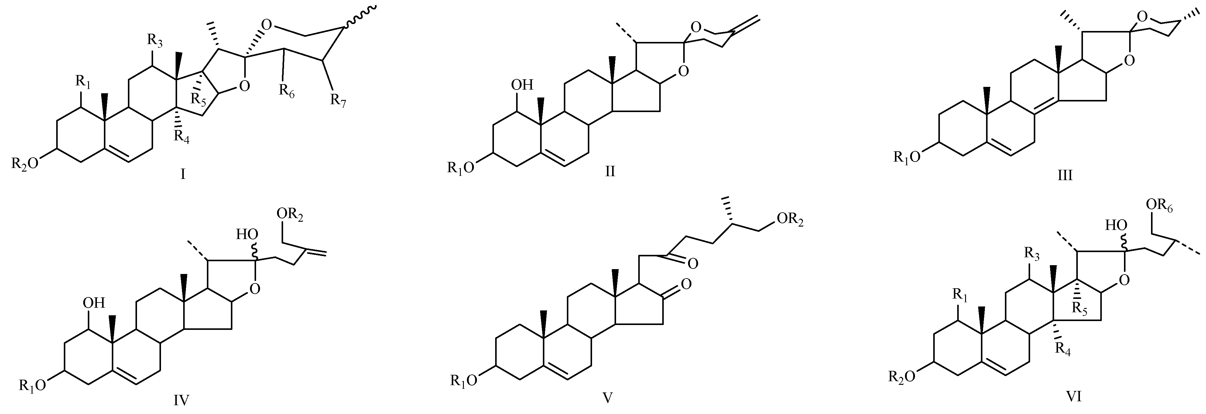
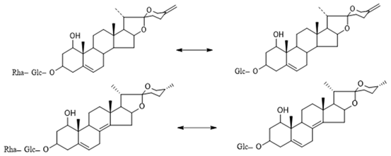
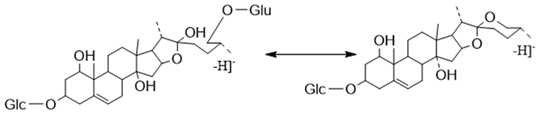
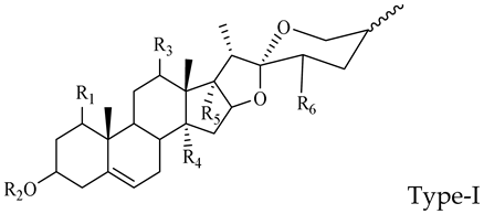
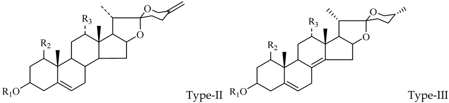
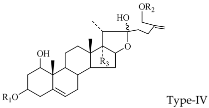
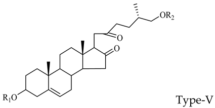
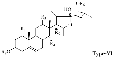
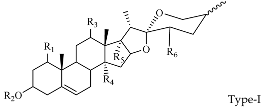
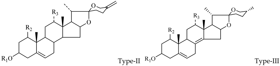
The steroidal saponins of OR are usually oligoglycosides of either spirostanol or furostanol (aglycone). According to the different structures of these steroidal saponins, they can be classified into ruscogenin, nitogenin, pennogenin, diosgenin, neoruscogenin, and furostanol saponins, etc. Rhamnose (Rha), fucose (Fuc), glucose (Glc), xylose (Xyl), and arabinose (Ara) are the main monosaccharides present in OR steroidal saponins. Different monosaccharides have unique connections in steroidal saponins [16,17,18,19,20]: (1) Fuc and Glc have priority when connecting steroidal saponins. (2) The monosaccharides in steroidal saponins appear in the following probability sequence: Rha > Fuc > Xyl > Glc > Ara. (3) Spirostanol saponins contain only monosaccharides at C3, and the maximum number of sugar units is 3. Arabionose is present only in spirostanol saponins. (4) Furostanol steroidal saponins generally contain two sugars, 1 to 2 Glc at C26 and an acetyl group (AC) at C3. Therefore, in this study, six steroidal saponins (as shown in Figure 2) were used as the core structure, and Rha (0–2), Fuc (0–2), Xyl (0–2), Glc (0–4), Ara (0–1), and AC (0–1) were used as substituents in the molecular design.
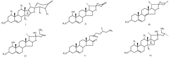
Figure 2.
Core structures of six steroidal saponins. Type-I: spirostanol; Type-II and Type-III: deformed spirostanol with a C5-C6 double bond and a carbohydrate at C3; Type-IV: deformed furostanol with a C5-C6 double bond and two carbohydrates at C3 and C26; Type-V: deformed furostanol with ring opening producing two carbonyls in the E-ring.
2.1.3. Construction of Ion Lists
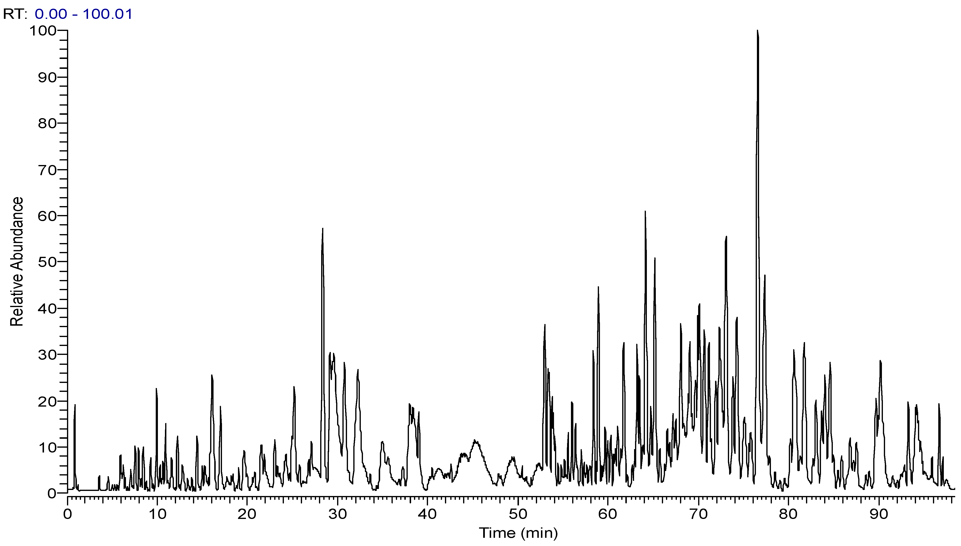
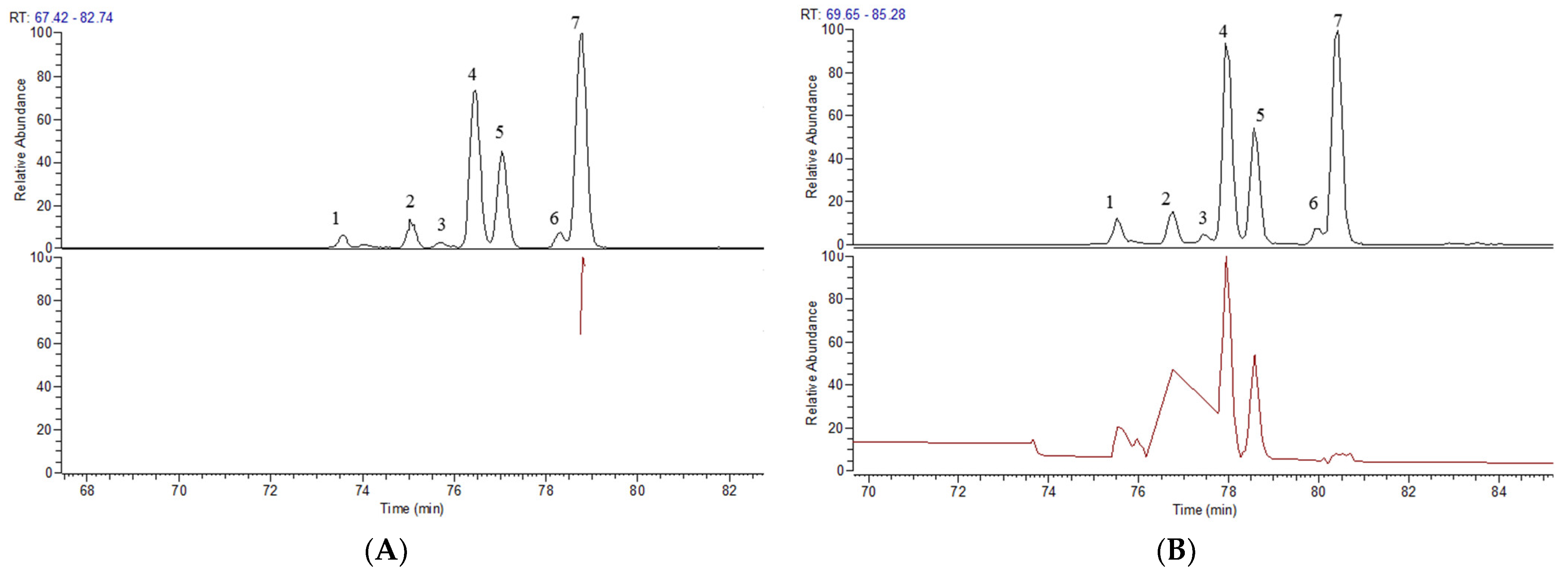
The Thermo Xcalibur 2.1 software was used to accurately calculate the molecular weights of the above candidate formulas with an error of ±10 ppm. Ion peaks with an intensity greater than 1.0 × 10⁴ in the full scan map (Figure 3) were extracted as potential steroidal saponins of OR. For example, taking compounds with a molecular weight at m/z 721.4157, seven chromatographic peaks were extracted from the HR-MS1. Only the secondary mass spectrum of peak-7 was obtained (Figure 4A), while, in the PIL-DE scan mode, the ESI-MS/MS spectra of these seven peaks were obtained through one data acquisition (Figure 4B). The PIL-DE scan mode dramatically increases the information acquisition efficiency of multi-stage mass spectra.

Figure 3.
The total ion chromatograms of SF-OR were obtained in full scan mode.
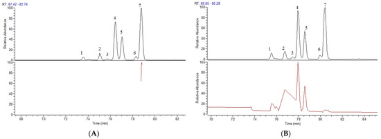
Figure 4.
Data acquisition efficiency of compound m/z 721.4157 in full scan mode (A) and PIL-DE mode (B).
2.1.4. Analysis of the Characteristic Fragmentation Mechanism of Steroidal Saponins
The DPIs and NLFs were speculated according to the summarized fragmentation behaviors of known compounds, which supplied tremendous help in identifying the secondary metabolites in Ophiopogonis Radix.
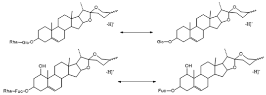
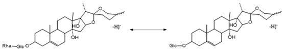
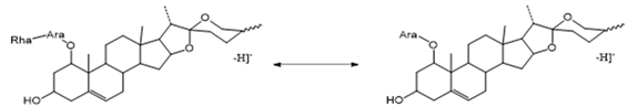
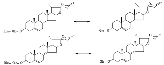
The Characteristic Fragmentation Mechanism of Type-I Steroidal Saponins
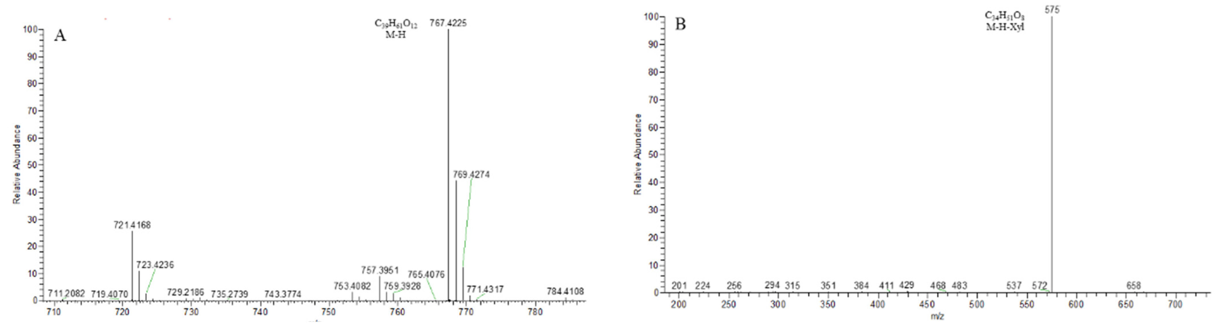
As shown in Figure 5, the core structure of Type-I steroidal saponins was spirostanol, which generally had only one carbohydrate chain attached to the C3 position. In ESI− mode, carbohydrates were removed one by one until a monosaccharide remained on the core structure; hence, most of their aglycon fragment ions were not observed. Take ophiopogonin C′ as an example (Figure 6); it gave rise to a [M − H]− ion at m/z 721.4157 (C39H61O12, <5 ppm) in negative mode. In its ESI-MS/MS spectrum, the product-ion at m/z 575 ([M − H − Rha]−) indicated the presence of the Rha group. According to the literature [21,22,23], there was a Glc group in ophiopogonin C′, while NLF of 162 Da was not observed in this experiment. This observation indicated that the Glc group should be directly attached to the core structure, and Rha was attached to the Glc. Based on the above analysis, the molecular formula of the core structure was deduced to be C27H42O3 with a molecular weight of 414 Da. Similarly, ophiopogonin B possessed the same [M − H]− ion at m/z 721.4170 in the ESI-MS spectrum and a fragment ion at m/z 575 ([M − H − Fuc]−) in the ESI-MS/MS spectrum with ophiopogonin C′ in the negative mode, as shown in Figure S1. The fucose disaccharide was present at C1 and the hydroxyl group at C3 of ophiopogonin B [23]. Therefore, the molecular formula of the core structure of ophiopogonin B was C27H42O4 with a molecular weight of 430 Da.
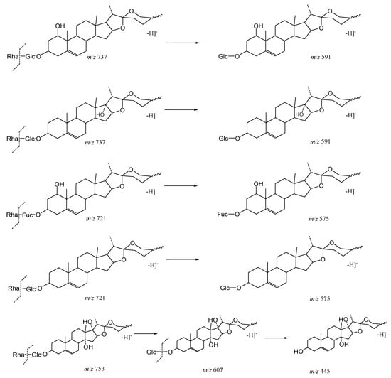
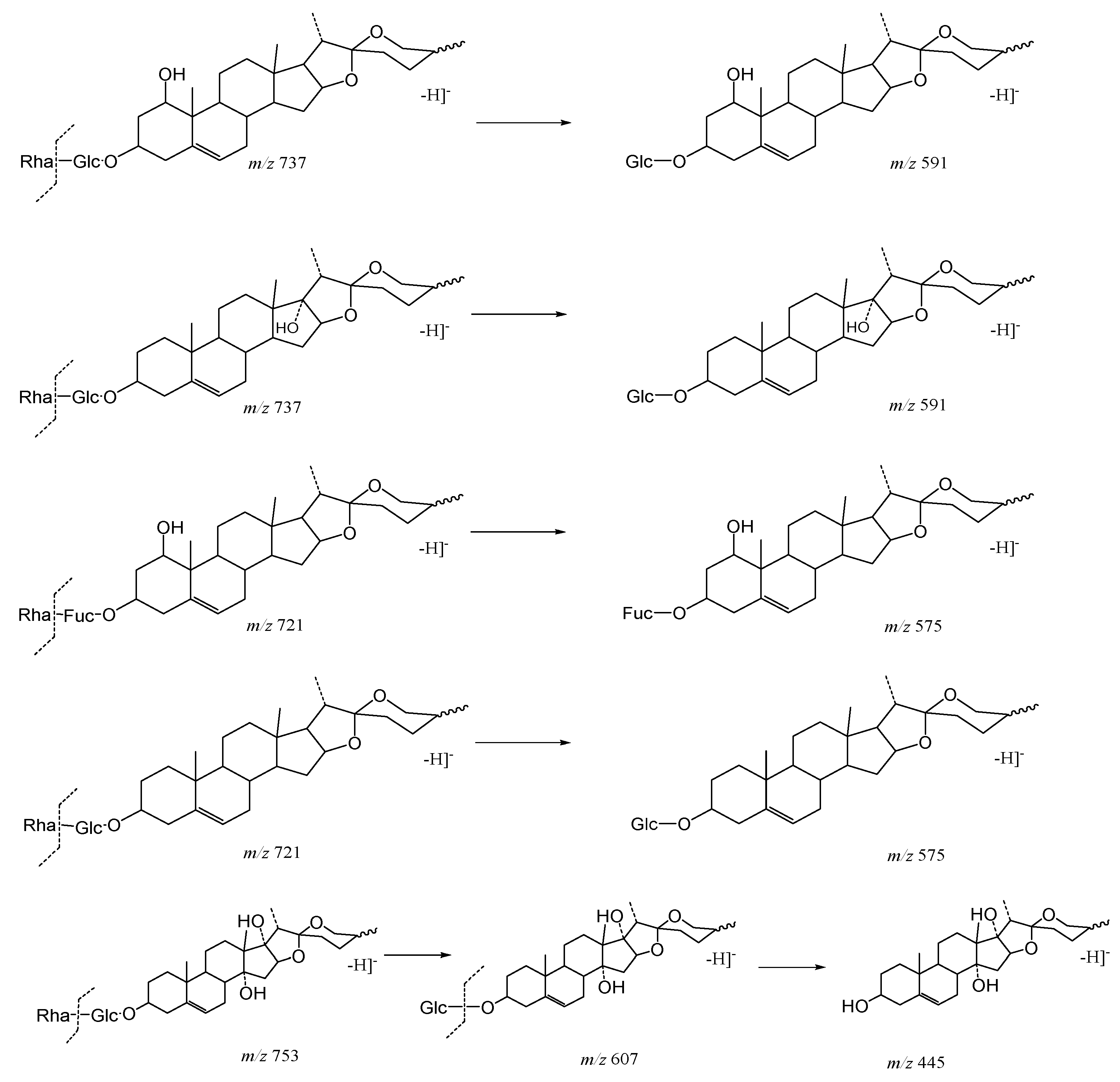
Figure 5.
The characteristic fragmentation mechanism of Type-I steroidal Saponins.
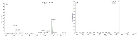
Figure 6.
The ESI-MS spectrum (A) and ESI-MS/MS spectrum (B) of ophiopogonin C′ in negative ion mode.
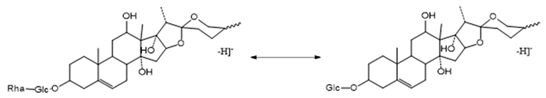
14-hydroxydiosgenin 3-O-α-l-rha-(1→2)-β-d-glc showed its [M − H]− ion at m/z 737.4106 (C7H5O3) with a mass error within 5 ppm. On account of the consecutive neutral loss of Rha and Glc, the fragment ions at m/z 591 and m/z 429 were generated in its ESI-MS/MS spectrum, suggesting the presence of a disaccharide chain. Thus, the molecular formula of the core structure was deduced to be C27H42O4 (430 Da), which was consistent with ophiopogonin B and had one more hydroxyl group than that of ophiopogonin C′. The ESI-MS and ESI-MS/MS spectra of 14-hydroxydiosgenin 3-O-α-l-rha-(1→2)-β-d-glc are shown in Figure 7. Ophiogenin 3-O-α-l-rha-(1→2)-β-d-glc afforded the [M − H]− ion at m/z 753.4055 (C39H61O14, <5 ppm) in negative ion mode. In the ESI-MS/MS spectrum, it produced fragment ions at m/z 607 ([M − H − Rha]−) and m/z 445 ([M − H − Rha-Glc]−) with consecutive neutral losses of 146 Da (Rha) and 162 Da (Glc) (Figure S2). The molecular formula of the core structure was eventually identified as C27H42O5 (446 Da), which had one more hydroxyl group than that of ophiopogonin B. Due to the polyhydroxy substitution, these compounds generated the fragment ion of the core structure in the ESI− mode, which could be used as a particular fragmentation behavior to provide a reference for subsequent identification.
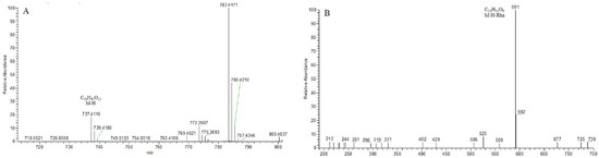
Figure 7.
The ESI-MS spectrum (A) and ESI-MS/MS spectrum (B) of 14-hydroxydiosgenin 3-O-α-l-rha-(1→2)-β-d-glc in negative ion mode.
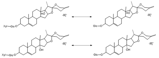
The Characteristic Fragmentation Mechanism of Type-II and Type-III Steroidal Saponins
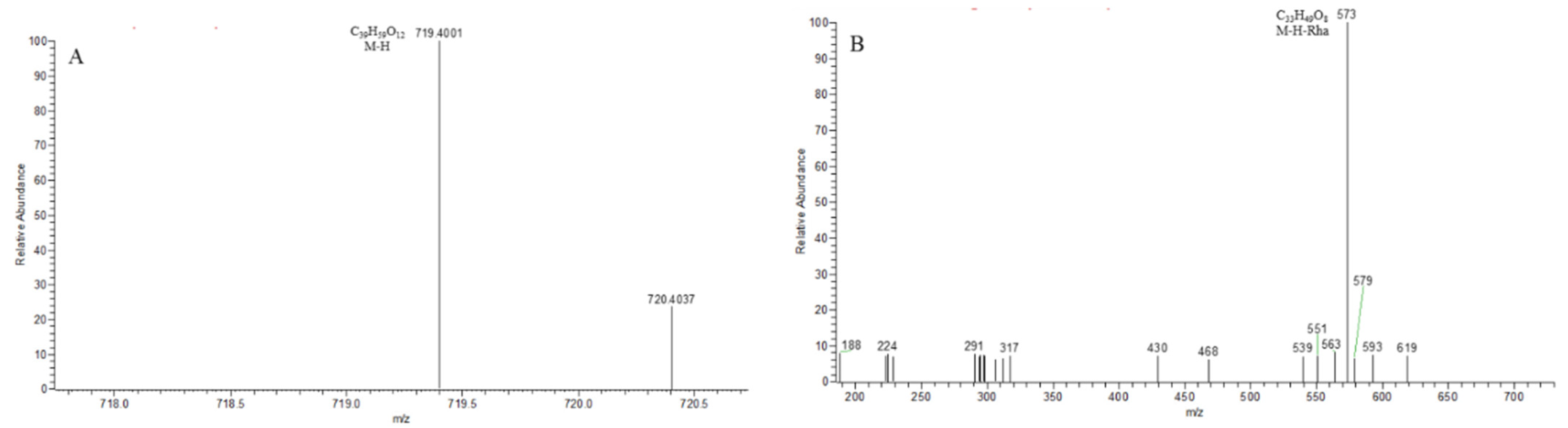
The core structures of Type-II and Type-III were deformed spirostanol with a C5-C6 double bond and a carbohydrate at C3. Similarly, most of their aglycon fragment ions were not observed. (1β,3β)-3-hydroxyspirost-5, 25(27)-dien-1-yl-O-6-deoxy-α-l-Rha-(1→2)-β-d-glc presented the [M − H]− ion at m/z 719.4008 (C39H59O12, <5 ppm). In the ESI-MS/MS spectrum, the fragment ion at m/z 573 ([M − H − Rha]−) indicated the presence of the Rha group (Figure 8). According to the literature [24], a Glc group indicated that the Glc group should be directly attached to the core structure, and Rha was attached to the Glc. It was concluded that the molecular formula of the core was C27H40O3 with a molecular weight of 412 Da.
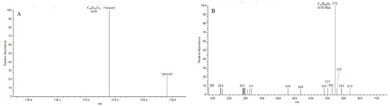
Figure 8.
The ESI-MS spectrum (A) and ESI-MS/MS spectrum (B) of (1β,3β)-3-hydroxyspirost-5,25(27)-dien-1-yl-O-6-deoxy-α-l-Rha-(1→2)-β-d-glc in negative ion mode.
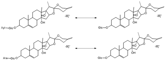
The Characteristic Fragmentation Mechanism of Type-IV Steroidal Saponins
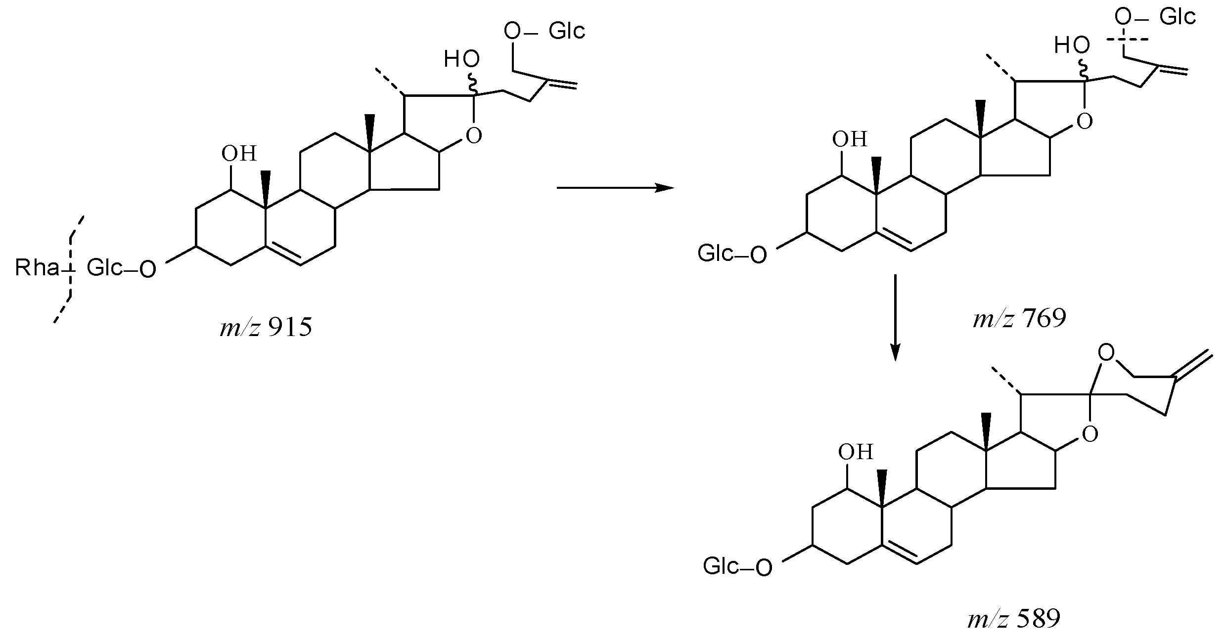
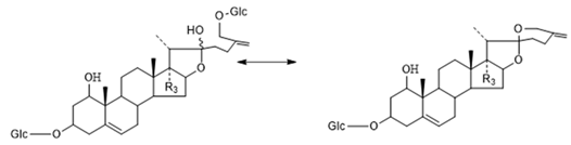
The core structure of Type-IV was deformed furostanol with a C5-C6 double bond and two carbohydrates at C3 and C26 (shown in Figure 9). It is worth noting that the carbohydrate chain at position C26 was generally substituted by Glc, which appeared as a specific NLF of 180 Da in the ESI-MS2 spectrum. 26-O-β-d-Glc-20α-hydroxyfurost-25, 27-dine-3-α-l-Rha-β-d-Glc generated the [M − H]− ion at m/z 915.4584 (C45H71O19, <5 ppm). In the ESI-MS/MS spectrum, characteristic fragment ions at m/z 769 ([M − H − Rha]−) and m/z 589 ([M − H − Rha-180]−) were observed, which could be conducted as paired-DPI (pDPI) for these types of compounds (Figure 10).
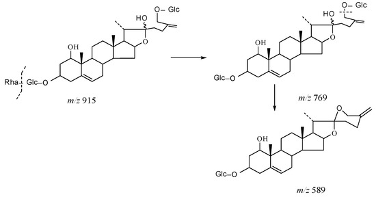
Figure 9.
The characteristic fragmentation mechanism of Type-IV steroidal Saponins.
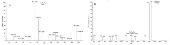
Figure 10.
The ESI-MS spectrum (A) and ESI-MS/MS spectrum (B) of 26-O-β-d-Glc-20α-hydroxyfurost-25, 27-dine-3-α-l-Rha-β-d-Glc in negative ion mode.
The Characteristic Fragmentation Mechanism of Type-V Steroidal Saponins
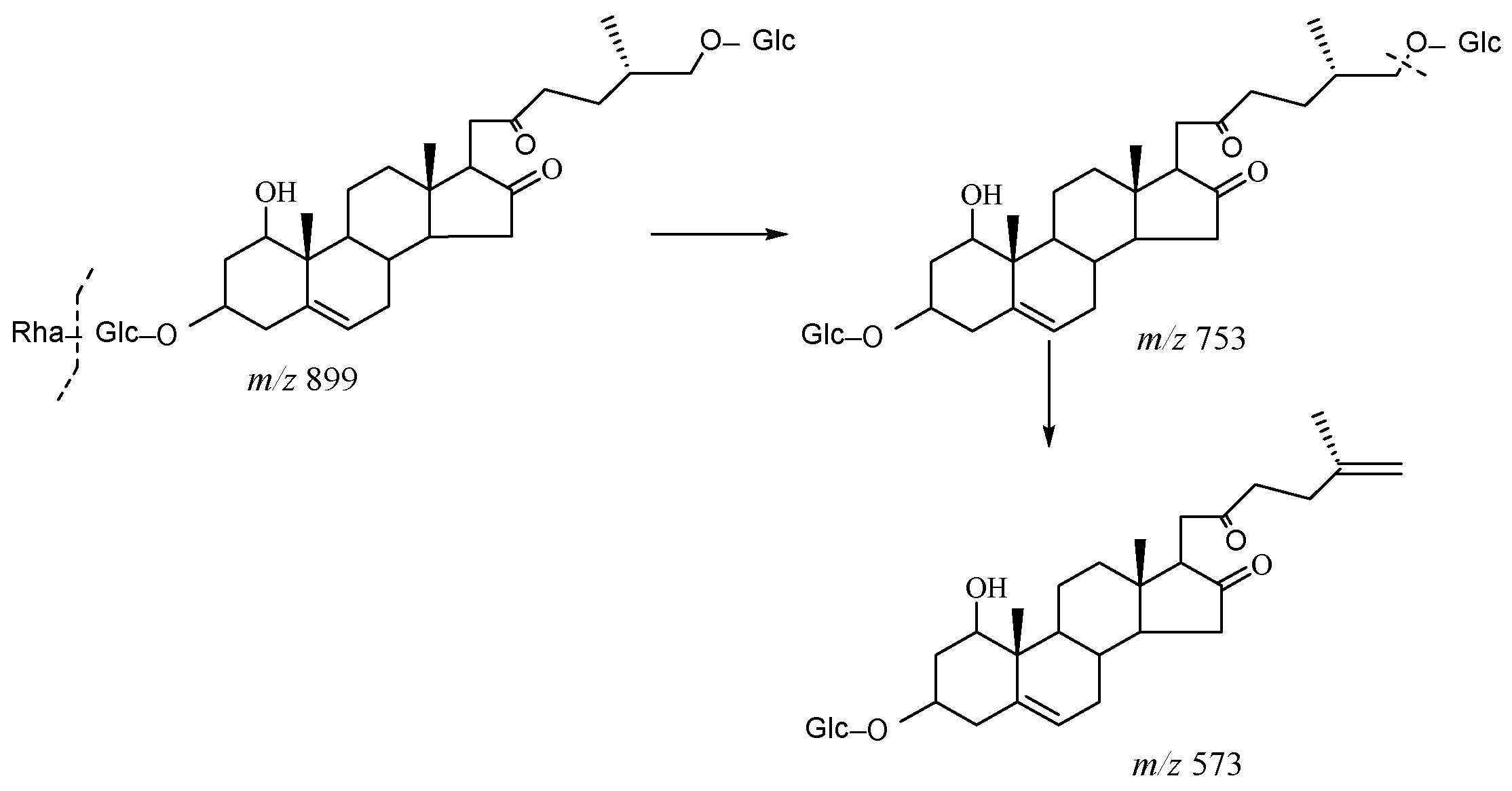
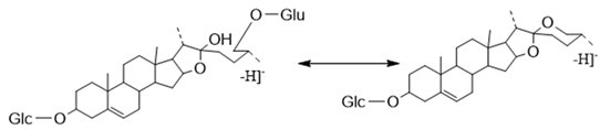
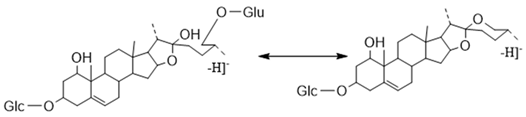
As shown in Figure 11, the core structure of Type-V was deformed furostanol with a ring opening producing two carbonyls in the E-ring. There were generally two carbohydrates at C3 and C26; the carbohydrate chain at position C26 was generally substituted by Glc, which appeared as a specific NLF of 180 Da in the ESI-MS2 spectrum. For example, (20R,25R)-26-O-β-d-glc-3β, 26-dihydroxycholest-5-en-16, 22-dioxo-3-O-α- L-rha(1→2)-β-d-Glc possessed the [M − H]− ion at m/z 899.4634 (C45H71O18, mass error within 5 ppm). It also produced ions at m/z 753 ([M − H − Rha]−) and m/z 573 ([M − H − Rha-180]−), suggesting the presence of Type-V pDPIs (Figure S3).
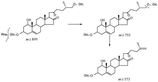
Figure 11.
The characteristic fragmentation mechanism of Type-V steroidal Saponins.
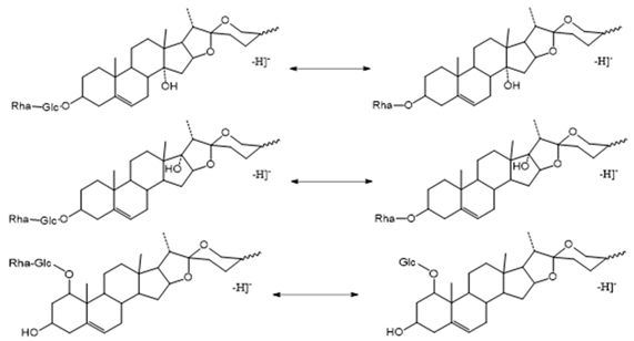
The Characteristic Fragmentation Mechanism of Type-VI Steroidal Saponins
The core structure of Type-VI was furostanol with two carbohydrate chains at C3 and C26. In negative mode, the carbohydrates were removed one by one until no monosaccharides remained on the core structure; hence, their aglycon fragment ions could be observed. The carbohydrate chain at C26 generally contained 1–2 Glc, which would appear as a specific NLF of 180 Da in MS2 spectra. The characteristic fragmentation mechanism of Type-V steroidal Saponins is shown in Figure 12. Ophiofurspiside M generated the [M − H]− ion at m/z 917.4741, with the molecular formula of C45H73O19 and a mass error within 5 ppm. In the ESI-MS2 spectrum, a battery of fragment ions at m/z 771 ([M − H − Rha]−), m/z 591 ([M − H − Rha-180]−), and m/z 591 ([M − H − Rha-180-Glc]−) were all observed (Figure S4). Xyl-Ophiofurspiside M, with a mass error within 5 ppm, gave rise to the accurate [M − H]− ion at m/z 1049.5163 (C50H81O23). Fragment ions at m/z 917 ([M − H-Xyl]−), m/z 771 ([M − H-Xyl-Rha]−), m/z 591 ([M − H-Xyl-Rha-180]−), and m/z 429 ([M − H-Xyl-Rha-180-Glc]−) were generated in the ESI-MS/MS spectrum, which was formed by the consecutive loss of Xyl, Rha, Glc, and Glc (Figure S5). Ophiofurspiside A, with the [M − H]− ion at m/z 1033.5214 (C50H81O22, <5 ppm), presented fragment ions at m/z 901 ([M − H-Xyl]−), m/z 755 ([M − H-Xyl-Rha]−), and m/z 575 ([M − H − Rha-180]−) (Figure S6). In addition, ophiopogonin H possessed the [M − H]− ion at m/z 1063.5319 (C51H83O23, mass error < 5 ppm). It generated ESI-MS2 DPIs at m/z 901 ([M − H-Glc]−), m/z 755 ([M − H-Glc-Rha]−), and m/z 575 ([M − H-Xyl-Rha-180]−) (Figure S7). Therefore, the pDPIs at m/z 771/591 and m/z 755/575 could be used to identify Type-VI rapidly.
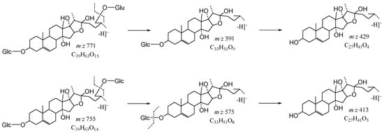
Figure 12.
The characteristic fragmentation mechanism of Type-VI steroidal Saponins.
2.1.5. Determination and Verification of NLFs and DPIs of OR Steroidal Saponins
The different core structures of the six types of steroidal saponins, combined with the relevant information reported in the literature [15], allow the fragmentation mechanism of these compounds in negative ion mode to be analyzed and the NLFs and DPIs to be summarized. The potential NLFs of the OR Steroidal Saponins are summarised in Table 1.

Table 1.
NLFs of OR Steroidal Saponins.
Congeneric compounds generally have a similar MS fragmentation regularity, thereby generating characteristic DPIs that can represent the structure of such compounds. However, it is difficult to accurately identify the structure of natural compounds with a large molecular weight and relatively complex structures based on one DPI. Therefore, in this experiment, the concept of pDPI was proposed to provide meaningful guidance for the rapid identification of OR steroidal saponins. According to the components identified and reported in the literature, the pDPIs of the six types of core structures are summarized, as shown in Table 2. A total of 9 pDPIs of Type-I, 2 pDPIs of Type-II and Type-III, 1 pDPI of Type IV, 1 pDPI of Type-V, and 3 pDPIs of Type-VI were found.
Table 2.
pDPIs of OR Steroidal Saponins.
2.1.6. Detection and Structural Elucidation of OR Steroidal Saponins
Based on the established pDPIs and NLFs, 191 steroidal saponins were quickly identified from OR. There were 105 Type-I, 12 Type-II and Type-III, 13 Type-IV, 18 Type-V, and 42 Type-VI OR steroidal saponin compounds screened and identified. The detailed MS data information is shown in Table 3.
Table 3.
Identification results of prototype components of Steroid Saponins in SF-OR.
Taking the pDPI at m/z 707/575 of Type-I as an example, Sprengerinin A with the [M − H]− ion at m/z 707.4011 (C38H59O12, mass error within 5 ppm) generated the DPI at m/z 575 ([M − H-Xyl]−). The NLF of 132 Da (m/z 707→m/z 575) indicated the presence of Xyl. The molecular formula corresponding to m/z 575 was inferred to be C33H51O8 ([Aglycon-H + Glc]−/[Aglycon-H + Rha]−). In addition, compound S39 generated its [M − H]− ion at m/z 839.4423; its molecular formula was deduced to be C43H67O16. In its ESI-MS/MS spectrum, the DPI at m/z 707 ([M − H-Xyl]−) certified the loss of Xyl from the [M − H]− ion. Further, the DPI at m/z 561 (C32H49O8, [Ruscogenin-H + Ara]−) was formed due to the neutral loss of 146 Da from the DPI at m/z 707. Therefore, S39 was tentatively identified as Ruscogenin 1-O-α-l-Xyl (1→3) Rha (1→2) Ara, and the pDPI at m/z 707/561 was initially assigned as Ruscogenin-Rha (1→2) Ara.
S109–S112 possessed identical [M − H]− ions at m/z 851.4418 (C44H67O16). The DPIs at m/z 719 ([M − H-Xyl]−) and m/z 573 ([M − H-Xyl-Rha]−) were generated in their ESI-MS/MS spectra, suggesting the presence of Xyl and Rha. The molecular formula corresponding to m/z 573 was inferred to be C33H51O8 ([Aglycon-H + Glc]−/[Aglycon-H + Rha]−). The core structures of these two aglycones differed by one hydroxyl substitution. Thus, S109–S112 were tentatively characterised as (1β,3β)-3-hydroxyspirosta-5,25(27)-dien-1-yl-O-6-deoxy-α-l-Rha-(1→2)-O-[β-d-Xyl-(1→4)]-β-d-Fuc or 25(R)-spirost-5, 8-diene-3β-ol-3-O-α-l-Rha(1→2)-β-d-Xyl(1→4)-β-d-Glc. The pDPI at m/z 719/573 was used for the preliminary identification of Type-II and Type-III compounds, which was one less hydroxyl than the pDPI at m/z 735/589.
S118 produced the [M − H]- ion at m/z 915.4584 (C45H71O19) in the negative ion mode. The pDPIs at m/z 769 ([M − H − Rha]−)/589 ([M − H − Rha-180]−) were, respectively, generated in its ESI-MS2 spectrum. The neutral loss of 180 Da indicated the existence of Glc in the carbohydrate chain at the position of C26. The corresponding molecular formula of m/z 589 was C33H49O9, which was conjectured to be [Aglycon-H + Glc]−. Since the neutral loss of Glc was not observed in the ESI-MS/MS spectrum, it was speculated that the other Glc was at C3. Thus, S118 could be deduced as 26-O-β-d-Glc-20α-hydroxyfurost-25,27-dine-3-α-l-Rha-β-d-Glc or its isomer. The structure represented by the pDPI at m/z 769/569 of Type-IV is shown in Table 2.
S131 afforded the [M − H]− ion at m/z 899.4634 (C45H71O18). In the MS2 spectrum, the DPI at m/z 753 ([M − H − Rha]−) indicated the presence of Rha, and the DPI at m/z 573 ([M − H − Rha-180]−) indicated the presence of Glc at C26 of Rha. The corresponding molecular formula of m/z 573 was C33H49O8 ([Aglycon-H + Glc]−). Since the neutral loss of 162 Da or 180 Da was not observed in the ESI-MS/MS spectrum, it was speculated that the other Glc was directly attached to the core. Therefore, S131 was tentatively identified as (20R,25R)-26-O-β-d-Glc-3β,26-dihydroxycholest-5-en-16,22-dioxo-3-O-α-l-Rha(1→2)-β-d-Glc or its isomer. The structure represented by the pDPI at m/z 753/573 of Type-V is shown in Table 2.
S149–S150 showed their theoretical deprotonated molecular ions at m/z 901.4791 (C45H73O18, mass error within ±5.00 ppm). The pDPI of Type-VI at m/z 755 ([M − H − Rha]−)/m/z 575 ([M − H − Rha-180]−) indicated the presence of Glc and Rha substitution at C26. The corresponding molecular formula of m/z 575 was C33H51O8, which was conjectured to be [Aglycone-H + Glc]−. Based on the above analysis, the structures of compounds S149–S150 were initially identified. The structure represented by the pDPI at m/z 755/575 was shown in Table 2. S151–S153 possessed the theoretical [M − H]− ions at m/z 917.4740 (C45H73O19, mass error within ±5.00 ppm). Another pDPI of Type-VI at m/z 771 ([M − H − Rha]−)/m/z 591 ([M − H − Rha-180]−) was formed by the neutral loss of Rha and Glc in their ESI-MS/MS spectra. The molecular formula of m/z 591 was C33H51O9, which should be [Aglycon-H + Glc]−. Compared with the pDPI at m/z 755/575, the pDPI at m/z 771/591 had more than one hydroxyl substitution. In addition, S154–S156 provided theoretical [M − H]− ions at m/z 933.4689 (C45H73O20, mass error within ±5.00 ppm). In the ESI-MS2 spectra, the DPIs at m/z 787 ([M − H − Rha]−) and 607 ([M − H − Rha-180]−) indicated the core structure was furostanol, and Glc was present at C26. The pDPI at m/z 771/591 could also be applied to identify Type-VI steroidal saponins rapidly.
2.2. Identification of Sulfur-Containing Derivatives of Steroidal Saponins from SF-OR
2.2.1. Molecular Design of Sulfur-Containing Derivatives of Steroidal Saponins
The reported pathways of ginsenosides during sulfur fumigation are sulfation and sulfite [25]. In addition, sulfur generates SO2 at high temperatures to lower the pH, making the glycosides easily hydrolyzed. Therefore, to fully characterize the sulfur-containing derivatives of steroidal saponins in OR, six types of steroidal saponins were used as the core structure, and Rha (0–2), Fuc (0–2), Xyl (0–2), Glc (0–4), Ara (0–1), Ac (0–2), SO2 (0–1), and SO3 (0–1) were used as substituents for molecular design.
2.2.2. Screening of the Candidate Molecular Weight of Sulfur-Containing Derivatives of Steroidal Saponins
The accurate [M − H]− of the candidate molecular formula was calculated in Section 2.2.1, the chromatographic peak from the ESI-MS spectra was extracted, and the peaks with an intensity > 1.0 × 10⁴ were selected as the potential sulfur-containing derivatives of Steroidal Saponins. According to the established PIL-DE scan mode, MS2 data collection was performed.
2.2.3. Identification of Sulfur-Containing Derivatives of Steroidal Saponins
Based on the established structure identification strategy with pDPIs and NLFs, rapid screening of the steroidal saponins’ sulfur-containing derivatives was carried out. In addition, the isotope peak [M − H+2]− of the sulfur-containing derivatives under ultra-high resolution split into two peaks with a mass difference of 0.01 Da. Therefore, 12Cx1Hy16Oz32S13C218O and 12Cx+21Hy16Oz+134S were separated, which could provide a basis for the further confirmation of sulfur-containing derivatives [26].
Identification of Type-I Sulfur-Containing Derivatives of Steroidal Saponins
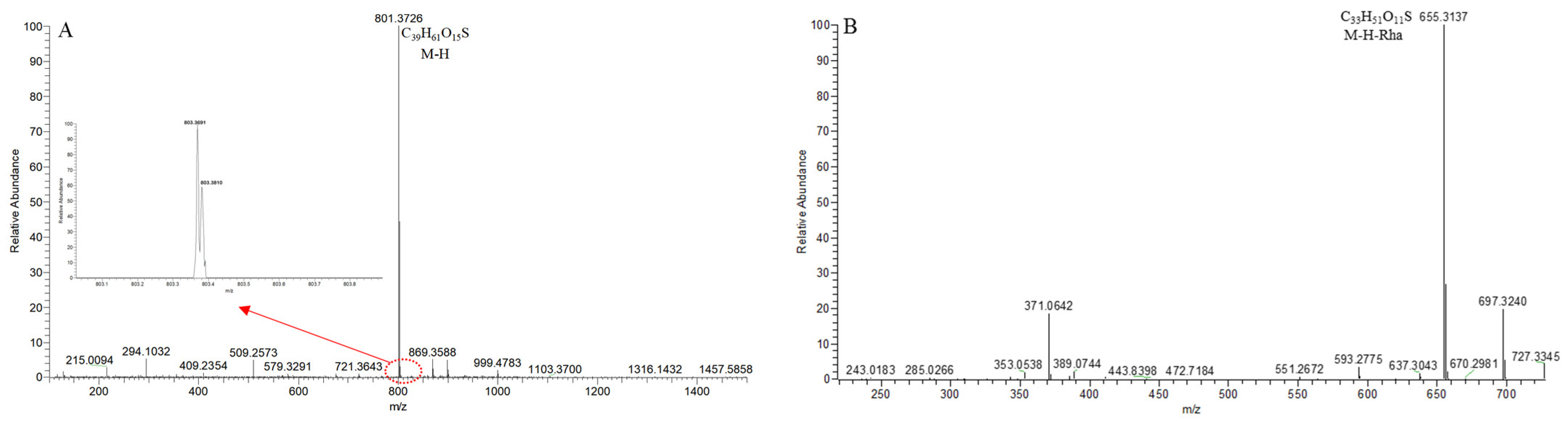
SS9, with the [M − H]− ion at m/z 801.3714 (C39H61O15S, <5 ppm), was 79.95 Da (SO3) more than that of S2–S8. In the 100,000 FWHM @ 400 m/z ultra-high resolution mode, its isotope peak at [M − H+2]− formed two peaks at m/z 803.3691 and m/z 803.3810, further confirming that SS9 was a sulfur compound. In its ESI-MS/MS spectrum, the DPI at m/z 655 ([M − H − Rha]−) was consistent with S2–S8 (Figure 13). The pDPI it generated at m/z 801/655 was 79.95 Da more than that of m/z 721/575 of S2–S8. Therefore, the compound SS9 was identified as the sulfated product of the compound S2–S8.
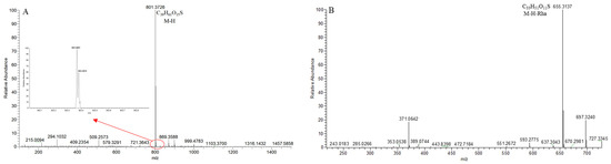
Figure 13.
The ESI-MS spectrum (A) and ESI-MS/MS spectrum (B) of SS9 in negative ion mode.
As shown in Figure S8, SS10–SS14 displayed the [M − H]− ion at m/z 817.3663 (C39H61O16S, 5 ppm). This was 79.95 Da greater than S11–S15 in negative ion mode, which implied that SS10–SS14 might be the sulfated product of S11–S15. Two isotope peaks at m/z 819.3615 and m/z 819.3772 further indicated that SS10–SS14 were sulfur-containing compounds. The DPI at m/z 671 ([M − H − Rha]−) was consistent with S11–S15. The pDPI at m/z 817/671 was 79.95 Da greater than the pDPI at m/z 721/575 of S11–S15. Therefore, the compounds SS10–SS14 were presumed to be the sulfated products of S11–S15.
According to the characteristic fragmentation mechanism of Type-I steroidal saponins and the specificity of the sulfur-containing compound isotope peaks, a total of 24 Type-I sulfur-containing derivatives of steroidal saponins were identified from SF-OR. They were all sulfated products, as shown in Table 4.
Table 4.
Identification results of sulfur-containing derivatives of Steroid Saponins in SF-OR.
Identification of Type-II and Type-III Sulfur-Containing Derivatives of Steroidal Saponins
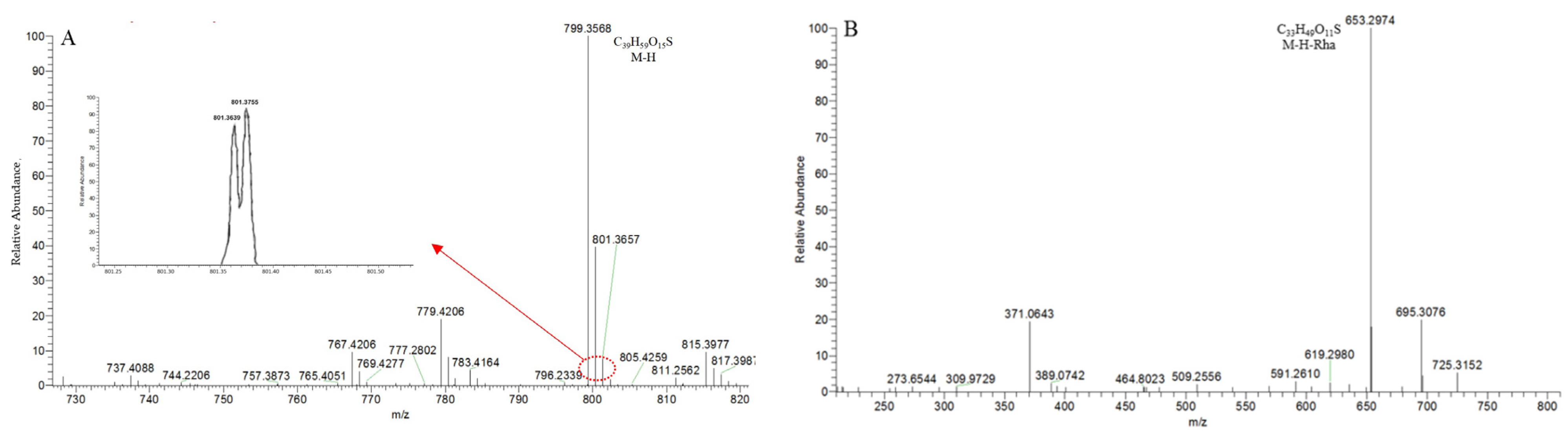
SS30, with the [M − H]− ion at m/z 799.3563 (C39H59O15S), was 79.95 Da greater than S106 (Figure 14A). In the 100,000 FWHM @ 400 m/z ultra-high resolution mode, its isotope peak at [M − H+2]− showed two peaks at m/z 803.3691 and m/z 803.3810 due to the existence of element “S”. In the MS2 spectrum (Figure 14B), the DPI at m/z 653 ([M − H-146]−) indicated the presence of a Rha or a Fuc. The pDPI at m/z 799/653 of SS30 was 79.95 Da more than the pDPI at m/z 719/573 of S106, indicating that these two compounds had the exact fragmentation mechanism. Therefore, SS30 was identified as the sulfated product of S106.
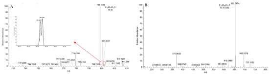
Figure 14.
The ESI-MS spectrum (A) and ESI-MS/MS spectrum (B) of SS30 in negative ion mode.
According to the characteristic fragmentation mechanism of Type-II and Type-III steroidal saponins and the specificity of the sulfur-containing compound isotope peaks, a total of three Type-II and Type-III sulfur-containing derivatives of steroidal saponins were identified and targeted from SF-OR. They were all sulfated products, as shown in Table 4.
Identification of Type-IV Sulfur-Containing Derivatives of Steroidal Saponins
There were no Type-IV sulfur-containing derivatives of steroidal saponins detected in SF-OR.
Identification of Type-V Sulfur-Containing Derivatives of Steroidal Saponins
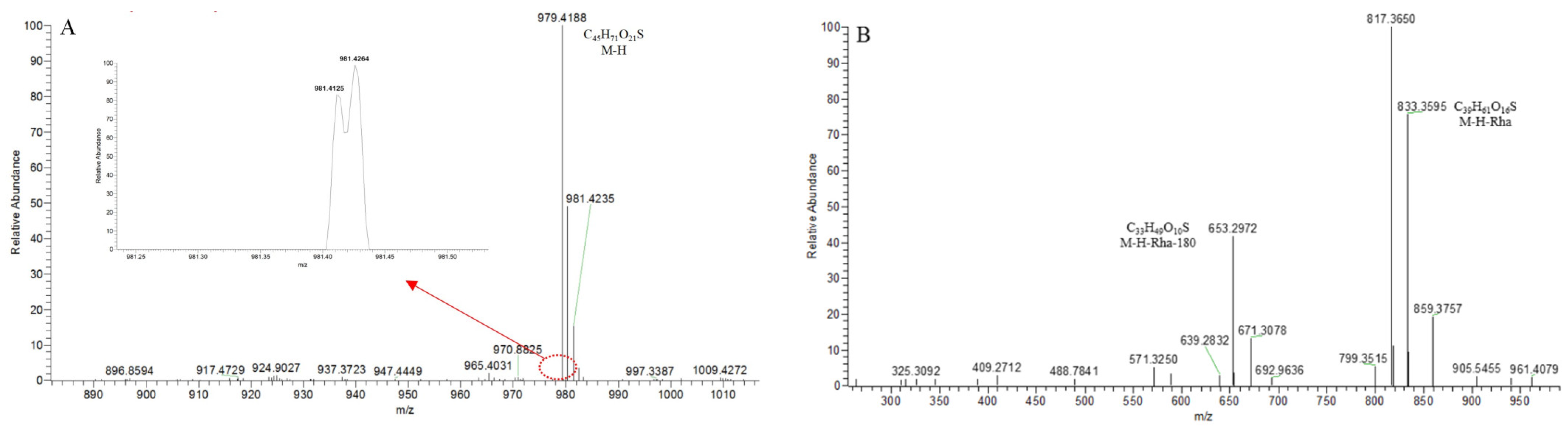
SS40 provided the [M − H]− ion at m/z 979.4192 (C45H71O21S) in the ESI-MS spectrum (Figure 15A). It was 79.95 Da greater than S131, indicating that it probably was the sulfated product of S131. Two isotope peaks at m/z 981.4125 and m/z 981.4264 proved that element “S” existed. The pDPI at m/z 817 ([M − H-146]−)/m/z 637 ([M − H-146-180]−) was 79.95 Da more than that of S106 (Figure 15B), indicating that SS40 was the sulfonation product of S106.
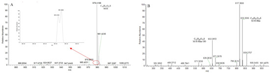
Figure 15.
The ESI-MS spectrum (A) and ESI-MS/MS spectrum (B) of SS40 in negative ion mode.
Based on the mass characteristic fragmentation mechanism of Type-V steroidal saponins, a total of eight Type-V steroidal saponins’ sulfur derivatives were identified from the targeted screening of OR, all of which belonged to sulfated products, as shown in Table 4.
Identification of Type-VI Sulfur-Containing Derivatives of Steroidal Saponins
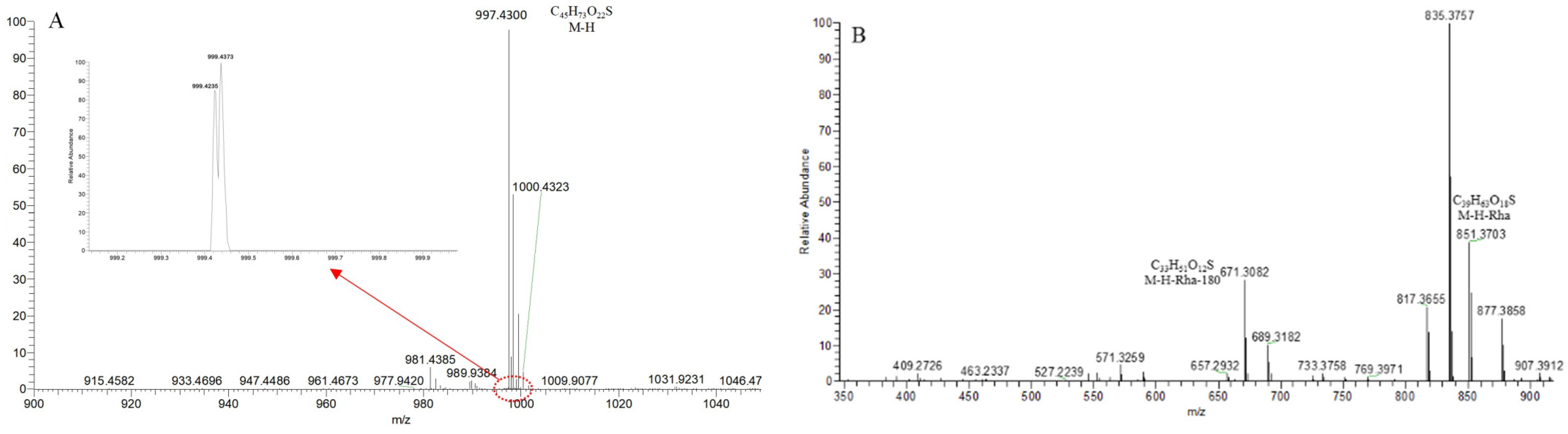
SS61, with the [M − H]− ion at m/z 997.4303 (C45H73O22S), was 79.95 Da (SO3) greater than Ohiopojaponin B and showed two isotope peaks at m/z 999.4235 and m/z 999.4373 (Figure 16A). In the ESI-MS2 spectrum (Figure 16B), the pDPIs at m/z 851 ([M − H-146]−)/m/z 671 ([M − H-146-180]−) were, respectively, attributed to the neutral losses of Rha and Glc. The core structure was inferred as furostanol or pseudo-furostanol. The pDPI at m/z 851/671 was 79.95 Da more than m/z 771/591 of S106, confirming that SS61 and Ohiopojaponin B had the exact fragmentation mechanism. Ultimately, SS61 was identified as the sulfated product of Ohiopojaponin B.

Figure 16.
The ESI-MS spectrum (A) and ESI-MS/MS spectrum (B) of SS61 in negative ion mode.
For compound SS72–SS74, the ESI- fragment ion with m/z 1127.4726 was designated as the quasi-molecular ion [M − H]−, which indicated that the possible elemental composition was C51H83O25S. In the 100,000 FWHM @ 400 m/z ultra-high resolution mode, two isotope peaks at m/z 1129.4956 and m/z 1129.5038 split from the ion [M − H+2]− of element “S”, and DPIs at m/z 997 ([M − H − Rha]−), m/z 981 ([M − H-Glc]−), m/z 917 ([M − H-Glc-SO2]−), and m/z 801 ([M − H-Glc-180]−) were observed (Figure S9), meaning that their fragmentation mechanism was consistent with that of Trigoneoside IVa. Therefore, SS72–SS74 was eventually identified as Trigoneoside IVa sulfite.
A total of 55 Type-VI steroidal saponins’ sulfur derivatives were identified from the targeted screening of OR based on the fragmentation mechanism of Type-VI steroidal saponins, in which 10 compounds were sulfite products and 45 were sulfate products, as shown in Table 4.
3. Discussion
UHPLC-LTQ-Orbitrap has become a powerful tool for drug analysis with its high sensitivity, accuracy, and separation ability [27]. However, the spectral information contained in existing chemical standards and databases is minimal. The LC-MS technique alone cannot satisfy the structural characterization of complex and diverse TCM [28]. Therefore, more new techniques and research strategies are needed to meet this challenge. Based on this, the study proposes a combined full scan-parent ions list-dynamic exclusion acquisition-diagnostic product ions analytical strategy for revealing the chemical composition of sulfur-fumigated TCM. The core technique of the strategy is to predict the possible molecular structures based on the structural characteristics of the compounds. A UHPLC-LTQ-Orbitrap high-resolution mass spectrometer is then utilized for data acquisition. After the initial screening of candidate compounds, multi-stage mass spectrometry data are targeted based on the PIL-DE scanning mode [12]. By studying the mass spectral cleavage behavior of the representative compounds, the characteristic cleavage patterns of compounds with different parent nucleus structures are summarized, and the DPIs and NLFs of such compounds are inferred. In addition, for natural products with a high molecular weight and relatively complex structures, it is often difficult to accurately characterize their structures based on one DPI [29]. Therefore, the concept of pDPI is proposed in the strategy. Accordingly, the rapid identification of chemical components is performed.
The proposed strategy was extensively investigated for the steroidal saponins of SF-OR. The successful implementation of the strategy accelerated the identification of the steroidal saponin components, expanded the search scope, and ensured the accuracy of the component identification. Although the strategy has many advantages, there are still some limitations. First, complex matrices and impurities in the samples may interfere with the analytical results, affecting accuracy and sensitivity. Second, the parent ion scanning technique has certain requirements for the sample concentration, as too high or too low will affect the detection sensitivity. Nevertheless, this innovative measure has broad application value in scientific research and medical diagnosis, while also providing a basis for the identification of compounds and their pharmacological activity research.
In this study, we summarized 6 types of 191 steroidal saponins of SF-OR based on the identified components and research reports [30,31,32,33]. Most of them were dominated by Type-I spirostanol matric structures, which generally had only one sugar chain. The representative compounds Ophiopogonin A, B, C, and D have a wide range of pharmacological effects, such as hypoglycemia [34], antitumor [35], the protection of myocardial ischemia [36], regulation of immunity [37], and resistance to myocardial infarction [38]. It was found that the steroidal saponin constituents are susceptible to the partial removal of sugar groups during SF and the further formation of corresponding sulfur-containing derivatives. A total of 95 sulfur-containing derivatives of steroidal saponin were found in the identification of SF-OR. It has been shown that SF decreases the total content of polysaccharides and increases the content of oligosaccharides and free monosaccharides in TCM. Although SO2 residues decreased during storage, chemical transformations of non-saccharide and sugar components continued to occur in the TCM [39]. In addition, SF induces chemical transformations of the bioactive components in TCM. A reduction in the bioactive components may affect their efficacy, and the generation of new sulfur-containing derivatives may also affect their safety [40]. For example, three highly toxic and carcinogenic components to humans were found in the volatile oil of sulfur-fumigated Radix Angelicae Dahuricae [41]. The anti-inflammatory and anti-tumor effects of sulfur-fumigated Astragalus membranaceus were substantially reduced [42]. The analgesic effect of Paeoniae Radix Alba was reduced after SF [43]. Therefore, when applying sulfur-fumigated TCM, attention should be paid to the transformation of its chemical composition and safety evaluation.
So far, there is very limited information on SF-induced changes in the steroidal saponin composition of OR, which can be analyzed quickly and efficiently using our proposed strategy. It provides a model for the identification and pharmacological activity studies of other sulfur-fumigated TCM in the future.
4. Materials and Methods
4.1. Chemicals and Reagents
The NSF-OR sample was purchased from Beijing Tong Ren Tang in China. The voucher specimen of NSF-OR was deposited in the School of Chinese Pharmacy, Beijing University of Chinese Medicine, Beijing, China.
Methanol and acetonitrile were MS-grade and purchased from Fisher Scientific (Fair Lawn, NJ, USA). Formic acid (MS grade) was provided by Sigma Aldrich (St. Louis, MO, USA). All the other analytical-grade chemicals were available at the workstation of Beijing Chemical Works (Beijing, China). The deionised water used throughout the experiment was purified using the Milli-Q Gradient A 10 System (Millipore, Billerica, MA, USA). Grace Pure SPE C18-Low solid-phase extraction cartridges (200 mg/3 mL, 59 μm, 70 Å) were purchased from Grace Davison Discovery Science (Deerfield, IL, USA).
Reference information is provided below. Ophiogenin 3-O-α-l-rhamnopyranosyl-(1→2)-β-d-glucopyranoside (CAS:128502-94-3; purity ≥ 98%); 14-hydroxy Sprengerinin C (CAS:1111088-89-1; ≥98%); Pennogenin 3-O-[α-l-rhamnopyranosyl-(1→2)][β-d-xylopyranosyl-(1→4)]-β-d-glucopyranoside (CAS:1029017-75-1; ≥98%); Sprengerinin C (CAS:88861-91-0; ≥98%); Ophiopogonin D(CAS:41753-55-3; ≥98%); Ophiopogonin D’(CAS:65604-80-0; ≥98%); Ophiopojaponin C(CAS:911819-08-4; ≥98%).
12-hydroxy Ophiogenin 3-O-α-l-rhamnopyranosyl-(1→2)-β-d-glucopyranoside, 14-hydroxydiosgenin-3-O-α-l-rhamnopyranosyl-(1→2)-β-d-glucopyranoside, and Pennogenin 3-O-[2-O-acetyl-α-l-rhamnopyranosyl-(1→2)][β-d-xylopyranosyl-(1→4)]-β-d-glucopyranoside were isolated and identified by the team during the preliminary work. Their structures were fully elucidated by comparing their spectral data (ESI-MS and 1H, 13C-NMR spectroscopy) with the literature [44].
4.2. Preparation of SF-OR Using Laboratory Simulation Method
NSF-OR (100 g) was wetted with 100 mL of water and then left to stand for 0.5 h. Sulfur powders (10 g) were heated until they burned, and then the burning sulfur and wetted OR samples were, respectively, put into the lower and upper layer of a desiccator. The desiccator was kept closed for 24 h. The SF-OR samples were dried in a ventilated drying oven at 50 °C for 24 h [5,45].
4.3. Preparation of SF-OR Extract Samples
The SF-OR samples were pulverised into powder (80 mesh). The SF-OR powder was accurately weighed (3 g) and then ultrasonically extracted (250 W, 40 kHz) with 30 mL of 70% methanol for 30 min at room temperature. The supernatant of the extracts was filtered and concentrated into a volume of 3 mL.
4.4. Solution Preparation
4.4.1. Sample Solution
The SF-OR extract samples (1 mL) were added to solid-phase extraction (SPE) cartridges pretreated with methanol (5 mL) and deionised water (5 mL), respectively. Then, the SPE cartridges were successively washed with deionised water (3 mL) and methanol (2 mL). The methanol eluent was collected and filtered by a 0.22 μm syringe filter before the UHPLC-LTQ-Orbitrap-MS/MS analysis.
4.4.2. Standard Solution
Each reference standard was accurately weighed and dissolved in methanol to produce the standard solutions, which were stored in the refrigerator at 4 °C before analysis. The mixed standard solution was diluted into a suitable working solution before using it.
4.5. Instruments and Analytical Conditions
Chromatographic separation was performed on a Thermo Fisher DIONEX Ultimate 3000 UHPLC system (Thermo Fisher Company, Waltham, MA, USA). An ACQUITY UPLC HSS T3 column (2.1 mm × 100 mm, 1.8 µm) was used. The separation flow rate was 0.3 mL/min and the column temperature was maintained at 25 °C. The mobile phase was water containing 0.1% formic acid (A) and acetonitrile containing 25% methanol (B). The gradient elution conditions were set as follows: 0–2 min, 0–10% B; 2–10 min, 10–20% B; 10–28 min, 20–26% B; 28–31 min, 26% B; 31–45 min, 26–32% B; 45-55 min, 32–46% B; 55–90 min, 46–67% B; 90–93 min, 67–80% B. Mass spectrometry analyses were performed on an LTQ XL™ linear ion trap mass spectrometer (Thermo Fisher Company, Waltham, MA, USA) using an electrospray ionisation (ESI) source. The ion source parameters are listed as follows: the sheath gas and auxiliary gas at a flow rate of 30 and 10 (arbitrary units), respectively, capillary temperature of 350 °C, capillary voltage of −35 V, spray voltage of −3000 V, and collision energy of 35%. HRMS were obtained with full scans at a 30,000 resolution in the mass range of m/z 100–800.
4.6. Peak Selections and Data Processing
A Thermo Xcalibur 2.1 workstation was used for the data acquisition and processing. Peaks with an intensity over 10,000 in negative ion mode were selected as fragment ions. The chemical formulas attributed to the selected peaks were calculated using a formula predictor by setting the parameters as follows: C [5–60], H [5–100], O [1–40], S [0–2], and the ring double-bond (RDB) equivalent value [3–30]. The maximum mass errors between the measured and calculated values were fixed within 5 ppm. All the relevant data, including the peak number, retention time, accurate mass, predicted chemical formula, and corresponding mass error, were recorded.
5. Conclusions
In this study, an analytical strategy for PIL-DE acquisition combined with DPIs and NLFs based on UHPLC-LTQ-Orbitrap MS was proposed. Steroidal saponins and their transformed components in SF-OR were analyzed according to the method. As a result, 191 steroidal saponins were quickly screened and identified from SF-OR. There were 105 Type-I, 12 Type-II and Type-III, 13 Type-IV, 18 Type-V, and 43 Type-VI. In addition, the occurrence of sulfate or sulfite esterification reactions led to the emergence of many new sulfur-containing derivatives in the SF process. Based on the summarized MS cleavage patterns and specific isotope peaks of the steroidal saponins, 95 sulfated and sulfite-esterified components of steroidal saponins were identified from the SF-OR, including 29 Type-I, 3 Type-II and Type-III, 8 Type-V, and 55 Type-VI. The implementation of this new strategy comprehensively characterized the compositional profile of the steroidal saponins in SF-OR. Meanwhile, this case also confirmed the feasibility of the new strategy for the identification of the chemical compositions of SF-herbs, which provides a reference for the compositional identification of other Chinese herbal medicines.
Supplementary Materials
The following supporting information can be downloaded at: https://www.mdpi.com/article/10.3390/molecules29030702/s1, Figure S1: The ESI-MS spectrum (A) and ESI-MS/MS spectrum (B) of ophiopogonin B in negative ion mode; Figure S2: The ESI-MS spectrum (A) and ESI-MS/MS spectrum (B) of Ophiogenin 3-O-α-l-rha-(1→2)-β-d-glc in negative ion mode; Figure S3: The ESI-MS spectrum (A) and ESI-MS/MS spectrum (B) of (20R,25R)-26-O-β-d-glc-3β,26-dihydroxycholest-5-en-16,22-dioxo-3-O-α-l-rha(1→2)-β-d-Glc in negative ion mode; Figure S4: The ESI-MS spectrum (A) and ESI-MS/MS spectrum (B) of Ophiofurspiside M in negative ion mode; Figure S5: The ESI-MS spectrum (A) and ESI-MS/MS spectrum (B) of Xyl-Ophiofurspiside M in negative ion mode; Figure S6: The ESI-MS spectrum (A) and ESI-MS/MS spectrum (B) of Ophiofurspiside A in negative ion mode; Figure S7: The ESI-MS spectrum (A) and ESI-MS/MS spectrum (B) of Ophiopogonin H in negative ion mode; Figure S8: The ESI-MS spectrum (A) and ESI-MS/MS spectrum (B) of SS10–SS14 in negative ion mode; Figure S9: The ESI-MS spectrum (A) and ESI-MS/MS spectrum (B) of SS72–SS74 in negative ion mode.
Author Contributions
Conceptualization, Y.L. and P.D.; methodology, Z.S.; software, Y.L.; data curation, Y.L. and P.D.; writing—original draft preparation, Y.L.; writing—review and editing, L.D. and S.W.; project administration, J.Z.; funding acquisition, J.Z. All authors have read and agreed to the published version of the manuscript.
Funding
This research was funded by National Natural Science Foundation of China, grant number 82174039; Natural Science Foundation of Shandong Province, grant number ZR2020MH371; Taishan Young Scholar Program of Shandong, grant number TSQN202103110; Chinese Medicine Science and Technology Program of Shandong Province, grant number Z-2022085; and Matching Support Program for Provincial and Above Leading Talents in Yantai City, grant number 10073801.
Institutional Review Board Statement
Not applicable.
Informed Consent Statement
Not applicable.
Data Availability Statement
Data are contained within the article and supplementary materials.
Conflicts of Interest
The authors declare no conflicts of interest.
References
- Liu, H.; Wang, S.Y.; Zhu, J.H.; Kong, M.; Zhou, S.S.; Li, S.L.; Zhu, H. Effects and contributory factors of sulfur-fumigation on the efficacy and safety of medicinal herbs evaluated by meta-analysis. J. Ethnopharmacol. 2022, 293, 115250. [Google Scholar] [CrossRef]
- Jiang, X.; Huang, L.F.; Zheng, S.H.; Chen, S.L. Sulfur fumigation, a better or worse choice in preservation of Traditional Chinese Medicine? Phytomedicine 2013, 20, 97–105. [Google Scholar] [CrossRef] [PubMed]
- Zhang, W.H.; Luo, H.Y.; Fang, J.; Zhao, C.L.; Chan, K.C.; Chan, Y.M.; Dong, C.X.; Chen, H.B.; Zhao, Z.Z.; Li, S.L.; et al. Impact of Sulfur Fumigation on Ginger: Chemical and Biological Evidence. J. Agric. Food Chem. 2022, 70, 12577–12586. [Google Scholar] [CrossRef] [PubMed]
- Shen, H.; Zhang, L.; Xu, J.D.; Ding, Y.F.; Zhou, J.; Wu, J.; Zhang, W.; Mao, Q.; Liu, L.F.; Zhu, H.; et al. Effect of sulfur-fumigation process on ginseng: Metabolism and absorption evidences. J. Ethnopharmacol. 2020, 256, 112799. [Google Scholar] [CrossRef] [PubMed]
- Duan, S.M.; Xu, J.; Bai, Y.J.; Ding, Y.; Kong, M.; Liu, H.H.; Li, X.Y.; Zhang, Q.S.; Chen, H.B.; Liu, L.F.; et al. Sulfur dioxide residue in sulfur-fumigated edible herbs: The fewer, the safer? Food Chem. 2016, 192, 119–124. [Google Scholar] [CrossRef]
- Kan, W.L.; Ma, B.; Lin, G. Sulfur fumigation processing of traditional chinese medicinal herbs: Beneficial or detrimental? Front. Pharmacol. 2011, 2, 84. [Google Scholar] [CrossRef]
- He, J.; Ye, L.; Li, J.; Huang, W.; Huo, Y.; Gao, J.; Liu, L.; Zhang, W. Identification of Ophiopogonis Radix from different producing areas by headspace-gas chromatography-ion mobility spectrometry analysis. J. Food Biochem. 2022, 46, e13850. [Google Scholar] [CrossRef] [PubMed]
- Li, S.L.; Shen, H.; Zhu, L.Y.; Xu, J.; Jia, X.B.; Zhang, H.M.; Lin, G.; Cai, H.; Cai, B.C.; Chen, S.L.; et al. Ultra-high-performance liquid chromatography-quadrupole/time of flight mass spectrometry based chemical profiling approach to rapidly reveal chemical transformation of sulfur-fumigated medicinal herbs, a case study on white ginseng. J. Chromatogr. A 2012, 1231, 31–45. [Google Scholar] [CrossRef]
- Ma, X.Q.; Leung, A.K.; Chan, C.L.; Su, T.; Li, W.D.; Li, S.M.; Fong, D.W.; Yu, Z.L. UHPLC UHD Q-TOF MS/MS analysis of the impact of sulfur fumigation on the chemical profile of Codonopsis Radix (Dangshen). Analyst 2014, 139, 505–516. [Google Scholar] [CrossRef]
- Li, Z.Y.; Gao, H.M.; Sun, J.; Chen, L.M.; Wang, Z.M.; Zhang, Q.W. Secoiridoid sulfonates from the sulfiting-processed buds of Lonicera japonica. Helv. Chim. Acta 2012, 95, 1144–1151. [Google Scholar] [CrossRef]
- Zhang, J.Y.; Wang, Z.J.; Li, Y.; Liu, Y.; Cai, W.; Li, C.; Lu, J.Q.; Qiao, Y.J. A strategy for comprehensive identification of sequential constituents using ultra-high-performance liquid chromatography coupled with linear ion trap-Orbitrap mass spectrometer, application study on chlorogenic acids in Flos Lonicerae Japonicae. Talanta 2016, 147, 16–27. [Google Scholar] [CrossRef]
- Pan, H.; Yang, W.; Yao, C.; Shen, Y.; Zhang, Y.; Shi, X.; Yao, S.; Wu, W.; Guo, D. Mass defect filtering-oriented classification and precursor ions list-triggered high-resolution mass spectrometry analysis for the discovery of indole alkaloids from Uncaria sinensis. J. Chromatogr. A 2017, 1516, 102–113. [Google Scholar] [CrossRef]
- Yang, M.; Zhou, Z.; Yao, S.; Li, S.; Yang, W.; Jiang, B.; Liu, X.; Wu, W.; Qv, H.; Guo, D.A. Neutral Loss Ion Mapping Experiment Combined with Precursor Mass List and Dynamic Exclusion for Screening Unstable Malonyl Glucoside Conjugates. J. Am. Soc. Mass. Spectrom. 2016, 27, 99–107. [Google Scholar] [CrossRef]
- Zhang, J.Y.; Wang, Z.J.; Zhang, Q.; Wang, F.; Ma, Q.; Lin, Z.Z.; Lu, J.Q.; Qiao, Y.J. Rapid screening and identification of target constituents using full scan-parent ions list-dynamic exclusion acquisition coupled to diagnostic product ions analysis on a hybrid LTQ-Orbitrap mass spectrometer. Talanta 2014, 124, 111–122. [Google Scholar] [CrossRef]
- Shang, Z.P.; Wang, F.; Zhang, J.Y.; Wang, Z.J.; Lu, J.Q.; Wang, H.Y.; Li, N. The genus Liriope: Phytochemistry and pharmacology. Chin. J. Nat. Med. 2017, 15, 801–815. [Google Scholar] [CrossRef]
- Ivanchina, N.V.; Kalinin, V.I. Triterpene and Steroid Glycosides from Marine Sponges (Porifera, Demospongiae): Structures, Taxonomical Distribution, Biological Activities. Molecules 2023, 28, 2503. [Google Scholar] [CrossRef] [PubMed]
- Tian, Y.; Gong, P.; Wu, Y.; Chang, S.; Xu, J.; Yu, B.; Qi, J. Screening and identification of potential active components in Ophiopogonis Radix against atherosclerosis by biospecific cell extraction. J. Chromatogr. B Anal. Technol. Biomed. Life Sci. 2019, 1133, 121817. [Google Scholar] [CrossRef] [PubMed]
- Liu, J.Y.; Lu, L.; Kang, L.P.; Liu, Y.X.; Zhao, Y.; Xiong, C.Q.; Zhang, Y.Q.; Yu, L.Y.; Ma, B.P. Selective glycosylation of steroidal saponins by Arthrobacter nitroguajacolicus. Carbohydr. Res. 2015, 402, 71–76. [Google Scholar] [CrossRef] [PubMed]
- Xie, T.; Liang, Y.; Hao, H.; Jiye, A.; Xie, L.; Gong, P.; Dai, C.; Liu, L.; Kang, A.; Zheng, X.; et al. Rapid identification of ophiopogonins and ophiopogonones in Ophiopogon japonicus extract with a practical technique of mass defect filtering based on high resolution mass spectrometry. J. Chromatogr. A. 2012, 1227, 234–244. [Google Scholar] [CrossRef] [PubMed]
- Chen, M.H.; Chen, X.J.; Wang, M.; Lin, L.G.; Wang, Y.T. Ophiopogon japonicus—A phytochemical, ethnomedicinal and pharmacological review. J. Ethnopharmacol. 2016, 181, 193–213. [Google Scholar] [CrossRef] [PubMed]
- Wu, Y.; Dong, Z.J.; Wu, H.Z.; Ding, W.J.; Zhao, M.M.; Shi, Q.W.; Wang, Q. Comparative studies on Ophiopogonis and Liriopes based on the determination of 11 bioactive components using LC-MS/MS and Hierarchical clustering analysis. Food Res. Int. 2014, 57, 15–25. [Google Scholar] [CrossRef]
- Fu, X.; Li, T.; Yao, Q. The Effect of Ophiopogonin C in Ameliorating Radiation-Induced Pulmonary Fibrosis in C57BL/6 Mice: An Update Study. Front. Oncol. 2022, 12, 811183. [Google Scholar] [CrossRef] [PubMed]
- Liu, C.H.; Qi, J.; Zhou, D.Z.; Ju, A.C.; Yu, B.Y. Influence of ultrafiltration membrane on ophiopogonins and homoisoflavonoids in Ophiopogon japonicus as measured by ultra-fast liquid chromatography coupled with ion trap time-of-flight mass spectrometry. Chin. J. Nat. Med. 2017, 15, 121–141. [Google Scholar] [CrossRef]
- Qi, J.; Hu, Z.F.; Zhou, Y.F.; Hu, Y.J.; Yu, B.Y. Steroidal sapogenins and glycosides from the fibrous roots of Ophiopogon japonicus and Liriope spicata var. prolifera with anti-inflammatory activity. Chem. Pharm. Bull. 2015, 63, 187–194. [Google Scholar] [CrossRef] [PubMed]
- Sun, X.; Cui, X.B.; Wen, H.M.; Shan, C.X.; Wang, X.Z.; Kang, A.; Chai, C.; Li, W. Influence of sulfur fumigation on the chemical profiles of Atractylodes macrocephala Koidz. evaluated by UFLC-QTOF-MS combined with multivariate statistical analysis. J. Pharm. Biomed. Anal. 2017, 141, 19–31. [Google Scholar] [CrossRef] [PubMed]
- Yang, M.; Zhou, Z.; Guo, D.A. A strategy for fast screening and identification of sulfur derivatives in medicinal Pueraria species based on the fine isotopic pattern filtering method using ultra-high-resolution mass spectrometry. Anal. Chim. Acta 2015, 894, 44–53. [Google Scholar] [CrossRef] [PubMed]
- Sun, X.; Xue, S.; Cui, Y.; Li, M.; Chen, S.; Yue, J.; Gao, Z. Characterization and identification of chemical constituents in Corni Fructus and effect of storage using UHPLC-LTQ-Orbitrap-MS. Food Res. Int. 2023, 164, 112330. [Google Scholar] [CrossRef] [PubMed]
- Hou, J.; Yao, C.; Li, Y.; Yang, L.; Chen, X.; Nie, M.; Qu, H.; Ji, S.; Guo, D.A. A MS-feature-based medicinal plant database-driven strategy for ingredient identification of Chinese medicine prescriptions. J. Pharm. Biomed. Anal. 2023, 234, 115482. [Google Scholar] [CrossRef] [PubMed]
- Griaud, F.; Denefeld, B.; Kao-Scharf, C.Y.; Dayer, J.; Lang, M.; Chen, J.Y.; Berg, M. All Ion Differential Analysis Refines the Detection of Terminal and Internal Diagnostic Fragment Ions for the Characterization of Biologics Product-Related Variants and Impurities by Middle-down Mass Spectrometry. Anal. Chem. 2019, 91, 8845–8852. [Google Scholar] [CrossRef] [PubMed]
- Masullo, M.; Pizza, C.; Piacente, S. Ruscus Genus: A Rich Source of Bioactive Steroidal Saponins. Planta Med. 2016, 82, 1513–1524. [Google Scholar] [CrossRef] [PubMed]
- Zhu, L.; Tan, J.; Wang, B.; Guan, L.; Liu, Y.; Zheng, C. In-vitro Antitumor Activity and Antifungal Activity of Pennogenin Steroidal Saponins from paris Polyphylla var. yunnanensis. Iran. J. Pharm. Res. 2011, 10, 279–286. [Google Scholar]
- Hernández Linares, M.G.; Bernès, S.; Flores-Alamo, M.; Guerrero-Luna, G.; Martínez-Gallegos, A.A. Diosgenin hemihydrate. Acta Crystallogr. Sect. E Struct. Rep. Online 2012, 68 Pt 8, o2357. [Google Scholar] [CrossRef]
- Yang, J.; Zhu, L.; Zhao, Y.; Xu, Y.; Sun, Q.; Liu, S.; Liu, C.; Ma, B. Separation of furostanol saponins by supercritical fluid chromatography. J. Pharm. Biomed. Anal. 2017, 145, 71–78. [Google Scholar] [CrossRef]
- Li, W.; Ji, L.; Tian, J.; Tang, W.; Shan, X.; Zhao, P.; Chen, H.; Zhang, C.; Xu, M.; Lu, R.; et al. Ophiopogonin D alleviates diabetic myocardial injuries by regulating mitochondrial dynamics. J. Ethnopharmacol. 2021, 271, 113853. [Google Scholar] [CrossRef]
- Zhu, X.; Wang, K.; Chen, Y. Ophiopogonin D suppresses TGF-β1-mediated metastatic behavior of MDA-MB-231 breast carcinoma cells via regulating ITGB1/FAK/Src/AKT/β-catenin/MMP-9 signaling axis. Toxicol. In Vitro 2020, 69, 104973. [Google Scholar] [CrossRef] [PubMed]
- Huang, X.; Wang, Y.; Wang, Y.; Yang, L.; Wang, J.; Gao, Y. Ophiopogonin D Reduces Myocardial Ischemia-Reperfusion Injury via Upregulating CYP2J3/EETs in Rats. Cell. Physiol. Biochem. 2018, 49, 1646–1658. [Google Scholar] [CrossRef] [PubMed]
- Tong, Y.N.; Yang, L.Y.; Yang, Y.; Song, Z.; Peng, L.S.; Gao, J.N.; Zeng, H.; Zou, Q.M.; Sun, H.W.; Mao, X.H. An immunopotentiator, ophiopogonin D, encapsulated in a nanoemulsion as a robust adjuvant to improve vaccine efficacy. Acta Biomater. 2018, 77, 255–267. [Google Scholar] [CrossRef] [PubMed]
- Jiang, M.; Kang, L.; Wang, Y.; Zhao, X.; Liu, X.; Xu, L.; Li, Z. A metabonomic study of cardioprotection of ginsenosides, schizandrin, and ophiopogonin D against acute myocardial infarction in rats. BMC Complement. Altern. Med. 2014, 14, 350. [Google Scholar] [CrossRef] [PubMed]
- Liu, H.; Wang, S.Y.; Zhu, J.H.; Xu, J.D.; Zhou, S.S.; Kong, M.; Mao, Q.; Li, S.L.; Zhu, H. Effects of sulfur-fumigated ginseng on the global quality of Si-Jun-Zi decoction, a traditional ginseng-containing multi-herb prescription, evaluated by metabolomics and glycomics strategies. J. Pharm. Biomed. Anal. 2022, 219, 114927. [Google Scholar] [CrossRef] [PubMed]
- Kang, C.; Zhao, D.; Kang, L.; Wang, S.; Lv, C.; Zhou, L.; Jiang, J.Y.; Yang, W.; Li, J.; Huang, L.Q.; et al. Elucidation of Characteristic Sulfur-Fumigated Markers and Chemical Transformation Mechanism for Quality Control of Achyranthes bidentate Blume Using Metabolome and Sulfur Dioxide Residue Analysis. Front. Plant Sci. 2018, 9, 790. [Google Scholar] [CrossRef]
- Cao, G.; Li, Q.; Zhang, J.; Cai, H.; Cai, B. A purge and trap technique to capture volatile compounds combined with comprehensive two-dimensional gas chromatography/time-of-flight mass spectrometry to investigate the effect of sulfur-fumigation on Radix Angelicae Dahuricae. Biomed. Chromatogr. 2014, 28, 1167–1172. [Google Scholar] [CrossRef] [PubMed]
- Xing, X.; Sun, Z.; Yang, M.; Zhu, N.; Yang, J.; Ma, G.; Xu, X. Quantitative Evaluation of Twelve Major Components of Sulfur-Fumigated Astragali Radix with Different Durations by UPLC-MS. Molecules 2018, 23, 2609. [Google Scholar] [CrossRef] [PubMed]
- Xu, Y.Y.; Long, F.; Zhang, Y.Q.; Xu, J.D.; Kong, M.; Li, S.L. Chemical markers for quality control of bran-fried sulfur-fumigated Paeoniae Radix Alba. J. Pharm. Biomed. Anal. 2018, 159, 305–310. [Google Scholar] [CrossRef]
- Li, N.; Zhang, L.; Zeng, K.W.; Zhou, Y.; Zhang, J.Y.; Che, Y.Y.; Tu, P.F. Cytotoxic steroidal saponins from Ophiopogon japonicus. Steroids 2013, 78, 1–7. [Google Scholar] [CrossRef]
- Li, S.L.; Song, J.Z.; Choi, F.F.; Qiao, C.F.; Zhou, Y.; Han, Q.B.; Xu, H.X. Chemical profiling of Radix Paeoniae evaluated by ultra-performance liquid chromatography/photo-diode-array/quadrupole time-of-flight mass spectrometry. J. Pharm. Biomed. Anal. 2009, 49, 253–266. [Google Scholar] [CrossRef] [PubMed]


Disclaimer/Publisher’s Note: The statements, opinions and data contained in all publications are solely those of the individual author(s) and contributor(s) and not of MDPI and/or the editor(s). MDPI and/or the editor(s) disclaim responsibility for any injury to people or property resulting from any ideas, methods, instructions or products referred to in the content. |
© 2024 by the authors. Licensee MDPI, Basel, Switzerland. This article is an open access article distributed under the terms and conditions of the Creative Commons Attribution (CC BY) license (https://creativecommons.org/licenses/by/4.0/).