Article

Anti-Inflammatory Effects of the Combined Treatment of Resveratrol- and Protopanaxadiol-Enriched Rice Seed Extract on Lipopolysaccharide-Stimulated RAW264.7 Cells

Department of Agriculture Life Science, Sunchon National University, Suncheon 59722, Republic of Korea
*
Author to whom correspondence should be addressed.
Molecules 2024, 29(18), 4343; https://doi.org/10.3390/molecules29184343
Submission received: 23 August 2024 / Revised: 9 September 2024 / Accepted: 11 September 2024 / Published: 13 September 2024
(This article belongs to the Special Issue Bioactive Phenolic and Polyphenolic Compounds, Volume III)

Abstract

:
The overproduction of proinflammatory cytokines triggers a variety of diseases. Protopanaxadiol (PPD) and resveratrol are naturally found in plants such as ginseng and have potential anti-inflammatory properties, and resveratrol- and PPD-enriched rice seeds have been previously successfully generated. Herein, the synergistic anti-inflammatory activities of extracts of these enriched seeds were assessed in lipopolysaccharide (LPS)-stimulated RAW264.7 cells. In comparison with treatment using extract prepared from PPD-producing transgenic rice (DJ-PPD) alone, cotreatment with DJ526 and DJ-PPD (TR_3) markedly enhanced the anti-inflammatory activities at a similar (compared to DJ526) or higher (compared to DJ-PPD) level. Cotreatment with DJ526 and DJ-PPD markedly inhibited the activation of nuclear factor kappa B (NF-κB) and mitogen-activated protein kinase (MAPK) signaling pathways. Thus, DJ526 and DJ-PPD in combination suppressed the expression of phosphorylated (p)-NF-κB p65, p-p38 MAPK, and p-ERK 1/2. Cotreatment with DJ526 and DJ-PPD downregulated the expression of proinflammatory cytokines (IL-1β, IL-6, and TNF-α), LPS receptor (toll-like receptor-4, TLR-4), proinflammatory mediators (nitric oxide and PGE2), and arachidonic acid pathway critical enzyme (COX-2). These findings demonstrate the synergistic potential anti-inflammatory activities of resveratrol- and PPD-enriched rice seed extract.

Graphical Abstract

1. Introduction

Inflammation is a biological response to harmful pathogens, microorganisms, stimuli, or infectious agents [1]. The activation of the mitogen-activated protein kinase (MAPK) and nuclear factor kappa B (NF-κB) pathways stimulates immune protection against pathogens [2]. The activation of these pathways causes the production of proinflammatory cytokines and mediators to eliminate infections [3]. Macrophages play an important role in establishing a controllable inflammatory response [4]. During an infection, macrophages produce proinflammatory cytokines, such as IL-6, IL-1β, and TNF-α, to promote B-cell differentiation [5] and are involved in cellular activities, such as cell apoptosis, cell differentiation, and cell proliferation [6]. The excess production of proinflammatory cytokines is associated with various diseases such as type 2 diabetes [7] and chronic inflammatory lung diseases [8]. Therefore, identifying natural agents to reduce inflammatory responses is important in developing new therapeutic approaches for such diseases [9,10].
The anti-inflammatory effect of resveratrol and protopanaxadiol (PPD) has been demonstrated in several studies. Resveratrol inhibits NF-κB signaling pathway activation, which has been shown to reduce IL-1α, IL-6, and TNF-α production in the cell culture supernatant of human T-cell leukemia virus-infected cells [11]. Treatment with resveratrol markedly decreased the expression of PGE2, COX-2, IL-1β, IL-8, and TNF-α in lipopolysaccharide (LPS)-stimulated BV-2 cells [12,13]. Resveratrol also suppressed the mRNA and protein expression of iNOS in intestinal cells (Caco-2 and SW480) following LPS stimulation of cells and decreased nitric oxide (NO) production [14]. Resveratrol downregulates the expression of iNOS and IL-6 via inhibiting the activation of NF-κB and JAK/STAT signaling pathways in LPS-treated RAW264.7 cells [15]. In addition, the inhibition of the arachidonic acid pathway related in the enhancement of anti-inflammatory activity [16,17]. Resveratrol was shown to inhibit the arachidonic acid pathway by decreasing PGD2 and PGE2 production and suppress the COX-2 expression level in the colon of rats [18,19] and inhibit neuronal TLR and COX-2 signaling pathways [20]. PPD (20(S)-PPD-type saponin) exhibited anti-inflammatory activities by inhibiting the LPS-induced NO production in RAW264.7 cells [21]. PPD from red ginseng (Panax ginseng C.A. Meyer) was reported to have anti-inflammatory effects by inhibiting the production of LPS-induced NO, expression of proinflammatory genes (IL-1β and monocyte chemotactic protein-1), and activity of the arachidonic acid pathway critical enzyme (COX-2) in RAW264.7 cells [22]. The inflammatory injury in murine colitis was significantly reduced with PPD (20(S)-PPD saponin) treatment via the reduction in TNF-α, IL-1β, and IL-6 expression [23].
Our previous studies show that DJ526 significantly suppressed the activation of NF-κB (p-NF-κB p65) and MAPK (p-ERK 1/2 and p-p38 MAPK) signaling pathways, downregulating the expression of proinflammatory-related mediators [24]. The effect of DJ-PPD in LPS-stimulated RAW264.7 cells [25,26] demonstrated an anti-inflammatory potential by inhibiting the activation of NF-κB and MAPK signaling pathways. In this study, we used extracts prepared from resveratrol-enriched rice (DJ526) and PPD-producing transgenic rice (DJ-PPD) [27,28] to assess their co-anti-inflammatory effects on LPS-stimulated murine RAW264.7 cells.

2. Results

2.1. Effect of Resveratrol- and PPD-Enriched Rice Extract on RAW264.7 Cell Viability

Treatments were evaluated for their effect on cell viability in LPS-stimulated RAW264.7 cells. The treatments were added to cells and then stimulated with 1 µg/mL of LPS. Figure 1 shows that treatments with 0.1% of DMSO, 200 ng/mL of aspirin, 100 µg/mL of DJ526, 100 µg/mL of DJ-PPD, 100 µg/mL of TR_1 (30% DJ526 and 70% DJ-PPD), 100 µg/mL of TR_2 (50% DJ526 and 50% DJ-PPD), and 100 µg/mL of TR_3 (70% DJ526 and 30% DJ-PPD) were noncytotoxic on LPS-stimulated RAW264.7 cells (p < 0.05). Therefore, all treatments were used for subsequent experiments in LPS-stimulated RAW264.7 cells.

2.2. Effect of Resveratrol- and PPD-Enriched Rice Extract on NO Production

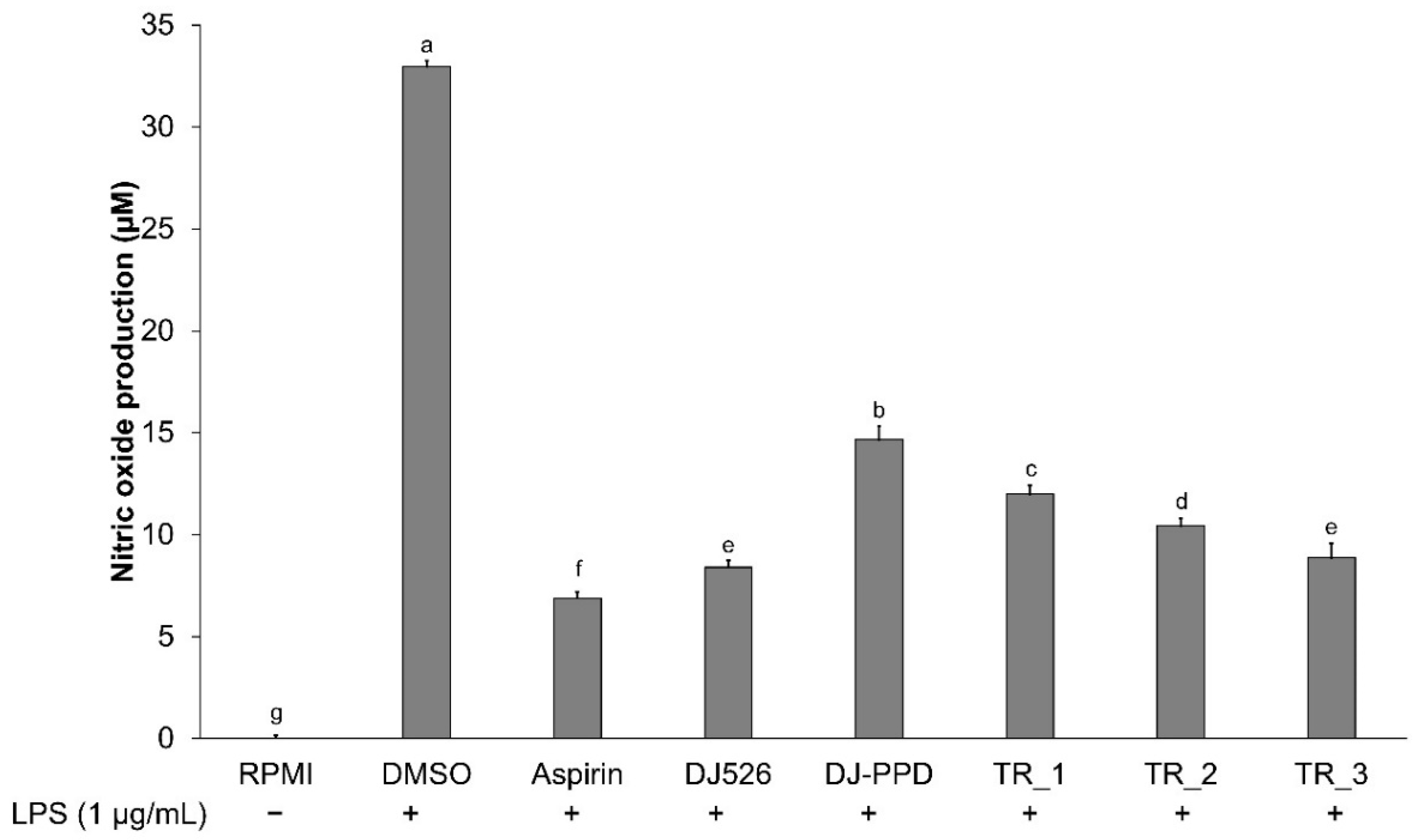
NO production in cultured media was measured using Griess reagent. Figure 2 shows that the production of NO was significantly higher in LPS-stimulated cells (DMSO group) (p < 0.05) than in the nontreated group (RPMI). However, the NO production in cell-cultured media significantly decreased in cells pretreated with aspirin, DJ526, DJ-PPD, TR_1, TR_2, and TR_3. As a positive control, aspirin at 200 ng/mL exhibited the highest inhibition of LPS-induced NO production in RAW264.7 cells among all treatments. The DJ526 treatment caused the highest inhibition of NO production among the treatments (74.49 ± 1.00% inhibition). For DJ-PPD, the LPS-induced NO production was significantly lower than that in the DMSO group (55.52 ± 2.04% reduction). Interestingly, the addition of DJ526 in the DJ-PPD treatment (TR_1 and TR_2) significantly reduced NO production in LPS-stimulated RAW264.7 cells when compared with that in cells treated with DJ-PPD alone. The TR_1 treatment markedly suppressed the LPS-induced NO production compared with that of DJ-PPD alone (18.22 ± 2.80% suppression). In addition, TR_2 markedly reduced NO production in LPS-stimulated RAW264.7 cells compared with that of DJ-PPD alone (28.77 ± 2.51% reduction). TR_3 treatment induced a similar inhibition of NO production compared with that after treatment with DJ526 alone. These results indicated that the addition of DJ526 in the DJ-PPD treatment significantly promoted LPS-induced NO production inhibition (p < 0.05).

2.3. Effect of Resveratrol- and PPD-Enriched Rice Extract on Inflammatory Cytokine Expression

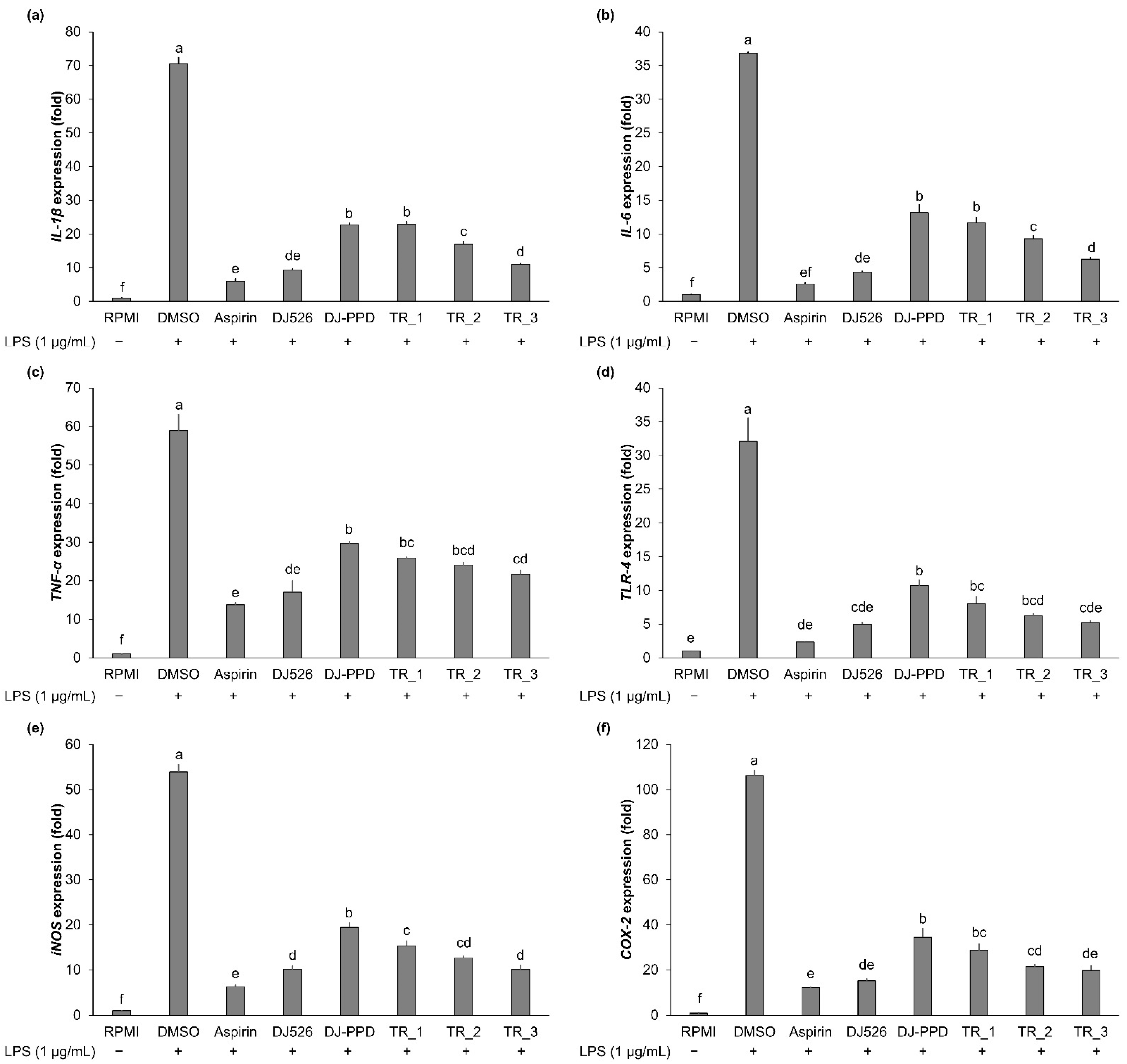
The expression level of immune-related genes was evaluated from cells stimulated with LPS for 6 h. The expression of these genes was upregulated in the LPS-stimulated RAW264.7 cells (DMSO group: Figure 3), whereas the expression of IL-1β, IL-6, TNF-α, TLR-4, iNOS, and COX-2 was downregulated in the rice-extract-treated cells. The DJ526 treatment at 100 µg/mL exhibited the highest suppression effect of immune-related genes among the treatments, which was similar to expression levels in aspirin-treated cells (positive control group). In comparison to the effect of the DJ526 treatment, treatment with TR_3 significantly decreased the expression of LPS-induced immune-related genes to the same levels as that of the DJ526 treatment. The TR_2 treatment markedly downregulated the expression of TNF-α, TLR-4, iNOS, and COX-2 to the same levels as that in the DJ526-treated cells. Moreover, the addition of 30% (w/w) of DJ526 in TR_1 significantly promoted the inhibited expression of LPS-induced immune-related genes to the same levels as found with DJ-PPD alone. These results indicate that the mixture of DJ526 and DJ-PPD significantly enhanced the inhibition of the LPS-induced immune-related gene expression.

2.4. Effect of Resveratrol- and PPD-Enriched Rice Extract on PGE2 Production

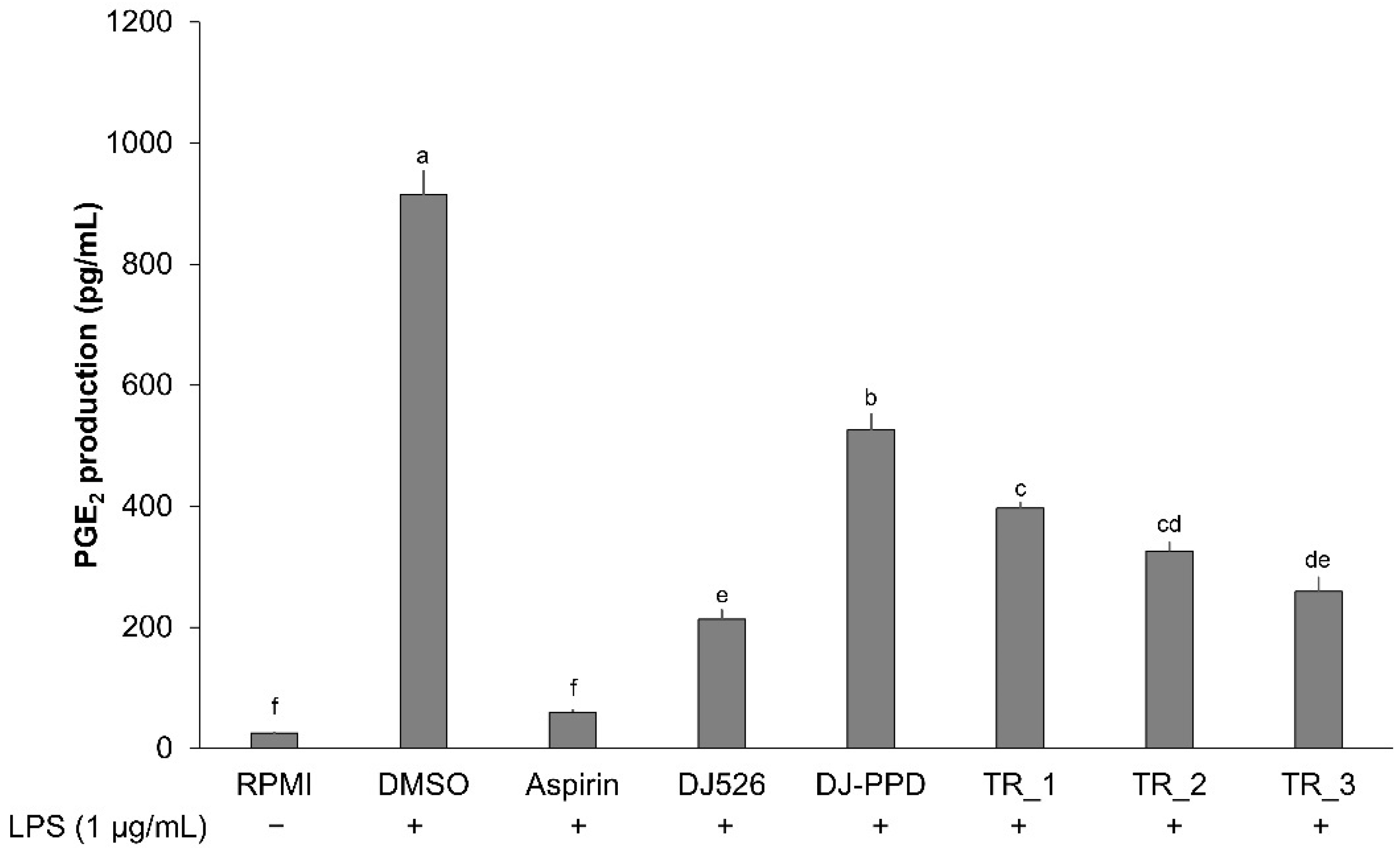
The production of PGE2 was increased when cells were stimulated with 1 µg/mL of LPS (Figure 4). The treatment of cells with rice seed extract significantly suppressed LPS-induced PGE2 production in comparison with that in cells treated with DMSO. The positive control (aspirin) inhibited PGE2 production to the same level as that in the negative control (RPMI)-treated cells. Treatment with DJ526 significantly decreased the inhibited production of PGE2 compared with that following treatment with DJ-PPD. In addition, treatment with TR_1, TR_2, and TR_3 significantly suppressed the production of PGE2 compared with that in the DJ-PPD group. Among the mixture samples, the highest inhibitory effect on PGE2 production was observed in the TR_3 group. These results indicate that the inhibitory effect on PGE2 production was correlated with increasing the DJ526 ratio in the DJ526/DJ-PPD mixture (r = −0.9883; Pearson’s correlation coefficient; degree of freedom at 10).

2.5. Effect of Resveratrol- and PPD-Enriched Rice Extract on Inflammatory Pathway Signaling

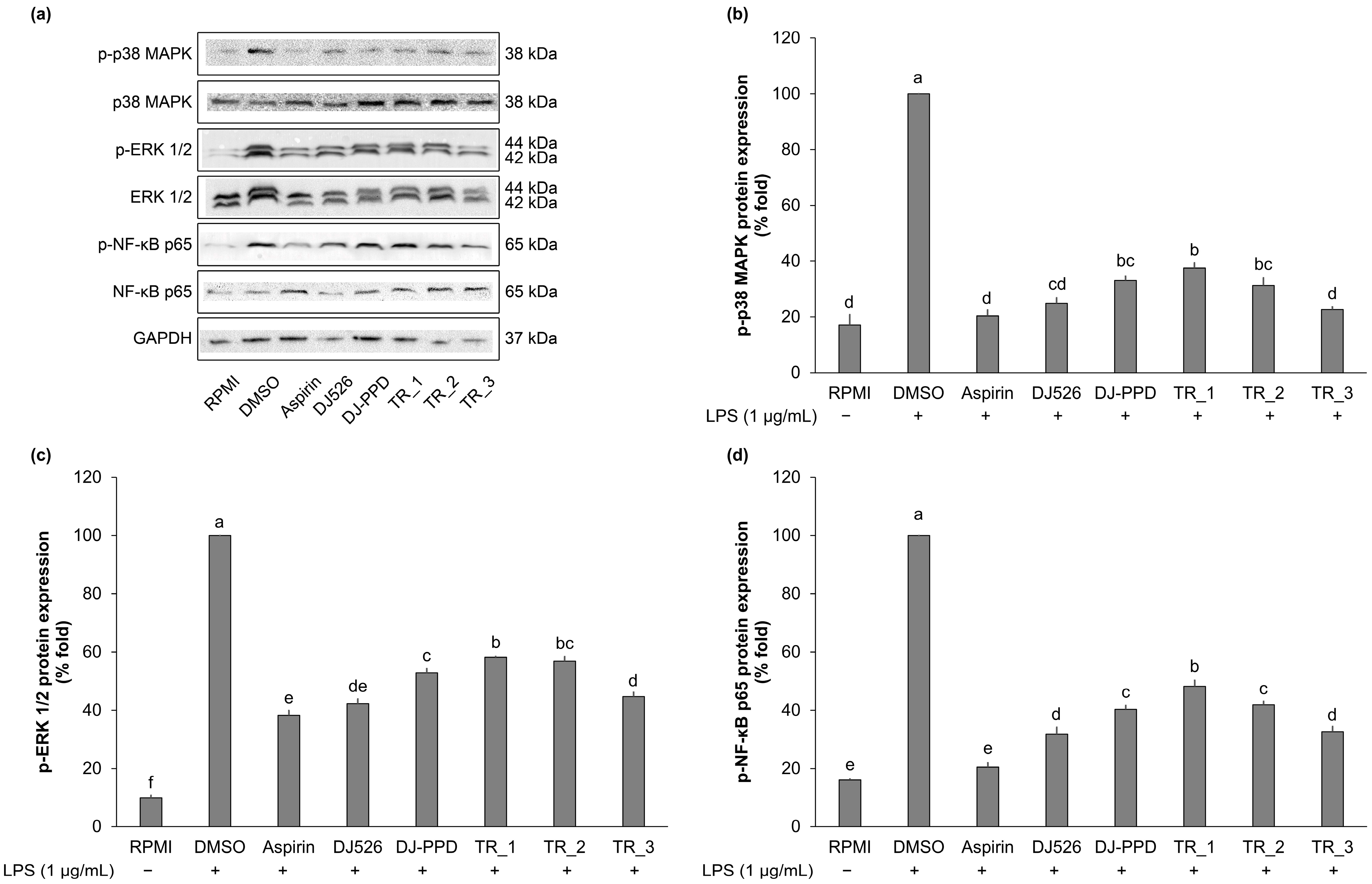
The highest expression of p-p38 MAPK, p-ERK 1/2, and p-NF-κB p65 was observed in DMSO-treated cells. Compared with that in DMSO-treated cells, the pretreatment with DJ526 and DJ-PPD and their mixture significantly inhibited the expression level of p-p38 MAPK, p-ERK 1/2, and p-NF-κB p65 (Figure 5). DJ526 produced the highest suppression of the expression of proteins associated with MAPK and NF-κB signaling pathways. Treatment with the mixture of DJ526 and DJ-PPD significantly decreased the expression of proteins associated with MAPK and NF-κB signaling pathways dose-dependently with the DJ526 ratio. Similar inhibition activities on p-p38 MAPK, p-ERK 1/2, and p-NF-κB p65 expression were observed in the TR_3 and DJ526 groups.

3. Discussion

LPS stimulation is frequently used to induce an in vitro inflammatory condition that mimics those in vivo [29,30,31,32,33]. LPS activates the NF-κB and MAPK signaling pathways via TLR-4 [34,35,36], an LPS receptor [37]. These pathways, when activated, generally result in an increase in the expression of known proinflammatory indicators, namely NO, iNOS, COX-2, IL-1β, IL-6, and TNF-α [38,39,40,41,42]. In addition, MAPK pathway also positively stimulates the production of prostaglandins and PGE2 [43,44]. The results of the present study showed synergistic anti-inflammatory regulation activity of resveratrol- and PPD-enriched rice seed extract against LPS-induced inflammatory conditions in RAW264.7 cells via the inhibition of the NF-κB and MAPK signaling pathways. Treatment of cells with TR_1-, TR_2-, and TR_3 downregulated the expression of the biomarkers p-p38 MAPK, p-ERK 1/2, and p-NF-κB p65 specifically with the DJ526 component in a dose-dependent manner (r = −0.9703; Pearson’s correlation coefficient). This inhibition reduced the proinflammatory biomarker content. The addition of DJ526 at 30%, 50%, and 70% (w/w) with DJ-PPD significantly inhibited the production of NO and PGE2 compared with that found after treatment with DJ-PPD alone (Figure 2 and Figure 4). In addition, the combination of 70% (w/w) of DJ526 and 30% (w/w) of DJ-PPD significantly inhibited the LPS-induced NO and PGE2 production at the same level as DJ526 treatment alone. The inhibitory effect on NO (r = −0.9479) and PGE2 (r = −0.9883) production correlated with the increase in the ratio of DJ526 in the DJ526/DJ-PPD mixture (Pearson’s correlation coefficient). Furthermore, treatment with DJ526 and DJ-PPD enhanced the inhibitory effect of the proinflammatory cytokines compared with that found with DJ-PPD treatment alone (Figure 3). The increasing amount of the DJ526 component in the DJ526/DJ-PPD mixture correlated with the level of inhibited expression of IL-1β (r = −0.9956), IL-6 (r = −0.9838), iNOS (r = −0.9645), COX-2 (r = −0.8849), TNF-α (r = −0.9553), and TLR-4 (r = −0.9204).
Our results are supported by those reported by Huangfu, et al. [45], who generated resveratrol and 20(S)-protopanaxadiol complex nanoparticles (RES@PPD, NPs) to investigate their anti-inflammatory and antioxidant activities. The treatment with NPs significantly suppressed the M1 polarization of macrophages, which are related to the secretion of proinflammatory cytokines [46], and promoted M2 polarization of macrophages, which are related to the inflammatory resolution or anti-inflammatory functions [47]. In addition, the NPs exhibited anti-inflammatory activities in the male Sprague–Dawley rat model of periodontitis by suppressing the expression of proinflammatory cytokines and increasing that of anti-inflammatory cytokines. Several studies have demonstrated the anti-inflammatory effects of resveratrol together with other compounds. Resveratrol combined with vitamin E significantly inhibited the phosphorylation of NF-κB p65, which then decreased the content of inflammatory mediators, such as NO, IL-1β, IL-6, TNF-α, and TLR-4 [48]. The combination of naproxen and resveratrol markedly promoted the anti-inflammatory activities by inhibiting the production of NO and inflammatory cytokines, such as TNF-α, COX-2, iNOS, and IL-6 by inhibiting the NF-κB, MAPK, and phosphoinositide-3 kinase (PI3K)/protein kinase B (Akt) signaling pathways [49]. The synergic effect of PPD has also been shown in several studies. Wang, et al. [50] reported that PPD effectively increased the effects of chemotherapy with 5-FU in patients with colorectal cancer by inhibiting the proliferation of HCT-116 human colorectal cancer cells when compared with the treatment of PPD or 5-FU alone. Ben-Eltriki, et al. [51] evaluated the combined effect of PPD and 1,25-dihydroxyvitamin D3 (calcitriol) and showed that treatment with PPD and calcitriol in combination significantly inhibited tumor growth in an androgen-independent C4-2 xenograft CRPC mouse model. The combination of PPD and cannabidiol (CBD-PPD coloading liposome, CP-liposomes) effectively inhibited the growth of the 4T1 breast cancer cell line more than that with CBD or PPD treatment alone [52]. In addition, administering resveratrol and Ligilactobacillus salivarius Li01 in combination significantly reduced the production of proinflammatory cytokines (IL-1β and IL-6) and increased that of an anti-inflammatory cytokine (IL-17A) in mouse serum [53].

4. Materials and Methods

4.1. Treatment Preparation

Treatments were prepared by DJ526 with DJ-PPD at various ratios (Table 1). DJ526 and DJ-PPD extracts are the same extracts as previously used [24,25]. DJ526 contained 4.724 and 2.605 ug/g dry weight of piceid and resveratrol, respectively [24], while DJ-PPD contained 7.28 ± 0.64 µg/g dry weight of protopanaxadiol [25]. After mixing, the treatments were prepared at 100 mg/mL in DMSO.

4.2. Cell Viability and NO Production

Cells in RPMI-1640 media (10% fetal bovine serum and 1% penicillin/streptomycin) at the concentration of 1 × 10⁵ cells were placed in a 96-well plate and the plate was incubated at 37 °C and 5% CO2 for 24 h. The cultured medium was discarded and the treatments (0.1% of DMSO, 200 ng/mL of aspirin, and 100 µg/mL of DJ526, DJ-PPD, TR_1, TR_2, and TR_3: 100 µL) were added into the specific wells. Aspirin at the concentration of 200 ng/mL served as a positive control. Aspirin, one of the most important members of nonsteroidal anti-inflammatory drugs, was used in treatment of inflammation [54]. Aspirin inhibits cyclooxygenase (COX), prostaglandins biosynthesis, and thromboxane biosynthesis [55]. Several studies demonstrated that aspirin is involved in anti-inflammatory processes through inhibiting the NF-κB and MAPK pathway [56,57,58], which led to the downregulation of NO, COX-2, PGE2, and proinflammatory cytokines [59,60,61]. After 1 h incubation, cells were stimulated with or without 1 µg/mL of LPS (Sigma-Aldrich, St. Louis, MO, USA). After 24 h stimulation, the cultured medium and stimulated cells were collected to assay NO production and cell viability. Culture media (100 µL) was transferred to a new 96-well plate and then mixed with a 100 µL working solution of Griess reagent (Sigma-Aldrich, St. Louis, MO, USA). The mixture was incubated for 15 min at room temperature in the dark and then the absorbance of the solution was measured at 540 nm. A standard curve of sodium nitrite was constructed for quantification of NO production (Figure 6). An EZ-Cytox Cell Viability Assay Kit (DoGenBio, Seoul, Republic of Korea) and stimulated cells were used for measuring the cytotoxicity of treatments. A working solution of an EZ-Cytox Cell Viability Assay Kit (10-fold dilution in 1× PBS: 110 µL) was added to stimulated cells, which were then incubated at 37 °C for 4 h. The solution (100 µL) was transferred to a new 96-well plate and the absorbance was measured at 450 nm. Cell viability was calculated by the following equation:
Cell   viability   ratio   ( % ) = An   absorbance   at   450   nm   of   treatment   group An   absorbance   at   450   nm   of   control   group   ×   100 ,
where control represents the untreated group.
The NO production in culture media from each treatment was calculated according to Equation (2).
Nitric   oxide   production   ( µ M ) = 91.084 x - 5.2501 ,  
where x represents the absorbance value at 540 nm.

4.3. RNA Extraction

Total RNA was extracted from each treatment group using a method described in the previous study [26]. Briefly, the treated cells were washed with ice-cold 1× PBS, lysed with TRI reagent™ (Invitrogen, Waltham, MA, USA), and then mixed with 200 µL of chloroform. After centrifugation, the upper-phase solution was transferred to the new tube. Total RNA was precipitated with isopropanol and the RNA pellet was washed with 70% EtOH. The RNA pellet was dissolved in nuclease-free water and kept at −80° until use.

4.4. RNA Quantification and cDNA Synthesis

The RNA concentration and RNA quality were evaluated using a SpectraMax® ABS Plus Microplate Reader (Molecular Device, San Jose, CA, USA) with a previously described method [62]. The RNA quality was shown as the ratio of A260:A280 and A260:A230 (acceptation range is 1.800–2.000; Table 2). The total RNA at 1 µg was used for the cDNA synthesis using a Power cDNA Synthesis Kit (Intron Biotechnology, Seongnam-si, Republic of Korea). The working cDNA solution was prepared at 5 ng/µL in nuclease-free water.

4.5. Inflammatory-Related Gene Expression Measurement

The mRNA expression levels were measured using a CFX Connect Real Time System (Bio-Rad, Hercules, CA, USA) in the total 20 µL of RealMOD™ Green W2 2 × qPCR mix (Intron Biotechnology). Specific primer sequences used were described in the previous study [62]. The PCR conditions are shown in Table 3.

4.6. PGE2 Production Assay

The cultured medium was collected and centrifuged at 3000 rpm for 10 min. The supernatant was used to determine the production of PGE2 with a PGE2 ELISA kit (Enzo Life Science, Farmingdale, NY, USA), according to the manufacturer’s instructions. Sample and standard solution (100 µL/well) were added into appropriate wells. Assay buffer was added into the nonsample binding (NSB: 150 µL/well) and maximum binding (Bo 100 µL/well) wells. The conjugate buffer (50 µL/well) was then added to each well, except for the total activity (TA) and blank wells, and then 50 µL of antibody was added per well, except for the blank, TA, and NSB wells. The plate was incubated at room temperature for 2 h at 500 rpm. Culture media were discarded and wells were washed with wash solution three times. After washing, the solution in the TA wells was mixed with conjugate solution (5 µL/well). The pNpp substrate solution (200 µL/well) was pipetted to each well and the plate was incubated at room temperature for 45 min. Finally, the stop solution (5 µL/well) was added per well and the absorbance was measured at 405 nm immediately. The PGE2 standard was generated by plotting the graph of percent bound versus concentration of PGE2 (Figure 7). The PGE2 production in the cultured medium was calculated according to Formula (3).
PGE 2   production   ( pg / mL )   =   543715 x 2 . 084 ,
where x represents the percent bound value.

4.7. Western Blot Analysis

The radioimmunoprecipitation assay buffer (Geneall Biotechnology, Seoul, Republic of Korea) was used for protein extraction from each treated culture. Cells were harvested using trypsin-EDTA and centrifugation at 13,000 rpm and 4 °C for 1 min. RIPA buffer supplemented with 1× Protease Inhibitor Cocktail Kit 5 (Bio-Medical Science Co., Ltd., Seoul, Republic of Korea) was added to the cell pellet. The solution was incubated on ice for 3 min and then centrifuged at 13,000 rpm and 4 °C for 30 min. The protein concentration in the resultant supernatant was measured using Bradford reagent (Sigma-Aldrich) by comparison with a bovine serum albumin standard curve. Protein sample (30 µg) from each treatment was separated in sodium dodecyl–sulfate polyacrylamide gel. The separated protein was transferred onto a nitrocellulose membrane (Immobilon®-P Transfer Membrane, Merck, Millipore, Burlington, MA, USA). The membrane was washed with 1× tris-buffered saline containing 0.1% Tween® 20 detergent (TBST) and was then incubated with 5% (w/v) skim milk in 1× TBST (blocking solution) at room temperature for 2 h. The membrane was incubated with antibodies in blocking solution at the ratio 1:1000 (v/v) specific for p38 MAPK (Santa Cruz Biotechnology, Dallas, TX, USA), NF-κB p65 (Santa Cruz Biotechnology), and ERK 1/2 (Santa Cruz Biotechnology). Membranes were incubated with antibodies in blocking solution at the ratio 1:2000 (v/v) specific for phosphorylation (p)-p38 MAPK (Cell Signaling Technology, Danvers, MA, USA), p-NF-κB p65 (Cell Signaling Technology), and p-ERK 1/2 (Cell Signaling Technology). Glyceraldehyde-3-phosphate dehydrogenase (GADPH, Santa Cruz Biotechnology) was used as the reference protein at 1:5000 (v/v) in TBST. Membranes were incubated with primary antibody at 4 °C overnight with mild agitation and then washed several times with 1× TBST at room temperature with strong agitation. Membranes were then incubated in 1:5000 (v/v: secondary antibody–blocking solution) of the secondary antibody (goat antirabbit IgG (H + L)-horseradish peroxidase (GenDEPOT, Barker, TX, USA) or m-IgG® BP-horseradish peroxidase (Santa Cruz Biotechnology)) at room temperature for 2 h. Membranes were incubated with Pierce ECL plus Western blotting substrate (Thermo Scientific™, Waltham, MA, USA) at room temperature for 10 min in the dark. Signals were captured, recorded, and the band intensity quantified using ChemiDoc Image System (Bio-Rad) and Image Lab Software (version 6, Bio-Rad).

4.8. Statistical Analysis

All calculations were performed using Statistix 8.1 (Statistix, Tallahassee, FL, USA). Treatment group means were compared by one-way analysis of variance followed by post hoc Duncan’s multiple range tests. A p-value < 0.05 was considered significant for all tests.

5. Conclusions

This study demonstrated the co-ordination anti-inflammatory effect of resveratrol- and PPD-enriched rice seed extracts on LPS-stimulated RAW264.7 cells. Treatment with DJ526 and DJ-PPD in combination significantly inhibited the MAPK and NF-κB signaling pathways. The cotreatment with DJ526 and DJ-PPD downregulated the production of NO and PGE2 and the expression of immune-associated genes in LPS-stimulated RAW264.7 cells. Therefore, we suggest that extracts of resveratrol- and PPD-enriched rice in a combination could be used as an anti-inflammatory agent.

Author Contributions

Conceptualization, S.-H.B.; Methodology, C.M.; Software, C.M.; Validation, C.M. and S.-H.B.; Formal Analysis, C.M.; Investigation, C.M.; Resources, S.-H.B.; Data Curation, C.M.; Writing—Original Draft Preparation, C.M.; Writing—Review and Editing, S.-H.B.; Visualization, S.-H.B.; Supervision, S.-H.B.; Project Administration, S.-H.B.; Funding Acquisition, S.-H.B. All authors have read and agreed to the published version of the manuscript.

Funding

This research received no external funding.

Institutional Review Board Statement

Not applicable.

Informed Consent Statement

Not applicable.

Data Availability Statement

All the applicable data have been provided in the manuscript. The authors will provide additional details if necessary.

Conflicts of Interest

The authors declare no conflicts of interest.

References

  1. Lee, E.-J.; Shin, S.-Y.; Kim, J.-K.; Woo, E.-R.; Kim, Y.-M. Anti-inflammatory effects of amentoflavone on modulation of signal pathways in LPS-stimulated RAW264.7 cells. Bull. Korean Chem. Soc. 2012, 33, 2878–2882. [Google Scholar] [CrossRef]
  2. Zhang, W.; Yan, J.; Wu, L.; Yu, Y.; Ye, R.D.; Zhang, Y.; Liang, X. In vitro immunomodulatory effects of human milk oligosaccharides on murine macrophage RAW264.7 cells. Carbohydr. Polym. 2019, 207, 230–238. [Google Scholar] [CrossRef] [PubMed]
  3. Dinarello, C.A. Proinflammatory and anti-inflammatory cytokines as mediators in the pathogenesis of septic shock. Chest 1997, 112 (Suppl. S6), 321s–329s. [Google Scholar] [CrossRef] [PubMed]
  4. Boniakowski, A.E.; Kimball, A.S.; Joshi, A.; Schaller, M.; Davis, F.M.; denDekker, A.; Obi, A.T.; Moore, B.B.; Kunkel, S.L.; Gallagher, K.A. Murine macrophage chemokine receptor CCR2 plays a crucial role in macrophage recruitment and regulated inflammation in wound healing. Eur. J. Immunol. 2018, 48, 1445–1455. [Google Scholar] [CrossRef]
  5. Chihara, N.; Aranami, T.; Sato, W.; Miyazaki, Y.; Miyake, S.; Okamoto, T.; Ogawa, M.; Toda, T.; Yamamura, T. Interleukin 6 signaling promotes anti-aquaporin 4 autoantibody production from plasmablasts in neuromyelitis optica. Proc. Natl. Acad. Sci. USA 2011, 108, 3701–3706. [Google Scholar] [CrossRef]
  6. Yencilek, F.; Yildirim, A.; Yilmaz, S.G.; Altinkilic, E.M.; Dalan, A.B.; Bastug, Y.; Isbir, T. Investigation of interleukin-1β polymorphisms in prostate cancer. Anticancer Res. 2015, 35, 6057–6061. [Google Scholar]
  7. Donath, M.Y.; Shoelson, S.E. Type 2 diabetes as an inflammatory disease. Nat. Rev. Immunol. 2011, 11, 98–107. [Google Scholar] [CrossRef]
  8. Conway, E.M.; Pikor, L.A.; Kung, S.H.; Hamilton, M.J.; Lam, S.; Lam, W.L.; Bennewith, K.L. Macrophages, Inflammation, and Lung Cancer. Am. J. Respir. Crit. Care Med. 2016, 193, 116–130. [Google Scholar] [CrossRef] [PubMed]
  9. Ramanan, M.; Sinha, S.; Sudarshan, K.; Aidhen, I.S.; Doble, M. Inhibition of the enzymes in the leukotriene and prostaglandin pathways in inflammation by 3-aryl isocoumarins. Eur. J. Med. Chem. 2016, 124, 428–434. [Google Scholar] [CrossRef]
  10. Kip, E.; Parr-Brownlie, L.C. Reducing neuroinflammation via therapeutic compounds and lifestyle to prevent or delay progression of Parkinson’s disease. Ageing Res. Rev. 2022, 78, 101618. [Google Scholar] [CrossRef]
  11. Fuggetta, M.P.; Bordignon, V.; Cottarelli, A.; Macchi, B.; Frezza, C.; Cordiali-Fei, P.; Ensoli, F.; Ciafrè, S.; Marino-Merlo, F.; Mastino, A.; et al. Downregulation of proinflammatory cytokines in HTLV-1-infected T cells by Resveratrol. J. Exp. Clin. Cancer Res. 2016, 35, 118. [Google Scholar] [CrossRef] [PubMed]
  12. Lopes Pinheiro, D.M.; Sales de Oliveira, A.H.; Coutinho, L.G.; Fontes, F.L.; de Medeiros Oliveira, R.K.; Oliveira, T.T.; Fonseca Faustino, A.L.; da Silva, V.L.; Araujo de Melo Campos, J.T.; Petta Lajus, T.B. Resveratrol decreases the expression of genes involved in inflammation through transcriptional regulation. Free Radic. Biol. Med. 2019, 130, 8–22. [Google Scholar] [CrossRef] [PubMed]
  13. Zhong, L.-M.; Zong, Y.; Sun, L.; Guo, J.-Z.; Zhang, W.; He, Y.; Song, R.; Wang, W.-M.; Xiao, C.-J.; Lu, D. Resveratrol inhibits inflammatory responses via the mammalian target of rapamycin signaling pathway in cultured LPS-stimulated microglial cells. PLoS ONE 2012, 7, e32195. [Google Scholar] [CrossRef] [PubMed]
  14. Panaro, M.A.; Carofiglio, V.; Acquafredda, A.; Cavallo, P.; Cianciulli, A. Anti-inflammatory effects of resveratrol occur via inhibition of lipopolysaccharide-induced NF-κB activation in Caco-2 and SW480 human colon cancer cells. Br. J. Nutr. 2012, 108, 1623–1632. [Google Scholar] [CrossRef]
  15. Ma, C.; Wang, Y.; Dong, L.; Li, M.; Cai, W. Anti-inflammatory effect of resveratrol through the suppression of NF-κB and JAK/STAT signaling pathways. Acta Biochim. Biophys. Sin. 2015, 47, 207–213. [Google Scholar] [CrossRef]
  16. Li, X.; Li, F.; Wang, F.; Li, J.; Lin, C.; Du, J. Resveratrol inhibits the proliferation of A549 cells by inhibiting the expression of COX-2. OncoTargets Ther. 2018, 11, 2981–2989. [Google Scholar] [CrossRef]
  17. Magrone, T.; Magrone, M.; Russo, M.A.; Jirillo, E. Recent advances on the anti-inflammatory and antioxidant properties of red grape polyphenols: In vitro and in vivo studies. Antioxidants 2020, 9, 35. [Google Scholar] [CrossRef]
  18. Martín, A.R.; Villegas, I.; La Casa, C.; de la Lastra, C.A. Resveratrol, a polyphenol found in grapes, suppresses oxidative damage and stimulates apoptosis during early colonic inflammation in rats. Biochem. Pharmacol. 2004, 67, 1399–1410. [Google Scholar] [CrossRef]
  19. Zykova, T.A.; Zhu, F.; Zhai, X.; Ma, W.Y.; Ermakova, S.P.; Lee, K.W.; Bode, A.M.; Dong, Z. Resveratrol directly targets COX-2 to inhibit carcinogenesis. Mol. Carcinog. 2008, 47, 797–805. [Google Scholar] [CrossRef]
  20. Ahmad, S.F.; Ansari, M.A.; Nadeem, A.; Alzahrani, M.Z.; Bakheet, S.A.; Attia, S.M. Resveratrol improves neuroimmune dysregulation through the inhibition of neuronal toll-like receptors and COX-2 signaling in BTBR T(+) Itpr3(tf)/J mice. Neuromol. Med. 2018, 20, 133–146. [Google Scholar] [CrossRef]
  21. Ruan, J.; Sun, F.; Zhang, Y.; Zheng, D.; Xiang, G.; Zhao, W.; Zhang, Y.; Wang, T. New 20(S)-protopanaxadiol type saponins from the leaves of Panax notoginseng and their potential anti-inflammatory activities. Steroids 2020, 162, 108696. [Google Scholar] [CrossRef] [PubMed]
  22. Lee, W.-M.; Kim, S.-D.; Kim, K.-S.; Kwak, Y.-S.; Park, H.-J.; Rhee, M.-H. Protopanaxadiol modulates LPS-induced inflammatory activity in murine macrophage RAW264.7 cells. J. Ginseng Res. 2006, 30, 181–187. [Google Scholar] [CrossRef]
  23. Lu, P.; Chen, J.; Chen, Y.; Quan, X.; Liu, J.; Han, Y.; Li, Y.; Yang, L.; Wan, J.-B.; Zhao, Y. 20(S)-Protopanaxadiol saponins isolated from Panax notoginseng target caveolin-1 against intestinal barrier dysfunction by alleviating inflammatory injury and oxidative stress in experimental murine colitis. Food Front. 2023, 4, 2081–2096. [Google Scholar] [CrossRef]
  24. Monmai, C.; Kim, J.-S.; Baek, S.-H. Use of germination to enhance resveratrol content and its anti-inflammatory activity in lipopolysaccharide-stimulated RAW264.7 cells. Molecules 2023, 28, 4898. [Google Scholar] [CrossRef] [PubMed]
  25. Monmai, C.; Kim, J.-S.; Baek, S.-H. Transgenic rice seed extracts exert immunomodulatory effects by modulating immune-related biomarkers in RAW264.7 macrophage cells. Nutrients 2022, 14, 4143. [Google Scholar] [CrossRef] [PubMed]
  26. Monmai, C.; Kim, J.-S.; Baek, S.-H. Effect of ginseng sapogenin protopanaxadiol-enriched rice (DJ-PPD) on immunomodulation. Plants 2023, 12, 767. [Google Scholar] [CrossRef]
  27. Baek, S.-H.; Shin, W.-C.; Ryu, H.-S.; Lee, D.-W.; Moon, E.; Seo, C.-S.; Hwang, E.; Lee, H.-S.; Ahn, M.-H.; Jeon, Y.; et al. Creation of resveratrol-enriched rice for the treatment of metabolic syndrome and related diseases. PLoS ONE 2013, 8, e57930. [Google Scholar] [CrossRef]
  28. Han, J.Y.; Baek, S.H.; Jo, H.J.; Yun, D.W.; Choi, Y.E. Genetically modified rice produces ginsenoside aglycone (protopanaxadiol). Planta 2019, 250, 1103–1110. [Google Scholar] [CrossRef]
  29. Cheng, D.; Zhu, C.; Liang, Y.; Xing, Y.; Shi, C. MiR-424 overexpression protects alveolar epithelial cells from LPS-induced apoptosis and inflammation by targeting FGF2 via the NF-κB pathway. Life Sci. 2020, 242, 117213. [Google Scholar] [CrossRef]
  30. Liu, X.; Yin, S.; Chen, Y.; Wu, Y.; Zheng, W.; Dong, H.; Bai, Y.; Qin, Y.; Li, J.; Feng, S.; et al. LPS-induced proinflammatory cytokine expression in human airway epithelial cells and macrophages via NF-κB, STAT3 or AP-1 activation. Mol. Med. Rep. 2018, 17, 5484–5491. [Google Scholar] [CrossRef]
  31. Ren, J.; Su, D.; Li, L.; Cai, H.; Zhang, M.; Zhai, J.; Li, M.; Wu, X.; Hu, K. Anti-inflammatory effects of aureusidin in LPS-stimulated RAW264.7 macrophages via suppressing NF-κB and activating ROS- and MAPKs-dependent Nrf2/HO-1 signaling pathways. Toxicol. Appl. Pharmacol. 2020, 387, 114846. [Google Scholar] [CrossRef] [PubMed]
  32. Tucureanu, M.M.; Rebleanu, D.; Constantinescu, C.A.; Deleanu, M.; Voicu, G.; Butoi, E.; Calin, M.; Manduteanu, I. Lipopolysaccharide-induced inflammation in monocytes/macrophages is blocked by liposomal delivery of G(i)-protein inhibitor. Int. J. Nanomed. 2018, 13, 63–76. [Google Scholar] [CrossRef] [PubMed]
  33. Zamyatina, A.; Heine, H. Lipopolysaccharide recognition in the crossroads of TLR4 and Caspase-4/11 mediated inflammatory pathways. Front. Immunol. 2020, 11, 585146. [Google Scholar] [CrossRef]
  34. Lu, Y.-C.; Yeh, W.-C.; Ohashi, P.S. LPS/TLR4 signal transduction pathway. Cytokine 2008, 42, 145–151. [Google Scholar] [CrossRef]
  35. Grylls, A.; Seidler, K.; Neil, J. Link between microbiota and hypertension: Focus on LPS/TLR4 pathway in endothelial dysfunction and vascular inflammation, and therapeutic implication of probiotics. Biomed. Pharmacother. 2021, 137, 111334. [Google Scholar] [CrossRef] [PubMed]
  36. Ciesielska, A.; Matyjek, M.; Kwiatkowska, K. TLR4 and CD14 trafficking and its influence on LPS-induced pro-inflammatory signaling. Cell. Mol. Life Sci. 2021, 78, 1233–1261. [Google Scholar] [CrossRef]
  37. Beutler, B. Tlr4: Central component of the sole mammalian LPS sensor. Curr. Opin. Immunol. 2000, 12, 20–26. [Google Scholar] [CrossRef]
  38. Cho, J.W.; Lee, K.S.; Kim, C.W. Curcumin attenuates the expression of IL-1beta, IL-6, and TNF-alpha as well as cyclin E in TNF-alpha-treated HaCaT cells; NF-kappaB and MAPKs as potential upstream targets. Int. J. Mol. Med. 2007, 19, 469–474. [Google Scholar] [CrossRef]
  39. Guha, M.; Mackman, N. LPS induction of gene expression in human monocytes. Cell Signal. 2001, 13, 85–94. [Google Scholar] [CrossRef]
  40. Liu, T.; Zhang, L.; Joo, D.; Sun, S.-C. NF-κB signaling in inflammation. Signal Transduct. Target. Ther. 2017, 2, 17023. [Google Scholar] [CrossRef]
  41. Hayden, M.S.; Ghosh, S. NF-κB in immunobiology. Cell Res. 2011, 21, 223–244. [Google Scholar] [CrossRef] [PubMed]
  42. Arthur, J.S.; Ley, S.C. Mitogen-activated protein kinases in innate immunity. Nat. Rev. Immunol. 2013, 13, 679–692. [Google Scholar] [CrossRef] [PubMed]
  43. Cho, W.; Choe, J. Prostaglandin E2 stimulates COX-2 expression via mitogen-activated protein kinase p38 but not ERK in human follicular dendritic cell-like cells. BMC Immunol. 2020, 21, 20. [Google Scholar] [CrossRef]
  44. Takai, E.; Tsukimoto, M.; Kojima, S. TGF-β1 downregulates COX-2 expression leading to decrease of PGE2 production in human lung cancer A549 cells, which is involved in fibrotic response to TGF-β1. PLoS ONE 2013, 8, e76346. [Google Scholar] [CrossRef] [PubMed]
  45. Huangfu, H.; Du, S.; Zhang, H.; Wang, H.; Zhang, Y.; Yang, Z.; Zhang, X.; Ren, S.; Chen, S.; Wang, C.; et al. Facile engineering of resveratrol nanoparticles loaded with 20(S)-protopanaxadiol for the treatment of periodontitis by regulating the macrophage phenotype. Nanoscale 2023, 15, 7894–7908. [Google Scholar] [CrossRef]
  46. Lee, J.H.; Moon, S.H.; Kim, H.S.; Park, E.; Ahn, D.U.; Paik, H.D. Immune-enhancing activity of phosvitin by stimulating the production of pro-inflammatory mediator. Poult Sci. 2017, 96, 3872–3878. [Google Scholar] [CrossRef]
  47. Jablonski, K.A.; Amici, S.A.; Webb, L.M.; Ruiz-Rosado Jde, D.; Popovich, P.G.; Partida-Sanchez, S.; Guerau-de-Arellano, M. Novel markers to delineate murine M1 and M2 macrophages. PLoS ONE 2015, 10, e0145342. [Google Scholar] [CrossRef]
  48. Wang, N.; Xu, C.; Li, N.; Wang, F.; Wang, F.; Li, Z.; Yu, Q.; Zhang, G. Synergistic anti-inflammatory effects of resveratrol and vitamin E in lipopolysaccharide-induced RAW264.7 cells. Food Sci. Technol. 2022, 42, e24122. [Google Scholar] [CrossRef]
  49. Ou, Y.; You, Z.; Yao, M.; Cao, Y.; Xue, X.; Chen, M.; Wu, R.; Gan, L.; Li, D.; Wu, P.; et al. Naproxen-derived new compound inhibits the NF-κB, MAPK and PI3K/Akt signaling pathways synergistically with resveratrol in RAW264.7 cells. Molecules 2023, 28, 3395. [Google Scholar] [CrossRef]
  50. Wang, C.Z.; Zhang, Z.; Wan, J.Y.; Zhang, C.F.; Anderson, S.; He, X.; Yu, C.; He, T.C.; Qi, L.W.; Yuan, C.S. Protopanaxadiol, an active ginseng metabolite, significantly enhances the effects of fluorouracil on colon cancer. Nutrients 2015, 7, 799–814. [Google Scholar] [CrossRef]
  51. Ben-Eltriki, M.; Deb, S.; Shankar, G.; Meckling, G.; Hassona, M.; Yamazaki, T.; Fazli, L.; Chin, M.Y.; Tomlinson Guns, E.S. Anti-tumor effects of ginsenoside 20(S)-protopanaxadiol and 1,25-dihydroxyvitamin D3 combination in castration resistant prostate cancer. Medicines 2021, 8, 28. [Google Scholar] [CrossRef] [PubMed]
  52. Fu, J.; Zhang, K.; Lu, L.; Li, M.; Han, M.; Guo, Y.; Wang, X. Improved therapeutic efficacy of CBD with good tolerance in the treatment of breast cancer through nanoencapsulation and in combination with 20(S)-protopanaxadiol (PPD). Pharmaceutics 2022, 14, 1533. [Google Scholar] [CrossRef] [PubMed]
  53. Fei, Y.; Zhang, S.; Han, S.; Qiu, B.; Lu, Y.; Huang, W.; Li, F.; Chen, D.; Berglund, B.; Xiao, H.; et al. The role of dihydroresveratrol in enhancing the synergistic effect of ligilactobacillus salivarius Li01 and resveratrol in ameliorating colitis in mice. Research 2022, 2022, 9863845. [Google Scholar] [CrossRef] [PubMed]
  54. Uzzaman, M.; Mahmud, T. Structural modification of aspirin to design a new potential cyclooxygenase (COX-2) inhibitors. Silico Pharmacol. 2020, 8, 1. [Google Scholar] [CrossRef] [PubMed]
  55. Chiang, N.; Serhan, C.N. Aspirin triggers formation of anti-inflammatory mediators: New mechanism for an old drug. Discov. Med. 2004, 4, 470–475. [Google Scholar]
  56. Liu, Y.; Fang, S.; Li, X.; Feng, J.; Du, J.; Guo, L.; Su, Y.; Zhou, J.; Ding, G.; Bai, Y.; et al. Aspirin inhibits LPS-induced macrophage activation via the NF-κB pathway. Sci. Rep. 2017, 7, 11549. [Google Scholar] [CrossRef]
  57. Vartiainen, N.; Goldsteins, G.; Keksa-Goldsteine, V.; Chan, P.H.; Koistinaho, J. Aspirin inhibits p44/42 mitogen-activated protein kinase and is protective against hypoxia/reoxygenation neuronal damage. Stroke 2003, 34, 752–757. [Google Scholar] [CrossRef]
  58. Zeng, Y.P.; Yang, C.; Li, Y.; Fan, Y.; Yang, H.J.; Liu, B.; Sang, H.X. Aspirin inhibits osteoclastogenesis by suppressing the activation of NF-κB and MAPKs in RANKL-induced RAW264.7 cells. Mol. Med. Rep. 2016, 14, 1957–1962. [Google Scholar] [CrossRef]
  59. Kim, J.; Lee, K.-S.; Kim, J.-H.; Lee, D.-K.; Park, M.; Choi, S.; Park, W.; Kim, S.; Choi, Y.K.; Hwang, J.Y.; et al. Aspirin prevents TNF-α-induced endothelial cell dysfunction by regulating the NF-κB-dependent miR-155/eNOS pathway: Role of a miR-155/eNOS axis in preeclampsia. Free Radic. Biol. Med. 2017, 104, 185–198. [Google Scholar] [CrossRef]
  60. Darby, I.A.; Weller, C.D. Aspirin treatment for chronic wounds: Potential beneficial and inhibitory effects. Wound Repair Regen. 2017, 25, 7–12. [Google Scholar] [CrossRef]
  61. Müller, N. COX-2 inhibitors, aspirin, and other potential anti-inflammatory treatments for psychiatric disorders. Front. Psychiatry 2019, 10, 375. [Google Scholar] [CrossRef] [PubMed]
  62. Monmai, C.; Kim, J.-S.; Baek, S.-H. Anti-inflammatory efficacy of resveratrol-enriched rice callus extract on lipopolysaccharide-stimulated RAW264.7 macrophages. Immuno 2024, 4, 131–146. [Google Scholar] [CrossRef]
Figure 1. Effect of resveratrol- and PPD-enriched rice seed extracts on the viability of LPS-stimulated RAW264.7 cells. The concentrations of DMSO, aspirin, and treatments (rice seed extract and mixture) were 0.1%, 200 ng/mL, and 100 µg/mL, respectively.
Molecules 29 04343 g001
Figure 2. Effect of resveratrol- and PPD-enriched rice seed extracts (DJ526 and DJ-PPD, respectively) on the NO production in LPS-stimulated RAW264.7 cells. The concentrations of DMSO, aspirin, and treatments (rice seed extract and mixture) were 0.1%, 200 ng/mL, and 100 µg/mL, respectively. The significant differences in NO production are presented as lowercase letters at p < 0.05.
Molecules 29 04343 g002
Figure 3. Effect of resveratrol- and PPD-enriched rice seed extracts (DJ526 and DJ-PPD, respectively) on the immune-related gene expression in LPS-stimulated RAW26.7 cells. The effect on (a) IL-1β, (b) IL-6, (c) TNF-α, (d) TLR-4, (e) iNOS, and (f) COX-2 expression. The concentrations of DMSO, aspirin, and treatments (rice seed extract and mixture) were 0.1%, 200 ng/mL, and 100 µg/mL, respectively. The significant differences in gene expression levels are presented as lowercase letters at p < 0.05.
Molecules 29 04343 g003
Figure 4. Effect of resveratrol- and PPD-enriched rice seed extracts (DJ526 and DJ-PPD, respectively) on PGE2 production in LPS-stimulated RAW264.7 cells. The concentrations of DMSO, aspirin, and treatments (rice seed extract and mixture) were 0.1%, 200 ng/mL, and 100 µg/mL, respectively. The significant differences in PGE2 production are presented as lowercase letters at p < 0.05.
Molecules 29 04343 g004
Figure 5. Effect of resveratrol- and PPD-enriched rice seed extracts (DJ526 and DJ-PPD, respectively) on the expression of immune-associated proteins in LPS-stimulated RAW264.7 cells. (a) Representative of blot analysis, (b) densitometric analyses of p-p38 MAPK protein expression, (c) densitometric analyses of p-ERK 1/2 protein expression, and (d) densitometric analyses of p-NF-κB p65 protein expression. The concentrations of DMSO, aspirin, and treatments (rice seed extract and mixture) were 0.1%, 200 ng/mL, and 100 µg/mL, respectively. The significant differences in protein expression are presented as lowercase letters at p < 0.05.
Molecules 29 04343 g005
Figure 6. The calibration curve of NaNO2 (0.00–100.00 µM).
Molecules 29 04343 g006
Figure 7. Standard curve of PGE2 production over the range of 39.0625–2500 pg. OD = optical density.
Molecules 29 04343 g007
Table 1. Treatment preparation used in this study.
TreatmentDJ526
(% w/w)
DJ-PPD
(% w/w)
Piceid Content
(×10−2 ng)
Resveratrol Content
(×10−2 ng)
PPD Content
(×10−2 ng)
DJ526100047.226.10.0
DJ-PPD01000.00.072.8
TR_1307014.27.851.0
TR_2505023.613.136.4
TR_3703033.018.321.8
Table 2. The quantity and quality of total RNA.
TreatmentA260:A280A260:A230Concentration (ng/µL)SD *CV **
Nontreatment (RPMI)1.9741.851351.498.122.31
DMSO1.9961.832389.9712.883.30
Aspirin1.9931.942802.4029.993.74
DJ5262.0011.905826.9317.332.10
DJ-PPD1.9891.988885.2330.783.48
TR_11.9691.841827.0311.241.36
TR_21.9971.915836.1917.222.06
TR_31.9751.887958.7912.051.26
* Standard deviation (SD). ** Coefficient of variation (CV).
Table 3. PCR reaction conditions used in this experiment.
ProcessTemperature (°C)TimeNumber of Cycles
Predenaturation9510 min1 cycle
Denaturation9520 s40 cycles
Annealing6020 s
Elongation7230 s
Final elongation725 min1 cycle
Disclaimer/Publisher’s Note: The statements, opinions and data contained in all publications are solely those of the individual author(s) and contributor(s) and not of MDPI and/or the editor(s). MDPI and/or the editor(s) disclaim responsibility for any injury to people or property resulting from any ideas, methods, instructions or products referred to in the content.