Article

A Phytotoxin with Selective Herbicidal Activity and Related Metabolites from the Phytopathogenic Fungus Bipolaris cookei SYBL03

by
Haiyan Li
1,2,
Jingzhuo Hou
2,
Bing Li
2,
Lizhong Zhang
3 and
Zhiguo Yu
1,*
1
College of Plant Protection, Shenyang Agricultural University, Shenyang 110866, China
2
College of Environmental and Safety Engineering, Shenyang University of Chemical Technology, Shenyang 110142, China
3
College of Computer Science and Technology, Shenyang University of Chemical Technology, Shenyang 110142, China
*
Author to whom correspondence should be addressed.
Molecules 2024, 29(13), 3040; https://doi.org/10.3390/molecules29133040
Submission received: 22 May 2024 / Revised: 21 June 2024 / Accepted: 24 June 2024 / Published: 26 June 2024
(This article belongs to the Section Natural Products Chemistry)

Abstract

:
Weeds are a serious threat to crop production, and the utilization of secondary metabolites of phytopathogenic fungi is considered to be an effective method of weed control. In this study, eight compounds were isolated and purified from the mycelium and fermentation broth extracts of Bipolaris cookei SYBL03. The compounds (18), except 2 and 6, are reported for the first time from this genus. The herbicidal activities of compounds 18 were studied by evaluating their effects on the seed germination and seedling growth of monocotyledonous and dicotyledonous weeds. The results indicated that compound 7 (Cyclo-N-methylphenylalanyltryptophenyl, cNMPT) exhibited a concentration-dependent dual effect on the growth of weed seedlings and selective herbicidal activity against dicotyledonous weeds. We further investigated the morphological and physiological responses of roots of Amaranthus retroflexus, a dicotyledonous weed, to compound 7. Some changes were found in seedlings grown in 400 μg/mL compound 7 solution for 96 h, such as shortening and swelling of elongation zone cells, reduced number and length of root hairs, damage and wrinkling of the root surface, occurrence of electrolyte leakage, and an increase in ethylene content. These results suggest that compound 7 may exert herbicidal activity by causing stress to weed seedlings. Increased ethylene production could be involved in the response of plants to compound 7.

1. Introduction

It is estimated that the global population will reach 9.7 billion by 2050 [1], leading to increased demand for food production. According to the Food and Agriculture Organization of the United Nations (FAO), at least a 50% increase in agricultural food production will be needed to meet the demand [2]. In agricultural production, various factors like animal pests, plant pathogens, and weeds can impact on grain yield, and weeds are responsible for the highest potential loss (34%) [3]. Currently, herbicides are widely used as the most economical method for controlling weeds all over the world [4].
Herbicides are employed to effectively manage and eliminate weeds, with the goal of protecting arable land, improving crop productivity, and promoting agricultural sustainable development [5]. Additionally, they can also reduce labor and machinery costs and increase profits. Nevertheless, the prevalent utilization of chemical herbicides leads to significant problems, including ecological degradation, soil pollution, destruction of beneficial microorganisms, pesticide resistance, and contamination of agricultural products, posing substantial health risks to humans and animals [6]. Consequently, there is an immediate necessity to explore newer environmentally friendly herbicides from natural resources, including metabolites from animals, plants, and microbes [7].
Many plant pathogens have been researched and identified as potential agents for controlling weeds in various arable crops [8]. Research indicates that the action of various phytopathogenic fungi is attributed to secondary metabolites, including terpenoids, polyketides, nonribosomal peptides, and compounds produced from shikimic acid [9]. Many of them are non-host-specific and have inhibitory effects, not only on host plants but also on others [10], so they have the potential to be developed into natural herbicides [11]. So far, several non-host-specific phytotoxins have been afforded by the genus Bipolaris [12], such as cochlioquinone derivatives [13,14], prehelminthosporol, dihydroprehelminthosporol, victoxinine, and prehelminthosporolactone [15].
Bipolaris cookei is a necrotrophic fungus that causes a sorghum disease called target leaf spot, and it can give rise to a 50% reduction in production during a serious outbreak [16,17]. The current research on the B. cookei mainly focuses on the pathogenic mechanism, gene sequence determination, and the process of host cell invasion [16,17,18]. B. cookei not only infects sorghum but also other species, such as rice [19], wheat [20], and maize [21]. Winder and Dyke [22] prepared a solution of B. cookei (=B. sorghicola) (isolated BS1) spores and sprayed it on two-week-old Johnson grass. Seven days after the inoculation, BS1 killed 100% of the Johnson grass. This report suggests that B. cookei may produce potent phytotoxic metabolites. In 1987, Sugawara et al. [23] isolated six ophiobolin compounds from B. cookei (=Drechslera sorghicola). These were all capable of causing brownish lesions in corn, Johnson grass, and sorghum. In 1989, Pena-Rodriguez and Chilton [24] isolated four ophiobolin compounds from the ethyl acetate (EtOAc) extract of the fermentation broth of B. cookei (=B. sorghicola). The lesions caused by these compounds on maize (Zea mays), bentgrass (Agrostis alba), sicklepod (Cassia obtusifolia), and morning glory (Ipomoea nil) were all necrotic spots similar to those found on sorghum. These results indicate that B. cookei has the ability to produce non-host-specific toxins for weed control. However, there has been no further research on the toxins produced by this plant pathogenic fungus except ophiobolin compounds.
To discover more structurally intriguing and bioactive metabolites from this fungus, we isolated and purified the EtOAc extracts from broth and mycelia of B. cookei SYBL03. And then, we selected six common weed species from different genera in Northeast China as targets to evaluate the herbicidal activity of the isolated compounds.

2. Results

2.1. Isolation, Purification, and Structure Elucidation

The EtOAc extracts of mycelia and fermentation supernatant of B. cookei SYBL03 were subjected to column chromatography (CC) on silica gel to provide six (Fr. A–F) and five fractions (Fr. A′–E′), respectively. The fractions were further separated by CC over silica gel, Sephadex LH-20 and semi-preparative HPLC, and recrystallization to afford eight compounds. Their structures were identified as 9,11-dehydroergosterol peroxide (=5α,8α-epidioxyergosta-6,9(11),22-trien-3β-ol) (1) [25], ergosterol peroxide (=5α,8α-epidioxyergosta-6,22-dien-3β-ol) (2) [25], (–)-ditryptophenaline (3) [26,27], 1,3,9-trimethyluric acid (4), cerebroside A (5) [28], cerebroside B (6) [29], cyclo-N-methylphenylalanyltryptophenyl (7) [26], and (2S)-3-di-1H-indol-3-yl-1,2-propanediol (8) [30] (Figure 1), based on their spectroscopic data (Figures S1–S25, Supplementary Materials) and compared with reported values in the previous literature.
According to the 1H and 13C NMR data, the structure of compound 4 was very similar to that of 8-oxocaffeine [31]. But the analysis of the HMBC correlations allowed for the position of three methyl groups at N-1, N-3 and N-9, respectively (Figure S12).

2.2. Phytotoxic Activity of Compounds 18

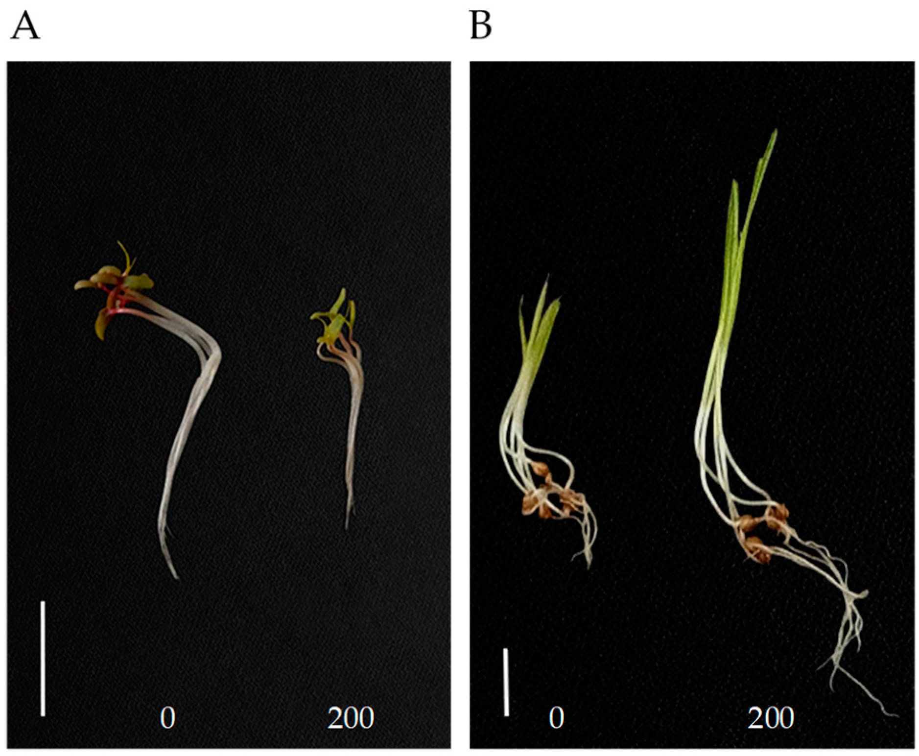
We investigated the effects of eight compounds at 200 μg/mL on the seedling growth of Amaranthus retroflexus and Echinochloa crus-galli. Compounds 36 and 8 showed no phytotoxicity to the seedling growth of two weeds. Compound 1 had weaker inhibitory activity against the seedling growth of A. retroflexus. Compounds 2 and 7 had obvious inhibitory effects on A. retroflexus, with inhibition rates of 39% and 46% against shoot growth, and 56% and 52% against root growth, respectively (Table 1 and Figure 2A). Interestingly, compound 7 significantly promoted the growth of E. crus-galli; the shoot and root length increased by 46% and 135% as compared with control, respectively (Table 1 and Figure 2B). These results indicated that the effects of compound 7 on seedling growth may be different between monocots and dicots.

2.3. Effect of Compound 7 on the seedling growth of Monocots and Dicots

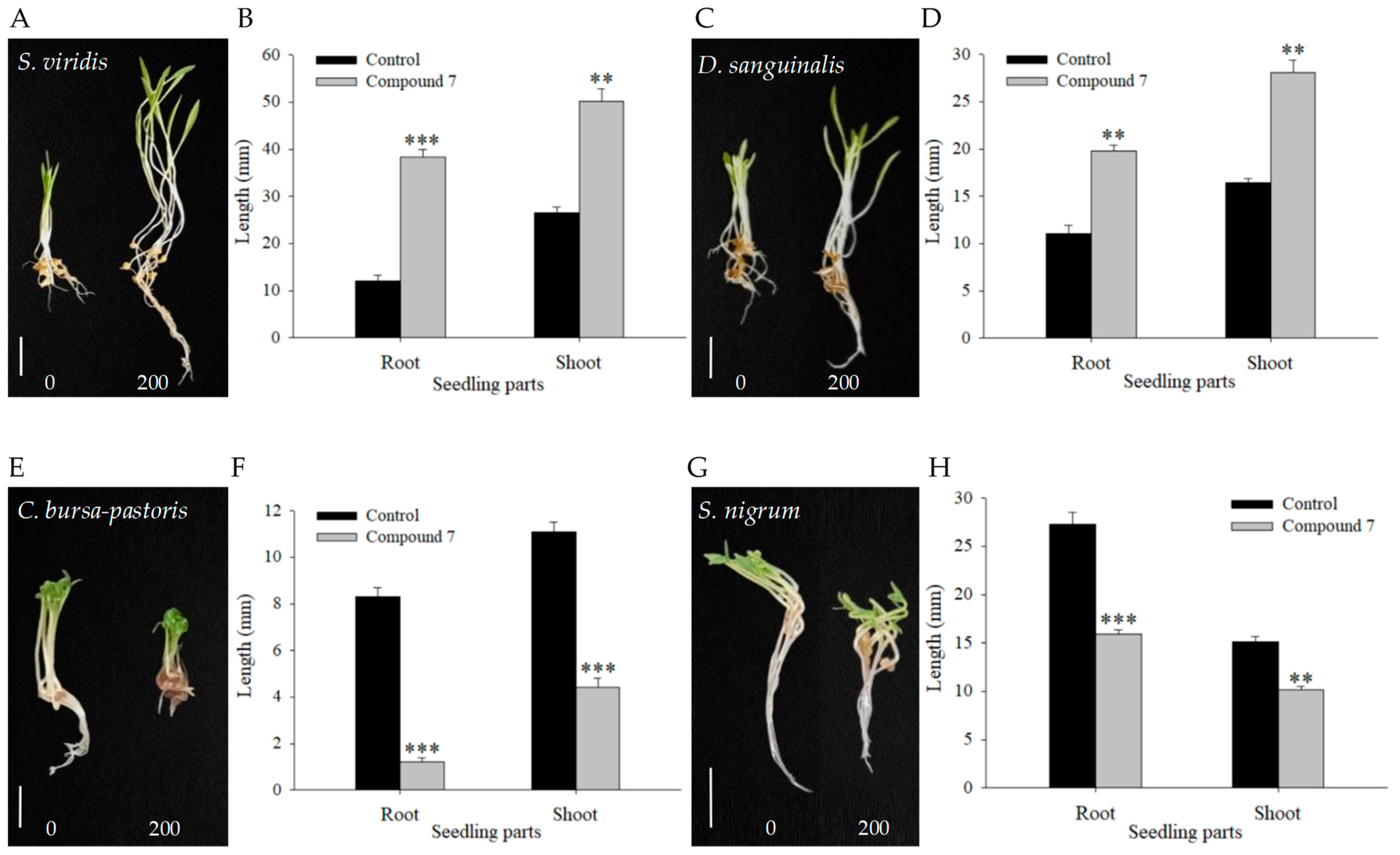
The following experiment aims to further ascertain whether the different effects of compound 7 on monocots and dicots are universal. Two dicots (Solanum nigrum, Capsella bursa-pastoris) and two monocots (Setaria viridis, Digitaria sanguinalis) were selected as test weeds for the bioassay to estimate the influence of compound 7 on seedling growth at 200 μg/mL. The results showed that compound 7 had a growth-promoting effect on monocots. At 96 h after treatment, the shoot and root length of S. viridis increased by 89% and 216% (Figure 3A,B), and those of D. sanguinalis increased by 71% and 80% compared with the corresponding controls, respectively (Figure 3C,D). However, compound 7 showed a growth inhibitory effect on the seedling growth of dicots. The shoot and root lengths of C. bursa-pastoris were inhibited by 60% and 86% at 144 h after treatment, and those of S. nigrum inhibited by 32% and 42% at 96 h following treatment compared with the corresponding controls, respectively (Figure 3E–H). To sum up, compound 7 had different effects on monocots and dicots, promoting the growth of monocots and inhibiting the growth of dicots at a certain concentration.

2.4. Effect of Compound 7 at Different Concentrations on Dicotyledons

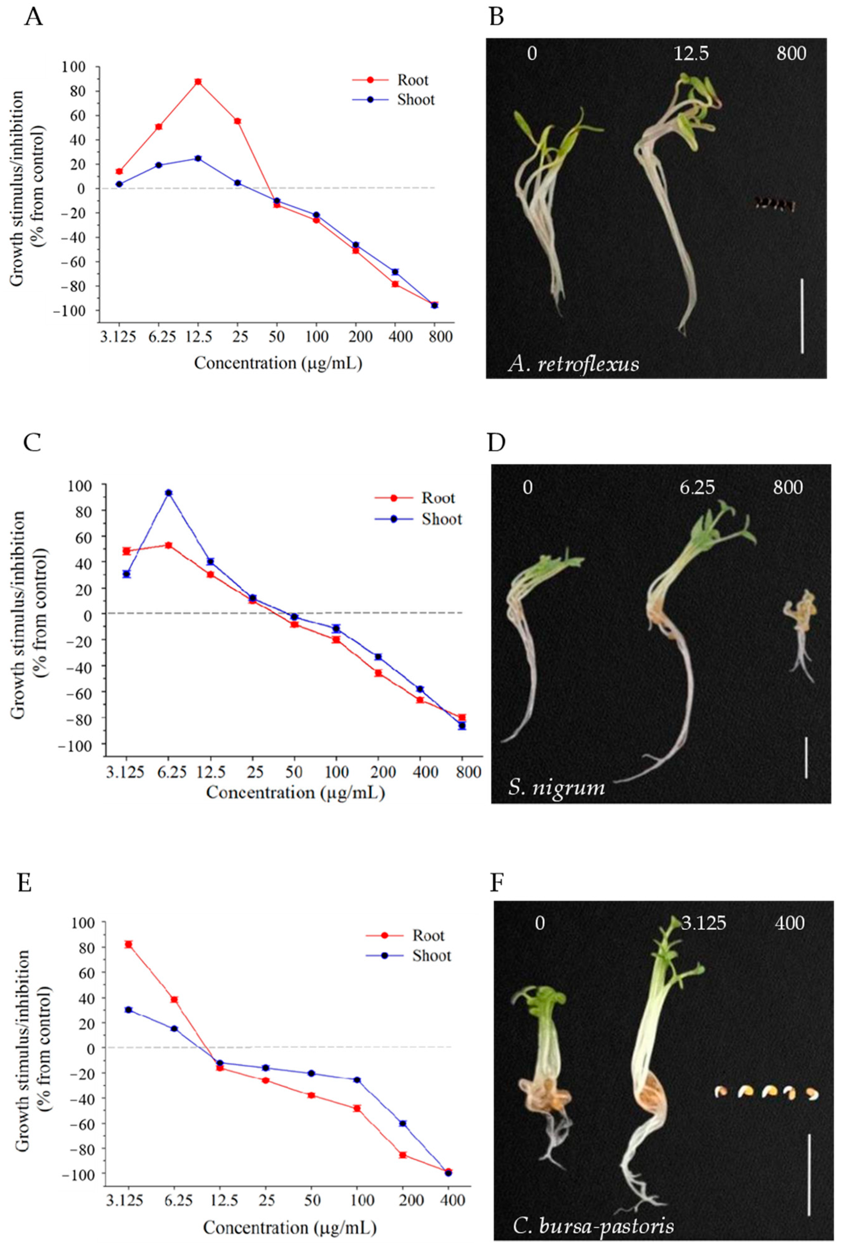
We further investigated the effect of compound 7 on the growth of dicotyledonous weeds (A. retroflexus, S. nigrum and C. bursa-pastoris) in a relatively wide concentration range (3.25–800 µg/mL). With increased treatment concentration, the root and shoot growth of three dicotyledonous weeds changed from promotion to inhibition compared with the control group (Figure 4A,C,E). Compound 7 at lower concentrations enhanced the growth of A. retroflexus (≤25 μg/mL), S. nigrum (≤25 μg/mL), and C. bursa-pastoris (≤6.25 μg/mL). These concentrations were significantly lower than the growth-promoting concentration (200 μg/mL) of three monocotyledonous weeds. The maximum growth promotion effects on A. retroflexus, S. nigrum, and C. bursa-pastoris seedlings were observed at concentrations of 12.5, 6.25, and 3.125 μg/mL, respectively. At the optimal growth-promoting concentration, the growth of roots and shoots separately increased by 88% and 25% for A. retroflexus (Figure 4A), 53%, and 93% for S. nigrum (Figure 4C), and 82% and 30% for C. bursa-pastoris (Figure 4E) compared with control. At a relatively high concentration, these seedlings began to exhibit concentration-dependent growth inhibition. At the highest treatment concentration (800 μg/mL), the seedling growth of A. retroflexus and C. bursa-pastoris almost completely stopped (Figure 4A,E), and the root and shoot growth of S. nigrum seedlings reduced by 80% and 86% compared with control (Figure 4C), respectively. The optimal promotion and inhibition effects of compound 7 on the three dicotyledons are shown in Figure 4B,D,F. At moderate inhibitory concentrations, compound 7 showed stronger inhibition against roots than shoots for the three dicotyledonous weeds, suggesting that roots are more susceptible to compound 7 than shoots. The results indicate that the effect of compound 7 on plants depends on its concentration, which promotes growth at low concentrations, inhibits growth at high concentrations, and completely blocks growth at higher concentrations.

2.5. Effect of Compound 7 on Seed Germination

An investigation was conducted to examine the effect of compound 7 on seed germination at 200 µg/mL. The results showed that the germination process and rate of the monocots (S. viridis, D. sanguinalis and E. crus-galli) were not significantly affected. In contrast, among the dicots (A. retroflexus, S. nigrum, and C. bursa-pastoris), the seed germination of A. retroflexus and S. nigrum was delayed, although the germination rate after 7 d remained unaffected (Figure 5A,B). However, the germination of C. bursa-pastoris seeds was significantly inhibited (Figure 5C).
Therefore, we further investigated the effects of different concentrations (3.125–400 µg/mL) of compound 7 on the seed germination of C. bursa-pastoris. The results showed that the seed germination of C. bursa-pastoris was inhibited by compound 7 in a concentration-dependent manner at concentrations greater than 6.25 µg/mL, and completely inhibited at a concentration of 400 µg/mL (Figure 5D,E).

2.6. Inhibitory Mechanism of Compound 7 on A. retroflexus Root Growth

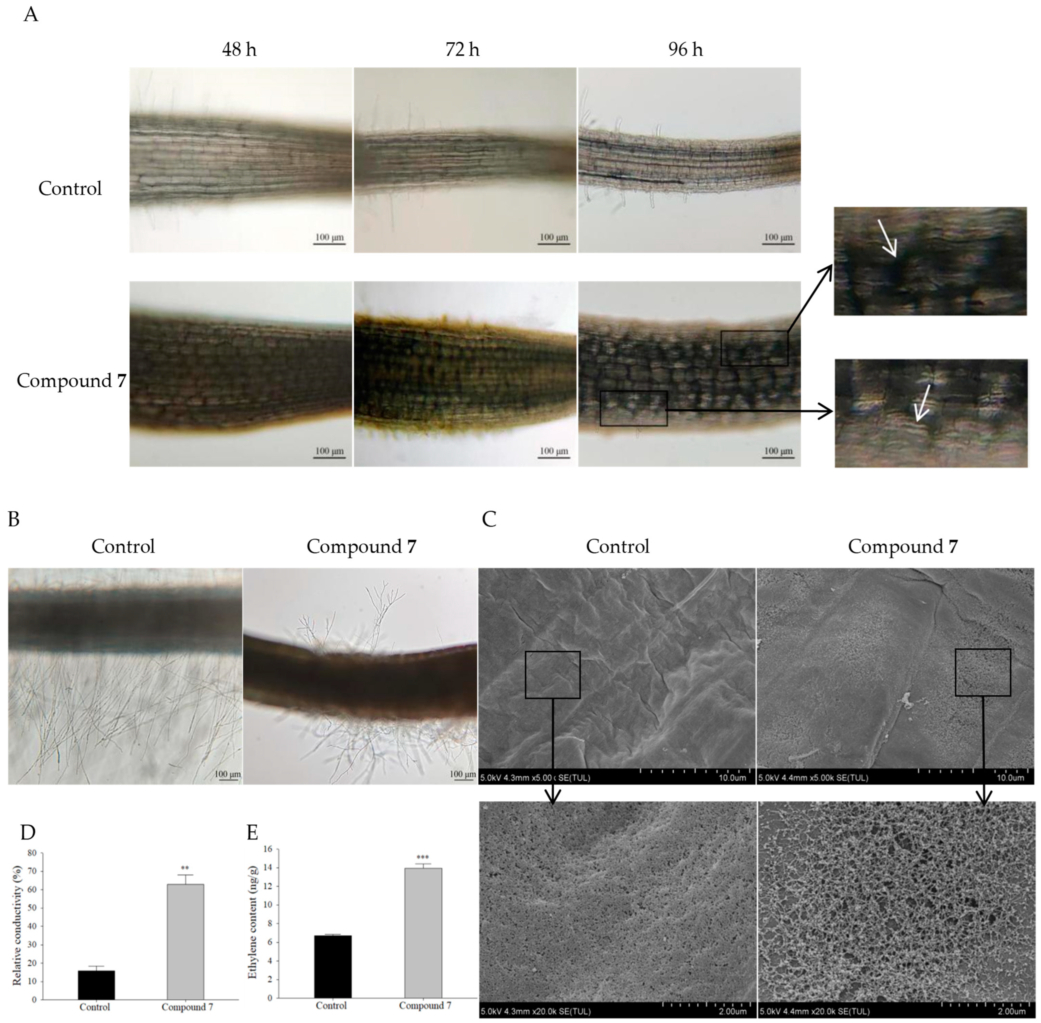
In order to better understand the inhibitory capacity of compound 7, we further investigated the morphological and physiological effects of compound 7 on A. retroflexus roots. A. retroflexus seedlings grown in 400 μg/mL of compound 7 solution for 48, 72, and 96 h were observed using an optical microscope. It is clear that compound 7 caused the radial swelling in the root phenotype and a reduction in cell length in the root elongation zone compared with control (Figure 6A). We further observed the radial expansion of the treated cells under scanning electron microscope (SEM) (Figure 6C). In addition, the SEM images revealed the damage effects of compound 7 on the elongation-zone cells, which were visible as cracks/fractures, breakages, and wrinkles relative to an indistinct, smooth/even, and unbroken shape, as shown in the control root surface (Figure 6A,C). Consistent with this finding, the relative conductivity of the seedlings grown in 400 μg/mL compound 7 for 96 h was 62.84%, which was much higher than that of the control (15.76%) (Figure 6D). These results indicate that the cell membrane can be damaged by compound 7, leading to the release of intracellular electrolytes.
Additionally, we found that compound 7 delayed the root hair initiation and significantly inhibited root hair growth (Figure 6A,B). In the control group, the development of root hairs entered the elongation stage at 48 h after sowing, but in the experimental group, the root hair cell wall began to bulge out by 72 h (Figure 6A). Notably, branched root hairs were found in the treatment group (Figure 6B). This finding was similar to previous reports in which branched root hairs will form under abiotic stress or nutrient deficiency [32,33]. These results suggest that the delayed and abnormal development of root hairs may be due to the effects of stress caused by compound 7.
In plants, ethylene production often enhances the tolerance to given environmental conditions [34]. Here, the ethylene content in A. retroflexus roots treated with compound 7 for 96 h was doubled compared with the control group (Figure 6E), which may be associated with the inhibition of root growth, the delayed development of root hairs, and the stress response.

3. Discussion

The utilization of fungal phytotoxins for the generation of environmentally benign bioherbicides is undergoing a period of accelerated development [10,11,35]. In this study, eight compounds were isolated and purified from the mycelium and fermentation broth extracts of B. cookei SYBL03, an important phytopathogenic fungus causing sorghum leaf diseases. The compounds (18), except 2 and 6, are reported for the first time from this genus. Compound 1 was first isolated in 1983 from the mycelium of Guignardia laricina by Otomo et al. [36] and showed potential anti-inflammatory and antitumor activities [37]. In our study, compound 1 exhibited weaker inhibitory activity against the seedling growth of A. retroflexus (Table 1). Compound 3, initially isolated in 1977 from the secondary metabolites of A. flavus by Springer et al. [38], demonstrated significant analgesic and anti-inflammatory activities [27]. Compound 4 displayed antioxidant activity. Although this structure has been synthesized before, NMR data from a natural source have not been reported. Compound 5, obtained for the first time in 1987 from the cell extract of Pachyhasium sp. by Sitrin et al. [39], has exhibited a variety of biological activities, including anti-inflammatory activity [40], analgesia [41], plant defense induction [42], and nematicidal activities against Bursaphelenchus xylophilus [43], highlighting its application prospects in agriculture and medicine. Compound 6, first isolated in 1983 from Schizophyllum commune by Kawai and Ikeda [44], has showed neuroprotection [45], analgesia [41], and plant resistance induction [46]. Compound 8, isolated for the first time in 1977 from Balansia epichloe (Weese) by Porter et al. [47], exhibited antibacterial activity against Clostridium perfringens [48]. In the present study, compounds 36 and 8 did not show any remarkable phytotoxicity to A. retroflexus and E. crus-galli.
Here, only compounds 2 and 7 showed obvious inhibitory effects on A. retroflexus. Compound 2, isolated for the first time in 1947 from the mycelium of Aspergillus fumigatus by Wieland and Prelog [49], has exhibited a wide range of biomedical activities, including antiviral [50], antitumor [51], anti-inflammatory [52], immunomodulatory [50], antifungal, and cytotoxic [53] activities. In addition, Macías et al. [54] reported its phytotoxicity against the seedling growth of E. crus-galli. In contrast, we found that compound 2 had no significant effect on the seedling growth of E. crus-galli. This inconsistency may be due to the differences in treatment dosage and culture conditions. It is noteworthy that compound 7 exhibited preferable selective herbicidal activity against dicotyledonous weeds. Compound 7 is a cyclic dipeptide compound, which was first separated from the culture filtrate of Aspergillus sp. by Luo et al. [55] and showed no obvious effect on the growth of bacteria and nematodes. Subsequently, Ma et al. [26] demonstrated that 400 µg/mL of compound 7 is an effective inhibitor of seed germination in A. thaliana. Similarly, our study also revealed that compound 7 at a concentration of 400 μg/mL inhibited the seed germination of C. bursa-pastoris by 100% (Figure 5A). Furthermore, our findings indicated that it significantly affected the seedling growth of A. retroflexus, S. nigrum, and C. bursa-pastoris.
Further research demonstrated that the effect of compound 7 on the growth of weed seedlings exhibited a concentration-dependent dual role. At low concentrations, the treatment promoted growth, whereas with an increasing concentration, growth was inhibited (Figure 4). It was found that the monocots and dicots exhibited differential sensitivity to compound 7 (Figure 2 and Figure 3), with seedling growth promoted in monocots (the increase ranging from 46% to 216% in shoot and root growth of S. viridis, D. sanguinalis and E. crus-galli) while inhibited in dicots (the decrease ranging from 32% to 86% in shoot and root growth of A. retroflexus, C. bursa-pastoris and S. nigrum) at a concentration of 200 μg/mL. The difference in sensitivity to plant toxic compounds between monocots and dicots has been widely reported in auxin herbicides [56,57,58]. Auxin herbicides exert selective action, with a preference for dicotyledonous weeds in cereal crops [59]. These findings showed that the effect of compound 7 on weeds might be comparable to that of auxin herbicides.
Based on the herbicidal activity of compound 7, we further investigated its mechanism of growth inhibition. The results showed that the application of compound 7 inhibited the root elongation and caused shortening and swelling of elongation-zone cells of A. retroflexus, which led to a swollen root phenotype (Figure 6A,C). This is similar to what DeBolt et al. [60] observed in A. thaliana seedlings, where the cells expanded manifold in diameter relative to controls, resulting in swollen roots.
Root hairs are long tubular-shaped outgrowths from root epidermal cells [61]. In Arabidopsis thaliana, the mechanism of the root epidermis is based on the positional relationship between epidermal cells and underlying cortical cells. The cells located outside an anticlinal cortical cell wall will undergo three processes: change in root hair cell fate, root hair initiation, and root hair elongation, leading to the development of mature root hairs [62]. Here, the root hair initiation was delayed, and the growth of root hairs was inhibited in the treatment group, showing a reduction in number and shortening in length (Figure 6A,B). In previous studies, herbicides have been found to inhibit the growth of plant root hairs. Zhou et al. [63] treated maize with imazethapyr, which impaired root hair growth, showing significantly sparser and shorter root hairs. Interestingly, it was observed that the branching of root hairs occurred, which may be attributed to the influence of stress (Figure 6B). Previous work revealed that branched root hairs will form under abiotic stress or nutrient deficiency [32,33]. In addition, electrolyte leakage and cell death accompany plant response to stresses, such as drought, salt stress, heavy metal contamination, extreme temperatures, and compounds with herbicidal activity [64,65]. Some natural phytotoxins, such as cercosporin and syringomycin, cause electrolyte leakage through both light-dependent and non-light-dependent mechanisms, ultimately resulting in plant death [66]. In our study, microscopic observation showed the cracks/fractures, breakages, and wrinkles on the root surface (Figure 6A,C), and, meanwhile, we also detected the occurrence of electrolyte leakage (Figure 6D), which further suggests that the roots were subjected to drug stress.
Previous work has shown that the plant hormone ethylene is involved in plant growth and development [67]. Ethylene also plays a crucial role in plant response or adaptation to biotic and abiotic stress [34]. In our study, the phytohormone detection revealed an increase in ethylene content from 6.7 to 13.9 ng/g. This finding is in accordance with those previously reported. Excessive stimulation of ethylene production through the induction of 1-aminocyclopropane-1-carboxylic acid (ACC) synthase in biosynthesis is a well-known early event of auxin herbicides [57,58,67]. Ethylene treatment induced root shortening and swelling in Arabidopsis seedlings [68]. These results indicate that ethylene may be involved in the root growth inhibition of A. retroflexus seedlings induced by compound 7.
This is the first report showing that compound 7 has the selective effect against monocotyledonous and dicotyledonous weeds. This selectivity may be related to the differential sensitivity of monocots and dicots to compound 7. Monocotyledonous weeds are less sensitive to compound 7 than dicotyledonous weeds. The herbicidal activity may be associated with the compound 7-induced stress. Increased ethylene production could be involved in the response of plants to compound 7. There should be further investigations to elucidate the mechanism of compound 7 on plants, with a particular focus on the proteomic, transcriptomic, and biochemical analyses.

4. Materials and Methods

4.1. General Experimental Procedures

Optical rotation was obtained on a JH-P100 digital automatic polarimeter (Shanghai Jiahang Instruments Co., Ltd., Shanghai, China) at 20 °C. The 1H- and 13C-NMR spectra were recorded on a Bruker Avance III 600 MHz NMR spectrometer using the solvent peak as an internal standard (Bruker BioSpin, Rheinstetten, Germany). The HMBC experiment was recorded using the pulsed-field gradients. Electrospray ionization mass spectra (ESI-MS) were performed on Waters ACQUITY RDa mass detector (Waters Co., Milford, MA, USA). The Amberlite XAD-16N resin (560–710 µm, DuPont, Wilmington, DE, USA) was used for isolating and concentrating organic compounds from fermentation supernatant. Column chromatography was performed using silica gel (100–200 and 200–300 mesh, Qingdao Haiyang Chemical Co., Ltd., Qingdao, China) and Sephadex LH-20 (25–100 µm, GE Healthcare, Uppsala, Sweden). Analytical and preparative thin-layer chromatography (TLC and PTLC) was carried out on pre-coated silica gel GF254 plates (0.25 and 0.5 mm, Qingdao Haiyang Chemical Co., Ltd., Qingdao, China). Vanillin-sulfuric acid was used as the spray reagent for TLC. Analytical and semi-preparative high-performance liquid chromatography (HPLC and semi-preparative HPLC) was performed using the C18 column (4.6 × 250 and 9.4 × 250 mm, 5 μm, Agilent) on an Agilent 1260 series system (Agilent, Palo Alto, CA, USA). Morphology of root tip cells was observed by Leica GALEN-III optical microscope (Nanjing Jiangnan Photoelectric (Group) Co., Ltd., Nanjing, China) and SU8020 scanning electron microscope (Hitachi, Tokyo, Japan). Relative conductivity was measured using DDSJ-308F conductivity meter (INESA Scientific Instrument Co., Ltd., Shanghai, China). The ethylene content was tested using GC-2010Pro gas chromatograph (Shimadzu, Kyoto, Japan). All chemical agents were purchased from Sinopharm Chemical Reagent company (Shanghai, China).

4.2. Fungal Material

The phytopathogenic fungus B. cookei SYBL03 was provided from Institute of Plant Protection, Liaoning Academy of Agricultural Sciences, China. The strain was isolated from the diseased leaves of target leaf spot of sorghum, a crop growing in Shenyang, Liaoning province, China. On the basis of the colony and spore morphology as well as the sequence analysis of the internal transcribed spacer (ITS) of nuclear ribosomal DNA, the strain SYBL03 was identified as B. cookei [69]. Working stocks were prepared on the slants containing potato dextrose agar (PDA) medium [70] and stored at 4 °C.

4.3. Fermentation and Extraction

B. cookei SYBL03 culture was initiated by transferring pieces of PDA medium containing mycelium to a fresh PDA slant. A loop full of the well-grown culture was transferred to a 250 mL Erlenmeyer flask with 100 mL modified PD (PDm) medium containing infusion from 20 g potatoes, dextrose 2 g, peptone 0.1 g, K2HPO4 0.1 g, MgSO4 0.05 g, Vitamin B1 1 mg in distilled water at pH 7.0. The flasks were incubated in a rotary incubator shaker at 28 °C and 180 rpm for 48 h and were used as seed culture for subsequent experiments. Modified potato sugar (PSm) medium (infusion from 200 g potatoes, sucrose 20 g, NaNO3 1 g, KCl 0.5 g, MgSO₄·7H₂O 0.5 g, FeSO4·7H₂O 0.02 g, CuSO4·5H₂O 0.02 g and distilled water up to 1 L, natural pH value) was used for fermentation production. The seed cultures (14 mL) were inoculated in 500 mL flasks containing 200 mL of PSm medium each. The flasks were incubated in rotary shakers at 28 °C and 180 rpm for 7 d. A total of 150 L fermentation culture was obtained. The broth and mycelia were collected by centrifugation at 9000 rpm for 20 min, respectively.
Mycelia were dried, crushed into powder, and extracted five times in 3 volumes of 80% ice-cold acetone. The organic solution was collected by filtration, and the combined filtrates were concentrated to remove acetone. The aqueous residue was repeatedly extracted with an equal volume of EtOAc. The organic layer was concentrated under reduced pressure to produce 9.7 g of the mycelium extract.
Twelve grams of the Amberlite XAD-16N resin was added to each 500 mL Erlenmeyer flask with 300 mL of the cell-free supernatant. The resin was collected by filtration after shaking at 20 °C and 120 rpm for 12 h, dried in an oven at 25 °C, and desorbed five times with methanol (MeOH). Desorbed solution was pooled and concentrated by rotary evaporator under vacuum to yield the total residue, which was then suspended in water and repeatedly extracted with EtOAc and then concentrated, as described above. Finally, the broth extract (27 g) was obtained.

4.4. Fractionation and Purification of Compounds

The EtOAc extract of mycelia was fractionated by column chromatography (CC) over silica gel using an increasing gradient of EtOAc-MeOH (99:1, 95:5, 90:10, 80:20, 50:50, 0:100, v/v) to afford six main fractions (A–F). Fraction B (298 mg) was purified by CC over Sephadex LH-20 eluted with CH2Cl2-MeOH (1:1, v/v) to yield two subfractions (B1 and B2). Subfraction B1 (180 mg) was dissolved in CH2Cl2-MeOH (1:1, v/v). The insoluble part (61 mg) was isolated by semi-preparative HPLC (MeOH-H2O, 93:7, v/v) to yield compounds 1 (8.2 mg, tR = 23.34 min) and 2 (32 mg, tR = 31.21 min). Fraction C (105 mg) was dissolved in methanol. The insoluble part (52 mg) was recrystallized from MeOH and further purified by Sephadex LH-20 column eluted with CH2Cl2-MeOH (1:1, v/v) to give compound 3 (35 mg). Fraction D (158 mg) was repeatedly purified by Sephadex LH-20 column eluted with CH2Cl2-MeOH (1:1, v/v) and was recrystallized from MeOH to obtain compound 4 (11 mg). Fraction E (75 mg) was subjected to CC over Sephadex LH-20 eluted with CH2Cl2-MeOH (1:1, v/v) to yield subfractions E1 and E2. As the solvent evaporated, some components precipitated out of the eluting solvent of subfraction E1. The precipitate (30 mg) was further purified by semi-preparative HPLC (100% MeOH) to afford compounds 5 (8.2 mg, tR = 16.10 min) and 6 (12 mg, tR = 18.88 min).
The EtOAc extract of the cell-free supernatant was subjected to silica gel CC and eluted with a gradient of PE-EtOAc (9:1, 8:2, 7:3, 5:5, 4:6, 2:8, 0:10, v/v) to obtain five fractions (A′–E′). Fraction C′ (2.78 g) was purified by CC over silica gel eluted with a step-wise gradient of PE-EtOAc (7:3 to 4:6, v/v) to yield compound 3 (19 mg) and mixed subfraction C1′ (1.25 g). Subfraction C1′ was further separated through Sephadex LH-20 column eluted with CH2Cl2-MeOH (1:1, v/v) and PTLC (PE-CH2Cl2-EtOH, 3:8:2, v/v), to afford compound 7 (752 mg). Fraction D′ (1.75 g) was applied to CC over silica gel eluted with PE-EtOAc (2:8, v/v) to give subfractions D1′ and D2′. Subfraction D1′ (125 mg) was repeatedly purified via Sephadex LH-20 column with CH2Cl2-MeOH (1:1, v/v) and semi-preparative HPLC (MeOH-H2O, 7:3, v/v) to afford compound 8 (23 mg, tR = 29.56 min).

4.4.1. 5α,8α-Epidioxyergosta-6,9(11),22-trien-3β-ol (1)

White powder; 1H-NMR (600 MHz, CD3OD): δH 6.65 (1H, d, J = 8.6 Hz, H-7), 6.30 (1H, d, J = 8.6 Hz, H-6), 5.48 (1H, dd, J = 6.1, 1.9 Hz, H-11), 5.26 (1H, dd, J = 15.3, 7.7 Hz, H-23), 5.20 (1H, dd, J = 15.3, 8.3 Hz, H-22), 3.80 (1H, m, H-3), 1.10 (3H, s, H-19), 1.02 (3H, d, J = 6.6 Hz, H-21), 0.93 (3H, d, J = 6.8 Hz, H-28), 0.86 (3H, d, J = 6.8 Hz, H-26), 0.84 (3H, d, J = 6.8 Hz, H-27), 0.77 (3H, s, H-18); 13C-NMR (150 MHz, CD3OD): δC 144.2 (s, C-9), 136.9 (d, C-6), 136.7 (d, C-22), 133.6 (d, C-23), 131.8 (d, C-7), 120.7 (d, C-11), 84.0 (s, C-5), 79.7 (s, C-8), 66.8 (d, C-3), 57.2 (d, C-17), 49.6 (d, C-14), 44.8 (s, C-13), 44.3 (d, C-24), 42.4 (t, C-12), 41.3 (d, C-20), 39.2 (s, C-10), 36.9 (t, C-4), 34.4 (d, C-25), 33.7 (t, C-1), 31.3 (t, C-2), 29.8 (t, C-16), 25.9 (q, C-19), 21.8 (q, C-21), 21.2 (t, C-15), 20.4 (q, C-26), 20.1 (q, C-27), 18.2 (q, C-28), 13.4 (q, C-18); ESI-MS m/z 449.31 [M + Na]+ (calcd. for C28H42O3Na+, 449.3032).

4.4.2. Ergosterol Peroxide (2)

Colorless needle crystals; 1H-NMR (600 MHz, CD3OD): δH 6.53 (1H, d, J = 8.5 Hz, H-6), 6.25 (1H, d, J = 8.5 Hz, H-7), 5.24 (1H, dd, J = 15.2, 7.7 Hz, H-23), 5.18 (1H, dd, J = 15.2, 8.3 Hz, H-22), 3.77 (1H, m, H-3), 1.01 (3H, d, J = 6.6 Hz, H-21), 0.93 (3H, d, J = 6.8 Hz, H-28), 0.90 (3H, s, H-19), 0.85 (3H, s, H-18), 0.85 (3H, d, J = 6.8 Hz, H-26), 0.83 (3H, d, J = 6.8 Hz, H-27); 13C-NMR (150 MHz, CD3OD): δC 136.8 (d, C-6, C-22), 133.5 (d, C-23), 131.7 (d, C-7), 83.5 (s, C-5), 80.7 (s, C-8), 67.0 (d, C-3), 57.6 (d, C-17), 53.1 (d, C-14), 52.7 (d, C-9), 45.8 (s, C-13), 44.3 (d, C-24), 41.1 (d, C-20), 40.7 (t, C-12), 38.2 (s, C-10), 37.8 (t, C-4), 35.9 (t, C-1), 34.4 (d, C-25), 30.9 (t, C-2), 29.8 (t, C-16), 24.4 (t, C-11), 21.6 (q, C-21), 21.4 (t, C-15), 20.5 (q, C-26), 20.1 (q, C-27), 18.6 (q, C-19), 18.2 (q, C-28), 13.3 (q, C-18): ESI-MS m/z 429.31 [M + H]+ (calcd. for C28H45O3+, 429.3369).

4.4.3. (–)-Ditryptophenaline (3)

White crystal; 1H-NMR (600 MHz, CD3OD): δH 7.57 (4H, dd, J = 7.6, 7.4 Hz, H-20, H-22, H-20′, H-22′), 7.50 (2H, t, J = 7.4 Hz, H-21, H-21′), 7.15 (4H, d, J = 7.6 Hz, H-19, H-23, H-19′, H-23′), 7.04 (2H, dd, J = 7.9, 7.4 Hz, H-7, H-7′), 6.95 (2H, d, J = 7.5 Hz, H-5, H-5′), 6.65 (2H, dd, J = 7.5, 7.4 Hz, H-6, H-6′), 6.57 (2H, d, J = 7.9 Hz, H-8, H-8′), 5.00 (2H, s, H-2, H-2′), 4.40 (2H, m, H-15, H-15′), 3.63 (2H, br dd, J = 12.0, 4.8 Hz, H-11, H-11′), 3.49 (2H, dd, J = 14.3, 2.8 Hz, H-17a, H-17a′), 3.30 (2H, overlapped, H-17b, H-17b′), 3.03 (6H, s, H-24, H-24′), 1.88 (2H, dd, J = 12.1, 4.8 Hz, H-12a, H-12a′), 1.46 (2H, dd, J = 12.1, 12.0 Hz, H-12b, H-12b′);13C-NMR (150 MHz, CD3OD): δC 167.6 (s, C-13, C-13′), 165.5 (s, C-16, C-16′), 152.7 (s, C-9, C-9′), 136.1 (s, C-18, C-18′), 130.7 (d, C-7, C-7′), 130.5 (d, C-19, C-23, C-19′, C-23′), 130.4 (d, C-20, C-22, C-20′, C-22′), 129.2 (d, C-21, C-21′), 127.7 (s, C-4, C-4′), 126.4 (d, C-5, C-5′), 119.1 (d, C-6, C-6′), 110.2 (d, C-8, C-8′), 79.9 (d, C-2, C-2′), 64.4 (d, C-15, C-15′), 60.4 (s, C-3, C-3′), 59.6 (d, C-11, C-11′), 37.4 (t, C-12, C-12′), 36.9 (t, C-17, C-17′), 33.1 (q, C-24, C-24′); ESI-MS m/z 693.34 [M + H]+ (calcd. for C42H41N6O4+, 693.3189).

4.4.4. 1,3,9-Trimethyluric Acid (4)

White powder; 1H-NMR (600 MHz, CD3OD): δH 3.73 (3H, s, H-11), 3.60 (3H, s, H-12), 3.32 (3H, s, H-10); 13C-NMR (150 MHz, CD3OD): δC 154.7 (s, C-6), 154.0 (s, C-8), 152.5 (s, C-2), 138.5 (s, C-4), 99.6 (s, C-5), 31.9 (q, C-11), 30.4 (q, C-12), 28.6 (q, C-10); ESI-MS m/z 211.05 [M + H]+ (calcd. for C8H11N4O3+, 211.0831).

4.4.5. Cerebroside A (5)

White powder; 1H-NMR (600 MHz, CD3OD): δH 7.71 (1H, d, J = 9.4 Hz, NH), 5.83 (1H, dt, J = 15.3, 6.6 Hz, H-4′), 5.71 (1H, dt, J = 15.4, 6.3 Hz, H-5), 5.49 (1H, dd, J = 15.3, 6.0 Hz, H-3′), 5.45 (1H, dd, J = 15.4, 7.4 Hz, H-4), 5.14 (1H, t, J = 6.6 Hz, H-8), 4.43 (1H, d, J = 6.0 Hz, H-2′), 4.27 (1H, d, J = 7.8 Hz, H-1′’), 4.13 (1H, dd, J = 10.4, 5.6 Hz, H-1a), 4.12 (1H, m, H-3), 3.97 (1H, m, H-2), 3.86 (1H, br d, J = 11.9 Hz, H-6′’a), 3.70 (1H, dd, J = 10.4, 3.3 Hz, H-1b), 3.66 (1H, dd, J = 11.9, 4.6 Hz, H-6″b), 3.17–3.37 (4H, m, H-2″, H-3″, H-4″, H-5″), 2.00–2.09 (6H, m, H-6, H-7, H-5′), 1.97 (2H, t, J = 7.5 Hz, H-10), 1.59 (3H, br s, H-19), 1.21–1.42 (34H, m, H-11–H-17, H-6′–H-15′), 0.89 (6H, t, J = 6.8 Hz, H-18, H-16′); 13C-NMR (150 MHz, CD3OD): δC 175.4 (s, C-1′), 136.7 (s, C-9), 134.7 (d, C-5), 134.5 (d, C-4′), 131.0 (d, C-4), 129.0 (d, C-3′), 124.9 (d, C-8), 104.7 (d, C-1″), 78.0 (d, C-5″), 77.9 (d, C-3″), 75.0 (d, C-2″), 74.1 (d, C-2′), 72.9 (d, C-3), 71.6 (d, C-4″), 69.7 (t, C-1), 62.7 (t, C-6″), 54.6 (d, C-2), 40.8 (t, C-10), 33.8 (t, C-6), 33.4 (t, C-5′), 33.1 (t, C-16, C-14′), 30.2–30.8 (t, C-12–C-15, C-6′–C-13′), 29.1 (t, C-11), 28.8 (t, C-7), 23.8 (t, C-17, C-15′), 16.2 (q, C-19), 14.5 (q, C-18, C-16′); ESI-MS m/z 726.52 [M + H]+ (calcd. for C41H76NO9+, 726.5520).

4.4.6. Cerebroside B (6)

White powder; 1H-NMR (600 MHz, CD3OD): δH 7.71 (1H, d, J = 9.4 Hz, NH), 5.73 (1H, dt, J = 15.3, 6.3 Hz, H-5), 5.48 (1H, dd, J = 15.3, 7.4 Hz, H-4), 5.14 (1H, t, J = 6.5 Hz, H-8), 4.26 (1H, d, J = 7.8 Hz, H-1″), 4.13 (1H, m, H-3), 4.11 (1H, dd, J = 10.3, 5.6 Hz, H-1a), 3.98 (2H, m, H-2, H-2′), 3.86 (1H, br d, J = 11.8 Hz, H-6″a), 3.70 (1H, dd, J = 10.3, 3.3 Hz, H-1b), 3.66 (1H, dd, J = 11.8, 4.4 Hz, H-6″b), 3.16–3.37 (4H, m, H-2″, H-3″, H-4″, H-5″), 2.06 (4H, m, H-6, H-7), 1.97 (2H, t, J = 7.5 Hz, H-10), 1.70 (1H, m, H-3′a), 1.59 (3H, br s, H-19), 1.55 (1H, m, H-3′b), 1.20–1.44 (38H, m, H-11–H-17, H-4′–H-15′), 0.89 (6H, t, J = 6.9 Hz, H-18, H-16′); 13C-NMR (150 MHz, CD3OD): δC 177.2 (s, C-1′), 136.8 (s, C-9), 134.7 (d, C-5), 131.1 (d, C-4), 124.8 (d, C-8), 104.7 (d, C-1″), 78.0 (d, C-5″), 77.9 (d, C-3″), 75.0 (d, C-2″), 73.1 (d, C-2′), 72.9 (d, C-3), 71.6 (d, C-4″), 69.7 (t, C-1), 62.7 (t, C-6″), 54.6 (d, C-2), 40.8 (t, C-10), 35.9 (t, C-3′), 33.8 (t, C-6), 33.1 (t, C-16, C-14′), 30.4–30.8 (t, C-12–C-15, C-5′–C-13′), 29.1 (t, C-11), 28.7 (t, C-7), 26.2 (t, C-4′), 23.8 (t, C-17, C-15′), 16.1 (q, C-19), 14.5 (q, C-18, C-16′); ESI-MS m/z 728.56 [M + H]+ (calcd. for C41H78NO9+, 728.5677).

4.4.7. Cyclo-N-methylphenylalanyltryptophenyl (7)

White powder; 1H-NMR (600 MHz, CD3OD): δH 7.52 (1H, d, J = 7.9 Hz, H-6), 7.34 (1H, d, J = 8.1 Hz, H-3), 7.23 (2H, dd, J = 7.5, 7.3 Hz, H-21, H-23), 7.18 (1H, t, J = 7.3 Hz, H-22), 7.12 (1H, dd, J = 8.1, 7.1 Hz, H-4), 7.04 (1H, dd, J = 7.9, 7.1 Hz, H-5), 6.95 (1H, s, H-9), 6.79 (2H, d, J = 7.5 Hz, H-20, H-24), 4.11 (1H, dd, J = 7.2, 3.9 Hz, H-11), 4.04 (1H, dd, J = 6.3, 4.3 Hz, H-15), 2.94 (1H, dd, J = 14.5, 3.9 Hz, H-10a), 2.72 (1H, dd, J = 14.2, 4.3 Hz, H-18a), 2.68 (3H, s, H-14), 2.19 (1H, dd, J = 14.5, 7.2 Hz, H-10b), 2.03 (1H, dd, J = 14.2, 6.3 Hz, H-18b); 13C-NMR (150 MHz, CD3OD): δC 168.9 (s, C-16), 168.4 (s, C-12), 138.1 (s, C-2), 138.0 (s, C-19), 130.8 (d, C-20, C-24), 129.7 (d, C-21, C-23), 128.7 (s, C-7), 128.2 (d, C-22), 125.6 (d, C-9), 122.7 (d, C-4), 120.1 (d, C-5), 119.7 (d, C-6), 112.6 (d, C-3), 109.9 (s, C-8), 64.9 (d, C-15), 57.2 (d, C-11), 39.4 (t, C-18), 34.3 (q, C-14), 31.7 (t, C-10); ESI-MS m/z 348.17 [M + H]+ (calcd. for C21H22N3O2+, 348.1712).

4.4.8. (2S)-3,3-di-1H-indol-3-yl-1,2-propanediol (8)

White powder; [α]D20 +39.4° (c 0.29, MeOH); 1H-NMR (600 MHz, CDCl3): δH 7.98, 8.06 (each 1H, br s, NH-1′, NH-1″), 7.62, 7.67 (each 1H, d, J = 8.0 Hz, H-4′, H-4″), 7.31, 7.33 (each 1H, d, J = 8.2 Hz, H-7′, H-7″), 7.15–7.20 (2H, m, H-6′, H-6″), 7.09, 7.19 (each 1H, br s, H-2′, H-2″), 7.05–7.11 (2H, m, H-5′, H-5″), 4.73 (1H, d, J = 7.1 Hz, H-1), 4.53 (1H, ddd, J = 7.1, 6.8, 3.4 Hz, H-2), 3.75 (1H, dd, J = 11.3, 3.4 Hz, H-3a), 3.63 (1H, dd, J = 11.3, 6.8 Hz, H-3b); 13C-NMR (150 MHz, CDCl3): δC 136.3, 136.4 (s, C-7a′, C-7a″), 126.8, 127.4 (s, C-3a′, C-3a″), 122.3, 122.8 (d, C-2′, C-2″), 122.1, 122.3 (d, C-6′, C-6″), 119.5, 119.7 (d, C-5′, C-5″), 119.2, 119.4 (d, C-4′, C-4″), 115.3, 116.8 (s, C-3′, C-3″), 111.2 (d, C-7′, C-7″), 74.8 (d, C-2), 65.3 (t, C-3), 36.9 (d, C-1); ESI-MS m/z 305.11 [M – H] (calcd. for C19H17N2O2, 305.1290).

4.5. Herbicidal Activity Assay

The compounds to be tested were dissolved in dimethyl sulfoxide (DMSO) at a concentration of 80 mg/mL, and solutions were diluted with 0.1% Tween 80 to make a stock solution concentration of 800 µg/mL. The required dilutions were subsequently made from the stock solution by adding 1% DMSO water with 0.1% Tween 80. The solution containing the same concentration of DMSO and Tween 80 was used as the control treatment.
Mature weed seeds of six species (E. crus-galli, S. viridis, D. sanguinalis, A. retroflexus, S. nigrum and C. bursa-pastoris) were collected from the suburban roadsides, hillsides, and wetlands of Liaoning Province, China. Seeds were kept at 4 °C until further use. Weed seeds were surface sterilized by treatment with 0.2% sodium hypochlorite solution for 10 min, followed by rinsing in sterile distilled water at least three times. Except for C. bursa-pastoris (40 seeds), 30 seeds were equidistantly placed in a Petri dish (diameter = 90 mm) containing double-layer filter paper. Afterward, each Petri dish was moistened with the solution to be tested (5 mL) and kept at 26 ± 1 °C, with 85% humidity and a 12 h photoperiod. At least three replicates were taken for each treatment of each compound. Seeds were considered to have germinated if the root extended at least 2 mm long. The number of germinated seeds was recorded continuously for seven days. Except for C. bursa-pastoris (144 h), the root and shoot lengths of germinated seeds were measured after 96 h. Germination rate was calculated as the total number of germinated seeds at the end of experiment out of the total number of seeds tested. Inhibition rate is calculated by using Formula (1).
Inhibition (%) = (CT)/C × 100
where C represents parameters of the control group and T represents parameters of the treatment group.

4.6. Light Microscopy

After treatment with compound 7 for 96 h at concentration of 400 μg/mL, the roots of A. retroflexus seedlings were kept in distilled water to observe the elongation-zone cells and root hairs, and then photographed under a Leica GALEN-III optical microscope (Nanjing Jiangnan Photoelectric (Group) Co., Ltd., Nanjing, China). Then, 1% DMSO water with 0.1% Tween 80 was used as the control treatment.

4.7. Scanning Electron Microscopy (SEM)

In order to clarify the herbicidal mechanism of compound 7, the cellular morphological changes of A. retroflexus roots were observed using SEM. After treatment with or without compound 7 (400 μg/mL) for 96 h, the roots were fixed in 2.5% glutaraldehyde solution for 24 h at 4 °C, dehydrated with an ethanol series (50%, 70% and 100%), dried by critical point dryer (Tousimis Autosamdri-815, Series, Tousimis, Rockville, MD, USA) for 1 h. The sputter coater (HIYACHI MC1000, Hitachi, Tokyo, Japan) was used to deposit a thin gold coating. The samples were observed under SEM (Hitachi SU8020, Hitachi, Tokyo, Japan) at 5 kV.

4.8. Relative Conductivity Assay

Seedlings grown for 96 h in the presence or absence of compound 7 (400 μg/mL) were used to assay electrolyte leakage. Fresh root samples (150 mg) were suspended in a test tube containing 10 mL of deionized water and incubated at 25 °C for 12 h to measure the conductivity of the solution (R1). The test tubes were then heated into a thermostatic water bath at 100 °C for 15 min to completely kill the tissues, and the conductivity value of the same sample was measured again after being cooled down (R2). The experiment was run in triplicates. The relative conductivity (REC) was calculated using Formula (2).
REC (%) = R1/R2 × 100

4.9. Ethylene Content Assay

Seedlings of A. retroflexus were incubated for 96 h in the presence or absence of compound 7 (400 μg/mL). The root samples (100 mg) were sonicated for 30 min in a headspace bottle containing 0.1 g of NaCl, and 1 mL of 10 mol/L NaOH solution. After 24 h, ethylene content was determined by gas chromatography (GC) as described by Liu et al. [71].

4.10. Statistical Analyses

The measurement data were expressed as mean ± standard error of at least three biological replicates. One-way analysis of variance (ANOVA) with Holm–Sidak method or the two-tailed t-test was performed using SigmaPlot 12.5. Differences were considered significant at (*) p < 0.05, (**) p < 0.01 and (***) p < 0.001.

Supplementary Materials

The following supporting information can be downloaded at: https://www.mdpi.com/article/10.3390/molecules29133040/s1, Figures S1–S25: The 1D NMR spectra of compounds 18, 2D NMR spectra of 4.

Author Contributions

Conceptualization, H.L. and Z.Y.; methodology, H.L.; validation, H.L., J.H. and B.L.; formal analysis, H.L.; investigation, H.L., J.H. and B.L.; resources, H.L.; data curation, H.L., J.H. and L.Z.; writing—original draft preparation, H.L. and J.H.; writing—review and editing, L.Z.; supervision, Z.Y.; project administration, H.L.; funding acquisition, H.L. and Z.Y. All authors have read and agreed to the published version of the manuscript.

Funding

This research was funded by the National Key Research and Development Program of China (2018YFD0800401), and the Cultivation Project for Postgraduate of Shenyang University of Chemical Technology in 2021.

Institutional Review Board Statement

Not applicable.

Informed Consent Statement

Not applicable.

Data Availability Statement

The original contributions presented in the study are included in the article/Supplementary Materials, further inquiries can be directed to the corresponding author.

Acknowledgments

The authors thank Yongming Yan of Shenzhen University for technical assistance with NMR and MS spectra. Thanks also to Han Yan of Liaoning Academy of Agricultural Sciences for providing the fermentation strain.

Conflicts of Interest

The authors declare no conflicts of interest.

References

  1. United Nations. World Population Prospects: 2019: Highlights; United Nations: New York, NY, USA, 2019; No. 423. [Google Scholar]
  2. FAO. The Future of Food and Agriculture—Alternative Pathways to 2050; FAO: Rome, Italy, 2018. [Google Scholar]
  3. Oerke, E.C. Crop losses to pests. J. Agric. Sci. 2005, 144, 31–43. [Google Scholar] [CrossRef]
  4. Gianessi, L.P. The increasing importance of herbicides in worldwide crop production. Pest Manag. Sci. 2013, 69, 1099–1105. [Google Scholar] [CrossRef] [PubMed]
  5. Council for Agricultural Science and Technology (CAST). The Contributions of Pesticides to Pest Management in Meeting the Global Need for Food Production by 2050; Issue Paper #55; CAST: Ames, IA, USA, 2014. [Google Scholar]
  6. Sun, Y.; Han, J.; Chen, X.; Guo, H. Advances in measures of reducing chemical pesticides to control plant diseases. Plant Dis. Pests 2021, 12, 1–6. [Google Scholar] [CrossRef]
  7. Deng, X. A mini review on natural safeners: Chemistry, uses, modes of action, and limitations. Plants 2022, 11, 3509. [Google Scholar] [CrossRef] [PubMed]
  8. Li, J.; Ye, Y.; Wang, X.; Dong, L. Herbicidal activity of curvulinic acid isolated from Nimbya alternantherae. Nat. Prod. Commun. 2012, 7, 51–52. [Google Scholar] [CrossRef] [PubMed]
  9. Elhamouly, N.A.; Hewedy, O.A.; Zaitoon, A.; Miraples, A.; Elshorbagy, O.T.; Hussien, S.; El-Tahan, A.; Peng, D. The hidden power of secondary metabolites in plant-fungi interactions and sustainable phytoremediation. Front. Plant Sci. 2022, 13, 1044896. [Google Scholar] [CrossRef] [PubMed]
  10. Gan, D.; Liu, J.Q.; Yang, Y.J.; Wang, C.Y.; Zhu, L.; Li, C.Z.; Cai, L.; Ding, Z.T. Phytotoxic meroterpenoids with herbicidal activities from the phytopathogenic fungus Pseudopestalotiopsis theae. Phytochemistry 2023, 206, 113522. [Google Scholar] [CrossRef]
  11. Cimmino, A.; Masi, M.; Evidente, M.; Superchi, S.; Evidente, A. Fungal phytotoxins with potential herbicidal activity: Chemical and biological characterization. Nat. Prod. Rep. 2015, 32, 1629–1653. [Google Scholar] [CrossRef] [PubMed]
  12. Khiralla, A.; Spin, R.; Saliba, S.; Laurain-Mattar, D. Diversity of natural products of the genera Curvularia and Bipolaris. Fungal Biol. Rev. 2019, 33, 101–122. [Google Scholar] [CrossRef]
  13. Lim, C.H.; Miyagawa, H.; Akamatsu, M.; Nakagawa, Y.; Ueno, T. Structures and biological activities of phytotoxins produced by the plant pathogenic fungus Bipolaris cynodontis cynA. J. Pestic. Sci. 1998, 23, 281–288. [Google Scholar] [CrossRef]
  14. Lim, C.H.; Miyagawa, H.; Tsurushima, T.; Ueno, T.; Sato, M. Cochlioquinol: A new cochlioquinone derivative produced by the plant pathogenic fungus Bipolaris cynodontis. Biosci. Biotechnol. Biochem. 1996, 60, 724–725. [Google Scholar] [CrossRef]
  15. Pena-Rodriguez, L.M.; Chilton, W.S. Victoxinine and prehelminthosporolactone, two minor phytotoxic metabolites produced by Bipolaris sp., a pathogen of Johnson grass. J. Nat. Prod. 1989, 52, 899–901. [Google Scholar] [CrossRef]
  16. Arnold, D.; Yazawa, T.; Kawahigashi, H.; Matsumoto, T.; Mizuno, H. Simultaneous transcriptome analysis of sorghum and Bipolaris sorghicola by using RNA-seq in combination with De Novo transcriptome assembly. PLoS ONE 2013, 8, e62460. [Google Scholar] [CrossRef]
  17. Peng, C.; Ge, T.T.; He, X.L.; Huang, Y.H.; Xu, Z.L.; Zhang, D.Y.; Shao, H.B.; Guo, S.W. Process of Bipolaris sorghicola invasion of host cells. Genet. Mol. Res. 2016, 15, 15016781. [Google Scholar] [CrossRef] [PubMed]
  18. Zaccaron, A.Z.; Bluhm, B.H. The genome sequence of Bipolaris cookei reveals mechanisms of pathogenesis underlying target leaf spot of sorghum. Sci. Rep. 2017, 7, 17217. [Google Scholar] [CrossRef]
  19. Moriwaki, A.; Kihara, J.; Kobayashi, T.; Tokunaga, T.; Arase, S.; Honda, Y. Insertional mutagenesis and characterization of a polyketide synthase gene (PKS1) required for melanin biosynthesis in Bipolaris oryzae. FEMS Microbiol. Lett. 2004, 238, 1–8. [Google Scholar] [CrossRef] [PubMed]
  20. Nizam, S.; Verma, S.; Singh, K.; Aggarwal, R.; Srivastava, K.D.; Verma, P.K. High reliability transformation of the wheat pathogen Bipolaris sorokiniana using Agrobacterium tumefaciens. J. Microbiol. Methods 2012, 88, 386–392. [Google Scholar] [CrossRef]
  21. Gogoi, R.; Singh, S.; Singh, P.; Sakkaravarthi, K.; Rai, S. Genetic variability in the isolates of Bipolaris maydis causing maydis leaf blight of maize. Afr. J. Agric. Res. 2014, 9, 1906–1913. [Google Scholar] [CrossRef]
  22. Winder, R.S.; Van Dyke, C.G. The pathogenicity, virulence, and biocontrol potential of two Bipolaris species on John-songrass (Sorghum halepense). Weed Sci. 1990, 38, 89–94. [Google Scholar] [CrossRef]
  23. Sugawara, F.; Strobel, G.; Strange, R.N.; Siedow, J.N.; Van Duyne, G.D.; Clardy, J. Phytotoxins from the pathogenic fungi Drechslera maydis and Drechslera sorghicola. Proc. Natl. Acad. Sci. USA 1987, 84, 3081–3085. [Google Scholar] [CrossRef]
  24. Pena-Rodriguez, L.M.; Chilton, W.S. 3-Anhydroophiobolin A and 3-Anhydro-6-epi-ophiobolin A, phytotoxic metabolites of the Johnson grass pathogen Bipolaris sorghicola. J. Nat. Prod. 1989, 52, 1170–1172. [Google Scholar] [CrossRef]
  25. Kim, J.W.; Ryu, J.; Shim, S.H. Chemical investigation on an endophytic fungus Gibberella moniliformis JS1055 derived from a halophyte Vitex rotundifolia. Nat. Prod. Sci. 2018, 24, 189–193. [Google Scholar] [CrossRef]
  26. Ma, J.; Lu, C.; Tang, Y.; Shen, Y. Phytotoxic metabolites isolated from Aspergillus sp., an endophytic fungus of Crassula arborescens. Molecules 2022, 27, 7710. [Google Scholar] [CrossRef] [PubMed]
  27. Saruwatari, T.; Yagishita, F.; Mino, T.; Noguchi, H.; Hotta, K.; Watanabe, K. Cytochrome P450 as dimerization catalyst in diketopiperazine alkaloid biosynthesis. ChemBioChem 2014, 15, 656–659. [Google Scholar] [CrossRef] [PubMed]
  28. Koga, J.; Yamauchi, T.; Shimura, M.; Ogawa, N.; Oshima, K.; Umemura, K.; Kikuchi, M.; Ogasawara, N. Cerebrosides A and C, sphingolipid elicitors of hypersensitive cell death and phytoalexin accumulation in rice plants. J. Biol. Chem. 1998, 273, 31985–31991. [Google Scholar] [CrossRef] [PubMed]
  29. Kang, H.S.; Choi, J.H.; Cho, W.K.; Park, J.C.; Choi, J.S. A sphingolipid and tyrosinase inhibitors from the fruiting body of Phellinus linteus. Arch. Pharmacal Res. 2004, 27, 742–750. [Google Scholar] [CrossRef] [PubMed]
  30. Sato, S.; Sato, T. A mild and environmentally friendly scandium(III) trifluoromethanesulfonate-catalyzed synthesis of bis(3′-indolyl)alkanes and bis(3′-indolyl)-1-deoxyalditols. Carbohydr. Res. 2005, 340, 2251–2255. [Google Scholar] [CrossRef] [PubMed]
  31. She, G.M.; Chen, K.; Zhang, Y.-J.; Yang, C.R. The occurrence of 8-oxocaffeine and pyrimidine alkaloids in Pu-er ripe tea. Acta Bot. Yunnanica 2007, 29, 713–716. [Google Scholar] [CrossRef]
  32. Bobrownyzky, J. Production of branched root hairs under progressive drought stress in Arabidopsis thaliana. Cytol. Genet. 2016, 50, 324–329. [Google Scholar] [CrossRef]
  33. Zhang, Y.; Yang, Z.; Zhang, Z.; Li, Y.; Guo, J.; Liu, L.; Wang, C.; Fan, H.; Wang, B.; Han, G. Root hair development and adaptation to abiotic stress. J. Agric. Food Chem. 2023, 71, 9573–9598. [Google Scholar] [CrossRef]
  34. Iqbal, N.; Trivellini, A.; Masood, A.; Ferrante, A.; Khan, N.A. Current understanding on ethylene signaling in plants: The influence of nutrient availability. Plant Physiol. Biochem. 2013, 73, 128–138. [Google Scholar] [CrossRef] [PubMed]
  35. Qu, R.Y.; He, B.; Yang, J.F.; Lin, H.Y.; Yang, W.C.; Wu, Q.Y.; Li, Q.X.; Yang, G.F. Where are the new herbicides? Pest Manag. Sci. 2021, 77, 2620–2625. [Google Scholar] [CrossRef] [PubMed]
  36. Otomo, N.; Sato, H.; Sakamura, S. Novel phytotoxins produced by the causal fungus of the shoot blight of larches. Agric. Biol. Chem. 1983, 47, 1115–1119. [Google Scholar] [CrossRef]
  37. Parhira, S.; Zhu, G.Y.; Li, T.; Liu, L.; Bai, L.P.; Jiang, Z.H. Inhibition of IKK-β by epidioxysterols from the flowers of Calotropis gigantea (Niu jiao gua). Chin. Med. 2016, 11, 9. [Google Scholar] [CrossRef] [PubMed]
  38. Springer, J.P.; Bűchi, G.; Kobbe, B.; Demain, A.L.; Clardy, J. The structure of ditryptophenaline—A new metabolite of Aspergillus flavus. Tetrahedron Lett. 1977, 18, 2403–2406. [Google Scholar] [CrossRef]
  39. Sitrin, R.D.; Chan, G.; Dingerdissen, J.; DeBrosse, C.; Mehta, R.; Roberts, G.; Rottschaefer, S.; Staiger, D.; Valenta, J.; Snader, K.M.; et al. Isolation and structure determination of Pachybasium cerebrosides which potentiate the antifungal activity of aculeacin. J. Antibiot. 1988, 41, 469–480. [Google Scholar] [CrossRef] [PubMed]
  40. Chiu, C.P.; Liu, S.C.; Tang, C.H.; Chan, Y.; El-Shazly, M.; Lee, C.L.; Du, Y.C.; Wu, T.Y.; Chang, F.R.; Wu, Y.C. Anti-inflammatory cerebrosides from cultivated Cordyceps militaris. J. Agric. Food Chem. 2016, 64, 1540–1548. [Google Scholar] [CrossRef] [PubMed]
  41. Chen, W.; Hu, Q.B. Secondary metabolites of Purpureocillium lilacinum. Molecules 2021, 27, 18. [Google Scholar] [CrossRef] [PubMed]
  42. Umemura, K.; Ogawa, N.; Yamauchi, T.; Iwata, M.; Shimura, M.; Koga, J. Cerebroside elicitors found in diverse phytopathogens activate defense responses in rice plants. Plant Cell Physiol. 2000, 41, 676–683. [Google Scholar] [CrossRef]
  43. Zhang, Y.G.; Yuan, W.P.; Xia, X.K.; Liu, X.; Meng, X.M.; Wang, X.J.; Zhang, M.S.; Liu, C.H. Isolation and identification of the nematicidal secondary metabolitesfrom one strain of entomogenous fungi. Chin. J. Pestic. Sci. 2010, 12, 225–228. [Google Scholar] [CrossRef]
  44. Kawai, G.; Ikeda, Y. Chemistry and functional moiety of a fruiting-inducing cerebroside in Schizophyllum commune. Biochim. Biophys. Acta (BBA) Lipids Lipid Metab. 1983, 754, 243–248. [Google Scholar] [CrossRef]
  45. Li, L.; Yang, R.; Sun, K.; Bai, Y.; Zhang, Z.; Zhou, L.; Qi, Z.; Qi, J.; Chen, L. Cerebroside-A provides potent neuroprotection after cerebral ischaemia through reducing glutamate release and Ca2+ influx of NMDA receptors. Int. J. Neuropsychopharmacol. 2012, 15, 497–507. [Google Scholar] [CrossRef] [PubMed]
  46. Umemura, K.; Tanino, S.; Nagatsuka, T.; Koga, J.; Iwata, M.; Nagashima, K.; Amemiya, Y. Cerebroside elicitor confers resistance to fusarium disease in various plant species. Phytopathology 2004, 94, 813–818. [Google Scholar] [CrossRef] [PubMed]
  47. Porter, J.K.; Bacon, C.W.; Robbins, J.D.; Himmelsbach, D.S.; Higman, H.C. Indole alkaloids from Balansia epichloe (Weese). J. Agric. Food Chem. 1977, 25, 88–93. [Google Scholar] [CrossRef] [PubMed]
  48. Yang, C.L.; Han, Y.; Wang, Y.; Zhang, X.H.; Zhu, W.M. Bis- and tris- indole alkaloids from Edwardsiella tarda. Microbiol. China 2010, 37, 1325–1330. [Google Scholar] [CrossRef]
  49. Wieland, P.; Prelog, V. On the isolation of ergosterol, ergosterol palmitate and ergosterol peroxide from the mycelium of Aspergillus fumigatus, mut. helvola, Yuill. Helv. Chim. Acta 1947, 30, 1028–1030. [Google Scholar] [CrossRef] [PubMed]
  50. Duan, C.; Ge, X.; Wang, J.; Wei, Z.; Feng, W.H.; Wang, J. Ergosterol peroxide exhibits antiviral and immunomodulatory abilities against porcine deltacoronavirus (PDCoV) via suppression of NF-κB and p38/MAPK signaling pathways in vitro. Int. Immunopharmacol. 2021, 93, 107317. [Google Scholar] [CrossRef] [PubMed]
  51. Rhee, Y.H.; Jeong, S.J.; Lee, H.J.; Lee, H.J.; Koh, W.; Jung, J.H.; Kim, S.H.; Sung-Hoon, K. Inhibition of STAT3 signaling and induction of SHP1 mediate antiangiogenic and antitumor activities of ergosterol peroxide in U266 multiple myeloma cells. BMC Cancer 2012, 12, 28. [Google Scholar] [CrossRef] [PubMed]
  52. Kobori, M.; Yoshida, M.; Ohnishi-Kameyama, M.; Shinmoto, H. Ergosterol peroxide from an edible mushroom suppresses inflammatory responses in RAW264.7 macrophages and growth of HT29 colon adenocarcinoma cells. Br. J. Pharmacol. 2007, 150, 209–219. [Google Scholar] [CrossRef]
  53. Wang, F.W. Bioactive metabolites from Guignardia sp., an endophytic fungus residing in Undaria pinnatifida. Chin. J. Nat. Med. 2012, 10, 72–76. [Google Scholar] [CrossRef]
  54. Macías, F.A.; Chinchilla, N.; Varela, R.M.; Molinillo, J.M. Bioactive steroids from Oryza sativa L. Steroids 2006, 71, 603–608. [Google Scholar] [CrossRef] [PubMed]
  55. Luo, S.L.; Li, G.H.; Liu, F.F.; Lei, L.P.; Xia, Z.Y.; Zhang, K.Q. A new sesquiterpene from endophytic fungus Aspergillus sp. Nat. Prod. Res. 2012, 26, 1334–1338. [Google Scholar] [CrossRef] [PubMed]
  56. Cobb, A.H.; Reade, J.P.H. (Eds.) Auxin-type herbicides. In Herbicides and Plant Physiology, 2nd ed.; Wiley-Blackwell: West Sussex, UK, 2010; pp. 133–155. [Google Scholar]
  57. Grossmann, K. Mediation of herbicide effects by hormone interactions. J. Plant Growth Regul. 2003, 22, 109–122. [Google Scholar] [CrossRef]
  58. Sterling, T. Mechanism of action of natural auxins and the auxinic herbicides. Rev. Toxicol. 1997, 1, 111–141. [Google Scholar]
  59. Grossmann, K. Auxin herbicides: Current status of mechanism and mode of action. Pest Manag. Sci. 2010, 66, 113–120. [Google Scholar] [CrossRef] [PubMed]
  60. DeBolt, S.; Gutierrez, R.; Ehrhardt, D.W.; Melo, C.V.; Ross, L.; Cutler, S.R.; Somerville, C.; Bonetta, D. Morlin, an inhibitor of cortical microtubule dynamics and cellulose synthase movement. Proc. Natl. Acad. Sci. USA 2007, 104, 5854–5859. [Google Scholar] [CrossRef] [PubMed]
  61. Dolan, L. Root hair development in grasses and cereals (Poaceae). Curr. Opin. Genet. Dev. 2017, 45, 76–81. [Google Scholar] [CrossRef] [PubMed]
  62. Grierson, C.; Nielsen, E.; Ketelaarc, T.; Schiefelbein, J. Root Hairs. Arab. Book 2014, 12, e0172. [Google Scholar] [CrossRef]
  63. Zhou, Q.; Xu, C.; Zhang, Y.; Liu, W. Enantioselectivity in the phytotoxicity of herbicide imazethapyr. J. Agric. Food Chem. 2009, 57, 1624–1631. [Google Scholar] [CrossRef]
  64. Demidchik, V.; Straltsova, D.; Medvedev, S.S.; Pozhvanov, G.A.; Sokolik, A.; Yurin, V. Stress-induced electrolyte leakage: The role of K+-permeable channels and involvement in programmed cell death and metabolic adjustment. J. Exp. Bot. 2014, 65, 1259–1270. [Google Scholar] [CrossRef]
  65. Zhang, H.; Zhao, Y.; Zhu, J.K. Thriving under stress: How plants balance growth and the stress response. Dev. Cell 2020, 55, 529–543. [Google Scholar] [CrossRef] [PubMed]
  66. Dayan, F.E.; Watson, S.B. Plant cell membrane as a marker for light-dependent and light-independent herbicide mechanisms of action. Pestic. Biochem. Physiol. 2011, 101, 182–190. [Google Scholar] [CrossRef]
  67. Abeles, F.B.; Morgan, P.W.; Saltveit, M.E. Ethylene in Plant Biology, 2nd ed.; Academic Press Inc.: San Diego, CA, USA, 1992. [Google Scholar]
  68. Wang, Y.; Ji, Y.; Fu, Y.; Guo, H. Ethylene-induced microtubule reorientation is essential for fast inhibition of root elongation in Arabidopsis. J. Integr. Plant Biol. 2018, 60, 864–877. [Google Scholar] [CrossRef] [PubMed]
  69. Zhang, Y.Y.; Xu, X.D.; Xu, J.; Liu, K.J.; Jiang, Y.; Hu, L. Pathogen population diversity of target leaf spot in sorghum. J. Shenyang Agric. Univ. 2012, 43, 163–167. [Google Scholar] [CrossRef]
  70. Dhingra, O.D.; Sinclair, J.B. Basic Plant Pathology Method, 2nd ed.; CRC Press, Inc.: Boca Raton, FL, USA, 1995. [Google Scholar]
  71. Liu, W.W.; Qi, H.Y.; Xu, B.H.; Li, Y.; Tian, X.B.; Jiang, Y.Y.; Xu, X.F.; Lv, D.Q. Ethanol treatment inhibits internal ethylene concentrations and enhances ethyl ester production during storage of oriental sweet melons (Cucumis melo var. makuwa Makino). Postharvest Biol. Technol. 2012, 67, 75–83. [Google Scholar] [CrossRef]
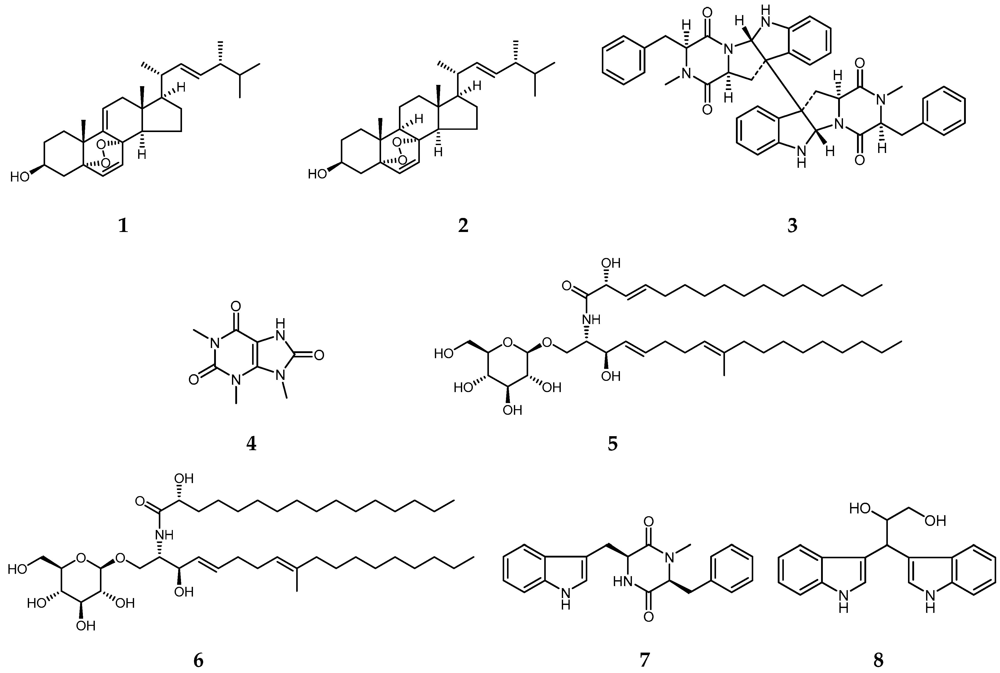
Figure 1. Chemical structures of compounds 18 isolated from B. cookei.
Molecules 29 03040 g001
Figure 2. A. retroflexus (A) and E. crus-galli (B) seedlings grown with 0 (control) and 200 μg/mL compound 7 for 96 h. Scale bar = 1 cm.
Molecules 29 03040 g002
Figure 3. Effect of compound 7 on the seedling growth of monocots (AD) and dicots (EH). Seedlings were grown with 0 (control) and 200 μg/mL compound 7 for 96 h for all except C. bursa-pastoris (144 h). The growth of S. viridis (A,B) and D. sanguinalis (C,D) was promoted, while that of C. bursa-pastoris (E,F) and S. nigrum (G,H) was inhibited by compound 7. Data presented as mean ± SE, and asterisks indicate a significant difference (two-tailed Student’s t-test, ** p < 0.01 and *** p < 0.001) as compared with the control group. Scale bar = 1 cm.
Molecules 29 03040 g003
Figure 4. Effect of compound 7 at different concentrations on the seedling growth of three dicotyledonous weeds, A. retroflexus (A), S. nigrum (C) and C. bursa-pastoris (E). The photographs show the optimal promotion and inhibition effects of compound 7 on A. retroflexus (B), S. nigrum (D) and C. bursa-pastoris (F), respectively. Scale bar = 5 mm.
Molecules 29 03040 g004
Figure 5. Effect of compound 7 on seed germination. (AC) The inhibition of compound 7 at 200 µg/mL against seed-germination process of three dicots, A. retroflexus (A), S. nigrum (B), and C. bursa-pastoris (C). (D) Compound 7 dose-response curve of C. bursa-pastoris gemination inhibition after 7 d; (E) Germination inhibition of compound 7 against C. bursa-pastoris at 400 µg/mL for 7 d.
Molecules 29 03040 g005
Figure 6. Phenotypic, cell morphological and physiological effects of compound 7 on A. retroflexu root at 400 μg/mL. (A) Root phenotype and cell shape of the elongation zone. White arrows in the picture showed damaged and wrinkled cell surfaces under the light microscope; (B) The morphology of root hairs; (C) The surface morphology of roots under SEM. Seedlings were grown for 96 h, and the root elongation zone was observed; (D) Root conductivity at 96 h following application of compound 7. (E) Ethylene content in roots at 96 h following application of compound 7. Data presented as mean ± SE, and asterisks indicate a significant difference (two-tailed Student’s t-test, ** p < 0.01 and *** p < 0.001) as compared with the control group.
Molecules 29 03040 g006
Table 1. Effects of compounds 18 at 200 μg/mL on the seedling growth of A. retroflexus and E. crus-galli.
CompoundsA. RetroflexusE. crus-galli
Shoot Length
(mm)
Root Length
(mm)
Shoot Length
(mm)
Root Length
(mm)
111.0 ± 0.3 b20.3 ± 0.3 b30.2 ± 2.1 b9.6 ± 0.3 b
28.7 ± 0.3 c11.1 ± 0.3 c33.3 ± 2.5 b9.1 ± 0.7 b
315.0 ± 0.6 a24.0 ± 0.4 a31.9 ± 1.6 b9.6 ± 0.9 b
414.4 ± 0.4 a24.8 ± 0.5 a32.8 ± 2.2 b9.8 ± 1.1 b
516.1 ± 0.3 a25.4 ± 0.2 a31.4 ± 2.1 b9.8 ± 1.6 b
615.7 ± 0.4 a26.9 ± 0.3 a31.0 ± 2.9 b10.2 ± 1.0 b
77.7 ± 0.3 c12.2 ± 0.3 c46.0 ± 2.7 a22.8 ± 1.7 a
814.8 ± 0.5 a23.9 ± 0.3 a32.3 ± 2.4 b10.1 ± 1.6 b
Control14.3 ± 0.4 a25.2 ± 0.7 a31.4 ± 2.7 b9.7 ± 0.7 b
Data shown are the mean of three independent experiments and presented as mean ± SE. Means followed by the different letters in the same column indicate significant differences (p < 0.05) among groups using one-way analysis of variance (ANOVA).
Disclaimer/Publisher’s Note: The statements, opinions and data contained in all publications are solely those of the individual author(s) and contributor(s) and not of MDPI and/or the editor(s). MDPI and/or the editor(s) disclaim responsibility for any injury to people or property resulting from any ideas, methods, instructions or products referred to in the content.