Article

[FeIIICl(TMPPH2)][FeIIICl4]2: A Stand-Alone Molecular Nanomedicine That Induces High Cytotoxicity by Ferroptosis

1
College of Chemistry, Chemical Engineering, and Materials Science, Soochow University, Suzhou 215123, China
2
NMPA Key Laboratory for Research and Evaluation of Drug Metabolism, Guangdong Provincial Key Laboratory of New Drug Screening, School of Pharmaceutical Sciences, Southern Medical University, Guangzhou 510515, China
3
Glasgow College UESTC, University of Electronic Science and Technology of China, Chengdu 611731, China
*
Authors to whom correspondence should be addressed.
These authors contributed equally to this work.
Molecules 2024, 29(11), 2495; https://doi.org/10.3390/molecules29112495
Submission received: 28 April 2024 / Revised: 22 May 2024 / Accepted: 23 May 2024 / Published: 24 May 2024

Abstract

:
Developing clinically meaningful nanomedicines for cancer therapy requires the drugs to be effective, safe, simple, cheap, and easy to store. In the present work, we report that a simple cationic Fe(III)-rich salt of [FeIIICl(TMPPH2)][FeIIICl4]2 (Fe-TMPP) exhibits a superior anticancer performance on a broad spectrum of cancer cell lines, including breast, colorectal cancer, liver, pancreatic, prostate, and gastric cancers, with half maximal inhibitory concentration (IC50) values in the range of 0.098–3.97 μM (0.066–2.68 μg mL−1), comparable to the best-reported medicines. Fe-TMPP can form stand-alone nanoparticles in water without the need for extra surface modification or organic-solvent-assisted antisolvent precipitation. Critically, Fe-TMPP is TME-responsive (TME = tumor microenvironment), and can only elicit its function in the TME with overexpressed H2O2, converting H2O2 to the cytotoxic •OH to oxidize the phospholipid of the cancer cell membrane, causing ferroptosis, a programmed cell death process of cancer cells.

1. Introduction

Materials designed to target the unique features of the tumor microenvironment (TME), such as the over-expression of H2O2 and glutathione (GSH), weak acidity, hypoxia, and immunosuppression, are attracting intensive interest in the field of nanomedicine [1,2,3,4,5,6]. Materials that are responsive exclusively to the TME imply low systemic toxicity and thus fewer side effects. Disturbing the complex homeostasis of the TME can further result in effective cancer therapy by novel cell death modalities (e.g., ferroptosis, pyroptosis, and immunogenic cell death) [7,8,9], and the resensitization of cells to conventional therapies [2,9,10]. Molecular complexes of Pt [11], Au [12], Fe [13], Os [14], Re [15], Ru [16], and Ir [17] have been reported to induce ferroptosis; their molecular structures are shown in Scheme 1. Nevertheless, most of the current formulations are far from satisfactory for practical use due to their formidable structural complexity, high cost, and low cellular uptake that subsequently leads to low anticancer efficacy.
Some useful strategies have been proposed to promote cellular uptake and the accumulation of the drugs in organelles (particularly mitochondria) [18,19] and the synergistic operation between different therapeutic modalities, such as chemotherapy, chemodynamic therapy (CDT), and photodynamic therapy (PDT), by taking advantage of the chemical properties of the individual building blocks of nanomedicines. For example, Ke et al. designed a cyclometallated Ir(III) complex featuring a −SH terminal as the cationic and photoactive building block [20]. Upon polymerization by disulfide bond formation, followed by DSPE–PEG–Biotin coating to endow aqueous solubility and cell specificity, the particle (denoted as IrS NPs) could be used for TME-responsive cancer therapy by harnessing both apoptosis and ferroptosis pathways. When IrS NPs were taken up by the cells, the disulfide bond was cleavaged by cross-coupling with the intracellular GSH to release the cationic subcomponent, which in turn, selectively accumulated in mitochondria. Notably, the use of Ir(III)-based photosensitizers elicited type I PDT, which circumvented the need for molecular oxygen. The oxidative stress imposed by the generated reactive oxygen species (ROS) from PDT resulted in the apoptosis of the lung cancer A549 cell line, while the depletion of GSH by IrS NPs weakened the reductive defense of the cells, leading to the effective peroxidation of the phospholipid, causing ferroptosis. In a similar effort, Yuan et al. designed a small molecular drug, MitoIrL2, featuring a cyclometallated Ir(III) skeleton for the hybrid treatment of breast and pancreatic cancer via a shared mechanism (apoptosis from type I PDT and ferroptosis) [21]. In MitoIrL2, the presence of an additional cationic phosphonium center resulted in more efficient mitochondrial accumulation and, therefore, better treatment outcomes relative to its phosphonium-free counterpart.
In the present work, we report that a simple salt of [FeIIICl(TMPPH2)][FeIIICl4]2 (denoted as Fe-TMPP; TMPPH2 is used to differentiate the ligand from the free-base porphyrin precursor of meso-Tetrakis (6-methylpyridin-3-yl) porphyrin, H2TMPP), which can directly form nanoparticles of suitable size for cellular uptake, and exhibits superior cytotoxicity towards a broad spectrum of cancer cell lines, including 4T1 (breast cancer), HCT-116 (colorectal cancer), DLD-1 (colorectal cancer), HuH-7 (liver cancer), BXPC3 (pancreatic cancer), PC3 (prostate cancer), and AGS (gastric cancer), with half maximal inhibitory concentration (IC50) values in the range 0.098−3.97 μM (0.066−2.68 μg mL−1), comparable to the best-reported medicines, including cisplatin and those exhibiting multiple treatment modalities.
Iron(II)-/Iron(III)- based nanoformulations have been widely reported for anticancer application via catalytic Fenton chemistry to convert the overexpressed H2O2 in TME into cytotoxic hydroxyl radicals (•OH) [6,22,23]. In addition, Fe3+ can be further reduced by the excess intracellular GSH into Fe2+ to initiate the Fenton process, and the exhaustion of GSH may further facilitate the ferroptosis process [6,22,23]. Fe-TMPP, reported herein, offers some advantages, such as iron-based chemotherapy, including inexpensive and facile synthesis, and direct nanoparticle formation without the need for complicated and costly surface modification. This instant nanoparticle formation by adding Fe-TMPP to water also permits convenient storage and simplifies the nanomedicine preparation process.

2. Results and Discussion

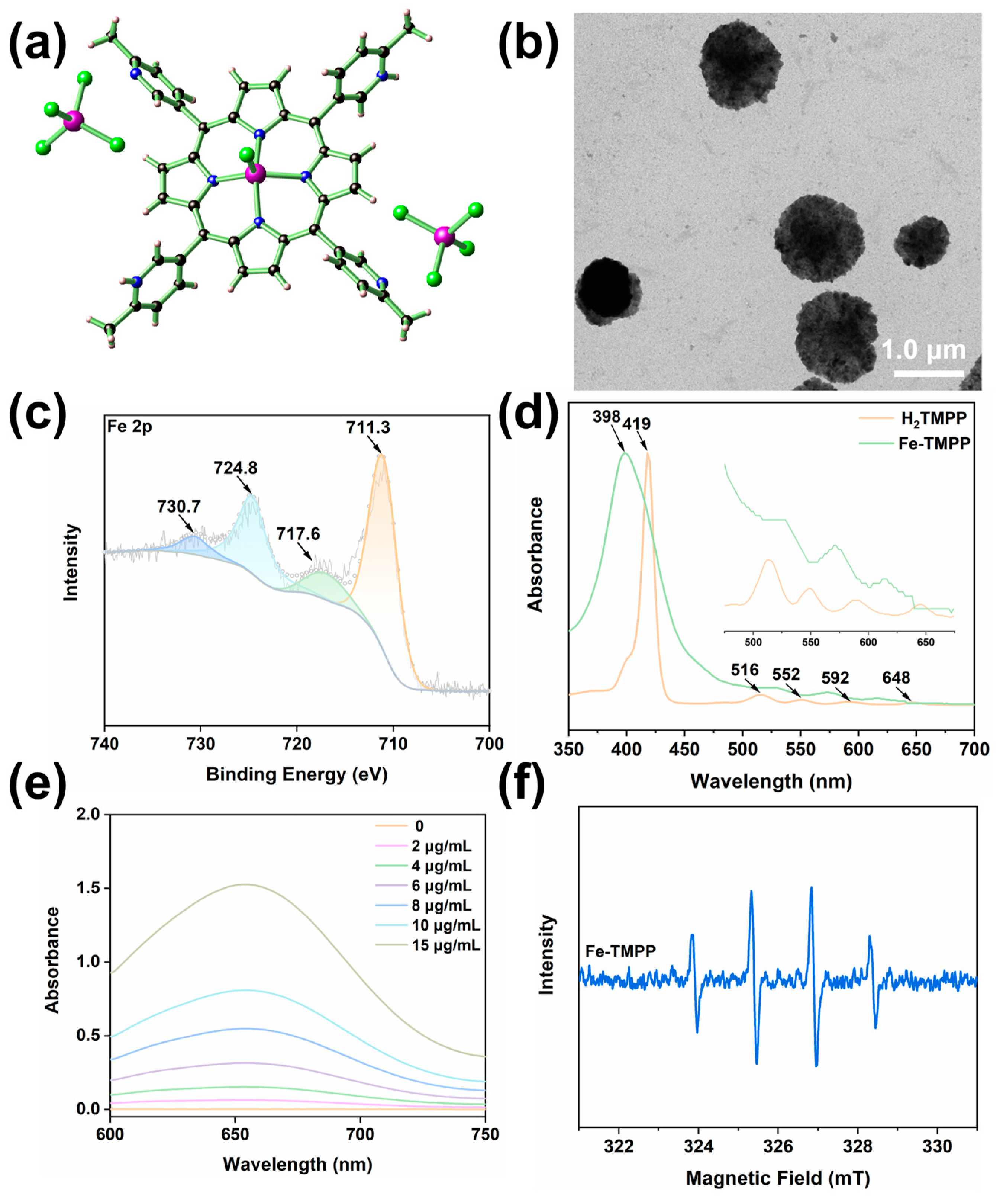
2.1. Material Synthesis and Structure Descriptions

The assembly of FeCl3·6H2O and H2TMPP under solvothermal conditions in CHCl3/EtOH using HAc as the modulating reagent resulted in the formation of Fe-TMPP. The successful synthesis was authenticated by single-crystal X-ray crystallographic analysis. Fe-TMPP crystallizes in the triclinic space group P−1 (Table 1), featuring a cation of [FeIIICl(TMPPH2)]2+ and a pair of [FeCl4] counterions (Figure 1a). The [FeIIICl(TMPPH2)]2+ cation features a ‘sit-atop’ Fe3+ which is chelated by four of the N atoms from the H2TMPP ligand and one Cl to give a five-coordinate square pyramidal configuration (Figure S1). Notably, of the four methylpyridyl peripheries of the H2TMPP ligand, the two N at the para-positions are protonated, as suggested by the presence of two [FeCl4] ions for each [FeIIICl(TMPPH2)]2+ cation, in addition to the relative dispositions of these methylpyridyl moieties in the molecular packing diagram (Figure S2). The structure, refined with full protonation of the methylpyridyl N sites or misplaced protons, inevitably led to short H∙∙∙H contacts.

2.2. Spectroscopic Characterizations of Fe-TMPP

Fe-TMPP was spectroscopically characterized and compared with its H2TMPP precursor. The in-plane N-H vibration at 964 cm−1 in the Fourier transform infrared (FT-IR) spectrum of the ligand disappeared (Figure S3), and a new peak at 997 cm−1 appeared in Fe-TMPP, indicating the metalation of the porphyrin center. Fe 2p X-ray photoelectron spectroscopy (XPS) of Fe-TMPP revealed two peaks at binding energies of 711.3 eV and 724.8 eV, which are assignable by the spin-orbit splitting of the Fe 2p3/2 and Fe 2p1/2 of Fe3+ (Figure 1c), while a pair of satellite peaks at around 717.6 eV and 730.7 eV were also present, presumably due to the photoreduction of Fe3+ during the measurement [24,25].
In the ultraviolet–visible (UV-Vis) spectra (Figure 1d), the Soret band of the free H2TMPP at 419 nm underwent a significant blueshift to 398 nm upon the chelation of Fe3+. In addition, the Q bands coalesced into two, due to the change of symmetry from D2h of H2TMPP to C4v of Fe-TMPP [26,27]. Notably, an additional absorption band at around 650 nm also appeared, presumably due to the charge transfer between Fe3+ and the ligand [28]. Notably, the UV-Vis spectrum of Fe-TMPP exhibited an undulating baseline, indicating lighting scattering by nanoparticle formation in an aqueous solution [29]. The formation of a stand-alone nanoparticle of Fe-TMPP could be directly observed with the Tyndall effect (Figure S4). Energy dispersive X-ray spectroscopy (EDS) showed an Fe:Cl:N atomic ratio of 1.0:2.6:3.0 in Fe-TMPP (Figure S5a), which is close to the value of 1.0:3.0:2.7 found in the single crystal data.

2.3. Characterizations of Fe-TMPP Nanoparticles

The direct nanoparticle formation of Fe-TMPP in water was observed in the transmission electron microscopic (TEM) images and also by dynamic light scattering (DLS) analysis. TEM showed that Fe-TMPP formed particles of variable sizes (Figure 1b). DLS gave different results from batch to batch but all contained considerable numbers of particles with sizes less than 100 nm (Figure S6). We argue that the accuracy of the TEM results may be compromised, as the particles of Fe-TMPP, without any surface modification, may undergo an Ostwald ripening process upon solvent evaporation during sample preparation [30]. The zeta potential of Fe-TMPP was 23.4 mV (Figure S7), which is ideal for ensuring good circulation stability with favorable adhesion to cell membranes for enhanced intracellular uptake [31].
Nanoparticles can be assembled and stored in an aqueous solution for short periods, or lyophilized for longer-term preservation. However, lyophilized particles can change their morphology, aggregate, or slowly degrade [32,33]. Antisolvent precipitation is an often-used alternative to prepare nanoparticles of organic drugs. A risk with this approach, however, is the encapsulation of toxic organic solvents, and sometimes the need for a supercritical antisolvent (e.g., CO2) [34,35]. The simple chemical formulation and good water-dispersing ability of Fe-TMPP is thus an advantage for storage and facile particle preparation.
Fresh samples of Fe-TMPP in aqueous solutions maintained their color and homogeneity under ambient conditions for three days (Figure S8), as also evidenced by their UV-Vis spectra (Figure S9). In addition, Fe-TMPP was stable in PBS (0.1×) for 0, 12, and 24 h, as indicated by only insignificant changes in particle size (Figure S10) and zeta potential (Figure S11). To further confirm the stability of Fe-TMPP in water, we sonicated the aqueous suspensions of Fe-TMPP and then lyophilized. The powder X-ray diffraction (PXRD) analysis revealed that the diffraction patterns of the water-treated samples resembled that of the as-synthesized crystalline samples (Figure S12). Moreover, the EDS spectra of the lyophilized sample of Fe-TMPP showed an Fe:Cl:N atomic ratio of 1.0:2.4:3.2 (Figure S5b), which was close to that of the as-synthesized crystalline sample (1.0:2.6:3.0), indicating that there was no obvious compositional change upon suspending Fe-TMPP in water. We postulate that the four methyl groups at the ortho position of the pyridyl N sites significantly boost the hydrophobicity of the molecule to facilitate the aggregation of the polar Fe-TMPP featuring multiple hydrophilic moieties (i.e., [FeCl4], protonated pyridyl ligands, the Fe-Cl, and free pyridyl N site) and thereby protecting the salt from hydrolyzation.

2.4. Detection of Fe-TMPP-induced Generation of ROS in Solution

High valent transition metal ions Fe3+ and Cu2+ exhibit peroxidase-like activity to generate reactive •OH, by utilizing the overexpressed H2O2 in the TME, thereby eliciting nonspecific damage inside cancer cells [6,7,8,26]. The TME also reduces Fe3+ and Cu2+ to Fe2+ and Cu+ through a reaction with the overexpressed GSH to initiate Fenton and Fenton-like reactions, which also generate •OH from H2O2. The peroxidase-like activity of the Fe3+-rich Fe-TMPP was therefore assayed using 3,3′,5,5′-tetramethylbenzidine (TMB) [36]. Different concentrations of Fe-TMPP were added to TMB solutions containing 100 μM H2O2, the approximate concentration in tumors, and five times higher than in normal cells [1,2]. It can be seen from Figure 1e that Fe-TMPP produced considerable •OH in a concentration-dependent manner upon incubating with H2O2 over 3 h. The generation of •OH was confirmed by electron paramagnetic resonance (EPR) spectroscopy, using DMPO as the spin trap. As shown in Figure 1f, the EPR spectrum of Fe-TMPP gave rise to a strong four-line signal with a 1:2:2:1 peak-to-peak intensity pattern, characteristic of the generation of •OH from H2O2 [4,5].
Fe-TMPP is supported by a porphyrin-based ligand, which is often used for type II PDT of cancer [3,7,37]. We employed diphenylisobenzofuran (DPBF) as a probe to compare the singlet oxygen’s (1O2) generating capacity with that of TMPP-F127 (H2TMPP encapsuled by Pluronic F-127, Figure S13) [38], using blank DPBF as a control [39,40]. DPBF was added to aqueous solutions of TMPP-F127 and Fe-TMPP, followed by laser irradiation (650 nm, 25 mW cm−2). As shown in Figure S14, at equivalent H2TMPP concentrations, neither species could generate sufficient 1O2 to degrade DPBF under laser irradiation. The 1O2 generated by Fe-TMPP was negligible, which might be due to the presence of redox-active Fe3+ that can quench the excited state of the molecule by electron transfer [41,42].

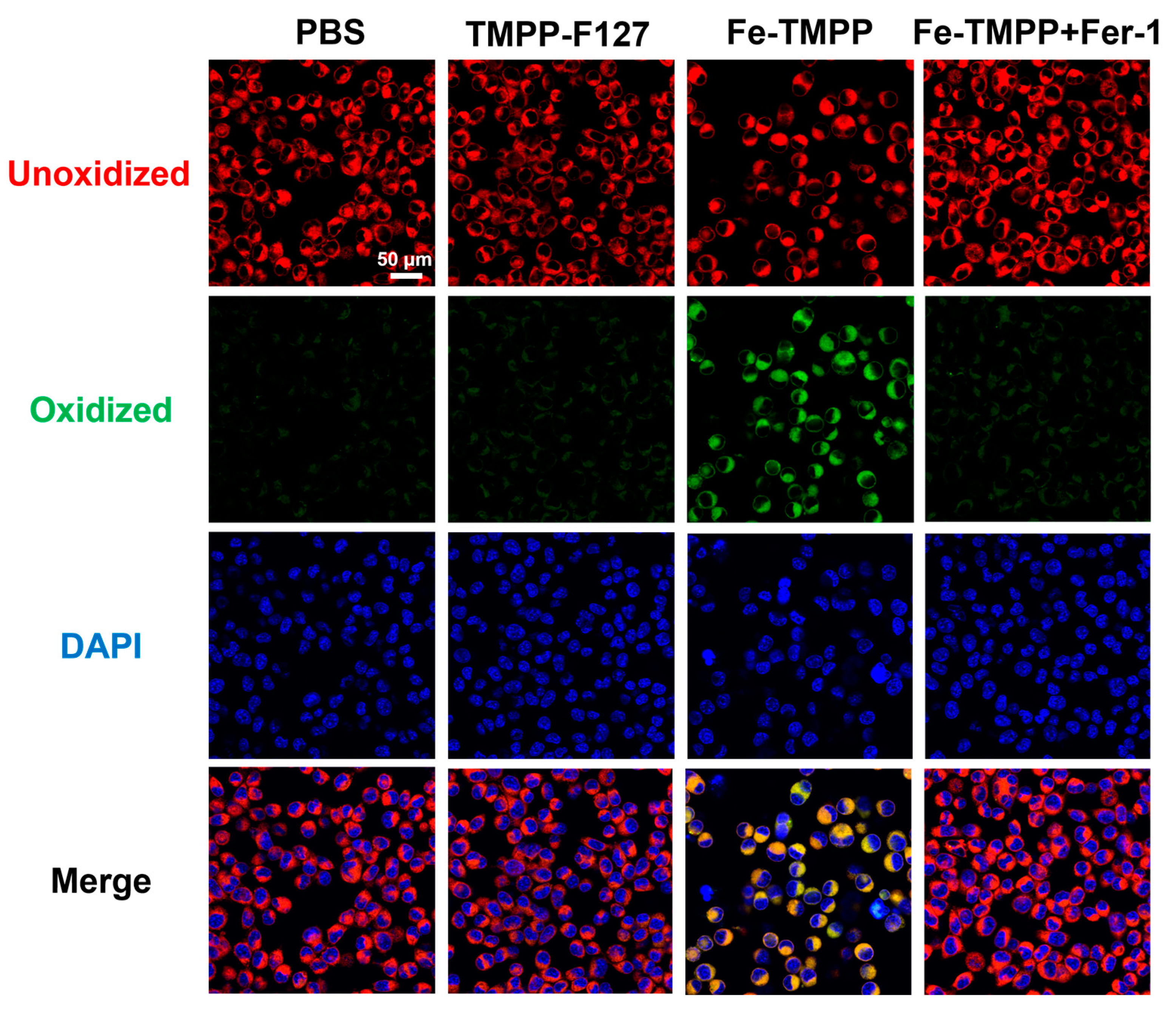
2.5. In Vitro ROS Detection and Cytotoxicity Assay

Given the efficient •OH production in water induced by Fe-TMPP, we next investigated in vitro ROS generation by Fe-TMPP in the representative 4T1 cell line using 2′,7′-dichlorodihydrofluorescein diacetate (DCFH-DA) as a fluorescence probe [4]. As shown in Figure 2a and Figure S15, green fluorescence was detected in the cells treated with Fe-TMPP, which indicated the production of ROS in the cells from the peroxidase-like activity of Fe3+. By comparison, TMPP-F127 produced few ROS, which may be due to the aggregation of the H2TMPP ligand within the hydrophobic core of the TMPP-F127 micelles.
The cytotoxicity of Fe-TMPP against diverse cell lines including 4T1 (breast cancer), HCT-116 (colorectal cancer), DLD-1 (colorectal cancer), HuH-7 (liver cancer), BXPC3 (pancreatic cancer), PC3 (prostate cancer), and AGS (gastric cancer), were evaluated using either 3-(4,5-dimethylthiazol-2-yl)-2,5-diphenyltetrazolium bromide (MTT) or cell counting kit-8 (CCK-8) assays. As shown in Figure 2b and Figure S16, Fe-TMPP exhibited concentration-dependent cytotoxicity against all tested cell lines. The half maximal inhibitory concentration (IC50) values for Fe-TMPP were relatively low, down to 0.0975 μM (0.0658 μg mL−1) for AGS and up to 3.97 μM (2.68 μg mL−1) for HCT-116 (Table 2). These IC50 values are comparable to cisplatin and to complex formulations that integrate multiple therapeutic modalities. The above results indicate that Fe-TMPP exhibits significant cytotoxicity against a broad spectrum of cancer cell lines.

2.6. Ferroptosis Assay

As noted above, when Fe-TMPP is internalized by cancer cells, Fe3+ can oxidize GSH to give glutathione disulfide (GSSG) and is concomitantly reduced to Fe2+. The exhaustion of GSH in these cells can downregulate the expression of glutathione peroxidase 4 (GPX4), making the cell membrane phospholipid more susceptible to peroxidation [49,50]. GSH depletion and phospholipid peroxidation are two characteristic features of ferroptosis, a novel form of iron-dependent programmed cell death [49,50]. We therefore examined the possibility of ferroptosis induced by Fe-TMPP using 4T1 cell line as a representative. Phospholipid peroxidation levels of 4T1 cells were measured using C11-BODIPY (BODIPY™ 581/591 C11) as a fluorescent probe, as observed under confocal laser scanning microscopy (Figure 3) [51]. We also used DAPI to stain the nucleus of the cells. The cells treated with Fe-TMPP showed significantly stronger lipid peroxidation than those treated with TMPP-F127 or PBS. In addition, lipid peroxidation can be inhibited by the presence of ROS scavengers, such as ferrostatin-1 (Fer-1), liproxstatin-1, and vitamin E [21,52]. In the present experiments, lipid peroxidation was completely inhibited by the introduction of Fer-1, as indicated by the restoration of the red emission of C11-BODIPY. These results indicated that Fe-TMPP can induce ferroptosis for cancer treatment. Flow cytometry using Annexin-V/PI double staining further confirmed that 4T1 cells treated with Fe-TMPP showed obviously late apoptosis relative to TMPP-F127 and PBS-treated cells, supporting this alternative cell death mechanism (Figure 2c) [53,54].

3. Materials and Methods

3.1. General

Ligand H2TMPP was synthesized as described in our previous reports [27]. 3-Aldehyde-6-methylpyridine (>98%, Macklin, Shanghai, China), ferric chloride hexahydrate (99%, Macklin), pluronic F-127 (average molecular weight 2000, Sigma-Aldrich, Darmstadt, Germany), 1,3-diphenylisobenzofuran (DPBF) (97%, Acros, Waltham, MA, USA), 3,3′,5,5′-tetramethylbenzidine (TMB) (99%, Shanghai Yuanye Bio-Technology Co., Ltd., Shanghai, China), and hydrogen peroxide (H2O2, 30%) (Yikewei Biological Technology Co., Ltd., Guangzhou, China) were obtained directly from commercial sources and used as received. Pyrrole (>95%, Macklin) was freshly distilled before use.
HCT-116, HuH-7, BXPC3, DLD-1, PC3, AGS, and 4T1 cell lines were purchased from the Shanghai Institute of Cell Biology, Chinese Academy of Sciences. Phosphate buffer solution (PBS) and fetal bovine serum (FBS) supplemented with penicillin and streptomycin were bought from Elabscience Crop (Guangzhou, China). 3-(4,5-Dimethylthiazol-2-yl)-2,5-diphenyltetrazolium bromide (MTT) was purchased from Beijing Solarbio Science & Technology Co., Ltd. (Beijing, China). The cell counting kit-8 (CCK-8) was commercially available from APEXBIO. 2′,7′-Dichlorodihydrofluorescein diacetate (DCFH-DA) and 4′,6-diamidino-2-phenylindole (DAPI) solution were obtained from Beijing Solarbio Science & Technology Co., Ltd. C11-BODIPY581/591 was bought from Invitrogen Corp. Ferrostatin-1 (Fer-1) was bought from Yike Biological Technology Co., Ltd. (Guangzhou, China). The Annexin FITC/PI apoptosis detection kit was purchased from Elabscience Crop.
Fourier transform infrared (FT-IR) spectra were recorded on a Bruker VERTEX 70+HYPERION 2000 spectrometer. Ultraviolet-visible spectroscopy (UV-Vis) absorption spectra were obtained on a Varian Cary-50 UV–Visible spectrophotometer (Varian, Inc., Palo Alto, CA, USA). X-ray photoelectron spectroscopy (XPS) was conducted on an EXCALAB 250 XI X-ray photoelectron spectrometer (Thermo Scientific, Waltham, MA, USA). The EPR measurement was conducted by a Bruker EMXnano spectrometer, with DMPO as the spin-trapping agent. The optical density (OD) values were collected using a TECAN M1000PRO microplate reader (Tecan, Zürich, Switzerland). The flow cytometry was performed using a BD FACS Caliber instrument (BD Biosciences, San Jose, CA, USA). The transmission electron microscopic (TEM) images were obtained by dropping the sample in water onto a copper net under a HITACHI HT7700 transmission electron microscope. Zeta potentials were measured on an LA-95052 laser particle size analyzer using dynamic light scattering (DLS). The cytotoxicity assay was conducted on a multifunction microplate detector by recording the absorption at 570 nm using a TECAN M1000PRO microplate reader. The cell staining results were observed with a confocal laser scanning microscope (CLSM, AxioObserver A1, Germany). The EDS and elemental mapping diagrams were obtained on the EVO18 scanning electron microscope.

3.2. Synthesis and Characterization of TMPP-F127

Pluronic F-127 (10.6 mg) was introduced to 1 mL of DMSO solution containing H2TMPP (4.5 mg), and the homogenous mixture formed was then added dropwise into 3.5 mL of deionized water with tip sonication. The micelle formed was dialyzed using a dialysis bag (molecular weight cut-off of 2000 Da) for 24 h to obtain TMPP-F127. TEM indicated that the particle sizes for TMPP-F127 were 170 ± 25 nm (Figure S13), which is conducive to cellular uptake.

3.3. Synthesis and Characterization of Fe-TMPP

FeCl3·6H2O (2.4 mg, 0.009 mmol) and H2TMPP (2 mg, 0.003 mmol) in 2 mL of CHCl3/EtOH (v:v = 3:1) were added into a Pyrex glass tube, followed by HAc (50 μL) as a regulator. The resulting mixture was then transferred to a programmable oven, heated to 120 °C over 4 h, and maintained at that temperature for 48 h, before cooling to room temperature over 24 h to yield purple block crystals of Fe-TMPP, which were collected through filtration, washed thoroughly with anhydrous ether, and dried in vacuo. Yield: 0.84 mg, 24.2% based on Fe. Anal. Calcd (%) for C44H34Cl9Fe3N8: C 45.50%, H 2.95%, N 9.65%; found: C 41.87%, H 2.74%, and N 8.70%, corresponding to Fe-TMPP∙CHCl3 (C 42.20%, H 2.75%, and N 8.75%). FT-IR (KBr disc, cm−1): 3372(w), 2521(w), 2162(w), 2029(w), 1607(m), 1552(m), 1458(m), 1377(w), 1329(w), 1288(s), 1258(w), 1204(m), 1139(s), 1085(m), 1047(m), 997(vs), 890(w), 854(w), 801(vs), 744(s), 721(s), 662(m), and 624(w).

3.4. Single Crystal X-ray Crystallography

Data collections were performed on a Bruker APEX II CCD X-ray diffractometer using Mo Kα (λ = 0.71073 Å) irradiation. Refinement and reduction of the collected data were achieved using the program Bruker SAINT and absorption corrections were performed using a multi-scan method [55]. Crystal structures were solved by direct methods and refined on F2 using full-matrix least-squares techniques with SHELXTL-2016 [56].
For Fe-TMPP, the [FeIIICl4] anion displays a conformational disorder with a relative ratio of 0.73:0.27 refined for the disordered domains. The [FeIIICl(TMPPH2)]2+ cation also has two conformations due to the presence of ‘sit-atop’ Fe3+, and the disorder is at about the center of the porphyrin ligand. Thus, the occupancy factor of the coordinated FeCl moiety was fixed at 0.5. For the porphyrin ligand, only two pyridyl groups were protonated to balance the charge of the anion. The protonation of half of the four pyridyls can also be judged from the packing of the molecules. In the packing diagram, two pyridyls were brought to proximity by an N-H∙∙∙N bond, thereby excluding the full protonation of the pyridyls and corroborating the charge counting. A small amount of spatially delocalized electron density in the lattice was found but acceptable refinement results could not be obtained for this electron density. The solvent contribution was then modeled using SQUEEZE in the Platon program suite [57].
Crystallographic data for Fe-TMPP have been deposited in the Cambridge Crystallographic Data Center (CCDC) as supplementary publication number 2330690. These data can be obtained free of charge from the CCDC via www.ccdc.cam.ac.uk/data_request/cif accessed on 3 February 2024) or from the Supplementary Materials. A summary of the key crystallographic data for Fe-TMPP is listed in Table 1.

3.5. •OH Detection

Different concentrations of Fe-TMPP were added to a TMB solution containing 100 μM H2O2. After reacting for 3 h at room temperature, the solution was centrifuged to remove the influence of nanoparticles on absorbance. The ultraviolet absorption curve of the supernatant at 652 nm was determined.

3.6. 1O2 Detection

Identical concentrations of DPBF were added to aqueous solutions of TMPP-F127 and Fe-TMPP relative to the concentration of TMPP, to give uniform mixtures, with the concentrations of DPBF and TMPP being 33 μg mL−1 and 7 μg mL−1, respectively. The solutions were then irradiated by a laser (650 nm, 25 mW cm−2). After each irradiation for 30 s, the absorption of DPBF was immediately measured. By monitoring the decreasing rate of the ultraviolet absorption peak at 416 nm, the 1O2 generation capacities of these materials could be evaluated.

3.7. MTT or CCK-8 Cytotoxicity Assay

The MTT assay of 4T1 cells is detailed as an example. The 4T1 cell line was cultured in Roswell Park Memorial Institute (RPMI) 1640 media supplemented with 10% FBS and 1% penicillin-streptomycin at 37 °C in 5% CO2. Cells grew as a monolayer and were detached upon confluence using trypsin (0.5% w/v in PBS). The cells were harvested from the cell culture medium by incubating in trypsin solution for 2 min, and then centrifuged with the supernatant subsequently discarded. A 3 mL portion of serum-supplemented cell culture medium was added to neutralize any residual trypsin. The cells were re-suspended in serum-supplemented RPMI 1640 at a concentration of 5 × 10⁴ cells/mL. Cells were cultured at 37 °C and 5% CO2 for the MTT studies.
4T1 cells were inoculated into 96-well plates (5 × 10³ cells per well) and incubated for 24 h. After 24 h of incubation, the cells were treated with different concentrations of drugs for 24 h, and, upon completion of the treatment, the MTT solution was added and incubated for 4 h, and DMSO was added and shocked for 15 min. The cell viability was determined using an enzyme marker.
The relative cell viability (%) related to control cells was calculated using the following equation:
V % = [ A ] e x p e r i m e n t a l [ A ] b l a n k [ A ] c o n t r o l [ A ] b l a n k × 100 %
where V% is the percentage of cell viability, [A]experimental is the absorbance of the wells culturing the treated cells, [A]blank is the absorbance of the blank, and [A]control is the absorbance of the wells culturing untreated cells.
The cell viability data for HCT-116 (CCK-8), DLD-1 (CCK-8), HuH-7 (CCK-8), BXPC3 (CCK-8), PC3 (CCK-8), and AGS (CCK-8) were obtained in a similar manner using protocols listed in Table S1.

3.8. Intracellular ROS Detection

4T1 cells were inoculated in 12-well plates (1 × 10⁵ cells per well), incubated for 24 h and then treated with different drugs for 24 h. After completion of the treatment, the cells were treated with DCFH-DA, and the cellular reactive oxygen species results were determined using an inverted fluorescence microscopy.

3.9. Flow Cytometric Apoptosis Assay

4T1 cells were inoculated in 6-well plates (3 × 10⁵ cells per well) and incubated for 24 h. After 24 h of incubation, the cells were treated with different drugs for 24 h. After the completion of the treatment, the cells were processed using an Annexin V-FITC/PI apoptosis kit, and the results of apoptosis were determined using flow cytometry.

3.10. Ferroptosis Assay

4T1 cells were inoculated in confocal glass dishes (1 × 10⁵ cells per dish) and incubated for 24 h. Fer-1 (10 μM), an inhibitor of ferroptosis, was added and incubated for 12 h. The cells were then treated with different drugs for 24 h. After the completion of the treatment, the cells were incubated for 30 min with the ferroptosis probe C11-BODIPY581/591 (10 μM) and the results of the ferroptosis were observed using CLSM.

4. Conclusions

In this work, a simple ferroptosis inducer of Fe-TMPP was readily synthesized from earth-abundant metal and common organic materials. Fe-TMPP can directly form nanoparticles without requiring extra surface modification or organic-solvent-assisted antisolvent precipitation. Compared to the reported ferroptosis inducers based on noble metals such as oxaliplatin [11] and other coordination complexes derived from Au [12], Os [14], Ru [16], and Ir [17], the advantages of using Fe-TMPP include inexpensive fabrication, in addition to its cancer-specific activity and promising cytotoxicity, as described herein. The instant nanoparticle formation by adding Fe-TMPP to water also ensures convenient storage and simplifies the potential nanomedicine preparation process. All these features are important for potential clinical use. However, it should also be noted that iron overloading can potentially lead to cardiotoxicity, also caused by the ferroptosis process [58,59]. Nevertheless, the unexpectedly high cytotoxicity originating from ROS alone inspires us to investigate the possibility of other structure analogs based on metal ions, such as Cu2+, and to further explore their mechanisms of action by examining the sub-organelle locations of these metallodrugs.

Supplementary Materials

The following supporting information can be downloaded at: https://www.mdpi.com/article/10.3390/molecules29112495/s1.

Author Contributions

Conceptualization, W.-H.Z. and J.-X.C.; methodology, X.W. and J.-H.F.; software, W.-H.Z. and C.-M.Z.; validation, C.-M.Z., Z.-S.Z. and F.-L.C.; formal analysis, X.W. and J.-H.F.; data curation, W.-H.Z., J.-X.C. and D.J.Y.; writing—original draft preparation, X.W. and J.-H.F.; writing—review and editing, W.-H.Z., J.-X.C. and D.J.Y.; supervision, W.-H.Z.; project administration, W.-H.Z. and J.-X.C.; funding acquisition, W.-H.Z. and J.-X.C. All authors have read and agreed to the published version of the manuscript.

Funding

This research was funded by the National Natural Science Foundation of China (21871203), the Natural Science Foundation from Guangdong Science and Technology Department of China (2023A1515012633), and the Project of Medical Innovation Application Research of Suzhou (SKY2022094).

Institutional Review Board Statement

Not applicable.

Informed Consent Statement

Not applicable.

Data Availability Statement

The data that support the findings of this study are available from the corresponding authors upon request.

Acknowledgments

We are grateful to Yiming Mao from Suzhou Kowloon Hospital, Shanghai Jiaotong University School of Medicine, for his insightful comments on this manuscript.

Conflicts of Interest

The authors declare no conflicts of interest.

References

  1. Chu, Z.; Yang, J.; Zheng, W.; Sun, J.; Wang, W.; Qian, H. Recent advances on modulation of H2O2 in tumor microenvironment for enhanced cancer therapeutic efficacy. Coord. Chem. Rev. 2023, 481, 215049. [Google Scholar] [CrossRef]
  2. Wang, Y.; Gao, F.; Li, X.; Niu, G.; Yang, Y.; Li, H.; Jiang, Y. Tumor microenvironment-responsive Fenton nanocatalysts for intensified anticancer treatment. J. Nanobiotechnology 2022, 20, 69. [Google Scholar] [CrossRef] [PubMed]
  3. Zou, Y.-M.; Li, R.-T.; Yu, L.; Huang, T.; Peng, J.; Meng, W.; Sun, B.; Zhang, W.-H.; Jiang, Z.-H.; Chen, J.; et al. Reprogramming of the tumor microenvironment using a PCN-224@IrNCs/d-Arg nanoplatform for the synergistic PDT, NO, and radiosensitization therapy of breast cancer and improving anti-tumor immunity. Nanoscale 2023, 15, 10715–10729. [Google Scholar] [CrossRef] [PubMed]
  4. Li, W.; Li, R.; Ye, Q.; Zou, Y.; Lu, X.; Zhang, W.; Chen, J.; Zhao, Y. Mn3O4 nanoshell coated metal–organic frameworks with microenvironment-driven O2 production and GSH exhaustion ability for enhanced chemodynamic and photodynamic cancer therapies. Adv. Healthc. Mater. 2023, 12, 2202280. [Google Scholar] [CrossRef]
  5. Huang, N.; Tang, X.-Y.; Meng, W.; Lai, Y.-H.; Zhou, X.; Yu, X.-Z.; Zhang, W.-H.; Chen, J.-X. Immunogenic radiation therapy for enhanced antitumor immunity via a core–shell nanosensitizer-mediated immunosuppressive tumor microenvironment modulation. ACS Nano 2023, 17, 19853–19864. [Google Scholar] [CrossRef]
  6. Hou, Y.-K.; Zhang, Z.-J.; Li, R.-T.; Peng, J.; Chen, S.-Y.; Yue, Y.-R.; Zhang, W.-H.; Sun, B.; Chen, J.-X.; Zhou, Q. Remodeling the tumor microenvironment with core–shell nanosensitizer featuring dual-modal imaging and multimodal therapy for breast cancer. ACS Appl. Mater. Interfaces 2023, 15, 2602–2616. [Google Scholar] [CrossRef]
  7. Pan, W.-L.; Tan, Y.; Meng, W.; Huang, N.-H.; Zhao, Y.-B.; Yu, Z.-Q.; Huang, Z.; Zhang, W.-H.; Sun, B.; Chen, J.-X. Microenvironment-driven sequential ferroptosis, photodynamic therapy, and chemotherapy for targeted breast cancer therapy by a cancer-cell-membrane-coated nanoscale metal-organic framework. Biomaterials 2022, 283, 121449. [Google Scholar] [CrossRef] [PubMed]
  8. Zhang, Y.; Yu, W.; Chen, M.; Zhang, B.; Zhang, L.; Li, P. The applications of nanozymes in cancer therapy: Based on regulating pyroptosis, ferroptosis and autophagy of tumor cells. Nanoscale 2023, 15, 12137–12156. [Google Scholar] [CrossRef] [PubMed]
  9. Wen, Y.; Chen, X.; Zhu, X.; Gong, Y.; Yuan, G.; Qin, X.; Liu, J. Photothermal-chemotherapy integrated nanoparticles with tumor microenvironment response enhanced the induction of immunogenic cell death for colorectal cancer efficient treatment. ACS Appl. Mater. Interfaces 2019, 11, 43393–43408. [Google Scholar] [CrossRef]
  10. Huang, N.; Qian, A.; Zou, Y.; Lin, M.; Pan, W.; Chen, M.; Meng, W.; Zhang, W.; Chen, J. Immunogenic radiation therapy for enhanced anti-tumor immunity via core-shell nanocomposite-mediated multiple strategies. Theranostics 2023, 13, 4121–4137. [Google Scholar] [CrossRef]
  11. Liu, B.; Wang, H. Oxaliplatin induces ferroptosis and oxidative stress in HT29 colorectal cancer cells by inhibiting the Nrf2 signaling pathway. Exp. Ther. Med. 2022, 23, 394. [Google Scholar] [CrossRef] [PubMed]
  12. Yang, L.; Wang, H.; Yang, X.; Wu, Q.; An, P.; Jin, X.; Liu, W.; Huang, X.; Li, Y.; Yan, S.; et al. Auranofin mitigates systemic iron overload and induces ferroptosis via distinct mechanisms. Signal Transduct. Target. Ther. 2020, 5, 138. [Google Scholar] [CrossRef] [PubMed]
  13. Zhang, L.; Ma, S.; Wang, T.; Li, S.; Wang, L.; Li, D.; Tian, Y.; Zhang, Q. Four-photon absorption iron complex for magnetic resonance/photoacoustic dual-model imaging and an enhanced ferroptosis process. Anal. Chem. 2023, 95, 1635–1642. [Google Scholar] [CrossRef] [PubMed]
  14. Lu, N.; Deng, Z.; Gao, J.; Liang, C.; Xia, H.; Zhang, P. An osmium-peroxo complex for photoactive therapy of hypoxic tumors. Nat. Commun. 2022, 13, 2245. [Google Scholar] [CrossRef] [PubMed]
  15. Zhu, J.; Ouyang, A.; He, J.; Xie, J.; Banerjee, S.; Zhang, Q.; Zhang, P. An ultrasound activated cyanine-rhenium(i) complex for sonodynamic and gas synergistic therapy. Chem. Commun. 2022, 58, 3314–3317. [Google Scholar] [CrossRef] [PubMed]
  16. Qi, F.; Yuan, H.; Chen, Y.; Peng, X.-X.; Wu, Y.; He, W.; Guo, Z. Type I photoreaction and photoinduced ferroptosis by a Ru(II) complex to overcome tumor hypoxia in photodynamic therapy. CCS Chem. 2022, 5, 1583–1591. [Google Scholar] [CrossRef]
  17. Wang, X.; Chen, F.; Zhang, J.; Sun, J.; Zhao, X.; Zhu, Y.; Wei, W.; Zhao, J.; Guo, Z. A ferroptosis-inducing iridium(III) complex. Sci. China Chem. 2020, 63, 65–72. [Google Scholar] [CrossRef]
  18. Liu, J.; Kang, D.W.; Fan, Y.; Nash, G.T.; Jiang, X.; Weichselbaum, R.R.; Lin, W. Nanoscale covalent organic framework with staggered stacking of phthalocyanines for mitochondria-targeted photodynamic therapy. J. Am. Chem. Soc. 2024, 146, 849–857. [Google Scholar] [CrossRef]
  19. Sun, Q.; Yang, J.; Shen, W.; Lu, H.; Hou, X.; Liu, Y.; Xu, Y.; Wu, Q.; Xuan, Z.; Yang, Y.; et al. Engineering mitochondrial uncoupler synergistic photodynamic nanoplatform to harness immunostimulatory pro-death autophagy/mitophagy. Biomaterials 2022, 289, 121796. [Google Scholar] [CrossRef]
  20. Ke, L.; Wei, F.; Xie, L.; Karges, J.; Chen, Y.; Ji, L.; Chao, H. A biodegradable iridium(III) coordination polymer for enhanced two-photon photodynamic therapy using an apoptosis–ferroptosis hybrid pathway. Angew. Chem. Int. Ed. 2022, 61, e202205429. [Google Scholar] [CrossRef]
  21. Yuan, H.; Han, Z.; Chen, Y.; Qi, F.; Fang, H.; Guo, Z.; Zhang, S.; He, W. Ferroptosis photoinduced by new cyclometalated iridium(III) complexes and its synergism with apoptosis in tumor cell inhibition. Angew. Chem. Int. Ed. 2021, 60, 8174–8181. [Google Scholar] [CrossRef] [PubMed]
  22. Li, S.-L.; Chu, X.; Dong, H.-L.; Hou, H.-Y.; Liu, Y. Recent advances in augmenting Fenton chemistry of nanoplatforms for enhanced chemodynamic therapy. Coord. Chem. Rev. 2023, 479, 215004. [Google Scholar] [CrossRef]
  23. Tang, Z.; Zhao, P.; Wang, H.; Liu, Y.; Bu, W. Biomedicine meets Fenton chemistry. Chem. Rev. 2021, 121, 1981–2019. [Google Scholar] [CrossRef] [PubMed]
  24. Liu, Z.; Liu, S.; Liu, B.; Bian, Y.; Yuan, M.; Yang, C.; Meng, Q.; Chen, C.; Ma, P.a.; Lin, J. Fe(III)-naphthazarin metal–phenolic networks for glutathione-depleting enhanced ferroptosis–apoptosis combined cancer therapy. Small 2023, 19, 2207825. [Google Scholar] [CrossRef] [PubMed]
  25. Meda, L.; Ranghino, G.; Moretti, G.; Cerofolini, G.F. XPS detection of some redox phenomena in Cu-zeolites. Surf. Interface Anal. 2002, 33, 516–521. [Google Scholar] [CrossRef]
  26. Li, Q.; Xu, B.-W.; Zou, Y.-M.; Niu, R.-J.; Chen, J.-X.; Zhang, W.-H.; Young, D.J. Nanoscale two-dimensional FeII- and CoII-based metal−organic frameworks of porphyrin ligand for the photodynamic therapy of breast cancer. Molecules 2023, 28, 2125. [Google Scholar] [CrossRef] [PubMed]
  27. Niu, R.-J.; Zhou, W.-F.; Liu, Y.; Yang, J.-Y.; Zhang, W.-H.; Lang, J.-P.; Young, D.J. Morphology-dependent third-order optical nonlinearity of a 2D Co-based metal–organic framework with a porphyrinic skeleton. Chem. Commun. 2019, 55, 4873–4876. [Google Scholar] [CrossRef] [PubMed]
  28. Valicsek, Z.; Horváth, O. Application of the electronic spectra of porphyrins for analytical purposes: The effects of metal ions and structural distortions. Microchem. J. 2013, 107, 47–62. [Google Scholar] [CrossRef]
  29. Basalla, A.J.; Kendrick, B.S. Correcting ultraviolet-visible spectra for baseline artifacts. J. Pharm. Sci. 2023, 112, 3240–3247. [Google Scholar] [CrossRef]
  30. Zhang, Q.; Peng, X.; Nie, Y.; Zheng, Q.; Shangguan, J.; Zhu, C.; Bustillo, K.C.; Ercius, P.; Wang, L.; Limmer, D.T.; et al. Defect-mediated ripening of core-shell nanostructures. Nat. Commun. 2022, 13, 2211. [Google Scholar] [CrossRef]
  31. Pochapski, D.J.; Carvalho dos Santos, C.; Leite, G.W.; Pulcinelli, S.H.; Santilli, C.V. Zeta potential and colloidal stability predictions for inorganic nanoparticle dispersions: Effects of experimental conditions and electrokinetic models on the interpretation of results. Langmuir 2021, 37, 13379–13389. [Google Scholar] [CrossRef]
  32. Howard, M.D.; Lu, X.; Jay, M.; Dziubla, T.D. Optimization of the lyophilization process for long-term stability of solid–lipid nanoparticles. Drug Dev. Ind. Pharm. 2012, 38, 1270–1279. [Google Scholar] [CrossRef] [PubMed]
  33. Hussain, Z.; Rahman, S. Storage stabilisation of albumin-loaded chitosan nanoparticles by lyoprotectants. Trop. J. Pharm. Res. 2013, 2, 135–142. [Google Scholar]
  34. Mahesh, K.V.; Singh, S.K.; Gulati, M. A comparative study of top-down and bottom-up approaches for the preparation of nanosuspensions of glipizide. Powder Technol. 2014, 256, 436–449. [Google Scholar] [CrossRef]
  35. Zhao, X.; Wang, W.; Zu, Y.; Zhang, Y.; Li, Y.; Sun, W.; Shan, C.; Ge, Y. Preparation and characterization of betulin nanoparticles for oral hypoglycemic drug by antisolvent precipitation. Drug Deliv. 2014, 21, 467–479. [Google Scholar] [CrossRef] [PubMed]
  36. Yuan, B.; Chou, H.-L.; Peng, Y.-K. Disclosing the origin of transition metal oxides as peroxidase (and catalase) mimetics. ACS Appl. Mater. Interfaces 2022, 14, 22728–22736. [Google Scholar] [CrossRef] [PubMed]
  37. Li, R.-T.; Zhu, Y.-D.; Li, W.-Y.; Hou, Y.-K.; Zou, Y.-M.; Zhao, Y.-H.; Zou, Q.; Zhang, W.-H.; Chen, J.-X. Synergistic photothermal-photodynamic-chemotherapy toward breast cancer based on a liposome-coated core–shell AuNS@NMOFs nanocomposite encapsulated with gambogic acid. J. Nanobiotechnology 2022, 20, 212. [Google Scholar] [CrossRef] [PubMed]
  38. Cacaccio, J.; Durrani, F.; Cheruku, R.R.; Borah, B.; Ethirajan, M.; Tabaczynski, W.; Pera, P.; Missert, J.R.; Pandey, R.K. Pluronic F-127: An efficient delivery vehicle for 3-(1′-hexyloxy)ethyl-3-devinylpyropheophorbide-a (HPPH or Photochlor). Photochem. Photobiol. 2020, 96, 625–635. [Google Scholar] [CrossRef] [PubMed]
  39. Entradas, T.; Waldron, S.; Volk, M. The detection sensitivity of commonly used singlet oxygen probes in aqueous environments. J. Photochem. Photobiol. B Biol. 2020, 204, 111787. [Google Scholar] [CrossRef] [PubMed]
  40. Çol, S.; Emirik, M.; Alım, Z.; Baran, A. Physical–chemical studies of new, versatile carbazole derivatives and zinc complexes: Their synthesis, investigation of in vitro inhibitory effects on α-glucosidase and human erythrocyte carbonic anhydrase I and II isoenzymes. Appl. Organomet. Chem. 2022, 36, e6799. [Google Scholar] [CrossRef]
  41. Rozenberga, L.; Skinner, W.; Lancaster, D.G.; Bloch, W.M.; Blencowe, A.; Krasowska, M.; Beattie, D.A. A europium metal–organic framework for dual Fe3+ ion and pH sensing. Sci. Rep. 2022, 12, 11982. [Google Scholar] [CrossRef] [PubMed]
  42. Yuan, F.-L.; Yuan, Y.-Q.; Chao, M.-Y.; Young, D.J.; Zhang, W.-H.; Lang, J.-P. Deciphering the structural relationships of five Cd-based metal–organic frameworks. Inorg. Chem. 2017, 56, 6522–6531. [Google Scholar] [CrossRef] [PubMed]
  43. Zhu, H.; Li, Q.; Shi, B.; Ge, F.; Liu, Y.; Mao, Z.; Zhu, H.; Wang, S.; Yu, G.; Huang, F.; et al. Dual-emissive platinum(II) metallacage with a sensitive oxygen response for imaging of hypoxia and imaging-guided chemotherapy. Angew. Chem. Int. Ed. 2020, 59, 20208–20214. [Google Scholar] [CrossRef] [PubMed]
  44. Zhao, H.; Xu, J.; Huang, W.; Zhao, Y.; Yang, X. Thermosensitive nanogels with cross-linked Pd(II) ions for improving therapeutic effects on platinum-resistant cancers via intratumoral formation of hydrogels. Chem. Mater. 2019, 31, 5089–5103. [Google Scholar] [CrossRef]
  45. Yue, Z.; Wang, H.; Li, Y.; Qin, Y.; Xu, L.; Bowers, D.J.; Gangoda, M.; Li, X.; Yang, H.-B.; Zheng, Y.-R. Coordination-driven self-assembly of a Pt(IV) prodrug-conjugated supramolecular hexagon. Chem. Commun. 2018, 54, 731–734. [Google Scholar] [CrossRef] [PubMed]
  46. Lan, G.; Ni, K.; Xu, Z.; Veroneau, S.S.; Song, Y.; Lin, W. Nanoscale metal-organic framework overcomes hypoxia for photodynamic therapy primed cancer immunotherapy. J. Am. Chem. Soc. 2018, 140, 5670–5673. [Google Scholar] [CrossRef] [PubMed]
  47. Liang, H.; Wu, X.; Zhao, G.; Feng, K.; Ni, K.; Sun, X. Renal clearable ultrasmall single-crystal Fe nanoparticles for highly selective and effective ferroptosis therapy and immunotherapy. J. Am. Chem. Soc. 2021, 143, 15812–15823. [Google Scholar] [CrossRef]
  48. Wu, M.; Ling, W.; Wei, J.; Liao, R.; Sun, H.; Li, D.; Zhao, Y.; Zhao, L. Biomimetic photosensitizer nanocrystals trigger enhanced ferroptosis for improving cancer treatment. J. Control. Release 2022, 352, 1116–1133. [Google Scholar] [CrossRef] [PubMed]
  49. Chen, Z.; Wang, W.; Abdul Razak, S.R.; Han, T.; Ahmad, N.H.; Li, X. Ferroptosis as a potential target for cancer therapy. Cell Death Dis. 2023, 14, 460. [Google Scholar] [CrossRef]
  50. Lei, G.; Zhuang, L.; Gan, B. Targeting ferroptosis as a vulnerability in cancer. Nat. Rev. Cancer 2022, 22, 381–396. [Google Scholar] [CrossRef]
  51. Drummen, G.P.C.; van Liebergen, L.C.M.; Op den Kamp, J.A.F.; Post, J.A. C11-BODIPY581/591, an oxidation-sensitive fluorescent lipid peroxidation probe: (micro)spectroscopic characterization and validation of methodology. Free Radic. Biol. Med. 2002, 33, 473–490. [Google Scholar] [CrossRef] [PubMed]
  52. Li, J.; Cao, F.; Yin, H.-l.; Huang, Z.-j.; Lin, Z.-t.; Mao, N.; Sun, B.; Wang, G. Ferroptosis: Past, present and future. Cell Death Dis. 2020, 11, 88. [Google Scholar] [CrossRef] [PubMed]
  53. Liang, X.; Chen, M.; Bhattarai, P.; Hameed, S.; Tang, Y.; Dai, Z. Complementing cancer photodynamic therapy with ferroptosis through iron oxide loaded porphyrin-grafted lipid nanoparticles. ACS Nano 2021, 15, 20164–20180. [Google Scholar] [CrossRef] [PubMed]
  54. Wan, X.; Song, L.; Pan, W.; Zhong, H.; Li, N.; Tang, B. Tumor-targeted cascade nanoreactor based on metal–organic frameworks for synergistic ferroptosis–starvation anticancer therapy. ACS Nano 2020, 14, 11017–11028. [Google Scholar] [CrossRef] [PubMed]
  55. Sheldrick, G.M. SADABS (Version 2.03): Program for Empirical Absorption Correction of Area Detector Data; University of Göttingen: Göttingen, Germany, 1996. [Google Scholar]
  56. Sheldrick, G.M. Crystal structure refinement with SHELXL. Acta Crystallogr. Sect. C 2015, 71, 3–8. [Google Scholar] [CrossRef]
  57. Spek, A.L. PLATON SQUEEZE: A tool for the calculation of the disordered solvent contribution to the calculated structure factors. Acta Crystallogr. Sect. C 2015, 71, 9–18. [Google Scholar] [CrossRef]
  58. Conrad, M.; Proneth, B. Broken hearts: Iron overload, ferroptosis and cardiomyopathy. Cell Res. 2019, 29, 263–264. [Google Scholar] [CrossRef]
  59. Fang, X.; Cai, Z.; Wang, H.; Min, J.; Wang, F. Role of iron overload and ferroptosis in heart disease. Chin. Sci. Bull. 2019, 64, 2974–2987. [Google Scholar]
Scheme 1. The structures of some reported ferroptosis inducers.
Molecules 29 02495 sch001
Figure 1. The crystal structure of Fe-TMPP with the disordered domain omitted for clarity ((a); Fe dark magenta, Cl green, N blue, C black, H light pink). TEM image (b) and the Fe 2p XPS spectra (c) of Fe-TMPP. A comparison of the Ultraviolet-visible (UV-Vis) spectra of H2TMPP (in N,N-dimethylformamide) and Fe-TMPP (in water) ((d); inset: the absorption in the range of 475−675 nm as amplified 10 times). Change of UV-Vis absorption intensity at 652 nm (oxTMB) upon treatment with different concentrations of Fe-TMPP in the presence of 100 μM H2O2 (e). Electron paramagnetic resonance (EPR) spectrum showing •OH production from H2O2 as induced by Fe-TMPP with 5,5-dimethyl-1-pyrroline-N-oxide (DMPO) as the spin trap (f).
Molecules 29 02495 g001
Figure 2. The generation of intracellular ROS by TMPP-F127, Fe-TMPP, and PBS control, as measured by DCF fluorescence (a). A comparison of the 4T1 cell viability when treated with gradient concentrations of TMPP-F127 and Fe-TMPP (b). Flow cytometric evaluation of 4T1 cell apoptosis by Fe-TMPP, TMPP-F127, and a PBS using the Annexin V-PI double staining method (c).
Molecules 29 02495 g002
Figure 3. The confocal laser scanning microscopic images of the accumulation of lipid peroxidation in 4T1 cells when treated with PBS control, TMPP-F127, Fe-TMPP, and Fe-TMPP in the presence of Fer-1 as the ferroptosis inhibitor using C11- BODIPY as the fluorescent probe.
Molecules 29 02495 g003
Table 1. Crystal data and structure refinement parameters for Fe-TMPP.
Fe-TMPP
CCDC number2330690
FormulaC44H34Cl9Fe3N8
Formula weight1161.39
Crystal systemTriclinic
Space groupP − 1
T (K)213(2)
a9.5980(6)
b11.0408(7)
c14.4682(10)
α70.702(3)
β86.792(3)
γ75.171(2)
V31398.16(16)
Z1
Dc/(g cm−3)1.379
F(000)585
μ (Mo–Kα)/mm−11.234
Total reflections29,635
Unique reflections4006
Observed reflections2844
No parameters339
Rint0.0778
R a0.1007
wR b0.2811
GOF c1.055
aR = Σ||Fo| − |Fc||/Σ|Fo|. b wR = {Σw(Fo2Fc2)2w(Fo2)2}1/2. c GOF = {Σw((Fo2Fc2)2)]/(np)}1/2, where n = number of reflections and p = total number of parameters refined.
Table 2. A comparison of the IC50 values of cell lines induced by the reported drugs and Fe-TMPP.
Drug FormulationCell LineIC50Reference
Cisplatin4T17.43 μM[43]
CisplatinMCF743.0 μM[44]
CisplatinA54916.4 μM[45]
CisplatinHT2924.8 μM[45]
Fe-TBPCT263.10 μM[46]
bcc-USINPsHep-G215.7 μg mL−1[47]
AE@RBC/Fe NCsHSC-37.80 μM[48]
Fe-TMPPHCT-1163.97 μMThis work
Fe-TMPP4T12.00 μMThis work
Fe-TMPPHuH-73.68 μMThis work
Fe-TMPPBXPC33.11 μMThis work
Fe-TMPPDLD-10.231 μMThis work
Fe-TMPPPC30.213 μMThis work
Fe-TMPPAGS0.0975 μMThis work
Corresponding IC50 values in μg mL−1: HCT-116 (2.68); 4T1 (1.35); HuH-7 (2.49); BXPC3 (2.10); DLD-1 (0.156); PC3 (0.144); AGS (0.0658).
Disclaimer/Publisher’s Note: The statements, opinions and data contained in all publications are solely those of the individual author(s) and contributor(s) and not of MDPI and/or the editor(s). MDPI and/or the editor(s) disclaim responsibility for any injury to people or property resulting from any ideas, methods, instructions or products referred to in the content.

