Article

Evaluation of Biological Activity of New 1,2,4-Triazole Derivatives Containing Propionic Acid Moiety

by
Renata Paprocka
1,*,†,
Małgorzata Wiese-Szadkowska
2,†,
Przemysław Kołodziej
3,
Jolanta Kutkowska
4,
Sara Balcerowska
2 and
Anna Bogucka-Kocka
3
1
Department of Organic Chemistry, Faculty of Pharmacy, Collegium Medicum in Bydgoszcz, Nicolaus Copernicus University in Toruń, Jurasza Str. 2, 85-089 Bydgoszcz, Poland
2
Department of Immunology, Faculty of Pharmacy, Collegium Medicum in Bydgoszcz, Nicolaus Copernicus University in Toruń, M. Curie-Sklodowska Str. 9, 85-094 Bydgoszcz, Poland
3
Chair and Department of Biology and Genetics, Faculty of Pharmacy, Medical University in Lublin, Chodźki Str. 4A, 20-093 Lublin, Poland
4
Department of Genetics and Microbiology, Institute of Biological Sciences, Maria Curie-Skłodowska University, Akademicka Str. 19, 20-033 Lublin, Poland
*
Author to whom correspondence should be addressed.
These authors contributed equally to this work.
Molecules 2023, 28(9), 3808; https://doi.org/10.3390/molecules28093808
Submission received: 31 March 2023 / Revised: 21 April 2023 / Accepted: 26 April 2023 / Published: 29 April 2023
(This article belongs to the Special Issue Design, Synthesis, and Analysis of Potential Drugs, 2nd Edition)

Abstract

:
To this day, the quest to find new drugs is still a challenge due to the growing demands of patients suffering from chronic inflammatory diseases and the need for the individualization of therapy. The aim of this research was to synthesize new 1,2,4-triazole derivatives containing propanoic acid moiety and to investigate their anti-inflammatory, antibacterial and anthelmintic activity. Compounds 3a3g were obtained in reactions of amidrazones 1a1g with succinic anhydride. Several analyses of proton and carbon nuclear magnetic resonance (1H NMR, 13C NMR, respectively), as well as high-resolution mass spectra (HRMS), confirmed the structures of 1,2,4-triazole derivatives 3a3g. Toxicity, antiproliferative activity and influence on cytokine release (TNF-α: Tumor Necrosis Factor-α, IL-6: Interleukin-6, IFN-γ: Interferon-γ, and IL-10: Interleukin-10) of the compounds 3a3g were evaluated in peripheral blood mononuclear cells culture. Moreover, mitogen-stimulated cell culture was used for biological activity tests. The antimicrobial and anthelmintic activity of derivatives 3a3g were studied against Gram-positive and Gram-negative bacterial strains and Rhabditis sp. culture. Despite the lack of toxicity, compounds 3a3g significantly reduced the level of TNF-α. Derivatives 3a, 3c and 3e also decreased the release of IFN-γ. Taking all of the results into consideration, compounds 3a, 3c and 3e show the most beneficial anti-inflammatory effects.

1. Introduction

Inflammatory symptoms, such as redness, pain, fever and swelling, have been known for centuries. They are caused by the action of many molecules, including prostaglandins, leukotrienes, cytokines and others, which are the key players in the pathomechanism of inflammation [1,2,3,4,5]. Leukotrienes and prostaglandins are biologically active metabolites derived from arachidonic acid. Cytokines are glycoproteins, and due to their biological functions, they are also called signaling proteins. All of these mediators work as chemoattractants for immunological cells. The migration of neutrophils, lymphocytes and other leucocytes can stop the process of inflammation because they eliminate the spreading of bacteria or viruses and start the process of healing the injured tissue [6,7]. However, high levels of these pro-inflammatory factors (e.g., IL-6—Interlukin-6, TNF-α—Tumor Necrosis Factor-α, IL-1β—Interleukin-1β, prostaglandins, leukotrienes) result in severe tissue damage, organ failure, and even death [8]. Moreover, chronic inflammation, in which a permanent increase in the concentration of pro-inflammatory cytokines is observed, could be involved in the development of many conditions, including Alzheimer’s disease, rheumatoid arthritis, cancer, type 2 diabetes and many others [4,9]. The commonly used non-steroidal anti-inflammatory drugs (NSAID) can indirectly block the production of prostaglandins by nonselective inhibition of the cyclooxygenase enzymes [2].
In our previous works, we used lipopolysaccharide (LPS)—stimulated peripheral blood mononuclear cells (PBMC) as an in vitro model of inflammatory processes. This model is commonly used to study the immunomodulatory properties of compounds [10,11,12,13,14,15]. The PBMC produce pro-inflammatory cytokines spontaneously after contact with bacteria and/or endotoxin. Interestingly, LPS applied in the same doses as in the presented experiment is used to mimic the model of sepsis [16]. LPS exposure activates nuclear factors, including (NF)-κB, and induces rapid release of IL-8 (Interleukin-8), IL-6, TNF-α and IL-10 (Interleukin-10) [17]. The other mitogen—phytohaemagglutinin (PHA)—was also used for testing the anti-inflammatory activities of compounds. PHA stimulates the proliferation of T cells [18], contrary to LPS, which is a potent stimulant of B cells, monocytes and macrophages [19].
The 1,2,4-triazole system is a well-known pharmacophore present in many drugs with different effects, e.g., antifungal (fluconazole), antiviral (ribavirin), sedative (alprazolam), anticancer (vorozole, anastrozole), antimigraine (rizatriptan), antihypertensive (trapidil) [20] and in other compounds with antitubercular and antibacterial activity [21,22]. Our previous studies showed that 1,2,4-triazole derivatives are an important group of anti-inflammatory compounds, particularly when linked to two aryl rings and an acrylic acid [23,24]. Since many NSAID such as ibuprofen, naproxen, fenoprofen, ketoprofen, flurbiprofen and oxaprozin contain a propionic acid moiety (Figure 1), we decided to obtain new derivatives containing this scaffold as a potential anti-inflammatory agent. Since amidrazone and 1,2,4-triazole derivatives can exhibit various biological activities [20,25,26,27], we also extended our research to antibacterial and anthelmintic activity.
The aim of this work was to obtain new 1,2,4-triazole derivatives substituted with propanoic acid moiety and to investigate their anti-inflammatory, antimicrobial and anthelmintic activity.

2. Results

2.1. The Synthesis of Compounds 3a3g

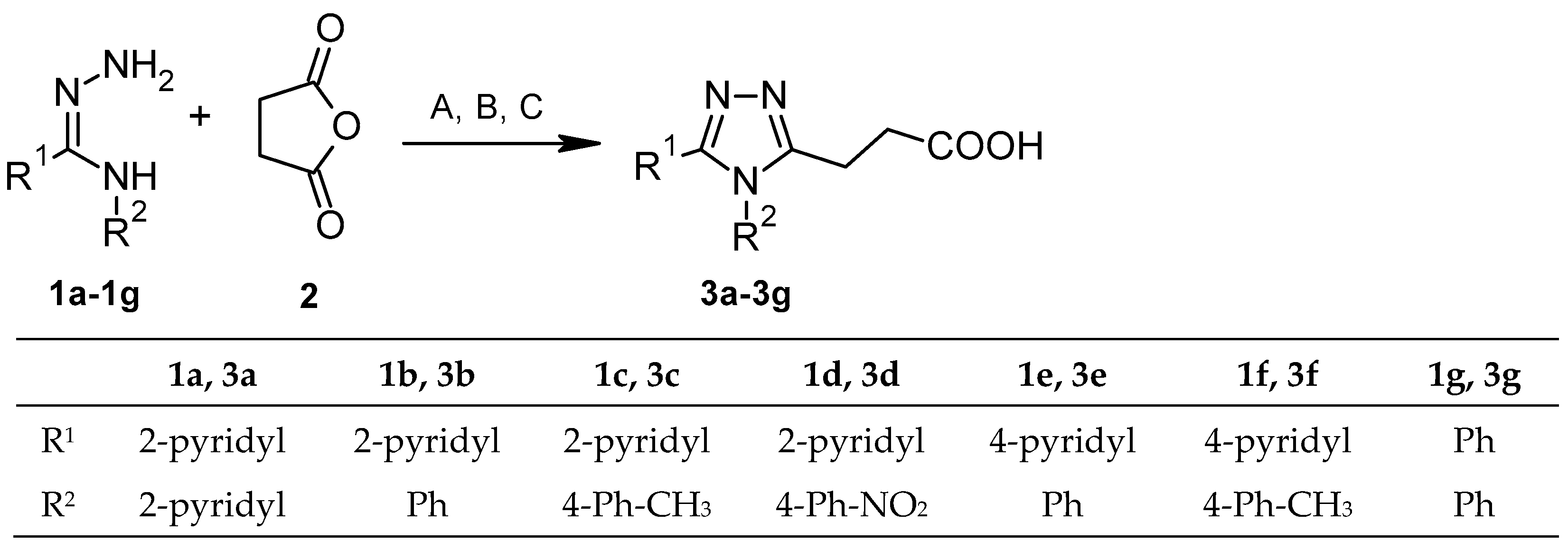
The reactions of amidrazones 1a1g [28] with succinic anhydride 2, leading to 1,2,4-triazole derivatives 3a3g, could be carried out under different conditions, for instance, using catalyst HClO4-SiO2 without solvent [21]. In our earlier works [23,24], we mainly used diethyl ether as a solvent. This time we extended the research to toluene and chloroform (methods A–C, Scheme 1). For example, using diethyl ether as a solvent at room temperature (method A) obtained the best yield (85.76%) for compound 3b, while the yields of the other reactions in this method were in the range of about 55.4–80.8% (Table S1, Supplementary Materials). However, carrying out the same reaction in boiling toluene (method B) allowed better yields to be obtained for 3e and 3a (85.71% and 69.36%, respectively) than for the same reactions carried out in diethyl ether. Due to the shorter reaction time, the use of boiling toluene as a solvent may be a good alternative to diethyl ether for the synthesis of 1,2,4-triazole derivatives. Boiling chloroform (method C) was less preferable as a solvent, probably due to its lower boiling point compared to toluene; only for compound 3e was a yield above 65% obtained. However, other obtained yield values for chloroform were lower than those obtained in methods A and B (about 30–44.12%).
Synthesis in diethyl ether (method A) failed to obtain compound 3d containing an electron-withdrawing -NO2 group in the para position of the phenyl ring, the presence of which may hinder cyclization [22]. The intermediate compound 4d, obtained in diethyl ether from amidrazone 1d, was subjected to cyclization in an alkaline medium as previously described [29], obtaining compound 3d (method D, Scheme 2).
The structures of derivatives 3a3g were confirmed by 1H NMR and 13C NMR spectra (Supplementary Materials, Figures S1–S14), high-resolution mass spectra (HRMS) (Supplementary Materials, Figures S15–S21) and elemental analyses, which are consistent with expected molecular structures. For example, in compound 3e, the presence of a carboxyl group is confirmed by a broad signal at ca. 12.28 ppm in the 1H NMR spectrum and a signal at 173.60 ppm in the 13C NMR spectrum. The presence of methylene carbons –CH2- of derivative 3e are characterized by two signals at 30.74 ppm and 20.72 ppm in the 13C NMR spectrum and a single peak at 2.73 ppm in the 1H NMR spectrum corresponding to the four hydrogen atoms attached to it.
In the 1H NMR spectra, a signal at ca. 8.55 ppm corresponds to two hydrogen atoms attached to aromatic carbon atoms attached to a nitrogen atom (in R1 substituent). The signals in the range of 7.62–7.26 ppm correspond to the remaining seven CH hydrogens attached to the pyridyl and phenyl rings. The 13C NMR spectrum is complemented by a group of signals in the range of ca. 156–122 ppm corresponding to aromatic carbon atoms. The value of the signal in the HRMS 295.1196 spectrum confirms the predicted molecular weight and composition of compound 3e.

2.2. Biological Activity of Compounds 3a3g in PBMC

First, the toxicity of compounds 3a3g at the highest dose of 100 µg/mL was tested in a 24-h human PMBC culture. All derivatives (3a3g) showed very low toxicity: the viable cell counts were in the range 94.71–96.72% and were comparable to the control culture with DMSO (94.19%) and culture with reference non-steroidal anti-inflammatory drug ibuprofen (IBU, 96.01%, Table S2, Supplementary Materials).
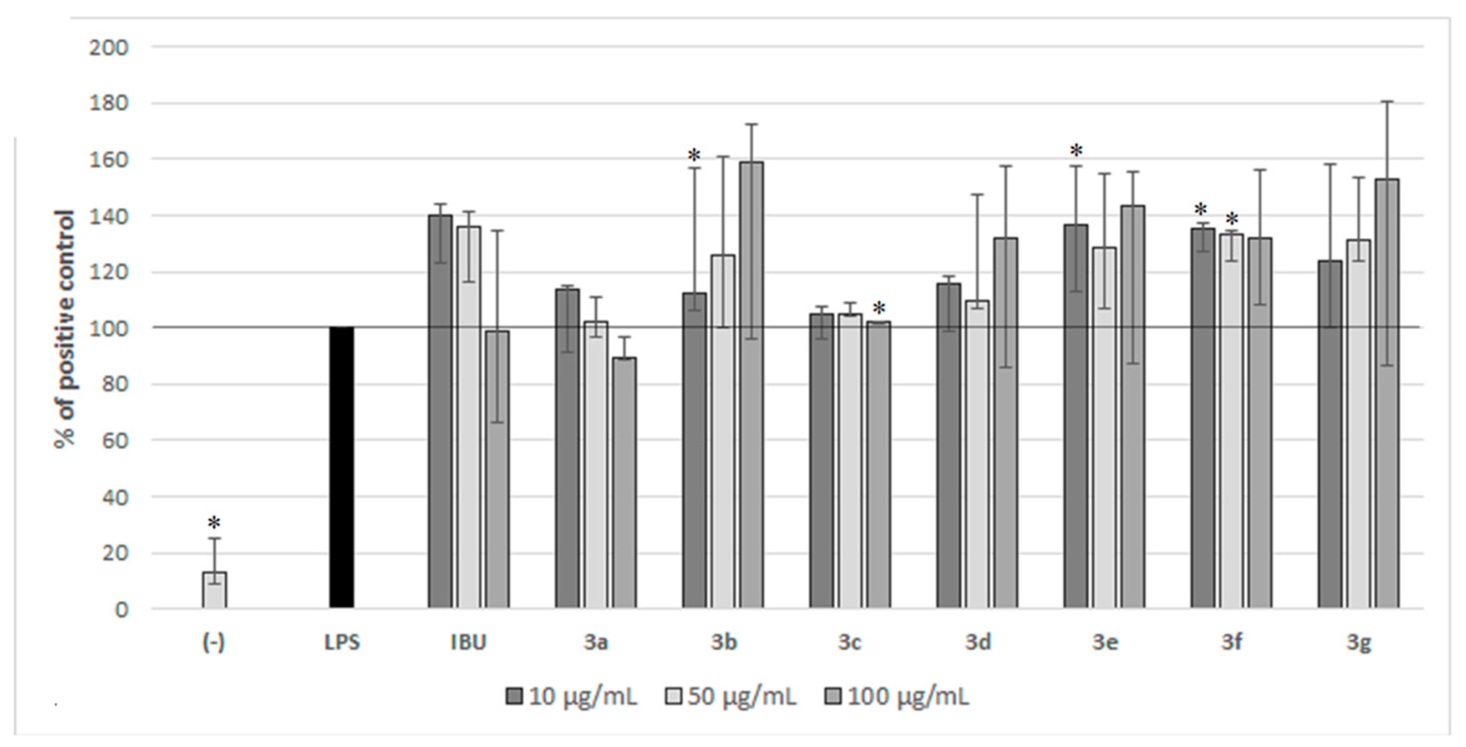
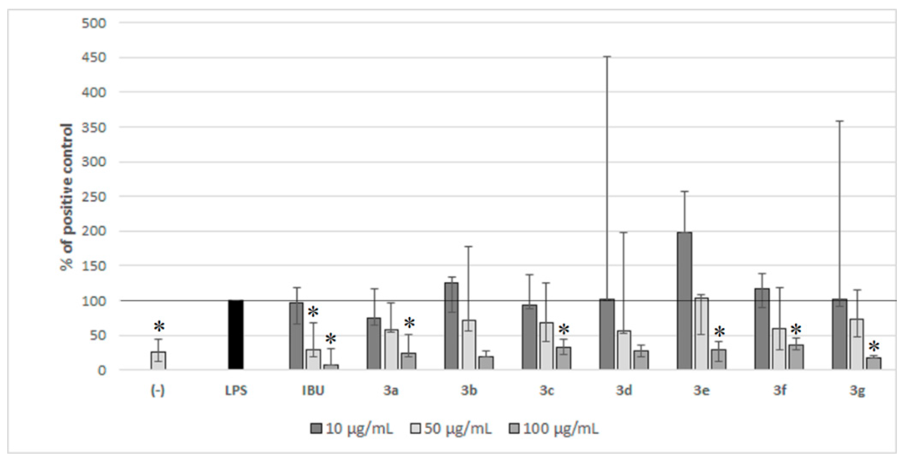
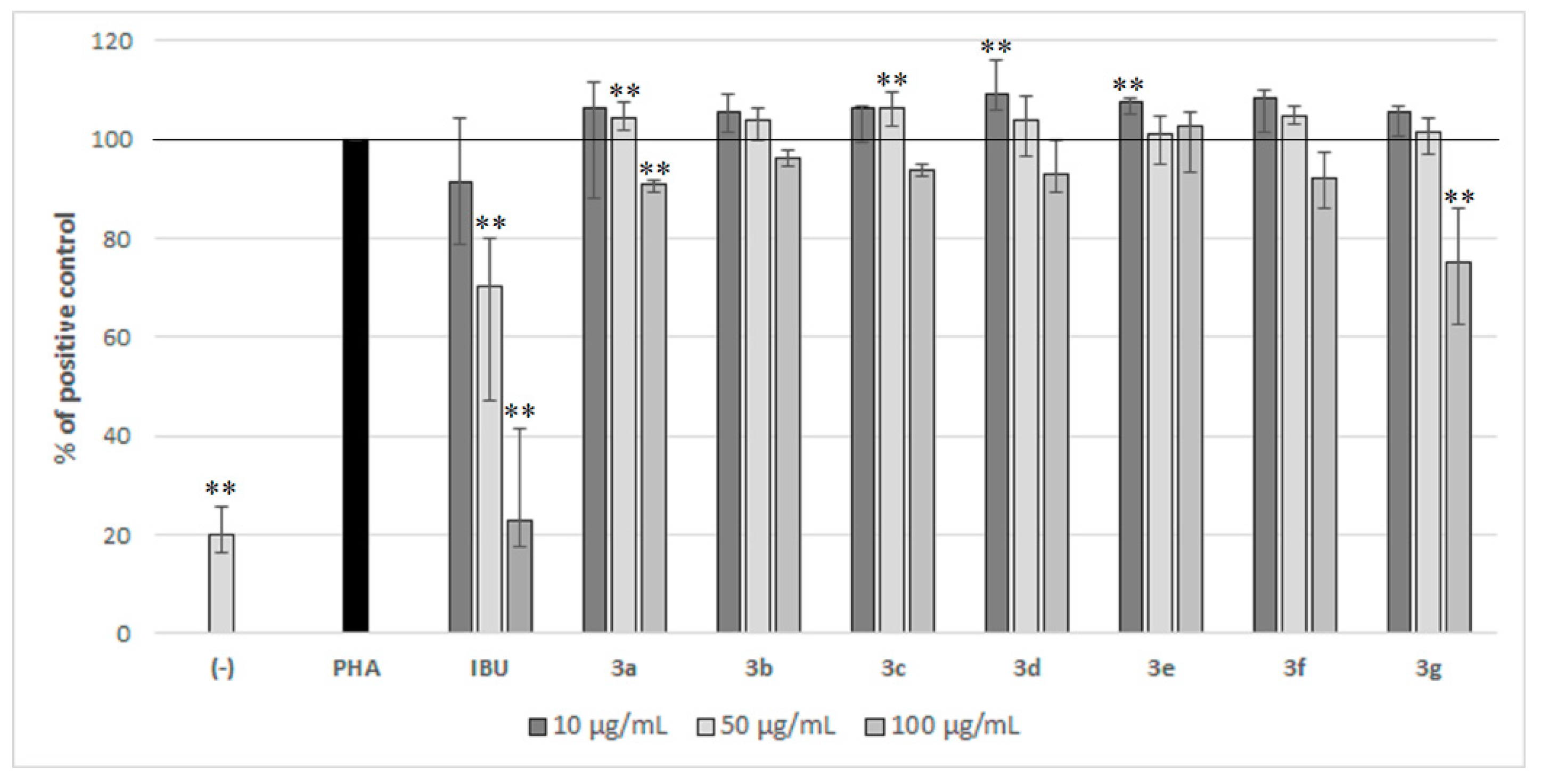
The influence of compounds 3a3g on cytokine release (TNF-α, IL-6, IFN-γ, and IL-10), as well as their antiproliferative activity, was evaluated in PBMC stimulated by LPS and PHA (respectively). The results of cytokine response and proliferation assays in PBMC cultures of compounds 3a3g are presented in Figure 2, Figure 3, Figure 4, Figure 5 and Figure 6.
All compounds 3a3f in the highest dose decreased production of TNF-α by about 44–60%, leading to a similar level of this cytokine as in control cells without stimulation (Figure 2). The strongest effects were exhibited by compounds 3a and 3c, which significantly inhibited TNF-α production in a medium dose of 50 µg/mL and, unlike compounds 3b and 3d3f, did not increase the amount of TNF-a at the lowest dose.
Interestingly, only compounds 3a and 3c did not modulate LPS-induced IL-6 levels in PBMC culture (Figure 3). The rest of the tested compounds, including the reference ibuprofen, acted rather additively with LPS, increasing the level of this pro-inflammatory cytokine.
Compounds 3a, 3c, 3e and 3g inhibited the release of IFN-γ in the highest dose by about 44-79% (Figure 4). On the contrary, derivatives 3b, 3d and 3f increased production of IFN-γ at the medium and low concentration
Ibuprofen significantly inhibited the production of IL-10 at medium and high doses by about 71% and 92%, respectively (Figure 5). At medium doses, all studied compounds were less effective than IBU, causing only about 27–45% inhibition of IL-10 release. Most of the studied compounds at the highest dose decreased the level of IL-10 by about 72–82%; only compounds 3e and 3c showed a slightly weaker effect (about a 65–68% decrease). Interestingly, LPS induced PBMC together with low doses of compounds 3b3g and IBU resulting in significantly higher levels of IL-10 than in the control cells without stimulation. The most favorable effect on the release of IL-10 was observed for compound 3e (R1= 4-Pyr, R2 = Ph), which at the lowest dose increased the level of IL-10—this could be beneficial in relieving chronic inflammation.
IBU in the concentration of 100 µg/mL decreased the proliferation of PBMC to a level similar to the negative control (Figure 6). Studied compounds were less effective, only compounds 3g and 3a significantly inhibited the proliferation of PBMC in the highest dose by about 25% and 9% (respectively).

2.3. Anthelmintic Activity

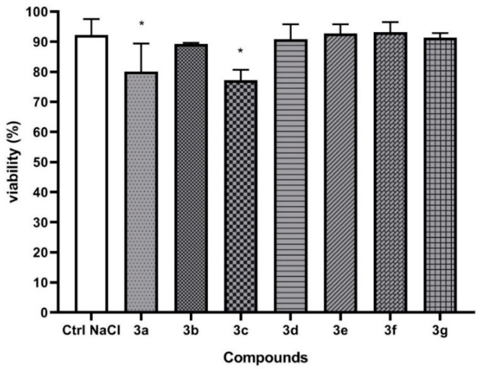
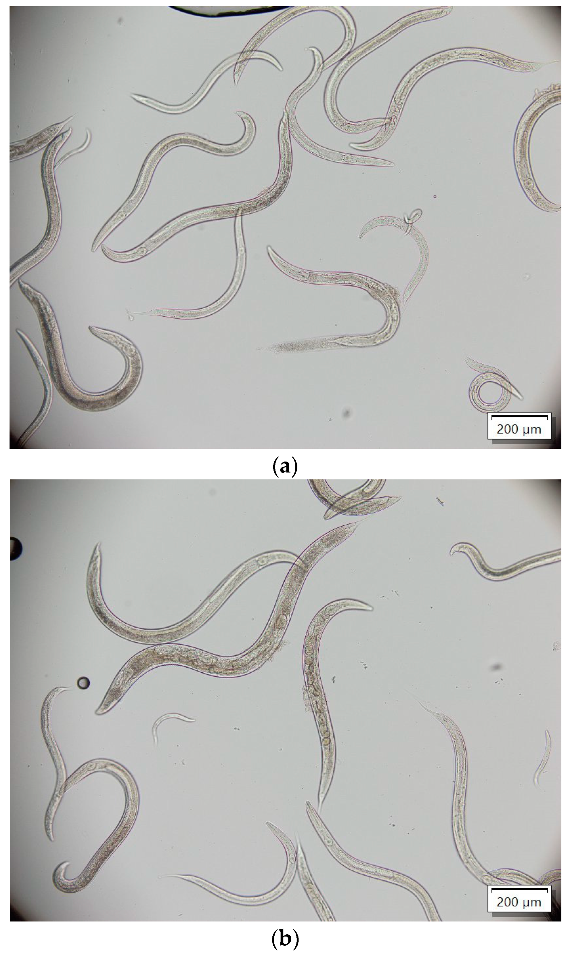
The studies of anthelmintic activity were carried out on a model of nematodes of the genus Rhabditis sp. The tested compounds 3a and 3c showed very low anthelmintic activity. The remaining compounds 3b and 3d3g showed a lack of anthelmintic activity. Due to the high viability of the nematodes, the LC50 dose could not be determined (Figure 7 and Figure 8).

2.4. Antibacterial Activity

Tested compounds 3a3g showed low antimicrobial activity (MIC values ≥ 512 μg/mL) (Table S3, Supplementary Materials) and were less effective than ciprofloxacin (CIP) and amphotericin B (AmB) used as antibacterial and antifungal reference drugs. However, five compounds exhibited anti-mycobacterial activity: 3a and 3e at a concentration of 128 μg/mL and 3b, 3c and 3f at a concentration of 256 μg/mL. Only the derivative 3g inhibited the growth of E. coli and S. aureus at a concentration of 256 μg/mL.

3. Discussion

The toxicity of all obtained compounds 3a3g was tested and was comparable with the control culture (PBMC without additions). This confirms that we work with non-toxic compounds (viability of cells around 90%).
The promising compounds could be 3a containing two 2-pyridine (2-Pyr) substituents, 3c (R1 = 2-Pyr, R2 = 4-Ph-CH3) and 3e (R1 = 4-Pyr, R2 = Ph) since these compounds inhibit the TNF-α and IFN-γ release. Additionally, derivative 3e showed favorable activity in inducting the IL-10 production. It should be noted that these promising compounds immunomodulate innate and adaptive immunity alike since both levels of TNF-α and INF-γ were altered. This is certainly related to the activity of lymphocytes and monocytes [30]. The anti-inflammatory and immunomodulatory activity observed in compounds 3a3f is not related to the inhibition of the proliferation of the test cells because the level of this inhibition is very low (Figure 6).
In previous in vitro studies of acetylsalicylic acid and ibuprofen, they inhibited the production of IL-17 and TNF-α when used at a concentration range of 25–100 µg/mL. However, no significant effect on IFN-γ release was observed [31]. It may suggest that compounds 3a, 3c and 3e could possess an additional beneficial component of the mechanism (IFN-γ inhibition) compared to the anti-inflammatory drugs mentioned above.
Comparing the biological activity of the obtained derivatives 3a3g, some crucial relationships could be observed between the different R1 and R2 substitutions in the 1,2,4-triazole ring, kind of aliphatic acid moiety and the biological effects of those compounds.
  • The presence of 2-pyridyl substituents in R1 position increased TNF-α inhibitory activity of compounds 3a and 3c;
  • The presence of two 2-pyridyne substituents (3a), 4-pyridyl substituent in R1, and phenyl ring in R2 (3e) or two phenyl rings in R1 and R2 (3g) improved the ability to decrease IFN-γ levels. Observations for the last two types of substitutions are consistent with the previously described observations for methacrylic acid derivatives [25];
  • The presence of 4-pyridyne substituent in R1 and phenyl ring in R2 positions in 3e derivative had beneficial influence on the level of IL-10 cytokine;
  • The presence of two phenyl or two 2-pyridyl substituents on R1 and R2 positions seemed to increase the antiproliferative activity of 3g and 3a derivatives;
  • The presence of two 2-pyridyne substituents in R1 and R2 or 2-pyridyne substituent in R1 and 4-methylphenyl substituent in R2 seemed to increase the anthelmintic potential of compounds 3a and 3c;
  • The presence of two 2-pyridyl substituents or 4-pyridyl and phenyl substituents in R1 and R2 positions increased the antituberculosis activity;
  • The presence of the propionic acid group reduced the antibacterial activity of compounds 3a3g compared to analogous 1,2,4-triazole derivatives substituted with methacrylic acid [32].
Comparing the antibacterial and anthelmintic activity of compounds 3a3g with the activity of the previously described 1,2,4-triazole derivatives with similar structures containing the propenoic [33] or methacrylic acid system [32], it can be concluded that the lower activity of the obtained compounds is probably related to the lack of a double bond in the acid chain in their molecules. For example, compounds 3c and 3e were less effective against nematodes than their methacrylic acid analogues. Also, the previously described 1,2,4-triazole derivatives showed a stronger inhibitory effect on PBMC proliferation and TNF-α release [23,24], which emphasizes the beneficial effect of the aliphatic acid substituent containing a double bond and a branched chain. However, the strongest anti-inflammatory, anti-mycobacterial and anthelmintic activities among derivatives 3a3g were observed for compounds 3a and 3c, which confirmed that they have the highest biological potential among the studied compounds.
However, there are some limitations to this immunomodulatory study. The PBMC culture is taken from the vein blood of healthy donors, but there are some individual factors that could influence the presented results. However, this is a useful test system for the investigation of immune modulatory effects of newly synthesized chemical compounds. For future investigation, we plan to additionally use human cell lines. Furthermore, we check the limited number of pro- and anti-inflammatory factors, such as IL-6, TNF-α, IFN-γ and IL-10. There are more factors that affect the inflammatory system, and this may be a new direction for future study (for example, prostaglandins and leukotrienes or cytokines such as IL-17).

4. Materials and Methods

4.1. General Information

1H NMR and 13C NMR spectra: the Bruker Avance 300, 400 and 700 apparatus (TMS as an internal standard). Melting points: MEL-Temp apparatus (Electrothermal, Stone, UK). Elemental analyses: CHN Vario MACRO analyzer (Elementar Analysensysteme GmbH, Langenselbold, Germany). HRMS (high-resolution mass spectrometry): Synapt G2 Si mass spectrometer (Waters). The retention factors: reverse-faced plates (nano-silica gel RP-18W on alu. foil with fluorescent indicator (Merck, Darmstadt, Germany) and a methanol-water (1:1) mixture. All reactions were controlled by TLC chromatography. Received yields and melting points using particular methods of synthesis are presented in Table S1 in Supplementary Materials. The 1H NMR and 13C NMR spectra (Figures S1–S14) and their HRMS spectra (Figures S15–S21) are present in Supplementary Materials.

4.2. General Procedures of Synthesis

4.2.1. Method A (Synthesis of Compounds 3a3c and 3e3g)

In total, 1.2 mmol of amidrazones 1a1c and 1e1g [28] and 1.2 mmol (0.12 g) of succinic anhydride 2 were dissolved in 30 mL of anhydrous diethyl ether, stirred for 2 h, then left for 2 days in ambient temperature. Obtained crude compounds 3a3c and 3e3g were filtered and washed with anhydrous diethyl ether and products were crystallized from 1:1 methanol/water mixture.

4.2.2. Method B (Synthesis of Compounds 3a3g)

In total, 1.2 mmol of amidrazones 1a1g [28] and 1.2 mmol (0.12 g) of succinic anhydride 2 were dissolved in 30 mL of anhydrous toluene. The mixtures were heated under reflux for 1–1.5 h, then filtered out. Crude compounds 3a3g obtained by evaporation of toluene were washed with anhydrous diethyl ether. Received products 3a3g were crystallized (3a, 3c and 3g: 1:1 ethanol/water mixture, 3b, 3d and 3e: water).

4.2.3. Method C (Synthesis of Compounds 3a3c and 3e)

In total, 1.2 mmol of amidrazones 1a1c and 1e [28] and 1.2 mmol (0.12 g) of succinic anhydride 2 were dissolved in 30 mL of anhydrous chloroform. The mixtures were heated under reflux for 1–4 h, then filtered out. Crude compounds obtained by partial evaporation of chloroform were filtered and dried. Received products 3a3c were crystallized (3a: ethanol, 3b3c: water).

4.2.4. Method D (Compound 3d)

In total, 1.2 mmol of amidrazone 1d [28] and 1.2 mmol (0.12 g) of succinic anhydride 2 were dissolved in 30 mL of anhydrous diethyl ether, stirred for 2, then left for 2 days in ambient temperature. Obtained intermediate 4d was filtered and washed with anhydrous diethyl ether and dried. Then the cyclization procedure was undertaken by heating in 2% aqueous sodium hydroxide solution for 2 h. After filtration and cooling obtained solution and neutralization with 2% aqueous hydrochloric acid solution. Obtained compound 3d was crystalized by isopropanol.
  • 3-(4,5-di(pyridin-2-yl)-4H-1,2,4-triazol-3-yl)propanoic acid (3a): yield 55.39%, m.p. 197–199 °C. 1H NMR (700 MHz, DMSO-d6, δ ppm): 12.25 (sb, 1H, COOH), 8.56 (d, 1H, J = 4.0 Hz), 8.23, (d, 1H, J = 4.0 Hz), 8.07–7.89 (m, 3H), 7.58–7.50 (m, 2H), 7.38–7.33 (m, 1H), 2.85–2.67 (m, 4H). 13C NMR (100 MHz, DMSO-d6, δ ppm): 173.6, 155.3, 152.8, 149.7, 149.3, 148.9, 146.8, 139.6, 137.8, 125.0, 124.8, 123.6, 122.8, 30.8, 20.9. HR-MS m/z: 296.1150 [M+ + 1] (calculated for C15H14N5O2: 296.1147). Rf = 0.53. Elem. Anal. for C15H13N5O2 calculated: C, 61.01; H, 4.44; N, 23.72%; obtained C, 61.18; H, 4.67; N, 23.63%.
  • 3-(4-phenyl-5-(pyridin-2-yl)-4H-1,2,4-triazol-3-yl)propanoic acid (3b): yield 85.76%, m.p. 178–180 °C. 1H NMR (400 MHz, DMSO-d6, δ ppm): 12.26 (sb, 1H, COOH), 8.30–8.28, (m, 1H), 7.97–7.87 (m, 2H), 7.52–7.47 (m, 3H), 7.37–7.32 (m, 3H), 2.72 (s, 4H). 13C NMR (100 MHz, DMSO-d6, δ ppm): 173.6, 155.7, 152.9, 149.4, 147.2, 137.6, 135.5, 129.8 (2×), 129.5, 127.8 (2x), 124.6, 124.1, 30.8, 20.8. HR-MS m/z: 29.1197 [M+ + 1] (calculated for C16H15N4O2: 295.1195). Rf = 0.41. Elem. anal. for C16H14N4O2 * H2O calculated: C, 61.54; H, 5.13; N, 17.95%; obtained C, 61.89; H, 5.08; N, 17.72%.
  • 3-(5-(pyridin-2-yl)-4-p-tolyl-4H-1,2,4-triazol-3-yl)propanoic acid (3c): yield 73.53%, m.p. 157–160 °C. 1H NMR (700 MHz, DMSO-d6, δ ppm): 12.24 (sb, 1H, COOH), 8.32 (d, 1H, J = 4.2 Hz), 7.92 (d, 1H, J = 7.7 Hz), 7.89–7.86 (m, 1H), 7.35–7.32 (m, 1H), 7.28 (d, 2H, J = 8.4 Hz), 7.22 (d, 2H, J = 8.4 Hz), 2.72–2.67 (m, 4H), 2.36 (s, 3H). 13C NMR (100 MHz, DMSO-d6, δ ppm): 173.6, 155.7, 153.0, 149.5, 147.3, 139.0, 137.5, 132.9, 130.3 (2×), 127.6 (2×), 124.6, 124.2, 30.8, 21.2, 20.8. HR-MS m/z: 309.1357 [M+ + 1] (calculated for C17H17N4O2: 309.1352). Rf = 0.32. Elem. Anal. for C17H16N4O2 calculated: C, 66.22; H, 5.23; N, 18.17%; obtained C, 66.56; H, 5.25; N, 18.17%.
  • 3-(4-(4-nitrophenyl)-5-(pyridin-2-yl)-4H-1,2,4-triazol-3-yl)propanoic acid (3d): yield 32.83%, m.p. 189–191 °C. 1H NMR (400 MHz, DMSO-d6, δ ppm): 12.29 (sb, 1H, COOH), 8.36 (d, 2H, J = 9.2 Hz), 8.26 (d, 1H, J = 4.4 Hz), 8.09 (d, 1H, J = 8.0 Hz), 7.93 (td, 1H, J1 = 8 Hz, J2 = 1.6 Hz), 7.75–7.69 (m, 2H), 7.39–7.34 (m, 1H), 2.75 (s, 4H). 13C NMR (100 MHz, DMSO-d6, δ ppm): 173.3. 155.2, 152.2, 148.9, 147.5, 146.4, 141.1, 137.5, 129.2 (2x), 124.7 (2×), 124.5, 123.4, 30.6, 20.5. HR-MS m/z: 340.1049 [M+ + 1] (calculated for C16H14N5O4: 340.1046). ESI—MS m/z: (M + 1): 340. Rf = 0.38. Elem. Anal. for C16H13N5O4 calculated: C, 56.64; H, 3.86; N, 20.64%; obtained C, 56.56; H, 3.92; N, 20.51%.
  • 3-(4-phenyl-5-(pyridin-4-yl)-4H-1,2,4-triazol-3-yl)propanoic acid (3e): yield 80.82%, m.p. 235–237 °C. 1H NMR (700 MHz, DMSO-d6, δ ppm): 12.28 (sb, 1H, COOH), 8.54–8.55 (m, 2H), 7.59–7.63 (m, 3H), 7.47–7.52 (m, 2H), 7.25–7.29 (m, 2H), 2.73 (s, 4H). 13C NMR (100 MHz, DMSO-d6, δ ppm): 173.6, 156.1, 151.5, 150.5 (2×), 134.8, 134.3, 130.7 (2×), 130.7, 128.0 (2×), 122.1 (2×), 30.7, 20.7. HR-MS m/z: 295.1196 [M+ + 1] (calculated for C16H15N4O2: 295.1195). Rf = 0.38. Elem. Anal. for C16H14N4O2 calculated: C, 61.54; H, 5.13; N, 17.95%; obtained C, 61.58; H, 5.12; N, 17.96%.
  • 3-(5-(pyridin-4-yl)-4-p-tolyl-4H-1,2,4-triazol-3-yl)propanoic acid (3f): yield 64.30%, m.p. 208–211 °C. 1H NMR (300 MHz, DMSO-d6, δ ppm): 12.26 (sb, 1H, COOH), 8.56–8.52, (m, 2H), 7.41–7.24 (m, 6H), 2.70 (s, 4H), 2.39 (s, 3H). 13C NMR (100 MHz, DMSO-d6, δ ppm): 173.6, 156.2, 151.5, 150.5 (2×), 140.5, 134.9, 131.7, 131.2 (2×), 127.8 (2×), 122.1 (2×), 30.8, 21.2, 20.7. Rf = 0.28. HR-MS m/z: 309.1355 [M+ + 1] (calculated for C17H17N4O2: 309.1352). Elem. Anal. for C17H16N4O2 * ½ H2O calculated: C, 64.40; H, 5.36; N, 17.68%; obtained C, 64.58; H, 5.47; N, 17.36%.
  • 3-(4,5-diphenyl-4H-1,2,4-triazol-3-yl)propanoic acid (3g): yield 76.47%, m.p. 251–254 °C. 1H NMR (400 MHz, DMSO-d6, δ ppm): 12.23 (sb, 1H, COOH), 7.57–7.52 (m, 3H), 7.43–7.29 (m, 7H), 2.70 (s, 4H). 13C NMR (100 MHz, DMSO-d6, δ ppm): 173.6, 155.0, 153.6, 134.8, 130.5 (2×), 130.2, 129.9, 129.0 (2×), 128.4 (2×), 128.1 (2×), 127.5, 30.8, 20.8. Rf = 0.31. HR-MS m/z: 294.1240 [M+ + 1] (calculated for C17H16N3O2: 294.1243). Elem. Anal. for C17H15N3O2 calculated: C, 69.61; H, 5.15; N, 14.33%; obtained C, 69.69; H, 5.26; N, 14.33%.

4.3. Biological Activity Evaluation for Compounds 3a3g

4.3.1. Cell Culture Preparation and Toxicity of Compounds 3a3g towards PBMC

Biological activity was evaluated on fresh peripheral blood taken from healthy donors. Informed consent was obtained from donors at the Occupational Medicine Clinic located in Dr. Antoni Jurasz University Hospital in Bydgoszcz, Poland. Immediately after the blood was taken, the cell isolation was performed by using the density gradient centrifugation (Lymphosept, BioWest, Nuaillé, France) as previously described [23]. After isolation, PBMC were used to conduct experiments which assessed toxicity of compounds and their impact on proliferation and cytokine production. The preparation of cell culture was described previously [23]. PBMC were stimulated with all tested compounds in highest doses (100 µg/mL). Additionally, we used ibuprofen as a reference drug in this study. For checking the number of viable cells, the Annexin V Apoptosis Detection Kit I (Becton Dickinson, Franklin Lakes, NJ, USA) was used. The procedure of staining was described previously [23].

4.3.2. The Influence of Compounds 3a3g on Cytokines Production in PBMC

Cytokines (TNF-α, IL-6, IL-10 and IFN-γ) supernatant levels were measured as previously described [23] using the commercially available ELISA kits (DuoSet, BD Bioscience, San Diego, CA, USA). Tests were conducted according to the manufacturer’s instructions. The supernatants from cell culture were analyzed with iEMS Reader MF (Labsystems, Vantaa, Finland) and date were calculated by Ascent Software AscSW26 (London, UK).

4.3.3. The Influence of Compounds 3a3g on PBMC Proliferation

Lymphocyte proliferation was evaluated by flow cytometry using BD HorizonTM Violet Proliferation Dye 450 (VPD450, BD Pharmingen, San Diego, CA, USA). Violet Proliferation Dye 450 is a tool for evaluation of cell division. VPD450-stained cells were cultured for 72 h with the mitogen, phytohemagglutinin (PHA, Sigma-Aldrich, 1 μg/mL, positive control), and/or increasing concentrations of compounds (10, 50 and 100 μg/mL). The procedure of staining with VPD450 was described previously [23]. The cells were analyzed by flow cytometry (FACSCanto II flow cytometer, Becton Dickinson, Franklin Lakes, NJ, USA). Twenty thousand events were collected and analyzed with FlowJo software (v 7.6.1, Tree Star, Ashland, OR, USA).

4.4. Anthelmintic Activity of Compounds 3a3g

In vitro experiments on anthelmintic activity were carried out using a nematode model of the genus Rhabditis sp. The tests were conducted in accordance with the patented procedure patent No. PL232918. The compounds 3a3g were tested in five concentrations: 0.2; 1.1; 3.3; 5.5; 11.1 mg/mL. The exact methodology of testing anthelmintic activity was described in our earlier publication [23].

4.5. Antibacterial Activity of Compounds 3a3g

The antibacterial activity of compounds 3a3g was tested by the broth microdilution method against bacteria Staphylococcus aureus ATCC 25923, Escherichia coli ATCC 25922, Yersinia enterocolitica O3 and Mycobacterium smegmatis and fungal strain Candida albicans ATCC 90028. Compounds 3a3g were dissolved, serially diluted to concentrations from 512 to 0.25 μg/mL and then inoculated with bacterial cultures as described previously [34].
In addition, the sensitivity of the tested bacterial strains to ciprofloxacin (quinolone antibiotic) and the yeast strain to amphotericin B was determined. After 18 h of incubation at 37 °C, the minimum inhibitory concentration (MIC) of the compounds was defined. The experiments were carried out three times.

4.6. Data Analysis

The statistical analyses were performed using Statistica 13.3. software (StatSoft, Cracow, Poland). Shapiro-Wilka test and Mann-Whitney U test were calculated. All p-values represented the nonparametric Mann–Whitney U test. The p value was set at 0.05.

5. Conclusions

Seven new 1,2,4-triazole derivatives 3a3g were obtained through the reaction of amidrazones 1a1g with succinic anhydride. It was shown that the use of boiling toluene is a good alternative for the synthesis of this type of cyclic compound in relation to the classical reaction carried out in diethyl ether. All obtained derivatives 3a3g showed anti-inflammatory and immunomodulatory activity. The strongest effect was shown by derivatives 3a, 3c and 3e, which decreased levels of TNF-α and IFN-γ in mitogen-stimulated PBMC cultures. Compared to the previously described 1,2,4-triazole derivatives containing the methacrylic acid system, lower antibacterial, antiproliferative and anthelmintic activity was observed for compounds 3a3g. This decrease in biological activity could be connected to the lack of a double bond and chain branching in the aliphatic side chain of the acid. The mechanisms of observed effects require further investigation.

Supplementary Materials

The following supporting information can be downloaded at: https://www.mdpi.com/article/10.3390/molecules28093808/s1. Table S1. A summary of the yield and melting point of compounds 3a3g obtained under different reaction conditions (methods A–D). Figure S1. 1H NMR spectrum of compound 3a (in DMSO-d6). Figure S2. 1H NMR spectrum of compound 3b (in DMSO-d6). Figure S3. 1H NMR spectrum of compound 3c (in DMSO-d6). Figure S4. 1H NMR spectrum of compound 3d (in DMSO-d6). Figure S5. 1H NMR spectrum of compound 3e (in DMSO-d6). Figure S6. 1H NMR spectrum of compound 3f (in DMSO-d6). Figure S7. 1H NMR spectrum of compound 3g (in DMSO-d6). Figure S8. 13C NMR spectrum of compound 3a (in DMSO-d6). Figure S9. 13C NMR spectrum of compound 3b (in DMSO-d6). Figure S10. 13C NMR spectrum of compound 3c (in DMSO-d6). Figure S11. 13C NMR spectrum of compound 3d (in DMSO-d6). Figure S12. 13C NMR spectrum of compound 3e (in DMSO-d6). Figure S13. 13C NMR spectrum of compound 3f (in DMSO-d6). Figure S14. 13C NMR spectrum of compound 3g (in DMSO-d6). Figure S15. HRMS spectrum of compound 3a. Figure S16. HRMS spectrum of compound 3b. Figure S17. HRMS spectrum of compound 3c. Figure S18. HRMS spectrum of compound 3d. Figure S19. HRMS spectrum of compound 3e. Figure S20. HRMS spectrum of compound 3f. Figure S21. HRMS spectrum of compound 3g. Table S2. The results of cell apoptosis assay of compounds 3a3g and ibuprofen at a concentration of 100 µg/mL in PBMC 24 h cultured measured by flow cytometry. Table S3. Minimal inhibitory concentrations of compounds 3a3g, ciprofloxacin (CIP) and amphotericin B (AmB).

Author Contributions

Conceptualization, R.P.; methodology, M.W.-S., A.B.-K., P.K. and J.K.; validation, M.W.-S. and P.K.; formal analysis, R.P.; investigation, R.P. and P.K., M.W.-S., S.B. and J.K.; resources, R.P.; writing—original draft preparation, R.P.; writing—review and editing, A.B.-K., P.K., M.W.-S., S.B. and J.K.; visualization, R.P.; supervision R.P. and M.W.-S.; project administration, R.P.; funding acquisition, R.P. and A.B.-K. All authors have read and agreed to the published version of the manuscript.

Funding

This work has been supported by Nicolaus Copernicus University, (IDUB DEBIUTY4) and funds for the statutory activity of the Medical University of Lublin DS43 (A.B.-K.).

Institutional Review Board Statement

Experiments using peripheral blood mononuclear cells (PBMC) ware conducted according to the guidelines of the Declaration of Helsinki and approved by the by the Collegium Medicum of Nicolaus Copernicus University Bioethical Com-mission (KB 39/2019).

Informed Consent Statement

Informed consent was obtained from healthy blood donors involved in the study.

Data Availability Statement

Data available from the authors.

Conflicts of Interest

The authors declare no conflict of interest.

Sample Availability

Samples of the compounds 3a3g are available from the authors.

References

  1. Mohammed, A.; Kalle, A.M.; Reddanna, P. Managing SARS-CoV2 Infections Through Resolution of Inflammation by Eicosanoids: A Review. J. Inflamm. Res. 2022, 15, 4349–4358. [Google Scholar] [CrossRef] [PubMed]
  2. Sokolowska, M.; Rovati, G.E.; Diamant, Z.; Untersmayr, E.; Schwarze, J.; Lukasik, Z.; Sava, F.; Angelina, A.; Palomares, O.; Akdis, C.A.; et al. Effects of non-steroidal anti-inflammatory drugs and other eicosanoid pathway modifiers on antiviral and allergic responses: EAACI task force on eicosanoids consensus report in times of COVID-19. Allergy 2022, 77, 2337–2354. [Google Scholar] [CrossRef]
  3. Patil, K.R.; Mahajan, U.B.; Unger, B.S.; Goyal, S.N.; Belemkar, S.; Surana, S.J.; Ojha, S.; Patil, C.R. Animal Models of Inflammation for Screening of Anti-inflammatory Drugs: Implications for the Discovery and Development of Phytopharmaceuticals. Int. J. Mol. Sci. 2019, 20, 4367. [Google Scholar] [CrossRef] [PubMed]
  4. Zhao, H.; Wu, L.; Yan, G.; Chen, Y.; Zhou, M.; Wu, Y.; Li, Y. Inflammation and tumor progression: Signaling pathways and targeted intervention. Signal Transduct. Target Ther. 2021, 6, 263. [Google Scholar] [CrossRef] [PubMed]
  5. Maddipati, K.R. Non-inflammatory Physiology of “Inflammatory” Mediators—Unalamation, a New Paradigm. Front. Immunol. 2020, 11, 580117. [Google Scholar] [CrossRef]
  6. Ferrero-Miliani, L.; Nielsen, O.H.; Andersen, P.S.; Girardin, S.E. Chronic inflammation: Importance of NOD2 and NALP3 in interleukin-1β generation. Clin. Exp. Immunol. 2007, 147, 227–235. [Google Scholar] [CrossRef]
  7. Nathan, C.; Ding, A. Nonresolving inflammation. Cell 2010, 140, 871–882. [Google Scholar] [CrossRef]
  8. Franceschi, C.; Garagnani, P.; Parini, P.; Giuliani, C.; Santoro, A. Inflammaging: A new immune–metabolic viewpoint for age-related diseases. Nat. Rev. Endocrinol. 2018, 14, 576–590. [Google Scholar] [CrossRef]
  9. Rohm, T.V.; Meier, D.T.; Olefsky, J.M.; Donath, M.Y. Inflammation in obesity, diabetes, and related disorders. Immunity 2022, 55, 31–55. [Google Scholar] [CrossRef]
  10. Jenny, M.; Klieber, M.; Zaknun, D.; Schroecksnadel, S.; Kurz, K.; Ledochowski, M.; Schennach, H.; Fuchs, D. In vitro testing for anti-inflammatory properties of compounds employing peripheral blood mononuclear cells freshly isolated from healthy donors. Inflamm. Res. 2011, 60, 127–135. [Google Scholar] [CrossRef]
  11. Van Belleghem, J.D.; Clement, F.; Merabishvili, M.; Lavigne, R.; Vaneechoutte, M. Pro- and anti-inflammatory responses of peripheral blood mononuclear cells induced by Staphylococcus aureus and Pseudomonas aeruginosa phages. Sci. Rep. 2017, 7, 8004. [Google Scholar] [CrossRef]
  12. Dargahi, N.; Johnson, J.C.; Apostolopoulos, V. Immune Modulatory Effects of Probiotic Streptococcus thermophilus on Human Monocytes. Biologics 2021, 1, 396–415. [Google Scholar] [CrossRef]
  13. Dargahi, N.; Johnson, J.; Apostolopoulos, V. Streptococcus thermophilus alters the expression of genes associated with innate and adaptive immunity in human peripheral blood mononuclear cells. PLoS ONE 2020, 15, e0228531. [Google Scholar] [CrossRef]
  14. Mączyński, M.; Artym, J.; Kocięba, M.; Kochanowska, I.; Ryng, S.; Zimecki, M. Anti-inflammatory properties of an isoxazole derivative—MZO-2. Pharmacol. Rep. 2016, 68, 894–902. [Google Scholar] [CrossRef]
  15. Díaz, I. Rules of thumb to obtain, isolate, and preserve porcine peripheral blood mononuclear cells. Vet. Immunol. Immunopathol. 2022, 251, 110461. [Google Scholar] [CrossRef]
  16. Ertel, W.; Kremer, J.-P.; Kenney, J.; Steckholzer, U.; Jarrar, D.; Trentz, O.; Schildberg, F.W. Downregulation of Proinflammatory Cytokine Release in Whole Blood from Septic Patients. Blood 1995, 85, 1341–1347. [Google Scholar] [CrossRef]
  17. Liu, X.; Yin, S.; Chen, Y.; Wu, Y.; Zheng, W.; Dong, H.; Zhao, P. LPS-induced proinflammatory cytokine expression in human airway epithelial cells and macrophages via NF-κB, STAT3 or AP-1 activation. Mol. Med. Rep. 2018, 17, 5484–5491. [Google Scholar] [CrossRef]
  18. Siedlik, J.A.; Deckert, J.A.; Benedict, S.H.; Bhatta, A.; Dunbar, A.J.; Vardiman, J.P.; Gallagher, P.M. T cell activation and proliferation following acute exercise in human subjects is altered by storage conditions and mitogen selection. J. Immunol. Methods 2017, 446, 7–14. [Google Scholar] [CrossRef]
  19. Lawlor, N.; Nehar-Belaid, D.; Grassmann, J.D.S.; Stoeckius, M.; Smibert, P.; Stitzel, M.L.; Pascual, V.; Banchereau, J.; Williams, A.; Ucar, D. Single Cell Analysis of Blood Mononuclear Cells Stimulated Through Either LPS or Anti-CD3 and Anti-CD28. Front. Immunol. 2021, 12, 636720. [Google Scholar] [CrossRef]
  20. Abdelli, A.; Azzouni, S.; Plais, R.; Gaucher, A.; Efrit, M.L.; Prim, D. Recent advances in the chemistry of 1,2,4-triazoles: Synthesis, reactivity and biological activities. Tetrahedron Lett. 2021, 86, 153518. [Google Scholar] [CrossRef]
  21. Wen, Y.; Lun, S.; Jiao, Y.; Zhang, W.; Hu, T.; Liu, T.; Yang, F.; Tang, J.; Zhang, B.; Bishai, W.R.; et al. Design, synthesis, and biological evaluation of 1,2,4-triazole derivatives as potent antitubercular agents. Chin. Chem. Lett. 2023, 108464, in press. [Google Scholar] [CrossRef]
  22. Li, J.; Zhang, J. The Antibacterial Activity of 1,2,3-triazole- and 1,2,4-Triazole-containing Hybrids against Staphylococcus aureus: An Updated Review (2020-Present). Curr. Top. Med. Chem. 2022, 22, 41–63. [Google Scholar] [CrossRef]
  23. Paprocka, R.; Kołodziej, P.; Wiese-Szadkowska, M.; Helmin-Basa, A.; Bogucka-Kocka, A. Evaluation of Anthelmintic and Anti-Inflammatory Activity of 1,2,4-Triazole Derivatives. Molecules 2022, 27, 4488. [Google Scholar] [CrossRef] [PubMed]
  24. Paprocka, R.; Wiese, M.; Eljaszewicz, A.; Helmin-Basa, A.; Gzella, A.; Modzelewska-Banachiewicz, B.; Michalkiewicz, J. Synthesis and anti-inflammatory activity of new 1,2,4-triazole derivatives. Bioorg. Med. Chem. Lett. 2015, 25, 2664–2667. [Google Scholar] [CrossRef] [PubMed]
  25. Paprocka, R.; Wiese-Szadkowska, M.; Kosmalski, T.; Frisch, D.; Ratajczak, M.; Modzelewska-Banachiewicz, B.; Studzińska, R. A Review of the Biological Activity of Amidrazone Derivatives. Pharmaceuticals 2022, 15, 1219. [Google Scholar] [CrossRef]
  26. Paprocka, R.; Wiese-Szadkowska, M.; Helmin-Basa, A.; Mazur, L.; Kutkowska, J.; Michałkiewicz, J.; Modzelewska-Banachiewicz, B.; Pazderski, L. Synthesis and evaluation of new amidrazone-derived hydrazides as a potential anti-inflammatory agents. Monatsh Chem. 2018, 149, 1493–1500. [Google Scholar] [CrossRef] [PubMed]
  27. Gupta, O.; Pradhan, T.; Chawla, G. An updated review on diverse range of biological activities of 1,2,4-triazole derivatives: Insight into structure activity relationship. J. Mol. Struct. 2023, 1274, 134487. [Google Scholar] [CrossRef]
  28. Modzelewska, B.; Pyra, E. Synthesis of N3-substituted amidrazones. Ann. UMCS Sec. AA 1996, 50, 111–116. [Google Scholar]
  29. Modzelewska-Banachiewicz, B.; Paprocka, R.; Mazur, L.; Saczewski, J.; Kutkowska, J.; Stępień, D.K.; Cyrański, M. Experimental and theoretical study on the reaction of N3-phenyl-(pyridin-2-yl)carbohydrazonamide with itaconic anhydride. J. Mol. Struct. 2012, 1022, 211–219. [Google Scholar] [CrossRef]
  30. Sibi, J.M.; Mohan, V.; Munisankar, S.; Babu, S.; Aravindhan, V. Augmented Innate and Adaptive Immune Responses Under Conditions of Diabetes-Filariasis Comorbidity. Front. Immunol. 2021, 12, 716515. [Google Scholar] [CrossRef]
  31. Sayın, N.; Uygun, D.F.K.; Sallakçı, N.; Filiz, S.; Yeğin, O. Inhibitory Effects of Acetylsalicylic Acid and Ibuprofen on Interleukin-17 Production. Turk. J. Immunol. 2013, 1, 42–46. [Google Scholar] [CrossRef]
  32. Paprocka, R.; Modzelewska-Banachiewicz, B.; Kutkowska, J.; Pawłowski, K.; Piątkowska-Chmiel, I.; Jagiełło-Wójtowicz, E. Antibacterial and central nervous system activity of 4,5-diaryl-4H-1,2,4-triazol-3-yl)methylacrylic acid derivatives. Acta Pol. Pharm. 2017, 74, 289–292. [Google Scholar]
  33. Kutkowska, J.; Modzelewska-Banachiewicz, B.; Ziółkowska, G.; Rzesko, W.; Urbanik-Sypniewska, T.; Zwolska, Z.; Prus, M. Antimicrobial Activity of 3,4-disubstituted-1,2,4-triazole derivatives. Acta Pol. Pharm. 2005, 62, 303–306. [Google Scholar]
  34. Paprocka, R.; Pazderski, L.; Mazur, L.; Wiese-Szadkowska, M.; Kutkowska, J.; Nowak, M.; Helmin-Basa, A. Synthesis and Structural Study of Amidrazone Derived Pyrrole-2,5-Dione Derivatives: Potential Anti-Inflammatory Agents. Molecules 2022, 27, 2891. [Google Scholar] [CrossRef]
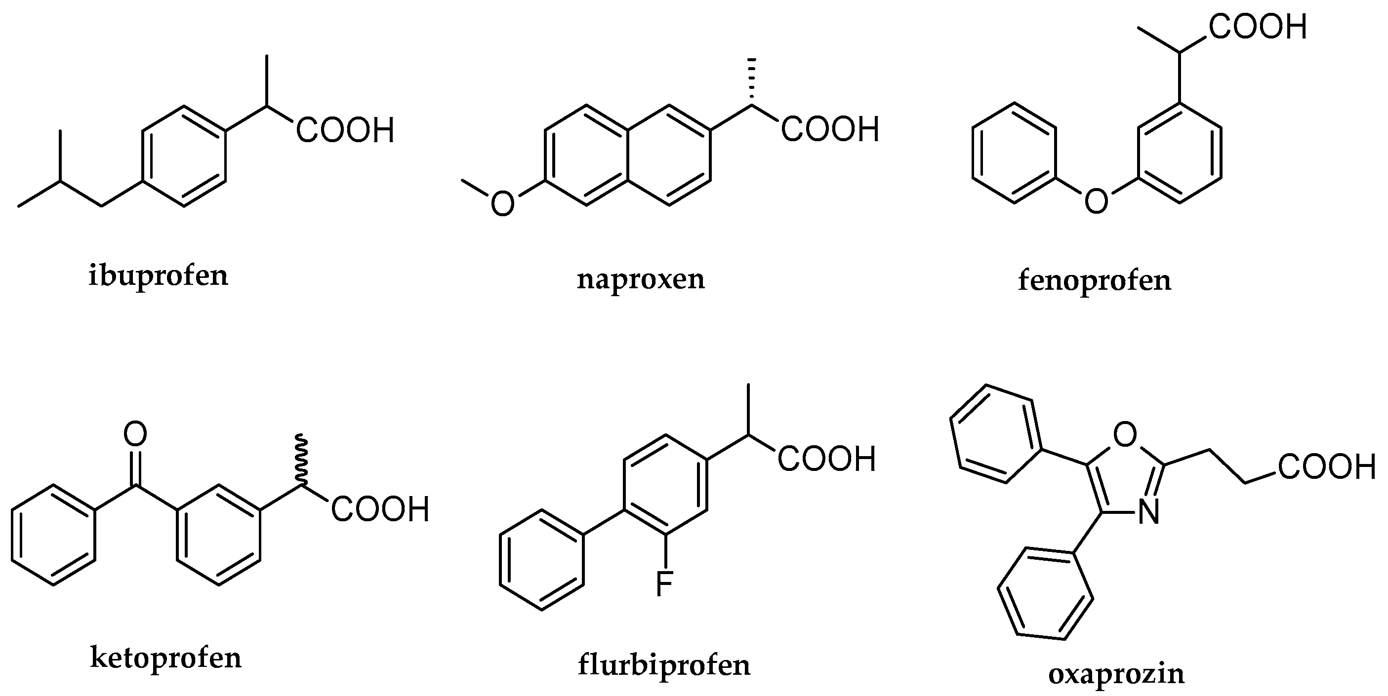
Figure 1. Selected structures of non-steroidal anti-inflammatory drugs (NSAID) containing propionic acid system.
Molecules 28 03808 g001
Scheme 1. The general synthesis of compounds 3a3g. Method A: diethyl ether, ambient temp., 2 days; method B: boiling toluene 1–4 h; method C: boiling chloroform 1–1.5 h.
Molecules 28 03808 sch001
Scheme 2. Synthesis of compound 3d (method D).
Molecules 28 03808 sch002
Figure 2. The effect of compounds 3a3g and ibuprofen (IBU) on the LPS (lipopolysaccharide)-induced production of TNF-α (Tumor Necrosis Factor-α) in 24 h PBMC (Peripheral Blood Mononuclear Cell) cultures; the results are shown as percentage of positive control—LPS alone; values are expressed as medians from four independent experiments and interquartile ranges [Q1, Q3]; * indicates a significant difference compared to the positive control at p < 0.05.
Molecules 28 03808 g002
Figure 3. The effect of compounds 3a3g and ibuprofen (IBU) on the LPS (lipopolysaccharide)-induced production of IL-6 (Interleukin-6) in 24 h PBMC (Peripheral Blood Mononuclear Cell) cultures; the results are shown as percentage of positive control—LPS alone; values are expressed as medians from five independent experiments and interquartile ranges [Q1, Q3]; * indicates a significant difference compared to positive control at p < 0.05.
Molecules 28 03808 g003
Figure 4. The effect of compounds 3a3g on the LPS (lipopolysaccharide)-induced production of IFN-γ (Interferon-γ) in 24 h PBMC (Peripheral Blood Mononuclear Cell) cultures; the results are shown as percentage of positive control—LPS alone; values are expressed as medians from three independent experiments and interquartile ranges [Q1, Q3]; ^ indicates a tendency compared to positive control at p = 0.0518.
Molecules 28 03808 g004
Figure 5. The effect of compounds 3a3g and ibuprofen (IBU) on the LPS (lipopolysaccharide)-induced production of IL-10 (Interleukin-10) in 24 h PBMC (Peripheral Blood Mononuclear Cell) cultures; the results are shown as percentage of positive control—LPS alone; values are expressed as medians from five independent experiments and interquartile ranges [Q1, Q3]; * indicates significant difference comparing to positive control at p < 0.05.
Molecules 28 03808 g005
Figure 6. The effect of compounds 3a3g and ibuprofen (IBU) on the PHA (phytohaemagglutinin) -induced PBMC (Peripheral Blood Mononuclear Cell) proliferation in 72 h cell cultures; the results are shown as percentage of positive control—PHA alone; values are expressed as medians from six independent experiments and interquartile ranges [Q1, Q3]; ** indicates a significant difference compared to positive control at p < 0.01.
Molecules 28 03808 g006
Figure 7. Nematodes of the genus Rhabditis sp. after 24 h of exposure to the highest concentration (11.1 mg/mL) of the tested compounds. * indicates significant difference comparing to control 0.6% NaCl (sodium chloride) at p < 0.05.
Molecules 28 03808 g007
Figure 8. Example culture of Rhabditis sp.—alive and motile nematodes are visible. (a) control 0.6% NaCl (sodium chloride), (b) compound 3a.
Molecules 28 03808 g008
Disclaimer/Publisher’s Note: The statements, opinions and data contained in all publications are solely those of the individual author(s) and contributor(s) and not of MDPI and/or the editor(s). MDPI and/or the editor(s) disclaim responsibility for any injury to people or property resulting from any ideas, methods, instructions or products referred to in the content.