Article

Physico-Chemical Study of Mn(II), Co(II), Cu(II), Cr(III), and Pd(II) Complexes with Schiff-Base and Aminopyrimidyl Derivatives and Anti-Cancer, Antioxidant, Antimicrobial Applications

by
Maged S. Al-Fakeh
1,2,*,
Maha A. Alsikhan
1 and
Jawza Sh Alnawmasi
1
1
Department of Chemistry, College of Science, Qassim University, Buraidah 51452, Saudi Arabia
2
Taiz University, Taiz 3086, Yemen
*
Author to whom correspondence should be addressed.
Molecules 2023, 28(6), 2555; https://doi.org/10.3390/molecules28062555
Submission received: 20 February 2023 / Revised: 7 March 2023 / Accepted: 9 March 2023 / Published: 11 March 2023

Abstract

:
A new class of biologically active mineral complexes was synthesized by reacting the following metal salts: MnCl2·4H2O, CoCl2·6H2O, CuCl2·2H2O, CrCl3·6H2O, and PdCl2 respectively with 2-amino-4,6-dimethyl pyrimidine (ADMPY) and Schiff’s base resulting from the condensation reaction between benzaldehyde with p-phenylenediamine and 2-hydroxy-1-naphthaldehyde as ligands have been synthesized and characterized on the basis of their CHN, thermal analysis, XRD, SEM and magnetic measurements along with their FT-IR and UV-vis spectra. The scanning electron microscope SEM measurements and the calculations on the powder XRD data indicate the nano-sized nature of the prepared complexes (average size 32–88 nm). The spectral data confirmed the coordinated ligand (HL) via a nitrogen atom of an azomethine group (-C=N-) and phenolic -OH group and NH2-ADMPY ligand with the metal ions. An octahedral geometry for all complexes has been proposed based on magnetic and electronic spectral data except Pd(II) complex, which has a tetrahedral geometry. Molecular modeling was performed for Cu(II) complex using the density functional method DFT/B3LYP to study the structures and the frontier molecular orbitals (HOMO and LUMO). The antioxidant of the complexes was studied using the 2,2-diphenyl-1-picrylhydrazyl (DPPH)-free radical-scavenging assays. The metal complexes were tested in vitro for anticancer activities against two cancer lines A-549 and MRC-5 cells. Cu(II) and Pd(II) complexes showed the highest cytotoxicity effect, comparable to that of other cis-platinum-based drugs. The complexes showed significant activity against fungi and bacteria.

1. Introduction

Recently, the focus has been on mixed chelated mineral complexes due to their importance and biological activity against microbes, fungi, cancerous tumors, and others. Schiff bases are the most common coordination compounds. They were named Schiff bases in relation to their discovery by the German scientist Hugo Schiff, who discovered them in 1864. They were simply synthesized by condensing primary amines and carbonyl compounds [1]. The azomethine (anils, imines) group (-HC=N-) is a structural feature shared by these compounds. This active group carries a potential binding site for metal ions through the lone pair of electrons (unlinked) in a hybridized orbital SP2 of the nitrogen atom. The (C=N) linkage is required for biological activity in azomethine derivatives; numerous azomethines have been reported to have strong antifungal, antibacterial, anticancer, and antimalarial properties. Numerous Schiff bases and their complexes have been investigated for their noteworthy and intriguing qualities, such as their photochromic qualities and capacity to bind to some toxic metals [2]. Here we find Schiff’s base acting as a toothed flexible ligand and coordination is carried out through the nitrogen atom of the azomethine group and the oxygen atom of the phenolic group. It is close to the azomethine group [3], so it was shown in studies that Schiff bases are excellent chelating agents, and they also provide a great attraction factor due to the ease of preparation, structural flexibility, and the special properties of the C=N group [4,5]. In addition, pyridine derivatives are among the important categories in inorganic chemistry, where 2-amino-4,6-dimethyl pyrimidine is one of its important derivatives, which plays an important vital role, as it participates in coordination. Pyrimidine is a member of the heterocyclic compound family and is a common substance found in living things. It holds a special position in the field of heterocyclic and medicinal chemistry because of its beneficial biological properties and therapeutic uses. Since the structural subunits of the pyrimidine compound are found in many naturally occurring products, including vitamins and antibiotics, it is widely distributed in nature and is extremely important to human health. Consequently, they pay close attention to the development of both biologically energetic compounds and antibiotics. Tri-substituted pyrimidine containing an electron-withdrawing group such as the amino group in pyrimidine shows more potent in vitro antimicrobial activity [6,7,8,9]. Thus, the synthesis and screening of Schiff base ligand, 2-((E)-((4-(((E)-benzylidene)amino)phenyl)imino)methyl)-naphthalene-1-ol with transition metal complexes ability, anticancer activity, antioxidant properties, and antibacterial properties would be of substantial biological importance [10]. Previously, complexes of Cr(III), Mn(II), Ni(II), Fe(III), and Zn(II) were reported to show anticancer, antioxidant, and antibacterial activity [10]. Therefore, we speculate that complexes of Mn(II), Co(II), Cu(II), Cr(III), and Pd(II) with 2-amino-4,6-dimethyl pyrimidine (ADMPY) and Schiff’s base will show more anticancer, antibacterial, and antioxidant activity. These findings prompted us to start a project on metal-ADMPY and Schiff’s base complexes, involving an investigation of their nano-sized, anticancer, antibacterial, and antioxidant properties. The main objective of this research is to prepare and study the physico-chemical composition of the new nano-sized complexes of manganese(II), cobalt(II), copper(II), chromium(III), and palladium(II), which are studied in solution and in the solid state. The stability constants are evaluated and structure of the studied complexes is elucidated using elemental analyses, FT-IR, UV-vis spectra, magnetic moment, molar conductance, and thermal analyses measurements. In the anticancer studies, antibacterial, antifungal, and antioxidant properties of the metal complexes are reported. The Schiff base ligand used in this study are HL (Figure 1a) and ADMPY (Figure 1b).

2. Results and Discussion

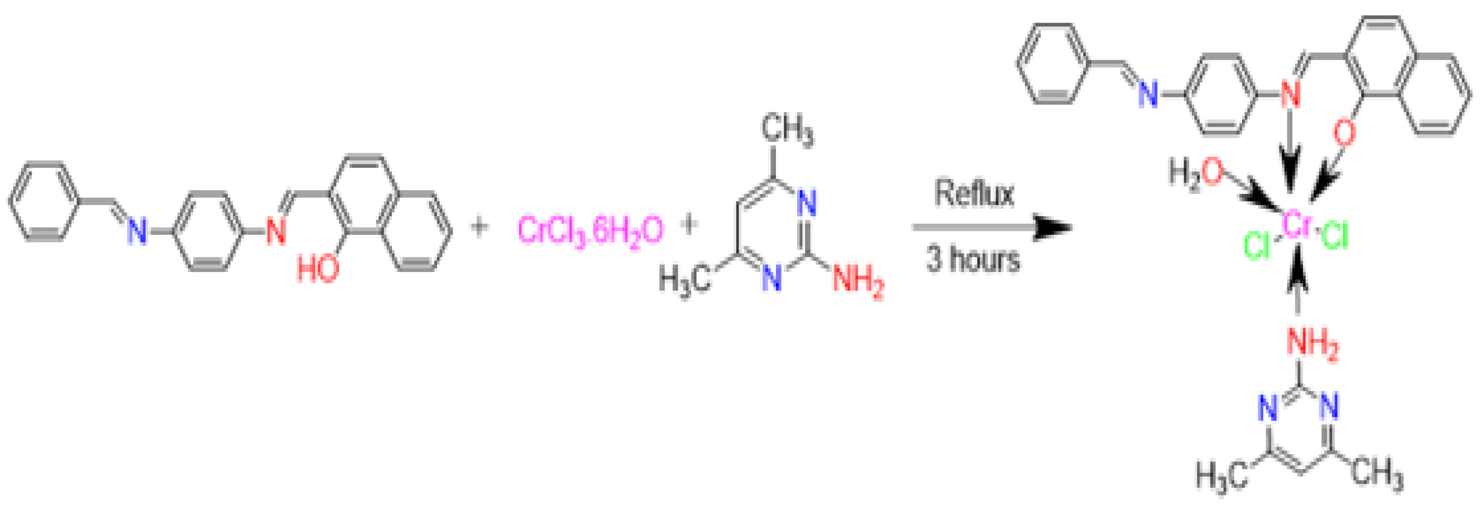
The reactions of manganese(II), cobalt(II), copper(II), chromium(III), and palladium(II) with HL and (ADMPY) ligands proceed readily to form the corresponding mixed ligand complexes. The metal mixed-bonding complexes were respectively prepared at a ratio of M:L1:L2 (1:1:1). The complexes are air-stable, insoluble in common organic solvents but soluble in dimethylformamide (DMF) and dimethylsulphoxide (DMSO). The molar conductivities of the complexes in DMSO at 25 °C are in the 11.4–28.2 Ω−1cm2 mol−1 range indicating the non-electrolytic nature of the compounds [11,12,13]. The following figure (Figure 2) illustrates these suggested summary formulas.

2.1. Fourier Transform Infrared Spectra

The IR spectra provide important information about the nature of the functional group attached to the metal. The IR spectrum of the free ligands is compared to the spectra of the complexes to study the bonding mode of Schiff base and (ADMPY) ligands to metal complexes. Table 1 shows the main IR bands and their assignments. FTIR spectrum of ligand (HL) did not show the stretching frequency of phenolic (O-H) because of its involvement in hydrogen bonding (intra-ligand hydrogen bond) with lone electron pair of an azomethine nitrogen atom (O-H……N) [14]. The υ(O-H) vibrations of complexes 1, 2, 3, 4 and 5 were observed as strong bands at 3410, 3345, 3438, 3326, and 3354 cm−1 respectively. Strong bands appear at 1618, 1604 cm−1 belonging to the two azomethine groups υ(C=N) [15]; it also shows a medium band of the υ(C=C) at 1540 cm−1 [16]; the stretching frequency of the phenolic υ(C-O) bond has been recorded as 1310 cm−1. The spectrum of ligand (ADMPY) observed the medium intensity absorption bands 3396 cm−1 and 3310 cm−1 are due to asymmetric and symmetric vibrations of υ(NH2) stretching frequency, respectively, while the bending band-specific scissors at 1633 cm−1 appears [17], C-N. It is found in two places; the base ring and the primary amine group. Comparisons were made by contrasting the compound’s spectrum with published literature on similar systems. It was found that the stretching frequency of the phenolic υ(C–O) bond has been shifted in spectra of prepared complexes of the υ(C-O) stretching band, which provides evidence that the phenolic-OH group is involved in chelation. This chelation is further reinforced also by the appearance of new peaks in complexes due to δ(OH) rocking [18], its blue-shift indicates the sharing of phenolic oxygen atom in coordination with metal ions after losing its proton. The azomethine group υ(C=N) in all complexes undergoes a shift, necessitating its coordination with the metal center. Because the NH2 group of the ADMPY ligand will be overlapping with those of the water molecules, the presence of water coordination in the complexes’ spectra makes it challenging to conclude them. Clarifying how chelation affects the in-plane bending, or δ(NH2) vibration, adds to the evidence that the NH2 group is involved. This band’s shift from 1633 cm−1 in the free ADMPY ligand to 1667–1698 cm−1 in the complexes shows that the NH2 group was involved in complex formation [19]. The υ(OH) in the lattice H2O in complexes (2) and (4) may be ascribed to a broad diffused band with medium intensity situated at 3547 and 3548 cm−1 respectively [20,21]. The coordinated H2O υ(OH) stretching vibration for the complexes (1) through (5) appears at a wavelength of 3302–3330 cm−1 ranges in each case. A band at 732–747 cm−1 in the IR spectra of the complexes 15 appears as (H2O), this suggests that coordinated water is present [22]. New bands are found in the spectra of the complexes in the regions 640–612 cm−1 which are assigned to υ(M-O). The bands at 566–544 cm−1 have been assigned to υ (M-N) of the amino mode [23]. The υ(M-Cl) bands appeared at 442–419 cm−1 [24].

2.2. Magnetic Moments

At room temperature, the magnetic moment (μeff) values for the complexes (14) are given in Table 2, and the results are 5.84 B.M, 4.90 B.M, 1.72 B.M, and 3.70 B.M. respectively. Mn(II) and Co(II) complexes are 5.84 and 4.90 B.M, this indicates high-spin. Mn(II) and Co(II) ions have five and three unpaired electrons in each compound’s respective outer valence shell [25,26]. The magnetic moment of the Cu(II) complex was 1.72 BM, which is consistent with an octahedral shape and one unpaired electron [27]. Indicating the presence of three unpaired electrons in its outer valence shell and creating an octahedral geometry around the Cr(III) ion, the Cr(III) complex displayed a magnetic moment of 3.70 BM [28]. The diamagnetic behavior of the Pd(II) complex suggests a square-planar geometry [29,30].

2.3. Electronic Spectra

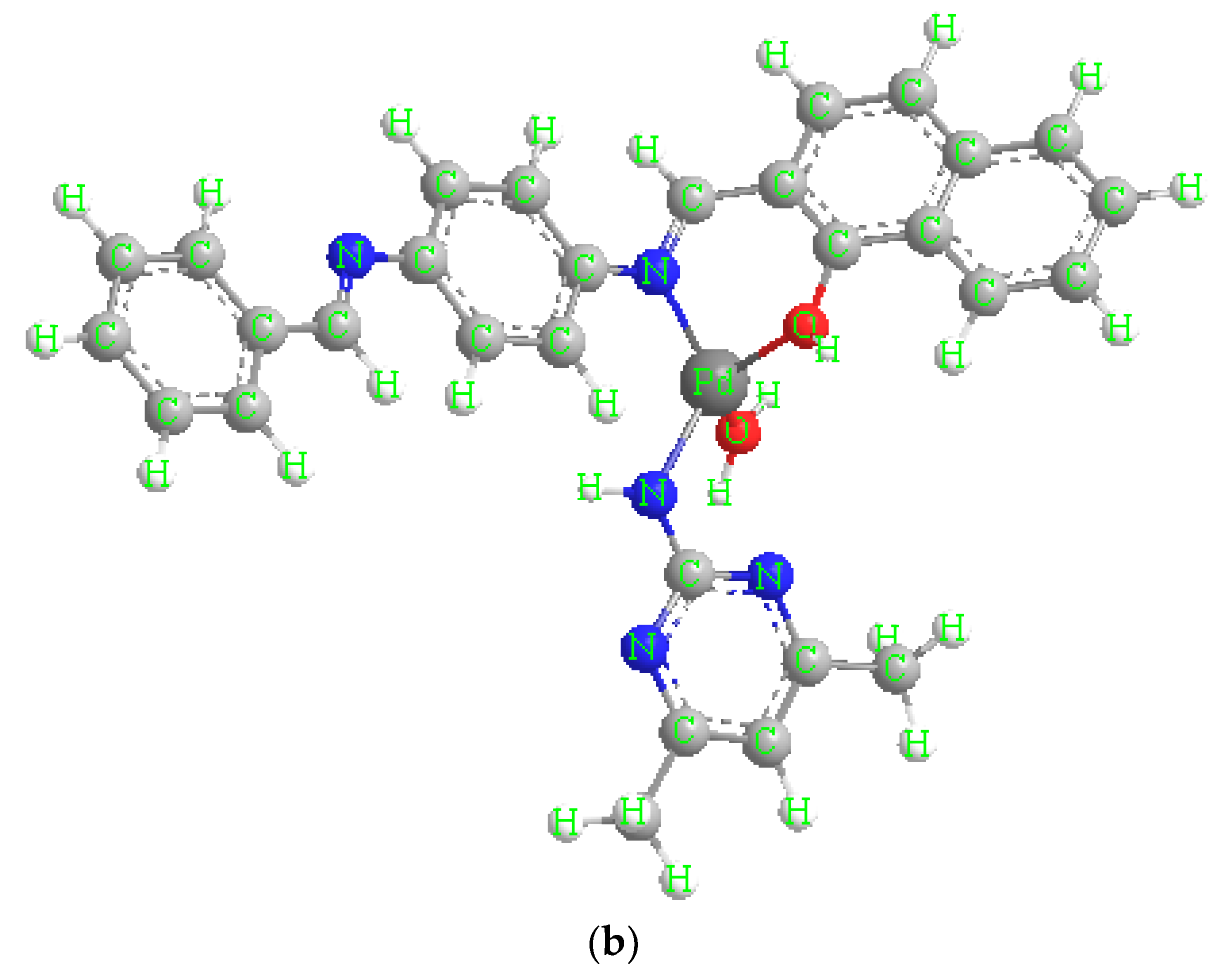
Electronic spectra of metal complexes and free ligands (HL), (ADMPY) use DMSO (10−3 M) as solvent (Table 2). The ligand UV–Vis spectrum (HL) peaks at 259 nm are assigned to (π→π∗) [10]. The ligand (ADMPY) showed absorbance bands at 314 assigned to (n→π∗) [6]. As for the electronic spectra of complexes for Mn(II), Co(II), Cu(II), Cr (III), and Pd(II), it has distinct bands in the visible region of spectra attributed to d-d transitions ranges at 415–455 nm. As for the bands corresponding to the π→π∗ and n→π∗, transitions appeared in the ranges of 252–308 nm and 300–355 nm, respectively, which indicates a bonding between the ligand and the metal elements. We may suggest the following chemical formulae for the produced five metal mixed ligand complexes based on the previously collected data elemental analyses, infrared and electronic spectra, magnetic moment measurements, and molar conductance measurements (Figure 3).

2.4. Theoretical Study

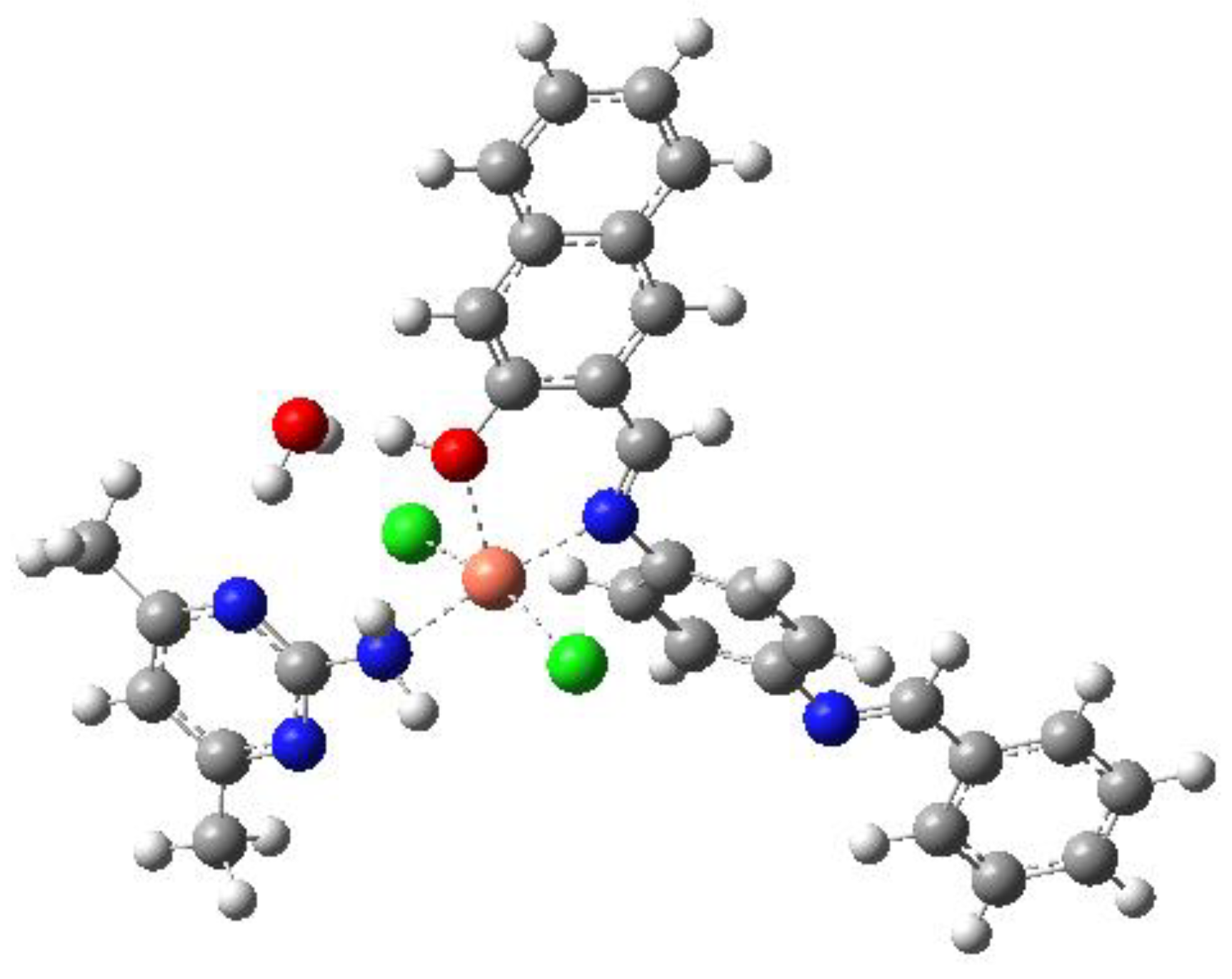
We carried out the DFT optimization through PBE1PBE functional using 6–31G (d)/Lanl2dz basis sets. The copper center is five coordinated (Figure 4) and bonded to the first ligand with Cu–Ni (2.02 Å) and Cu-OH (2.23 Å). It is also bonded to 2 Cl (2.32 Å and 2.32 Å). For the second ligand, it is bonded to NH2 with a distance Cu-NH2 = 2.13 Å. The electron density is distributed on the first ligand for the HOMO (Figure 5) but it is mainly distributed on the metal for the LUMO.

2.5. Thermal Analysis

The DTA and TGA curves in the temperature range 20–550 °C for the complexes appear at a heating rate of 10 °C/min; they showed an agreement in weight loss between their results obtained from the thermal decomposition and the calculated values, which supports the results of elemental analysis and confirms the suggested formulae. TG analysis results of mixed ligand complexes are summarized in Table 3, it was possible to determine the following characteristic thermal parameters for each reaction step: Initial point temperature of decomposition (Ti): the point at which TG (Tm) curve starts deviating from its base line. Final point temperature of decomposition (Tf): the point at which TG curve returns to its base line.

[Pd(HL)(ADMPY)(H2O)]·Cl2

One water molecule’s loss (calculated at 2.69%, found at 2.51%) and the observed mass loss of this initial stage are closely correlated. Two chlorine atoms (calculated at 10.60%, measured at 10.34%) are responsible for the observed mass reduction in the second step.
The final step (calculated at 70.83%, discovered at 69.56%) represents the breakdown of the remaining organic ligands. A large exothermic peak at 360 °C on the DTA curve designates this stage. The ultimate product at 450 °C is consistent with the formation of palladium oxide as a residual part was found to be (17.98%), compared to the (18.30%) calculated (Figure 6).

2.6. X-ray Powder Diffraction (XRD)

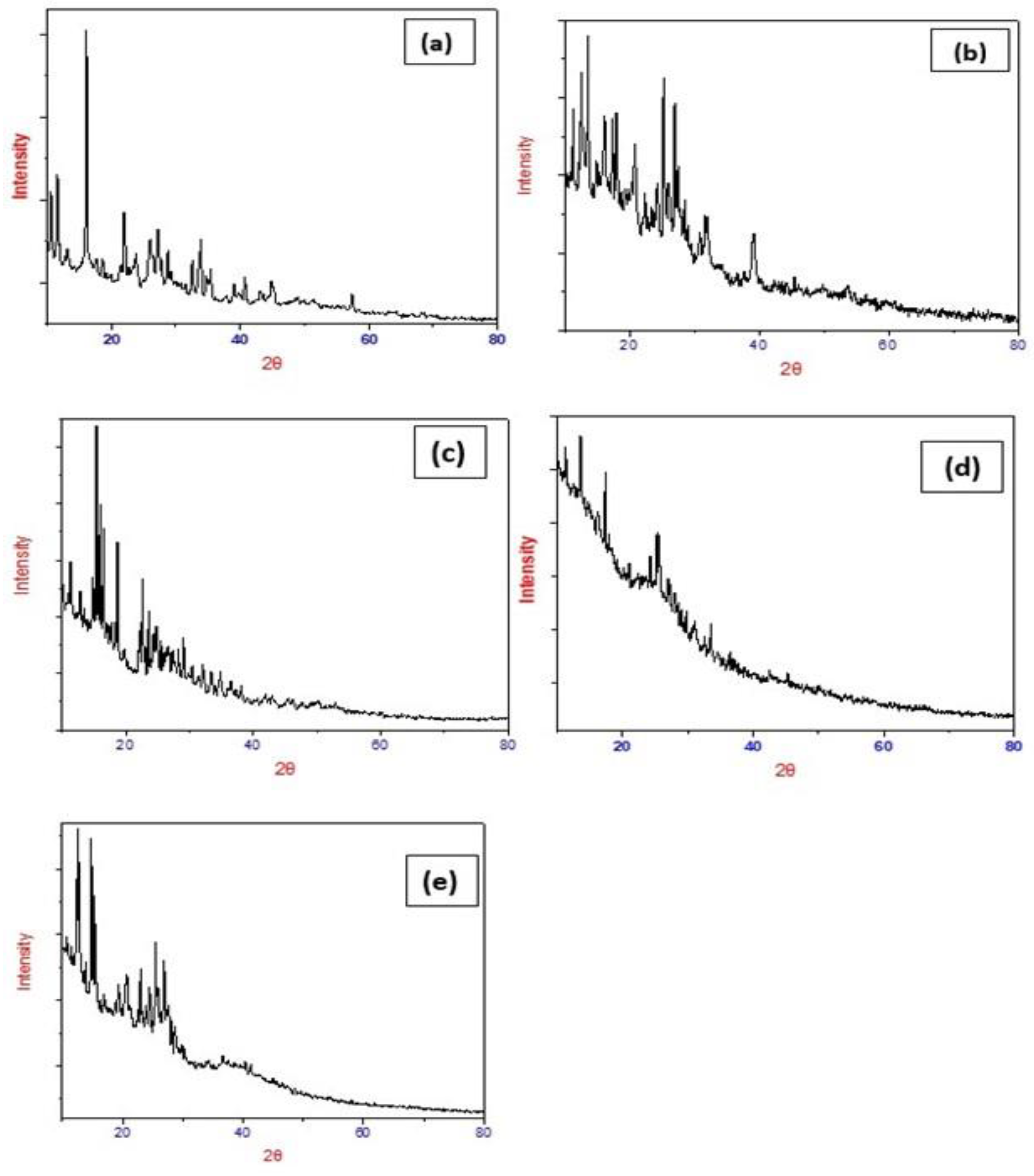
This XRD study is also one of the pieces of evidence about the formation of metal-ligand complexes. The manganese(II), cobalt(II), copper(II), chromium(III) and palladium(II) complexes were chosen for X-ray powder diffraction studies. The samples are irradiated with a beam of monochromatic X-rays over a variable incident angle range. The X-rays were detected using a fast-counting detector based on silicon strip technology (Bruker LynxEye detector). Interaction with atoms in the sample results in diffracted X-rays when the Bragg equation is satisfied. The X-rays were detected using a fast-counting detector based on silicon strip technology (Bruker LynxEye detector). XRD patterns are shown in Figure 7. All the samples are characterized at room temperature by X-ray diffraction using Cu Kα radiation. The diffraction pattern of complexes is recorded between 2θ ranging from 10° to 80°. The particle size of the samples is estimated using Scherrer’s formula. According to Scherrer’s equation, the particle size is given by t = 0.9 λ/Bcosθ, where t is the crystal thickness (in nm), B is half width (in radians), θ is the Bragg angle, and λ is the wavelength. The particle size corresponding to each diffraction maxima is determined from the measurement of the half-width of the diffraction peak. The value of the Lattice parameter and the particle size are shown in Table 4 for all five complexes. The Mn(II) complex is monoclinic, that of the Pd(II) and Co(II) complexes are tetragonal, and those of the Cu(II) and Cr(III) complexes belong to the triclinic crystal system. All metal complexes exhibit sharp peaks. The particle size was found to be within the range of 32–88 nm for all the complexes.

2.7. Morphological and Structural Properties of Synthesized Metal-Ligand Complexes

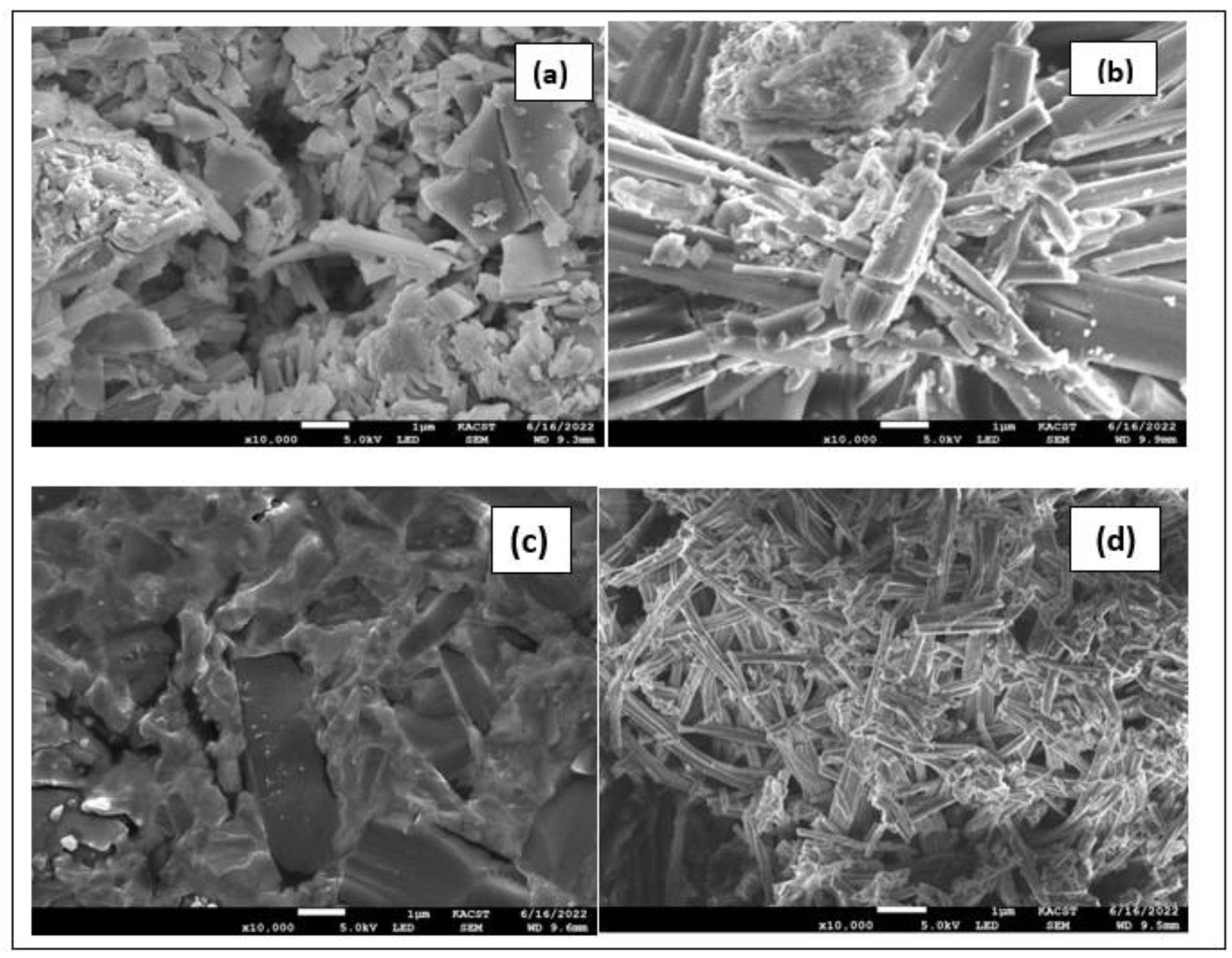
Scanning electron microscopy (SEM) was used to assess the mixed ligand metal complexes’ morphological and structural characteristics, as morphology changes when the metal ions are changed. The SEM image of Mn(II) complex revealed small particles of irregular shape. The Co(II) complex shows road-shaped particles, whereas Cr(III) complex exhibits granular texture. Finally, The Pd(II) complex shows a figure of branched nanowires. The SEM micrographs are shown in Figure 8. Thus, these SEM results confirmed the nano-structured behavior of the synthesized metal complexes.

2.8. Biological Activity

2.8.1. In Vitro Anticancer Activities

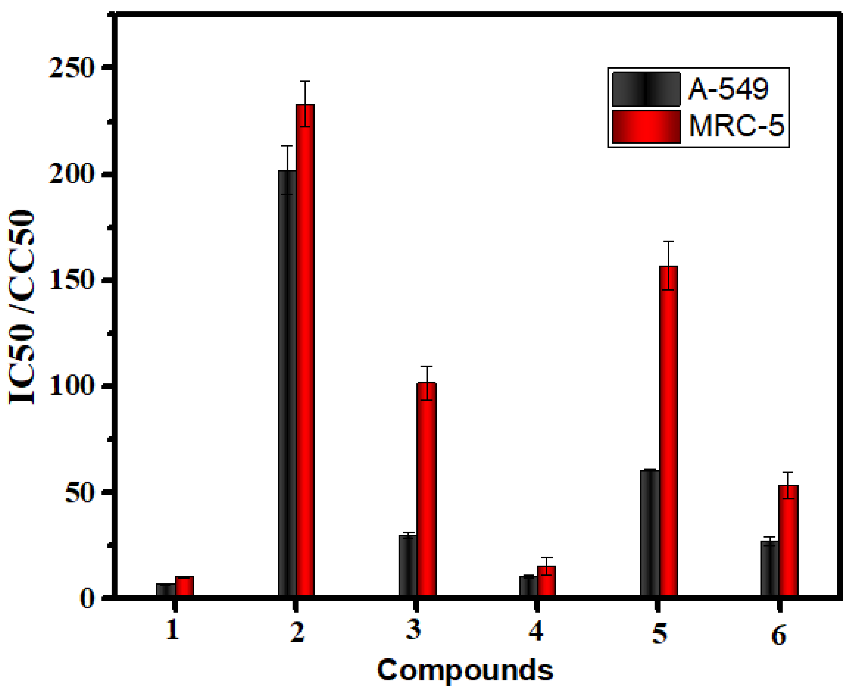
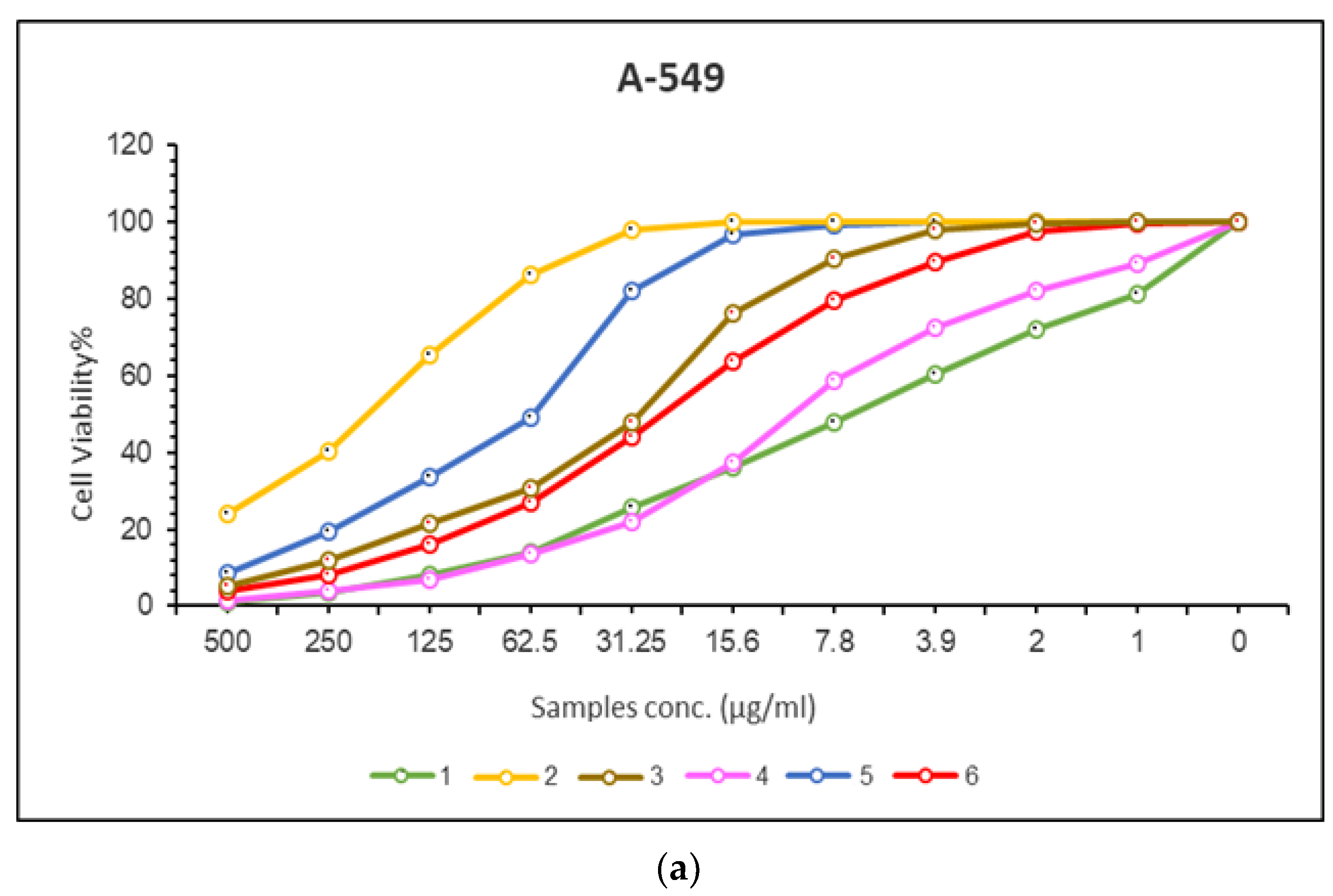
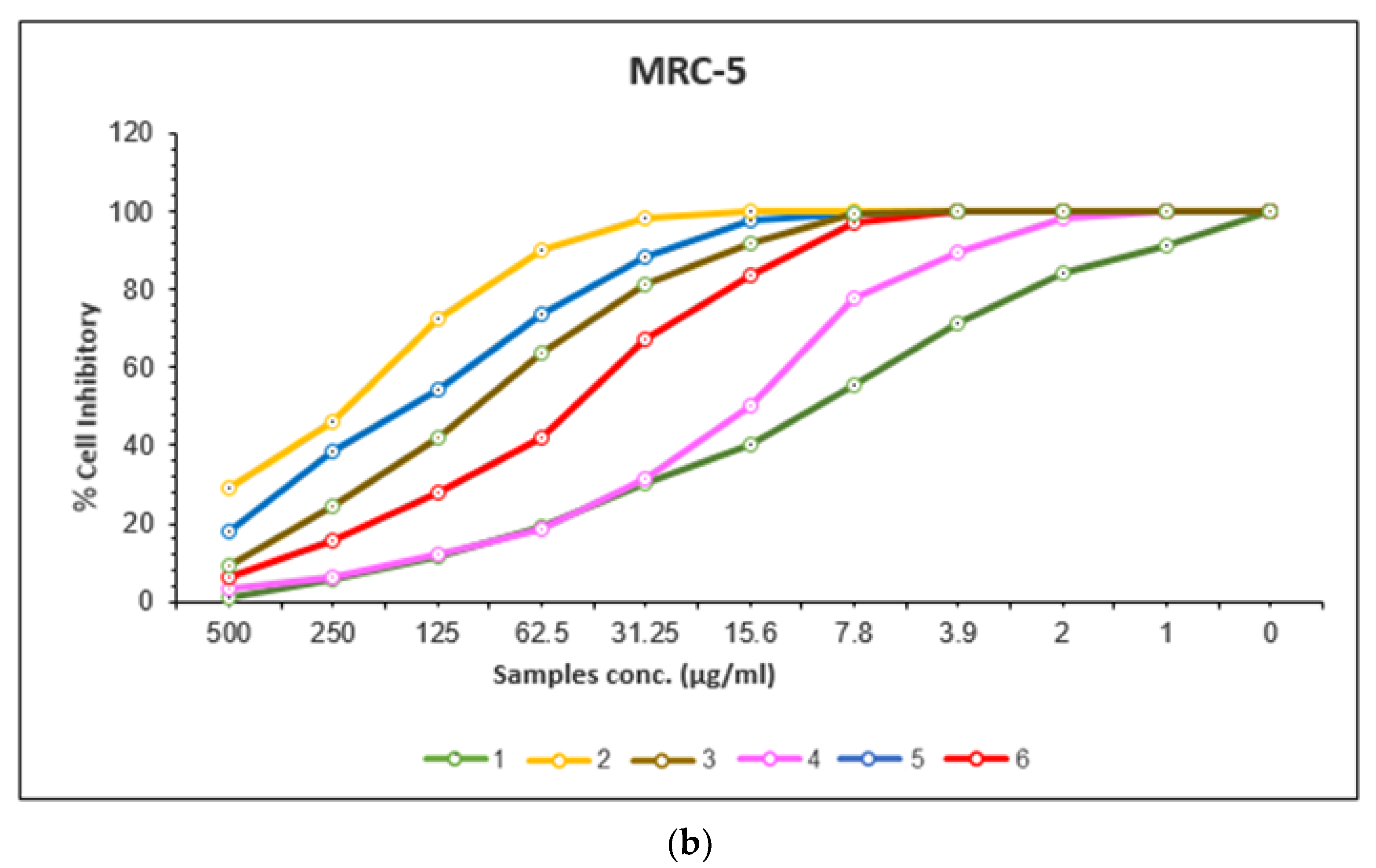
Cancer is a type of malignant disease in which a group of cells appears to grow in an irregular pattern. The only way to treat this is through surgical intervention or chemotherapy, both of which have negative side effects. Although platinum-based complexes such as cis-platin have been used to treat cancer, it is not effective, so in vitro tests for complexes of manganese, cobalt, copper, chromium, and palladium were conducted against two cancer lines, respectively. A-549 and MRC-5 cells were used as test subjects for the cytotoxic activity of Schiff base and aminopyrimidyl complexes in the concentration range of 0–500 (g/mL). Results are provided in Figure 9, which shows the IC50 and CC50 values for each compound (Table 5). According to cytotoxicity results, all tested complexes showed strong cytotoxicity against A-549 cancer cells (IC50 = 10.97–201.86 μg/µL) and strong cytotoxicity against MRC-5 cancer cells (CC50 = 15.41–233.01 μg/µL). Copper(II) complex showed the highest cytotoxicity effect with an IC50 value of 10.97 μg/µL, followed by palladium(II) complex with an IC50 value of 26.54 μg/µL and then Cobalt(II) complex with an IC50 value of 29.94 μg/µL and then chromium(III) complex with IC50 value 61.46 μg/µL and then manganese (II) complex with IC50 value 201.86 μg/µL in case of A-549 cancer cells. Copper(II) complex showed the highest cytotoxicity effect with a CC50 value of 15.41 μg/µL, followed by palladium(II) complex with a CC50 value of 53 μM, cobalt(II) complex with a CC50 value of 101.72 μM, then chromium(III) complex with a CC50 value 157.65 μg/µL, then manganese(II) complex with CC50 value 233.01 μg/µL in case of MRC-5 cancer cells. As the concentration of the complexes increased, cell viability decreased (Figure 10).

2.8.2. Antimicrobial Activity

Antifungal Screening

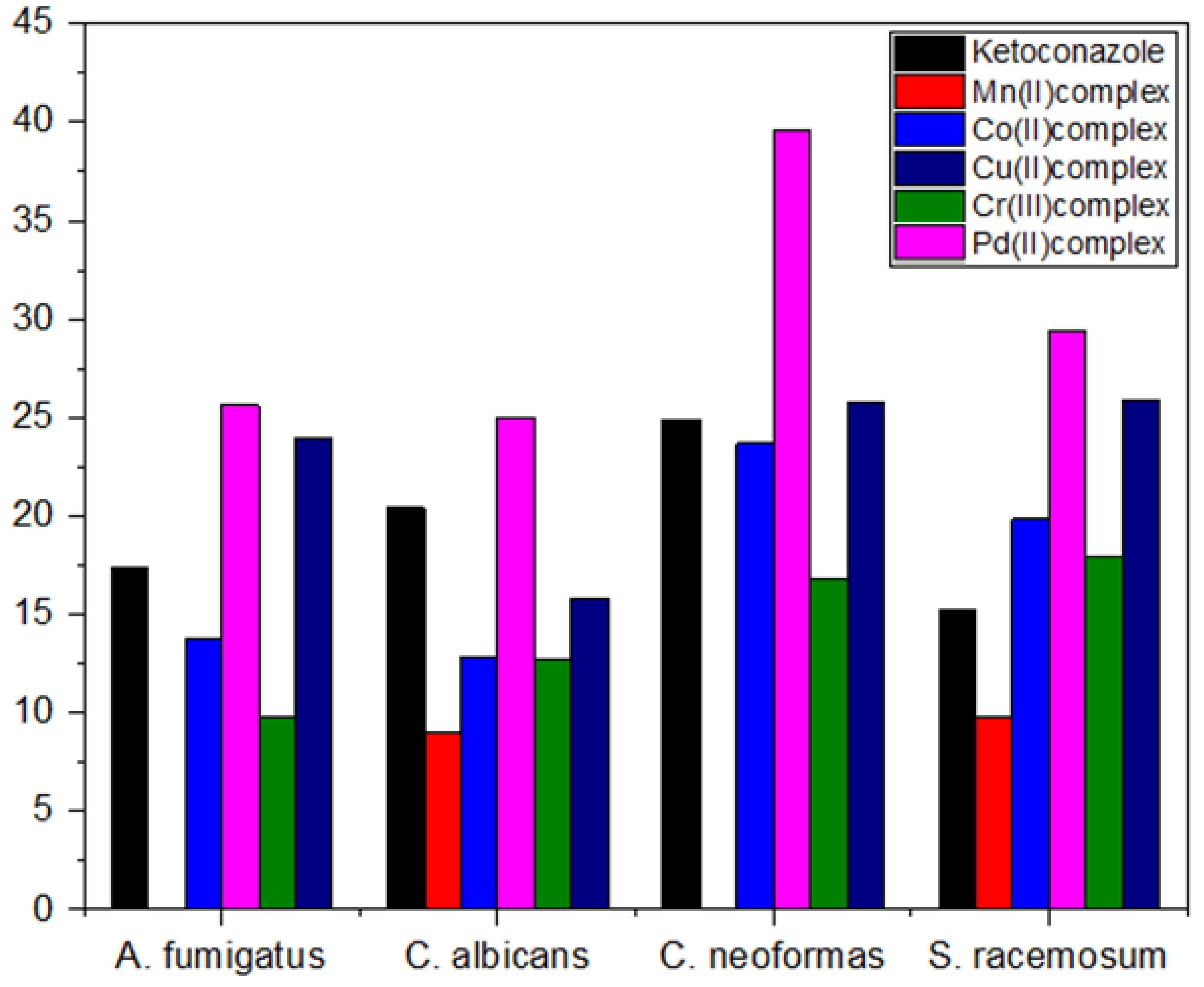
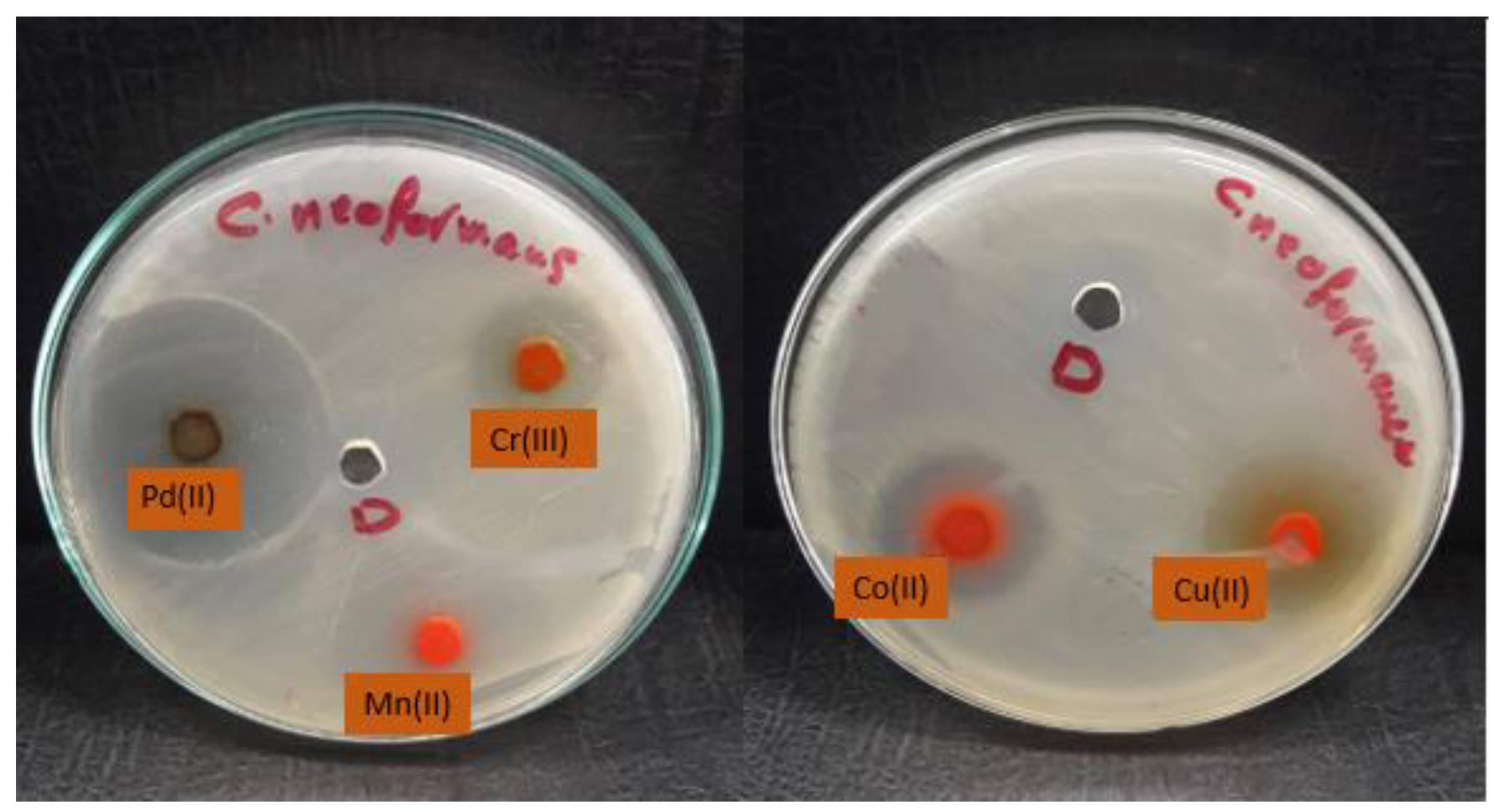
The data shown in Table 6 showed excellent activity against fungal strains, specifically against the fungal strain Cryptococcus nanoforms. We note that the growth inhibition zone ranged between 9 and 40. We also note that many mineral complexes showed higher activity than the control sample compared to the lower antifungal activity of the free ligand or the previous complexes [10]. We found that palladium(II) and copper(II) complexes are the most active against fungi which showed impressive activity, while the manganese(II) complex is the least effective (Figure 11, Figure 12 and Figure 13). The complexes were arranged according to their activity against fungal strains as follows: Pd(II) > Cu(II) > Co(II) > Cr(III) > Mn(II).

Antibacterial Screening

The data in Table 7 showed activity against bacterial strains, where the palladium(II) and copper(II) complexes showed the highest zone of inhibition compared to the rest of the complexes, and manganese(II) complexes were found to be the least effective (Figure 14). The complexes were arranged in the following order based on their activity against bacterial strains: Pd(II) > Cu(II) > Co(II) > Cr(III) > Mn(II)
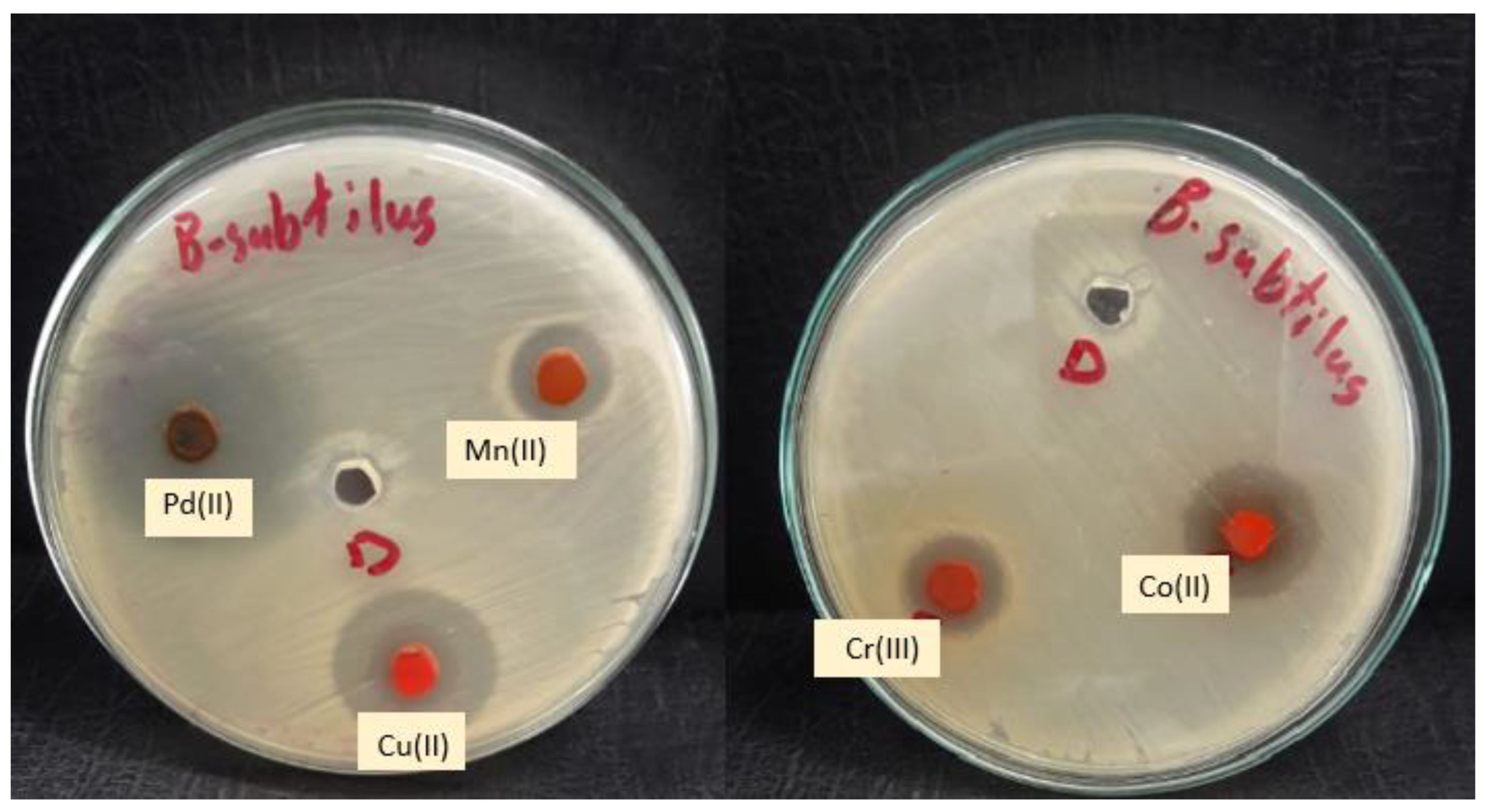
Complexes are typically more effective and active against Gram-positive bacteria than Gram-negative bacteria compared to the lower antibacterial activity of the free ligand or the previous complexes [10]. This could be explained by the fact that Gram-positive bacteria have dense cell walls with many layers of teichoic acids and peptidoglycan. Gram-negative bacteria, on the other hand, have a relatively thin cell wall made up of a few peptidoglycan layers that are encircled by a second lipid membrane that contains lipoproteins and lipopolysaccharides. The variance in the structure of the cell wall led to differences in antibacterial activity. Figure 15 and Figure 16 demonstrate that the tested complexes are effective against Gram-positive and negative bacteria.

2.8.3. Antioxidant Assays

Oxidation in living organisms caused by free radicals are usually undesirable processes damaging such important biomolecules as DNA, proteins, and lipids [31].
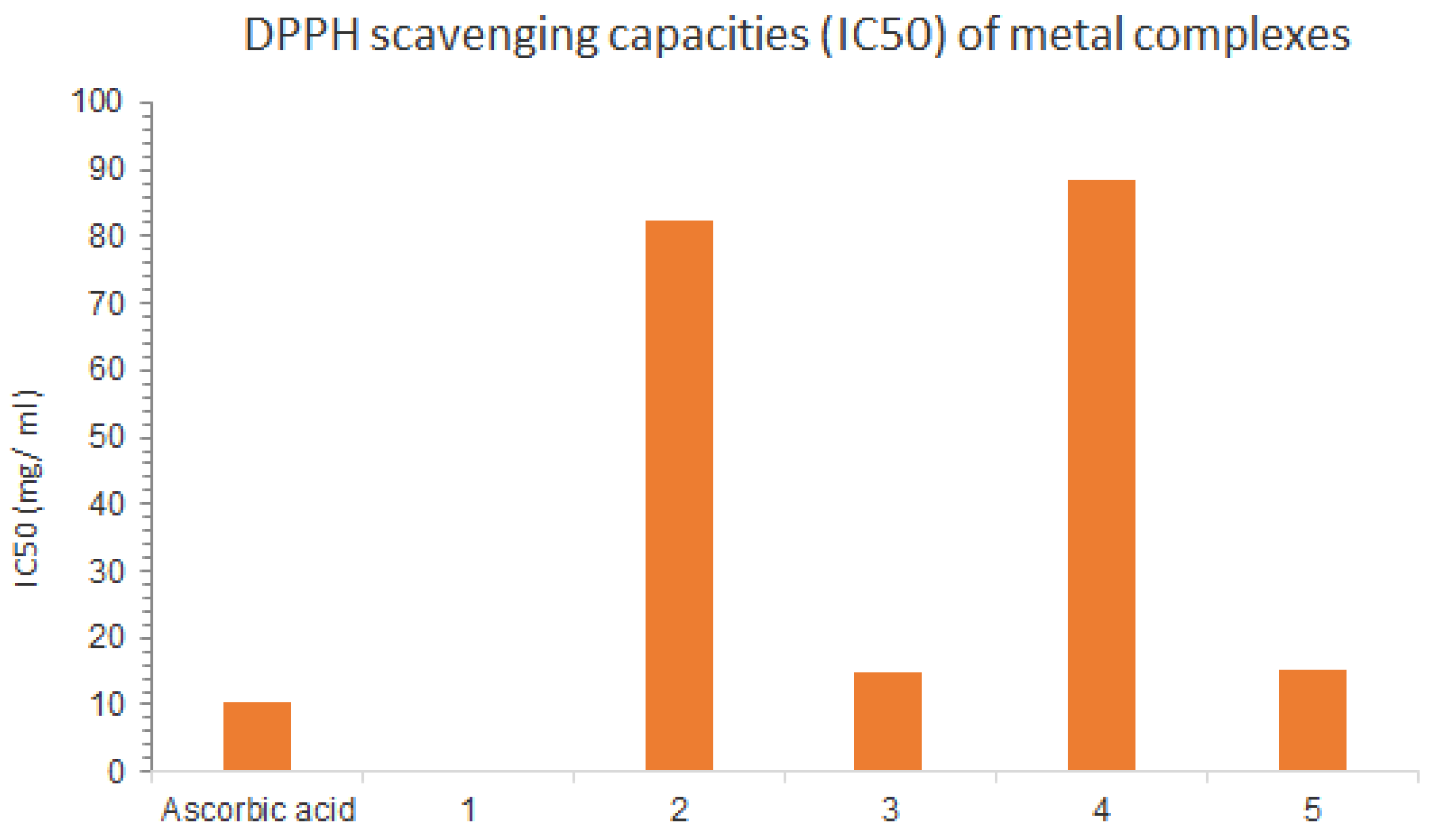
The application of antioxidants is one of the most straightforward means to protect these biomolecules from oxidative stress. Many assays are available for the determination of antioxidant potential in vitro. These assays are based on the reduction of stable radicals (DPPH). DPPH test is one of the most commonly performed antioxidant assays with natural and (semi)synthetic biologically active compounds. Although it has no direct physiological relevance, this assay allows quick comparison of the free radical scavenging potential of new derivatives as this activity has been published for many compounds [32]. We have therefore tested our mixed ligand complexes of Schiff bases and aminopyrimidyl derivatives for DPPH scavenging and compared their activity with the activity of the parent compounds, that is, Cu(II) and Pd(II) complexes showing high activity, Co(II) and Cr(III) complexes showing moderate activity, and all of them showing less activity when compared to the standard (ascorbic acid). Under these experimental conditions, the Mn(II) complexes demonstrated weak antioxidant activity. The IC50 values for all the collectors were determined and compared with the standard as listed in the Table 8 and Figure 17.

3. Materials and Methods

3.1. Physical Measurements for Complexes

The FTIR spectra of the ligand and its metal complexes in the solid state were measured using Agilent technologies/Gladi-ATR, USA) Cary 600 Series FTIR Spectrometer in the wave range 400–4000 cm−1. The carbon, hydrogen, and nitrogen contents were determined for the prepared ligand and its metallic complexes using a Eurovector CHN (EA3000, Italy) analyzer. Magnetic moments were measured using a Sherwood Scientific magnetic susceptibility balance (MSB-Auto) (UK). Electron-visible and UV-visible absorption spectra of (DMSO, 1 × 10⁻³ M) solutions were also recorded on a Shimadzu 1650-Spec UV-Vis spectrometer (Shimadzu, Duisburg, Germany). Thermal analysis of the compounds was carried out in dynamic air on a Shimadzu (DTG 60-H) thermal analyzer at a heating rate of 10 °C/min, the temperature range was 20–600 °C. XRD Model (PW 1710) control unit Philips with a Cu Kα (l = 1.54180 Å) anode at 40 K.V 30 M. A scanning electron microscopy (SEM) was used to examine the morphology of the complexes (JEOL JSM-5400-LV Field Emission SEM, Tokyo, Japan).

3.2. Microbial Strains and Culture Media

The complexes were tested in vitro using a diffusion agar technique; well diameter: 6.0 mm (100 μL tested) on four fungi (Aspergillus fumigatus (RCMB 002008), Candida albicans (RCMB 005003 (1) ATCC 10231), Cryptococcus neoformas (RCMB 0049001) and Syncephalastrum racemosum (RCMB 016001 (1)). The positive control was a fungicide (ketoconazole, 100 μg/mL) as well as four bacteria, two Gram-positive strains (Staphylococcus aureus (ATCC 25923) and Bacillus subtilis (RCMB 015 (1) NRRL B-543)), and two Gram-negative strains (Escherichia coli (ATCC 25922) and Proteus Vulgaris (RCMB 004 (1) ATCC 13315)). The positive control was Streptococcus (gentamycin, 4 μg/mL). The inhibitory activity of a compound against an organism is indicated by a clear area around the disc.

3.3. Computational Studies

The density functional theory (DFT) was performed by using Gaussian09 program. The optimization of the diverse Cu(II) complex was done by DFT through the functional B3LYP and Lanl2dz/6-31G(d) basis sets. LANL2DZ basis set was limited for the treatment copper(II) atom and 6-31G(d) basis set for all other atoms. To confirm the stability of the structures, we calculated the vibrational frequencies at the same level of theory.

3.4. Antioxidant Activity

The antioxidant activity of the extract was determined at the Regional Center for Mycology and Biotechnology (RCMB) at Al-Azhar University by the DPPH free radical scavenging assay in triplicate, and average values were considered.

(DPPH) Radical Scavenging Assay

Freshly prepared (0.004% w/v) methanol solution of 2,2-diphenyl-1-picrylhydrazyl (DPPH) radical was prepared and stored at 10 °C in the dark. A methanol solution of the test compound was prepared. About 40 uL aliquot of the methanol solution was added to 3 mL of DPPH solution. Absorbance measurements were recorded immediately with a UV-visible spectrophotometer (Milton Roy, Spectronic 1201). The decrease in absorbance at 515 nm was determined continuously, with data being recorded at 1 min intervals until the absorbance stabilized (16 min). The absorbance of the DPPH radical without antioxidant (control) and the reference compound ascorbic acid were also measured. All the determinations were performed in three replicates and averaged. The percentage inhibition (PI) of the DPPH radical was calculated according to the formula:
PI = [{(AC − AT)/AC} × 100]
where AC = absorbance of the control at t = 0 min and AT = absorbance of the sample + DPPH at t = 16 min. A 50% inhibitory concentration (IC50) or the concentration required for 50% DPPH radical scavenging activity was calculated.

3.5. Cytotoxicity Testing

3.5.1. Mammalian Cell Lines

A-549 cells (human Lung cancer cell line) were obtained from VACSERA Tissue Culture Unit.

3.5.2. Mammalian Cell Lines

MRC-5 cells (Normal human Lung fibroblast cells), were obtained from the American Type Culture Collection (ATCC, Rockville, MD, USA).

3.5.3. Chemicals Used

Dimethyl sulfoxide (DMSO), crystal violet, and trypan blue dye were purchased from Sigma (St. Louis, MO, USA). Fetal bovine serum, DMEM, RPMI-1640, HEPES buffer solution, L-glutamine, gentamycin, and 0.25% trypsin-EDTA were purchased from Lonza.

3.5.4. Crystal Violet Stain (1%)

It is composed of 0.5% (w/v) crystal violet and 50% methanol then made up to volume with ddH2O and filtered through a Whatmann No.1 filter paper.

3.5.5. Cell Line Propagation

The cells were propagated in Dulbecco’s modified Eagle’s medium (DMEM) supplemented with 10% heat-inactivated fetal bovine serum, 1% l-glutamine, HEPES buffer, and 50 µg/mL gentamycin. All cells were maintained at 37 °C in a humidified atmosphere with 5% CO2 and were subcultured two times a week.

3.6. Synthesis of the Metals Mixed Ligand Complexes

Synthesis of 2-((E)-((4-(((E)-benzylidene)amino)phenyl)imino)methyl)-nap hthalene-1-ol (HL) ligand was carried out as in the previous method [10]. Mn(II), Co(II), Cu(II), Cr(III), and Pd(II) mixed-bonding complexes were respectively prepared at a ratio of M: L1:L2 (1:1:1).

3.6.1. [Mn(HL)(ADMPY)(H2O)Cl2] (1)

Mn(II) complex was synthesized by dissolving MnCl2·4H2O (0.169 gm, 0.9 mmol, in 10 mL) of distilled water and adding it to an ethanolic solution of 2-((E)-((4-(((E)-benzylidene)amino)phenyl)imino)methyl)-nap hthalene-1-ol (HL) ligand (0.3 gm, 0.9 mmol, in 20 mL), followed by the addition of the second ligand 2-amino-4,6-dimethyl pyrimidine (ADMPY) (0.105 gm, 0.9 mmol, in 10 mL) of methanol. The resulting mixtures were stirred for 3 h under refluxing conditions at 60 °C. The mixture was allowed to cool to room temperature after the reflux process. A light burgundy product precipitated, which was filtered and washed with distilled water and ethanol before transferring it to the oven for drying. Elemental analysis (%) Calc. for C30H29N5O2MnCl2: C, 58.35; H, 4.74; N, 11.34. Found (%): C, 58.42; H, 4.09; N,11,68. Yield: (67%). Molar conductivity in DMSO, ΛM = 22.1 ohm−1 cm2 mol−1. M.p.: 272 °C.

3.6.2. [Co(HL)(ADMPY)(H2O)Cl2]·H2O (2)

About 0.8 gm, 2.3 mmol of HL is dissolved in 30 mL of ethanol in a 250 mL beaker, then 0.54 gm, 2.3 mmol of CoCl2·6H2O is added and allowed to dissolve in 20 mL of distilled water and then continue to add. Then immediately add (0.28 gm, 2.3 mmol) of ADMPY dissolved in 10 mL of methanol. It is transferred to refluxing for 3 h. A reddish-brown precipitate was formed, and the precipitate was filtered, washed with distilled water and ethanol to remove unreacted organic materials, and then dried in the oven at 50 °C for 2 h. Elemental analysis (%) Calc. for C30H31N5O3CoCl2: C, 56.34; H, 4.89; N, 10.95. Found (%): C, 56.60; H, 4.97; N,10,89. Yield: (83%). Molar conductivity in DMSO, ΛM = 26.6 ohm−1 cm2 mol−1. M.p.: 280 °C.

3.6.3. [Cu(HL)(ADMPY)(H2O)Cl2] (3)

It was made by combining (0.4 g, 1.1 mmol) HL Schiff base with (0.193 g, 1.1 mmol) CuCl2·2H2O, then adding ADMPY (0.140 g, 1.1 mmol) and transferred to refluxing for three hours at 60 °C. The mixture is then cooled to laboratory temperature before being filtered, washed, and dried in the oven. Finally, burgundy powder is formed. Elemental analysis (%) Calc. for C30H29N5O2CuCl2: C, 57.58; H, 4.68; N, 11.19. Found (%): C, 57.64; H, 4.96; N,11,85. Yield: (72%). Molar conductivity in DMSO, ΛM = 28.2 ohm−1 cm2 mol−1. M.p.: 266 °C.

3.6.4. [Cr(HL)(ADMPY)(H2O)Cl2]·H2O (4)

About 10 mL of an aqueous solution of CrCl3·6H2O (0.304 g, 1.1 mmol) mineral salt is dissolved in a circular flask and added to 20 mL ethanolic solution of HL (0.4 gm, 1.1 mmol), then immediately add (0.140 gm, 1.1mmol) of ADMPY dissolved in 10 mL of methanol. It is transferred to refluxing for 3 h. The light brown color was produced, filtered, washed with distilled water and ethanol, and dried in the oven at 50 °C for 2 h. Elemental analysis (%) Calc. for C30H31N5O3CrCl2: C, 57.08; H, 4.80; N, 11.09. Found (%): C, 56.42; H, 4.98; N,11,42. Yield: (82%). Molar conductivity in DMSO, ΛM = 11.4 ohm−1 cm2 mol−1. M.p.: 246 °C.

3.6.5. [Pd(HL)(ADMPY)(H2O)]·Cl2 (5)

Prepared by adding PdCl2 (0.3 gm, 1.3 mmol) dissolved in acetonitrile to a solution of the Schiff base ligand HL (0.468 gm, 1.3 mmol). The second ligand (0.164 gm, 1.3 mmol) was then dissolved in methanol. After 3 h of stirring and heating the mixture formed, and the dark brown precipitate was filtered and washed several times until the filters became clear, dried, and collected. Elemental analysis (%) Calc. for C30H29N5O2PdCl2: C, 53.88; H, 4.38; N, 10.47. Found (%): C, 53.04; H, 4.80; N,10.89. Yield: (79%). Molar conductivity in DMSO, ΛM = 12.3 ohm−1 cm2 mol−1. M.p.: >300 °C.

4. Conclusions

In summary, anticancer, antibacterial, antifungal, and antioxidant activities of previously reported of Mn(II), Co(II), Cu(II), Cr(III), and Pd(II) complexes were examined. All synthesized metal complexes showed moderate cytotoxicity against ovarian cancer cells (A-549 and MRC-5) even in the presence of DMSO in cell media. In vitro biological studies showed that the Cu(II) and Pd(II) complexes have the best anticancer activity against A-549 cells with an IC50 of 10.97 and 26.54 μM respectively, and against MRC-5 cells with a CC50 of 15.41 and 53 μM which is close to the activity of cisplatin. Out of the all complexes, the Mn(II), Co(II), and Cu(II) compounds demonstrated the highest biological activities against G+ and G- bacteria in comparison to the standard drug, indicating their promising therapeutic potential. The Cu(II) and Cr(III) complexes demonstrated the highest potency against fungi. The complexes showed significant DPPH scavenging activity, and the experimental data revealed that they could be good antioxidants.

Author Contributions

Formal analysis, J.S.A.; Methodology, M.S.A.-F.; Software, M.S.A.-F.; Writing—original draft, M.A.A.; Writing—review & editing, M.S.A.-F. All authors have read and agreed to the published version of the manuscript.

Funding

This research was funded by the Deanship of Scientific Research, Qassim University for the financial support for this research under the number (COS-2022-1-1-J-29314) during the academic year 1444 AH/2022 AD.

Institutional Review Board Statement

Not applicable.

Informed Consent Statement

Not applicable.

Data Availability Statement

By e-mail: [email protected].

Acknowledgments

The author(s) gratefully acknowledge Qassim University, represented by the Deanship of Scientific Research, for the financial support for this research under the number (COS-2022-1-1-J-29314) during the academic year 1444 AH/2022 AD.

Conflicts of Interest

The authors declare no conflict of interest.

References

  1. Uddin, M.N.; Ahmed, S.S.; Alam, S.M.R. REVIEW: Biomedical applications of Schiff base metal complexes. J. Coord. Chem. 2020, 73, 3109–3149. [Google Scholar] [CrossRef]
  2. Omar, M.; El-Halim, H.F.A.; Khalil, E.A. Synthesis, characterization, and biological and anticancer studies of mixed ligand complexes with Schiff base and 2,2′-bipyridine. Appl. Organomet. Chem. 2017, 31, e3724. [Google Scholar] [CrossRef]
  3. Xavier, A.; Srividhya, N. Synthesis and Study of Schiff base Ligands. IOSR J. Appl. Chem. 2014, 7, 6–15. [Google Scholar] [CrossRef]
  4. Al Zoubi, W.; Al-Hamdani, A.; Ahmed, S.D.; Ko, Y.G. Synthesis, characterization, and biological activity of Schiff bases metal complexes. J. Phys. Org. Chem. 2018, 31, e3752. [Google Scholar] [CrossRef]
  5. Chaudhary, N.K.; Guragain, B.; Chaudhary, S.K.; Mishra, P. Schiff base metal complex as a potential therapeutic drug in medical science: A critical review. Bibechana 2021, 18, 214–230. [Google Scholar] [CrossRef]
  6. Jaziri, E.; Louis, H.; Gharbi, C.; Unimuke, T.O.; Agwamba, E.C.; Mathias, G.E.; Fugita, W.; Nasr, C.B.; Khedhiri, L. Synthesis, X-ray crystallography, molecular electronic property investigation, and leukopoiesis activity of novel 4,6-dimethyl-1,6-dihydropyridin-2-amino nitrate hybrid material. J. Mol. Struct. 2022, 1268, 133733. [Google Scholar] [CrossRef]
  7. Al-Fakeh, M.S.; Al-Otaibi, N.F. Nd2O3, Cr2O3, and V2O3 Nanoparticles via Calcination: Synthesis, Characterization, and Antimicrobial and Antioxidant Activities. J. Nanotechnol. 2022, 2022, 7794939. [Google Scholar] [CrossRef]
  8. Al-Fakeh, M.S.; Allazzam, G.A.; Yarkandi, N.H. Preparation, Properties and Antimicrobial evaluation of a new metal complex with Diphosphine ligand and 2-Aminopyridine. SYLWAN 2021, 9, 165. [Google Scholar]
  9. Al-Fakeh, M.S.; Allazzam, G.A.; Yarkandi, N.H. Ni(II), Cu(II), Mn(II) and Fe(II) Metal Complexes Containing 1,3-Bis(diphenylphosphino)-propane and Pyridine Derivative: Synthesis, Characterization, and Antimicrobial Activity. Int. J. Biomater. 2021, 2021, 4981367. [Google Scholar] [CrossRef] [PubMed]
  10. Alorini, T.A.; Al-Hakimi, A.N.; Saeed, S.E.-S.; Alhamzi, E.H.L.; Albadri, A.E. Synthesis, characterization, and anticancer activity of some metal complexes with a new Schiff base ligand. Arab. J. Chem. 2022, 15, 103559. [Google Scholar] [CrossRef]
  11. Al-Fakeh, M.S.; Messaoudi, S.; Alresheedi, F.I.; Albadri, A.E.; El-Sayed, W.A.; Saleh, E.E. Preparation, Characterization, DFT Calculations, Antibacterial and Molecular Docking Study of Co(II), Cu(II), and Zn(II) Mixed Ligand Complexes. Crystals 2023, 13, 118. [Google Scholar] [CrossRef]
  12. Al-Fakeh, M.S. Supramolecular metal coordination polymers containing from 1,3-di(4-pyridyl)-propane and 2-amino4-methylthiazole. Biochem. Biotechnol. Res. 2017, 5, 30–38. [Google Scholar]
  13. Al-Fakeh, M.S. Sonochemical Synthesis and Characterizations of Nanosized metal coordination polymers derived from 5-(3-pyridyl)-1,3,4-oxadiazole-2-thiole and 2-aminothiazole. Eur. Chem. Bull. 2016, 5, 450–455. [Google Scholar] [CrossRef]
  14. Al-Zaidi, B.H.; Hasson, M.M.; Ismail, A.H. New complexes of chelating Schiff base: Synthesis, spectral investigation, antimicrobial, and thermal behavior studies. J. Appl. Pharm. Sci. 2019, 9, 45–57. [Google Scholar] [CrossRef] [Green Version]
  15. Rehman, M.; Ali, S.; Munawar, K.S. Synthesis, spectroscopic characterization and biological applications of novel zinc complexes with schiff bases. Key Eng. Mater. 2018, 778, 293–300. [Google Scholar] [CrossRef] [Green Version]
  16. El-Boraey, H. Structural and thermal studies of some aroylhydrazone Schiff’s bases-transition metal complexes. J. Therm. Anal. Calorim. 2005, 81, 339–346. [Google Scholar] [CrossRef]
  17. Thilagavathi, G.; Arivazhagan, M. Density functional theory calculation and vibrational spectroscopy study of 2-amino-4,6-dimethyl pyrimidine (ADMP). Spectrochim. Acta Part A Mol. Biomol. Spectrosc. 2011, 79, 389–395. [Google Scholar] [CrossRef]
  18. Panchal, P.K.; Patel, D.H.; Patel, M. Preparation and thermal, spectroscopic, and antibacterial studies of some mixed-ligand complexes. Synth. React. Inorg. Met. Org. Chem. 2004, 34, 1223–1235. [Google Scholar] [CrossRef]
  19. Mohamed, G.G.; Omar, M.; Ibrahim, A.A. Biological activity studies on metal complexes of novel tridentate Schiff base ligand. Spectroscopic and thermal characterization. Eur. J. Med. Chem. 2009, 44, 4801–4812. [Google Scholar] [CrossRef]
  20. Aly, A.A.; Al-Fakeh, M.S.; Ghandour, M.A.; Abu-Zied, B.M. New nano-sized supramolecular metal coordination polymers derived from 1,2-bis (2-pyridyl)-ethene and benzimidazole. Nano Sci. Nano Technol. Indian J. NSNTAIJ 2013, 7, 197–203. [Google Scholar]
  21. Al-Fakeh, M.S. Synthesis, thermal stability and kinetic studies of copper(II) and cobalt(II) complexes derived from 4-aminobenzohydrazide and 2-mercaptobenzothiazole. Eur. Chem. Bull. 2020, 9, 403–409. [Google Scholar] [CrossRef]
  22. Al-Fakeh, M.S. Synthesis, characterization and anticancer activity of NiO nanoparticles from a Ni(II) complex derived from chitosan and pyridine derivative. Bulg. Chem. Commun. 2021, 53, 321–326. [Google Scholar] [CrossRef]
  23. Aly, A.A.M.; Ghandour, M.; Alfakeh, M.S. Synthesis and characterization of transition metal coordination polymers derived from 1,4-Benzenedicarboxylic acid and Azoles. Turk. J. Chem. 2012, 36, 69–79. [Google Scholar]
  24. Al-Fakeh, M.S. Synthesis and characterization of coordination polymers of 1, 3-di (4-pyridyl)-propane and 2-aminobenzothiazole with Mn(II), Co(II), Cu(II) and Ni(II) ions. J. Chem. Pharm. Res. 2018, 10, 77–83. [Google Scholar]
  25. Bargujar, S.; Chandra, S.; Chauhan, R.; Rajor, H.K.; Bhardwaj, J. Synthesis, spectroscopic evaluation, molecular modelling, thermal study and biological evaluation of manganese(II) complexes derived from bidentate N,O and N,S donor Schiff base ligands. Appl. Organomet. Chem. 2021, 32, e4149. [Google Scholar] [CrossRef]
  26. Kumar, S.; Pandey, P.K.; Sinha, N.; Chaudhari, S.; Sharma, S. Spectroscopic characterisation of metal complexes with tetradentate ligand. J. Phys. Sci. 2018, 29, 1–11. [Google Scholar] [CrossRef]
  27. Moustafa, I.M.I.; Magda, H. Synthesis, Spectroscopic Studies and Biological Evaluation of Co(II), Ni(II), Cu(II) and Zr(IV) Complexes of Azo Dyes and Thiamine Hydrochloride as Antimicrobial Agents. Mod. Chem. Appl. 2017, 5, 1–7. [Google Scholar] [CrossRef]
  28. Al-zaidi, B.H.; Rasheed, A.M.; Al-bayati, S.M.; Hussein, A.M. Preparation and Characterization of Some Chelate Complexe with New bi-dentate (N, O) bis-Schiff base Ligand Among different types of coordination ligands. J. Kufa Chem. Sci. 2016, 2, 49–61. [Google Scholar]
  29. Al-Nahary, T.T. Synthesis and characterization of metal complexes of Cr(III), Mn(II), Fe(III), Co(II), Ni(II), Cu(II), Ru(III), Rh(III) and Pd(II) with derivatives of 1,3,4-thiadiazole-2,5-dithiol as new ligands. J. Saudi Chem. Soc. 2009, 13, 253–257. [Google Scholar] [CrossRef] [Green Version]
  30. El-Boraey, H.A. Coordination behavior of tetraaza [N4] ligand towards Co(II), Ni(II), Cu(II), Cu(I) and Pd(II) complexes: Synthesis, spectroscopic characterization and anticancer activity. Spectrochim. Acta Part A Mol. Biomol. Spectrosc. 2012, 97, 255–262. [Google Scholar] [CrossRef]
  31. Ntonga, A.P.; Baldovini, N.; Mouray, E.; Mambu, L.; Belong, P.; Grellier, P. Activity of Ocimum basilicum, Ocimum canum and Cymbopogon citratus essential oils against Plasmodium falciparum and mature-stage larvae of Anopheles funestus ss. Parasite 2014, 21, 33. [Google Scholar] [CrossRef] [PubMed] [Green Version]
  32. Huang, D.; Ou, B.; Prior, R.L. The chemistry behind antioxidant capacity Assays. J. Agric. Food Chem. 2005, 53, 1841–1856. [Google Scholar] [CrossRef] [PubMed]
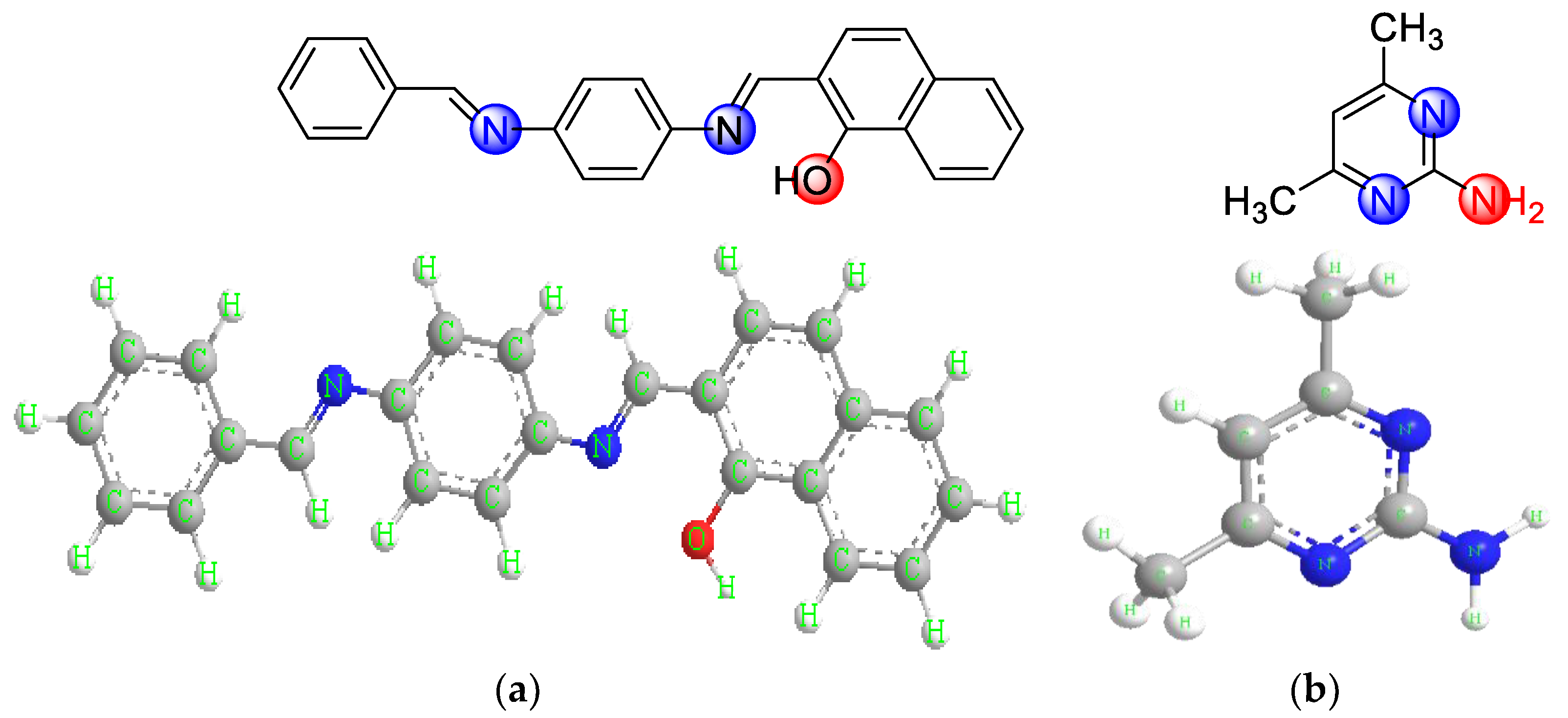
Figure 1. Structure of the ligands. (a) 2-((E)-((4-(((E) benzylidene)amino)phenyl) imino)methyl)-naphthalene-1-ol. (b) 2-amino-4,6-dimethyl pyrimidine.
Molecules 28 02555 g001
Figure 2. Preparation of the Cr(III) complex (4).
Molecules 28 02555 g002
Figure 3. A perspective view of the complete coordination around (a) Mn(II) complex. (b) Pd(II) complex.
Molecules 28 02555 g003aMolecules 28 02555 g003b
Figure 4. DFT Optimized structure of Cu(II) complex model.
Molecules 28 02555 g004
Figure 5. Frontier orbitals and their energies for the Cu(II) complex (a,b).
Molecules 28 02555 g005
Figure 6. TGA and DTA thermograms of Pd(II) complex.
Molecules 28 02555 g006
Figure 7. X-ray powder diffraction pattern for the (a) Mn(II), (b) Co(II), (c) Cu(II), (d) Cr(III), and (e) Pd(II) complexes.
Molecules 28 02555 g007
Figure 8. SEM micrographs of (a) Mn(II), (b) Co(II), (c) Cr(III), and (d) Pd(II) complexes.
Molecules 28 02555 g008
Figure 9. The comparison of anticancer activity for complexes (26) with standard drug cisplatin against (1) against selected human cell lines.
Molecules 28 02555 g009
Figure 10. (a) A-549. The dose-response curves of the cytotoxicity of compounds (16). Cells were exposed to different concentrations of the compounds for 72 h. Cell viability was determined by a colorimetric method. (b) MRC-5cell lines. The dose-response curves of the cytotoxicity of compounds (16) Cells were exposed to different concentrations of the compounds for 72 h. Cell viability was determined by a colorimetric method.
Molecules 28 02555 g010aMolecules 28 02555 g010b
Figure 11. Antifungal activities of the Ketoconazole and five metal complexes.
Molecules 28 02555 g011
Figure 12. Microbiological screening of coordination compounds of Mn(II), Co(II), Cu(II), Cr(III), and Pd(II) toward Cryptococcus neoformas.
Molecules 28 02555 g012
Figure 13. Microbiological screening of coordination compounds of Mn(II), Co(II), Cu(II), Cr(III), and Pd(II) toward Candida albicans.
Molecules 28 02555 g013
Figure 14. Antibacterial activities of the gentamycin and five metal complexes against S. aureus, B. subtilis, Escherichia coli and P. vulgaris.
Molecules 28 02555 g014
Figure 15. Microbiological screening of coordination compounds of Mn(II), Co(II), Cu(II), Cr(III), and Pd(II) toward Bacillus Subtilis.
Molecules 28 02555 g015
Figure 16. Microbiological screening of coordination compounds of Mn(II), Co(II), Cu(II), Cr(III), and Pd(II) toward Proteus vulgaris.
Molecules 28 02555 g016
Figure 17. Variation of IC50 values for all complexes and standards.
Molecules 28 02555 g017
Table 1. FT-IR details spectral data (cm−1) of free ligands and its metal compounds.
Group(HL)(ADMPY)12345
υ(OH)3350-34103345343833263354
υ(OH) lattice water---3547-3548-
υ(OH) coordinated water--33303305331433123302
υas/s (NH2)-3396, 3310DisappearDisappearDisappearDisappearDisappear
δ(NH2)-163316801666168016681698
υ(C=N)1618, 1604-1616, 15481625, 15921617, 15501613, 15881610, 1575
δ(OH) in-plane850-834826833821819
υ(C=C)1540-15301509152615381536
υ(C-O)1310-12971316129713091305
υ(M-O)--640612634626620
υ(M-N) --566547560554544
υ(M-Cl)--442419440430424
υ: stretching, δ:bending, υas: antisymmetric stretching υS: symmetric stretching; (HL): C24H18N2O, (ADMPY): C6H9N3; 1: C30H29N5O2MnCl2; 2: C30H31N5O3CoCl2; 3: C30H29N5O2CuCl2; 4: C30H31N5O3CrCl2; 5: C30H29N5O2PdCl2
Table 2. Electronic spectral data of free ligand and its complexes and magnetic moments of the compounds.
Ligands and ComplexesVmax (nm)vmax (cm−1)Assignmentμeff. (B.M)Geometrydn
(HL)25938,610ππ---
(ADMPY)31431,847nπ---
Mn(II) complex27037,037ππ5.84
31931,347nπOctahedrald5
45521,978d-d transition
Co (II) complex25239,682ππ4.90
30932,620nπOctahedrald7
42623,474d-d transition
Cu(II) complex26138,314ππ1.72
30033,333nπOctahedrald9
41524,096d-d transition
Cr(III) complex29533,898ππ3.70
34528,985nπOctahedrald3
41923,866d-d transition
Pd(II) complex30832,468ππDiamagnetic
35528,169nπsquare planard8
45422,026d-d transition
n = number of electrons.
Table 3. Thermal decomposition data of complexes.
CompoundStepTemp. Range °CAssignmentTGA (Wt. loss %)
Found (calcd.)
Mn(II) complex1st
 
2nd
3rd
4th
80–140
 
142–200
200–302
304–550
Loss of coordinated water molecules (H2O)
Loss of two chloride atoms
Loss of ligand (ADMPY)
Decomposition the rest of the organic ligand (HL)
final product (manganese oxide)
2.59 (2.91)
 
11.30 (11.48)
19.72 (19.94)
55.86 (56.75)
10.53 (11.48)
Co(II) complex1st
 
2nd
3rd
4th
78–150
 
152–248
250–396
398–550
Loss two water molecules, one crystalline and one coordinated.
Loss of two chloride atoms
Loss of ligand (ADMPY)
Decomposition with the formation of
final product (cobalt oxide).
5.40 (5.62)
 
10.98 (11.08)
19.10 (19.26)
53.11 (54.80)
15.70 (15.93)
Cu(II) complex1st
 
2nd
3rd
4th
5th
83–154
 
156–270
272–362
364–420
422–550
Loss of coordinated water molecules (H2O)
Loss of two chloride atoms
Loss of ligand (ADMPY)
Loss of ligand (HL)
formation of copper oxide.
2.67 (2.87)
 
10.96 (11.33)
19.47 (19.68)
55.80 (56.01)
12.02 (12.71)
Cr(II) complex1st
 
2nd
3rd
4th
81–158
 
160–221
223–353
355–550
Loss two water molecules, one crystalline and one coordinated.
Loss of two chloride atoms
Loss of ligand (ADMPY)
Decomposition with the formation of
final product chromium oxide.
5.13 (5.68)
 
10.98 (11.21)
19.14 (19.48)
54.87 (55.38)
10.56 (10.75)
Pd(II) complex1st
 
2nd
3rd
82–160
 
162–254
256–550
Loss of coordinated water molecules (H2O)
Loss of two chloride atoms
Thermal decomposition of the rest of the complex and forming palladium oxide.
2.51 (2.69)
 
10.34 (10.60)
69.56 (70.83)
17.98 (18.30)
Table 4. X-ray powder diffraction crystal data of complexes.
ParametersMn(II) ComplexCo(II) ComplexCu(II) ComplexCr(III) ComplexPd(II) Complex
Empirical formulaC30H29N5O2MnCl2C30H31N5O3CoCl2C30H29N5O2CuCl2C30H31N5O3CrCl2C30H29N5O2PdCl2
Formula weight617.42639.42625.72632.16668.60
Crystal systemMonoclinicTetragonalTriclinicTriclinicTetragonal
a (Å)17.16012.5507.0445.38213.281
b (Å)5.86112.55010.2983.34513.281
c (Å)16.84013.09013.41914.5326.228
Alfa (°)90.00090.00055.91491.74990.000
Beta (°)101.31090.00071.22626.75790.000
gamma (°)90.00090.00050.08289.81790.000
Volume of unit cell (Å3)
Particle
Size (nm)
1661
 
 
72
2064
 
 
67
617.34
 
 
38
117.58
 
 
32
1098.6
 
 
88
Table 5. The inhibitory effects (IC50%) (CC50%) of cisplatin compound and Mn(II), Co(II), Cu(II), Cr(III), and Pd(II) complexes against different types of cell lines.
No.Compoundsa A-549b MRC-5
c IC50 Value (µg/mL)±S.Dd CC50 Value (µg/mL)±S.D
1cisplatin7.110.3910.510.37
2Mn(II) complex201.8611.48233.0110.74
3Co(II) complex29.941.14101.728.26
4Cu(II) complex10.970.6715.414
5Cr(III) complex61.460.21157.6511.28
6Pd(II) complex26.542.08536.09
a Human lung cancer cell line, b normal human lung fibroblast cells, c average IC50 values from three independent experiments for drug concentration μg/mL of 50% cell death following 72 h exposure, d the concentration required to cause toxic effects in 50% of intact cells.
Table 6. Biological activities of the complexes against fungal.
Mean Zone of Inhibition in mm for Three Replicates
CompoundsAspergillus fumigatusCandida albicansCryptococcus neoformasSyncephalastrum racemosum
1 Ketoconazole17.37±0.5520.40±0.8224.87±0.3115.27±0.57
Mn(II) complex2 NA-8.97± 0.21NA2-9.80±0.36
Co(II) complex13.73±0.3212.90±0.2623.67±0.4719.87±0.38
Cu(II) complex23.97±0.4715.80±0.3625.73±0.4025.87±0.21
Cr(III) complex9.83±0.3212.77±0.4016.83±0.3117.90±0.30
Pd(II) complex25.63±0.4925±0.4639.57±0.5729.40±0.44
1 control of antifungus, 2 no activity.
Table 7. Biological activities of the complexes against Gram-positive bacteria.
Mean Zone of Inhibition in mm for Three Replicates
(G+) Gram-Positive Bacteria (G−) Gram-Negative Bacteria
CompoundsStaphylococcus aureusBacillus subtilisEscherichia coliProteus vulgaris
3 Gentamycin24.33±0.1525.93±0.3229.83±0.3225.20±0.26
Mn(II) complex8.87±0.4210.73±0.424 NA-10.85±0.35
Co(II) complex12.83±0.4215.77±0.4211.93±0.3517.60±0.62
Cu(II) complex13±0.4618.70±0.4413.90±0.2621.20±0.40
Cr(III) complex10.87±0.3112±0.4010.80±0.5215.93±0.55
Pd(II) complex14.10±0.2629.60±0.5217.67±0.6021.83±0.42
3 Control of antibacterial, 4 no activity.
Table 8. IC50 values for the antioxidant activities of the mineral complexes and the standard.
CompoundsIC50 (mg/mL)
Ascorbic acid10.21 ± 0.77
Mn(II) complexweak antioxidant activity
Co(II) complex82.43 ± 1.85
Cu(II) complex13.98 ± 0.52
Cr(III) complex 87.76 ± 5.11
Pd(II) complex15.03 ± 0.36
Disclaimer/Publisher’s Note: The statements, opinions and data contained in all publications are solely those of the individual author(s) and contributor(s) and not of MDPI and/or the editor(s). MDPI and/or the editor(s) disclaim responsibility for any injury to people or property resulting from any ideas, methods, instructions or products referred to in the content.