Article

Chemical Composition, Antimicrobial and Antioxidant Bioautography Activity of Essential Oil from Leaves of Amazon Plant Clinopodium brownei (Sw.)

1
Master Program of Natural Pharmaceutical Products, Universidad Politécnica Salesiana, Avenida 12 de Octubre N2422 y Wilson, Quito 170109, Ecuador
2
Group of Research and Development in Science Applied to Biological Resourses, Universidad Politécnica Salesiana, Avenida 12 de Octubre N2422 y Wilson, Quito 170109, Ecuador
*
Author to whom correspondence should be addressed.
Molecules 2023, 28(4), 1741; https://doi.org/10.3390/molecules28041741
Submission received: 23 December 2022 / Revised: 25 January 2023 / Accepted: 6 February 2023 / Published: 11 February 2023
(This article belongs to the Special Issue Terpenes: Flavours, Fragrances and Pharmaceuticals)

Abstract

:
The Amazonian region of Ecuador has an extremely rich vegetal biodiversity, and its inhabitants have proven to have a millennial ancestral knowledge of the therapeutic and medicinal use of these resources. This work aimed to evaluate the chemical composition and biological activity of the essential oil obtained from the medicinal plant Clinopodium brownei (Sw.) Kuntze, which is widely spread in tropical and subtropical America. This species is traditionally used for treating respiratory and digestive diseases and is also known for its analgesic properties. Most of the molecules detected on a non-polar column were ethyl cinnamate 21.4%, pulegone 20.76%, methyl cinnamate 16.68%, caryophyllene 8.17%, β-selinene 7.92% and menthone 7.51%, while those detected on a polar column were: pulegone 29.90%, ethyl cinnamate 18.75%, methyl cinnamate 13.82%, caryophyllene 10.0% and menthone 8.04%. The antioxidant activity by the assays, DPPH (2.2-diphenyl-1-picrylhydrazyl) and ABTS (2.2′-azino-bis (3-ethylbenzothiazoline-6-sulfonic acid)), shows the following values of 50% inhibition of oxidation, IC50 DPPH 1.77 mg/mL, IC50 ABTS 0.06 mg/mL, which, compared to the essential oil of Thymus vulgaris (natural positive control), turn out to be less active. Bioautography indicates that the molecules responsible for the antioxidant activity are derived from cinnamic acid: ethyl cinnamate and methyl cinnamate, and caryophyllene. The antimicrobial activity on the nine microorganisms evaluated shows bacterial growth inhibitory concentrations ranging from 13.6 mg/mL for Staphylococcus epidermidis ATCC 14990 to 3.1 mg/mL for Candida albicans ATCC 10231; the results are lower than those of the positive control. Bioautography assigns antimicrobial activity to caryophyllene. The results indicate a very interesting activity of the essential oil and several of its molecules, validating the traditional use and the importance of this medicinal plant from Ecuador.

Graphical Abstract

1. Introduction

The tropical and subtropical regions have the most significant biological biodiversity on the planet; a large part of the Ecuadorian territory is in tropical environments, except for the Andean mountains, which have a climate that ranges from temperate to cold in the highest parts [1,2]. Due to this peculiar biogeography of Ecuador, its biodiversity is considered one of the most important in the world, and it is included in the list of 17 megadiverse countries. In Ecuador, there are approximately 25.000 plant species, representing about 10% of all plants in the world, despite occupying only 0.06% of the global land surface [3].
There are many medicinal plants in the tropics that are used for treating various diseases and symptoms [4,5,6,7,8,9,10,11], representing a source of secondary metabolites. Essential oils are among the secondary metabolites with the most significant potential and industrial interest due to their simple extraction method [12], the easy analysis method of their components [13], and their wide range of biological, pharmaceutical [14,15,16], and cosmetic applications [17,18].
Essential oils of several species from the tropical zones of Ecuador have been investigated, obtaining significant results, highlighting the evaluations of chemical composition and bioactivity; among the most interesting plants are Ocotea quixos [19,20,21], Piper aduncum [22,23], Piper carpunya [24,25], Siparuna aspera and Siparuna macrotepala [26], among others.
Among the Ecuadorian clinopidum species, Clinopidium nubigenum stands out for its antimicrobial respiratory activity [27]; and Clinopodium tomentosum is anti-inflammatory [28]. Clinopodium brownei, an aromatic, herbaceous species, is found in the southeastern United States, Central America, the Caribbean, and South America; in Ecuador, it is found in tropical and subtropical areas of several provinces [27]. It is traditionally known as “tipo pequeño”, “warmi poleo” and “poleo de la tierra”; it is used for treating respiratory diseases, diseases of the digestive system, dental cavities, and fever; it is also known for its analgesic properties [28,29,30]. Chemical studies of its essential oil show the presence of pulegone and menthone as the most abundant molecules, with smaller proportions of isomenthone, isopulegone, β-pinene, caryophyllene, and calcorene [27,31,32]. In-vitro assays of antioxidant and antimicrobial activity have been conducted with good results on its essential oil [26,27,28,29,30,31,32]. All the information on its ancestral uses and preliminary studies on C. brownei identify the plant as an excellent medicinal resource. For this reason, this research aims to evaluate the chemical compounds and bioactivity in the essential oil for pharmaceutical and cosmetic use.

2. Results

2.1. Clinopodiun Brownei Essential Oil Extraction

The average yield of the essential oil was 0.25 ± 0.01% w/w. Different extraction assays of C. brownei essential oil show yields ranging from 0.58% to 0.12% [29,33,34].

2.2. Chemical Composition

Twenty-four compounds were detected with the Termo Scientific TR-5MS (5%-phenyl-95% dimethyl) polysiloxane) column, of which 18 were identified for a total of 98.52%. The most abundant molecules were ethyl cinnamate 21.4%, pulegone 20.76%, methyl cinnamate 16.68%, caryophyllene 8.17%, β-selinene 7.92%, and menthone 7.51%. Using the Agilent DBWax (polyethylene glycol) column, 22 molecules were detected, and 17 were identified, for a total of 98.35%. The most abundant components were pulegone 29.90%, ethyl cinnamate 18.75%, methyl cinnamate 13.82%, caryophyllene 10.0%, and menthone 8.04%. Table 1 shows the chemical composition of the oil with the two columns used. Supplementary Materials (Figures S1 and S2) show the chromatograms for each analysis.

2.3. Antioxidant Activity

The results of antioxidant activity using the DPPH and ABTS methods, evaluated as the inhibition percentage at 50%, can be seen in Table 2. The IC50 values were calculated from the variation in inhibition percentage for each concentration of essential oil in contact with the radicals DPPH and ABTS. The essential oil of T. vulgaris (natural control) [19] and the chemical control butylated hydroxyanisole (BHA) were used as activity standards, showing the differences in activity with the natural positive standard and the chemical control.

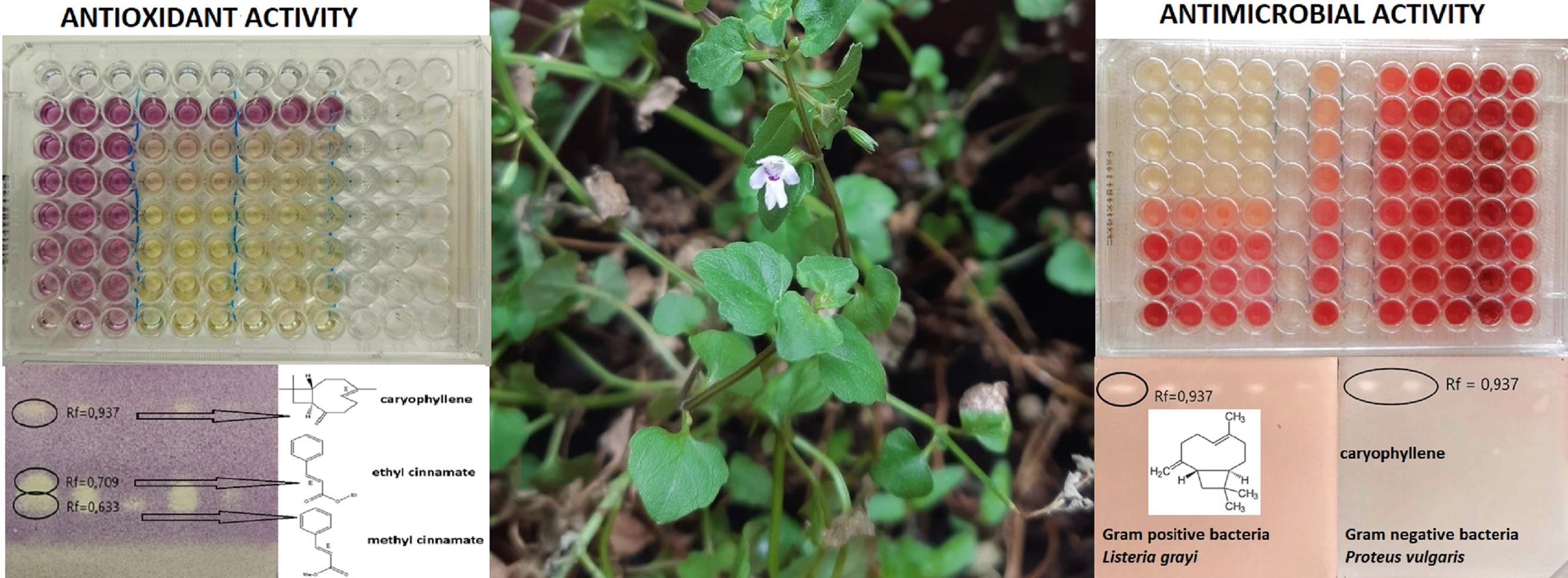
2.4. Bioautography Antioxidant

Chromatographic separation on HP-TLC plates and subsequent disclosure with DPPH reagent showed three components with antioxidant activity at the following retention factors: (Rf), 0.633, 0.709, and 0.937. Verification of their chemical identity by mass spectrometry yielded the following results: Rf 0.633 (methyl cinnamate), Rf 0.709 (ethyl cinnamate), and Rf 0.937 (caryophyllene). Figure 1 shows the individual activity with their antioxidant molecule. The mass spectrums of activity molecules can be appreciated in the Supplementary Materials (Figures S3–S5).

2.5. Antimicrobial Activity

The minimum inhibitory concentration results (MIC), evaluated in 4 Gram+ strains, 4 Gram-strains, and one yeast, are shown in Table 3 and Figure 2. The values are compared with the MIC values of T. vulgaris oil, known for its high antimicrobial activity.

2.6. Bioautography Antimicrobial

Bioautographic assays confirm the antimicrobial activity in a single molecule identified in the retention factor (Rf = 0.937), which corresponds to E-caryophyllene. Since the separation occurred in decreasing volumes of C. brownei essential oil solutions (25, 20, 15, 10, and 5 microliters), a higher activity on Gram-positive bacteria is observed. These results are shown in Figure 3.

3. Discussion

There are similarities and differences in the evaluation of the chemical composition of the essential oil. In all investigations, including this one, the percentage for pulogene and menthona is significant [29,33,34]. However, in our research, a high content of two esters derived from cinnamic acid, methyl cinnamate and ethyl cinnamate, can be observed. This suggests that the oil of this research can be considered a chemotype with a high percentage of cinnamic acid derivatives.
The antioxidant activity reported was lower than that observed in T. vulgaris oil (positive control). The only reference research on C. brownei oil, carried out in Colombia [34], proposes IC50 values between a range of 2.5 to 10 mg/mL, much less active than those found in our research, whose values are IC50 DPPH is 1.771 mg/mL, IC50 ABTS of 0.060 mg/mL. When the IC50 values are lower, the antioxidant activity is higher. This increase can be explained by the presence of cinnamic acid derivatives, which in the bioautographic assays, together with caryophyllene, proved to be the active antioxidant molecules. The high antioxidant activity of methyl cinnamate [37] and ethyl cinnamate [38] has been demonstrated in individual studies. Several studies on caryophyllene have evaluated its antioxidant, anti-inflammatory, and anticarcinogenic activity [39,40,41].
The activity in nine microorganisms shows minimum inhibitory concentration values ranging from 13.57 mg/mL for S. epidermidis to 3.11 mg/mL for C. albicans. The activity of the natural positive control is higher in all the tests; however, if values described in the scientific literature for known essential oils, such as chamomile [42], cinnamon [43], and Rosmarinus [44], are analyzed, the results are quite interesting. Comparing this research with another carried out in Venezuela, the results are quite similar for common bacteria: Staphylococcus aureus (7.921 mg/mL research in Ecuador and 6.250 mg/mL research in Venezuela); Enterococcus faecalis (5.540 mg/mL research in Ecuador and 3.125 mg/mL research in Venezuela); Escherichia coli (6.250 mg/mL research in Ecuador and 6.250 mg/mL research in Venezuela), and Pseudomonas aeruginosa (8.38 mg/mL research in Ecuador and 3.125 mg/mL research in Venezuela) [45]. Bioautography evidences the antimicrobial activity of caryophyllene; there is ample evidence of its action on this molecule, such as that performed on P. gingivalis [46]. Another research determines an inhibition value of 2.5% for B. cereus [47]. On the other hand, some essential oils in which caryophyllene is found in high concentrations have proven to have significant antimicrobial activity [48,49,50].

4. Materials and Methods

4.1. Plant Material

The plant material was collected in the parish of Rio Blanco, Morona Santiago, Ecuador, in January 2021, at the coordinates 2°28′54.0″ S and 78°09′03″ W. The plant was identified by botanist Marco Cerna, from the herbarium of Universidad Politécnica Salesiana.

4.2. Essential Oil Extraction

The extraction of the essential oil was carried out by hydrodistillation, using Clevenger equipment with 500 mL capacity. A hundred grams of fresh leaves, which were distilled for 3 h, were used in each distillation. The process was carried out in triplicate. Subsequently, the essential oil was stored in an amber bottle at a temperature of 4 °C.

4.3. GC/FID Analysis

Each individual molecule was quantified using GC/FID, triplicating each chromatographic test and calculating its relative area. A gas chromatograph Varian 3900, with FID detector, was used. The sample was prepared by diluting 10 µL of essential oil in 990 µL of dichloromethane. The operating conditions were as follows: analysis started at 45 °C, reaching 100 °C at a rate of 1 °C per minute; then, it reached 250 °C at a speed of 5 °C per minute, staying at this temperature for 15 min, for a total analysis time of 90 min. The haul gas was 99.9999% pure helium at a flow of 1 mL/min and split-ratio of 1:25. A chromatographic column Zebron ZB-5MS (5%-phenyl-95% dimethyl) polysiloxane) with a length of 30 m, a thickness of 0.25 mm and a film thickness of 0.25 m was used.

4.4. GC/MS Analysis

The first analysis was carried out with a Termo Scientific TR-5MS column (5%-phenyl-95% dimethyl) polysiloxane) with a length of 30 m, a thickness of 0.25 mm, and a film thickness of 0.25 m. A chromatograph Trace 1310 coupled to a mass spectrometer was used.
The operating conditions were as follows: analysis started at 60 °C for 5 min, reaching 100 °C at a rate of 2 °C per minute; then, it reached 200 °C at a speed of 3 °C per minute, after it reached 230 °C at a rate 5 °C per minute, staying at this temperature for 5 min, for a total analysis time of 60 min.
Mass spectrometer conditions were as follows: ionization energy: 70 eV; emission current: 10 µAmp; scan rate: 1 scan/s; mass range: 40–350 Da; trap temperature: 230 °C; transfer line temperature: 200 °C [26].
The second analysis was carried out with an Agilent DBWax column (polyethylene glycol) with a length of 20 m, a thickness of 0.10 mm, and a film thickness of 0.20 m. A chromatograph EVOQ 436 GC–TQ coupled to a mass spectrometer was used.
The operating conditions were as follows: analysis started at 50 °C for 2 min, reaching 80 °C at a rate of 5 °C per minute; then, it reached 250 °C at a speed of 5 °C per minute, staying at this temperature for 1 min, for a total analysis time of 49.50 min.
Mass spectrometer conditions were as follows: ionization energy: 70 eV; emission current: 10 µAmp; scan rate: 1 scan/s; mass range: 35–400 Da; trap temperature: 220 °C; transfer line temperature: 260 °C [51].

4.5. Identification of Compounds

The NIST 2001 Mass Spectra Database was used to identify molecules [52]. In addition, the retention index rates (RI) of each compound were calculated by comparing a series of C8–C30 alkanes, and comparing the theoretical arithmetic retention rates, contrasting them with the scientific literature of the Adams database for 5%-phenyl-95% dimethyl) polysiloxane) column [35], and the research of Babushok for Agilent DBWax column (polyethylene glycol) [36].

4.6. Antioxidant Activity, DPPH Assay

DPPH is a molecule that is already radicalized and directly allows measurement of the oxidation inhibition percentage [53]. The DPPH solution was prepared by weighing 40 mg and dissolving it in 100 mL of 96% ethanol. The essential oil of C. brownei and the natural control oil T. vulgaris were prepared by dissolving 100 µL of oil in 900 µL of absolute ethanol (99.9% purity). Butylated hydroxyanisole (BHA) was used as a positive chemical control at a concentration of 1 mg/mL. The reading was performed in a UV/VIS spectrophotometric reader for BIOTEK microplates, EPOCH model.
Using 96-well plates, the following volumes of the essential oil and BHA solutions were added to each well in increasing order: 0, 1, 2, 5, 10, 15, and 20 µL. It was added to each essential oil volume, DPPH solution to until reach 200 µL; the plates were shaken in the dark for 30 min, and their absorbance was read at a wavelength of 517 nm. The assay was performed in triplicate for each solution. The oxidation inhibition percentage was calculated according to the following equation:
%   i n h i b i t i o n   D P P H = ( b l a n k   a b s o r b a n c e S a m p l e   a b s o r b a n c e b l a n k   a b s o r b a n c e )   ×   100
With the data obtained from the calibration curve, the DPPH IC50 is calculated and compared with the controls used.

4.7. Antioxidant Activity, ABTS Assay

ABTS is a molecule that requires its radicalized form to be generated [54], which was carried out by mixing a solution of ABTS containing 1.1 mg/mL with 0.25 µL of a solution of K2S2O8 with 18.8 mg/mL concentration. The mixture was kept in the dark for 12 h, and its absorbance was adjusted to 0.700 ± 0.02. As in the DPPH assay, the essential oils of C. brownei and T. vulgaris (natural control) and BHA as a chemical control at a concentration of 1 mg/mL, were used in the assay.
The arrangement and preparation of the samples in the 96-well plates were the same as in the DPPH assay. The reading was taken 1 min after applying the radical ABTS and was determined at a wavelength of 734 nm in a UV/VIS spectrophotometer for BIOTEK microplates, EPOCH model.
The same formula and methodology, as described in the DPPH assay, Section 4.6, were used to calculate the inhibition percentages and the IC50 ABTS.

4.8. Antioxidant Bioautography

Several studies show that it is possible to identify the molecules responsible for biological activity in an essential oil using bioautographic techniques [21,26,55]. To separate the active compounds, a Merck silica gel HPTLC chromatographic plate was used, to which 25 µL of C. brownei oil dissolved in methanol at a concentration of 30 mg/mL was added. The mobile phase was composed of toluene, ethyl acetate and petroleum ether, in proportions of 97:7:20. Once the chromatographic plate was prepared, it was developed with DPPH at a concentration of 1% in ethanol, and the compounds that changed their coloration from violet to yellow were observed [21]. From these active fractions, the compounds were isolated by dissolving them in dichloromethane. The GC/MS assay was performed under the same conditions, as described in Section 4.4 for TR-5MS column.

4.9. Antimicrobial Activity

Assays were performed on 4 Gram-positive bacteria: Staphylococcus aureus ATCC 6328, Enterococcus faecalis ATCC 29212, Listeria grayi ATCC 1912, and Staphylococcus epidermidis ATCC 14990; 4 Gram-negative bacteria: Escherichia coli ATCC 25922, Proteus vulgaris ATCC 6380, Klebsiella oxytoca ATCC 8724 and Pseudomonas aeruginosa ATCC 9027; and one yeast Candida albicans ATCC 10231. The technique used to calculate the minimum inhibition concentration is known as broth micro dilution, which has been used in several studies of antimicrobial activity with essential oils [55,56].
Bacteria and yeast were reactivated in appropriate media according to growth specifications, reaching concentrations of 1 × 10⁸ CFU/mL for bacteria and 1 × 10⁶ CFU/mL for yeast. In a 96-well plate, 10 µL of C. brownei essential oil solution in DMSO (99.6%) + 150 µL of the medium with the microorganism, and 40 µL of the 2.5% TTC (Triphenyl tetrazolium chloride) dye were seeded. T. vulgaris essential oil was used as a positive control and DMSO as a negative control.
In the cases of the tested essential oils and the positive control, the concentration used is 5%, diluting it in descending ratios: 1:2, 1:4, 1:18, 1:16, 1:32, 1:64, and 1:128. Finally, plates were incubated for 24 h in agitation at a temperature of 35 ± 2 degrees Celsius for bacteria and for 48 h in agitation at a temperature of 25 ± 2 degrees Celsius for yeast. After this time, absorbance was measured at 615 nm in a UV/VIS spectrophotometer for BIOTEK microplates, EPOCH model.

4.10. Antimicrobial Bioautography

In this assay, two bacteria that needed the lowest concentrations of C. brownei essential oil to inhibit bacterial growth were selected; in this case: Listeria grayi ATCC 1912 (Gram-positive bacteria) and Proteus vulgaris ATCC 6380 (Gram-negative bacteria). The components of the essential oil were separated on a Merk silica gel HPTLC chromatographic plate; essential oil at a concentration of 30 mg/mL was incubated using a mobile phase composed of toluene, ethyl acetate and petroleum ether in proportions of 97:7:20. Disclosure was performed by covering the plate with culture medium, containing the target bacteria and the TTC dye. The plates were incubated for 24 h at 37 degrees Celsius, showing activity in those fractions that stain white [21]. Subsequently, the active fractions were extracted with dichloromethane and analyzed in a GC/MS, under the same conditions described in Section 4.4 for TR-5MS column.

4.11. Statistics

For all data collected, relative standard deviations and statistical significance (student’s test; p < 0.05), one-way ANOVA, and LSD post hoc Fisher’s honest significant difference test were taken where appropriate. All computations were collected using the statistical software STATISTICA 6.0.

5. Conclusions

The chemistry of the essential oil of C. brownei allows us to observe similarities and differences with the few studies previously carried out on the species. The most relevant result is that the Ecuadorian Amazonian variety contains high percentages of compounds derived from cinnamic acid, methyl cinnamate and ethyl cinnamate, both representing about 40% of all the oil, something that had not been observed in previous studies. Additionally, these molecules would be related to antioxidant activity as confirmed by bioautographic evaluations.
Another molecule involved in biological activity is caryophyllene, which affects both antimicrobial and antioxidant potential. This molecule has aroused pharmaceutical interest in recent times because it is a potential antimicrobial, antioxidant, anti-inflammatory, and anticarcinogenic molecule.
Based on the interesting chemical composition and biological activity, it is advisable to continue testing complementary biological activity, such as anti-inflammatory, cytotoxic, insect repellent, etc., thus having a validated biological resource that could be used as an alternative to treat various diseases and symptoms.

Supplementary Materials

The following supporting information can be downloaded at: https://www.mdpi.com/article/10.3390/molecules28041741/s1, Figure S1 Chromatogram used Zebron ZB-5MS column, Figure S2 Chromatogram used Agilent DBWax column, Figure S3 methyl cinnamate mass spectrum, Figure S4 ethyl cinnamate mass spectrum and Figure S5 caryophyllene mass spectrum.

Author Contributions

Conceptualization, P.N.; methodology, L.C. and A.O.; software, L.C. and A.O.; validation, P.N. and E.P.; formal analysis, L.C. and A.O.; investigation, P.N.; resources, P.N.; data curation, P.N. and E.P.; writing—original draft preparation, P.N.; writing—review and editing, P.N.; visualization, P.N.; supervision, E.P.; project administration, E.P.; funding acquisition, P.N. All authors have read and agreed to the published version of the manuscript.

Funding

This research received no external funding.

Institutional Review Board Statement

Not applicable.

Informed Consent Statement

Not applicable for studies not involving humans.

Data Availability Statement

Data are available in the manuscript.

Acknowledgments

Life Science Laboratories, Universidad Politécnica Salesiana.

Conflicts of Interest

The authors declare no conflict of interest.

Sample Availability

Samples of the compounds are not available from the authors.

References

  1. Cuesta, F.; Peralvo, M.; Merino-Viteri, A.; Bustamante, M.; Baquero, F.; Freile, J.; Muriel, P.; Torres-Carvajal, O. Priority areas for biodiversity conservation in mainland Ecuador. Neotrop. Biodivers. 2017, 3, 93–106. [Google Scholar] [CrossRef]
  2. Sierra, R.; Campos, F.; Chamberlin, J. Assessing biodiversity conservation priorities: Ecosystem risk and representativeness in continental Ecuador. Landsc. Urban Plan. 2002, 59, 95–110. [Google Scholar] [CrossRef]
  3. Mestanza-Ramón, C.; Henkanaththegedara, S.M.; Vásconez Duchicela, P.; Vargas Tierras, Y.; Sánchez Capa, M.; Constante Mejía, D.; Mestanza Ramón, P. In-situ and ex-situ biodiversity conservation in Ecuador: A review of policies, actions and challenges. Diversity 2020, 12, 315. [Google Scholar] [CrossRef]
  4. De la Torre, L.; Navarrete, H.; Macía, M.; Balslev, H.; Muriel, P. Enciclopedia de las Plantas Útiles del Ecuador (con Extracto de Datos), 1st ed.; Herbario QCA de la Escuela de Ciencias Biológicas de la Pontificia Universidad Católica del Ecuador & Herbario AAU del Departamento de Ciencias Biológicas de la Universidad de Aarhus: Quito, Ecuador, 2008. [Google Scholar]
  5. Giovannini, P. Medicinal plants of the Achuar (Jivaro) of Amazonian Ecuador: Ethnobotanical survey and comparison with other Amazonian pharmacopoeias. J. Ethnopharmacol. 2015, 164, 78–88. [Google Scholar] [CrossRef] [PubMed]
  6. Ballesteros, J.L.; Bracco, F.; Cerna, M.; Vita Finzi, P.; Vidari, G. Ethnobotanical research at the Kutukú Scientific Station, Morona-Santiago, Ecuador. BioMed Rev. Int. 2016, 2016, 9105746. [Google Scholar] [CrossRef]
  7. Cerón, C.; Montalvo, C.; Calazacón, A. Etnobotánica Tsáchila, Pichincha-Ecuador. Cinchonia 2004, 5, 109–194. [Google Scholar]
  8. Velásquez, G.; Moreira, W.; Caicedo, C.; Preciado, O.; Jaramillo, T.; Iturralde, G.; Aizaga, G. El uso de la etnobotánica como cura de eruptivas de la infancia por la etnia chachi. Esmeraldas. Ecuador. Rev. Investig. Talentos 2016, 3, 1–10. [Google Scholar]
  9. Cerón, C.; Reyes, C. Parches de bosque y etnobotánica Shuar en Palora, Morona Santiago-Ecuador. Cinchonia 2007, 8, 66–83. [Google Scholar]
  10. Zhiñin, H.R.; Poma, B.V.; González, L.P.; Quito, G.B. Etnobotánica y derechos de la naturaleza en el aja shuar: Caso de estudio parroquia Nankais, cantón Nangaritza, provincia Zamora Chinchipe, Ecuador. Siembra 2021, 8, 1–13. [Google Scholar] [CrossRef]
  11. Cerón, C. Etnobotánica Huaorani de Tivacuno-Tiputini Parque Nacional Yasuní. Cinchonia 2002, 3, 64–94. [Google Scholar]
  12. Noriega, P. Extracción, química, actividad biológica, control de calidad y potencial económico de los aceites esenciales. La Granja 2009, 10, 3–13. [Google Scholar] [CrossRef]
  13. Marriott, P.J.; Shellie, R.; Cornwell, C. Gas chromatographic technologies for the analysis of essential oils. J. Chromatogr. A 2001, 936, 1–22. [Google Scholar] [CrossRef] [PubMed]
  14. Baptista, S.; Borges, S.; Ramos, O.L.; Pintado, M.; Sarmento, B. The progress of essential oils as potential therapeutic agents: A review. J. Essent. Oil Res. 2020, 32, 279–295. [Google Scholar] [CrossRef]
  15. Aziz, Z.A.; Ahmad, A.; Setapar, S.H.; Karakucuk, A.; Azim, M.M.; Lokhat, D.; Ashraf, G.M. Essential oils: Extraction techniques, pharmaceutical and therapeutic potential-a review. Curr. Drug Metab. 2018, 19, 1100–1110. [Google Scholar] [CrossRef]
  16. Diniz do Nascimento, L.; Moraes, A.A.; Costa, K.S.; Pereira, J.M.; Taube, P.S.; Costa, C.M.; Faria, L.J. Bioactive natural compounds and antioxidant activity of essential oils from spice plants: New findings and potential applications. Biomolecules 2020, 10, 988. [Google Scholar] [CrossRef]
  17. Maccioni, A.M.; Anchisi, C.; Sanna, A.; Sardu, C.; Dessi, S. Preservative systems containing essential oils in cosmetic products. Int. J. Cosmet. Sci. 2002, 24, 53–59. [Google Scholar] [CrossRef]
  18. Vankar, P.S. Essential oils and fragrances from natural sources. Resonance 2004, 9, 30–41. [Google Scholar] [CrossRef]
  19. Bruni, R.; Medici, A.; Andreotti, E.; Fantin, C.; Muzzoli, M.; Dehesa, M.; Sacchetti, G. Chemical composition and biological activities of Ishpingo essential oil, a traditional Ecuadorian spice from Ocotea quixos (Lam.) Kosterm. (Lauraceae) flower calices. Food Chem. 2004, 85, 415–421. [Google Scholar] [CrossRef]
  20. Sacchetti, G.; Guerrini, A.; Noriega, P.; Bianchi, A.; Bruni, R. Essential oil of wild ocotea quixos (lam.) kosterm. (lauraceae) leaves from amazonian Ecuador. Flavour Fragr. J. 2006, 21, 674–676. [Google Scholar] [CrossRef]
  21. Noriega, P.; Mosquera, T.; Paredes, E.; Parra, M.; Zappia, M.; Herrera, M.; Osorio, E. Antimicrobial and antioxidant bioautography activity of bark essential oil from Ocotea quixos (Lam.) kosterm. J. Planar Chromatogr.-Mod. TLC 2018, 31, 163–168. [Google Scholar] [CrossRef]
  22. Guerrini, A.; Sacchetti, G.; Rossi, D.; Paganetto, G.; Muzzoli, M.; Andreotti, E.; Bruni, R. Bioactivities of Piper aduncum L. and Piper obliquum Ruiz & Pavon (Piperaceae) essential oils from eastern Ecuador. Environ. Toxicol. Pharmacol. 2009, 27, 39–48. [Google Scholar] [PubMed]
  23. Scalvenzi, L.; Radice, M.; Toma, L.; Severini, F.; Boccolini, D.; Bella, A.; Di Luca, M. Larvicidal activity of Ocimum campechianum, Ocotea quixos and Piper aduncum essential oils against Aedes aegypti. Parasite 2019, 26, 23. [Google Scholar] [CrossRef]
  24. Ballesteros, J.L.; Tacchini, M.; Spagnoletti, A.; Grandini, A.; Paganetto, G.; Neri, L.M.; Sacchetti, G. Rediscovering medicinal Amazonian aromatic plants: Piper carpunya (Piperaceae) essential oil as paradigmatic study. Evid. Based Complement. Altern. Med. 2019, 2019, 6194640. [Google Scholar] [CrossRef] [PubMed]
  25. Valarezo, E.; Rivera, J.X.; Coronel, E.; Barzallo, M.A.; Calva, J.; Cartuche, L.; Meneses, M.A. Study of volatile secondary metabolites present in Piper carpunya leaves and in the traditional ecuadorian beverage Guaviduca. Plants 2021, 10, 338. [Google Scholar] [CrossRef] [PubMed]
  26. Noriega, P.; Guerrini, A.; Sacchetti, G.; Grandini, A.; Ankuash, E.; Manfredini, S. Chemical composition and biological activity of five essential oils from the Ecuadorian Amazon rain forest. Molecules 2019, 24, 1637. [Google Scholar] [CrossRef]
  27. Noriega, P.; Mosquera, T.; Osorio, E.; Guerra, P.; Fonseca, A. Clinopodium nubigenum (Kunth) Kuntze essential oil: Chemical composition, antioxidant activity, and antimicrobial test against respiratory pathogens. J. Pharmacogn. Phytother. 2018, 10, 149–157. [Google Scholar]
  28. Salto, M.; Puente, B.; Malafronte, N.; Braca, A. Phenolic compounds from Clinopodium tomentosum (Kunth) govaerts (Lamiaceae). J. Braz. Chem. Soc. 2014, 25, 2121–2124. [Google Scholar]
  29. Matailo, A.; Bec, N.; Calva, J.; Ramírez, J.; Andrade, J.M.; Larroque, C.; Vidari, G.; Armijos, C. Selective BuChE inhibitory activity, chemical composition, and enantiomer content of the volatile oil from the Ecuadorian plant Clinopodium brownei. Rev. Bras. Farmacogn. 2019, 29, 749–754. [Google Scholar] [CrossRef]
  30. Armijos, C.; Ramírez, J.; Salinas, M.; Vidari, G.; Suárez, A.I. Pharmacology and Phytochemistry of Ecuadorian Medicinal Plants: An Update and Perspectives. Pharmaceuticals 2021, 14, 1145. [Google Scholar] [CrossRef]
  31. Vandebroek, I.; Picking, D. Clinopodium brownei (Sw.) Kuntze (Lamiaceae). In Popular Medicinal Plants in Portland and Kingston, Jamaica, 1st ed.; Springer: Cham, Switzerland; Kingston: Fantinwali, CA, USA, 2020; pp. 89–94. [Google Scholar]
  32. Ansaloni, R.; Wilches, I.; León, F.; Peñaherrera, E.; Orellana, A.; Tobar, V.; De Witte, P. Estudio preliminar sobre plantas medicinales utilizadas en algunas comunidades de las provincias de Azuay, Cañar y Loja, para afecciones del aparato gastrointestinal. Rev. Tecnológica-ESPOL 2010, 23, 89–97. [Google Scholar]
  33. Rojas, L.B.; Usubillaga, A. Composition of the essential oil of Satureja brownei (SW.) Briq. from Venezuela. Flavour Fragr. J. 2000, 15, 21–22. [Google Scholar] [CrossRef]
  34. Jaramillo, B.E.; Stashenko, E.; Martínez, J.R. Composición química volátil de Satureja brownei (Sw.) Briq. colombiana y determinación de su actividad antioxidante. Rev. Cuba. Plantas Med. 2010, 15, 52–63. [Google Scholar]
  35. Adams, R.P. Identification of Essential Oils by Ion Trap Mass Spectroscopy, 1st ed.; Academic Press: San Diego, CA, USA, 2012. [Google Scholar]
  36. Babushok, V.I.; Linstrom, P.J.; Zenkevich, I.G. Retention Indices for Frequently Reported Compounds of Plant Essential Oils. J. Phys. Chem. Ref. Data 2011, 40, 043101–043102. [Google Scholar] [CrossRef]
  37. Suryanti, V.; Wibowo, F.R.; Khotijah, S.; Andalucki, N. Antioxidant activities of cinnamaldehyde derivatives. In IOP Conference Series: Materials Science and Engineering; IOP Publishing: Surakarta, Indonesia, 2018. [Google Scholar]
  38. Oladimeji, O.H.; Essien, A.E.; Sheriff, A.J.; Silaev, T.E. Evaluation of antioxidant activity of cinnamic acid and some of its derivatives. Eur. Chem. Bull. 2019, 8, 224–226. [Google Scholar] [CrossRef]
  39. Dahham, S.S.; Tabana, Y.M.; Iqbal, M.A.; Ahamed, M.B.; Ezzat, M.O.; Majid, A.S.; Majid, A.M. The anticancer, antioxidant and antimicrobial properties of the sesquiterpene β-caryophyllene from the essential oil of Aquilaria crassna. Molecules 2015, 20, 11808–11829. [Google Scholar] [CrossRef] [PubMed]
  40. Calleja, M.A.; Vieites, J.M.; Montero-Meterdez, T.; Torres, M.I.; Faus, M.J.; Gil, A.; Suárez, A. The antioxidant effect of β-caryophyllene protects rat liver from carbon tetrachloride-induced fibrosis by inhibiting hepatic stellate cell activation. Br. J. Nutr. 2013, 109, 394–401. [Google Scholar] [CrossRef]
  41. Gushiken, L.F.; Beserra, F.P.; Hussni, M.F.; Gonzaga, M.T.; Ribeiro, V.P.; De Souza, P.F.; Pellizzon, C.H. Beta-caryophyllene as an antioxidant, anti-inflammatory and re-epithelialization activities in a rat skin wound excision model. Oxid. Med. Cell. Longevity. 2022, 2022, 9004014. [Google Scholar] [CrossRef] [PubMed]
  42. Aguilar, I.E.; Galviz, R.A. Determinación de la Composición Química y Actividad Antibacteriana del Aceite Esencial extraído de la planta Satureja brownei (Briq.) cultivada en el Jardín de plantas Medicinales “Dr. Luis Ruiz Terán” de la Facultad de Farmacia y Bioanálisis, Universidad de los Andes. Bachelor’s Thesis, Universidad de los Andes, Mérida, Venezuela, 2015. [Google Scholar]
  43. Yoo, H.J.; Jwa, S.K. Efficacy of β-caryophyllene for periodontal disease related factors. Arch. Oral. Biol. 2019, 100, 113–118. [Google Scholar] [CrossRef]
  44. Moo, C.L.; Yang, S.K.; Osman, M.A.; Yuswan, M.H.; Loh, J.Y.; Lim, W.M.; Lai, K.S. Antibacterial Activity and Mode of Action of β-caryophyllene on. Pol. J. Microbiol. 2020, 69, 49–54. [Google Scholar] [CrossRef]
  45. Mahdavi, B.; Ghorat, F.; Nasrollahzadeh, M.S.; Hosseyni-Tabar, M.; Rezaei-Seresht, H. Chemical composition, antioxidant, antibacterial, cytotoxicity, and hemolyses activity of essential oils from flower of Matricaria chamomilla var. chamomilla. Anti-Infect. Agents 2020, 18, 224–232. [Google Scholar] [CrossRef]
  46. El-Baroty, G.S.; Abd El-Baky, H.H.; Farag, R.S.; Saleh, M.A. Characterization of antioxidant and antimicrobial compounds of cinnamon and ginger essential oils. Afr. J. Biochem. Res. 2010, 4, 167–174. [Google Scholar]
  47. Garzoli, S.; Laghezza Masci, V.; Franceschi, S.; Tiezzi, A.; Giacomello, P.; Ovidi, E. Headspace/GC–MS analysis and investigation of antibacterial, antioxidant and cytotoxic activity of essential oils and hydrolates from Rosmarinus officinalis L. and Lavandula angustifolia Miller. Foods 2021, 10, 1768. [Google Scholar] [CrossRef]
  48. Sabulal, B.; Dan, M.; Kurup, R.; Pradeep, N.S.; Valsamma, R.K.; George, V. Caryophyllene-rich rhizome oil of Zingiber nimmonii from South India: Chemical characterization and antimicrobial activity. Phytochemistry 2006, 67, 2469–2473. [Google Scholar] [CrossRef] [PubMed]
  49. Novak, J.; Zitterl-Eglseer, K.; Deans, S.G.; Franz, C.M. Essential oils of different cultivars of Cannabis sativa L. and their antimicrobial activity. Flavour Fragr. J. 2001, 16, 259–262. [Google Scholar] [CrossRef]
  50. Montanari, R.M.; Barbosa, L.C.; Demuner, A.J.; Silva, C.J.; Carvalho, L.S.; Andrade, N.J. Chemical composition and antibacterial activity of essential oils from Verbenaceae species: Alternative sources of (E)-caryophyllene and germacrene-D. Quim. Nova 2011, 34, 1550–1555. [Google Scholar] [CrossRef]
  51. Balcerzak, L.; Gibka, J.; Sikora, M.; Kula, J.; Strub, D.J. Minor constituents of essential oils and aromatic extracts. Oximes derived from natural flavor and fragrance raw materials–Sensory evaluation, spectral and gas chromatographic characteristics. Food Chem. 2019, 301, 125283. [Google Scholar] [CrossRef]
  52. NIST/02, Mass Spectral Library; United States Government: Washington, DC, USA, 2001. Available online: https://chemdata.nist.gov (accessed on 22 December 2022).
  53. Sirivibulkovit, K.; Nouanthavong, S.; Sameenoi, Y. Based DPPH assay for antioxidant activity analysis. Anal. Sci. 2018, 34, 795–800. [Google Scholar] [CrossRef]
  54. Re, R.; Pellegrini, N.; Proteggente, A.; Pannala, A.; Yang, M.; Rice-Evans, C. Antioxidant activity applying an improved ABTS radical cation decolorization assay. Free Radical Biol. Med. 1999, 26, 1231–1237. [Google Scholar] [CrossRef]
  55. Noriega, P.; Ballesteros, J.; De la Cruz, A.; Veloz, T. Chemical composition and preliminary antimicrobial activity of the hydroxylated sesquiterpenes in the essential oil from Piper barbatum Kunth leaves. Plants 2020, 9, 211. [Google Scholar] [CrossRef] [PubMed] [Green Version]
  56. Vanegas, D.; Abril-Novillo, A.; Khachatryan, A.; Jerves-Andrade, L.; Peñaherrera, E.; Cuzco, N.; Wilches, I.; Calle, J.; León, F. Validation of a method of broth microdilution for the determination of antibacterial activity of essential oils. BMC Res. Notes 2021, 14, 1–7. [Google Scholar] [CrossRef]
Figure 1. DPPH Bioautographic assay of Clinopodium brownei essential oil, bioactive molecules: Rf 0.633 methyl cinnamate, Rf 0.709 ethyl cinnamate and Rf 0.937 caryophyllene.
Molecules 28 01741 g001
Figure 2. Graphical comparison of minimum inhibitory concentration (MIC) values between the essential oils of C. brownei and T. vulgaris (natural control). Mean ± SD (n = 3), p < 0.05.
Molecules 28 01741 g002
Figure 3. Bioautographic antibacterial assay of Clinopodium brownei essential oil, bioactive molecule Rf 0.937 caryophyllene.
Molecules 28 01741 g003
Table 1. Chemical composition of the essential oil of Clinopodium brownie, using two chromatographic systems, with the polar and non-polar columns.
Components
(Non-Polar Column)
TR5MS RI cal aRI Lit b%RDAComponents
(Polar Column)
DB5-Wax RI cal cRT Lit d% RDA
3-octanone9929790.243-octanone126312550.22
menthone116411487.512-pentanone,
4-hydroxy-4-methyl
1371-1.42
iso-isopulegol117211591.18menthone147114658.04
neoiso.pulegol117611670.49α-copaene149614913.07
pulegone1252123320.76β-elemene159415910.64
α-copaene137613742.87(E)-caryophyllene1602159910.0
(E)-methyl cinnamate1401137616.68pulegone1653165529.9
(E)-caryophyllene142114178.17β-humulene167216671.96
(E)-ethyl cinnamate1465144321.4γ-gurjunene169216680.49
β-humulene145814362.17α-terpinyl acetate169716950.31
γ-gurjunene148714750.81β-selinene172217176.46
β-selinene149314897.92α-selinene172617251.46
α-selinene149914982.12α-panasinsene1764-0.34
δ-selinene149514920.31Caryophyllene oxide19861986-
spathulenol158515770.33(E)-cinnamaldehyde203120330.93
Caryophyllene oxide158915822.33(E)-mehtyl cinnamate2056207513.82
benzyl benzoate177217590.17(E)-ethyl cinnamate2110-18.75
Total amount of identified compounds 98.52 98.35
a Calculated retention index on a DB-5MS capillary column; b Retention indices on a DB-5MS column from reference (Adams 2012) [35]; c Calculated retention indices on a HP-INNOWax capillary column; d Retention indices on a HP-INNOWax column from reference (Babushok 2011) [36].
Table 2. Antioxidant activity as IC50 DPPH and IC50 ABTS of C. brownei essential oil, natural control T. vulgaris essential oil, and (BHA) chemical control. Mean ± SD (n = 3), p < 0.05.
Essential Oils/Antioxidant MoleculeDPPH IC50 (mg/mL)ABTS IC50 (mg/mL)
Clinopodium brownei1.771 ± 0.2600.060 ± 0.002
Thymus vulgaris0.010 ± 0.0050.008 ± 0.002
butylated hydroxyanisole (BHA)0.004 ± 0.0000.002 ± 0.000
Table 3. MIC values of C. brownei essential oil and T. vulgaris essential oil (natural control) [22] on various microorganisms. Mean ± SD (n = 4), p < 0.05.
MicroorganismC. brownie
MIC (mg/mL)
T. vulgaris
MIC (mg/mL)
Gram Positive Bacteria
Staphylococcus aureus ATCC 63287.92 ± 0.213.02 ± 0.16
Enterococcus faecalis ATCC 292125.54 ± 0.252.89 ± 0.14
Listeria grayi ATCC 19123.84 ± 0.162.28 ± 0.11
Staphylococcus epidermidis ATCC 1499013.57 ± 0.217.98 ± 0.37
Gram negative Bacteria
Escherichia coli ATCC 259226.22 ± 0.162.13 ± 0.09
Proteus vulgaris ATCC 63804.62 ± 0.182.77 ± 0.08
Klebsiella oxytoca ATCC 87247.19 ± 0.351.30 ± 0.05
Pseudomonas aeruginosa ATCC 90278.38 ± 0.413.10 ± 0.12
Yeast
Candida albicans ATCC 102313.11 ± 0.101.45 ± 0.07
Disclaimer/Publisher’s Note: The statements, opinions and data contained in all publications are solely those of the individual author(s) and contributor(s) and not of MDPI and/or the editor(s). MDPI and/or the editor(s) disclaim responsibility for any injury to people or property resulting from any ideas, methods, instructions or products referred to in the content.

