Article

Effects of Dietary Ferulic Acid on Intestinal Health and Ileal Microbiota of Tianfu Broilers Challenged with Lipopolysaccharide

1
Department of Basic Veterinary Medicine, Sichuan Agricultural University, Chengdu 611130, China
2
College of Animal Science and Technology, Sichuan Agricultural University, Chengdu 611130, China
3
Center for Veterinary Sciences, Zhejiang University, Hangzhou 310030, China
4
Department of Small Animal Clinical Science, Virginia-Maryland College of Veterinary Medicine, Blacksburg, VA 24062, USA
5
Farm Animal Genetic Resources Exploration and Innovation Key Laboratory of Sichuan Province, Sichuan Agricultural University, Chengdu 611130, China
*
Author to whom correspondence should be addressed.
These authors contributed equally to this work.
Molecules 2023, 28(4), 1720; https://doi.org/10.3390/molecules28041720
Submission received: 1 January 2023 / Revised: 9 February 2023 / Accepted: 9 February 2023 / Published: 10 February 2023
(This article belongs to the Special Issue Bioactive Compounds: From Extraction to Biological Evaluations)

Abstract

:
Lipopolysaccharide (LPS) has been considered the primary agent to establish animal models of inflammation, immunological stress, and organ injury. Previous studies have demonstrated that LPS impaired gastrointestinal development and disrupted intestinal microbial composition and metabolism. Ferulic acid (FA) isolated from multiple plants exhibits multiple biological activities. This study investigated whether FA ameliorated intestinal function and microflora in LPS-challenged Tianfu broilers. The results showed that LPS challenge impaired intestinal function, as evidenced by decreased antioxidant functions (p < 0.05), disrupted morphological structure (p < 0.05), and increased intestinal permeability (p < 0.05); however, these adverse effects were improved by FA supplementation. Additionally, FA supplementation preserved sIgA levels (p < 0.05), increased mRNA expression levels of CLDN and ZO-1 (p < 0.05), and enhanced epithelial proliferation (p < 0.05) in the ileal mucosa in LPS-challenged chickens. Moreover, FA supplementation rectified the ileal microflora disturbances in the LPS-challenged broilers. The results demonstrate that dietary FA supplementation decreased LPS-induced intestinal damage by enhancing antioxidant capacity and maintaining intestinal integrity. Furthermore, FA supplementation protects intestinal tight junctions (TJs), elevates secretory immunoglobulin A (sIgA) levels, and modulates ileal microflora composition in LPS-challenged broilers.

1. Introduction

Broilers are raised under an intensive husbandry environment in commercial poultry farms, which may increase the risk of ingesting various toxins, such as mycotoxin, bacteriotoxin, and zootoxin. Lipopolysaccharide (LPS) is a crucial bacterial endotoxin produced by most gram-negative bacteria [1]. It has been considered the primary agent to establish animal models of inflammation, immunological stress, and organ injury [2,3,4,5,6,7]. Moreover, the accumulation of excessive free radicals [8] induced by LPS damaged the intestinal barrier [9] and decreased the growth performance of broilers [10] by triggering inflammation of the gastrointestinal tract [11], initiating oxidative damage and apoptosis of intestinal epithelial cells [12,13], and disrupting intestinal microbial composition and metabolism [14,15].
A number of studies indicated active plant components enhanced growth performance and combated various diseases, therefore, had great potential for generating revenue in the poultry industry [16,17,18,19]. An active ingredient of phenolic, ferulic acid (FA, 4-hydroxy-3-methoxycinnamic acid), is isolated from multiple plants and has been recognized as an antioxidant. A series of recent studies indicated that FA shows low toxicity [20] and possesses varieties of biological activities, including antibacterial [21], antioxidant [22], anti-inflammatory [23], antithrombotic [24], and anticancer [25] properties. Additionally, it has been reported that FA supplementation improves the immune functions and growth performance in male adult zebrafish by elevating the number of intestinal goblet cells and ameliorating the intestinal microbiota composition [26]. In piglets, dietary FA supplementation exhibited a similar effect by promotion of Claudin-1 (CLDN-1) and Occludin (OLDN) expression, increasing the Firmicutes/Bacteroidetes ratio (F/B), the abundance of the Lachoiraceaea family, and reduction in the abundance of the Prevotellaceae family in the cecum [27]. Another example of this is that FA reduces atherosclerotic injury by regulating lipid metabolism and intestinal microbiota [28]. However, information with regard to the mechanism of action and how FA protects the intestinal health and microbial composition of broilers remains unreported.
On account of the multiple benefits on intestinal FA, this study was carried out to explore whether dietary 100 mg/kg FA supplementation ameliorates the intestinal injury and intestinal microflorae of broilers challenged with LPS.

2. Results

2.1. Intestinal Morphological Analysis

Figure 1 represents the morphological characteristics of the ileum among groups. The ileal VH and CD in the LPS group were significantly lower (p < 0.05) than in the CON group. While the contrary, it increased significantly (p < 0.05) with dietary FA supplementation. Birds in the FL group had the highest VH/CD (p < 0.05) among the groups. Similarly, LPS challenge significantly decreased (p < 0.05) duodenal VH and VH/CD, while significantly increasing (p < 0.05) the CD in the duodenum and jejunum, which were modified by FA supplementation (Supplementary Materials, Figure S1).

2.2. Intestinal Permeability Biochemical Analysis

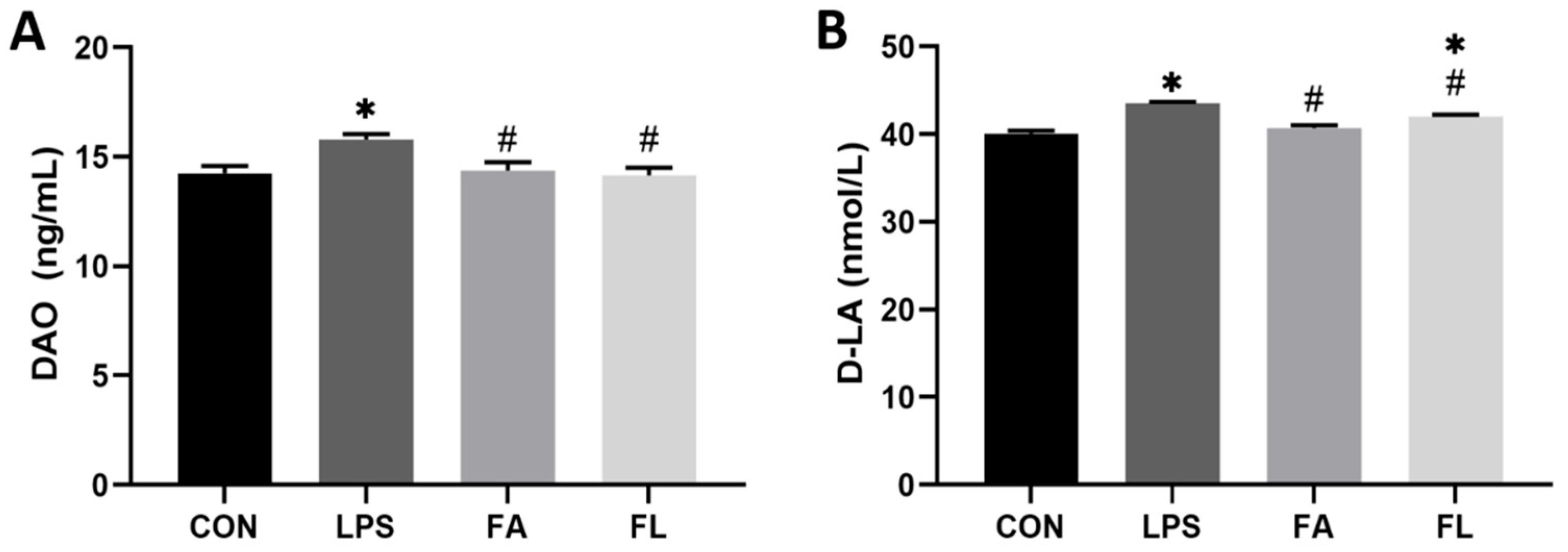
Figure 2 shows the parameters indicating intestinal permeability. The levels of DAO and D-LA in the serum were significantly increased (p < 0.05) after LPS challenge; however, FA significantly decreased the serum DAO activity and D-LA levels (p < 0.05) in chickens challenged with LPS.

2.3. Antioxidant Parameters of Intestinal Mucosa

The levels of antioxidants in intestinal mucosa were examined and shown in Figure 3. The LPS group had significantly lower levels of SOD, T-AOC, and GSH in the mucosa than the CON group (p < 0.05). Conversely, levels of SOD, T-AOC, and GSH were significantly elevated (p < 0.05) in the FL group when compared to the LPS group.

2.4. sIgA Content in Ileal Mucosa

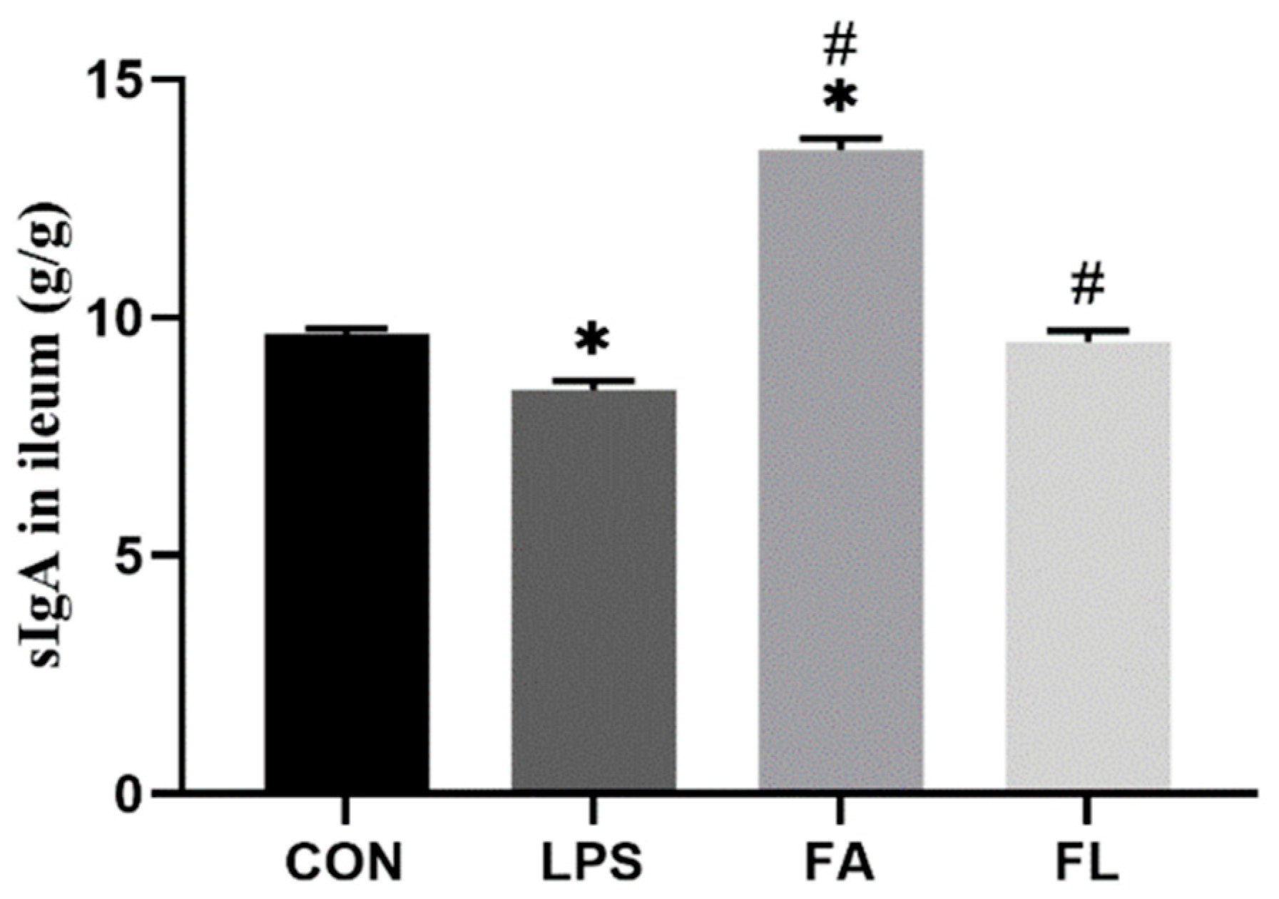
Figure 4 shows that ileal sIgA contents in LPS-challenged chickens were the lowest (p < 0.05); however, dietary FA supplementation increased (p < 0.05) the ileal sIgA contents in the FL group compared to the LPS group.

2.5. Life Cycle of the Ileal Epithelium

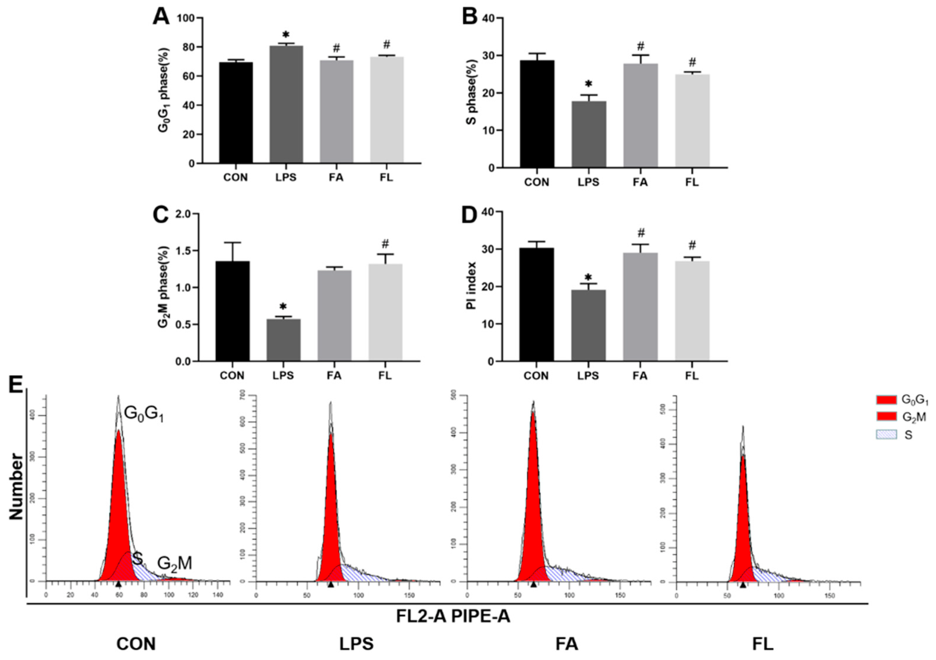
Figure 5 shows an increased ileal epithelium percentage in the G0/G1 phase (p < 0.05), a reduced percentage of cells in the S and G2M phases, and a lower PI index in the LPS group (p < 0.05) compared to the CON group. However, more ileal epithelium entered the S and G2M phase (p < 0.05) in the FL group compared to the LPS group.

2.6. Relative mRNA Expressions of Intestinal Tight Junction Proteins

Figure 6 depicts the relative mRNA expressions of intestinal TJs. The OCLN expression did not significantly change, but the CLDN-1 and ZO-1 decreased significantly (p < 0.05) in the LPS group when compared to the CON group. Significant upregulations (p < 0.05) of CLDN-1 and ZO-1 were noticed in the FL group compared to the LPS group.

2.7. Ileal Microbiota Composition

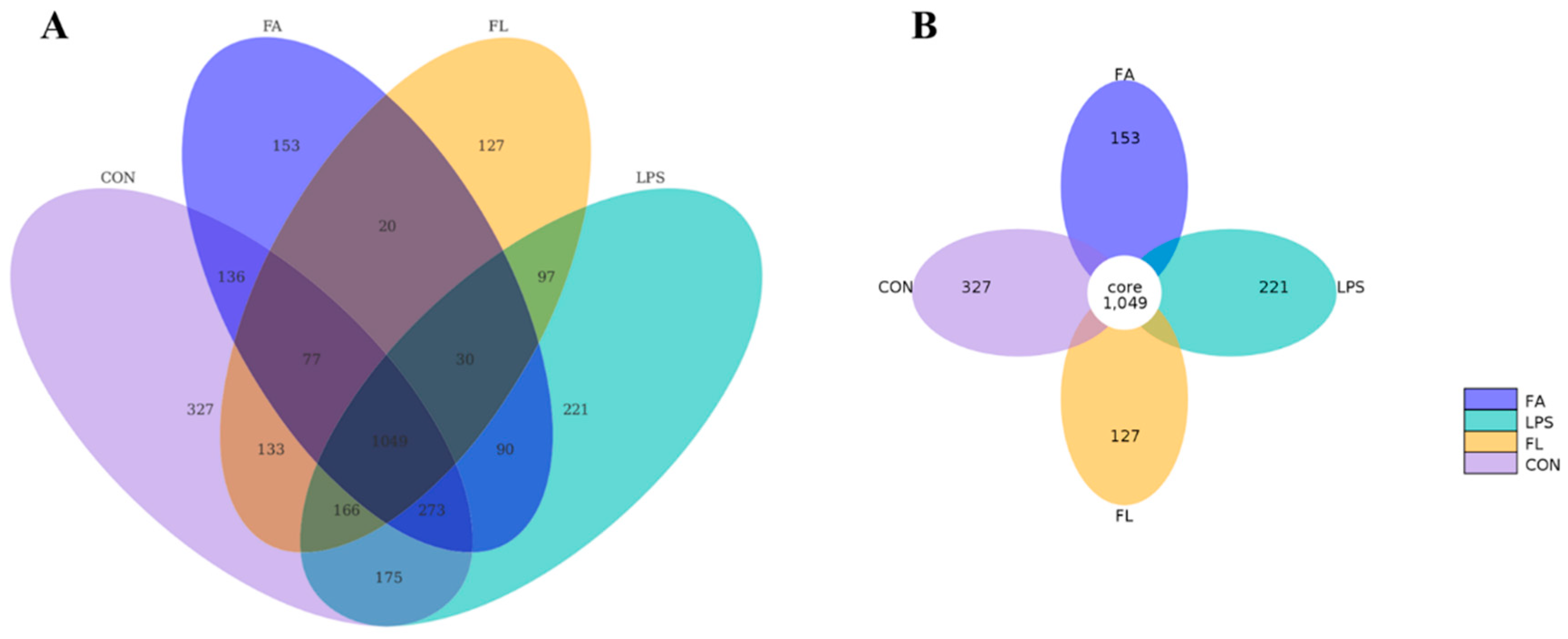
The Venn diagram (Figure 7A) and flower diagram (Figure 7B) showed the unique and shared OTUs of the different microbiome groups in the ileum. The diagrams illustrate that 1049 OTUs (the core) were shared by four groups. In total, 327, 221, 153, and 127 unique OTUs were discovered in the CON, LPS, FA, and FL groups, respectively (Figure 7B). Interestingly, chickens in the LPS and FL groups shared fewer OTUs (1342) than those in the CON and LPS groups (1663). Among these unique OTUs, 175 OTUs were discovered in both the CON and LPS groups; 136 OTUs were discovered in both the CON and FA groups; 133 OTUs were discovered in both the CON and FL groups; 90 OTUs were discovered in both the LPS and FA groups; and 97 OTUs were discovered in both the LPS and FL groups.
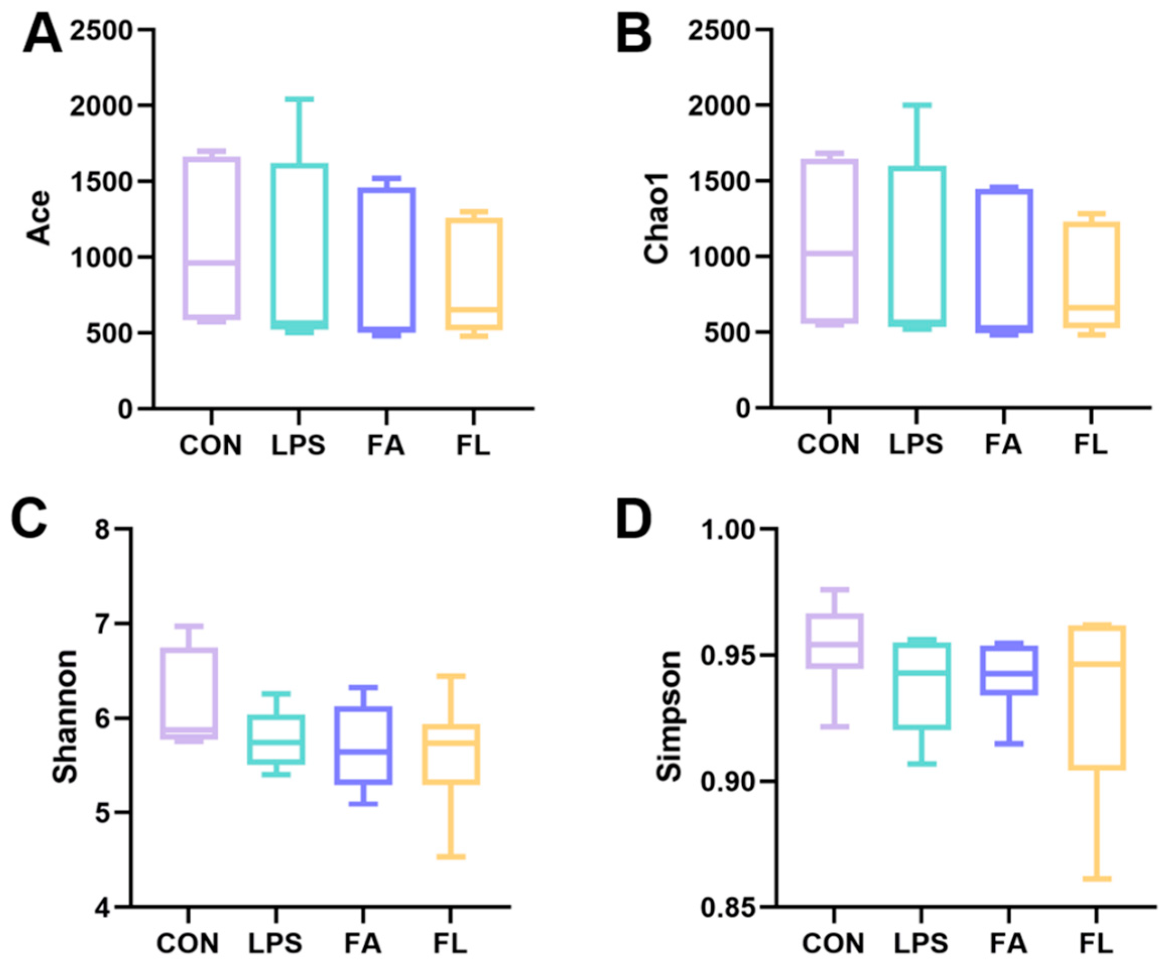
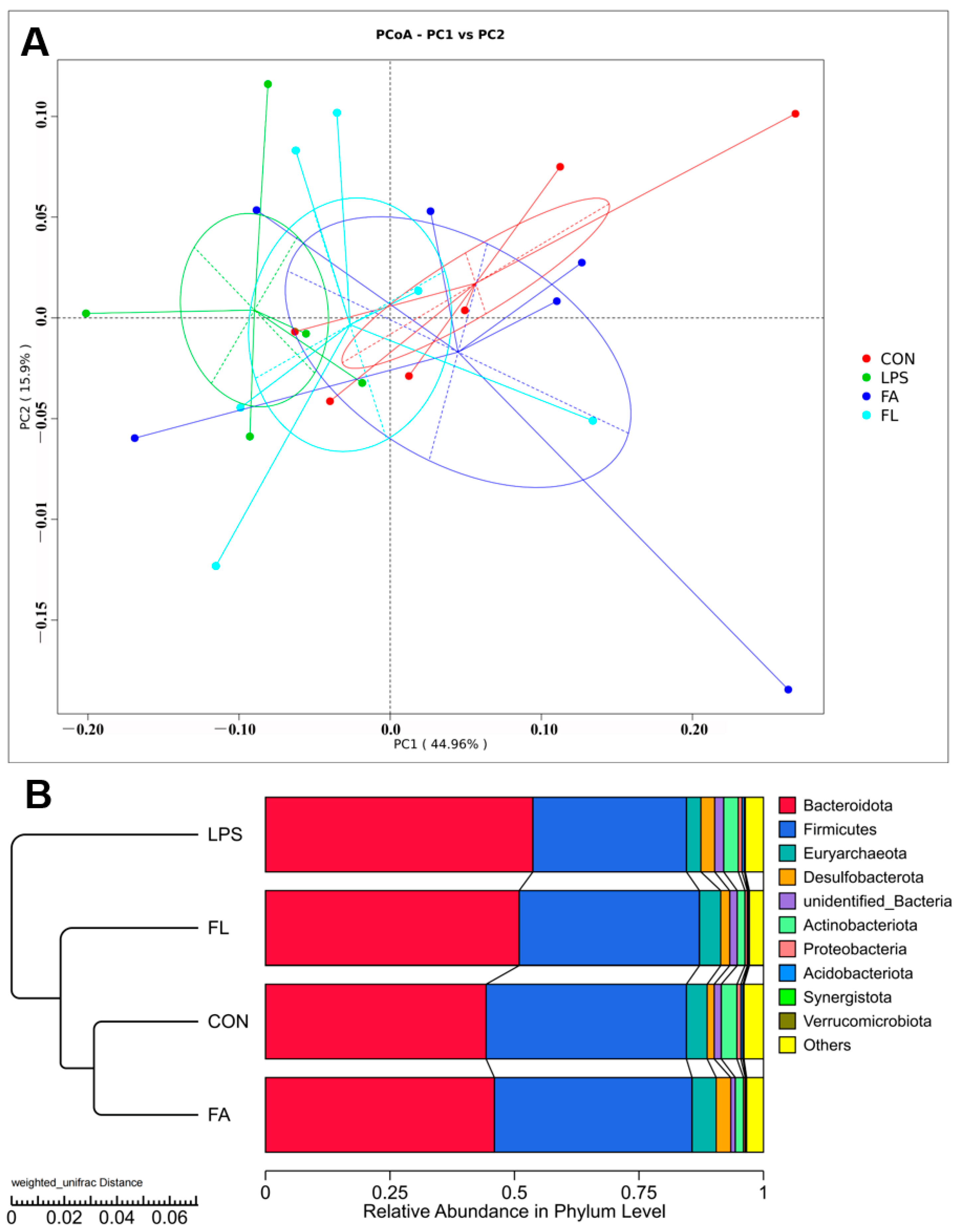
The alpha diversity (Figure 8), a parameter that evaluates diversity and uniformity of the bacterial species, of the ileal microbiota was not significantly (p > 0.05) different among the four groups. The ileal microbiota was obviously different among the four groups (PC1, 44.96%; PC2, 15.9%) based on the results of principal coordinate analysis (PCoA) with weighted unifrac distances (Figure 9A). Furthermore, the results (Figure 9B) indicated that the FL group had a similar ileal microbiota composition to the CON and FA groups compared to the LPS group.
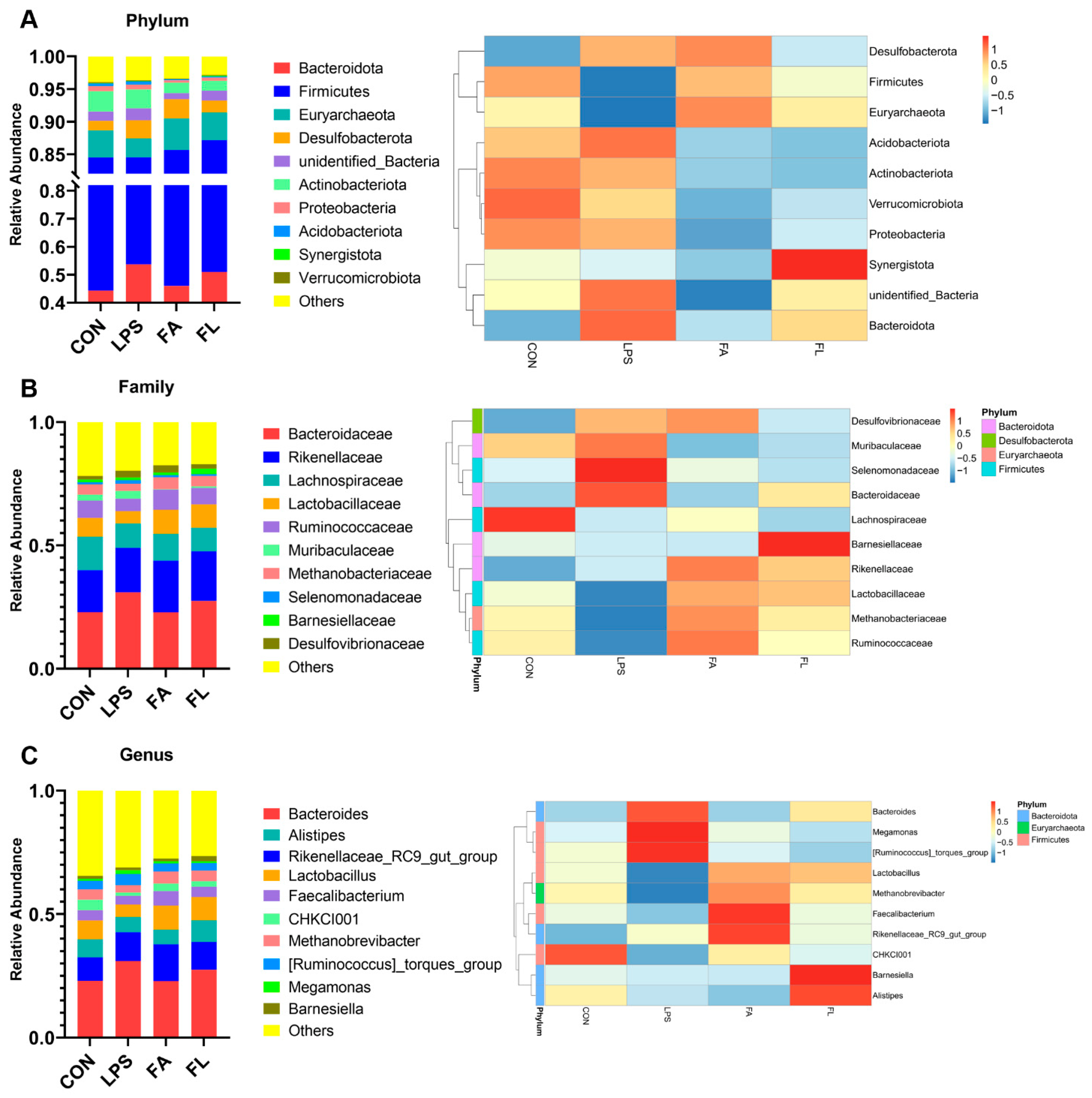
Figure 10 presents the composition of microbiota in Tianfu broilers. At the phylum level (Figure 10A), the ileal microbiota of the Tianfu broilers mainly was composed of Bacteroiota (44.3%), Firmicutes (40.23%), Euryarchaeota (4.16%), Desulfobacterota (1.41%), unidentified_bacterira (1.42%), and Antinobacteriota (3.15%). After LPS challenge, the ileal microbiota exhibited an increment of Bacteroidota, Desulfobacterota, and Actinobacteriota, but a decrement of Firmicutes and Euryarchaeota in comparison to the CON group. However, the results showed an increment of Firmicutes and Euryarchaeota, and a decrement of Bacteroidota, Desulfobacterota, and Actinobacteriota in the FL group when compared to the LPS group.
At the family level (Figure 10B), Bacteroidaceae (22.96%) was the most abundant family followed by Rikenellaceae (16.94%), Lachnospiraceae (13.63%), Lactobacillaceae (7.56%), Ruminococcaceae (7.05%), and Muribaculaceae (2.45%). LPS reduced the abundance of Lactobacillaceae and Ruminococcaceae, whereas both of them increased in the FL group in comparison to the LPS group. Moreover, LPS increased the abundance of Bacteroidaceae and Muribaculaceae compared to the CON group, but dietary 100 mg/kg FA supplementation decreased their abundance in comparison to the LPS group.
At the genus level (Figure 10C), the ileal microbiota mainly was composed of Bacteroides (22.96%), Rikenellaceae_RC9_gut_group (9.57%), Alistipes (7.32%), Lactobacillus (7.56%), Faecalibacterium (4.14%), CHKCI001 (4.28%), Methanobrevibacter (4.16%), [Ruminococcus]_torques_group (3.50%), Barnesiella (1.18%), and Megamonas (0.82%). The challenge of LPS decreased the abundance of Lactobacillus, Faecalibacterium, CHKCI001, and Methanobrevibacter, but increased the abundance of Bacteroides, [Ruminococcus]_torques_group, and Megamonas. However, dietary 100 mg/kg FA supplementation increased the relative abundance of Lactobacillus, Faecalibacterium, CHKCI001, and Methanobrevibacter, while decreasing the proportions of Bacteroides, [Ruminococcus]_torques_group and Megamonas compared to the LPS group.

3. Discussion

The gastrointestinal tract is an essential organ for nutrient absorption and a fundamental protective barrier against incursions of bacteria, pathogens, and toxins; therefore, maintaining intestinal homeostasis is of great importance [29]. There is increasing evidence that suggests LPS, a common toxin in the poultry industry [30], leads to loss of body weight [31], intestinal epithelium injury [32], increased intestinal permeability [33,34], and microflora dysbiosis [35]. Bioactive agents with various biological benefits [36], such as FA, may represent an interesting solution.
The intestinal epithelium is renewed from the proliferation and differentiation of multipotential stem cells located in the crypt. At the tips of the villus, fully differentiated cells are extruded into the lumen [37]. Higher VH, VH/CD, and lower CD result in a greater mucosa surface area and faster migration, which is helpful to improve the capacity of intestinal digestion and absorption [38,39]. In this study, LPS significantly impaired the intestinal morphological structure through diminishing ileal VH. This result indicates that LPS impaired intestinal digestion and absorption, which is the leading cause of decreased body weight and average daily feed intake reported in our previous study [40]. Previous studies by Gadde et al. [41] and An et al. [42] reported similar findings. It shows that FA improves growth performance by maintaining the intestinal structure of LPS-challenged broilers. However, the protective effect of FA on intestinal morphology and barrier function was reported by a previous study [43] and was possibly related to its activation of the Nrf/HO-1 signaling pathway [44], as well as its radial scavenging property [45], as evidenced by the enhanced antioxidant ability of the intestinal mucosa in this study.
Intestinal permeability is a functional indicator to reflect the integrity of the intestinal wall and the extent of bacteria translocation. The increased level of serum DAO or D-LA has been considered a token of intestinal structural damage. DAO is an intracellular enzyme that exists in intestinal mucosa [46]. Similarly, D-LA is located in the gut and is produced by various bacteria as a fermentation product. Increased serum levels of DAO and D-LA reflect the alteration of intestinal permeability as a consequence of intestinal barrier dysfunction [47]. The results in the present study showed an increased serum activity of DAO and level of D-LA in the broilers challenged with LPS, which was consistent with the reports by Yang’s study [48]. In comparison, FA-supplemented chickens decreased the activity of DAO and the levels of D-LA, indicating an augment of intestinal wall stability, which was also observed in tilapia fed with an oxidized fish oil diet [49].
Interestingly, this study found that LPS blocks ileal cell entry into the S and G2/M phase, leading to an aggregation of G0G1 cells and a remarkable reduction in the PI index; however, FA supplementation promoted the proliferation of the ileal cells. In the present study, LPS downregulated the expression of CLDN-1, OCDN, and ZO-1 in the ileum. CLDN-1, OCDN, and ZO-1 belong to the tight junction proteins (TJs) and are responsible to seal the paracellular space in the epithelial cells in the gut [50]. TJs regulate ion and solute diffusion through the intercellular space and prevent the translocation of luminal antigens, microorganisms, and related toxins [51]. FA could alleviate LPS-induced TJ function loss, as evidenced by upregulated expression of CLDN-1 and ZO-1 in the ileum. Secretory immunoglobulin A (sIgA), the first line of defense of the gastrointestinal epithelium, is one of the most abundant antibodies in mucosal secretions. It binds to viruses and bacteria and prevents them from attaching to and invading the epithelial cells [52]. Furthermore, sIgA is reported to help with the stabilization of the microbial composition by binding commensal microbiota [53]. In line with a present study, a significant decrease in sIgA level was noticed in the LPS group [54]. Dietary FA supplementation instead increased the sIgA levels in the LPS-challenged chickens and maintained the mucosal immune response.
Intestinal microflora hemostasis provides essential health benefits to animals by promoting food digestion and nutrient absorption, influencing immune development, and providing colonization resistance against pathogens [55,56,57]. However, the beneficial effects of the intestinal microbiota depend on its composition, which have been shown to vary with a number of factors including diet, disease, and stress conditions [58,59]. In the present study, the diversity and uniformity of ileal microflora are different among the four groups. However, the composition of the ileal microflora in the FL group is highly similar to those in the CON group but differs from the LPS group, indicating that dietary FA supplementation reduces LPS-induced intestinal bacterial disturbance. Bacteroidota and Firmicutes are the two dominant phyla of bacteria in the intestine [60,61], and the F/B ratio was closely dependent on body weight because Firmicutes has higher efficiency of absorbing calorie than Bacteroidetes [62]. In this study, FA shows positive effects on the F/B ratio in the ileum of LPS-challenged chickens. Interestingly, the BW of LPS-challenged chickens was positively correlated with FA, as reported in our previous study [44]. The relative abundance of phylum Bacteroidota was found positively associated with oxidative stress [63] and was consistent with what we found in the present study, as levels of GSH and T-AOC, and the activity of SOD in the intestinal mucosa, were decreased, but the relative abundance of phylum Bacteroidetes in the ileum was increased in the LPS group, which was improved by FA. This finding is in agreement with previous observations that FA scavenges free radicals [64] and increases antioxidant function [65]. Increased relative abundance of Proteobacteria suggests a non-homeostasis of the intestinal environment [66]. In this study, chickens that receive FA supplementation showed a decrease in the relative abundance of Proteobacteria compared to the CON and LPS groups at the phylum level, which comprises several pathogens, such as Escherichia, Salmonella, and Helicobacter [67]. It is widely acknowledged that Lactobacillus improves intestinal health by preventing the colonization of pathogens [68]. The present results reveal that dietary FA supplementation increased the relative abundance of the family Lactobacillaceae and the genus Lactobacillus compared to the LPS group. Faecalibacterium has been recognized as a probiotic that improves epithelial proliferation and is a major butyrate producer [69]. At the genus level, FA-supplemented chickens shows an increased relative abundance of Faecalibacterium in the ileum compared to the ones challenged by LPS, which is in agreement with a previous study [70].

4. Materials and Methods

4.1. Reagents

Ferulic acid (C10H10O4, CAS number: 1135-24-6) was purchased from Chengdu Herbpurify Co. Ltd. (Chengdu, China) with 99.91% purity assessed by high-performance liquid (Welch C18 column (4.6 × 250 mm, 5 μm) with mobile phase acetonitrile: 20% phosphoric acid (22:78), Palo Alto, CA, USA). Lipopolysaccharide from Escherichia coli O55:B5 (LPS, L2880) was purchased from Sigma-Aldrich Corp (St. Louis, MO, USA) with ≥98% purity and ≥500,000 EU/mg titer. Then, LPS was suspended in sterile saline at a concentration of 100 μg/mL.

4.2. Experimental Design

Tianfu broiler chickens (n = 160, male, 25-day-old initial body weight 184.33 ± 3.34 g), were divided into four groups at random. Each group had four identical cages with 10 chickens per cage. Figure 11 represents the overview of the experimental design: CON group (basal diet and non-LPS challenge), LPS group (basal diet and 1.0 mg/kg LPS challenge), FA group (100 mg/kg FA + basal diet and non-LPS challenge). and FL group (100 mg/kg FA + basal diet and 1.0 mg/kg LPS challenge). On days 14, 16, 18, and 20, the birds were administered 1.0 mg/kg LPS of body weight intraperitoneally or an equal volume of normal saline based on a protocol described in a previous study [71].
All chickens were provided by and fed in the Poultry Breeding Research Unit of Sichuan Agricultural University (Ya’an, China) and were housed in an environment-controlled room with a relative humidity of 50–55%, room temperature of 22 °C, and 16 h light:8 h dark, and food and water ad libitum. The basic nutrient fact composition and of the basal diet are listed in Table 1, and the nutritional requirement was formulated according to the National Research Council requirements for chickens.

4.3. Sample Collection and Measurement

On day 21 of the study, eight chickens per group were selected randomly for blood sample collection. Isolated serum samples were stored at −20 °C. Intestinal samples were collected after birds were slaughtered under anesthesia. Part of the intestine was fixed in 4% (wt/vol) paraformaldehyde. Some ileal mucosa was scraped and stored at −80 °C, and another was crushed, centrifuged at 600× g for 5 min, and suspended at a density of 1 × 10⁶ cells/mL in the phosphate-buffered saline (PBS; Sigma-Aldrich, MO, USA) for subsequent flow cytometry analysis.

4.4. Morphological Analysis

Fixed intestinal samples were dehydrated using graded ethanol, and vitrificated by dimethylbenzene. Then samples were embedded in paraffin, sliced with a Lecia RM2235 microtome (Leica Biosystems Inc., Buffalo Grove, IL, USA), and stained by hematoxylin-eosin (HE). The villus height (VH) and crypt depth (CD) were measured using a microscope imaging system (DM 1000, Leica, Germany) and Image-Pro Plus software 6.0 (Media Cybernetics, Inc., Washington, DC, USA) to calculate the ratio of the villus height to the crypt depth (VH/CD).

4.5. Permeability and sIgA Content Analysis

The levels of diamine oxidase (DAO), d-lactate acid (D-LA) in serum, and sIgA content in ileal mucosa were determined by the chicken-specific enzyme-linked immunosorbent assay (ELISA) kits (Shanghai Enzyme-linked Biotechnology Co., Ltd., Shanghai, China).

4.6. Antioxidant Parameters

Biochemistry kits (Nanjing Jiancheng Bioengineering Institute, Nanjing, China) were used to measure the concentrations of superoxide dismutase (SOD), total antioxidant (T-AOC), and glutathione (GSH) in the ileal mucosa.

4.7. Life Cycle of Ileum Epithelium

After being fixed in cold ethanol, the cell suspension was stained with PI/Rnase Staining Buffer (BD Bioscience, Franklin Lakes, NJ, USA), resuspended in PSB, and detected by a CytoFLEX flow cytometer (Backman Coulter, Brea, CA, USA). The formula used to determine the proliferation index (PI) was (S1 + G2M)/(G0/1 + S + G2M).

4.8. Real-Time Quantificative PCR (RT-qPCR)

The isolation of total ileal RNA followed the user’s guidance of RNAiso Plus (TaKaRa Bio Inc., Shiga, Japan). The PrimeScript™ reagent Kit with gDNA Eraser Kit (TaKaRa Bio Inc., Shiga, Japan) was used for the synthesis of the first strand (cDNA). A CFX96 Real-Time PCR Detection System (Bio-Rad, Hercules, CA, USA) with a SYBR Premix Ex Taq II (TaKaRa Bio Inc., Shiga, Japan) was used for RT-qPCR and gene expression was analyzed using the 2−∆∆Ct method [72]. The primers for the target genes and β-actin (as a housekeeping gene) are listed in Table 2.

4.9. 16S rRNA Sequencing and Data Analysis

Ileal digesta contents were collected randomly from six broilers per group. Total genome DNA was extracted using the SDS method. PCR reactions were carried out using a Phusion® High-Fidelity PCR Master Mix (New England Biolabs, Ipswich, MA, USA). Distinct regions V3-V4 of 16S rRNA genes were amplified using a specific primer. Sequencing libraries were generated using a TruSeq® DNA PCR-Free Sample Preparation Kit (Illumina, San Diego, CA, USA) and sequenced on an Illumina NovaSeq platform after quality assessment. Paired-end reads assembly used FLASH and quality control through data filtration and chimera removal. Effective tags were finally obtained. The same operational taxonomic units (OTUs) were formed from sequences that shared a minimum 97% similarity. The QIIME software (version 1.9.1; GitHub, San Francisco, CA, USA) was used to determine Alpha diversity including ACE, Chao1, Shannon, Simpson, and Beta diversity, including PCA and PCoA.

4.10. Statistical Analyses

All data were analyzed by SPSS 27.0 (SPSS Inc., Chicago, IL, USA) and compared significant differences among groups with one-way analysis of variance (ANOVA) and Tukey’s multiple comparison tests. Results were presented as mean ± standard error of mean (SEM). Values of p < 0.05 were considered significant.

5. Conclusions

In conclusion, LPS decreased the intestinal antioxidant ability and impaired the intestinal morphological structure, resulting in increased intestinal permeability, diminished levels of sIgA, debilitated TJ function, impaired enterocytes’ ability to proliferate, and induced gut microbiota dysbiosis in the ileum. The aforementioned detrimental effects were ameliorated by dietary FA supplementation through enhancement of antioxidant ability, augmentation of intestinal barrier integrity, improvement of the mucosal immune response, and elevated stability of ileal microflora.

Supplementary Materials

The following supporting information can be downloaded at https://www.mdpi.com/article/10.3390/molecules28041720/s1, Figure S1: The data and histology of duodenum and jejunum.

Author Contributions

Conceptualization, G.S. and X.Z.; methodology, Y.Z.; software, H.D.; validation, F.X. and H.L.; formal analysis, Z.T. and F.K.A.; supervision, P.O. and J.L.; project administration, H.F. and W.Z.; data curation, C.L.; writing—original draft preparation, Z.T. and G.S.; writing—review and editing, Y.Z. and L.-J.C. All authors have read and agreed to the published version of the manuscript.

Funding

This work was funded by the National Natural Science Foundation of China (Grant No. 31872347), the projects funded by the Central Government to Gui Local Scientific and Technological Development from Guizhou Province (QIANKEZHONGYINDI (2021) 4003), the projects funded by the Central Government to Gui Local Scientific and Technological Development from Guizhou Province (QIANKEZHONGYINDI (2021) 4018).

Institutional Review Board Statement

The study was conducted according to the guidelines of the Animal Ethics Committee of Sichuan Agricultural University (protocol#DYY-2019303099, approved 21 September 2019).

Informed Consent Statement

Not applicable.

Data Availability Statement

The raw data supporting the conclusions of this article will be made available by the authors, without reservation.

Conflicts of Interest

The authors declare no conflict of interest.

Sample Availability

Not available from the authors.

References

  1. Di Lorenzo, F.; Duda, K.A.; Lanzetta, R.; Silipo, A.; De Castro, C.; Molinaro, A. A journey from structure to function of bacterial lipopolysaccharides. Chem. Rev. 2021, 122, 15767–15821. [Google Scholar] [CrossRef]
  2. Geng, Y.; Ma, Q.; Wang, Z.; Guo, Y. Dietary vitamin d3 supplementation protects laying hens against lipopolysaccharide-induced immunological stress. Nutr. Metab. 2018, 15, 58. [Google Scholar] [CrossRef]
  3. Anders, L.C.; Lang, A.L.; Anwar-Mohamed, A.; Douglas, A.N.; Bushau, A.M.; Falkner, K.C.; Hill, B.G.; Warner, N.L.; Arteel, G.E.; Cave, M.; et al. Vinyl chloride metabolites potentiate inflammatory liver injury caused by lps in mice. Toxicol. Sci. 2016, 151, 312–323. [Google Scholar] [CrossRef]
  4. Zhang, Y.; Liu, L.; Peng, Y.L.; Liu, Y.Z.; Wu, T.Y.; Shen, X.L.; Zhou, J.R.; Sun, D.Y.; Huang, A.J.; Wang, X.; et al. Involvement of inflammasome activation in lipopolysaccharide–induced mice depressive–like behaviors. CNS Neurosci. Ther. 2014, 20, 119–124. [Google Scholar] [CrossRef]
  5. Zeng, Y.; Zhao, H.; Zhang, T.; Zhang, C.; He, Y.; Du, L.; Zuo, F.; Wang, W. Lung-protective effect of punicalagin on lps-induced acute lung injury in mice. Biosci. Rep. 2022, 42, BSR20212196. [Google Scholar] [CrossRef]
  6. Tgkagi, T.; Taguchi, O.; Aoki, S.; Toda, M.; Yamaguchi, A.; Fujimoto, H.; Bobeda-Ruiz, D.; Gil-Bernabe, P.; Ramirez, A.Y.; Naito, M.; et al. Direct effects of protein s in ameliorating acute lung injury. J. Thromb. Haemost. 2009, 7, 2053–2063. [Google Scholar] [CrossRef]
  7. Plotnikov, E.; Pevzner, I.; Zorova, L.; Chernikov, V.; Prusov, A.; Kireev, I.; Silachev, D.; Skulachev, V.; Zorov, D. Mitochondrial damage and mitochondria-targeted antioxidant protection in lps-induced acute kidney injury. Antioxidants 2019, 8, 176. [Google Scholar] [CrossRef]
  8. Bhattacharyya, J.; Biswas, S.; Datta, A.G. Mode of action of endotoxin: Role of free radicals and antioxidants. Curr. Med. Chem. 2004, 11, 359–368. [Google Scholar] [CrossRef]
  9. Jiang, J.; Qi, L.; Wei, Q.; Shi, F. Maternal stevioside supplementation ameliorates intestinal mucosal damage and modulates gut microbiota in chicken offspring challenged with lipopolysaccharide. Food Funct. 2021, 12, 6014–6028. [Google Scholar] [CrossRef]
  10. Yan, L.; Lv, Z.Z.; An, S.; Xing, K.; Wang, Z.G.; Lv, M.B.; Choct, M.; Guo, Y.M.; Zhou, G.L. Effects of rearing system and narasin on growth performance, gastrointestinal development, and gut microbiota of broilers. Poult. Sci. 2021, 100, 100840. [Google Scholar] [CrossRef]
  11. Zhou, X.; Zhang, Y.; He, L.; Wan, D.; Liu, G.; Wu, X.; Yin, Y. Serine prevents lps-induced intestinal inflammation and barrier damage via p53-dependent glutathione synthesis and ampk activation. J. Funct. Food 2017, 39, 225–232. [Google Scholar] [CrossRef]
  12. Ozdemir, D.; Uysal, N.; Tugyan, K.; Gonenc, S.; Acikgoz, O.; Aksu, I.; Ozkan, H. The effect of melatonin on endotoxemia-induced intestinal apoptosis and oxidative stress in infant rats. Intensive Care Med. 2007, 33, 511–516. [Google Scholar] [CrossRef] [PubMed]
  13. Gu, Y.F.; Chen, Y.P.; Jin, R.; Wang, C.; Wen, C.; Zhou, Y.M. Dietary chitooligosaccharide supplementation alleviates intestinal barrier damage, and oxidative and immunological stress in lipopolysaccharide-challenged laying hens. Poult. Sci. 2022, 101, 101701. [Google Scholar] [CrossRef] [PubMed]
  14. Yu, Y.; Li, Q.; Zeng, X.; Xu, Y.; Jin, K.; Liu, J.; Cao, G. Effects of probiotics on the growth performance, antioxidant functions, immune responses, and caecal microbiota of broilers challenged by lipopolysaccharide. Front. Vet. Sci. 2022, 9, 846649. [Google Scholar] [CrossRef] [PubMed]
  15. Chen, J.Y.; Yu, Y.H. Bacillus subtilis-fermented products ameliorate the growth performance and alter cecal microbiota community in broilers under lipopolysaccharide challenge. Poult. Sci. 2021, 100, 875–886. [Google Scholar] [CrossRef]
  16. Ullah, A.; Anjum, A.A.; Rabbani, M.; Nawaz, M.; Ashraf, M.; Ijaz, M.; Ali, A.; Rashid, A.; Najeeb, I.; Pervez, A. Phytochemical composition and in-vitro activity of ethanolic extract of eucalyptus globulus leaves against multidrug resistant poultry pathogens. Cell. Mol. Biol. 2021, 67, 159–164. [Google Scholar] [CrossRef]
  17. Zhang, P.; Sun, D.; Shi, B.; Faucitano, L.; Guo, X.; Li, T.; Xu, Y.; Yan, S. Dietary supplementation with artemisia argyi extract on inflammatory mediators and antioxidant capacity in broilers challenged with lipopolysaccharide. Ital. J. Anim. Sci. 2020, 19, 1091–1098. [Google Scholar] [CrossRef]
  18. Konieczka, P.; Szkopek, D.; Kinsner, M.; Fotschki, B.; Juskiewicz, J.; Banach, J. Cannabis-derived cannabidiol and nanoselenium improve gut barrier function and affect bacterial enzyme activity in chickens subjected to C. Perfringens challenge. Vet. Res. 2020, 51, 141. [Google Scholar] [CrossRef]
  19. Csernus, B.; Biro, S.; Babinszky, L.; Komlosi, I.; Javor, A.; Stundl, L.; Remenyik, J.; Bai, P.; Olah, J.; Pesti-Asboth, G.; et al. Effect of carotenoids, oligosaccharides and anthocyanins on growth performance, immunological parameters and intestinal morphology in broiler chickens challenged with escherichia coli lipopolysaccharide. Animals 2020, 10, 347. [Google Scholar] [CrossRef]
  20. Choi, J.H.; Park, J.K.; Kim, K.M.; Lee, H.J.; Kim, S. In vitro and in vivo antithrombotic and cytotoxicity effects of ferulic acid. J. Biochem. Mol. Toxicol. 2018, 32, e22004. [Google Scholar] [CrossRef]
  21. Shi, C.; Zhang, X.; Sun, Y.; Yang, M.; Song, K.; Zheng, Z.; Chen, Y.; Liu, X.; Jia, Z.; Dong, R.; et al. Antimicrobial activity of ferulic acid against cronobacter sakazakii and possible mechanism of action. Foodborne Pathog. Dis. 2016, 13, 196–204. [Google Scholar] [CrossRef] [PubMed]
  22. Chowdhury, S.; Ghosh, S.; Das, A.K.; Sil, P.C. Ferulic acid protects hyperglycemia-induced kidney damage by regulating oxidative insult, inflammation and autophagy. Front. Pharmacol. 2019, 10, 27. [Google Scholar] [CrossRef] [PubMed]
  23. Rehman, S.U.; Ali, T.; Alam, S.I.; Ullah, R.; Zeb, A.; Lee, K.W.; Rutten, B.; Kim, M.O. Ferulic acid rescues lps-induced neurotoxicity via modulation of the tlr4 receptor in the mouse hippocampus. Mol. Neurobiol. 2019, 56, 2774–2790. [Google Scholar] [CrossRef]
  24. Liu, H.; Chen, Z.; Shen, L.; Guo, X.; Ji, D.; Sun, S.; Edwards, M.G.; Frank, F.; Li, J.F.; Salama, A.; et al. Well modelling methods in thermal reservoir simulation. Oil Gas Sci. Technol. 2020, 75, 63. [Google Scholar] [CrossRef]
  25. Zheng, Y.; You, X.; Guan, S.; Huang, J.; Wang, L.; Zhang, J.; Wu, J. Poly(ferulic acid) with an anticancer effect as a drug nanocarrier for enhanced colon cancer therapy. Adv. Funct. Mater. 2019, 29, 1808646. [Google Scholar] [CrossRef]
  26. Yin, X.; Liu, W.; Chen, H.; Qi, C.; Chen, H.; Niu, H.; Yang, J.; Kwok, K.W.H.; Dong, W. Effects of ferulic acid on muscle development and intestinal microbiota of zebrafish. J. Anim. Physiol. Anim. Nutr. 2022, 106, 429–440. [Google Scholar] [CrossRef]
  27. Hu, R.; Wu, S.; Li, B.; Tan, J.; Yan, J.; Wang, Y.; Tang, Z.; Liu, M.; Fu, C.; Zhang, H.; et al. Dietary ferulic acid and vanillic acid on inflammation, gut barrier function and growth performance in lipopolysaccharide-challenged piglets. Anim. Nutr. 2022, 8, 144–152. [Google Scholar] [CrossRef]
  28. Gu, Y.; Zhang, Y.; Li, M.; Huang, Z.; Jiang, J.; Chen, Y.; Chen, J.; Jia, Y.; Zhang, L.; Zhou, F. Ferulic acid ameliorates atherosclerotic injury by modulating gut microbiota and lipid metabolism. Front. Pharmacol. 2021, 12, 621339. [Google Scholar] [CrossRef]
  29. Furness, J.B.; Kunze, W.A.; Clerc, N. Nutrient tasting and signaling mechanisms in the gut. Ii. The intestine as a sensory organ: Neural, endocrine, and immune responses. Am. J. Physiol. 1999, 277, G922–G928. [Google Scholar]
  30. Gautam, R.; Heo, Y.; Lim, G.; Song, E.; Roque, K.; Lee, J.; Kim, Y.; Cho, A.; Shin, S.; Kim, C.; et al. Altered immune responses in broiler chicken husbandry workers and their association with endotoxin exposure. Ind. Health 2018, 56, 10–19. [Google Scholar] [CrossRef]
  31. Tan, J.; Liu, S.; Guo, Y.; Applegate, T.J.; Eicher, S.D. Dietary l-arginine supplementation attenuates lipopolysaccharide-induced inflammatory response in broiler chickens. Br. J. Nutr. 2014, 111, 1394–1404. [Google Scholar] [CrossRef]
  32. Xiong, W.; Ma, H.; Zhang, Z.; Jin, M.; Wang, J.; Xu, Y.; Wang, Z. The protective effect of icariin and phosphorylated icariin against lps-induced intestinal epithelial cells injury. Biomed. Pharmacother. 2019, 118, 109246. [Google Scholar] [CrossRef]
  33. Rizzo, V.; Ferlazzo, N.; Currò, M.; Isola, G.; Matarese, M.; Bertuccio, M.P.; Caccamo, D.; Matarese, G.; Ientile, R. Baicalin-induced autophagy preserved lps-stimulated intestinal cells from inflammation and alterations of paracellular permeability. Int. J. Mol. Sci. 2021, 22, 2315. [Google Scholar] [CrossRef]
  34. Yan, J.; Gong, Z.; Zhang, T.; Cai, W. Sodium butyrate attenuates soybean oil-based lipid emulsion-induced increase in intestinal permeability of lipopolysaccharide by modulation of p-glycoprotein in caco-2 cells. Biochem. Biophys. Res. Commun. 2017, 482, 791–795. [Google Scholar] [CrossRef]
  35. Kong, Y.; Yan, T.; Tong, Y.; Deng, H.; Tan, C.; Wan, M.; Wang, M.; Meng, X.; Wang, Y. Gut microbiota modulation by polyphenols fromaronia melanocarpa of lps-induced liver diseases in rats. J. Agric. Food Chem. 2021, 69, 3312–3325. [Google Scholar] [CrossRef]
  36. Li, D.; Rui, Y.; Guo, S.; Luan, F.; Liu, R.; Zeng, N. Ferulic acid: A review of its pharmacology, pharmacokinetics and derivatives. Life Sci. 2021, 284, 119921. [Google Scholar] [CrossRef]
  37. Sato, T.; Vries, R.G.; Snippert, H.J.; van de Wetering, M.; Barker, N.; Stange, D.E.; van Es, J.H.; Abo, A.; Kujala, P.; Peters, P.J.; et al. Single lgr5 stem cells build crypt-villus structures in vitro without a mesenchymal niche. Nature 2009, 459, 262–265. [Google Scholar] [CrossRef]
  38. Awad, W.A.; Ghareeb, K.; Abdel-Raheem, S.; Böhm, J. Effects of dietary inclusion of probiotic and synbiotic on growth performance, organ weights, and intestinal histomorphology of broiler chickens. Poult. Sci. 2009, 88, 49–56. [Google Scholar] [CrossRef]
  39. Jia, G.; Yan, J.Y.; Cai, J.Y.; Wang, K.N. Effects of encapsulated and non-encapsulatedcompound acidifiers on gastrointestinal ph andintestinal morphology and function in weaningpiglets. J. Anim. Feed Sci. 2010, 19, 81–92. [Google Scholar] [CrossRef]
  40. Shu, G.; Tang, Z.; Du, H.; Zheng, Y.; Chang, L.; Li, H.; Xu, F.; Fu, H.; Zhang, W.; Lin, J. Effects of dietary ferulic acid supplementation on hepatic injuries in tianfu broilers challenged with lipopolysaccharide. Toxins 2022, 14, 227. [Google Scholar] [CrossRef]
  41. Gadde, U.D.; Oh, S.; Lee, Y.; Davis, E.; Zimmerman, N.; Rehberger, T.; Lillehoj, H.S. Retracted: Dietary bacillus subtilis-based direct-fed microbials alleviate lps-induced intestinal immunological stress and improve intestinal barrier gene expression in commercial broiler chickens. Res. Vet. Sci. 2017, 114, 236–243. [Google Scholar] [CrossRef]
  42. An, J.; Shi, J.; Liu, K.; Li, A.; He, B.; Wang, Y.; Duan, T.; Wang, Y.; He, J. Effects of solid-state fermented wheat bran on growth performance, immune function, intestinal morphology and microflora in lipopolysaccharide-challenged broiler chickens. Animals 2022, 12, 1100. [Google Scholar] [CrossRef] [PubMed]
  43. He, S.; Liu, F.; Xu, L.; Yin, P.; Li, D.; Mei, C.; Jiang, L.; Ma, Y.; Xu, J. Protective effects of ferulic acid against heat stress-induced intestinal epithelial barrier dysfunction in vitro and in vivo. PLoS ONE 2016, 11, e145236. [Google Scholar] [CrossRef] [PubMed]
  44. Li, X.; Zhang, J.; Rong, H.; Zhang, X.; Dong, M. Ferulic acid ameliorates mpp+/mptp-induced oxidative stress via erk1/2-dependent nrf2 activation: Translational implications for parkinson disease treatment. Mol. Neurobiol. 2020, 57, 2981–2995. [Google Scholar] [CrossRef] [PubMed]
  45. Yang, J.; Chen, J.; Hao, Y.; Liu, Y. Identification of the dpph radical scavenging reaction adducts of ferulic acid and sinapic acid and their structure-antioxidant activity relationship. LWT 2021, 146, 111411. [Google Scholar] [CrossRef]
  46. Thompson, J.S.; Vaughan, W.P.; Forst, C.F.; Jacobs, D.L.; Weekly, J.S.; Rikkers, L.F. The effect of the route of nutrient delivery on gut structure and diamine oxidase levels. J. Parenter. Enter. Nutr. 1987, 11, 28–32. [Google Scholar] [CrossRef]
  47. Xu, J.; Liu, Z.; Zhan, W.; Jiang, R.; Yang, C.; Zhan, H.; Xiong, Y. Recombinant tsp53 modulates intestinal epithelial barrier integrity via upregulation of zo1 in lpsinduced septic mice. Mol. Med. Rep. 2018, 17, 1212–1218. [Google Scholar]
  48. Yang, L.; Liu, G.; Lian, K.; Qiao, Y.; Zhang, B.; Zhu, X.; Luo, Y.; Shang, Y.; Gu, X. Dietary leonurine hydrochloride supplementation attenuates lipopolysaccharide challenge-induced intestinal inflammation and barrier dysfunction by inhibiting the nf-κb/mapk signaling pathway in broilers. J. Anim. Sci. 2019, 97, 1679–1692. [Google Scholar] [CrossRef]
  49. Yu, L.; Wen, H.; Jiang, M.; Wu, F.; Tian, J.; Lu, X.; Xiao, J.; Liu, W. Effects of ferulic acid on intestinal enzyme activities, morphology, microbiome composition of genetically improved farmed tilapia (oreochromis niloticus) fed oxidized fish oil. Aquaculture 2020, 528, 735543. [Google Scholar] [CrossRef]
  50. Groschwitz, K.R.; Hogan, S.P. Intestinal barrier function: Molecular regulation and disease pathogenesis. J. Allergy Clin. Immunol. 2009, 124, 3–20. [Google Scholar] [CrossRef]
  51. Zihni, C.; Mills, C.; Matter, K.; Balda, M.S. Tight junctions: From simple barriers to multifunctional molecular gates. Nat. Rev. Mol. Cell Biol. 2016, 17, 564–580. [Google Scholar] [CrossRef] [PubMed]
  52. Mestecky, J.; Russell, M.W.; Elson, C.O. Intestinal iga: Novel views on its function in the defence of the largest mucosal surface. Gut 1999, 44, 2–5. [Google Scholar] [CrossRef] [PubMed]
  53. Kaetzel, C.S. Cooperativity among secretory iga, the polymeric immunoglobulin receptor, and the gut microbiota promotes host-microbial mutualism. Immunol. Lett. 2014, 162, 10–21. [Google Scholar] [CrossRef] [PubMed]
  54. Lv, Z.; Dai, H.; Wei, Q.; Jin, S.; Wang, J.; Wei, X.; Yuan, Y.; Yu, D.; Shi, F. Dietary genistein supplementation protects against lipopolysaccharide-induced intestinal injury through altering transcriptomic profile. Poult. Sci. 2020, 99, 3411–3427. [Google Scholar] [CrossRef] [PubMed]
  55. Yu, Q.; Jia, A.; Li, Y.; Bi, Y.; Liu, G. Microbiota regulate the development and function of the immune cells. Int. Rev. Immunol. 2018, 37, 79–89. [Google Scholar] [CrossRef]
  56. Zhao, J.; Zhang, X.; Liu, H.; Brown, M.A.; Qiao, S. Dietary protein and gut microbiota composition and function. Curr. Protein Pept. Sci. 2018, 20, 145–154. [Google Scholar] [CrossRef]
  57. Schmidt, F.; Dahlke, K.; Batra, A.; Ring, C.; Keye, J.; Loh, G.; Schumann, M.; Kühl, A.A.; Blaut, M.; Siegmund, B. Impact of microbiota on intestinal immune cell composition and intestinal barrier function. Gastroenterology 2017, 152, S1000. [Google Scholar] [CrossRef]
  58. De Filippo, C.; Cavalieri, D.; Di Paola, M.; Ramazzotti, M.; Poullet, J.B.; Massart, S.; Collini, S.; Pieraccini, G.; Lionetti, P. Impact of diet in shaping gut microbiota revealed by a comparative study in children from europe and rural africa. Proc. Natl. Acad. Sci. USA 2010, 107, 14691–14696. [Google Scholar] [CrossRef]
  59. Sonalio, K.; Almeida, H.M.S.; Mechler-Dreibi, M.L.; Storino, G.Y.; Haesebrouck, F.; Maes, D.; de Oliveira, L.G. Infuence of mycoplasma hyopneumoniae natural infection on the respiratory microbiome diversity of fnishing pigs. Vet. Res. 2022, 53, 20. [Google Scholar] [CrossRef]
  60. Bäckhed, F.; Ley, R.E.; Sonnenburg, J.L.; Peterson, D.A.; Gordon, J.I. Host-bacterial mutualism in the human intestine. Science 2005, 307, 1915–1920. [Google Scholar] [CrossRef]
  61. Eckburg, P.B.; Bik, E.M.; Bernstein, C.N.; Purdom, E.; Dethlefsen, L.; Sargent, M.; Gill, S.R.; Nelson, K.E.; Relman, D.A. Diversity of the human intestinal microbial flora. Science 2005, 308, 1635–1638. [Google Scholar] [CrossRef] [PubMed]
  62. Koliada, A.; Syzenko, G.; Moseiko, V.; Budovska, L.; Puchkov, K.; Perederiy, V.; Gavalko, Y.; Dorofeyev, A.; Romanenko, M.; Tkach, S.; et al. Association between body mass index and firmicutes/bacteroidetes ratio in an adult ukrainian population. BMC Microbiol. 2017, 17, 120. [Google Scholar] [CrossRef] [PubMed]
  63. Ortega-Hernandez, A.; Martinez-Martinez, E.; Gomez-Gordo, R.; Lopez-Andres, N.; Fernandez-Celis, A.; Gutierrrez-Miranda, B.; Nieto, M.L.; Alarcon, T.; Alba, C.; Gomez-Garre, D.; et al. The interaction between mitochondrial oxidative stress and gut microbiota in the cardiometabolic consequences in diet-induced obese rats. Antioxidants 2020, 9, 640. [Google Scholar] [CrossRef] [PubMed]
  64. Liang, C.; Chang, C.; Liang, C.; Hung, K.; Hsieh, C. In vitro antioxidant activities, free radical scavenging capacity, and tyrosinase inhibitory of flavonoid compounds and ferulic acid from spiranthes sinensis (pers.) Ames. Molecules 2014, 19, 4681–4694. [Google Scholar] [CrossRef]
  65. Graf, E. Antioxidant potential of ferulic acid. Free Radic. Biol. Med. 1992, 13, 435–448. [Google Scholar] [CrossRef]
  66. Rizzatti, G.; Lopetuso, L.R.; Gibiino, G.; Binda, C.; Gasbarrini, A. Proteobacteria: A common factor in human diseases. BioMed Res. Int. 2017, 2017, 9351507. [Google Scholar] [CrossRef]
  67. Buton, A.; Bobay, L. Evolution of chi motifs in proteobacteria. G3 Genes Genom Genet 2021, 11, jkaa054. [Google Scholar] [CrossRef]
  68. Yang, K.M.; Jiang, Z.Y.; Zheng, C.T.; Wang, L.; Yang, X.F. Effect of lactobacillus plantarum on diarrhea and intestinal barrier function of young piglets challenged with enterotoxigenic escherichia coli k88. J. Anim. Sci. 2014, 92, 1496–1503. [Google Scholar] [CrossRef]
  69. Gangadoo, S.; Dinev, I.; Chapman, J.; Hughes, R.J.; Van, T.T.H.; Moore, R.J.; Stanley, D. Selenium nanoparticles in poultry feed modify gut microbiota and increase abundance of faecalibacterium prausnitzii. Appl. Microbiol. Biotechnol. 2018, 102, 1455–1466. [Google Scholar] [CrossRef]
  70. Liu, Y.; Lin, Q.; Huang, X.; Jiang, G.; Li, C.; Zhang, X.; Liu, S.; He, L.; Liu, Y.; Dai, Q.; et al. Effects of dietary ferulic acid on the intestinal microbiota and the associated changes on the growth performance, serum cytokine profile, and intestinal morphology in ducks. Front. Microbiol. 2021, 12, 698213. [Google Scholar] [CrossRef]
  71. Yang, L.; Liu, G.; Liang, X.; Wang, M.; Zhu, X.; Luo, Y.; Shang, Y.; Yang, J.Q.; Zhou, P.; Gu, X.L. Effects of berberine on the growth performance, antioxidative capacity and immune response to lipopolysaccharide challenge in broilers. Anim. Sci. J. 2019, 90, 1229–1238. [Google Scholar] [CrossRef] [PubMed]
  72. Livak, K.J.; Schmittgen, T.D. Analysis of relative gene expression data using real-time quantitative pcr and the 2(-delta delta c(t)) method. Methods 2001, 25, 402–408. [Google Scholar] [CrossRef] [PubMed]
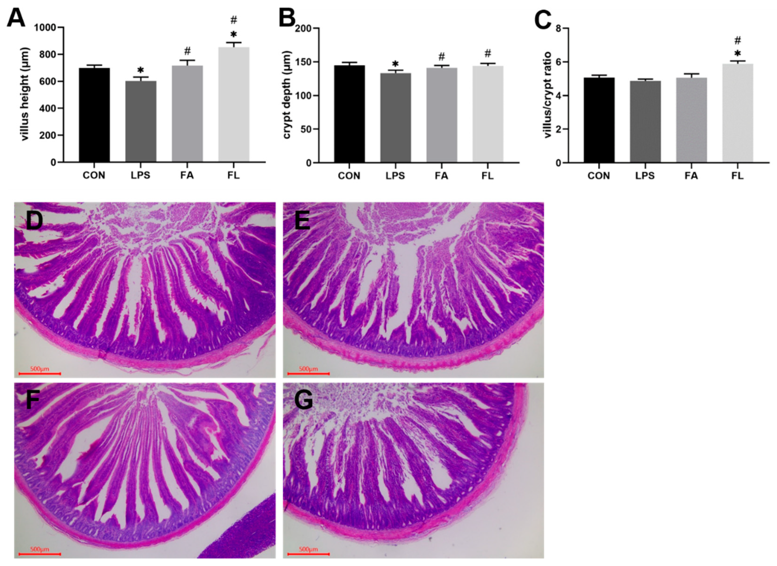
Figure 1. The data of villus height, crypt depth, and villus/crypt ratio. (A) Villus height (μm). (B) Crypt depth (μm). (C) Villus height/crypt depth ratio. Values are expressed as the mean ± SEM. n = 8. * means p < 0.05, compared with the CON group. # means p < 0.05, compared with the LPS group. (DG) The representative histological changes of the ileum villus height and crypt depth with HE staining (scale bar = 500 μm). (D) CON group, (E) LPS group, (F) FA group, (G) FL group.
Molecules 28 01720 g001
Figure 2. DAO activity and D-LA content in serum. (A) Diamine oxidase (DAO, ng/mL) (B) d-lactate acid (D-LA, nmol/L). Values are expressed as the mean ± SEM. n = 6. * means p < 0.05, compared with the CON group. # means p < 0.05, compared with the LPS group.
Molecules 28 01720 g002
Figure 3. Antioxidant parameters of the intestinal mucosa. (A) Superoxide dismutase (SOD, U/mg), (B) total antioxidant capacity (T-AOC, mmol/g), (C) glutathione (GSH, µmol/g). Values are expressed as the mean ± SEM, n = 3. * means p < 0.05, compared with the CON group. # means p < 0.05, compared with the LPS group.
Molecules 28 01720 g003
Figure 4. sIgA content in ileal mucosa. Values are expressed as the mean ± SEM. n = 4. * means p < 0.05, compared with the CON group. # means p < 0.05, compared with the LPS group.
Molecules 28 01720 g004
Figure 5. Ileal cell cycle. The percentage of the ileal cells in the (A) G0G1 phase (DNA pre-synthesis phase of the cell life cycle, %), (B) S phase (DNA synthesis phase of the cell life cycle, %), (C) G2M phase (prophase of division and division phase of the cell life cycle, %). (D) PI index (proliferation index). (E) The flow cytometry quadrant diagrams of the ileal cell life cycle among four groups. Values are expressed as the mean ± SEM. n = 6. * means p < 0.05, compared with the CON group. # means p < 0.05, compared with the LPS group.
Molecules 28 01720 g005
Figure 6. Relative mRNA expression levels of CLDN-1, OCDN, and ZO-1 in the ileum. Values are expressed as the mean ± SEM. n = 3. * means p < 0.05, compared with the CON group. # means p < 0.05, compared with the LPS group.
Molecules 28 01720 g006
Figure 7. Diagram of the operational taxonomic units (OTUs) distribution of the ileal digesta. (A) Venn diagram: each circle represents one group. The overlapping parts represent the same number of OTUs among groups. (B) Flower diagram: different colors represent different groups. The core means the same number of OTUs shared among four groups. n = 6.
Molecules 28 01720 g007
Figure 8. Alpha diversity of ileum microbiota. The alpha diversity was evaluated by the (A) Ace index, (B) Chao1 index, (C) Shannon index, and (D) Simpson index.
Molecules 28 01720 g008
Figure 9. Beta diversity of ileal microbiota (PCoA). (A) Principal coordinate analysis based on weighted unifrac distances. (B) Unweighted pair group method with arithmetic mean based on weighted unifrac distances.
Molecules 28 01720 g009
Figure 10. Microbial composition at the phylum, family, and genus levels in the ileum. Left: the histogram; Right: the heatmap. (A) Microbial structure at the phylum level. (B) Microbial structure at the family level. (C) Microbial structure at the genus level.
Molecules 28 01720 g010
Figure 11. Experimental design. Four treatment groups were randomly assigned to broilers of similar body weights. CON, birds received basal diet and intraperitoneal injection of saline. LPS, birds received basal diet and intraperitoneal injection of LPS. FA, birds received basal diet supplemented with FA and intraperitoneal injection of saline. FL, birds received basal diet supplemented with FA and intraperitoneal injection with LPS.
Molecules 28 01720 g011
Table 1. Ingredient composition and nutrient content of the basal diet.
Ingredient%Calculated Nutrients%
Corn59.50Metabolizable energy (MJ kg−1)12.80
Soybean meal32.90Crude protein19.70
Vegetable oil4.65Lysine1.08
CaCO30.50Methionine0.40
CaHPO41.60Methionine + Cystine0.74
NaCl0.30Calcium0.77
Choline0.10Nonphytate P0.40
DL-Met0.12
Premix 10.33
Total100
1 Provided per kg for diet: vitamin A (all-trans retinol acetate), 12,500 IU; cholecalciferol, 2500 IU; vitamin E (all-rac-a-tocopherol acetate), 18.75 IU; vitamin K (menadione Na bisulfate), 5.0 mg; thiamine (thiamine mononitrate), 2.5 mg; riboflavin, 7.5 mg; vitamin B6, 5.0 mg; vitamin B12, 0.0025 mg; pantothenate, 15 mg; niacin, 50 mg; folic acid, 1.25 mg; biotin, 0.12 mg; Cu (CuSO4·5H2O), 10 mg; Mn (MnSO4·H2O), 100 mg; Zn (ZnSO4·7H2O), 100 mg; Fe (FeSO4·7H2O), 100 mg; I (KI), 0.4 mg; and Se (Na2SeO3), 0.2 mg.
Table 2. Primers used for qRT-PCR.
GeneAccession NumberPrimer Sequence (5′–3′)Product Length (bp)
CLDN-1XM_001013611.2F: CATACTCCTGGGTCTGGTTGGT
R: GACAGCCATCCGCATCTTCT
100
OCLNNM_205128.1F: CTCAATCAGCTCAGCCGAC
R: TCTCCTGCTTCTTGCTTTGGTA
130
ZO-1NM_040706827.1F: GTAAACCACTGCCTACACC
R: ATATCTTAACTCTACTTCGCACA
90
β-actinNM_205518.1F: AAGGATCTGTATGCCAACACA
R: AGACAGAGTACTTGCGCTCA
148
CLDN-1: Claudin-1, OCLN: Occludin, ZO-1: zonula occkudens-1, β-actin: reference gene.
Disclaimer/Publisher’s Note: The statements, opinions and data contained in all publications are solely those of the individual author(s) and contributor(s) and not of MDPI and/or the editor(s). MDPI and/or the editor(s) disclaim responsibility for any injury to people or property resulting from any ideas, methods, instructions or products referred to in the content.