Abstract
The continuously increasing flow of toxic heavy metals to the environment due to intensive industrial activity and tightening requirements with regard to the content of metal ions in drinking and discharged waters urges the development of affordable and sensitive devices to the field control of pollutants. Here, we report a new thiated Rhodamine-lactam probe for Hg2+ detection and demonstrate how its sensitivity can be increased via the incorporation of the probe molecules into the optically transparent siloxane-acrylate coatings on polymethyl methacrylate and, alternatively, into the water-dispersible light-harvesting FRET nanoparticles (NPs), in which dye cations are separated by fluorinated tetraphenylborate anions. We have shown that the optimization of the FRET NPs composition had allowed it to reach the antenna effect of ~300 and fabricate “off/on” sensor for Hg2+ ion determination in aqueous solutions with the detection limit of ~100 pM, which is far below the maximum permissible concentration (MPC) of mercury in drinking water recommended by the World Health Organization. Although this work is more proof-of-concept than a ready-to-use analytical procedure, the suggested approaches to fabrication of the FRET NPs based on the popular rhodamine-lactam platform can be used as a background for the development of low-cost portable sensing devices for the extra-laboratory determination of hazardous metal ions.
1. Introduction
Drinking water quality and safety is a highly sensitive issue for the population, even in developed countries with high standards of water treatment technologies. As a result of industrial activities, continuously increasing flow of wastewaters containing toxic heavy metals, which are already harmful to human health at ppb level, it is released into the environment. Moreover, the synergetic effects of several pollutants, especially the combination of toxic metals with antibiotics and the low efficacy of water treatment toward new classes of emerging pollutants, make on-site water quality monitoring extremally important. Although analytical methods are available for the majority of metal ions (Hg, Cd, Pb, As), the fabrication of highly sensitive and cost-effective devices is still a challenge. According to a World Health Organization (WHO) report [1], there are no extra-laboratory methods for determining mercury at the level of maximum permissible concentration (MPC). Lead and cadmium in the field can only be determined at levels close to the MPC. Despite the high sensitivity of electrochemical sensors described in the literature, the detection limit for Hg achieved to date is close to the MPC for mercury in water [2]. Microfluidic devices using optical sensors are characterized by a limit of detection (LOD), which is at least tenfold higher than the MPC values in drinking water recommended by WHO for the most toxic metals: Hg [3] and Pb [4]. Alternative ultrasensitive techniques such as surface-enhanced Raman scattering [5,6,7,8] or surface-enhanced fluorescence [9,10,11,12] allow for the determination of significantly lower analyte concentrations; however, they require powerful and expensive equipment, both for measurements and for the fabrication of nanostructured enhancing substrates, which limits the use of such sensors devices for monitoring water quality in problematic regions, as in the active gold mining and processing industry, for example.
To date, the most traditional and widely used detection method is based on the application of fluorescent probes, which have become an effective analytical tool due to the unique capability for sensitive monitoring of metal ions [13,14], anions [15,16], reactive oxygen species [17,18], or biomolecules [19,20]. With these benefits, the sensitivity of such fluorescent probes is strictly limited by their brightness (the product of absorption and the photoluminescence quantum yield). Thus, one of the brightest probes, which is based on the Rhodamine 6G scaffold [21,22], has binding constants ~103–105 that are similar to other types of fluorophores [23]; a molar absorptivity of ~105, and show a typical working range of sensor operation from ~1μm to ~0.1 mM.
To improve the performance of fluorescent probes, it is necessary to enhance the response signal significantly via the incorporation into the hydrophobic environment [24] or by pumping the probe with a much brighter quantum emitter via Förster resonance energy transfer (FRET). However, the required bright emitters are usually large nanoparticles (NPs) which are inefficient FRET donors, since their sizes are beyond the FRET radius (1–10 nm) [19]. An alternative approach based on pumping the probe with multiple molecular emitters was recently demonstrated in light-harvesting FRET NPs, in which dye cations are separated by fluorinated tetraphenylborate anions that prevent dye self-quenching [25,26,27,28]. In these NPs, a short inter-fluorophore distance controlled by the counterion enables ultrafast dye–dye excitation energy migration on a femtosecond time scale through the whole particle within the fluorescence lifetime until it reaches a donor close to the acceptor leading to the FRET. Therefore, the energy can be transferred beyond the Förster radius from multiple donors to a single acceptor, providing a basis for signal amplification [19]. In a recent work, we adopted this light harvesting approach to pump the probe for the determination of Cu2+ ions, which demonstrated a ~100 fold decrease of LOD [29]. Here, we demonstrate the further development of this method. Using a specially designed novel thiated Rhodamine-lactam probe for mercury detection, we have reoptimized the light harvesting system based on protonated Coumarin-30 (further referred to as C30) cation and sodium tetrakis [3,5-bis(1,1,1,3,3,3-hexafluoro-2-methoxy-2-propyl)phenyl] borate anion (further referred to as F12). As a result, an ultrasensitive “off/on” nanoprobe with an antenna effect of 300 and an impressive Hg2+ LOD of ~100 pM was obtained.
2. Results and Discussion
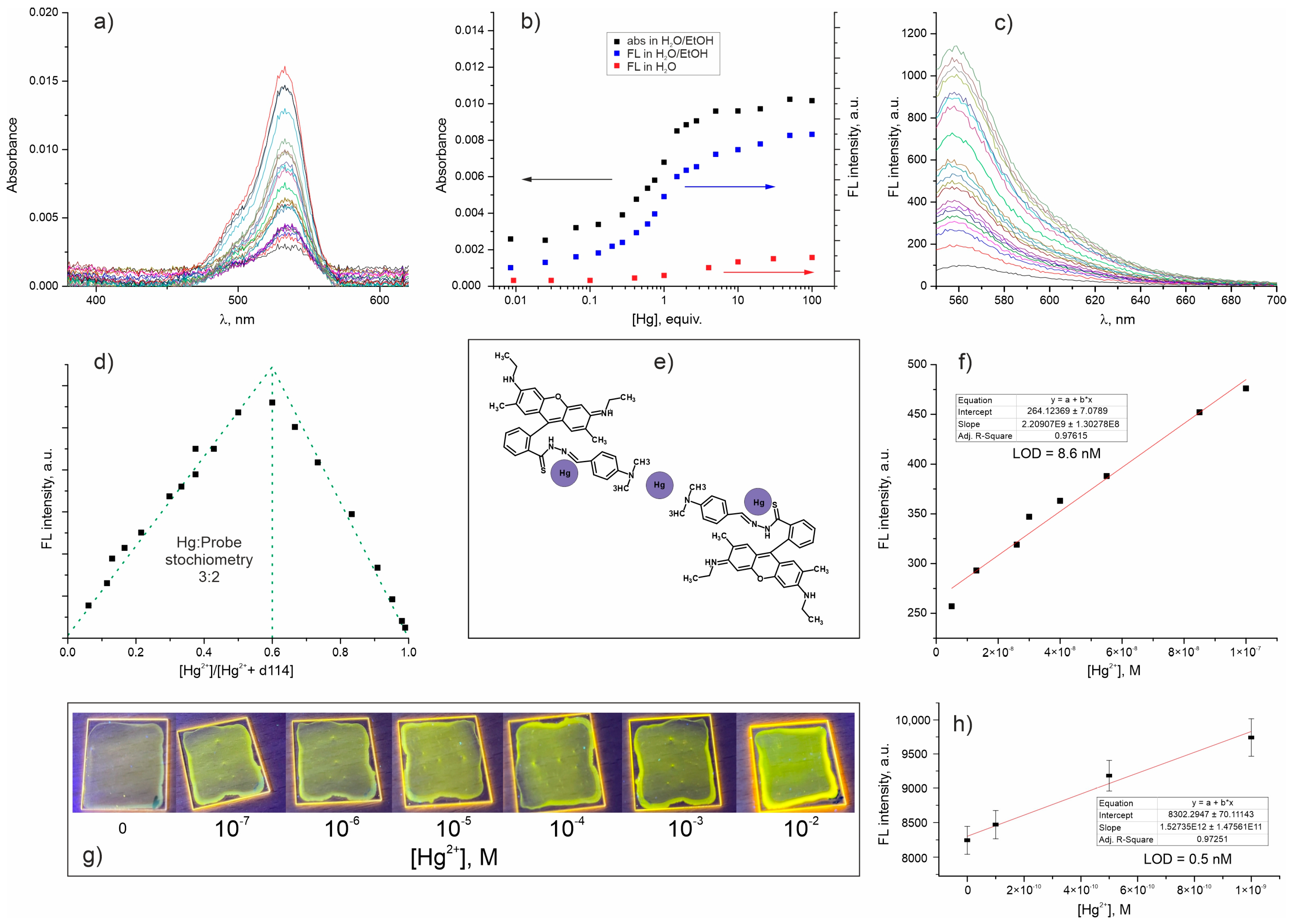
The development of an ultra-sensitive nanoprobe for detection of trace amounts of mercury was started from the design of a suitable molecular probe. The probe designed for Hg2+ detection (d114) is a derivative of d98 probe obtained by substitution amide oxygen with sulfur (Figure 1). We expected that d98 and d114 would be a pair of probes for Cu2+ and Hg2+ detection with properties similar to those of the already known rhodamine hydrazide and thiohydrazide probes. However, the characterization of d114 in solution revealed that its optical response has a fundamentally different character. In sharp contrast to d98, which forms a colored and non-luminescent 1:1 complex with Cu2+ [29], the successive addition of Hg2+ to d114 solution results in a simultaneous increase in absorption (Figure 2a,b) and luminescence (Figure 2b,c). The method of continuous variation revealed the formation of 3:2 Hg2+/d114 complex (Figure 2d,e), whose binding constant in H2O/C2H5OH (1/1, v/v), calculated as described in [30], was equal to 2.8·107 (Figure S3, Supplementary information). This value is higher than those usually reported for Rhodamine-based probes for metal ions [21,22] and other probes for Hg2+ [3].
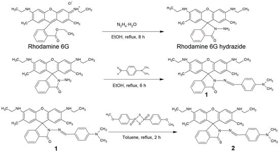
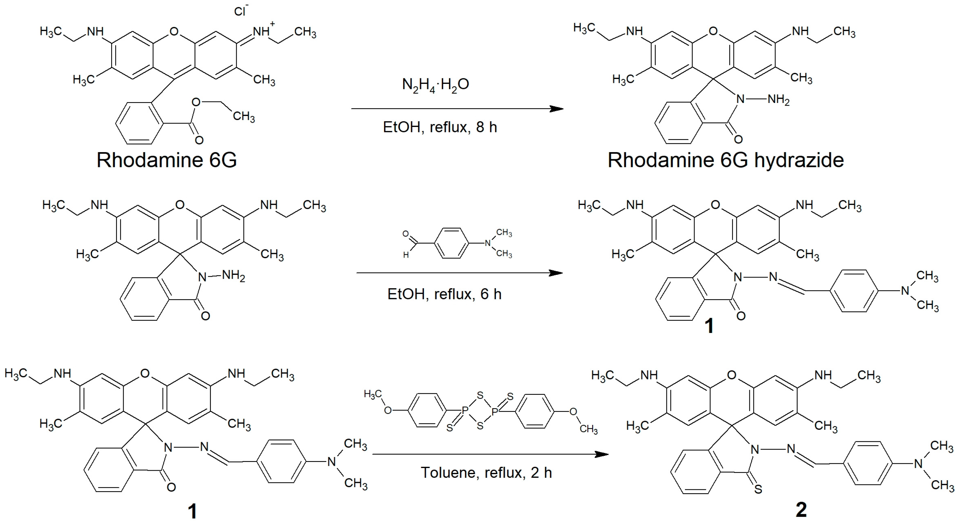
Figure 1.
The synthesis route of d114 (compound 2).
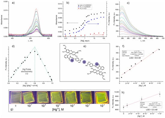
Figure 2.
Characterization of d114 probe in H2O/C2H5OH (1/1, v/v): the change of absorbance (a,b) and fluorescence (c,b) spectra upon successive addition of Hg2+ to d114 solution with concentration of 10−6 M; Job’s plots (method of continuous variation) showing formation of 3:2 Hg2+/d114 complexes in solution (d); proposed binding scheme between d114 and Hg2+ ions (e); dependence of the response signal on the concentration of the analyte and results of the Hg2+ LOD calculations (f). Photo of PMMA slides with latex coatings containing d114 probe immersed for 5 min in Hg2+ solutions (pH = 6) and excited with 365 nm LED (g); and results of the Hg2+ LOD calculations for d114-doped coating (h).
The LOD for mercury with d114 in H2O/C2H5OH (1/1, v/v) was 8.6 nM or 1.72 µg/L (Figure 2f) that is below mercury MPC in drinking water recommended by WOS (6 µg/L) [1] and the maximum concentration level (MCL) for the discharge to the aquatic environment regulated by US Environmental Protection Agency (2 µg/L) [31]. This makes the d114 probe superior to most of the reported optical sensors. However, the intensity of the d114 optical signal is solvent-dependent, as is often observed for most fluorophores [32]; it is much lower in water than it is in an H2O/C2H5OH mixture (Figure 2b) or in pure organic solvents, so the analyzed sample has to be diluted first. This increases LOD and somehow complicates express analysis if this probe is used in the devices for the field application.
In order to estimate the potential of d114 for the modification of channels in microfluidic sensors, we fabricated transparent latex coatings with a thickness of ~600 nm on PMMA slides (Figure 2g), which have demonstrated a fast response time in aqueous solutions with an LOD of ~0.5 nM or 0.1 µg/L (Figure 2h). Sensitivity enhancement of the probe after immobilization in the coatings can be related to the metal ions preconcentration on the carboxylic group of the siloxane-acrylate latex, which was earlier demonstrated by our group [33]. Furthermore, despite a hydrophobicity sufficient to prevent fluorophore release to the aqueous solution and the high stability of the siloxane-acrylate coatings in aqueous solutions at pH < 11, they were permeable for metal ions [34,35]. A large excess of carboxylic groups in the coating did not interfere with the sensory response due to the much higher binding constant of Hg2 with S-containing ligand (d114). Thus, the Hg2+ LOD reached using d114-doped siloxane-acrylate coatings was sufficient for application in extra-laboratory portable devices to control the mercury concentration in water below MPC [1] and MCL [31].
However, field monitoring of mercury concentrations in the areas of concern, e.g., water reservoirs in highly developed industrial areas and gold mining and processing sites requires more sensitive methods of detection due to the fast accumulation of mercury by living organisms and organic matter and its chronic toxic effects. Thus, the next step consisted in development of light-harvesting FRET NPs, which was done to reach Hg2+ LOD in water at the level far below ppb.
Our previous work [29] was focused on finding the conditions for the formation of the brightest coumarin-based NPs, which then were doped with a Cu2+-probe (d98) to yield a nanoprobe with FRET enhancement. In order to reach a high luminescence quantum yield of such NPs, they had to be assembled at a high counterion/dye ratio in the solution. However, at a large excess of counterion (F12) in water, amines and their derivatives (including lactams, i.e., all rhodamine-based probes) are protonated and associated into insoluble NPs. This makes the enhancing of the sensory response of classical rhodamine-based probes impossible, since “switching ON” the probe inside the NPs is determined by the NPs component (F12 counterion) rather than by the analyte (target ion). To overcome this limitation, we have modified a Cu2+-sensitive probe (d98) with an electron-donating amino aromatic group, which was introduced to change the optical response of the probe via photoinduced electron transfer (PET) fluorescence quenching that allowed Cu2+ detection via the quenching of the initially bright nanoparticles. Thus, d98 has two binding sites—hydrazone and amino aromatic fragments (see compound 1 structure in Figure 1). The hydrazone fragment has affinity for both Cu2+ and H+ ions, while the amino aromatic one only has an affinity for H+. When d98 was associated with an excess of F12 in acidic solution or inside sensitive NPs, both binding sites of the molecule were occupied by H+ ions, so the probe existed in colored and fluorescent form, which was efficiently pumped by the light harvesting matrix, since the emission band of Coumarin 30 significantly overlaps with excitation band of d98. The binding of the Cu2+ ion to d98 either dissolved or incorporated in NPs leads to the displacement of both protons from the probe molecule and a decrease in fluorescence intensity. Thus, the concentration of Cu2+ ions can be determined from the change in the fluorescence intensity of the solution at constant absorption (i.e., by decrease in FLQY).
Taking into account the stoichiometry of binding and the bright luminescence of the Hg2+/d114 complex, the approach suggested for Cu2+ determination using FRET NPs has to be modified. To obtain a system capable of modulating the d114 sensory response under the action of an ion analyte, it was necessary to reduce the excess of the counterion in NPs in order to minimize its effect on the d114 binding sites responsible for sensing properties, and thus maintain the ability of the probe to interact with the analyte.
For this, Coumarin-30/F12 (further referred to as C30/F12) nanoparticles doped with d114 were obtained at a C30/F12 ratio of 1:2. We showed earlier that C30/F12 NPs obtained at pH = 5 in sodium-acetate buffer solution have low colloidal stability. Here we have used Millipore ® water with a neutral pH that greatly improved the colloidal stability of the obtained solutions. According to the dynamic light scattering data, the size of the obtained NPs was ~250 nm, while the size of unstable particles obtained at the same ratio of precursors in the sodium acetate buffer was ~130 nm.
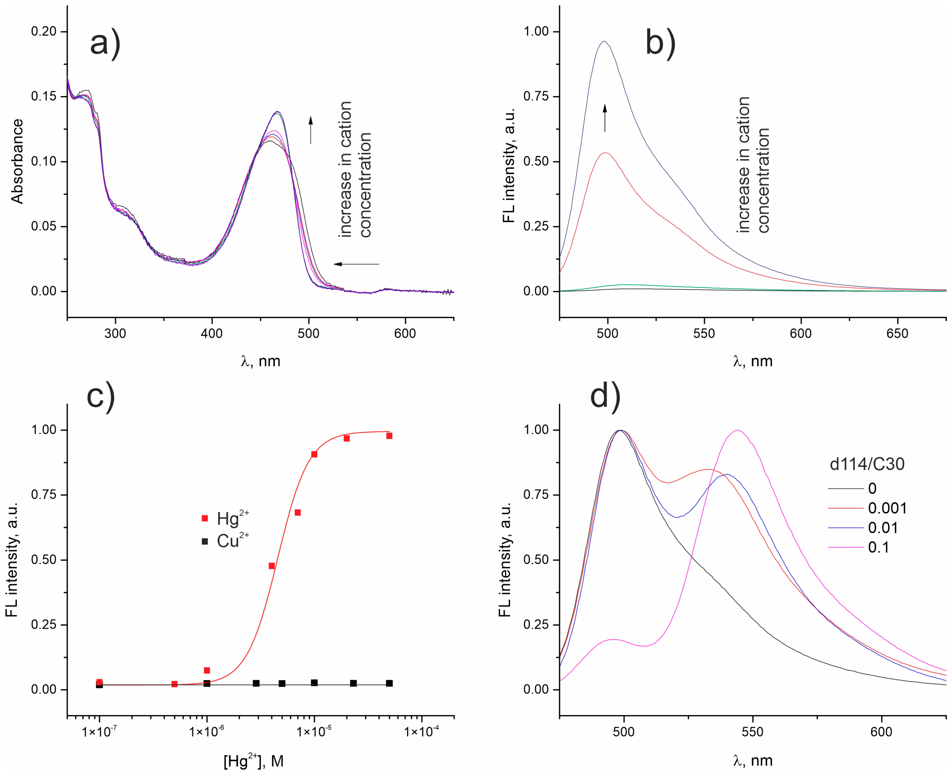
Since colloidal solutions, and especially ionic associates, are quite sensitive to changes in the ionic composition of the solution, we first tested pure C30/F12 NPs solution by titration with Hg2+ and Cu2+, i.e., cations with a high affinity to d114, to determine the concentration range over which C30/F12 NPs are stable. Figure 3 shows that at Hg2+ concentrations up to 10−6 M, the intensity of the luminescence of the C30/F12 NPs solution virtually did not change, while at higher concentrations a significant increase in luminescence intensity and a slight shift of the absorbance band was observed (Figure 3a–c). Next, the corresponding solutions of C30/F12 NPs doped with different amounts of d114 were obtained and tested by the addition of Hg2+ at a concentration of 10−6 M to establish the maximum C30/d114 ratio at which FRET from coumarin nanoantenna to d114 is observed (Figure 3d). The estimated FRET efficiency in NPs with the composition d114/C30/F12 of 0.001/1/2 was ~30%, which means that the antenna effect in the system is about 300. This value is rather close to that (1000) reported by the Klymchenko group [25].
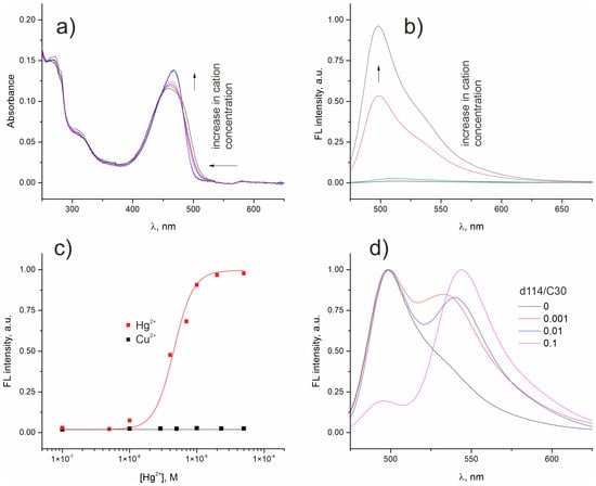
Figure 3.
The change in absorption (a) and luminescence (b) spectra of C30/F12 1/2 NPs in aqueous solution (pH = 6) upon addition of Hg2+. The dependence of luminescence intensity of C30/F12 1/2 NPs on Hg2+ and Cu2+ concentration (c). The luminescence spectra of C30/F12 NPs doped with different amount of d114 (d).
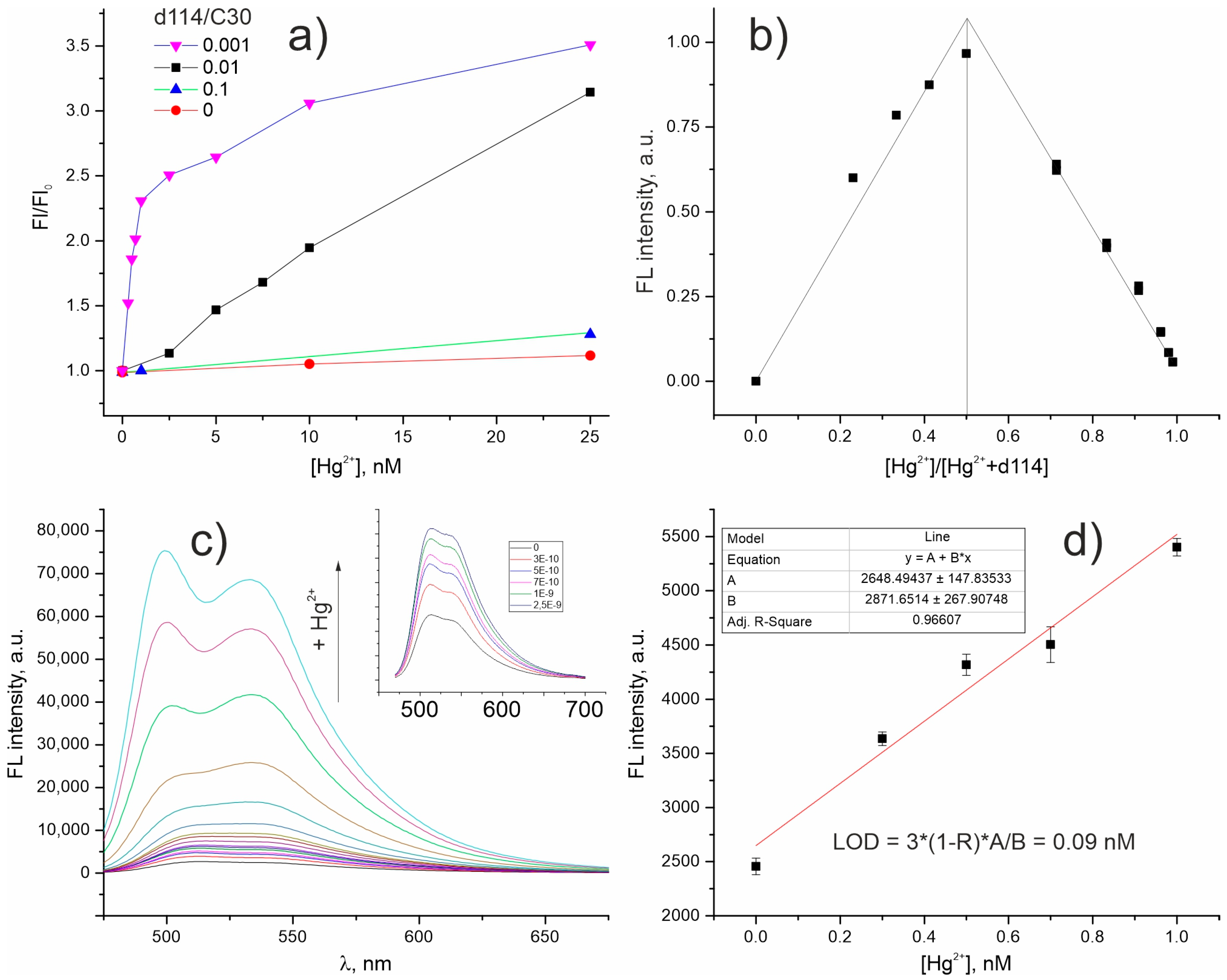
The titration of d114/C30/F12 NPs in aqueous solution with Hg2+ ions have shown that at a lower d114 content in the system, a sharper change in the signal intensity in the region of low concentrations is observed (Figure 4a). Additionally, the method of continuous variation was applied for d114/C30/F12 0.001/1/2 NPs to analyze binding stoichiometry between d114 and Hg2+ and demonstrated inflection at a 1:1 ratio of Hg/d114 (Figure 4b). The change in the signal intensity (Figure 4c) in the Hg2+ concentration range up to 1 equiv. of d114 was approximately linear (Figure 4a,c); the Hg2+ LOD calculated for nanoprobe d114/C30/F12 0.001/1/2 according to signal-to-noise ratio of 3 was 0.09 nM (Figure 4d). It is also worthy of note that, similarly to Cu2+-sensitive FRET NPs doped with d98 [29], the presence of low amounts (at least up to 10–6 M) of interfering ions, including Cu2+, does not significantly affect the sensitivity of mercury detection (Figure S4, Supplementary Information).
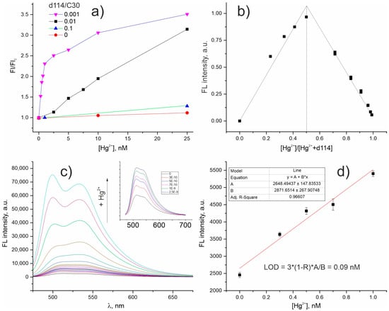
Figure 4.
Characterization of d114/C30/F12 NPs in aqueous solution (pH = 6): dependence of luminescence intensity change (F/F0) on Hg2+ concentration for NPs with different d114 content (a); Job’s plots showing 1:1 interaction stoichiometry of d114 and Hg2+ inside light-harvesting NPs d114/C30/F12 0.001/1/2 (b); the change in the luminescence spectrum of d114/C30/F12 0.001/1/2 solution upon the addition of Hg2+(the inset shows the spectra in Hg2+ concentration range up to 2.5 nM) (c); dependence of the response signal on the analyte concentration in the range up to 1nM (d).
3. Materials and Methods
3.1. Chemicals and Instruments
Rhodamine 6G (99%, Sigma Aldrich, St. Louis, MO, USA), Coumarin 30 (99%, Sigma Aldrich), 4-(dimethylamino)benzaldehyde (99%, Sigma Aldrich), Lawesson reagent 2,4-Bis-(4-methoxyphenyl)-1,3-dithia-2,4-diphosphetane 2,4-disulfide (97%, Sigma Aldrich), hexane (95%, Sigma Aldrich), chloroform (99%, Sigma Aldrich), ethyl acetate (99.8%, Sigma Aldrich), dichloromethane (99%, Sigma Aldrich), hydrazine monohydrate (98%, Sigma Aldrich), sodium tetrakis [3,5-bis(1,1,1,3,3,3-hexafluoro-2-methoxy-2-propyl)phenyl]borate trihydrate (99%, Sigma Aldrich), and silica gel (100/200 μm) were used as received. Siloxane-acrylate latex (KE 13-36) dispersion with a solid phase content of 46%, were produced by the Scientific Production Association “Astrokhim” (Elektrostal’, Moscow Region, Russia). All other reagents were of analytical grade and were used without purification. All aqueous solutions were prepared using Millipore ® water.
The Fourier transform infrared radiation (FT-IR) spectra of the compounds in the range 400–4000 cm−1 were recorded using a Perkin Elmer Spectrum 100BX II spectrometer in KBr pellets. 1H, 13C NMR spectra were performed on a Bruker Avance 400 with the frequency of proton resonance of 400 MHz using CDCl3 as the solvent and tetramethylsiliane as the internal reference. Mass spectrometry was performed on a Shimadzu LCMS-2010 LC-ESI/MS system. The UV-Vis spectra were obtained using a Shimadzu UV-2600 spectrophotometer equipped with a Shimadzu ISR 2600 Plus integrating sphere. The fluorescence spectra were obtained with a Shimadzu RF-6000 spectrofluorophotometer with a 1 cm standard quartz cell. For selectivity experiments, freshly prepared stock solutions of the nitrate salts of Hg2+, Cu2+, Ni2+, Mg2+, Al3+, Zn3+, Co2+, Ag+ in Millipore ® water were used. All of the titration experiments were recorded at room temperature. The size of fluorescent NPs was determined using a ZetaSizer Nano ZS analyzer (Malvern Instruments Ltd., UK). The analyzed solutions were filtered through a 0.8 µm syringe filter to remove dust particles, while the optical absorption of the solutions was monitored using UV-visible spectroscopy before and after filtration to ensure that the NPs were not retained by the membrane. The measurements were carried out in automatic operation mode at room temperature. The pH measurements were carried out using a Sartorius Professional Meter PP-50. The thickness of the sensing coatings was measured using an Auto SE spectroscopic ellipsometer (Horiba, Japan).
3.2. Synthesis and Characterization of d114
Rhodamine 6G hydrazide was synthesized using the similar procedure as described in [36] with minor modifications. Briefly, 300 mg, (6.3 · 10−4 mol) of rhodamine 6G and hydrazine monohydrate (1.0 mL, 1.9·10−2 mol) were dissolved in 20 mL of ethanol (95%). The mixture was refluxed for 12 h. The solvent was removed under the reduced pressure and the crude product was purified by flash column chromatography on a silica gel with dichloromethane/ethyl acetate (v/v = 4/1) as the eluent to afford the product as a crystal powder (220 mg, Yield 80%). If a larger amount of the substance needs to be synthesized, purification can be carried out by recrystallization from ethanol. 1H NMR (400 MHz, CDCl3, ppm, δ): 7.96 (m, 1H), 7.45 (m, 2H), 7.06 (m, 1H), 6.39 (s, 2H), 6.26 (s, 2H), 3.58 (s, 2H), 3.54 (br.s, 2H), 3.22 (q, 4H), 1.92 (s, 6H), 1.32 (t, 6H); 13C NMR (100 MHz, CDCl3, ppm, δ): 14.38, 16.34, 37.97, 65.66, 96.42, 104.47, 117.60, 122.65, 123.42, 127.31, 127.75, 129.45, 132.22, 147.15, 151.35, 151.83, 165.82; ESI-MS (m/z, +νe mode) 529.63 [M + H]+, calc. for C26H29N4O2+ is 529.22; Elemental Analysis data: Calc. C, 72.87; H, 6.59; N, 13.07; Expt. C, 72.97; H, 6.66; N, 12.89.
Compound 1 (d98) was synthesized as described in our previous work [29]. An amount of 300 mg (7 · 10−4 mol) of rhodamine 6G hydrazide and 220 mg (1.4 · 10−3 mol) of 4-(dimethylamino)benzaldehyde were dissolved in 15 mL of ethanol (95%). The mixture was refluxed for 6 h. The solvent was removed under reduced pressure and the crude product was purified by flash column chromatography on a silica gel with hexane/ethyl acetate (v/v = 2/1) as the eluent to afford the product as a crystal powder (290 mg, Yield 74%). 1H NMR (400 MHz, CDCl3, ppm, δ): 8.33 (s, 1H, H(24)), 8.03–8.04 (m, 1H, H(12)), 7.06–7.08 (m, 1H, H(9)), 7.45 (s, 1H, H(18)), 7.47 (s, 1H, H(19)), 7.61 (s, 1H, H(15)), 7.59 (s, 1H, H(22)), 7.47–7.49 (m, 2H, H(10), H(11)), 6.42 (br. s, 2H, H(26), H(30)), 6.40 (br. s, 2H, H(27), H(29)), 3.50 (br.s, 2H, H(33), H(36)), 3.21–3.26 (q, 4H, H(34), H(37)), 2.96 (s, 6H, H(41), H(42)), 1.89 (s, 6H, H(32), H(39)), 1.34–1.36 (t, 6H, H(35), H(38)); 13C NMR (100 MHz, CDCl3, ppm, δ): 14.77, 16.68, 22.70, 29.70, 31.94, 38.37, 40.23, 59.19, 65.72, 76.70, 77.03, 77.34, 96.78, 106.63, 111.51, 117.94, 123.21, 123.51, 127.76, 128.98, 133.00, 147.44, 151.17, 152.40, 164.83; ESI-MS (m/z, +νe mode) 560.81 [M + H]+, calc. for C35H38N5O2+ is 560.30; Elemental Analysis data: Calc. C, 75.11; H, 6.66; N, 12.51; Expt. C, 75.43; H, 6.70; N, 12.42.
Compound 2 (d114). The scheme of synthesis is presented in Figure 1. An amount of 30 mg (5.37 · 10−4 mol) of 1 and 22 mg (5.37 · 10−4 mol) of Lawesson reagent were mixed in 15 mL of absolute toluene. The mixture was refluxed for 1 h. An additional portion of Lawesson reagent (11 mg, 2.69 · 10−4 mol) was then added to the reaction mixture, and then the mixture was refluxed for 1 h. The solvent was removed under reduced pressure and the crude product was purified by flash column chromatography on a silica gel with hexane/ethyl acetate (v/v = 1/1) as the eluent to afford the product as a red powder (17.7 mg, 57%). 1H NMR (400 MHz, CDCl3, ppm, δ): 8.53 (s, 1H, H(24)), 8.12–8.14 (m, 1H, H(12)), 7.05–7.07 (m, 1H, H(9)), 7.37 (s, 1H, H(18)), 7.40 (s, 1H, H(19)), 7.66 (s, 1H, H(15)), 7.69 (s, 1H, H(22)), 7.40 (m, 2H, H(10), H(11)), 6.30 (br. s, 2H, H(26), H(30)), 6.62 (br. s, 2H, H(27), H(29)), 3.50 (br.s, 2H, H(33), H(36)), 3.20–3.21 (q, 4H, H(34), H(37)), 3.01 (s, 6H, H(41), H(42)), 1.92 (s, 6H, H(32), H(39)), 1.29–1.33 (t, 6H, H(35), H(38)); 13C NMR (100 MHz, CDCl3, ppm, δ): 14.36, 14.92, 17.02, 22.92, 29.93, 32.15, 38.78, 40.38, 96.75, 111.76, 118.32, 122.32, 127.29, 127.91, 130.39, 130.64, 132.12, 150.20, 152.39, 155.67, 159.72; ESI-MS (m/z, +νe mode) 576.34 [M + H]+, calc. for C35H38N5OS+ is 576.28; Elemental Analysis data: Calc. for C35H37N5OS: C, 73.01; H, 6.48; N, 12.16; S, 5.57; Expt. C, 73.33; H, 6.54; N, 12.18; S, 5.41. Mp: 208–210 °C (with decomposition). 1H NMR and 13C NMR spectra are shown in Figures S1 and S2, respectively (Supplementary information).
3.3. Fabrication of the Sensing Coating Containing d114 on PMMA
Transparent sensing coatings were fabricated by casting 0.156 mL of siloxane-acrylate latex dispersion in H2O/CH3OH (1/1, v/v) with solid content of 2% and d114 concentration of 10−5 M on the polymethyl methacrylate (PMMA) slides with the surface area of 4.05 cm2. Coatings were left for drying in the air at T = 25 °C for 24 h before use.
3.4. Preparation of Fluorescent NPs
1 mM of Coumarin-30 (further referred to as C30) in CH3CN, 1–100 µM of d114 in CH3CN and 1mM of sodium tetrakis [3,5-bis(1,1,1,3,3,3-hexafluoro-2-methoxy-2-propyl)phenyl]borate) (further referred to as F12) in CH3CN were used as stock solutions. Typically, 10 µL of C30, 0–10 µL of d114 and 20 µL of F12 stock solutions were mixed in a plastic vial and quickly added to 5 mL of deionized water under intensive stirring to yield a colloidal solution of fluorescent NPs. The resulting colloidal solutions were left for equilibration for 60 min before use.
4. Conclusions
Very strict limitations to the mercury content in discharge and drinking water results in the failure of the most fluorescent probes to provide the required detection limit. It is therefore necessary to enhance the response signal significantly so that it can be realized, for example, via pumping the probe with a much brighter quantum emitter via Förster resonance energy transfer (FRET). Pursuing the goal of developing optical sensors for the extra-laboratory determination of mercury at ultralow concentrations, we have designed a new thiated Rhodamine derivative (d114) with a luminescence response inverted from “on/off” to “off/on”, which demonstrated an Hg 2+ LOD of 8.6 nM in H2O/C2H5OH (1/1, v/v) solution and of 0.5 nM in water when used for doping transparent siloxane acrylate coating on PMMA.
To further improve the sensitivity of this fluorophore, we have modified our earlier developed strategy for the fabrication of Cu2+—sensitive probe-doped light-harvesting FRET nanoparticles (NPs) formed by Coumarin-30 (C30) and sodium tetrakis [3,5-bis(1,1,1,3,3,3-hexafluoro-2-methoxy-2-propyl)phenyl]borate) (F12). Due to the different nature of the sensory response and binding stoichiometry of the earlier developed probe for Cu2+ ions and d114 for Hg2+ ions, the composition of the FRET NPs has to be optimized. We have investigated the dependence of the fluorescence signal on the d114/C30 mol ratio and shown that a nanoprobe with the composition of d114/C30/F12 0.001/1/2 provided an antenna effect of ~300. Taking advantage of the FRET effect, ~100-fold decrease of mercury LOD was reached using a simple spectrofluorometer. Thus, the demonstrated approach makes possible the detection of extremely toxic Hg2+ cations in water at concentrations far below the maximum permissible concentration (MPC) in drinking water set by the World Health Organization and can be possibly extended to other optical sensors based on the Rhodamine-lactam platform.
Supplementary Materials
The following supporting information can be downloaded at: https://www.mdpi.com/article/10.3390/molecules28041633/s1, Figure S1: 1H NMR spectrum of d114 recorded in CDCl3; Figure S2: 13C NMR spectrum of d114; Figure S3: Fluorescence intensity of d114 solution versus free ligand concentration, [free Hg2+] calculated according to the law of mass action; Figure S4: Influence of the interfering metal ions (10−6 M) on response value of d114/C30/F12 0.001/1/2 NPs in the presence of 10−8 M Hg2+.
Author Contributions
Conceptualization, A.M.; Methodology, A.M. and S.B.; Formal analysis, A.C. and A.M.; Investigation, A.C., M.T. and D.B.; Writing—original draft preparation, review and editing, A.M. and S.B.; Visualization, A.C.; Supervision, S.B.; Funding acquisition, S.B. All authors have read and agreed to the published version of the manuscript.
Funding
This study was performed under the «ERA.Net RUS plus» program and funded by the Russian Foundation of Basic Research, project number 20-53-76016 ERA-t.
Institutional Review Board Statement
Not applicable.
Informed Consent Statement
Not applicable.
Data Availability Statement
Not applicable.
Conflicts of Interest
The authors declare that they have no conflict of interest.
References
- World Health Organization. Guidelines for Drinking-Water Quality: Fourth Edition Incorporating the First Addendum; WHO: Geneva, Switzerland, 2017; p. 541.
- Jaywant, S.A.; Mahmood Arif, K. A Comprehensive Review of Microfluidic Water Quality Monitoring Sensors. Sensors 2019, 19, 4781. [Google Scholar] [CrossRef] [PubMed]
- Li, M.; Li, X.-J.; Lu, H.-Y.; Chen, C.-F. Tetrahydro[5]helicene thioimide-based fluorescent and chromogenic chemodosimeter for highly selective and sensitive detection of Hg2+. Sens. Actuators B Chem. 2014, 202, 583–587. [Google Scholar] [CrossRef]
- Fan, C.; He, S.; Liu, G.; Wang, L.; Song, S. A Portable and Power-Free Microfluidic Device for Rapid and Sensitive Lead (Pb2+) Detection. Sensors 2012, 12, 9467–9475. [Google Scholar] [CrossRef] [PubMed]
- Mitsai, E.; Kuchmizhak, A.; Pustovalov, E.; Sergeev, A.; Mironenko, A.; Bratskaya, S.; Linklater, D.P.; Balčytis, A.; Ivanova, E.; Juodkazis, S. Chemically non-perturbing SERS detection of a catalytic reaction with black silicon. Nanoscale 2018, 10, 9780–9787. [Google Scholar] [CrossRef] [PubMed]
- Shinde, S.S.; Bhosale, C.H.; Rajpure, K.Y. Oxidative degradation of acid orange 7 using Ag-doped zinc oxide thin films. J. Photochem. Photobiol. B Biol. 2012, 117, 262–268. [Google Scholar] [CrossRef] [PubMed]
- Pavliuk, G.; Pavlov, D.; Mitsai, E.; Vitrik, O.; Mironenko, A.; Zakharenko, A.; Kulinich, S.A.; Juodkazis, S.; Bratskaya, S.; Zhizhchenko, A.; et al. Ultrasensitive SERS-based plasmonic sensor with analyte enrichment system produced by direct laser writing. Nanomaterials 2020, 10, 49. [Google Scholar] [CrossRef]
- Mitsai, E.; Naffouti, M.; David, T.; Abbarchi, M.; Hassayoun, L.; Storozhenko, D.; Mironenko, A.; Bratskaya, S.; Juodkazis, S.; Makarov, S.; et al. Si1-xGex nanoantennas with a tailored Raman response and light-to-heat conversion for advanced sensing applications. Nanoscale 2019, 11, 11634–11641. [Google Scholar] [CrossRef]
- Dostovalov, A.; Bronnikov, K.; Korolkov, V.; Babin, S.; Mitsai, E.; Mironenko, A.; Tutov, M.; Zhang, D.; Sugioka, K.; Maksimovic, J.; et al. Hierarchical anti-reflective laser-induced periodic surface structures (LIPSSs) on amorphous Si films for sensing applications. Nanoscale 2020, 12, 13431–13441. [Google Scholar] [CrossRef]
- Borodaenko, Y.; Gurbatov, S.; Tutov, M.; Zhizhchenko, A.; Kulinich, S.A.; Kuchmizhak, A.; Mironenko, A. Direct femtosecond laser fabrication of chemically functionalized ultra-black textures on silicon for sensing applications. Nanomaterials 2021, 11, 401. [Google Scholar] [CrossRef]
- Mironenko, A.Y.; Tutov, M.V.; Sergeev, A.A.; Mitsai, E.V.; Ustinov, A.Y.; Zhizhchenko, A.Y.; Linklater, D.P.; Bratskaya, S.Y.; Juodkazis, S.; Kuchmizhak, A.A. Ultratrace Nitroaromatic Vapor Detection via Surface-Enhanced Fluorescence on Carbazole-Terminated Black Silicon. ACS Sens. 2019, 4, 2879–2884. [Google Scholar] [CrossRef]
- Sergeeva, K.A.; Tutov, M.V.; Voznesenskiy, S.S.; Shamich, N.I.; Mironenko, A.Y.; Sergeev, A.A. Highly-sensitive fluorescent detection of chemical compounds via photonic nanojet excitation. Sens. Actuators B Chem. 2020, 305, 127354. [Google Scholar] [CrossRef]
- Tutov, M.V.; Sergeev, A.A.; Zadorozhny, P.A.; Bratskaya, S.Y.; Mironenko, A.Y. Dendrimeric rhodamine based fluorescent probe for selective detection of Au. Sens. Actuators B Chem. 2018, 273, 916–920. [Google Scholar] [CrossRef]
- Mironenko, A.Y.; Tutov, M.V.; Sergeev, A.A.; Bratskaya, S.Y. On/off rhodamine based fluorescent probe for detection of Au and Pd in aqueous solutions. Sens. Actuators B Chem. 2017, 246, 389–394. [Google Scholar] [CrossRef]
- Wang, L.; Ding, H.; Ran, X.; Tang, H.; Cao, D. Recent progress on reaction-based BODIPY probes for anion detection. Dye. Pigment. 2020, 172, 107857. [Google Scholar] [CrossRef]
- La, M.; Hao, Y.; Wang, Z.; Han, G.C.; Qu, L. Selective and Sensitive Detection of Cyanide Based on the Displacement Strategy Using a Water-Soluble Fluorescent Probe. J. Anal. Methods Chem. 2016, 2016, 1462013. [Google Scholar] [CrossRef] [PubMed]
- Wu, L.; Sedgwick, A.C.; Sun, X.; Bull, S.D.; He, X.-P.; James, T.D. Reaction-Based Fluorescent Probes for the Detection and Imaging of Reactive Oxygen, Nitrogen, and Sulfur Species. Acc. Chem. Res. 2019, 52, 2582–2597. [Google Scholar] [CrossRef]
- Nguyen, V.N.; Ha, J.; Cho, M.; Li, H.; Swamy, K.M.K.; Yoon, J. Recent developments of BODIPY-based colorimetric and fluorescent probes for the detection of reactive oxygen/nitrogen species and cancer diagnosis. Coord. Chem. Rev. 2021, 439, 213936. [Google Scholar] [CrossRef]
- Melnychuk, N.; Egloff, S.; Runser, A.; Reisch, A.; Klymchenko, A.S. Light-Harvesting Nanoparticle Probes for FRET-Based Detection of Oligonucleotides with Single-Molecule Sensitivity. Angew. Chemie Int. Ed. 2020, 59, 6811–6818. [Google Scholar] [CrossRef]
- Jurek, K.; Kabatc, J.; Kostrzewska, K.; Grabowska, M. New Fluorescence Probes for Biomolecules. Molecules 2015, 20, 13071–13079. [Google Scholar] [CrossRef]
- Kim, H.N.; Lee, M.H.; Kim, H.J.; Kim, J.S.; Yoon, J. A new trend in rhodamine-based chemosensors: Application of spirolactam ring-opening to sensing ions. Chem. Soc. Rev. 2008, 37, 1465. [Google Scholar] [CrossRef]
- Chen, X.; Pradhan, T.; Wang, F.; Kim, J.S.; Yoon, J. Fluorescent Chemosensors Based on Spiroring-Opening of Xanthenes and Related Derivatives. Chem. Rev. 2011, 112, 1910–1956. [Google Scholar] [CrossRef]
- Li, M.; Lu, H.-Y.; Liu, R.-L.; Chen, J.-D.; Chen, C.-F. Turn-On Fluorescent Sensor for Selective Detection of Zn 2+, Cd 2+, and Hg 2+ in Water. J. Org. Chem. 2012, 77, 3670–3673. [Google Scholar] [CrossRef] [PubMed]
- Liu, K.; Marin, L.; Cheng, X. Water-soluble β-cyclodextrin based turn-on amplifying fluorescent probes for sensitive and selective detection of Hg2+/Hg+ ions. Sens. Actuators B Chem. 2023, 377, 133060. [Google Scholar] [CrossRef]
- Trofymchuk, K.; Reisch, A.; Didier, P.; Fras, F.; Gilliot, P.; Mely, Y.; Klymchenko, A.S. Giant light-harvesting nanoantenna for single-molecule detection in ambient light. Nat. Photonics 2017, 11, 657–663. [Google Scholar] [CrossRef] [PubMed]
- Andreiuk, B.; Aparin, I.O.; Reisch, A.; Klymchenko, A.S. Bulky Barbiturates as Non-Toxic Ionic Dye Insulators for Enhanced Emission in Polymeric Nanoparticles. Chem. A Eur. J. 2021, 27, 12877–12883. [Google Scholar] [CrossRef]
- Shulov, I.; Oncul, S.; Reisch, A.; Arntz, Y.; Collot, M.; Mely, Y.; Klymchenko, A.S. Fluorinated counterion-enhanced emission of rhodamine aggregates: Ultrabright nanoparticles for bioimaging and light-harvesting. Nanoscale 2015, 7, 18198–18210. [Google Scholar] [CrossRef]
- Aparin, I.O.; Melnychuk, N.; Klymchenko, A.S. Ionic Aggregation-Induced Emission: Bulky Hydrophobic Counterions Light Up Dyes in Polymeric Nanoparticles. Adv. Opt. Mater. 2020, 8, 2000027. [Google Scholar] [CrossRef]
- Mironenko, A.Y.; Tutov, M.V.; Chepak, A.K.; Bratskaya, S.Y. FRET pumping of rhodamine-based probe in light-harvesting nanoparticles for highly sensitive detection of Cu2+. Anal. Chim. Acta 2022, 1229, 340388. [Google Scholar] [CrossRef]
- Mironenko, A.Y.; Tutov, M.V.; Chepak, A.K.; Zadorozhny, P.A.; Bratskaya, S.Y. A novel rhodamine-based turn-on probe for fluorescent detection of Au3+ and colorimetric detection of Cu2+. Tetrahedron 2019, 75, 1492–1496. [Google Scholar] [CrossRef]
- Water Quality Association. Mercury in Drinking Water. Available online: https://wqa.org/learn-about-water/common-contaminants/mercury (accessed on 30 October 2022).
- Adamoczky, A.; Nagy, L.; Nagy, M.; Zsuga, M.; Kéki, S. Conversion of isocyanide to amine in the presence of water and hg(Ii) ions: Kinetics and mechanism as detected by fluorescence spectroscopy and mass spectrometry. Int. J. Mol. Sci. 2020, 21, 5588. [Google Scholar] [CrossRef]
- Avramenko, V.; Bratskaya, S.; Zheleznov, V.; Sheveleva, I.; Voitenko, O.; Sergienko, V. Colloid stable sorbents for cesium removal: Preparation and application of latex particles functionalized with transition metals ferrocyanides. J. Hazard. Mater. 2011, 186, 1343–1350. [Google Scholar] [CrossRef] [PubMed]
- Bratskaya, S.; Mironenko, A.; Koivula, R.; Synytska, A.; Musyanovych, A.; Simon, F.; Marinin, D.; Göbel, M.; Harjula, R.; Avramenko, V. Polymer-inorganic coatings containing nanosized sorbents selective to radionuclides. 2. Latex/tin oxide composites for cobalt fixation. ACS Appl. Mater. Interfaces 2014, 6, 22387–22392. [Google Scholar] [CrossRef] [PubMed]
- Bratskaya, S.; Musyanovych, A.; Zheleznov, V.; Synytska, A.; Marinin, D.; Simon, F.; Avramenko, V. Polymer-inorganic coatings containing nanosized sorbents selective to radionuclides. 1. Latex/Cobalt Hexacyanoferrate(II) composites for cesium fixation. ACS Appl. Mater. Interfaces 2014, 6, 16769–16776. [Google Scholar] [CrossRef] [PubMed]
- Zhang, J.F.; Zhou, Y.; Yoon, J.; Kim, Y.; Kim, S.J.; Kim, J.S. Naphthalimide Modified Rhodamine Derivative: Ratiometric and Selective Fluorescent Sensor for Cu2+ Based on Two Different Approaches. Org. Lett. 2010, 12, 3852–3855. [Google Scholar] [CrossRef] [PubMed]
Disclaimer/Publisher’s Note: The statements, opinions and data contained in all publications are solely those of the individual author(s) and contributor(s) and not of MDPI and/or the editor(s). MDPI and/or the editor(s) disclaim responsibility for any injury to people or property resulting from any ideas, methods, instructions or products referred to in the content. |
© 2023 by the authors. Licensee MDPI, Basel, Switzerland. This article is an open access article distributed under the terms and conditions of the Creative Commons Attribution (CC BY) license (https://creativecommons.org/licenses/by/4.0/).