Review

Development and Application of Ruthenium(II) and Iridium(III) Based Complexes for Anion Sensing

School of Chemical Sciences, Indian Association for the Cultivation of Science, 2A & 2B Raja S. C. Mullick Road, Kolkata 700032, India
*
Author to whom correspondence should be addressed.
These authors contributed equally to this work.
Molecules 2023, 28(3), 1231; https://doi.org/10.3390/molecules28031231
Submission received: 28 December 2022 / Revised: 18 January 2023 / Accepted: 20 January 2023 / Published: 27 January 2023

Abstract

:
Improvements in the design of receptors for the detection and quantification of anions are desirable and ongoing in the field of anion chemistry, and remarkable progress has been made in this direction. In this regard, the development of luminescent chemosensors for sensing anions is an imperative and demanding sub-area in supramolecular chemistry. This decade, in particular, witnessed advancements in chemosensors based on ruthenium and iridium complexes for anion sensing by virtue of their modular synthesis and rich chemical and photophysical properties, such as visible excitation wavelength, high quantum efficiency, high luminescence intensity, long lifetimes of phosphorescence, and large Stokes shifts, etc. Thus, this review aims to summarize the recent advances in the development of ruthenium(II) and iridium(III)-based complexes for their application as luminescent chemosensors for anion sensing. In addition, the focus was devoted to designing aspects of polypyridyl complexes of these two transition metals with different recognition motifs, which upon interacting with different inorganic anions, produces desirable quantifiable outputs.

1. Introduction

Anions play vital roles in numerous physiological and industrial processes [1,2,3,4,5,6,7]. In addition, anionic species are omnipresent in the environment and act as an essential component to sustain growth and/or as pollutants [8,9,10]. So, the selective detection and sensing of anions are of immense interest in supramolecular chemistry. The more complex geometries, larger size, pH dependency, and higher hydration energies of the anions compared to cations make selective anion binding an additional challenging task. In the last few decades, significant progress has been made in the area of designing receptor molecules for the detection and quantification of anions [11,12,13,14,15,16,17].
In recent years, the development of phosphorescent compounds with unique photophysical properties is growing as a wide area of research. Such organometallic complexes usually consist of transition metal ions of the second and third row, such as rhenium(I), ruthenium(II), iridium(III), platinum(II), osmium(II) and gold(I), and organic ligand frameworks [18,19,20,21,22,23]. These metal complexes usually have a range of excited states, such as ligand-to-ligand charge transfer (LLCT), metal–metal-to-ligand charge transfer (MMLCT), ligand-to-metal charge transfer (LMCT), intra-ligand charge transfer (ILCT), metal-to-ligand charge transfer (MLCT), and ligand-to-metal–metal charge transfer (LMMCT) [19,24,25,26]. The emission properties of these complexes can be varied by coordinating different ligands and varying the local environments. Upon photoexcitation, the strong spin-orbit coupling in heavy metals facilitates rapid inter-conversion of the singlet excited states to the triplet excited state via effective intersystem crossing states. This triplet-state emission is responsible for the intense and long-lived phosphorescence in transition metal complexes [24] (Figure 1).
Among the various transition metal compounds, Ru(II) and Ir(III) complexes are receiving significant interest because of their application as molecular receptors and probes for anion sensing. In this regard, the fundamental criteria for the effective synthesis of such complexes are (i) high quantum yields at room temperature, (ii) appropriately large Strokes shift which limits inner filter effects, (iii) decent photochemical and photo-physical stability, (iv) easily tunable-photophysical properties with readily accessible near-infrared (NIR) emission,(v) ease of synthesis through ligand modification, and (vi) longer lifetime of emission which limits interference from autofluorescence [27,28].
A widely followed approach for the design of receptor host molecules is that the host must provide a binding site where the anion can coordinate/bind with the receptor molecule. Various supramolecular interactions, such as electrostatic interactions [29,30,31,32,33], hydrogen bonding [34,35], halogen bonding [36,37,38,39], anion -π interactions or π-π interactions [40,41,42,43,44], or the formation of a new host-guest conjugate species [13] (chemodosimeter) are commonly operational between the receptor molecule and the anion. Moreover, the anion binding event must be quantified by a detectable signaling output response which can be followed optically (naked eye, fluorescence, or phosphoresce color change), spectroscopically (through NMR, UV-Vis, PL), electrochemically (through an ion-selective electrode or cyclic voltammetry (CV)), through a sol-gel transformation or isothermal titration calorimetrically. The incorporation of an optical or electronic transducer into the receptor molecule responsive to a particular anion serves the above process and assists not only in detection but also in sensing.
Some of the review articles about transition metal-based chemosensors for anions, cations, and small molecules have been published [25,26,45,46,47,48,49]. However, some of them only focused on a specific motif-based (polar -NH moiety-based [46]; 1,10-phenanthroline-based [47]), while some of them published specific anions binding ruthenium or iridium complexes [48,49]. Here, this review seeks to summarize the recent advances in the development of luminescent ruthenium (II) and iridium (III) polypyridyl complexes for the purpose of anion sensing with different anion recognition motifs, which produces desirable results upon interacting with different inorganic anions. This review will focus on some recent development of polypyridyl complexes of ruthenium (II) and iridium (III) and their application as anion sensors.

2. Discussion

2.1. Ruthenium(II) Based Complexes for Sensing of Anion

These Ru(II) polypyridyl complexes reviewed as better chromophores have attracted tremendous interest owing to their notable large Stokes shift, excited state lifetime, and photophysical, electrochemical, and redox properties [46,50]. The amalgamation of ruthenium with a bipyridine (bpy) or phenanthroline (phen) moiety and a felicitous ligand could provide a binding site and a signaling unit for anions. In 1993, Beer and co-workers first reported [Ru(bpy)3]2+ complexes for anion sensing where amide substituents in bipyridine moiety acted as the anionic recognition site [51].

2.1.1. Triazole/Iodo-Triazole-Based Ru(II) Complexes as Chemosensors

In this part, we have concisely described triazole/iodo-triazole-based ruthenium(II) complexes for the “turn on” detection of anions through solitary C-H/C-I anion interaction. Triazole C-H is acidic in nature, and its incorporation with ruthenium metal makes it a suitable recognition site for anions. The previously described Ru(II) polypyridyl complexes worked as wonderful luminescent chemical sensors because of their distinctive optical properties such as large Stokes shift, extended excited-state lifetime, and excitation at visible region.
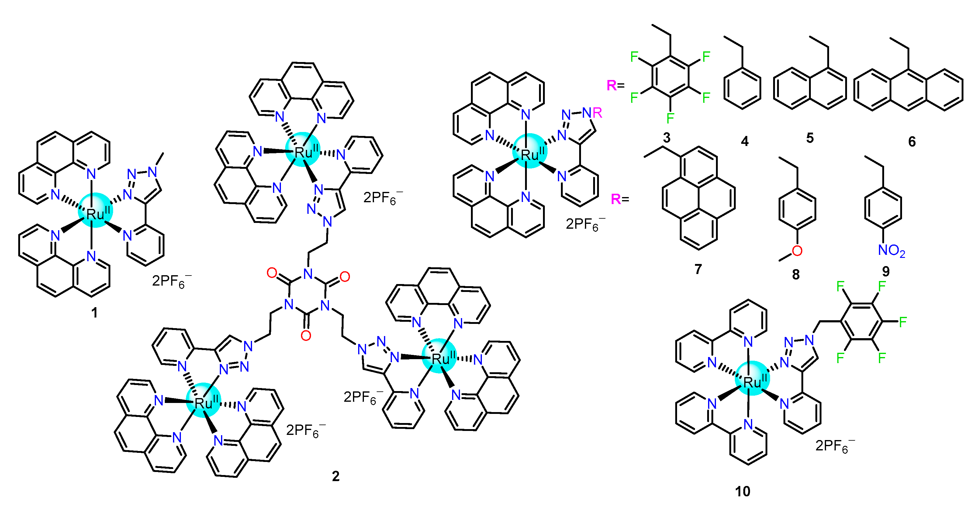
Since 2014, our group has systematically developed bis-heteroleptic ruthenium(II) complexes for selective sensing, recognition, and extraction of H2PO4 and HP2O73− anions. In this regard, mononuclear Ru(II) complex 1 and trinuclear electron-deficient cyanuric acid template-based Ru(II) complex 2 containing pyridine triazolium and phenanthroline units were synthesized for selective sensing of H2PO4 and HP2O73− (Figure 2) [52,53]. Both of these complexes showed distinctive MLCT charge transfer absorption bands that popped up at 403 nm [Ru(dπ)→phenanthroline] and 445 nm [Ru(dπ)→ triazole pyridine]. A weak luminescence band was observed at 590 nm upon excitation at either 403 or 445 nm for both complexes. Qualitative analysis of these two receptors by 1H NMR and UV-PL spectroscopies resulted in selective recognition of phosphates amid other examined anions (e.g., I, Br, Cl, F, NO3, HSO4, CH3CO2, ClO4, PhCO2, HCO3, etc.). Further PL titration of 1 showed an increase in emission intensity by 6-fold for H2PO4 and 3-fold with HP2O73− as compared to the free receptor, whereas 2 depicted 20-fold and 7-fold enhancement upon HP2O73− and H2PO4 binding, respectively. Through the –C-H unit of the triazole moiety mononuclear Ru(II) complex, 1, selectively binds with the singly charged and comparatively smaller H2PO4 anion, as established from X-ray crystallographic analysis. On the contrary, the trinuclear Ru(II) complex 2 was more flexible compared to 1, had numerous anion recognition sites and therefore preferred to bind with the larger and more negatively charged HP2O73− anion. Further, 2 exhibited more selectivity towards higher phosphate analogs (such as ATP, ADP, and AMP) over HP2O73− anions when a 10 mM concentrated solution of 10% Tris-HCl buffer in acetonitrile (having 1/9, v/v) medium was used. Excitingly, Job’s plot analysis revealed 1 and 2 formed 1:1 and 1:3 (H:G) complexes, respectively, with phosphates. The overall binding constant values in the presence of H2PO4 for 1 and 2 were calculated to be 5.28 × 10⁴ M−1 and 6.76 × 10¹³ M−3, respectively, and these values also revealed the trend of reversal of phosphate selectivity by 1 and 2. Remarkably, the lifetime of 2 was raised by 28-fold and 8-fold in the presence of HP2O73− and H2PO4, respectively, and it could be used as a potential sensor owing to their higher excited state lifetime.
In addition, our group developed an array of bis-heteroleptic Ru(II) complexes 310 (Figure 2), having the pyridine triazole unit as the anion binding site along with various other substituents which were involved in a meticulous comparative investigation for the development of superior selective probes for phosphates [54]. All these complexes showed photo-physical properties, which were similar to 1 and 2. UV/Vis spectra of these complexes (39) showed similar intense broad absorption peaks in the region 400–405 nm and 440–445 nm attributed to MLCT charge transfer from Ru-to-phenanthroline and Ru-to- triazole pyridine, respectively. In the case of 10, the absorption band appeared at 412 nm (Ru-to-bipyridine transition) and 440 nm (Ru-to- triazole pyridine transition). Probe 6 showed characteristic peaks at λmax391, 370, and 351 nm for the anthracene unit, whereas seven peaks at λmax 343, 327, and 313 nm were attributed to the pyrene unit, owing to π-π* transitions in both cases. Qualitative analysis showed that all these receptors were selective towards phosphates only, even in the presence of other competitive anions. Further, the gradual addition of phosphates to probes 39 individually resulted in a significant perturbation of the emission spectra reflected by increased emission intensity. In addition, the emission band was observed to be red-shifted by 15 nm and 50 nm in cases 6 and 7, respectively. In complexes 310, the acidic nature of the triazole C-H bond was changed by alternating the pendent substituents from electron-withdrawing to electron-donating, followed by polyaromatic substituents. Receptors 49 showed 5 to 10-fold enhancements of emission intensity in the presence of H2PO4 whereas 3 and 10 displayed ~22-fold and ~19-fold increases, respectively. Thus, it can be concluded that the pentafluorophenyl moiety in the substituent makes 3 and 10 comparatively superior sensors for phosphates accredited to the rigidification of molecular backbone assisted by supramolecular self-assembly, hydrogen bonding and anion-π interactions. The Job’s plot analysis suggested 1:1 (H:G) binding of receptors with H2PO4 where binding constant values ranged from 104 to 106 M−1. Among the receptors, receptor 3, having a pendent pentafluorophenyl moiety, showed the highest binding constant of 2.64 × 10⁶ M−1 and a very low detection limit of ~0.013 μM for H2PO4 Furthermore, the 1H NMR experiment revealed that the sharp singlet corresponding to the triazole C-H was found to be shifted to a more downfield region in the presence of H2PO4. Again, 19F NMR experimental results of 3 and 10 exposed an additional anion-π interaction between pentafluorophenyl moiety and H2PO4 which enabled them to be used as efficient sensors for phosphates. In addition, macroscopic morphological studies such as Transmission electron microscopy (TEM), Diffusion-ordered spectroscopy (DOSY) NMR experiments, and Dynamic light scattering (DLS) results disclosed that supramolecular aggregation occurred in solution and the solid state for all these complexes with H2PO4. The phosphate adducts of 3 and 10, as compared to other complexes, showed a maximum decrease in diffusion coefficient, i.e., from 7.188 × 10⁻¹⁰ to 1.659 × 10⁻¹⁰ m2/S for 3 and from 1.136 × 10⁻⁹ to 2.179 × 10⁻¹⁰ m2/S for 10. Thus, these macroscopic data results confirm the formation of higher-order supramolecular aggregates in solution, and this aggregate formation is assisted by anion-π interaction. Finally, the single crystal X-ray structure of phosphate adduct of 3 established the formation of supramolecular aggregates via anion-π and strong directional hydrogen-bonding interaction between triazole C-H and oxygen atoms of H2PO4. Electron-deficient pentafluorophenyl substituent, 3 was reported as a superior probe for H2PO4 in terms of a higher binding constant, larger enhancement of emission intensity, and lower detection limit over another aryl- and polyaromatic-substituted analogs (410). The superiority of 3 was realized because of the pentafluorophenyl substituent, which helped in the formation of supramolecular aggregation via both anion-fluorine/anion-π non-covalent and hydrogen bonding interactions.
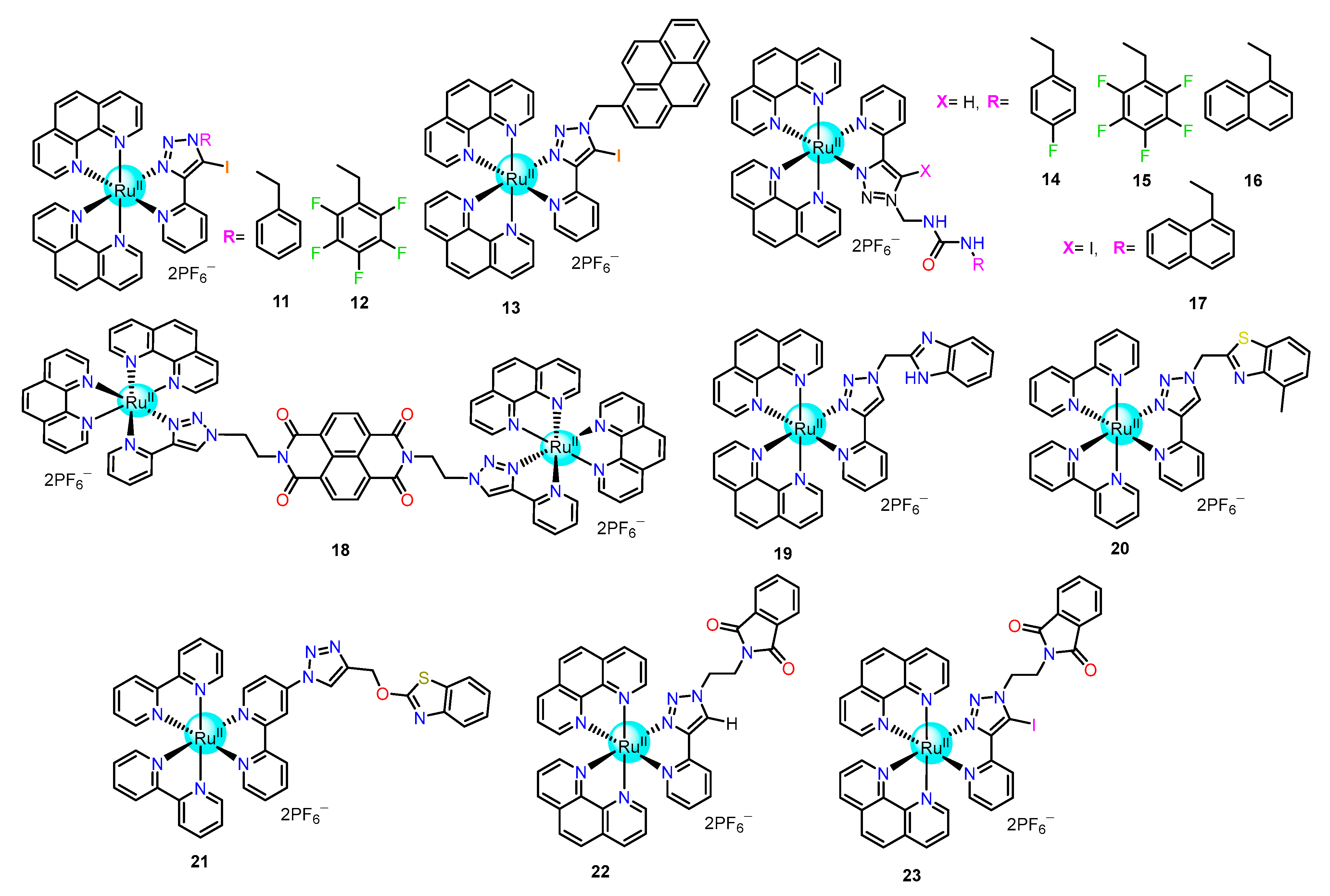
The significant phosphate selectivity via the C–H⋯ anion interactions by Ru (II)-polypyridyl complexes encouraged us to design halogen bonding-based receptors 11 and 12 (Figure 3) by replacing the hydrogen atom of C-H triazole bond with iodine [55,56]. Complexes 11 and 12 containing the iodotriazole (halogen bond unit) with phenyl and pentafluorophenyl (π-acidic) units as the pendent moiety for 11 and 12, respectively, showed superior dihydrogen phosphate sensing over their hydrogen bonding analogs. These receptors resembled the hydrogen bonding analogs in terms of UV-Vis absorption, emission spectra, and binding studies. The acetonitrile solution of receptors 11 and 12 showed ~16-fold and ~25-fold increments of emission intensities, respectively, in the presence of dihydrogen phosphate. The calculated binding constants for 11 and 12 were observed as 1.95 × 10⁵ and 2.76 × 10⁶ M−1, respectively, and the limit of detection of H2PO4 were 0.018 µM and 0.011 µM respectively. Furthermore, the DOSY NMR experiment and crystal structure analysis of receptors 11 and 12 exposed the formation of supramolecular polymeric structure in both solution and solid states through halogen bonding interaction and made them more sensitive and selective sensors for phosphates.
Between these two, 12 was found to be a superior sensor over 11 due to the presence of the pentafluorophenyl pendent moiety, which forms anion-π interaction with the dihydrogen phosphates, which additionally assisted in the formation of supramolecular aggregation. The DLS experiment further confirmed the formation of supramolecular aggregates in solution as the hydrodynamic diameter of free receptor 12 increased from ~130 nm to ~869 nm upon the addition of phosphates. In addition, due to the strong and highly directional nature of halogen bonding interaction, 12 could also be used as a perrhenate receptor in acetonitrile. To establish the origin of selective phosphate sensing phenomena by Ru(II)-polypyridyl, halogen bonding interaction-based receptor 13 (Figure 3) was designed, which contained an iodo-triazole unit as the anion binding unit and pendent pyrene as the fluorophoric as well as π-π stacking unit [57]. This receptor also showed a similar absorption and emission at 405 nm and 590 nm, respectively, and similar anion binding properties as observed previously. The presence of the pyrene unit caused characteristic absorption peaks at 314, 327, and 343 nm to appear, and the excitation at any of these peaks resulted in emission at 377, 390, 417, and 470 nm. These emission peaks corresponding to the pyrene moiety increased by ~2.8 fold upon the addition of H2PO4, and so this receptor could be used as a dual sensor for dihydrogen phosphates. Job’s plot analysis showed a 1:1 (H:G) interaction, and the calculated binding constant and detection limits were 8.1 × 10⁴ M−1 and ~0.10 μM, respectively. The solution state investigation was performed by DOSY NMR and DLS experiments. The results showed almost a 34% reduction in the diffusion coefficient and an increase in hydrodynamic radius from 140 nm to 889 nm of free probe 13 upon the addition of 1 equiv. of H2PO4 which additionally affirmed the formation of supramolecular aggregates into the solution. The macroscopic investigations by Scanning electron microscope (SEM) studies imparted that the free receptor formed a spherical vesicular structure with an approximate size of 200 nm, which transformed into a rod-like structure with an approximate size of 30 μM. Finally, solid-state PL and crystal structure analysis confirmed the formation of solid-state supramolecular polymeric assembly directed by strong C–I··· O halogen bonding interaction between the iodine of iodo-triazole unit and the oxygen atom of phosphate, π-π stacking of pyrene unit, and polymeric phosphates chain (Figure 4). The formation of supramolecular aggregates might rigidify the molecular backbone and cease the non-radiative decay, which subsequently increases the emission intensity.
After the efficacious establishment of the halogen bond into the triazole moiety of Ru(II)-polypyridyl, a pendent urea moiety was amalgamated with the triazole unit to enhance the binding ability and extraction applicability of phosphates. For this purpose, 1417 (Figure 3) were synthesized for recognition and sensing of phosphate via combined –C-H/–NH or –C-I/–NH anion hydrogen bonding or halogen bonding interactions [58,59,60]. A detailed comparative study exposed that the naphthyl urea-modified analogs 16 and 17 had the optimal acidity of the urea –NH protons which made them better sensors for H2PO4 compared to other urea and non-urea hydrogen analogs. Again, 17 exhibited the highest binding affinity, sensitivity, and lowest detection limit in comparison to 16 due to the combined effect of halogen bonding of idodo-triazole (C-I) moiety and hydrogen bonding interaction of urea –NH protons. The binding order of the receptors followed the order 17 > 16 > 14 > 15. The presence of acidic urea moiety enabled all these receptors to bind with other oxyanions, such as carboxylates (e.g., CH3CO2 and C6H5CO2).
Recently our group developed a molecular donor-acceptor-donor triad 18 (Figure 3), which encompassed an electron-scarce 1,4,5,8-naphthalene tetracarboxylic diimide (NDI) moiety covalently linked to two triazole-pyridine-based bis-heteroleptic Ru(II) complex of 1,10-phenanthroline [61]. The electron-poor NDI unit acted as an anion-binding host through anion-π interaction. Meticulous studies in solutions such as NMR, UV-Vis, and EPR displayed that F and CN selectively interacted with the NDI unit to form a radical anion, NDI•− which was further reduced to dianionic NDI2− only in the presence of F. Remarkably, this triad exhibited “OFF-ON” phosphorescence properties in the presence of fluoride ions by re-electing the 3MLCT emission of Ru(dπ)→triazole pyridine/phenanthroline whereas cyanide failed to show such similar results.
Similarly, Khatua and his group also developed benzimidazole substituted 1,2,3-triazole pyridyl Ru(II) complexes 19 and 20 (Figure 3), which imparted them as hydrogen bonding-based phosphates sensors [62,63]. Triazole–CH proton displayed downfield shifts in the 1H NMR experiment that indicated its interaction with phosphate ions. Receptor 19 showed around a 10-fold enhancement of emission intensity (at 583 nm), whereas receptor 20 showed a 57-fold increment of the same (at 575 nm) upon subsequent addition of two equiv. of H2PO4. The reported association constants for 19 were noted as 6.8 × 10³ M−1 and 3.3 × 10³ M−1, whereas the limit of detections (LOD) were 5.19 and 5.73 ppb for HP2O73− and H2PO4, respectively. Similarly, 20 exhibited strong binding towards HP2O73−/H2PO4 and was found to be superior to 19 due to higher binding constant (Ka = 8.7× 10⁵ M−1 and 2.4 ×10⁵ M−1 for H2PO4 and HP2O73− respectively) and lower LOD (0.48 μM and 0.43 μM for H2PO4 and HP2O73− respectively).
Again, Anandan et al. reported a polypyridine complex of Ru(II), 21 (Figure 3) comprising of 1,2,3-triazole linker holding a benzothiazole unit for the detection of phosphates and Cu2+ ions [64]. The 1H NMR titration exhibited that basic anions such as HP2O73− and H2PO4 upon interacting with the acidic triazole C–H showed significant sharp downfield shift (Δδ = 0.15 ppm) and broad downfield shift (Δδ = 0.15 ppm) of triazole C-H proton upon addition of 1.0 equiv. of H2PO4, and HP2O73−, respectively. Further, the PL titration of 21 exhibited a 2.3 and 1.9-fold increase in emission intensity in the presence of HP2O73− and H2PO4, respectively, in acetonitrile. The association constants were obtained from PL titration, providing values of 7.7 × 10⁵ M−1 and 4.2 × 10⁵ M−1, whereas detection limits were 0.22 μM and 0.31 μM for H2PO4 and HP2O73−, respectively. Again, receptor 21 showed one reversible oxidation peak at 1.13 V in Ag/AgCl reference electrode, which corresponds to RuII/RuIII oxidation. In addition, a new oxidation peak at 0.90 V, accompanied by an anodic shift of the oxidation peak at 1.13 V, was found due to the addition of 1 equiv. of H2PO4 and HP2O73−.
This group further developed non-luminescent hydrogen and halogen-bonded metalloreceptors 22 and 23 (Figure 3), respectively, where selective turn-on luminescence sensing was observed upon the addition of phosphate anions in acetonitrile [65]. Both metalloreceptors have brilliant sensing properties for H2PO4 and HP2O73− over other common inorganic anions in acetonitrile. The initial lifetimes of the non-luminescent receptors 22 and 23 were increased from ~2 ns to ~54 ns and from ~2 ns to ~61 ns, respectively, for the H2PO4 anion. Comparative studies (such as - 1H NMR titration, 31P NMR titration, lifetime studies, and electrochemical experiments) revealed that non-covalent interactions (halogen/hydrogen bonding) played a vital role in the sensing of phosphates and, moreover, the halogen bonding exhibited better recognition properties over hydrogen bonding. The LOD value of H2PO4 for 22 and 23 was calculated at 79 nM and 48 nM, respectively.

2.1.2. Imidazole/Benzimidazole-Based Ru(II) Complexes as Chemosensors

Since 2005, imidazole/benzimidazole-based ruthenium(II) complexes, especially bi(benz)imidazole ligand-based, were potentially used as anion receptors as their chromogenic and redox properties enabled them to be appropriate candidates for optical molecular recognition [66,67]. Imidazole and its derivatives have been broadly exploited for the recognition and sensing of basic anions such as F, H2PO4, CH3COO, etc. In general, the C-H or N-H moiety in the imidazole unit acted as recognition sites. Due to the more acidic nature of polarized -NH, it served as a better recognition center compared to the C-H group of imidazole. Further, the acidity of the -NH group could be easily altered by varying the electronic properties of neighboring substituents. In general, the recognition process proceeds via hydrogen bonding interaction between the acidic -NH proton of imidazole and basic anions or via the deprotonation of the imidazole -NH by the anions [47].
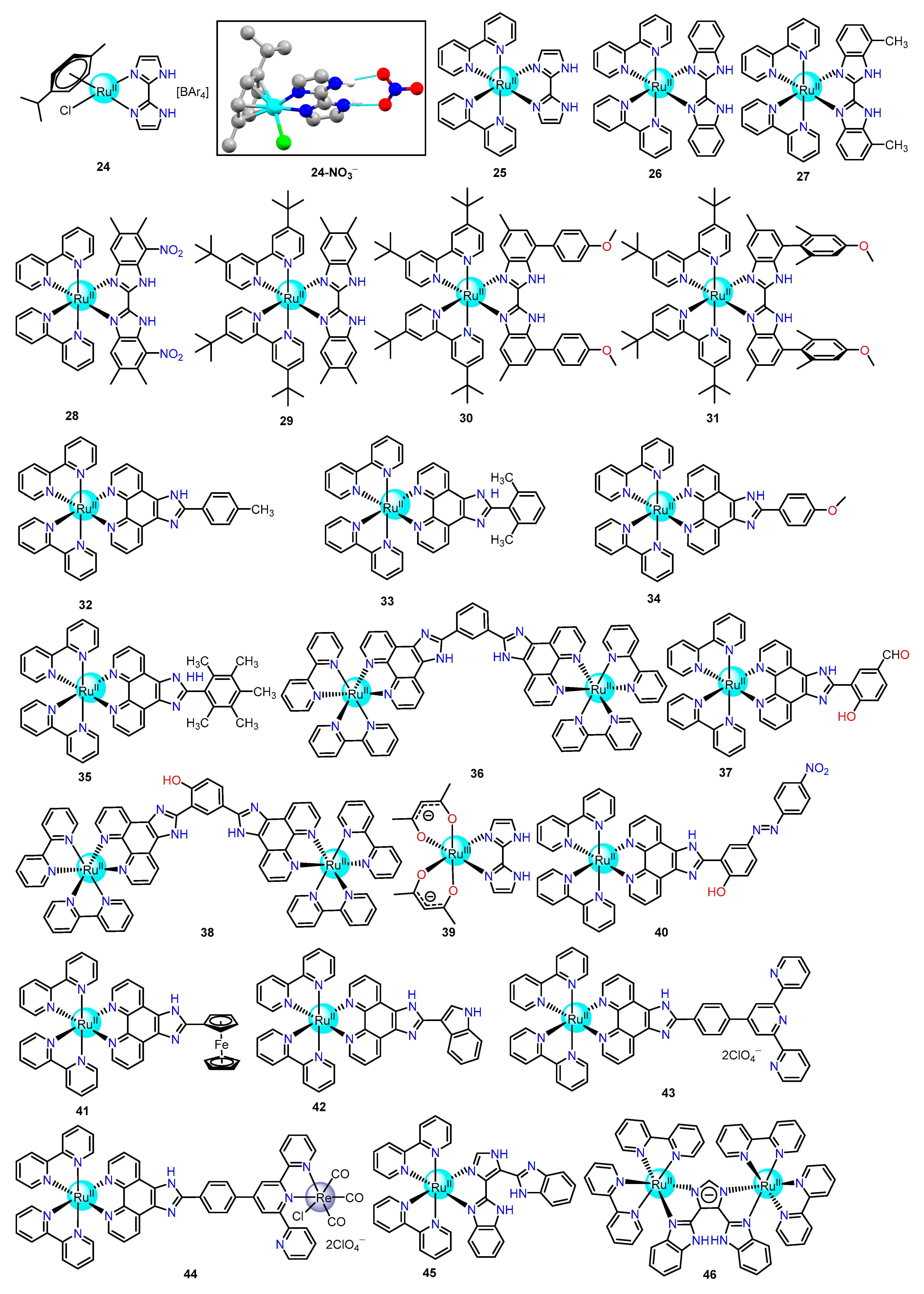
McPartlin and co-workers first investigated the solution state properties of chlorido para-cymol ruthenium (II) biimidazole complex 24 (Figure 5) and established strong, non-selective interactions with some common inorganic anions such as- Cl, Br, NO3, I or ReO4 [68]. The anion binding was investigated by 1H NMR titration with the above-mentioned inorganic anions, and the shift of the –NH proton was studied. According to the basicity of the anions, it was expected that Cl and Br would bind strongly with the receptor compared to NO3 and HSO4. However, the NO3 ion showed higher binding affinity due to the structural complementarity confirmed by the single-crystal X-ray structure (Figure 5).
The Ye group reported a series of bis-heteroleptic Ru(II)complexes, 2528 (Figure 5) containing 2,2-bisimidazole as the recognition site for anion and Ru(II)-bipyridine moiety as the signaling unit [69,70,71,72]. Remarkably, the bisimidazole acted as a bifunctional unit due to the presence of two lone pairs of electrons for coordination to the central metal ion, and the two imidazole –NH groups provided exclusive hydrogen bond for second coordination sphere recognition of anions. The detailed analysis demonstrated that the photophysical and electrochemical properties of 2528 depend on the protonated state of the –NH group of the bisimidazole moiety. The acidic nature of the bisimidazole –NH group may grant a hydrogen bonding interaction state, mono-deprotonated state, and double deprotonated state in the presence of anions, and these states were governed by numerous factors such as the acidity of the metallo-receptor, basicity of anions, strength, and the number of hydrogen bonds. Metallo-receptor 25 offered two –NH protons toward I, Br, Cl, H2PO4, CH3COO, HSO4, and NO3 for hydrogen bonding interactions, whereas only one of the –NH proton is deprotonated upon excess addition of CH3COO which altered the color of the solution from yellow to orange-brown. Conversely, fluoride formed highly stable HF2 by deprotonating both the acidic protons of bisimidazole –NH and altered the color of the solution from yellow to orange-brown followed by violet.
Again, ruthenium(II) 2,2- bisbenzimidazole complex 26 showed a similar type of anion binding properties towards I, Br, Cl, H2PO4, CH3COO, HSO4, and NO3 as observed in the case of 25. The 2,2-bisbenzimidazole unit in 26 operated as electron-withdrawing groups and made it a more acidic receptor (pKa 5.7) as compared to 25 (pKa 7.2). In the presence of both F and CH3COO, 26 formed a mono-deprotonated complex followed by the double-deprotonated complex at low anion and high anion concentrations, respectively. On the contrary, distinct responses were observed in the case of substituted acetate anions such as CCl3COO and CF3COO. Later this group introduced receptor 27, containing 7,7-dimethyl-2,2-bisbenzimidazole ligand, to make an intermediate acidic complex (pKa 6.2) between 25 and 26. This moderately acidic complex displayed significant sensing properties towards both the weakly basic anions (I, Br, Cl, H2PO4, HSO4, and NO3) as well as strongly basic anions (F andCH3COO). The emission intensity (at 617 nm) was improved by about 35–40% upon binding weakly basic anions through hydrogen bonding interactions which rigidified the molecular backbone and ceased the non-radiative decay of the 3MLCT state. On the contrary, the addition of strongly basic anions (such as CH3COO and F) caused the luminescence intensity of 27 to be quenched through the formation of doubly deprotonated species. Furthermore, the acidity of –NH protons was increased by incorporating the –NO2 group in the 2,2-bisbenzimidazole unit, which could form intramolecular hydrogen bonding and act as anion receptors. In this regard, the Ye group developed the ruthenium(II) bisimidazole-like complex 28, which showed a strong binding affinity towards Brand Cl, a weaker affinity towards I and no affinity towards other basic anions such as HSO4 and NO3. Interestingly, deprotonation of the bisimidazole –NH of 28 was observed in the presence of strong basic anions, such as CH3COO and F.
In addition, Rau et al. developed ruthenium(II) bibenzimidazole complexes 2931 (Figure 5) for the recognition and sensing of halide anions [73]. All the complexes 2931 showed a similar type of bathochromic shift of the 3MLCT band along with luminescence quenching upon the addition of F and OH. Due to p-anisyl substituents at 4,4’-positions of 30 and 31, a specified binding pocket for anion is generated at peripheral diamine functions. It should be noted that rotation of the aryl rings provides structural flexibility to 30, which allowed the positively polarized C-H edges to bind with the anion actively. On the contrary, the rotation of the p-anisyl moieties in 31 is prevented due to the presence of four methyl substituents, and thus, it formed a rigid structural conformation. Due to this structural rigidity,31 displayed an up-field shift of –NH proton, whereas a distinct shift of –NH signals (Δδ= 3–5 ppm) was observed in the case of other receptors in the presence of I, Br, and Cl.
Further, the Ye group assimilated the idea of multipoint hydrogen bonding interactions to improve the probability of anion binding affinity and solubility in water. In this regard, a set of complexes 3236 (Figure 5) were prepared and used for selective recognition of CN in water [74]. Amongst all the anions, only CN perturbed the emission spectra of 32 and 36 via the formation of hydrogen bonding with the receptor at physiological conditions in the water. Bimetallic ruthenium(II) complex 36 showed higher affinity and sensitivity towards CN in water (Figure 6). It might be due to the appropriate pKa’s and C-shape cavity structure of complex 36, which provided multiple hydrogen bonding interactions towards CN. The theoretical investigation also suggested CN was capped inside the C-shape cavity of the receptor through five-point hydrogen bonding interaction that provides an energy-minimized structure (Figure 6). The calculated binding constant of 36 was 2.5 times higher, and the detection limit was 20 times lower than 32.
Again, hydroxyphenyl imidazo appended ruthenium (II) complexes 37 and 38 (Figure 5) were presented by Ma et al. for “turn-off-on” sensing of CN [75]. The luminescence properties of both complexes were instantaneously quenched upon binding with Cu2+ in an aqueous buffer solution (20 mM HEPES, pH = 7.2) containing 1% (v/v) acetonitrile. Non-luminescent 37-Cu2+ and 38-Cu2+ adducts were formed and could selectively and sensitively detect the very low concentration of CN in 99% aqueous solutions through a “turn-on” fluorescent response (Figure 6). The calculated LOD for CN was 0.36 μM for 37-Cu2+ and 0.87 μM for 38-Cu2+, which were lesser than the World Health Organization (WHO) recommendation value (maximum admissible concentration of 1.90 μM in drinking water). So, these ruthenium (II) complex-based ensembles could be used as potential and efficient cyanide ion sensors in nearly 100% aqueous solutions.
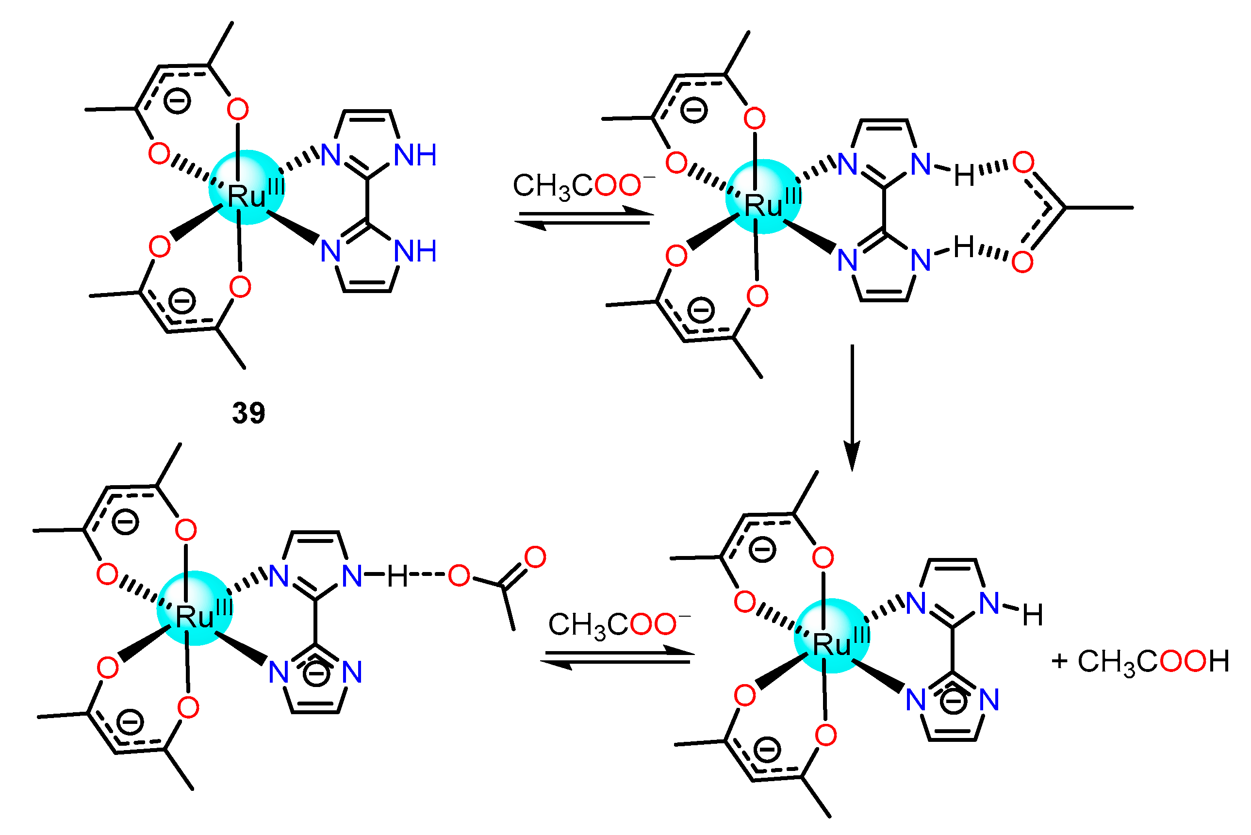
Colorimetric recognition of basic anions, for example, F, H2PO4, HSO4, Br, Cl, I, and CH3COO by non-luminescent paramagnetic bis(acetylacetonate) biimidazole Ru(III) complex 39 (Figure 5), was reported by Lahiri and his group [76]. A gradual color change of receptor 39 was observed in the presence of basic anions caused due to deprotonation, whereas weakly basic anions such as halides and sulphate formed only minor hydrogen bonding with the imidazole –NH proton of 39 (Scheme 1).
Again, azocoupled salicylaldehyde imidazole-based Ru(II) complex 40 (Figure 5) was documented by Khanmohammadi et al. for selective detection of basic anions such as F, H2PO4, CH3COO, etc. in DMSO [77]. UV-Vis spectra of 40 showed a decrease in band intensity at 355 nm and simultaneously the debut of a new band at 550 nm, followed by an alteration of the colour of the solution from light orange to dark brown. In contrast, the emission intensity at 735 nm was enhanced upon the addition of anions to the complex.
Molina et al. reported ruthenium(II)–polypyridyl complex 41 (Figure 5), containing ferrocene-imidazophenanthroline ligand and ruthenium bipyridine moiety for extremely selective detection of Cl over all other anions [78]. The sensing studies of this heterobimetallic ruthenium (II) complex were carried out in two ways: (i) cathodic redox shift of the Fe(II)/Fe(III) couple (ΔE1/2 = −80 mV) without changing the oxidation wave of the ruthenium (II) center; and (ii) a noticeable enhancement of red emission (CHEF = 30). The 1H NMR,31P NMR, and all the photo-physical and electrochemical data strongly suggested imidazole –NH proton bind with Cl through hydrogen bonding interactions.
Wang and co-workers synthesized Ru(II) complex 42 (Figure 5), encompassing both imidazole and indole groups, which acted as selective and naked eye detectors of CH3COO at much lower concentrations as compared to other anions such as H2PO4, I, Br, Cl, F, and NO3 [79]. Free receptor showed two absorption bands at 291 nm (bpy-based π-π*) and at 462 nm (3MLCT band), which gradually decreased upon sequential addition of CH3COO to 42 along with a notable amplification of the absorption band at 346 nm with the onset of an isosbestic point at 376 nm and a new peak at 430 nm. Further, the free Ru(II) complex showed a strong emission band at 618 nm at room temperature upon excitation in the visible-light region (at 462 nm) in DMSO, which was subsequently quenched upon the addition of CH3COO, F and H2PO4. However, CH3COO was found to bring more spectral changes and turn off the luminescence behavior of 42. Further mechanistic investigations pointed out that the triangular CH3COO (O-C-O angle of 120°) anion might be more favorable for the immediate binding to the imidazole and indole protons of 42 than tetrahedral anion such as H2PO4 (since O-P-O angle of 108°). Thus, it was concluded that the distance of the oxygen atoms in CH3COO was accurate for bridging with the imidazole and indole protons, as shown in Scheme 2.
Once more, this group systematically synthesized ruthenium (II) complex 43 and eventually prepared hetero bimetallic complex 44 (Figure 5) for the “turn-on” sensing of H2PO4 and “turn-off” sensing of CH3COO and F (Figure 7) [80,81]. The Job’s plots analysis resulted in 1:1 host-to-guest stoichiometry for CH3COO and F while a 1:2 stoichiometry was reported for H2PO4. The reason for 1:2 host-to-guest stoichiometry was attributed to the formation of two different types of hydrogen bonding by H2PO4. One of the H2PO4 ion formed an N–H⋯O hydrogen bond with the imidazole –NH, whereas the other formed a hydrogen bond with the pyridyl nitrogen atom of the receptor. Further, calculated binding constant values of 44 were 1.5 × 10¹⁰ M−2, 1.3 × 10⁵ M−1, and 5.5 × 10⁴ M−1 for H2PO4, CH3COO, and F respectively, which were comparatively greater than the calculated binding constant values for 43 (i.e., 7.6 × 10⁹ M−2, 3.7 × 10⁴ M−1, and 3.3 × 10⁴ M−1, for H2PO4, CH3COO, and F respectively). These binding constant results indicated 44 as a better sensor compared to its parent analog 43, and it was well matched with the enhanced acidity of -NH of 44 (pKa 6.84 and 9.07 for 44 and 43, respectively).
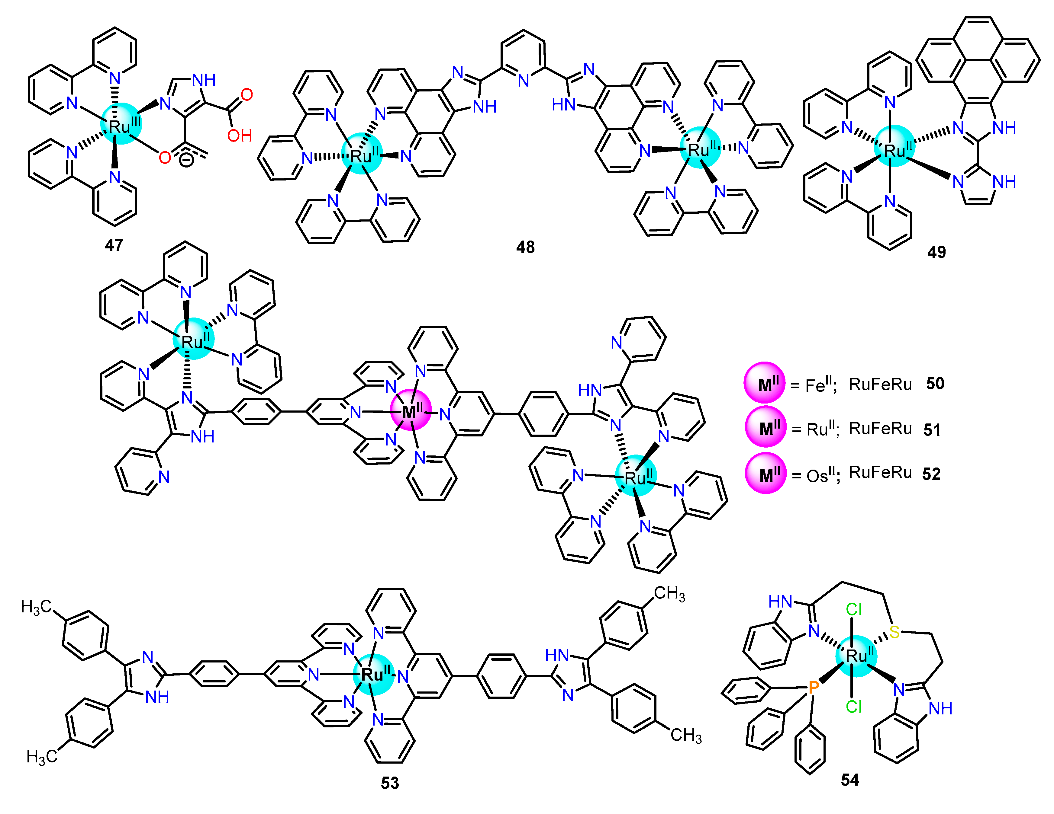
Further, Baitalik and his group developed monometallic and bimetallic ruthenium(II) complexes 45 and 46 (Figure 8) utilizing 4,5 bis(benzimidazol-2-yl)imidazole moiety as colorimetric sensors for anions [82]. Both complexes contained multipoint hydrogen bonds donating imidazole –NH protons for selective recognition and sensing of F and CH3COO. It was shown that both 45 and 46 formed 1:1 hosts to guest adduct at lower concentrations of anions through hydrogen bonding interaction, while stepwise deprotonation of the imidazole –NH protons took place at higher concentrations. Later, the same group employed imidazole-4,5-dicarboxylic acid with Ru(II)-bipyridine moiety to synthesize 47 (Figure 9) as a luminescence anions sensor [83]. The photoluminescence intensity of 47 was increased upon the addition of F, H2PO4, and CH3COO ions. A significant red shift of emission maxima from 690 to 740 nm was observed, and a distinct color change of 47 was noticed in the presence of excess anions, which might be due to the deprotonation of imidazole –NH.
Again, second coordination sphere recognition of F, H2PO4, and CH3COO ions by bimetallic ruthenium (II) complexes 36 and 48 (Figure 8) containing acidic imidazole –NH protons were reported by the same group [84,85]. Remarkably, photo-physical experimental results displayed increased emission intensity as well as lifetime by H2PO4 due to the rigidification of molecular backbone via hydrogen bonding interaction. However, a decrease in both was noted for F and CH3COOcaused due to the deprotonation of the imidazole -NH.
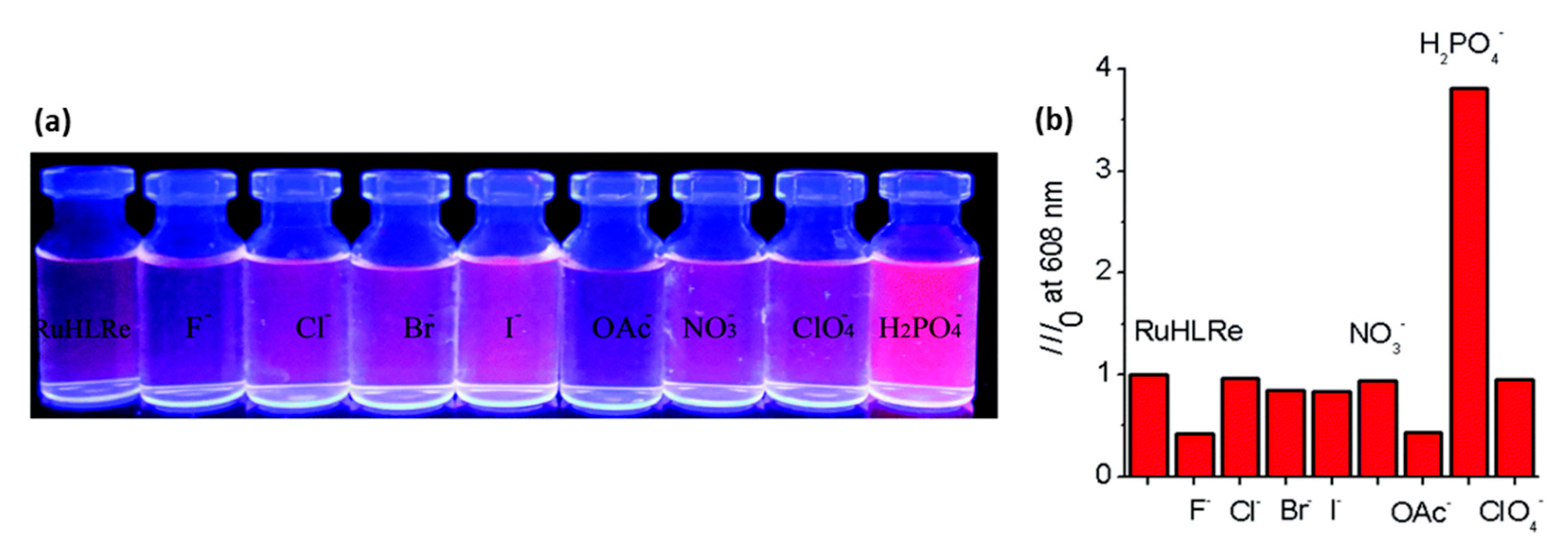
Pyrene-biimidazole-based Ru(II) chemosensor 49 (Figure 8) was developed by the same group for selective detection of CN in both acetonitrile and aqueous media [86]. The imidazole -NH protons of 49 were highly acidic, having pKa1 = 5.09 and pKa2 = 8.95. Deprotonation of these two -NHs by CN increased the electron density on the metal center. As a result, a red shift of absorption and quenching of emission of 49 was noted. The limit of detection of CN by 49 was 5.24 and 4.67 nM by the colorimetric and luminescent analyses methods, respectively.
Further, Baitalik et al. developed a trimetallic complex containing Fe(II) 50, Ru(II) 51, and Os(II) 52 (Figure 8) derived from a bipyridine-terpyridine heteroditopic type spanning ligand for sensing of selected anions in both organic and aqueous media [87]. All three triads acted as multichannel receptors for F, CN, H2PO4, and CH3COO in acetonitrile without much selectivity. However, the triads displayed higher sensitivity and selectivity towards CN and SCN in water with a lower detection limit of 10−8 M. Similarly, recently developed Ru(II)-terpyridine complex 53 (Figure 8) was also selective for CN and capable of detecting CN ion in water up to the concentration limit 10−8 M [88].
Recently, Ramos and co-workers reported 1,5-bis(benzimidazol-2yl)-3-thiapentane-based ruthenium (II) complex 54 (Figure 8) for selective detection of Cl as a “switch on” probe even in the presence of other interfering anions such as F, NO3, Br, HSO4, I, and H2PO4 [89]. The interaction of Cl with the receptor complex facilitated the electron transfer from the highest occupied molecular orbital (HOMO) of the donor to low-lying HOMO of the fluorophore, i.e., acceptor as a disfavored Photo Electron Transfer (PET) process in the excited state resulted in the increasing the fluorescence intensity.

2.1.3. Amide/Sulphonamide/Picolinamide Based Ru(II) Complexes as Chemosensors

Among the several polarized -NH functional groups, amide-based receptors are well known for sensing anions, while sulphonamide and picolinamide-based receptors are less explored.
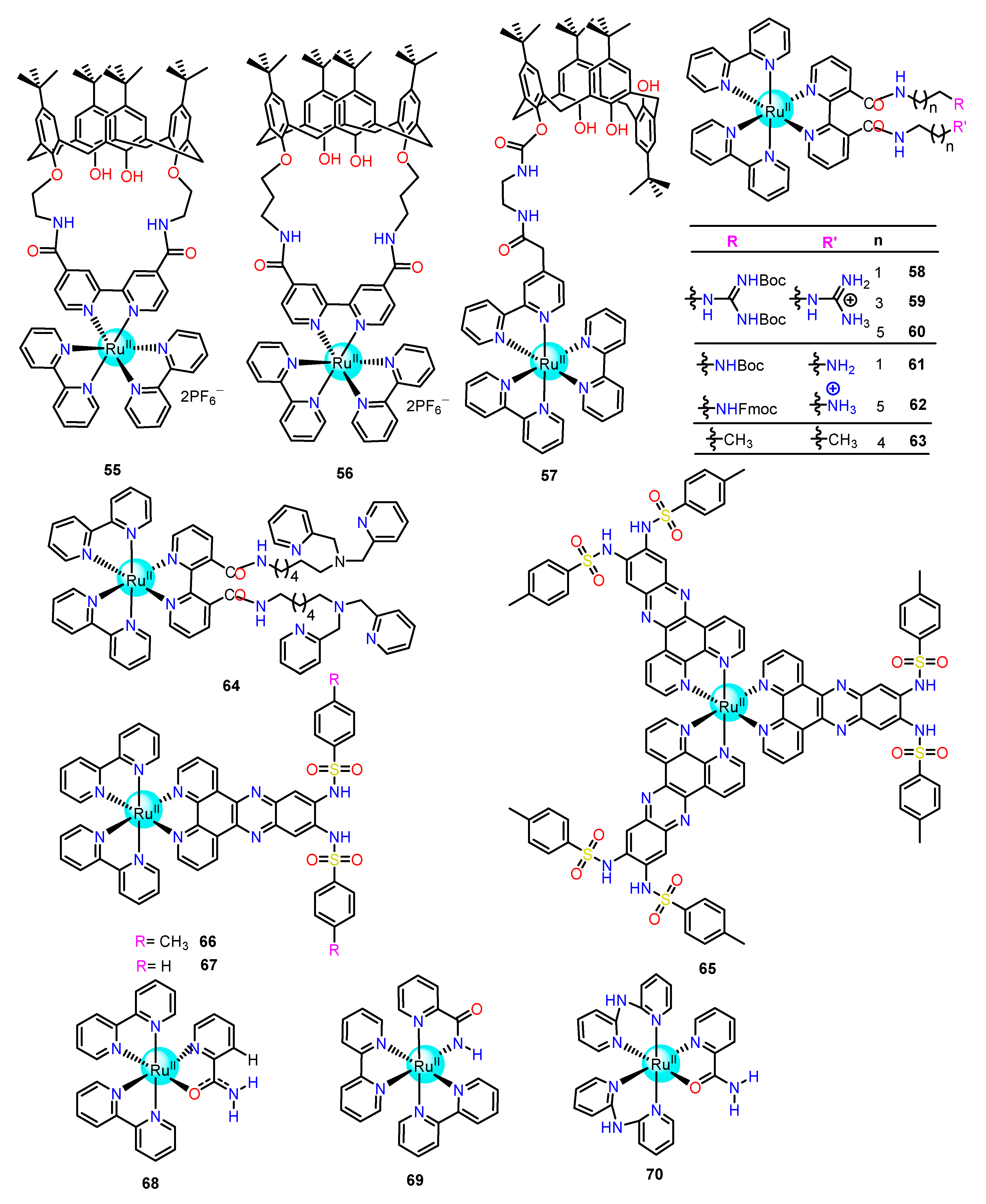
Through amendment of the [Ru(bpy)3]2+ with an amide containing t-butylcalix [4] arene moiety, Rajagopal et al. reported two new Ru(II) complexes 55 and 56 (Figure 9) for sensing of anions [90]. UV-Visible and PL emission studies revealed that 55 selectively recognized Cl, H2PO4, and CH3COO, while 56 recognized Br and CH3COO anions. In both cases, the emission intensity was quenched by CH3COO, where an increment of intensity was observed for other anions. Further, transient absorption and excited state lifetime studies were performed to find out the possible reasons for the quenching of emission in the case of acetate but enhancement for other anions. Through these studies, it was revealed that the excited states of both the receptors were stabilized by Cl, Br, and H2PO4 anions which facilitated the radiative decay and instigated enhancement, while CH3COO destabilized the excited state through the non-radiative deactivation pathway and promoted quenching.
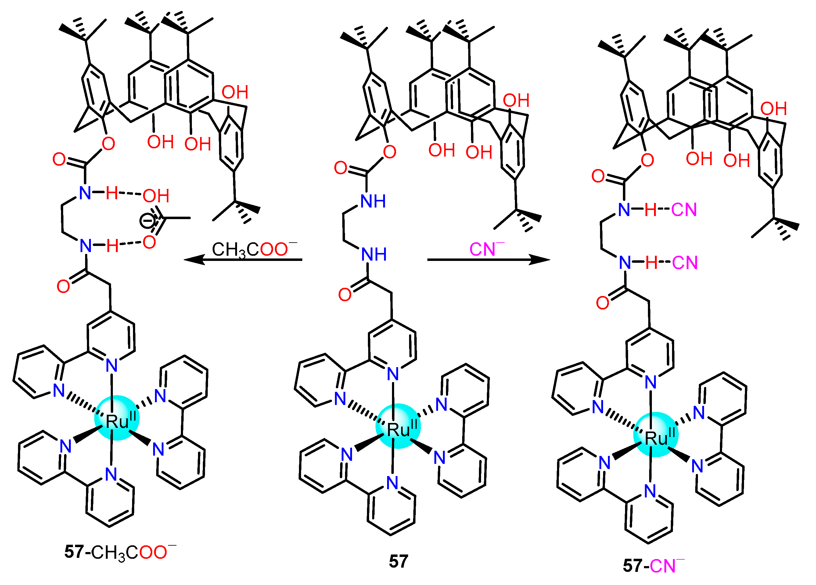
Another example of amide encompassing calixarene-based Ru(II) complex 57 (Figure 9) was developed by Maity et al. for selective recognition of CN and CH3COO [91]. The photo-luminescence studies of 57 in an aqueous organic medium (H2O:CH3CN 95:5) showed quenching for CN, and enhancement of luminescence property was noted for CH3COO. The mechanistic investigation exposed that CN resulted in deprotonation of amide –NH and increased the electron density in bipyridine ligand, and thus the intramolecular quenching was increased. On the contrary, bidentate CH3COO was bound with the amide –NH via weak hydrogen bonding interaction (Scheme 3). The hydrogen bounded CH3COO pulled the electron density and decreased the intramolecular quenching, thereby causing enhancement of luminescence intensity. The limit of detection value for CN calculated from luminescence response was 70 ppb.
Pinet et al. developed a series of modified 3,3′-bipyridyl-based novel ruthenium luminescent probes 5864 (Figure 9) containing guanidinium, ammonium, or zinc(II) dipicolylamine binding sites for selective detection of anions in acetonitrile [92]. Hypsochromic shifts with enhanced luminescence intensity were found upon the addition of anions. Interestingly, guanidinium-functionalized probes were unveiled as more efficient sensors for CH3COO. Receptor 60 showed higher selectivity towards glutamate preferentially to phosphates. Conversely, ammonium functionalized, 62, selectively detects phosphates derivative. However, these probes were unable to bind such species in aqueous media due to the higher solvation energy of anions.
Further, dipyridyl or phenanthroline moieties could be easily functionalized by the anion-sensitive bis(sulfonamide) group. Keeping this idea, Sun et al. developed Ru(II) complex 65 (Figure 9) comprising bis(sulfonamide) anion binding sites with highly chromophoric conjugated quinoxaline moieties for selective detection of F [93]. The Job’s plot analysis exposed a 1:1 host-guest complex for the receptor with F. It was revealed that one sulfonamide interacts with the F while two other –NH protons is deprotonated in DMSO. It was assumed that the metalloreceptor 65 became a neutral molecule with two negative charges generated by the deprotonation of two –NH protons while only one was left to interact with the F.
Besides, the Lin group reported sulfonamide-based ruthenium(II) complexes 66 and 67 (Figure 9) which displayed a strong affinity for F and CH3COO ions with adequate affinities for H2PO4 or OH and almost no affinity for other halides [94]. Different photo-physical, electrochemical, and NMR studies revealed the anion binding abilities of Ru(II) complexes were in the order of CH3COO > F > H2PO4≫ OH > Cl, Br, I. Further, receptor 67 showed higher anion binding affinity over 66. Thus, the relatively lower anion binding ability of the toluene-substituted complex 66 was ascribed to the better electron-donation of the methyl group compared to the hydrogen atom of the benzene analog.
Again, neutral or deprotonated 2-picolinamide (i.e., H2pia and Hpia, respectively) binds with many transition metal ions and has potential anticancer activity. It could bind with transition metal by two coordination modes: (I) pyridyl-N, and the deprotonated amide N jointly provided N, N’-coordination mode; (II) pyridyl N atom and the amide O provided the other N, O-coordination mode as shown in Figure 10a. However, the bis-heteroleptic ruthenium(II) complex with 2-picolinamide was scarcely used for the recognition and sensing of anions.
Endo group reported 2-picolinamide-based ruthenium complex 68 for naked-eye visual detection of F in acetonitrile [95]. Crystal structures of 68 revealed 2-picolinamide coordinated to Ru(II) via N,O coordination using N of the pyridyl group and O of the amide group. A visual color change from the red solution to dark-red was obtained upon the addition of F to 68 (Figure 10b). The technique 1H NMR and absorption spectroscopies exposed that the H near the pyridyl group of H2pia first formed a 1:1 adduct followed by the other H resulting in a 1:2 adduct as shown in Scheme 4.
Another interesting example of a 2-picolinamide-based Ru(II) complex 69 (Figure 9) was reported by Zhong and co-workers [96]. Single crystal X-ray characterization exhibited that the 2-picolinamide binds with Ru(II) center by the N atom of pyridyl and another N atom of the deprotonated amide group via N,N’-coordination mode. The spectroscopic and electrochemical studies revealed the receptor was selective for H2PO4 over other common inorganic anions such as I, Br, Cl, F, NO3, CH3COO, HSO4, and HP2O73−. The Job’s plot analysis showed 1:2 hosts to guest binding with 108 L2mol−2 global associations constant. The calculated limit of detection was 1.4 × 10⁻⁶ molL−1. Two wave behavior of Ru(II/III) redox couple of 69 was observed in the presence of H2PO4 with a large negative shift from +0.74 to +0.45 V vs. Ag/AgCl. Again 1H NMR experiment exposed that a strong hydrogen bonding interaction between 69 and H2PO4 plays a key role in the recognition process (Scheme 5).
Recently Nagao et al. reported 2-pycolinamide-based Ru (II) complex 70 (Figure 9) for selective colorimetric detection of F in DMSO [97]. Crystal structure analysis showed H2pia ligand and Ru(II) coordination via pyridyl-N and carbonyl-O, along with the carbonyl π-electron delocalized over the amide group. Further 1H NMR analysis and absorption spectroscopy showed that the added F did not differentiate amid the two amino groups of the di-2-pyridylamine and amide group of H2pia and thus formed a tris-F-adduct by turning the color from red to dark red.

2.1.4. Pyrrol-Based Ru(II) Complexes as Chemosensors

Among the various investigated anion receptors, pyrrol is one of the widely studied due to the presence of weakly acidic –NH proton (pKa of –NH proton is 16.5) that can form hydrogen bonding as well as become deprotonated by anions. Although, pyrrol or substituted pyrrol-based ruthenium (II) complex are scarcely reported in the literature as an anion sensor.
Fused dipyrrolylquinoxaline (DPQ) phenanthroline-based ruthenium(II) complex 71 (Figure 11) was designed and synthesized by Sessler and his group for selective detection of F in DMSO [98]. The acidic nature of the pyrrol –NH protons were increased upon binding the DPQ-phenanthroline ligand with the Ru(II) metal center. Further, Job’s plot analysis showed a 1:1 hosts-to-guest binding. The calculated binding constant was 1.2 × 10⁴ M−1, which is nearly 30 times greater than that calculated from the free ligand.
Thereafter the same group replaced the quinoxaline with a bipyridine moiety for the generation of a larger binding site and to achieve enhanced binding affinity towards larger anions. In this regard, dipyrrol-appended bipyridine-based Ru(II) complex 72 (Figure 11) was synthesized for emission-based selective sensing of H2PO4 [99]. Emission intensity at 630 nm of the receptor was decreased upon the addition of anions. The Job’s plot analysis resulted in 1:1 hosts to guest binding and the binding constant value for H2PO4 was 1.0 × 10⁵ M−1.
The development of time-resolved fluorescence method-based chemical sensing of anion is advantageous since the lifetime is independent of the excitation source, intensity fluctuations, total probe intensity, loss of light in the optical path, and sensitivity of the detector. Anzenbacher’s group first reported lifetime-based anion sensing by Ru(II) complex, 73 (Figure 11) having DPQ-phenanthroline as an anion coordinating site [100]. Photophysical studies showed a decrease in the absorption band corresponding to MLCT and the high-energy π-π* ligand in addition to quenching of luminescence intensity upon the addition of CN and F. The binding constants for CN and F were found to be 4.3 × 10⁵ M−1 and 6.4 × 10⁵ M−1, respectively, in acetonitrile solutions. Further luminescence lifetime of 73 was shortened minutely from 377 ± 20 ns to 341 ± 20 ns upon the addition of 6.20 µM of CN. Two lifetimes shortening was obtained ranging from 13 to 17 ns (short τ) and 320 to 370 ns (long τ) upon excessive addition of CN which was fitted in two exponential decay fitting equations.

2.1.5. Urea-Based Ru(II) Complexes as Chemosensors

Urea is mostly explored as a classical neutral anion receptor as it is capable of forming hydrogen bonding interactions with anions. Most of the halide could interact with urea through the formation of a six-membered chelate ring, whereas oxyanions (having nearby two oxygen) could form eight-membered chelate rings. In the last two decades, Wilcox [101] and Hamilton [102] synthesized an array of receptors comprising one or more urea entities aiming to control the substituents choice and the polarization of the –NH units.
Das et al. reported urea-based Ru(II) complex 74 (Figure 12) that acted as a colorimetric sensor for basic anions such as F, CH3COO, and H2PO4 [103]. Different spectroscopic techniques, including 1H NMR titration studies, concluded that the ligand is bound with H2PO4, F, and CH3COO more strongly than the other halides and oxyanions. Further photoluminescence studies showed the emission intensity of 74 is completely quenched by F, CH3COO, and H2PO4. It is because of the faster decay of the excited triplet state by reducing the energy gap (excited triplet state and ground singlet states) upon anion binding that increased the solvation of the anionic adducts or deprotonated state of 74.
Ru(II)-polypyridyl complex 75 (Figure 12), having pendent urea moiety as an anion recognition unit, was synthesized by Gunnlaugsson and co-workers, which worked as a long-wavelength fluorescent sensor for anions [104]. The 1H NMR titrations demonstrated the interaction between the urea moiety and anion via hydrogen bonding interaction which played an important role in the recognition process. Further, MLCT emission is sensitive towards the binding of CH3COO, H2PO4, and HP2O73− but not for F in the organic solvent. Remarkably 75 could differentiate between phosphate and pyrophosphate through “turn on’’ emission in the presence of H2PO4 and ‘‘turn-off’’ emission in the presence of HP2O73−.
Again, sensing and recognition properties of the urea subunit toward anions could be improved by linking with Ru(terpy)22+ instead of Ru(bpy)32+ units. Advantages of the use of Ru(terpy)22+ appeared to result from (i) a through-space electrostatic effect and (ii) a through-bond covalent effect. Further, a combination effect of Ru(II) polypyridine-metal complex and pendent substituents could enhance the anion binding affinity. In this regard, Fabbrizzi et al. developed terpyridine-based Ru(II) complex 76 and improved the acidity of the urea unit by substituting pendent benzene with nitrobenzene 77 (Figure 12) [105]. Hydrogen bonding interaction between urea proton and anions resulted in a 1:1 stoichiometry receptor-anion complex which was confirmed by Job’s plot. The log K values for the binding of Cl were 5.66 ± 0.01 and 6.32 ± 0.02, corresponding to 76 and 77, respectively. The binding affinity decreased across the series Cl > Br > I, which was parallel to the decreased charge density of the anion.
Yao and co-workers showed how the interaction of anion and urea could be engaged to control the electron transfer and electronic coupling between redox-active sites [106]. For this purpose, this group synthesized urea-functionalized diruthenium complex 78 (Figure 12). A detail electrochemical study along with DFT suggested that the electronic coupling between two cyclometalated ruthenium and urea might enhance the degree of binding of Cl or Br ions to urea units via hydrogen bonding interactions. Further, the redox wave reversibility of 78 was fully maintained in the presence of Br or Cl. However, strong basic anions, for example, F, CH3COO, and H2PO4 ruined the redox waves by making them highly irreversible.

2.1.6. Aldehyde Incorporated Ru(II) Complexes as Chemodosimeters

Chemodosimeter is a term first described by Chae and Czarnik [107]. It is an abiotic molecule achieved by analyte recognition through observable signal output. This approach involves the reaction of a molecular receptor, known as a chemidosimeter, with the analyte followed by substantial chemical transformation, including breaking and forming of the covalent bonds.
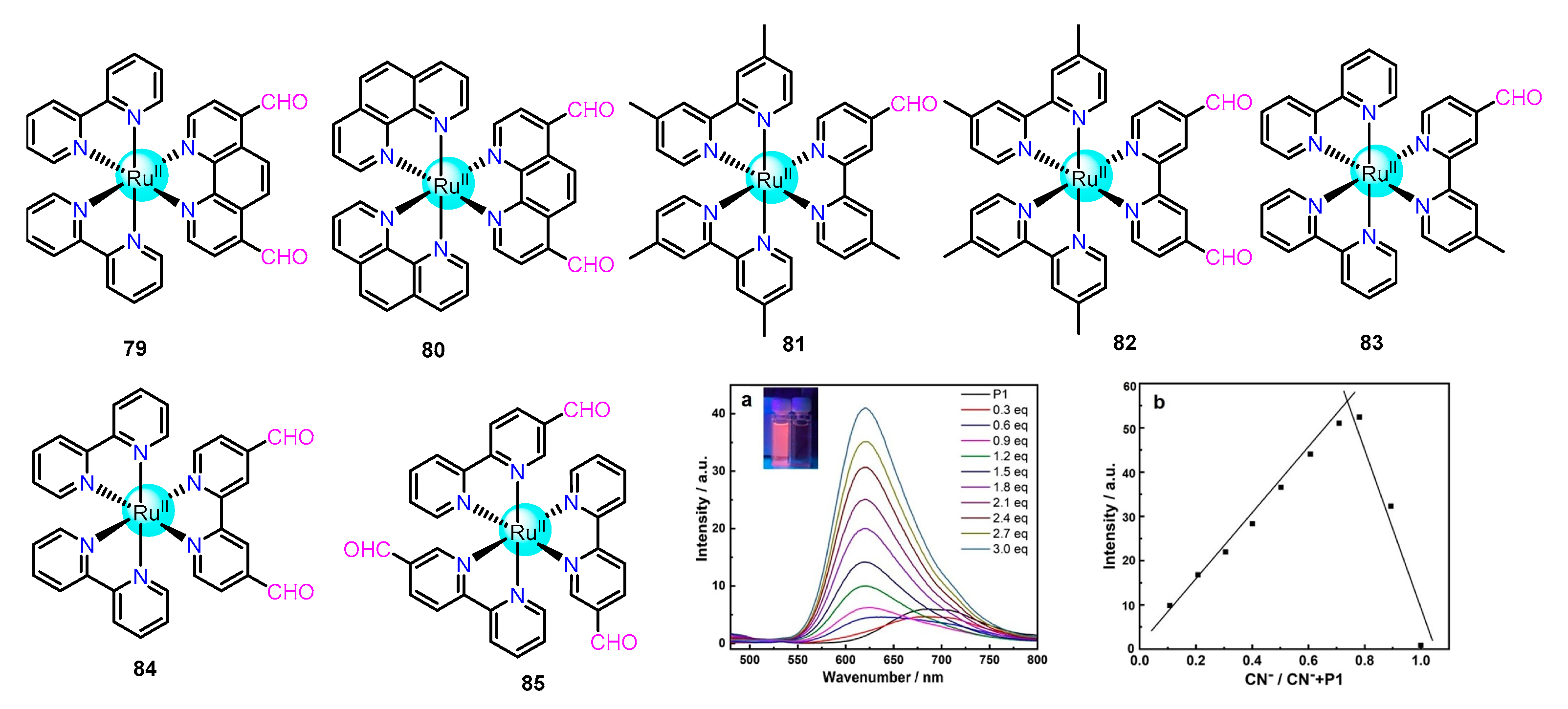
Schmittel’s group presented two new ruthenium complexes 79 and 80 (Figure 13) with 1,10-phenanthroline-4,7-dicarboxaldehyde (PDA) as a chelating ligand to detect CN based on the cyanohydrin’s formation [108]. The immediate change of color (orange-red to yellow) was detected through the naked eye after CN addition. Further, PL studies displayed a drastic blue shift of almost 100 nm and ~55-fold enhancement of emission intensity within 15 s in the presence of 2 equiv. of CN to both the receptor’s solutions. A large value of overall cyanohydrin formation constants was obtained from PL titration for the two receptors (log β[CN] = 15.36 ± 0.44 and log β[CN] = 16.37 ± 0.53 for 79 and 80, respectively).
Later Chen et al. reported four ruthenium(II) complexes 8184 (Figure 13) which selectively sensed CN by the formation of a well-known cyanohydrin complex [110]. All these complexes showed similar types of photo-physical properties. A visible naked-eye change of color (orange to yellow), along with a fluorescent distinction (dark red to red-orange), was perceived after the addition of CN to the Ru(II) complex solution. Further, a large blue shift of the absorption and emission spectra with a notable increment in the emission was observed after the addition of CN
Again, Yao et al. reported 5-aldehyde-2,20- bipyridine-based Ru(II) complex 85 (Figure 13) for selective detection of CN in an aqueous acetonitrile solution (60% water) [109]. Similar to the previous receptors, it was highly selective and sensitive toward CN. A visible color change (orange-red to yellow) was observed. It worked as a phosphorescence “turn-on” response probe with a ~30- fold increase in emission intensity for the formation of cyanohydrin through nucleophilic addition of CN to aldehyde (Figure 13a). The formation of cyanohydrin followed the 1:3 stoichiometry of host to guest, and a detection limit corresponding to 0.75 μM was calculated(Figure 13b).

2.1.7. Some Example of Bis-Heteroleptic Ru(II) Complexes as Chemosensor

In this category, we give some examples of bis-heteroleptic Ru(II) complexes that could act as chemosensors for different anions.
In 2007, Das et al. reported phenol and catechol-based Ru(II) polypyridile complexes 86 and 87 (Figure 14), which acted as efficient colorimetric sensors for F even in the presence of ~20% H2O (v/v) [111]. Different spectroscopic techniques and time-dependent density-functional theory (TDDFT) revealed that the hydrogen bonding interaction between F and O-H of phenol or catechol occurred at lower concentrations while higher concentrations of anions promoted deprotonation of O-H (Scheme 6).
Das and co-workers developed three mononuclear Ru(II) complexes 8890 (Figure 14) comprising 2,2’-bipyridine and 2,2’-dipyridylamine for selective detection of F and CN [113]. The spectrophotometry, electrochemistry, and 1H NMR spectroscopy studies revealed that all these complexes were selective for F and CN over other common inorganic anions (for example, PF6, Br, Cl, ClO4, NO3, HSO4, and CH3COO). The recognition and sensing phenomena occurred through the formation of sequentially deprotonated complexes for all three receptors, a hydrogen-bonded refereed adduct. The stoichiometry of binding was 1:1, 1:2, and 1:3, with 88, 89, and 90, respectively, for CN and F. Again, Chattopadhyay et al. developed six thiosemicarbazones containing Ru(II) complexes 9196 (Figure 14) for sensing F [112]. Cyclic voltammetric (CV) measurement showed all the complexes underwent two quasi-reversible oxidations on the positive side of the potential window (0 to +0.8 V) and three consecutive quasi-reversible/irreversible reductions on the negative side of the potential window (0 to 2 V) (Figure 14a). Further, it was found that the binding constants were practically high (logKa > 5) for complexes 9396.
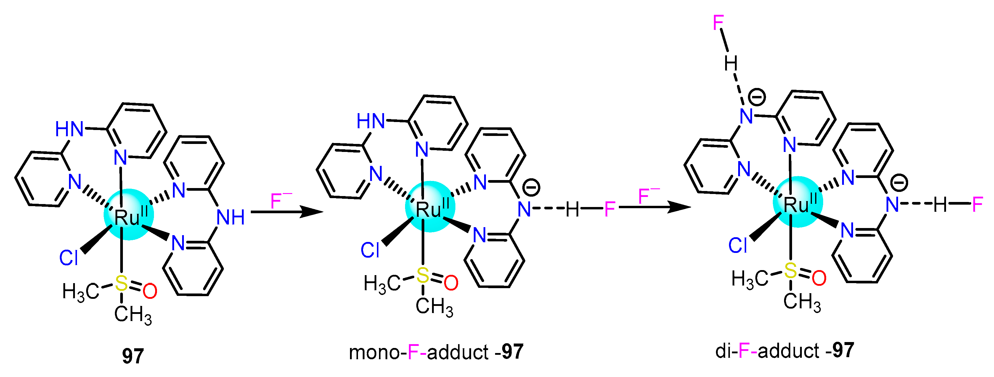
Nagao et al. reported C1-symmetricRu(II) complex 97, where two distinguishable –NH groups acted as Cl and F receptors via hydrogen bonding interactions [114]. The use of 1H NMR demonstrated that the–NH of di-2-pyridylamine formed selectively and successively mono F adduct followed by di-F adduct of 97 upon addition of F to the receptor solution in DMSO (Scheme 7).

2.2. Cyclometalated Iridium(III)-Based Complexes for Sensing of Anions

Complexes of iridium usually exhibit a high room temperature quantum efficiency, high thermal and electrochemical stabilities, good photo-stability, and large Stokes shift, which restricts self-quenching, readily tunable emission wavelengths (green to red), which could easily be altered by changing the nature of the secondary co-ligands. Owing to their ease of synthesis and their excellent photo-physical and electrochemical properties, cyclometalated iridium complexes have been receiving a lot of attention for their application as molecular sensors. This section of the review highlights the application of luminescent iridium complexes for sensing and recognition of anions.

2.2.1. Fluoride Sensing

It is widely known that fluoride anions (F) play vital roles in an extensive range of chemical, biological, and environmental processes. Medical practices such as the treatment of osteoporosis and those for dental care, environmental treatments such as fluorination of water supplies, and the use of F in chemical and nuclear warfare agents have resulted in increased human exposure [115]. High doses of F are, however, detrimental and can lead to dental or skeletal fluorosis. Therefore, the recognition and detection of this anion is an active area of research, and a great deal of effort has been devoted to the design of molecular receptors containing binding sites for fluoride.
Receptors based on Lewis acid-base interactions between boron and F were developed and established to accomplish highly efficient detection. In particular, sterically hindered boryl groups, for instance, dimesitylboryl moieties, have shown amended size-selectivity toward fluoride ions. The strong B–F interaction could hold up the π-conjugation of organoboron compounds, thus resulting in changes in absorption colorimetry, fluorescence emission, spectral shifts in NMR, and electrochemical properties. In this regard, numerous cyclometalated Ir(III) complexes conjugated with organoborane have drawn attention for their abilities as colorimetric chemosensors selective for fluoride anion.
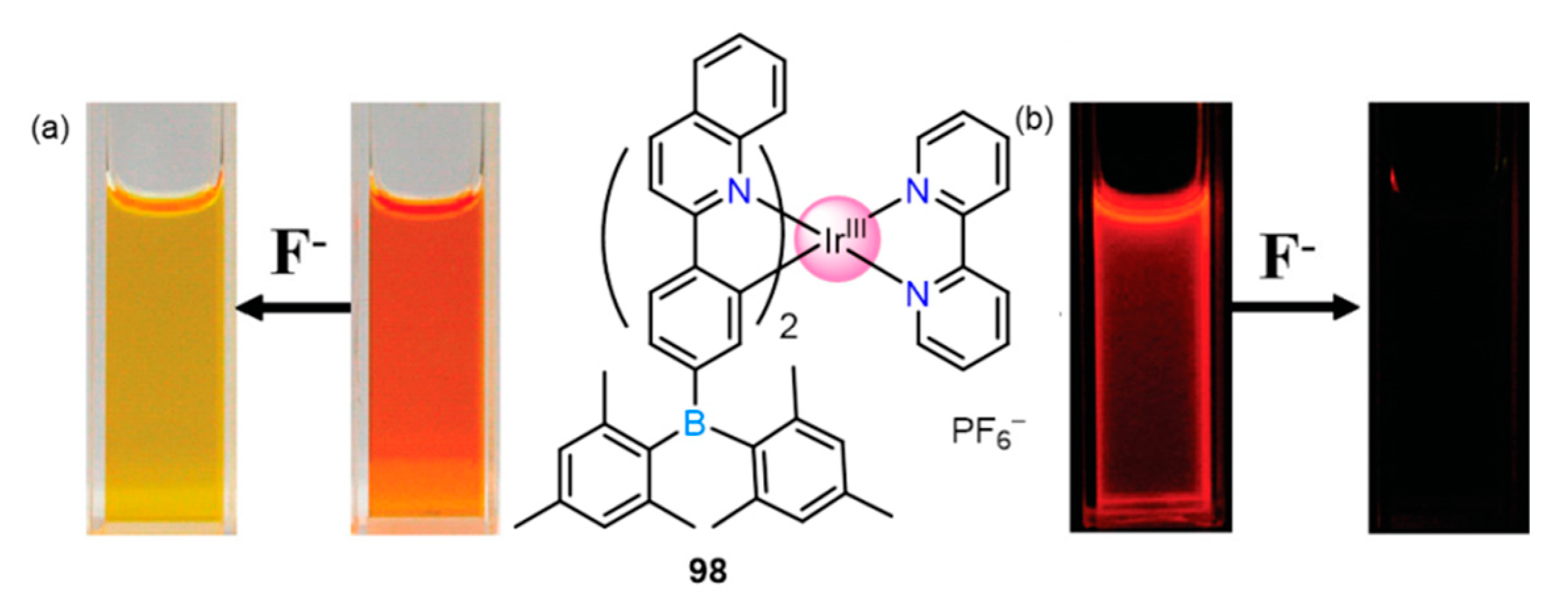
A naked eye chemosensor of F via a phosphorescent iridium complex 98, functionalized with an arylborane unit, was reported by Huang et al. A visible color change from yellow to reddish-orange was noticed in the presence of 3 equiv. of F anion with the subsequent red shifting (420−600 nm) of the absorption band (Figure 15) [116]. This shift was caused by the complexation of fluoride to cyclometalated arylborane ligand, which resulted in the excited-state switch from π-π* to CT transition of the adduct. The binding constant from UV-Vis titration data was put to 1:2 binding isotherm for complex-fluoride adduct, which awarded the K1 = 1.29 × 10⁶ and K2 = 4.27 × 10⁵ M−1. The photo-luminescence emission spectra showed a quenching phenomenon upon the 2 equiv. addition of F anion, thus making it an “ON−OFF” type probe.
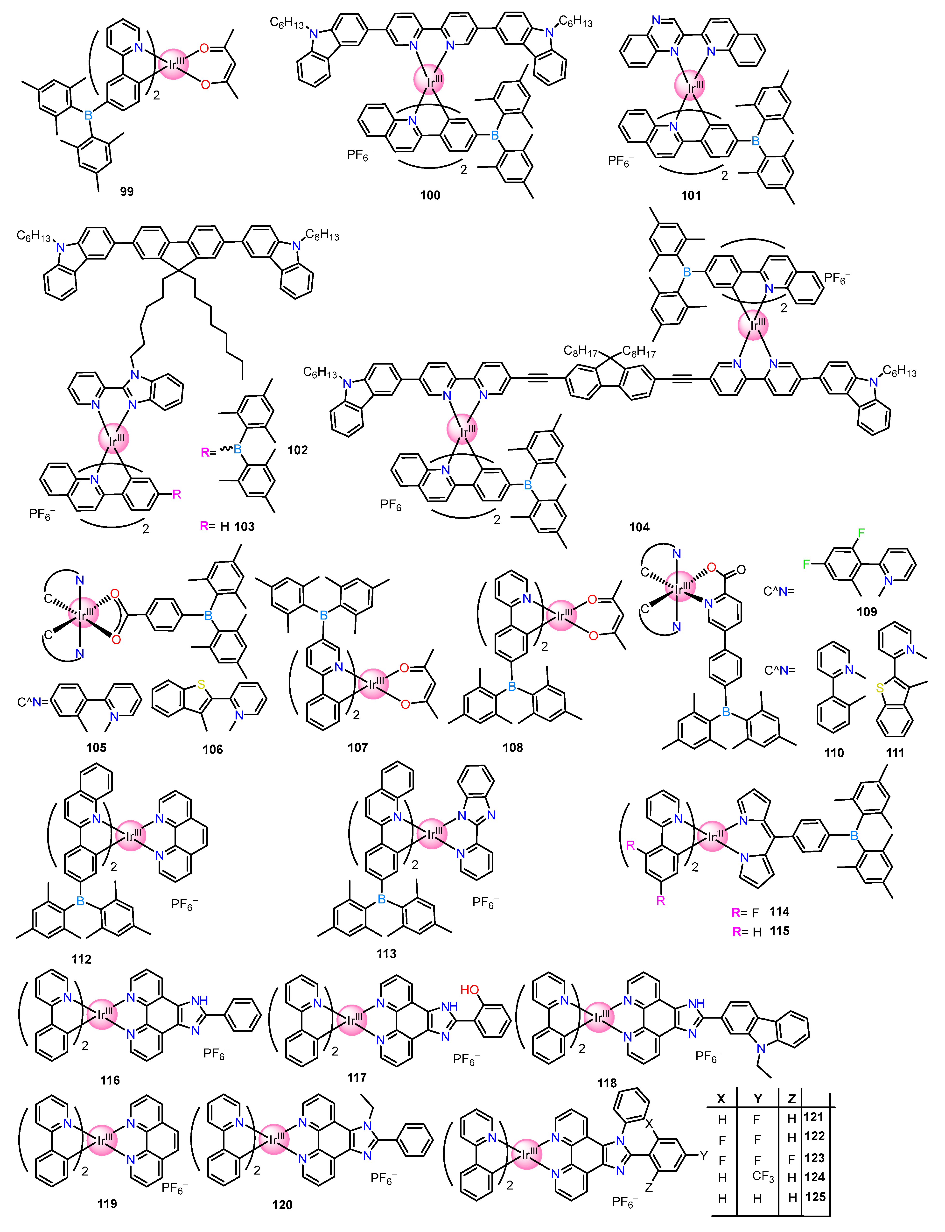
Again, the Park group in 2008 designed and synthesized a sterically congested dimesityboryl group-based phenylpyridine ligand and complexed it with an iridium center to produce a highly phosphorescent complex 99 (Figure 16), which served as a fluoride sensor via colorimetric and ratiometric responses [117]. The complex showed not only high selectivity for the fluoride ion but also an efficient two-color phosphorescence behavior. In addition, a high signal-to-noise ratio was reached through the time-gated acquisition of phosphorescence signals. Furthermore, successful aqueous medium detection of fluoride was realized by doping the iridium complex with PMMA.
A novel cationic iridium (III) complex 100 (Figure 16), containing carbazole and dimesitylboryl moieties, was synthesized by Huan et al. for colorimetric and ratiometric sensing of F [118]. A quenching in the orange-red phosphorescent emission of the complex was observed upon fluoride binding along with the switch on of fluorescent emission from the N^N ligand, which is reflected by a visual change in the emission color from orange-red to blue. The mechanistic studies of the fluoride adduct were conducted through TDDFT, which revealed that the free complex has a triplet emission state and the energy transfer occurred from the carbazolyl bipyridine moiety to the iridium (III) center. However, upon the interaction of F, the emission took place from the singlet excited state, and thus the energy transfer was inhibited. Hence the complex displayed fluoride sensing/interaction via a triplet to singlet switch-on behavior. Only fluoride was able to perturb the emission spectra of the complex, while the spectra remained unchanged in the presence of other anions. A low detection limit corresponding to the range 0–50 um was calculated with F anion.
The same group synthesized complex 101 (Figure 16), which was a near-infrared (NIR) phosphorescent chemosensor of F containing dimesitylboryl (Mes2B) groups and an iridium (III) center [119]. The selective binding of F caused quenching of the NIR phosphorescent emission with λem of 680 nm exhibited by the complex. From the emission titration data, the binding constants K1 and K2 were found to be 2.64 × 10⁶ and 2.12 × 10⁴ M−1, respectively.
Later this group developed a Förster (Fluorescence) Resonance Energy Transfer (FRET)-based F probe, 102 (Figure 16), comprising of Mes2B unit-functionalized with cationic Ir(III) complex as phosphorescent acceptor unit and carbazole-fluorene-carbazole as the fluorescent donor unit [120]. In addition, the group synthesized 103, which was similar to 102, but without the Mes2B group. The inception of Mes2B groups into the ligand part of the Ir(III) complex leads to red-shifting along with strong absorption and phosphorescence. Further, the efficacy of the FRET process from the donor of fluorescent to the acceptor of phosphorescent was increased significantly due to the Mes2B groups. The excited-state properties of Ir(III) perturbed and suppressed the FRET process from donor to acceptor upon binding of F, which improved the blue emission from the fluorescent donor. So, 102 could be used as switchable phosphorescence and fluorescence sensor due to its higher selectivity towards F.
Again, a new D-A-π-A-D dinuclear phosphorescent complex of iridium, 104 (Figure 16), containing dimesitylboryl groups on the cyclometalated C^N ligands (Bpq) was developed by Huang and co-workers by modification of 98 [121]. The introduction of extended conjugation in the ligand backbone made 104 a highly effective orange-red phosphorescent emitter (emission wavelength of 606 nm) and displayed a quantum efficiency of 0.13 at room temperature. Further, complex 104 showed two photon-absorption cross-sections having a maximum value of 481 GM which was much higher than the non-metallated ligand as well as from the mononuclear 98. Upon checking the complexing ability of 104 with F, it was observed that the absorption bands gradually decreased, and blue shifting was noted along with three isosbestic points. In addition, the SPEF and OPEP emission spectra showed quenching upon the addition of increasing concentration of F to 104, and thus it can be used as an “ON-OFF” switch probe for F.
Bis-heterolptic iridium (III) complexes 105 and 106 (Figure 16), having 4-(dimesitylboryl)benzoate as an auxiliary ligand, were reported by Lee and his group for a unique phosphorescence turn-on response in the presence of F [122]. It was proposed through experimental and DFT calculations that the complex originally exhibited a weak PL emission due to the forward PET from the iridium ppy center to the 4-(dimesitylboryl)benzoate, which subsequently is inhibited upon F binding and thus resulting in switching on the 3MLCT phosphorescence with a consequent increase in the intensity of the band (Scheme 8). A high binding constant corresponding to 8.0 × 10⁶ M−1 and 9.0 × 10⁶ M−1 for 105 and 106, respectively, was assessed from the UV−Vis titrations experiments when fitted in the 1:1 binding model.
Wong and group incorporated organoborane moiety into ppy ligand and complexed it with Ir(III) metal to give isomeric complexes 107 and 108 (Figure 16), which gave intense red phosphorescence [123]. In 107 and 108, the p-orbital of B(Mes)2 contributes to the lowest unoccupied molecular orbital (LUMO), which is traditionally different from the typical Ir-ppy complexes where the p-orbital on the pyridyl unit contributes predominantly. This caused a shift in the electron transfer in the MLCT process from pyridyl units to the B(Mes)2 moieties and induced stabilized low-energy 3MLCT states in both complexes. In the case of 107, the color changed from red to yellow, accompanied by a new band at 556 nm, probably caused by destabilizing the MLCT state owing to the repulsion interaction between the electron-rich boron center and F. In the second step, the addition of more F generated a new green phosphorescent band at 505 nm via a strong binding between F and the boron center, as the repulsion interaction between them is reduced due to the electron density transfer from the boron center to pyridyl unit. The above example demonstrated a reversible, highly sensitive, and selective chemosensor for F.
Lee et al. reported borane conjugated heteroleptic (C^N)2IrIII complexes 109111 (Figure 16), assisted by ancillary triarylborylpicolinate (Bpic) ligand as immensely sensitive, ratiometric and/or turn-on phosphorescence sensors for F [124]. Experimental and theoretical studies revealed the LUMO of 109 and 110 were mostly localized over Bpic ligand, which made weakly emissive 3ML′CT/3LL′CT (L = C^N; L′ = Bpic) states as the lowest-energy triplet excited state. However, binding of F to these probes induced the highly emissive 3MLCT/3ππ* (L = C^N) states which were centered on the (C^N)2IrIII moiety. Consequently, 109 and 110 became ratiometric turn-on phosphorescence sensors for F, while the phosphorescence property of 111 remained notably unaffected.
Utilizing molecules having optoelectronic properties as molecular switches have fascinated increasing attention for their applications in molecular photonic and electronic devices. It is observed that external physical and chemical stimuli can bring about fast and reversible responses in these molecular switches. In this regard, Huang and coworkers reported two new complexes of iridium 112 and 113 (Figure 16), possessing bulky dimesitylboryl (Mes2B) moieties, which prevented the boron center from nonspecific nucleophilic attack [125]. The complex in acetonitrile exhibited an orange-red wide emission band centered at 595 nm with a shoulder at 637 nm. Both complexes showed a quenching phenomenon upon F addition through the formation of B–F bonds. Further, DFT calculations were performed to understand the reason for the quenching behaviors, which showed that the HOMO of complex 112 is localized on the iridium atom and dimesitylboryl (Mes2B) moieties while (LUMO) resides on the N^N ligand. Thus, the phosphorescence in the complex originated from 3MLCT and 3LLCT transitions. Whereas for the fluoride adduct, the HOMO comes from the Mes2B–F fragments, and the LUMO resides on the N^N ligand hence the lowest triplet state originates from the HOMO/LUMO transition. Thus, upon complexation, the excited-state properties of the complex are changed, resulting in emission quenching. Interestingly, the quenched phosphoresce reappeared near the anode, and it is supposed that the electric field caused the rupture of the B–F bond, and hence the red-orange phosphorescence remerged. Thus, the complex acts as a reversible phosphorescence optoelectronic device by using the fluoride and electric field, and thus an INH logic gate is constructed by using F and an electric field.
Lee et al. reported two novel borane-coupled Ir(III)-complexes 114 and 115 (Figure 16), chelated by a dipyrromethene-based ancillary ligand as ratiometrically “turn-on” emissive probes for F in the near IR region. Both these receptors were well characterized by different spectroscopic and solid-state single-crystal X-ray diffraction measurements [126]. Further UV-Vis studies showed the typical 1MLCT absorption bands at ~483 nm, while with CT absorption band corresponding to the borane center appeared at ~326 nm. From the PL studies, the emissions were observed at ~675 nm corresponding to the 3MLCT emission. Further, the F titration experiment showed gradual blue-shifting of the emission spectra with corresponding Δλ values of 27 nm and 34 nm for 114 and 115, respectively, accompanied by a ratiometric turn-on of the emissive properties. The turn-on emissive response by 114 and 115 were established through theoretical calculations.
Amongst various heterocyclic units, the imidazole ring provided excellent hydrogen bond donor sites for anion binding. The acidity of the –NH proton in the imidazole ring can easily be modified by functionalizing the imidazole unit with different substituents having varied electronic properties. Tailoring the imidazole unit with a ligand having a donor pyridine-like nitrogen atom capable of complexing with phosphorescent metal centers converted the imidazole derivatives into excellent luminophores for anion binding.
Huang group reported five cationic iridium salts (116120) (Figure 16). Among them, the three complexes 116118 have distinctly substituted phenanthroline-imidazole units, with each containing anion binding –NH site, while 119 contains just the phenanthroline and 120 has imidazole phenanthroline moiety but lacks the –NH unit [127]. Photophysical and electrochemical studies showed 116118 were selective towards basic anions such as F, H2PO4, and CH3COO. Furthermore, the addition of these three basic anions to the DCM solution of 116118 showed an impressive naked-eye color change from yellow-green to brown. All three complexes showed intense luminescence emission in the range of 568–583 nm, and the addition of F to all these complexes resulted in phosphoresce quenching, probably caused by the PET from the lone pair of imidazolyl group after deprotonation. A similar change in absorbance and emission spectra was noted for these complexes upon the addition of H2PO4 and CH3COO, whereas other anions failed to show a similar change. Calculation of the binding constant in each case revealed that all three complexes prefer to bind F anion over H2PO4 and CH3COO. The extent of binding is preferably more for complex 116, and hence it can be concluded that 116 can act as an ideal phosphorescent probe for F.
A series of heteroleptic cationic salts of iridium having different phenanthroline derivatives with supremely electron retreating fluoro- and trifluoromethyl- substituents at preferred positions were developed, and their effects on the photophysical and electrochemical properties by anions were studied [128]. The complexing capabilities of 121125 (Figure 16) with different anions were probed through UV–Vis titration experimentation. These complexes showed a strong absorption band of ~263 nm and are allocated to spin-allowed π-π* transitions of phenanthroline ligands. The presence of F influenced the band at 263 nm to decrease gradually, accompanied by the appearance of new bands at 308, 442, and 478 nm, along with three different isosbestic points. A change in color from green to brown is also observed for 121. Complexes 122124 reported a similar change upon F addition. The complexes emit in the range of 564–582 nm in DCM solution, and the titration profile of all the complexes resulted in a quenching phenomenon. Interestingly, a similar result was obtained upon the addition of CH3COO and H2PO4. A binding constant of 4.92×10³, 2.63×10⁴, and 6.98× 10⁴ M−1 for H2PO4, CH3COO, and F, respectively were reported for 121. Further, it was concluded through a controlled experiment that the considerable optical response was due to the interaction of –NH of phenanthroline ligands with the anions. Additionally, the enhanced acidity of –NH and deprotonation trend was caused by the intramolecular N-H-F-P hydrogen bonds existing in these complexes.
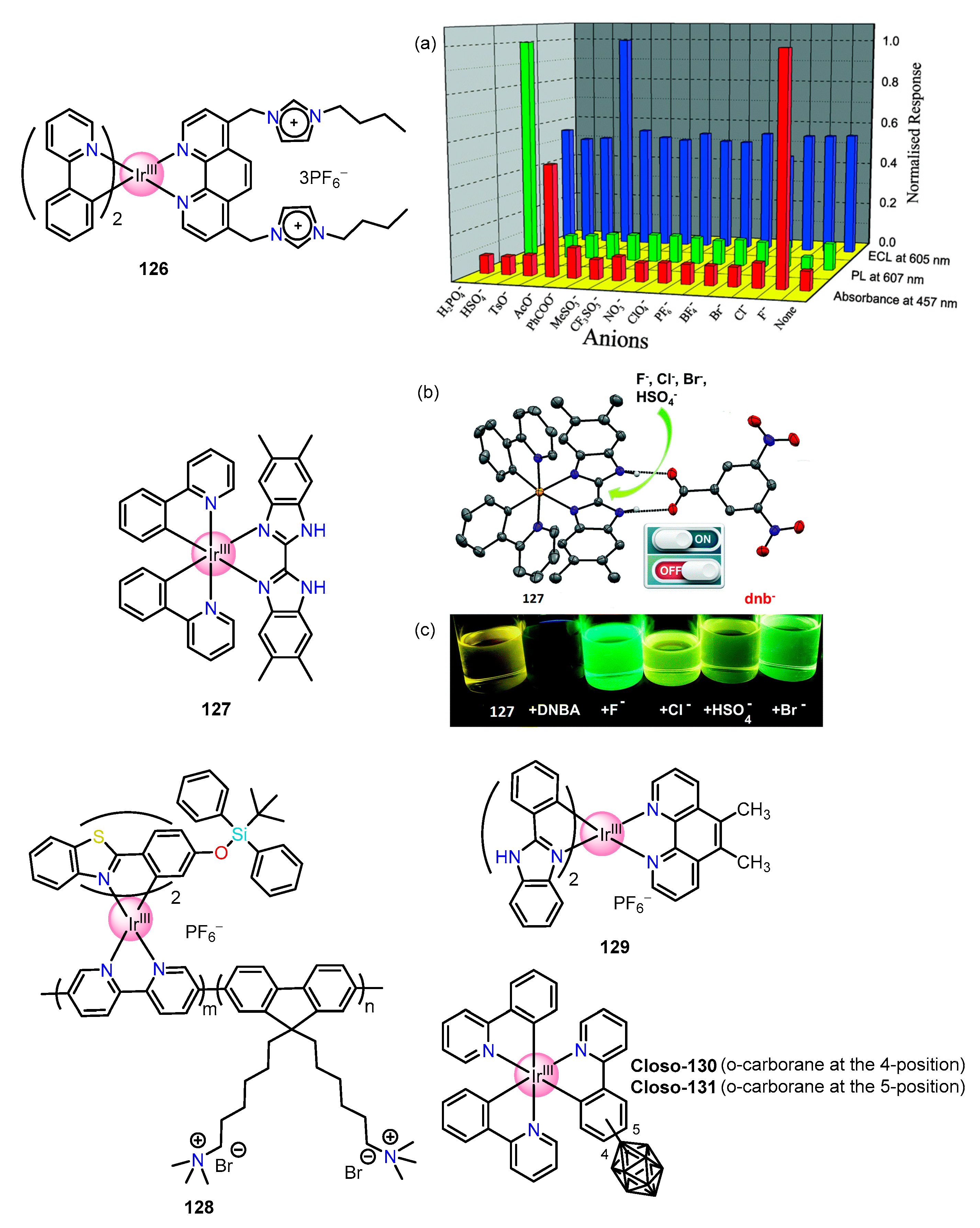
A competitive iridium-based quantification of three different anions in a mixture via three different interrogation techniques and depending upon the anion-imidazolium interaction was reported by Schmittel et al. [129]. The following anions H2PO4, HSO4, F, Cl, Br, PhCOO, BF4, PF6, ClO4, NO3, CF3SO3, MeSO3, CH3CO2, and TsO were tested in three different channels (Figure 17a). The addition of F to 126 caused a broadening and increase in absorption intensity at 457 nm, indicating a new electron-donating unit, whereas the addition of CH3CO2 caused a slight hyperchromic shift, whereas other anions did not induce any such similar changes. The 1H NMR analysis of complex 126 showed a downfield shift corresponding to Δδ = 0.62 ppm, indicating an ionic hydrogen-fluoride bond accompanied by H/D exchange of both 2-H and the methylene protons of 126 in a mixture of CD3CN/CD3OD (v/v, 9:1). The Job’s plot was fitted in a 1:2 binding model for UV-titration results which gave the binding constant as log b = 9.71 ± 0.30 and the limit of detection was calculated as 0.21 μM in the presence of F. Complex 126 emits at 660 nm, and the emission shifted to 607 nm with 7-fold increases in emission intensity upon the addition of 5 equiv. of H2PO4.From the PL and UV titration profile, the binding constant was calculated to be logβ = 7.18 ± 0.10, and the corresponding detection limit was 68 nM. Here the NMR showed a slight downfield shift in the imidazolium proton. In oxidative ECL scans, emission at 605 nm was displayed by complex 126. A highly selective ECL enhancement was shown by 126 in the presence of CH3CO2 while other anions failed to perturb the result. Here the limit of detection was calculated to be 0.17 mM. It is proposed from the results that the positively charged iridium center and the imidazolium units form a tricationic cavity that serves to bind a single CH3CO2 in a bifurcated hydrogen bond. Thus, three different anions were quantified using three techniques by receptor 126.
In 2016, Rau reported a cis-diamine bibenzimidazole functionalized iridium receptor 127 (Figure 17) as an anion sensor [131]. The hydrogen bonding interaction between complex 127 and the anion influenced its photophysical properties. Titration with fluoride caused blue sifting and an increase in emission intensity until 1 equiv. of F addition, beyond which further addition until 2 equiv. caused a constant decrease in intensity accompanied by a naked-eye color change from luminous yellow to bright green. The addition of 2 to 8 equiv.of F further caused an increase in emission, after which saturation is reached. From the changing nature of the emission curve, it was concluded that F caused deprotonation in the iridium complex. The two protons on the benzene ring of the complex are shifted upfield and after the addition of about 2 equiv. F caused the –NH signal to disappear. Strongly basic anions such as H2PO4 and CH3COO caused deprotonation of highly acidity N-H protons of 127 and showed results that are similar to F. Moderately basic anions such as Cl, Br, I, and HSO4 established remarkable H-bonding interactions without deprotonation taking place. The PL spectra were quite different from those of strongly basic anions and showed enhancement in the order Cl ˃ Br ˃ HSO4 ˃ I indicating H-bonding interaction, although blue shifting in the emission spectra was common for all the above-mentioned anions.
Again, 3,5-dinitro benzoate(DNBA) was able to quench the PL of complex 127 by 50% upon 1 equiv. of its addition, whereas upon 8 equiv. addition of the same reduced the PL intensity to 8% compared to the original complex. This “off-state” in situ formed 1:8 2-DNBZ complex served as an active sensor. The five different anions, namely F, Cl, Br, I, and HSO4, were able to restore the quenched PL by replacing DNBA from 127 via a competitive binding and thus were able to switch on the luminescence of 127 (Figure 17b) [130]. The addition of almost 120 equiv. of each anion caused the increase in PL intensity by about 750%, 450%, 405%, 220%, and 118% for fluoride, chloride, hydrogen sulfate, bromide, and iodide, respectively, and the changes were observed via the naked eye (Figure 17c). From the emission bands of the fluoride titration profile shape, it was concluded that increasing concentration of F- anion induced deprotonation in the iridium(III) complex (vide supra). Additionally, the association constants between 127-DNBA and the anions were determined from the 1:1 binding model and were found to be: Ka(Cl) = 33 195, Ka(HSO4) = 17 804, Ka(Br) = 1572. Thus, it was concluded that based on the association, constant values 127-DNBA discriminated between chloride and bromide.
There were a few more complexes of iridium(III) that could act as fluoride sensors. Huang et al. reported a dual emissive nanoprobe of iridium(III),128 (Figure 17), which could form ultrasmall polymer dots in aqueous media and used this nanoprobe for selective, sensitive, accurate, and rapid detection of F in aqueous media as well as in biological systems [132]. In this regard, the tert-butyldiphenylsilyl group was attached to the iridium(III) complex, which could act as the F sensing signaling unit via quenching the phosphorescence. Nanoprobe 128 acted as a dual emissive ratiometric probe and accurately detected the F in live cells by determining the change in the ratio of F-sensitive red phosphorescence from the iridium(III) complex to the F insensitive blue fluorescence from polyfluorene.
Again, Ma et al. reported a luminescent chemosensor, 129 (Figure 17), for tandem recognition of Al3+ and F. Photophysical studies revealed the quenching of luminescing property upon the addition of F to 129 via hydrogen bonding interaction [133]. Again, luminesce is turned on in the presence of Al3+. So chemosensor 129 acted as an on-off-on sensor for F and Al3+.
Again, an interesting fluoride sensor was reported by Lee and co-workers. In this regard, they synthesized two cyclometalated Ir(III) complexes 130 and 131 (Figure 17), containing an o-carborane at the 4- and 5-position, respectively, in the phenyl ring of the pipyridine ligand [134]. X-ray crystal structure analysis of the closo-130 revealed three C^N moiety chelates to Ir via fac-arrangement. Deboronation of the closo-carborane cage occurred upon the addition of F to both the non-emissive closo-130 and closo-131 complexes, which produced the corresponding emissive nido-carborane complexes (nido-130 and nido-131). Therefore, these two carborane complexes could be used as turn-on emissive sensors for F.

2.2.2. Sensing of Cyanide

The detection of cyanide is an area of immense interest due to its extreme toxicity, even at very low concentrations. The limit of cyanide in water is 70 μg/L (in accordance with the World Health Organization (WHO)). So selective sensing and determination of cyanide is a contemporary area of research.
Reddy and coworkers reported a new cyanohydrin-forming phosphorescent iridium(III) complex 132 (Figure 18), which contains bis-2-(2,4- difluorophenyl)-4-formylpyridine ligands as the CN detecting unit [135]. Addition of 2 equiv. CN to acetonitrile solution of 132 caused a prominent orange-to-yellow color change accompanied by a drastic blue shift corresponding to 100 nm in the absorption maxima in the UV spectra of 132. Other similar anions failed to cause a similar change, indicating a selective reaction of CN with the −CHO group. The association constant was estimated to be 1.029 × 10⁵ M−1 from the UV-titration experiments. The emission intensity of 132 was enhanced dramatically at 480 nm, with ~536-fold increment noted within 100 s, assisted with ~155 nm blue shift of the emission and a LOD value corresponding to 2.16 × 10⁻⁸ M. Furthermore, cyanohydrin formation was confirmed by 1H NMR, mass spectrometry, and FT-IR spectral studies which confirmed the vanishing of the aldehyde peak from the iridium complex and appearance of signals indicative of hydroxyl peak in the cyanide adduct.
Yao group reported a cyclometallated phosphorescent complex of iridium, 133 (Figure 18), which was exploited for CN detection [136]. The complex was synthesized by cyanide alcoholized reaction on the C^N ligand (2-phenyl pyridine) and ancillary N^N ligand (1,10-phenanthroline-5-carboxaldehyde). The addition of CN caused cyanohydrin formation as evidenced by the vanishing of -CHO proton peat at δ 10.56 ppm and the appearance of a new peak at δ 8.60 ppm corresponding to the cyanohydrin proton signal. Further, the cyanohydrin formation was confirmed by FT-IR, mass spectra studies, UV−vis, and PL investigations. The PL spectra showed a turn-on response with 15-fold enhancement upon the addition of varying CN concentrations from 0 to 2 equiv. along with an alteration of the color of the solution (pale yellow to bright orange). The PL study revealed that 133 was selective towards CN over other reactive anions, and an excellent sensitivity corresponding to LOD of 1.23 μM in acetonitrile and H2O (95/5) was calculated.
Hong and the group designed a specific molecular sensor 134 (Figure 18) with improved selectivity towards cyanide anion [137]. The complex offered a better reaction or binding site and could discriminate among various interfering anions. Through electrochemical manipulation, the two dicyanovinyl groups at the end of the phenylisoquinoline ligands became selective toward CN. A 1:2 adduct is formed between dicyanovinyl and CN anion. The absorbance at 375 nm was found to decrease upon CN addition accompanied by naked eye color change. The strong electron-withdrawing dicyanovinyl group makes the photoluminescence negligible. The nucleophilic attack of CN at the β-position of the dicyanovinyl to form anionic species resulted in PL enhancement and characteristic blue shifting. Although the dicyanovinyl group is reactive to sulfides and thiols, the same is not reflected in PL as the enhancement in PL intensity in the presence of these two anions is only 15% of that from 134-CN complex. ECL spectra of 134 are relatively weak. However, the addition of CN resulted in a strong ECL signal at 631 nm with 160-fold enhancement, whereas sulfides, thiols, and other anions failed to produce a similar pronounced response. The detection limit from the ECL experiment was estimated to be 0.04 μM. The ECL investigation technique was applied to tap water analysis which gave a rapid and successful detection of CN.
A ratiometric upconversion luminescence (UCL) probe was developed by Li and co-workers for the selective detection of cyanide. In this regard, they synthesized NaYF4 (20%Yb, 1.6%Er, 0.4%Tm)-coated iridium(III) complex 135 (Figure 18), which inhibited the FRET method from UCL emission of the nanocrystals to the absorbance of the chromophoric iridium complex [138]. The limit of detection of CN by this nanocrystal probe was 0.18 μM towards CN and could be practically used for detection of CN in drinking water. This nanocrystal UCL probe is the first example of sensing and bioimaging of CN in living cells.

2.2.3. Sensing of Phosphates

Phosphates are omnipresent in nature and are a fundamental part of our ecosystem. Its plays many vital roles in biological, environmental, and industrial processes. Due to the high hydration energies of phosphates, recognition and sensing was difficult task [139,140].
In 2022, our group synthesized a new bis-heteroleptic complex of iridium (III) 136 (Figure 19), which consisted of 1-ethyl-1H-imidazol-3-ium for the selective sensing of phosphates [141]. Complex 136 served as a lifetime-based sensor for phosphates in acetonitrile and was able to recognize and sense these two anions selectively. The lifetime of the free receptor complex 136 was 0.03543 μs which increased to 0.1323 μs and 0.2736 μs in the presence of HP2O73− and H2PO4, respectively. PL intensities showed 13.7- and 8.5-fold increments for H2PO4 and HP2O73− along with blue shifts in emission wavelength. Sensitive detection of these two anions was achieved with corresponding LOD values of 0.040 μM for H2PO4 and 0.035 μM for HP2O73−. From the NMR studies, it was established that the recognition occurred via CH···phosphate hydrogen bond (HB) interactions.
Again, PL-based detection of pyrophosphate via an iridium complex consisting of a new benzimidazole-substituted 1,2,3-triazole methanol ligand has been reported by Khatua and their group [142]. A three-fold increase in PL spectra of 137 (Figure 19) upon pyrophosphate titration was observed, and the subsequent binding constant of 8.6 × 10⁷ M−1 was calculated. Selective and sensitive detection of pyrophosphate was achieved in acetonitrile over competitive anions such as H2PO4, ADP, ATP, and AMP. The LOD was assessed to be ~127 nM which indicated that complex 137 could detect pyrophosphate in the nanomolar concentration range without interference from other anions. Furthermore, the mechanistic investigations revealed that the acidic protons in the complexes formed H bonding with the solvent molecules and counter anions, while only the long-chain anions could interact with the complex. In addition, the PL increment was attributed to H-bonding interactions of the triazole C–H, methylene hydrogen, imidazole N-H, and hydroxyl groups with the pyrophosphate anion, as confirmed by 1H NMR. In addition, from the TDDFT calculation, it was established that in the 137. Pyrophosphate adduct 137 becomes a better an efficient emitter as the energy gap between the 3MLCT–3MC is increased, and the emission occurs from the lower 3MLCT/3ILCT state. Furthermore, on performing cell imaging, it was found that the complex had low cytotoxicity.

2.2.4. Sensing of Perchlorate

Perchlorate anions are water soluble and have an ionic radius and charge similar to iodide and so it could prevent the uptake and iodide amassing by the thyroid gland competitively. As a result, the deficiency of iodide in the body can be observed, which leads to impairment of the brain and goiter [143]. In addition, the relative stability of perchlorate anions is such that their availability is quite high in groundwater and surface water. So, the detection of ClO4 in water is necessary because of its adverse effect on public health by taking food or drinking water [144].
A binuclear cationic iridium complex 138 (Figure 20) exhibited an efficient aggregation-induced emission in the presence of perchlorate anions [145]. The Schiff base complex displayed a fast, extremely selective, naked-eye, PL “turn on” response towards ClO4 in both an aqueous medium and HeLa cells. The binuclear complex adds an advantage over mononuclear analogs by providing conformational flexibility. Interestingly, ClO4-induced a phosphorescent turn-on in 138 with a decay lifetime of 0.24 μs, accompanied by a blueshift corresponding to 25 nm and 430 times increase in quantum yield. The other anions remarkably produced no substantial variation in the emission experiment performed in HEPES buffer. The phenomenon of the formation of ClO4 induced nano aggregates was confirmed with the help of TEM and DLS experiments. TEM experiment reveals the average size was increased in the presence of ClO4 while DLS showed an increase in the intensity of scattered light and particle size corresponding to 405 nm (Figure 20a–f). The LOD was calculated as 0.05 ppm from the intensity vs. concentration plot. Furthermore, confocal microscopy showed enhanced red phosphorescence in HeLa cells.
In 2016, Chao developed a strategy based on iridium(III) complex 139 (Figure 20) for imaging ClO4 in the cell with strong red emission along with selective sensing of ClO4 in an aqueous medium (1% DMSO) without the intervention of other ions [146]. The complex was weakly emissive at 595 nm. However, upon addition of ClO4, the complex became emissive with a calculated LOD of 43.8 mM (via S/N = 3 methods), and hence it can be used as a luminescence “turn–on” probe for ClO4 in HEPES buffer (10 mM, pH = 7.4). The UV absorbance titration spectra showed an increase in absorbance in the range of 400–550 nm, indicating a light scattering phenomenon caused due to nanoparticles formation in solution. The nanoparticle formation was further confirmed by DLS and SEM experiments which showed that the addition of 100 equiv. of ClO4 induced the formation of larger aggregated nanoparticles having a particle size of 215 nm. It was further proposed that ClO4 caused aggregation-induced emission enhancement (AIEE) of the weakly emissive iridium complex in water.
Further, Chao et al. modified and synthesized a water-soluble “turn-on” luminescence response iridium complex 140 (Figure 20) for ClO4 via AIE [147]. Compared to probe 139, which showed only 4-fold intensity enhancement with ClO4, 140 exhibited a significant 250–fold enhancement at 632 nm. This unprecedented enhancement in emission beyond a threshold concentration of ClO4 was credited to the electrostatic interaction between cationic 140 and anionic ClO4. Additionally, 1:3 stoichiometry of binding was found, and the LOD was estimated to be 9.6 × 10⁻⁷ M. As observed for the complex 140, the UV-Vis titration for 140 also showed an increase in absorption upon ClO4 addition. Luminescence responses of 140 remained unperturbed to other anions, and a substantial red emission in the presence of only ClO4 was observed. Further, macroscopic studies such as SEM, TEM, CLSM, and DLS were performed to obtain an understanding of the sensing mechanism, which unambiguously established the formation of induced aggregation nanoparticles due to ClO4. Last of all, 140 was employed in cell–imaging of the complex in the presence of ClO4 which displayed emission enhancement.

2.2.5. Sensing of Chloride

Williams et al. reported chloride induced luminesce quenching of isomeric bis-terpyridine functionalized iridium, complexes 141 and 142 (Figure 21) [148]. These two complexes differ based on the position of N-methylpyridyl substituents at the terpyridine units. Addition of aq. KCl led to a decrease in the PL intensities of both complexes, but the quenching was greater for complex 142 as compared to 141. The lifetimes of emission were also perturbed for both complexes but to different extents. Thus, the two complexes acted as selective chloride sensors.

2.2.6. Sensing of Nitrite

Two similar chloro-bridged iridium-based chemodosimeters 143 and 144 (Figure 21) for nitrite detection were reported by Schmittel and group [149]. The anion sensing occurs via the substitution of μ-dichloro diiridium complexes. The UV-Vis investigations of both the complexes were carried out in aqueous acetonitrile buffer, which showed absorption intensity enhancement in the presence of nitrite only, followed by an immediate color change for 143 (red to orangish yellow) and 144 (greenish to nearly colorless). The limit of detection from absorption was calculated to be 50 μM. The photoluminescence response of probes 143 and 144 to nitrite showed the opposite behavior. The addition of nitrite caused 350% enhancement in the PL spectra of 143 while 144 resulted in quenching of emission. Furthermore, the NMR and X-ray structure of NO2 adduct with 143 and 144 revealed that the binding of NO2 occurs via two different coordination modes (η1-nitrito-N and η2-nitrito-O, O’) and with distinct stoichiometries.

2.2.7. Sensing of Carbonate/Bicarbonate

Pu et al. developed a novel luminescent “turn on” iridium (III) complex 145 (Figure 21) for HCO3 and CO32− [150]. H-bonding interaction was operational between the hydroxyl group on 145 and the carbonate and bicarbonate anion, which caused the change in the PL spectra of the iridium(III) complex by altering the MLCT states of 145. Only CO32− and HCO3 were able to amplify the luminescence at 600 nm in THF at room temperature, while other anions failed to induce a similar response. The PL intensity was enhanced by 11 times and 14 times upon the addition of CO32− and HCO3 respectively, with alteration of color (colorless to orang red) in both cases. A very low detection limit corresponding to 3.95 × 10⁻⁸ mol L−1 and 1.51 × 10⁻⁷ mol L−1 was calculated for CO32− and HCO3. A 1:1 hosts-to-guest stoichiometry of binding was obtained from Job’s plot analysis and the mass spectrometry between 145 and the two anions separately. A quick response of <20 s was observed, and the association constants were calculated to be 2.67 × 10⁻⁴ and 2.27 × 10⁻⁴ for CO32− and HCO3, respectively. Moreover, fluorescent test strips were also formulated for a cost-effective, simple operation and convenient detection of these anions. Overall, selective and sensitive detection of CO32− and HCO3 was achieved for 145.

2.2.8. Sensing of Hypochlorite

Hypochlorite is recognized as a typical reactive oxygen species (ROS) and is crucial to various immune and pathological processes. It is widely utilized as a detergent, bleaching agent, disinfectant, and decontaminant. A conventional approach for designing ClO detecting probes includes a luminescent signaling unit (iridium), recognition site (ligand), and quencher (C=C, C=N), which controls the phosphorescent intensity of the probes to achieve the detection purpose. A series of “turn on” iridium-based receptors 146150 (Figure 21) tailored with a phosphorescent quencher has been reported in the literature recently [151,152,153,154,155]. Fluorogenic compounds having unbridged C=N and C=C are either weakly fluorescent or non-fluorescent on account of excited state non-radiative decay caused by the isomerization of these bonds. Subsequently, upon reaction with ClO specifically, the C=N and C=C bonds in these complexes were oxidized to C=O and thereby generating the luminous nature of the probe.

3. Conclusions

The prime interest of this review is to give a comprehensive idea of some of the most exciting and pioneering research works of already developed phosphorescent ruthenium (II) and iridium (III)-based complexes and their applications as chemosensor for biologically/chemically/industrially important anions. These chemosensors are considered versatile materials for the detection of anions because they possess excellent photophysical properties such as large Stoke shifts, high quantum yield, longer lifetime, lesser auto-fluorescence, adjustable excitation and emission colors, commendable stability, higher biocompatibility. Here we have concisely amassed heteroleptic ruthenium (II) and iridium (III)-based complexes based on their recognition units in Table 1, as well as anion selectivity in different organic, semi-aqueous and aqueous solvents.
Further, a substantial shortcoming in this direction is the decreased solubility of the designed organometallic receptor molecules in a 100% aqueous medium. A highly demanding research area of significant interest is the extraction of harmful anions generated as by-products from industrial wastes. However, fewer reports are documented in the literature for extraction or removal of inorganic anions from aqueous medium by ruthenium and iridium-based complexes as these anions have high hydration energy. Another drawback faced upon dealing with these complexes is the lower selectivity of iridium complexes which can be overcome in the future by proper modification of ligands. Therefore, this review is envisioned to provide extensive ideas and a significant amount of opportunity for researchers in the future to develop robust Ru(II)/Ir(III) complex-based chemosensors and their application for selective detection of anions in biological media, bioimaging, photo-activated therapeutic agents, and as a photo-activated catalyst.

Author Contributions

Conceptualization, S.M., A.R. and P.G.; methodology, A.R. and S.M.; data curation, A.R., S.M. and P.G.; writing—original draft preparation, S.M., A.R. and P.G.; writing—review and editing, S.M., A.R. and P.G.; visualization, S.M. and A.R.; supervision, P.G.; funding acquisition, P.G. All authors have read and agreed to the published version of the manuscript.

Funding

Science and Engineering Research Board (SERB; CRG/2019/002236), New Delhi.; J. C. Bose National Fellowship (JCB/2021/000032).

Institutional Review Board Statement

Not applicable.

Informed Consent Statement

Not applicable.

Data Availability Statement

Not applicable.

Acknowledgments

PG acknowledges the SERB and J. C. Bose National Fellowship for funding. AR and SM acknowledge CSIR for SRF and IACS for RA-I, respectively.

Conflicts of Interest

The authors declare no conflict of interest.

References

  1. Sessler, J.L.; Gale, P.A.; Cho, W.S. Anion Receptor Chemistry; Royal Society of Chemistry: Cambridge, UK, 2006. [Google Scholar]
  2. Sessler, J.L.; Camiolo, S.; Gale, P.A. Pyrrolic and polypyrrolic anion binding agents. Coord. Chem. Rev. 2003, 240, 17. [Google Scholar] [CrossRef]
  3. Kang, S.O.; Hossain, M.A.; Bowman-James, K. Influence of dimensionality and charge on anion binding in amide-based macrocyclic receptors. Coord. Chem. Rev. 2006, 250, 3038–3052. [Google Scholar] [CrossRef]
  4. Gale, P.A.; Quesada, R. Anion coordination and anion-templated assembly: Highlights from 2002 to 2004. Coord. Chem. Rev. 2006, 250, 3219–3244. [Google Scholar] [CrossRef]
  5. Gunnlaugsson, T.; Glynn, M.; Tocci, G.M.; Kruger, P.E.; Pfeffer, F.M. Anion recognition and sensing in organic and aqueous media using luminescent and colorimetric sensors. Coord. Chem. Rev. 2006, 250, 3094–3117. [Google Scholar] [CrossRef]
  6. Katayev, E.A.; Ustynyuk, Y.A.; Sessler, J.L. Receptors for tetrahedral oxyanions. Coord. Chem. Rev. 2006, 250, 3004–3037. [Google Scholar] [CrossRef]
  7. Gale, P.A.; Garcia-Garrido, S.E.; Garric, J. Anion receptors based on organic frameworks: Highlights from 2005 and 2006. Chem. Soc. Rev. 2008, 37, 151–190. [Google Scholar] [CrossRef] [PubMed]
  8. Kim, H.N.; Lee, M.H.; Kim, H.J.; Kim, J.S.; Yoon, J. A new trend in rhodamine-based chemosensors: Application of spirolactam ring-opening to sensing ions. Chem. Soc. Rev. 2008, 37, 1465–1472. [Google Scholar] [CrossRef] [PubMed]
  9. Caltagirone, C.; Gale, P.A. Anion receptor chemistry: Highlights from 2007. Chem. Soc. Rev. 2009, 38, 520–563. [Google Scholar] [CrossRef]
  10. Xu, Z.; Chen, X.; Kim, H.N.; Yoon, J. Sensors for the optical detection of cyanide ion. Chem. Soc. Rev. 2010, 39, 127–137. [Google Scholar] [CrossRef]
  11. Beer, P.D.; Gale, P.A. Anion recognition and sensing: The state of the art and future perspectives. Angew. Chem. Int. Ed. 2001, 40, 486–516. [Google Scholar] [CrossRef]
  12. Kubik, S. Anion recognition in water. Chem. Soc. Rev. 2010, 39, 3648–3663. [Google Scholar] [CrossRef] [PubMed]
  13. Gale, P.A.; Caltagirone, C. Anion sensing by small molecules and molecular ensembles. Chem. Soc. Rev. 2015, 44, 4212–4227. [Google Scholar] [CrossRef] [PubMed]
  14. Busschaert, N.; Caltagirone, C.; Rossom, W.V.; Gale, P.A. Applications of supramolecular anion recognition. Chem. Rev. 2015, 115, 8038–8155. [Google Scholar] [CrossRef]
  15. Langton, M.J.; Serpell, C.J.; Beer, P.D. Anion recognition in water: Recent advances from a supramolecular and macromolecular perspective. Angew. Chem. Int. Ed. 2016, 55, 1974–1987. [Google Scholar] [CrossRef] [PubMed] [Green Version]
  16. Gale, P.A.; Caltagirone, C. Fluorescent and colorimetric sensors for anionic species. Coord. Chem. Rev. 2018, 354, 2–27. [Google Scholar] [CrossRef]
  17. Hein, R.; Beer, P.D.; Davis, J.J. Electrochemical anion sensing: Supramolecular approaches. Chem. Rev. 2020, 120, 1888–1935. [Google Scholar] [CrossRef]
  18. Zhao, Q.; Li, F.; Huang, C. Phosphorescent chemosensors based on heavy-metal complexes. Chem. Soc. Rev. 2010, 39, 3007–3030. [Google Scholar] [CrossRef]
  19. Zhao, Q.; Huang, C.; Li, F. Phosphorescent heavy-metal complexes for bioimaging. Chem. Soc. Rev. 2011, 40, 2508–2524. [Google Scholar] [CrossRef] [Green Version]
  20. Lo, K.K.-W.; Choi, A.W.-T.; Law, W.H.-T. Applications of luminescent inorganic and organometallic transition metal complexes as biomolecular and cellular probes. Dalton Trans. 2012, 41, 6021–6047. [Google Scholar] [CrossRef]
  21. Baggaley, E.; Weinstein, J.A.; Williams, J.A.G. Lighting the way to see inside the live cell with luminescent transition metal complexes. Coord. Chem. Rev. 2012, 256, 1762–1785. [Google Scholar] [CrossRef]
  22. Qiu, K.; Chen, Y.; Rees, T.W.; Ji, L.; Chao, H. Organelle-targeting metal complexes: From molecular design to bio-applications. Coord. Chem. Rev. 2019, 378, 66–86. [Google Scholar] [CrossRef]
  23. Lee, L.C.-C.; Lo, K.K.-W. Luminescent and photofunctional transition metal complexes: From molecular design to diagnostic and therapeutic applications. J. Am. Chem. Soc. 2022, 144, 14420–14440. [Google Scholar] [CrossRef] [PubMed]
  24. Yam, V.W.-W.; Wong, K.M.-C. Luminescent metal complexes of d6, d8 and d10 transition metal centres. Chem. Commun. 2011, 47, 11579–11592. [Google Scholar] [CrossRef] [PubMed]
  25. Ma, D.-L.; Lin, S.; Wang, W.; Yang, C.; Leung, C.-H. Luminescent chemosensors by using cyclometalated iridium(III) complexes and their applications. Chem. Sci. 2017, 8, 878–889. [Google Scholar] [CrossRef] [PubMed] [Green Version]
  26. Shi, H.; Wang, Y.; Lin, S.; Lou, J.; Zhang, Q. Recent development and application of cyclometalated iridium(III) complexes as chemical and biological probes. Dalton Trans. 2021, 50, 6410–6417. [Google Scholar] [CrossRef] [PubMed]
  27. Arndtsen, B.A.; Bergman, R.G. Unusually mild and selective hydrocarbon CH bond activation with positively charged iridium (III) complexes. Science 1995, 270, 1970–1973. [Google Scholar] [CrossRef]
  28. Chepelin, O.; Ujma, J.; Wu, X.; Slawin, A.M.; Pitak, M.B.; Coles, S.J.; Michel, J.; Jones, A.C.; Barran, P.E.; Lusby, P.J. Luminescent, enantiopure, phenylatopyridine iridium-based coordination capsules. J. Am. Chem. Soc. 2012, 134, 19334–19337. [Google Scholar] [CrossRef] [Green Version]
  29. Llinares, J.M.; Powell, D.; Bowman-James, K. Ammonium based anion receptors. Coord. Chem. Rev. 2003, 240, 57–75. [Google Scholar] [CrossRef]
  30. Garcia-Espana, E.; Diaz, P.; Llinares, J.M.; Bianchi, A. Anion coordination chemistry in aqueous solution of polyammonium receptors. Coord. Chem. Rev. 2006, 250, 2952–2986. [Google Scholar] [CrossRef]
  31. Lu, L.; Wang, J.; Xie, B.; Liu, J.Q.; Yadav, R.; Singh, A.; Kumar, A. Fluorescence sensing of nitro-aromatics by Zn (II) and Cd (II) based coordination polymers having the 5-[bis (4 carboxybenzyl)-amino] isophthalic acid ligand. New J. Chem. 2017, 41, 3537–3542. [Google Scholar] [CrossRef]
  32. Qin, L.; Li, Y.; Liang, F.; Li, L.; Lan, Y.; Li, Z.; Lu, X.; Yang, M.; Ma, D. A microporous 2D cobalt-based MOF with pyridyl sites and open metal sites for selective adsorption of CO2. Micropor. Mesopor. Mat. 2022, 341, 112098. [Google Scholar] [CrossRef]
  33. Qin, L.; Liang, F.; Li, Y.; Wu, J.; Guan, S.; Wu, M.; Xie, S.; Luo, M.; Ma, D. A 2D porous zinc-organic framework platform for loading of 5-fluorouracil. Inorganics 2022, 10, 202. [Google Scholar] [CrossRef]
  34. Desiraju, G.R. A bond by any other name. Angew. Chem. Int. Ed. 2011, 50, 52–59. [Google Scholar] [CrossRef]
  35. Steiner, T. Unrolling the hydrogen bond properties of C–H•••O interactions. Chem. Commun. 1997, 8, 727–734. [Google Scholar] [CrossRef]
  36. Desiraju, G.R.; Ho, P.S.; Kloo, L.; Legon, A.C.; Marquardt, R.; Metrangolo, P.; Politzer, P.; Resnati, G.; Rissanen, K. Definition of the halogen bond (IUPAC Recommendations 2013). Pure Appl. Chem. 2013, 85, 1711–1713. [Google Scholar] [CrossRef]
  37. Gilday, L.C.; Robinson, S.W.; Barendt, T.A.; Langton, M.J.; Mullaney, B.R.; Beer, P.D. Halogen bonding in supramolecular chemistry. Chem. Rev. 2015, 115, 7118–7195. [Google Scholar] [CrossRef]
  38. Cavallo, G.; Metrangolo, P.; Milani, R.; Pilati, T.; Priimagi, A.; Resnati, G.; Terraneo, G. The halogen bond. Chem. Rev. 2016, 116, 2478–2601. [Google Scholar] [CrossRef] [Green Version]
  39. Beale, T.M.; Chudzinski, M.G.; Sarwar, M.G.; Taylor, M.S. Halogen bonding in solution: Thermodynamics and applications. Chem. Soc. Rev. 2013, 42, 1667–1680. [Google Scholar] [CrossRef]
  40. Schottel, B.L.; Chifotides, H.T.; Dunbar, K.R. Anion-π interactions. Chem. Soc. Rev. 2008, 37, 68–83. [Google Scholar] [CrossRef]
  41. Dawson, R.E.; Hennig, A.; Weimann, D.P.; Emery, D.; Ravikumar, V.; Montenegro, J.; Takeuchi, T.; Gabutti, S.; Mayor, M.; Mareda, J.; et al. Experimental evidence for the functional relevance of anion–π interactions. Nat. Chem. 2010, 2, 533–538. [Google Scholar] [CrossRef]
  42. Frontera, A.; Gamez, P.; Mascal, M.; Mooibroek, T.J.; Reedijk, J. Putting anion–π interactions into perspective. Angew. Chem. Int. Ed. 2011, 50, 9564–9583. [Google Scholar] [CrossRef] [PubMed]
  43. Chifotides, H.T.; Dunbar, K.R. Anion−π interactions in supramolecular architectures. Acc. Chem. Res. 2013, 46, 894–906. [Google Scholar] [CrossRef] [PubMed]
  44. Dutta, A.; Pan, Y.; Liu, J.Q.; Kumar, A. Multicomponent isoreticular metal-organic frameworks: Principles, current status and challenges. Coord. Chem. Rev. 2021, 445, 214074. [Google Scholar] [CrossRef]
  45. Wu, M.; Zhang, Z.; Yong, J.; Schenk, P.M.; Tian, D.; Xu, Z.P.; Zhang, R. Determination and imaging of small biomolecules and ions using ruthenium(II) complex-based chemosensors. Top. Curr. Chem. 2022, 380, 29. [Google Scholar] [CrossRef]
  46. Chang, K.-C.; Sun, S.-S.; Odago, M.O.; Lees, A.J. Anion recognition and sensing by transition-metal complexes with polarized N H recognition motifs. Coord. Chem. Rev. 2015, 284, 111–123. [Google Scholar] [CrossRef] [Green Version]
  47. Alreja, P.; Kaur, N. Recent advances in 1,10-phenanthroline ligands for chemosensing of cations and anions. RSC Adv. 2016, 6, 23169–23217. [Google Scholar] [CrossRef]
  48. Pal, S.; Ghosh, T.K.; Ghosh, R.; Mondal, S.; Ghosh, P. Recent advances in recognition, sensing and extraction of phosphates: 2015 onwards. Coord. Chem. Rev. 2020, 405, 213128. [Google Scholar] [CrossRef]
  49. Kumar, P.; Pachisia, S.; Gupta, R. Turn-on detection of assorted phosphates by luminescent chemosensors. Inorg. Chem. Front. 2021, 8, 3587–3607. [Google Scholar] [CrossRef]
  50. Juris, A.; Balzani, V.; Barigeletti, F.; Campagna, S.; Belser, P.; Zelewsky, A.V. Ru(II) polypyridine complexes: Photophysics, photochemistry, eletrochemistry, and chemiluminescence. Coord. Chem. Rev. 1988, 84, 85–277. [Google Scholar] [CrossRef]
  51. Beer, P.D.; Dickson, C.A.P.; Fletcher, N.; Goulden, A.J.; Grieve, A.; Hodacova, J.; Wear, T. New classes of anion receptor containing charged and neutral transition metal Lewis acidic recognition sites. J. Chem. Soc. Chem. Commun. 1993, 10, 828–830. [Google Scholar] [CrossRef]
  52. Chowdhury, B.; Khatua, S.; Dutta, R.; Chakraborty, S.; Ghosh, P. Bis-heteroleptic ruthenium(II) complex of a triazole ligand as a selective probe for phosphates. Inorg. Chem. 2014, 53, 8061–8070. [Google Scholar] [CrossRef] [PubMed]
  53. Chowdhury, B.; Dutta, R.; Khatua, S.; Ghosh, P. A cyanuric acid platform based tripodal bis-heteroleptic Ru(II) complex of click generated ligand for selective sensing of phosphates via C–H···anion interaction. Inorg. Chem. 2016, 55, 259–271. [Google Scholar] [CrossRef] [PubMed]
  54. Mondal, S.; Sarkar, K.; Ghosh, P. Influence of triazole substituents of bis-heteroleptic Ru(II) probes toward selective sensing of dihydrogen phosphate. Inorg. Chem. 2021, 60, 9084–9096. [Google Scholar] [CrossRef] [PubMed]
  55. Chowdhury, B.; Sinha, S.; Ghosh, P. Selective sensing of phosphates by a new bis-heteroleptic RuII complex through halogen bonding: A superior sensor over its hydrogen-bonding analogue. Chem. Eur. J. 2016, 22, 18051–18059. [Google Scholar] [CrossRef]
  56. Mondal, S.; Rashid, A.; Ghosh, P. Apentafluorophenyl functionalized RuII-probe having halogen bond center toward recognition and sensing of perrhenate and dihydrogen phosphate. J. Organomet. Chem. 2021, 952, 122027. [Google Scholar] [CrossRef]
  57. Mondal, S.; Ghosh, T.K.; Chowdhury, B.; Ghosh, P. Supramolecular self-assembly driven selective sensing of phosphates. Inorg. Chem. 2019, 58, 15993–16003. [Google Scholar] [CrossRef]
  58. Ghosh, T.K.; Chakraborty, S.; Chowdhury, B.; Ghosh, P. Bis-heteroleptic ruthenium(II) complex of pendant urea functionalized pyridyl triazole and phenanthroline for recognition, sensing, and extraction of oxyanions. Inorg. Chem. 2017, 56, 5371–5382. [Google Scholar] [CrossRef]
  59. Ghosh, T.K.; Ghosh, P. Balancing the acidity of the pendant urea arm of bis-heteroleptic ruthenium(II) complex containing pyridyl triazole for improved oxyanion recognition. Dalton Trans. 2018, 47, 7561–7570. [Google Scholar] [CrossRef]
  60. Ghosh, T.K.; Mondal, S.; Bej, S.; Nandi, M.; Ghosh, P. An integrated urea and halogen bond donor based receptor for superior and selective sensing of phosphates. Dalton Trans. 2019, 48, 4538–4546. [Google Scholar] [CrossRef]
  61. Chowdhury, B.; Sinha, S.; Dutta, R.; Mondal, S.; Karmakar, S.; Ghosh, P. Discriminative behavior of a donor–acceptor–donor triad toward cyanide and fluoride: Insights into the mechanism of naphthalene diimide reduction by cyanide and fluoride. Inorg. Chem. 2020, 59, 13371–13382. [Google Scholar] [CrossRef]
  62. Sheet, S.K.; Sen, B.; Thounaojam, R.; Aguan, K.; Khatua, S. Ruthenium(II) complex-based luminescent bifunctional probe for Ag+ and phosphate ions: Ag+-assisted detection and imaging of rRNA. Inorg. Chem. 2017, 56, 1249–1263. [Google Scholar] [CrossRef] [PubMed]
  63. Sen, B.; Kumar, S.P.; Rabha, M.; Sheet, S.K.; Aguan, K.; Samanta, D.; Khatua, S. Luminescence detection of Ag+ and phosphate ions by a ruthenium(II) complex-based multianalyte probe: A combined spectroscopic, crystallographic, and theoretical approach. Eur. J. Inorg. Chem. 2021, 35, 3549–3560. [Google Scholar] [CrossRef]
  64. Ramachandran, M.; Anandan, S. Triazole appending ruthenium(II) polypyridine complex for selective sensing of phosphate anions through C–H–anion interaction and copper(II) ions via cancer cells. New J. Chem. 2020, 44, 6186–6196. [Google Scholar] [CrossRef]
  65. Ramachandran, M.; Syed, A.; Marraiki, N.; Anandan, S. The aqueous dependent sensing of hydrazine and phosphate anions using a bis-heteroleptic Ru(II) complex with a phthalimide–anchored pyridinetriazole ligand. Analyst 2021, 146, 1430–1443. [Google Scholar] [CrossRef]
  66. Molina, P.; Tárraga, A.; Otón, F. Imidazole derivatives: A comprehensive survey of their recognition properties. Org. Biomol. Chem. 2012, 10, 1711–1724. [Google Scholar] [CrossRef]
  67. Rommel, S.A.; Sorsche, D.; Fleischmann, M.; Rau, S. Optical sensing of anions via supramolecular recognition with biimidazole complexes. Chem. Eur. J. 2017, 23, 18101–18119. [Google Scholar] [CrossRef]
  68. Ion, L.; Morales, D.; Perez, J.; Riera, L.; Riera, V.; Kowenicki, R.; McPartlin, M. Ruthenium biimidazole complexes as anion receptors. Chem. Commun. 2006, 1, 91–93. [Google Scholar] [CrossRef]
  69. Cui, Y.; Mo, H.-J.; Chen, J.-C.; Niu, Y.-L.; Zhong, Y.-R.; Zheng, K.-C.; Ye, B.-H. Anion-selective interaction and colorimeter by an optical metalloreceptor based on ruthenium (ii) 2, 2 ′-biimidazole: Hydrogen bonding and proton transfer. Inorg. Chem. 2007, 46, 6427–6436. [Google Scholar] [CrossRef]
  70. Cui, Y.; Niu, Y.-L.; Cao, M.-L.; Wang, K.; Mo, H.-J.; Zhong, Y.-R.; Ye, B.-H. Ruthenium (II) 2, 2′-bibenzimidazole complex as a second-sphere receptor for anions interaction and colorimeter. Inorg. Chem. 2008, 47, 5616–5624. [Google Scholar] [CrossRef]
  71. Mo, H.-J.; Niu, Y.-L.; Zhang, M.; Qiao, Z.-P.; Ye, B.-H. Photophysical, electrochemical and anion sensing properties of Ru(II) bipyridine complexes with 2,2′-biimidazole-like ligand. Dalton Trans. 2011, 40, 8218–8225. [Google Scholar] [CrossRef]
  72. Mo, H.-J.; Chao, H.-Y.; Ye, B.-H. A ruthenium biimidazole-like anion receptor with two chelating Nsingle bondH⋯O intramolecular hydrogen bonds. Inorg. Chem. Commun. 2013, 35, 100–103. [Google Scholar] [CrossRef]
  73. Sorsche, D.; Rommel, A.S.; Rau, S. Functional dimming of pincer-shaped bibenzimidazole ruthenium(II) complexes with improved anion-sensitive luminescence. Eur. J. Inorg. Chem. 2016, 10, 1503–1513. [Google Scholar] [CrossRef]
  74. Mo, H.-J.; Shen, Y.; Ye, B.-H. Selective recognition of cyanide anion via formation of multipoint NH and phenyl CH hydrogen bonding with acyclic ruthenium bipyridine imidazole receptors in water. Inorg. Chem. 2012, 51, 7174–7184. [Google Scholar] [CrossRef] [PubMed]
  75. Zheng, Z.-B.; Huang, Q.-Y.; Han, Y.-F.; Zuo, J.; Ma, Y.-N. Ruthenium(II) complex-based chemosensors for highly sensitive and selective sequential recognition of copper ion and cyanide. Sens. Actuators B 2017, 253, 203–212. [Google Scholar] [CrossRef]
  76. Kundu, T.; Mobin, S.M.; Lahiri, G. Paramagnetic ruthenium-biimidazole derivatives [(acac)2RuIII(LHn)]m, n/m = 2/+, 1/0, 0/-. Synthesis, structures, solution properties and anion receptor features in solution state. Dalton Trans. 2010, 39, 4232–4242. [Google Scholar] [CrossRef]
  77. Khanmohammadi, H.; Rezaeian, K. A catalyst-free approach to a novel imidazo [4,5-f][1,10] phenanthroline ligand and its corresponding ruthenium(II) complex: Insights into their applications in colorimetric anion sensing. New J. Chem. 2014, 38, 5536–5543. [Google Scholar] [CrossRef]
  78. Zapata, F.; Caballero, A.; Espinosa, A.; Tárraga, A.; Molina, P. Cation coordination induced modulation of the anion sensing properties of a ferrocene−imidazophenanthroline dyad: Multichannel recognition from phosphate-related to chloride anions. J. Org. Chem. 2008, 73, 4034–4044. [Google Scholar] [CrossRef]
  79. Yang, H.-X.; Liu, Y.-J.; Zhao, L.; Wang, K.-Z. Highly selective acetate optical sensing of a ruthenium(II) complex carrying imidazole and indole groups. Spectrochim. Acta Part A 2010, 76, 146–149. [Google Scholar] [CrossRef]
  80. Zheng, Z.-B.; Duan, Z.-M.; Ma, Y.-Y.; Wang, K.-Z. Highly sensitive and selective difunctional ruthenium(II) complexbased chemosensor for dihydrogen phosphate anion and ferrous cation. Inorg. Chem. 2013, 52, 2306–2316. [Google Scholar] [CrossRef]
  81. Zheng, Z.-B.; Wu, Y.-Q.; Wang, K.-Z.; Li, F. pH luminescence switching, dihydrogen phosphate sensing, and cellular uptake of a heterobimetallic ruthenium(II)–rhenium(I) complex. Dalton Trans. 2014, 43, 3273–3284. [Google Scholar] [CrossRef]
  82. Saha, D.; Das, S.; Bhaumik, C.; Dutta, S.; Baitalik, S. Monometallic and bimetallic ruthenium(II) complexes derived from 4,5-bis(benzimidazol-2-yl)imidazole (H3Imbzim) and 2,20-bipyridine as colorimetric sensors for anions: Synthesis, characterization, and binding studies. Inorg. Chem. 2010, 49, 2334–2348. [Google Scholar] [CrossRef] [PubMed]
  83. Das, S.; Saha, D.; Bhaumik, C.; Dutta, S.; Baitalik, S. Ru(II) and Os(II) mixed-chelates derived from imidazole-4,5-dicarboxylic acid and 2,2′-bipyridine as colorimetric sensors for anions: Synthesis, characterization and binding studies. Dalton Trans. 2010, 39, 4162–4169. [Google Scholar] [CrossRef] [PubMed]
  84. Saha, D.; Das, S.; Mardanya, S.; Baitalik, S. Structural characterization and spectroelectrochemical, anion sensing and so vent dependence photophysical studies of a bimetallic Ru(II) complex derived from 1,3–di (1H-imidazo [4,5-f][1,10]phenanthroline-2-yl)benzene. Dalton Trans. 2012, 41, 8886–8898. [Google Scholar] [CrossRef] [PubMed]
  85. Saha, D.; Das, S.; Karmakar, S.; Dutta, S.; Baitalik, S. Synthesis, structural characterization and anion-, cation- and solvent-induced tuning of photophysical properties of a bimetallic Ru(II) complex: Combined experimental and DFT/TDDFT investigation. RSC Adv. 2013, 3, 17314–17334. [Google Scholar] [CrossRef]
  86. Mardanya, S.; Karmakar, S.; Bar, M.; Baitalik, S. Pyrene-biimidazole based Ru(II) and Os(II) complexes as highly efficient probes for the visible and near-infrared detection of cyanide in aqueous media. Dalton Trans. 2015, 44, 21053–21072. [Google Scholar] [CrossRef]
  87. Paul, A.; Bar, M.; Ahmed, T.; Baitalik, S. Anion-sensitive photophysics of luminescent trimetallic complexes of Fe(II), Ru(II), and Os(II) with polarized NH motifs. Polyhedron 2020, 190, 114772. [Google Scholar] [CrossRef]
  88. Deb, S.; Sahoo, A.; Pal, P.; Baitalik, S. Exploitation of the second coordination sphere to promote significant increase of room-temperature luminescence lifetime and anion sensing in ruthenium–terpyridine complexes. Inorg. Chem. 2021, 60, 6836–6851. [Google Scholar] [CrossRef]
  89. Pineda, L.H.; Flores, E.D.T.; Hernandez, J.G.; Thangarasu, P.; Ramos, J.M.V. Ruthenium complex of bis(benzimidazole-yl-ethyl)sulfide as chemo-sensor for selective recognition of chloride ion, and its application in real bacterial samples. Inorg. Chim. Acta 2021, 522, 120354. [Google Scholar] [CrossRef]
  90. Mareeswaran, P.M.; Babu, E.; Rajagopal, S. Optical recognition of anions by ruthenium(II)-bipyridine-calix [4]arene system. J Fluoresc. 2013, 23, 997–1006. [Google Scholar] [CrossRef]
  91. Maity, D.; Vyas, G.; Bhatt, M.; Paul, P. Detection of NaCN in aqueous media using a calixarene-based fluoroionophore containing ruthenium(II)-bipyridine as the fluorogenic unit. RSC Adv. 2015, 5, 6151–6159. [Google Scholar] [CrossRef]
  92. Berni, E.; Henaff, L.L.; Jarrige, L.; Girard, E.; Jonusauskas, G.; Gosse, I.; Pinet, S. Functionalized ruthenium complexes: Selective “Turn-on” detection of biologically relevant anionic species. Eur. J. Org. Chem. 2017, 2017, 3620–3630. [Google Scholar] [CrossRef] [Green Version]
  93. Lin, T.-P.; Chen, C.-Y.; Wen, Y.-S.; Sun, S.-S. Synthesis, photophysical, and anion-sensing properties of quinoxalinebis(sulfonamide) functionalized receptors and their metal complexes. Inorg. Chem. 2007, 46, 9201–9212. [Google Scholar] [CrossRef] [PubMed]
  94. Shang, X.-F.; Li, J.; Lin, H.; Jiang, P.; Caia, Z.-S.; Lin, H.-K. Anion recognition and sensing of ruthenium(II) and cobalt(II) sulfonamido complexes. Dalton Trans. 2009, 12, 2096–2102. [Google Scholar] [CrossRef] [PubMed]
  95. Toyama, M.; Fujii, Y.; Endo, M. Bis-heteroleptic ruthenium(II) complex with 2-picolinamide: Synthesis, crystal structures, and spectroscopic study for anion recognition using the amide group. Inorg. Chim. Acta 2019, 486, 304–313. [Google Scholar] [CrossRef]
  96. Gong, Z.-L.; Zhong, Y.-W. Spectroscopic and electrochemical recognition of H2PO4− based on a ruthenium complex with 2-picolinamide. J. Organomet. Chem. 2021, 931, 121612. [Google Scholar] [CrossRef]
  97. Toyama, M.; Hasegaw, T.; Nagao, N. Colorimetric fluoride detection in dimethyl sulfoxide using a heteroleptic ruthenium (II) complex with amino and amide groups: X-ray crystallographic and spectroscopic analyses. RSC Adv. 2022, 12, 25227–25239. [Google Scholar] [CrossRef]
  98. Mizuno, T.; Wei, W.-H.; Eller, L.R.; Sessler, J.L. Phenanthroline complexes bearing fused dipyrrolylquinoxaline anion recognition sites: Efficient fluoride anion receptors. J. Am. Chem. Soc. 2002, 124, 1134–1135. [Google Scholar] [CrossRef]
  99. Plitt, P.; Gross, D.E.; Lynch, V.M.; Sessler, J.L. Dipyrrolyl-functionalized bipyridine-based anion receptors for emission based selective detection of dihydrogen phosphate. Chem. Eur. J. 2007, 13, 1374–1381. [Google Scholar] [CrossRef]
  100. Anzenbacher, P., Jr.; Tyson, D.S.; Jursıkova, K.; Castellano, F.N. Luminescence lifetime-based sensor for cyanide and related anions. J. Am. Chem. Soc. 2002, 124, 6232–6233. [Google Scholar] [CrossRef] [Green Version]
  101. Smith, P.J.; Reddington, M.V.; Wilcox, C.S. Ion pair binding by a urea in chloroform solution. Tetrahedron Lett. 1992, 33, 6085–6088. [Google Scholar] [CrossRef]
  102. Fan, E.; van Arman, S.A.; Kincaid, S.; Hamilton, A.D. Molecular recognition: Hydrogen-bonding receptors that function in highly competitive solvents. J. Am. Chem. Soc. 1993, 115, 369–370. [Google Scholar] [CrossRef]
  103. Ghosh, A.; Ganguly, B.; Das, A. Urea-based ruthenium(II)−polypyridyl complex as an optical sensor for anions: Synthesis, characterization, and binding studies. Inorg. Chem. 2007, 46, 9912–9918. [Google Scholar] [CrossRef] [PubMed]
  104. Kitchen, J.A.; Boyle, E.M.; Gunnlaugsson, T. Synthesis, structural characterisation and luminescent anion sensing studies of a Ru(II)polypyridyl complex featuring an aryl urea derivatised 2,2’-bpy auxiliary ligand. Inorg. Chim. Acta 2012, 381, 236–242. [Google Scholar] [CrossRef]
  105. Baggi, G.; Boiocchi, M.; Ciarrocchi, C.; Fabbrizzi, L. Enhancing the anion affinity of urea-based receptors with a Ru(terpy)22+ chromophore. Inorg. Chem. 2013, 52, 5273–5283. [Google Scholar] [CrossRef]
  106. Gong, Z.-L.; Deng, L.-Y.; Zhong, Y.-W.; Yao, J. Anion-regulated electronic communication in a cyclometalated diruthenium complex with a urea bridge. Phys. Chem. Chem. Phys. 2017, 19, 8902–8907. [Google Scholar] [CrossRef] [PubMed]
  107. Chae, M.Y.; Czarnik, A.W. Fluorometric chemodosimetry mercury (II) and silver (I) indication in water via enhanced fluorescence signaling. J. Am. Chem. Soc. 1992, 114, 9704–9705. [Google Scholar] [CrossRef]
  108. Khatua, S.; Samanta, D.; Bats, J.W.; Schmittel, M. Rapid and highly sensitive dual-channel detection of cyanide by bis-heteroleptic ruthenium (II) complexes. Inorg. Chem. 2012, 51, 7075–7086. [Google Scholar] [CrossRef]
  109. Zhu, J.-W.; Ou, H.-D.; Xu, N.; Deng, W.; Yao, Z.-J. Ruthenium-based phosphorescent probe for selective and naked-eye detection of cyanide in aqueous media. Dyes Pigments 2020, 176, 108196. [Google Scholar] [CrossRef]
  110. Li, M.-J.; Lin, Z.; Chena, X.; Chen, G. Colorimetric and luminescent bifunctional Ru(II) complexes for rapid and highly sensitive recognition of cyanide. Dalton Trans. 2014, 43, 11745. [Google Scholar] [CrossRef]
  111. Jose, D.A.; Kar, P.; Koley, D.; Ganguly, B.; Thiel, W.; Ghosh, H.N.; Das, A. Phenol- and catechol-based ruthenium(II) polypyridyl complexes as colorimetric sensors for fluoride ions. Inorg. Chem. 2007, 46, 5576–5584. [Google Scholar] [CrossRef]
  112. Ghosh, B.; Adak, P.; Naskar, S.; Pakhira, B.; Mitra, P.; Dinda, R.; Chattopadhyay, S.K. Ruthenium(II) complexes of thiosemicarbazones: Synthesis, X-ray crystal structures, spectroscopy, electrochemistry, DFT studies and fluoride sensing properties. Inorg. Chim. Acta 2017, 459, 1–14. [Google Scholar] [CrossRef]
  113. Patil, S.K.; Ghosh, R.; Kennedy, P.; Mobin, S.M.; Das, D. Potential anion sensing properties by a redox and substitution series of [Ru(bpy)3-n(Hdpa)n]2+, n ¼ 1–3; Hdpa = 2,2’-dipyridylamine: Selective recognition and stoichiometric binding with cyanide and fluoride ions. RSC Adv. 2016, 6, 62310–62319. [Google Scholar] [CrossRef]
  114. Toyama, M.; Mishima, D.; Suganoya, R.; Nagao, N. C1-symmetrical cis-bis(di-2-pyridylamine)chloro(dimethyl sulfoxide-S) ruthenium(II) complex: Synthesis, crystal structure, and anion recognition using the NH groups in the chelating ligands. Inorg. Chim. Acta 2018, 478, 104–111. [Google Scholar] [CrossRef]
  115. Everett, E.T. Fluoride’s effects on the formation of teeth and bones, and the influence of genetics. J. Dent. Res. 2011, 90, 552–560. [Google Scholar]
  116. Zhao, Q.; Li, F.; Liu, S.; Yu, M.; Liu, Z.; Yi, T.; Huang, C. Highly selective phosphorescent chemosensor for fluoride based on an iridium(III) complex containing arylborane units. Inorg. Chem. 2008, 47, 9256–9264. [Google Scholar] [CrossRef] [PubMed]
  117. You, Y.; Park, S.Y. A phosphorescent Ir(III) complex for selective fluoride ion sensing with a high signal-to-noise ratio. Adv. Mater. 2008, 20, 3820–3826. [Google Scholar] [CrossRef]
  118. Xu, W.-J.; Liu, S.-J.; Zhao, X.-Y.; Sun, S.; Cheng, S.; Ma, T.-C.; Sun, H.-B.; Zhao, Q.; Huang, W. Cationic iridium(III) complex containing both triarylboron and carbazole moieties as a ratiometric fluoride probe that utilizes a switchable triplet–singlet emission. Chem. Eur. J. 2010, 16, 7125–7133. [Google Scholar] [CrossRef]
  119. Xu, W.; Liu, S.; Zhao, Q.; Ma, T.C.; Sun, S.; Zhao, X.Y.; Huang, W. A near-infrared phosphorescent probe for F− based on a cationic iridium(III) complex with triarylboron moieties. Sci. China Chem. 2011, 54, 1750–1758. [Google Scholar]
  120. Xu, W.; Liu, S.; Sun, H.; Zhao, X.; Zhao, Q.; Sun, S.; Cheng, S.; Ma, T.; Zhou, L.; Huang, W. FRET-based probe for fluoride based on a phosphorescent iridium(III) complex containing triarylboron groups. J. Mater. Chem. 2011, 21, 7572–7581. [Google Scholar] [CrossRef]
  121. Xu, W.-J.; Liu, S.-J.; Zhao, X.; Zhao, N.; Liu, Z.-Q.; Xu, H.; Liang, H.; Zhao, Q.; Yu, X.-Q.; Huang, W. Synthesis, one- and two-photon photophysical and excited-state properties, and sensing application of a new phosphorescent dinuclear cationic iridium(III) complex. Chem. Eur. J. 2013, 19, 621–629. [Google Scholar] [CrossRef]
  122. Vadavi, R.S.; Kim, H.; Lee, K.M.; Kim, T.; Lee, J.; Lee, Y.S.; Lee, M.H. Turning On MLCT Phosphorescence of Iridium(III)−Borane Conjugates upon Fluoride Binding. Organometallics 2012, 31, 31–34. [Google Scholar] [CrossRef]
  123. Yang, X.; Huang, Z.; Ho, C.-L.; Zhou, G.; Whang, D.R.; Yao, C.; Xu, X.; Park, S.Y.; Chuid, C.-H.; Wong, W.-Y. Dynamic dual stage phosphorescence chromatic change in a diborylated iridium phosphor for fluoride ion sensing with concentration discriminating capability. RSC Adv. 2013, 3, 6553–6563. [Google Scholar] [CrossRef]
  124. Sharma, S.; Kim, H.; Lee, Y.H.; Kim, T.; Lee, Y.S.; Lee, M.H. Heteroleptic cyclometalated iridium(III) complexes supported by triarylborylpicolinate ligand: Ratiometric turn-on phosphorescence response upon fluoride binding. Inorg. Chem. 2014, 53, 8672–8680. [Google Scholar] [CrossRef]
  125. Lin, W.; Tan, Q.; Liang, H.; Zhang, K.Y.; Liu, S.; Jiang, R.; Hu, R.; Xu, W.; Zhao, Q.; Huang, W. Phosphorescence switch and logic gate of iridium(III) complexes containing a triarylboron moiety triggered by fluoride and an electric field. J. Mater. Chem. C 2015, 3, 1883–1887. [Google Scholar] [CrossRef]
  126. Hong, J.H.; Kim, S.; So, H.; Lee, J.H.; Hwang, H.; Lee, K.M. Intriguing ‘Turn-on’ phosphorescent response in the near infrared region upon fluoride binding: Dipyrromethene chelating-based triarylboryl-iridium(III) conjugates. Dyes Pigments 2020, 183, 108706. [Google Scholar] [CrossRef]
  127. Zhao, Q.; Liu, S.J.; Shi, M.; Li, F.Y.; Jing, H.; Yi, T.; Huang, C.H. Tuning photophysical and electrochemical properties of cationic iridium(III) complex salts with imidazolyl substituents by proton and anions. Organometallics 2007, 26, 5922–5930. [Google Scholar] [CrossRef]
  128. Jayabharathi, J.; Sathishkumar, R.; Thanikachalam, V.; Jayamoorthy, K. Tuning photophysical and electrochemical properties of phosphorescent heteroleptic iridium complex salts–as chemosensors. J. Fluoresc. 2014, 24, 445–453. [Google Scholar] [CrossRef]
  129. Chena, K.; Schmittel, M. A triple-channel lab-on-a-molecule for triple-anion quantification using an iridium(III)–imidazolium conjugate. Chem. Commun. 2014, 50, 5756–5759. [Google Scholar] [CrossRef] [Green Version]
  130. Rommel, S.A.; Sorschea, D.; Rau, S. A supramolecular H-bond driven light switch sensor for small anions. Dalton Trans. 2016, 45, 74–77. [Google Scholar] [CrossRef]
  131. Rommel, S.A.; Sorsche, D.; Dixit, A.; Rau, S. Interaction of an iridium(III)–bibenzimidazole complex with anions—Implications for luminescent sensing. Eur. J. Inorg. Chem. 2016, 2016, 40–48. [Google Scholar] [CrossRef]
  132. Zhao, Q.; Zhang, C.; Liu, S.; Liu, Y.; Zhang, K.Y.; Zhou, X.; Jiang, J.; Xu, W.; Yang, T.; Huang, W. Dual-emissive polymer dots for rapid detection of fluoride in pure water and biological systems with improved reliability and accuracy. Sci. Rep. 2015, 5, 16420. [Google Scholar] [CrossRef] [PubMed] [Green Version]
  133. Liu, J.-B.; Wang, W.; Li, G.; Wang, R.-X.; Leung, C.-H.; Ma, D.-L. Luminescent iridium(III) chemosensor for tandem detection of F and Al3+. ACS Omega 2017, 2, 9150–9155. [Google Scholar] [CrossRef] [PubMed] [Green Version]
  134. Nghia, N.V.; Oh, J.; Jung, J.; Lee, M.H. Deboronation-induced turn-on phosphorescent sensing of fluorides by iridium(III) cyclometalates with o-carborane. Organometallics 2017, 36, 2573–2580. [Google Scholar] [CrossRef]
  135. Bejoymohandas, K.S.; Kumar, A.; Sreenadh, S.; Varathan, E.; Varughese, S.; Subramanian, V.; Reddy, M.L.P. A highly selective chemosensor for cyanide derived from a formylfunctionalized phosphorescent iridium(III) complex. Inorg. Chem. 2016, 55, 3448–3461. [Google Scholar] [CrossRef] [PubMed]
  136. Lin, N.; Ou, H.-D.; Xu, Q.; Jin, Y.; Deng, W.; Yao, Z.-J. An efficient probe of cyclometallated phosphorescent iridium complex for selective detection of cyanide. ACS Omega 2020, 5, 4636–4645. [Google Scholar] [CrossRef] [Green Version]
  137. Kim, T.; Kim, H.J.; Shin, I.-S.; Hong, J.-I. Potential-dependent electrochemiluminescence for selective molecular sensing of cyanide. Anal. Chem. 2020, 92, 6019–6025. [Google Scholar] [CrossRef]
  138. Liu, J.; Liu, Y.i.; Liu, Q.; Li, C.; Sun, L.; Li, F. Iridium(III) complex-coated nanosystem for ratiometric upconversion luminescence bioimaging of cyanide anions. J. Am. Chem. Soc. 2011, 133, 15276–15279. [Google Scholar] [CrossRef]
  139. Marcus, Y. A simple empirical model describing the thermodynamics of hydration of ions of widely varying charges, sizes, and shapes. Biophys. Chem. 1994, 51, 111–127. [Google Scholar] [CrossRef]
  140. Zhao, J.; Yang, D.; Yang, X.-J.; Wu, B. Anion coordination chemistry: From recognition to supramolecular assembly. Coord. Chem. Rev. 2019, 378, 415–444. [Google Scholar] [CrossRef]
  141. Rashid, A.; Mondal, S.; Mondal, S.; Ghosh, P. A bis-heteroleptic imidazolium-bipyridine functionalized iridium(III)complex for fluorescence lifetime-based recognition and sensing of phosphates. Chem. Asian J. 2022, 17, e202200393. [Google Scholar] [CrossRef]
  142. Rabha, M.; Sen, B.; Sheet, S.K.; Aguan, K.; Khatua, S. Cyclometalated iridium(III) complex of a 1,2,3- triazole-based ligand for highly selective sensing of pyrophosphate ion. Dalton Trans. 2022, 51, 11372–11380. [Google Scholar] [CrossRef] [PubMed]
  143. Lewandowski, T.A.; Peterson, M.K.; Charnley, G. Iodine supplementation and drinking–water perchlorate mitigation. Food Chem. Toxicol. 2015, 80, 261–270. [Google Scholar] [CrossRef]
  144. Kucharzyk, K.H.; Crawford, R.L.; Cosens, B.; Hess, T. Development of drinking water standards for perchlorate in the United States. J. Environ. Manag. 2009, 91, 303–310. [Google Scholar] [CrossRef] [PubMed]
  145. Li, G.; Guan, W.; Du, S.; Zhu, D.; Shan, G.; Zhu, X.; Yan, L.; Su, Z.; Bryce, M.R.; Monkman, A.P. Anion-specific aggregation induced phosphorescence emission (AIPE) in an ionic iridium complex in aqueous media. Chem. Commun. 2015, 51, 16924–16927. [Google Scholar] [CrossRef] [PubMed] [Green Version]
  146. Chao, D.; Ni, S. Highly selective sensing of ClO4 in water with a simple cationic iridium(III) complex and its application in bioimaging. J. Photochem. Photobiol. 2016, 324, 1–7. [Google Scholar] [CrossRef]
  147. Chao, D.; Zhang, Y.; Chao, D.; Zhang, Y. A water–soluble cationic Ir(III) complex for turn–on sensing of ClO4 based on aggregation–induced emission. Sens. Actuators B 2017, 245, 599–604. [Google Scholar] [CrossRef]
  148. Goodall, W.; Williams, J.A.G. Iridium(III) bis-terpyridine complexes incorporating pendent N-methylpyridinium groups: Luminescent sensors for chloride ions. J. Chem. Soc. Dalton Trans. 2000, 17, 2893–2895. [Google Scholar] [CrossRef]
  149. Qinghai, S.; Bats, J.W.; Schmittel, M. Two closely related iridium(III) complexes as colorimetric and fluorometric chemodosimeters for nitrite in aqueous solution operating along different modes of action. Inorg. Chem. 2011, 50, 10531–10533. [Google Scholar] [CrossRef]
  150. Liu, Y.; Fan, C.; Pu, S. A cyclometalated iridium(III) complex-based luminescent probe for HCO3 and CO32− detection and its application by test strips. Microchem. J. 2020, 158, 105166. [Google Scholar] [CrossRef]
  151. Liu, J.; Shangguan, M.; Zeng, X.; Guo, Y.; Wang, T.; Hou, L. Phosphorescent iridium(III) complex for efficient sensing of hypochlorite and imaging in living cells. Anal. Biochem. 2020, 592, 113573. [Google Scholar] [CrossRef]
  152. Zhan, Z.X.; Su, Z.S.; Chai, L.; Li, C.H.; Liu, R.; Lv, Y. Multimodal imaging iridium (III) complex for hypochlorous acid in living systems. Anal. Chem. 2020, 92, 8285–8291. [Google Scholar] [CrossRef] [PubMed]
  153. Yi, S.; Lu, Z.; Lin, Y.; Wang, J.; Qiao, Z.; Shen, R.; Zhang, J.; Hou, L. A novel mitochondria-targeted phosphorescence probe for hypochlorite ions detection in living cells. Talanta 2020, 209, 120516. [Google Scholar] [CrossRef] [PubMed]
  154. Zhan, Z.X.; Zhang, K.X.; Zhang, L.C.; Li, Q.Y.; Lv, Y. Development of iridium(III) phosphorescent probe for hypochlorous acid detection in macrophages cells and cancer cells co-culture system and application in inflamed mouse model. Sens. Actuators B 2020, 303, 127016. [Google Scholar] [CrossRef]
  155. Hou, L.; Shangguan, M.; Lu, Z.; Yi, S.; Jiang, X.; Jiang, H. A cyclometalated iridium (III) complex-based fluorescence probe for hypochlorite detection and its application by test strips. Anal. Biochem. 2019, 566, 27–31. [Google Scholar] [CrossRef] [PubMed]
Figure 1. Simplified Jablonski diagram of RuII/IrIII complexes showing triplet-state emission mechanism.
Molecules 28 01231 g001
Figure 2. Structures of triazole-based Ru(II) complexes 110.
Molecules 28 01231 g002
Figure 3. Structures of triazole-based Ru(II) complexes 1123.
Molecules 28 01231 g003
Figure 4. X-ray crystal structure of H2PO4 adduct of 13 where supramolecular polymeric chain propagated via (a) C-I···O interaction through halogen bond; (b) combined C-I···O and π-π stacking interactions. [Adapted from reference [57]; Copyright © 2023, American Chemical Society].
Molecules 28 01231 g004
Figure 5. Structures of biimidazole/imidazole-based Ru(II) complexes 2446 and the crystal structure of 24-NO3 complex. [Adapted from ccdc no.279202].
Molecules 28 01231 g005
Figure 6. (a) Relative emission response of 36 (1.0 μM in pH = 7.00; 0.02 M HEPES buffer) at 593 nm in the presence of 5 mM potassium salts of the aforesaid anions; (b) Optimized structure of 36–CN at the B3LYP/6−31G* level for H, C, and N atoms and the SDD for Ru. [Adapted from reference [74]; Copyright © 2023, American Chemical Society]. (c) Emission color change under 365 nm UV lamp of 37–Cu2+ (10 μM) in the presence of 20 equiv. of different anions. [Adapted from reference [75]; © 2023 Elsevier B.V. All rights reserved].
Molecules 28 01231 g006
Scheme 1. Mode of hydrogen bonding interaction between 39 and CH3COO.
Molecules 28 01231 sch001
Scheme 2. Mode of hydrogen bonding interaction between 42 and CH3COO.
Molecules 28 01231 sch002
Figure 7. (a) Photographs of acetonitrile solutions of 44 (10μM) taken under a UV−lamp in the absence and presence of 10 equiv. of anions. (b) The PL emission intensity ratios of 44 were plotted in the presence and absence of the anions (λex = 460 nm). [Reproduced from reference [81]; Copyright © 2023, Royal Society of Chemistry].
Molecules 28 01231 g007
Figure 8. Structures of imidazole-based Ru(II) complexes 4754.
Molecules 28 01231 g008
Figure 9. Structures of Amide/Sulphonamide/Picolinamide-based Ru(II) complexes 5570.
Molecules 28 01231 g009
Scheme 3. Modes of binding of 57 with CH3COO and CN.
Molecules 28 01231 sch003
Figure 10. (a)Two coordination modes of 2-picolinamide: (I) H2pia-k2N,O (II) Hpia-k2N,N’; (b) The naked eye color of the solution of only 68 (left), and after adding TBAF (right). [Adapted from reference [91]; © 2023 Elsevier B.V. All rights reserved].
Molecules 28 01231 g010
Scheme 4. The stepwise reaction of 68 with F to give di-F adduct.
Molecules 28 01231 sch004
Scheme 5. Schematic representation of the formation of the 1:2 adduct of 69 and H2PO4.
Molecules 28 01231 sch005
Figure 11. Structures of dipyrrol-based Ru(II) complexes 7173.
Molecules 28 01231 g011
Figure 12. Structures of urea-based Ru(II) complexes 7478.
Molecules 28 01231 g012
Figure 13. Structures of Aldehyde group containing Ru(II) complexes 8184. (a) phosphorescence titration of 85 with CN (0.3 eq/time) (inset left: 85, right: 85 + 3CN). (b) Job′s plots of CN adduct complexes of 85. [Adapted from reference [109]; Copyright © 2023, Royal Society of Chemistry].
Molecules 28 01231 g013
Figure 14. Structures of 2,2’-dipyridylamine−based Ru(II) complexes 8896 and (a) cyclic voltammograms of 9196, recorded in acetonitrile solution with Ag/AgCl/KCl(std.) electrode. [Adapted from reference [112]; © 2023 Elsevier B.V. All rights reserved].
Molecules 28 01231 g014
Scheme 6. Mechanism of deprotonation of 86 and 87 via hydrogen bonding interaction.
Molecules 28 01231 sch006
Scheme 7. Formation of mono and di-F adduct of 97.
Molecules 28 01231 sch007
Figure 15. Structure of Ir(III) complex 98 and (a) naked eye color change; (b) emission color observed under a UV lamp in the absence and presence of F. [Adapted from reference [116]; Copyright © 2023, American Chemical Society].
Molecules 28 01231 g015
Figure 16. Structures of Ir(III) complexes 99125.
Molecules 28 01231 g016
Scheme 8. Mechanism of PET process of 105 with “OFF-ON” F sensor.
Molecules 28 01231 sch008
Figure 17. Structures of Ir(III) complexes 126131; (a)The comparative response of complex 126 in UV−Vis, PL, and ECL upon the addition of anions. [Adapted from reference [129]; Copyright © 2023, Royal Society of Chemistry]. (b) luminescent on/off sensor for anions; (c) picture of 127 in chloroform and excess anions. [adapted from reference [130]; Copyright © 2023, Royal Society of Chemistry].
Molecules 28 01231 g017
Figure 18. Structures of Ir(III) complexes 132135.
Molecules 28 01231 g018
Figure 19. Structures of Ir(III) complexes 136137.
Molecules 28 01231 g019
Figure 20. Structures of Ir(III) complexes 138140 and TEM images of nanoparticles found from anion titration of (a) Free 138 (b) 138 + 10 equiv. HPO42−; (c) 138 + 10 equiv. I; (d) 138 + 3 equiv. ClO4; (e) 138 + 5 equiv. ClO4; (f) 138 + 10 equiv. ClO4. [Adapted from reference [145]; Copyright © 2023, Royal Society of Chemistry].
Molecules 28 01231 g020
Figure 21. Structures of Ir(III) complexes 141150.
Molecules 28 01231 g021
Table 1. Summary of different recognition/sensing unit-based sensors with accompanying references.
Recognition/Sensing Units Authors and References
Triazole/iodo-triazoleGhosh [52,53,54,55,56,57,58,59,60,61], Khatua [62,63], Anandan [64,65]
Imidazole/benzimidazole

Amide/Sulphonamide/Picolinamide
Pyrrol
Urea
Aldehyde
phenol and catechol
thiosemicarbazones
dimesityboryl
dicyanovinyl
Ye [69,70,71,72,74], Rau [73,131],Wang [79,80,81], Baitalik [82,83,84,85,86,87], Huang [127], Ghosh [141], Khatua [142], Chao [147]
Rajagopal [90], Pinet [92], Sun [93], Lin [94], Zhong [96]
Sessler [98,99], Anzenbacher [100]
Das [103], Gunnlaugsson [104], Fabbrizzi [105], Yao [106]
Schmittel [108], Yao [109,136], Reddy [135]
Das [111]
Chattopadhyay [112]
Huang [116,121,125], Park [117], Huan [118,119,120], Lee [124,126]
Hong [137]
Disclaimer/Publisher’s Note: The statements, opinions and data contained in all publications are solely those of the individual author(s) and contributor(s) and not of MDPI and/or the editor(s). MDPI and/or the editor(s) disclaim responsibility for any injury to people or property resulting from any ideas, methods, instructions or products referred to in the content.