Article

Generation of Aurachin Derivatives by Whole-Cell Biotransformation and Evaluation of Their Antiprotozoal Properties

1
Laboratory of Technical Biology, Department of Biochemical and Chemical Engineering, TU Dortmund University, 44227 Dortmund, Germany
2
Institute of Bioorganic Chemistry, Heinrich Heine University Düsseldorf at Forschungszentrum Jülich and Bioeconomy Science Center (BioSC), 52426 Jülich, Germany
3
NMR Laboratory, Department of Chemistry and Chemical Biology, TU Dortmund University, 44227 Dortmund, Germany
4
Laboratory of Bioprocess Engineering, Department of Biochemical and Chemical Engineering, TU Dortmund University, 44227 Dortmund, Germany
5
Institute of Bio- and Geosciences: Biotechnology (IBG-1), Forschungszentrum Jülich, 52428 Jülich, Germany
6
Swiss Tropical and Public Health Institute, 4123 Allschwil, Switzerland
7
Swiss Tropical and Public Health Institute, University of Basel, Petersplatz 1, 4002 Basel, Switzerland
*
Author to whom correspondence should be addressed.
Molecules 2023, 28(3), 1066; https://doi.org/10.3390/molecules28031066
Submission received: 23 December 2022 / Revised: 16 January 2023 / Accepted: 18 January 2023 / Published: 20 January 2023
(This article belongs to the Special Issue Natural Secondary Metabolites II)

Abstract

:
The natural product aurachin D is a farnesylated quinolone alkaloid, which is known to possess activity against the causative agent of malaria, Plasmodium spp. In this study, we show that aurachin D inhibits other parasitic protozoa as well. While aurachin D had only a modest effect on Trypanosoma brucei rhodesiense, two other trypanosomatids, T. cruzi and Leishmania donovani, were killed at low micromolar and nanomolar concentrations, respectively, in an in vitro assay. The determined IC50 values of aurachin D were even lower than those of the reference drugs benznidazole and miltefosine. Due to these promising results, we set out to explore the impact of structural modifications on the bioactivity of this natural product. In order to generate aurachin D derivatives with varying substituents at the C-2, C-6 and C-7 position of the quinolone ring system, we resorted to whole-cell biotransformation using a recombinant Escherichia coli strain capable of aurachin-type prenylations. Quinolone precursor molecules featuring methyl, methoxy and halogen groups were fed to this E. coli strain, which converted the substrates into the desired analogs. None of the generated derivatives exhibited improved antiprotozoal properties in comparison to aurachin D. Obviously, the naturally occurring aurachin D features already a privileged structure, especially for the inhibition of the causative agent of visceral leishmaniasis.

1. Introduction

Many parasitic protozoa are found in tropical and subtropical areas, where they cause severe, often lethal human diseases [1]. Well-known are infections with Plasmodium spp., which are transmitted by mosquito bites. The parasites colonize and eventually destroy red blood cells, resulting in malaria [2]. Although malaria remains a scourge of humanity, a number of medications are available for its prevention as well as for suppressing its progression [2]. For several other protozoa-associated diseases, in particular those classified as neglected tropical diseases (NTDs) [3], there are fewer therapeutic options. Examples are African sleeping sickness, Chagas disease, and visceral leishmaniasis, which result from infections with trypanosomatids, namely Trypanosoma brucei, T. cruzi and Leishmania donovani [4,5,6]. The currently approved trypanocidal drugs exhibit severe side effects, which limit their use [7]. On the other hand, African sleeping sickness and visceral leishmaniasis are fatal if left untreated. This dilemma illustrates the urgent need for new antiprotozoal drugs with improved toxicity profiles.
Natural products represent a promising source for the discovery of drugs against infectious diseases, including those caused by protozoa [8]. An illustrative example is the quinoline alkaloid quinine, which was first isolated from the bark of a Cinchona tree [9]. Quinine interferes with the ability of the malarial pathogen to detoxify hemoglobin-derived metabolites. Furthermore, it served as a lead structure for the development of several synthetic antimalarial drugs featuring a quinoline scaffold, such as chloroquine [9]. In recent years, quinolines and quinolones have gained increasing attention as trypanocidal agents [10]. In this study, we set out to assess the activity of aurachin D against human infective forms of pathogenic trypanosomatids and malaria. Aurachin D (Figure 1, 1) belongs to a family of bacterial quinolone antibiotics, of which some members, including 1, were already demonstrated to possess antiprotozoal properties [11,12]. However, previous studies only included Plasmodium falciparum and T. brucei gambiense as test organisms. It was hence unclear whether these natural products would also be active against other protozoa. For us, 1 was an attractive test compound, because we had previously established a scalable process for its biocatalytic production using recombinant E. coli cells [13]. The production of 1 in a heterologous host eliminated the need for extensive downstream processing since the biosynthesis could be efficiently directed to the target molecule. Subsequent biosynthetic modifications are excluded, thereby circumventing complex chromatographic separations of byproducts [14]. Moreover, we expected that the described method for the production of 1 would also allow the generation of structural analogs by means of whole-cell biotransformation [15].
Here, we report the results of our in vitro testing, which revealed 1 as an extremely potent antiprotozoal agent, even in comparison with approved reference drugs. Due to this promising outcome, we subsequently generated several derivatives of 1 by feeding the aurachin-producing E. coli strain with appropriate precursor molecules. The resulting compounds were subjected to bioactivity testing following their purification. In this way, the impact of structural modifications on the antiprotozoal properties of aurachins could be explored.

2. Results

For in vitro bioactivity testing, 1 was recombinantly produced from 2-methyl-1H-quinolin-4-one with an E. coli strain expressing the aurachin farnesyltransferase AuaA [13,16]. The compound was purified by HPLC, and its purity was determined to be >95% by 1H NMR analysis. Subsequently, 1 was assayed against P. falciparum NF54, T. brucei rhodesiense STIB 900, T. cruzi Tulahuen C4 and L. donovani MHOM-ET-67/L82, using the approved drugs chloroquine, melarsoprol, benznidazole, and miltefosine as positive controls. Consistent with previous literature reports [11,12], 1 showed a potent antiplasmodial effect with an IC50 value of 0.012 μM against P. falciparum. In addition, we observed significant trypanocidal activities. The IC50 values of 1 against T. brucei rhodesiense and T. cruzi were in the lower micromolar range, i.e., 4.5 μM and 1.3 μM, respectively. In the case of L. donovani, 1 was even active at nanomolar concentrations (IC50 0.044 μM). It is noteworthy that 1 exhibited lower IC50 values than the reference drugs benznidazole (IC50 2.5 μM against T. cruzi) and miltefosine (IC50 0.732 μM against L. donovani). Since the aurachins, including 1, are known to inhibit electron transport processes in the respiratory chain not only of prokaryotic but also of eukaryotic organisms [17,18], it was necessary to evaluate possible toxic effects on mammalian cells. For this, we used rat skeletal myoblasts, and we determined an IC50 value of 0.131 mM for 1, which corresponds to a selectivity index of 2970 for the antileishmanial effect. Overall, this indicates a favorable window between antiprotozoal activity and toxicity. Due to these promising results, we set out to explore the impact of structural modifications on the bioactivity of 1. Although one study had already substantiated the importance of the farnesyl side-chain for antiplasmodial activity [12], the effects of substitutions on the quinolone nucleus had not been systematically interrogated. Recent investigations of structure–activity relationships (SAR) of aurachin-type compounds focused exclusively on their inhibitory effect on bacterial cytochrome bd oxidases [19,20]. It was hence questionable whether the SAR data reported in these studies would also be applicable to antiprotozoal activities.

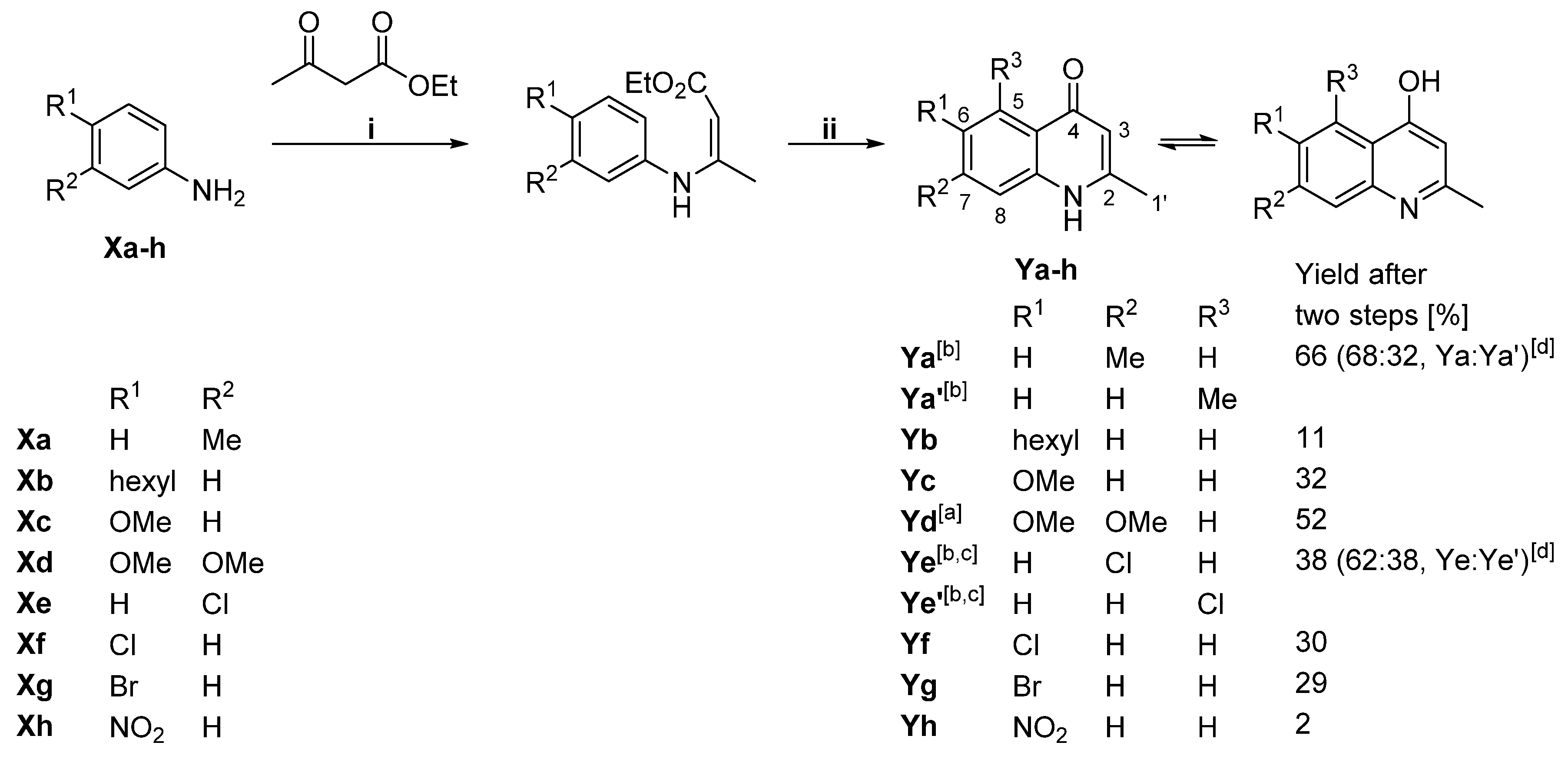
2.1. Generation of Aryl-Substituted Aurachin D Analogs by Whole-Cell Biotransformation

In contrast to previous SAR studies, which relied on total synthesis for the preparation of aurachin derivatives [12,19,20], we decided to pursue a hybrid strategy involving the biocatalytic conversion of synthetically generated quinolone precursor molecules [21]. The latter approach became a feasible option after the construction of an E. coli strain capable of regioselectively introducing a farnesyl moiety at C-3 of 2-methyl-1H-quinolin-4-one [13]. The substrates for the biotransformation experiments were prepared in a two-step sequence from ethyl acetoacetate and substituted aniline derivatives (Scheme 1, Figures S1−S17).
Previous in vitro studies already indicated that AuaA tolerates diverse substituents at C-2, C-6, or C-7 of a 1H-quinolin-4-one substrate, whereas the enzyme is much less permissive concerning variations in other positions [16]. This regiopreference was considered in the substrate selection. The generated compounds as well as four commercial substrates, 1H-quinolin-4-one, 2,6-dimethyl-1H-quinolin-4-one, 7-methoxy-2-methyl-1H-quinolin-4-one and 6-fluoro-2-methyl-1H-quinolin-4-one, were added to growing cultures of the auaA-expressing E. coli strain [13] to give a final substrate concentration of 10 or 20 mg/L. Afterward, the cultivation was resumed for 24 h before the biotransformation products were recovered with an adsorber resin and purified by HPLC. Molar yield coefficients (YP/S) were determined post-purification and then used to calculate the substrate conversion in relation to 2-methyl-1H-quinolin-4-one, which is farnesylated by AuaA to 1. Although no replicates were included in this analysis, the resulting values allow a cautious estimation of the efficiency of the whole-cell biotransformation. Overall, we observed satisfactory conversion (Table 1). Only three out of 12 tested unnatural substrates could not be converted into aurachins. While hexyl and nitro substituents are likely too bulky to be tolerated by AuaA, the installation of methoxy groups seems to be less efficient. The two tested monomethoxylated substrates were converted, albeit at a comparatively low level, whereas the quinolone precursor featuring two methoxy groups at C-6 and C-7 was not accepted. Interestingly, some substrates, such as 2,6-dimethyl-1H-quinolin-4-one and 7-chloro-2-methyl-1H-quinolin-4-one were converted much more efficiently into aurachins than anticipated from an in vitro analysis of AuaA [16]. This might be explained by the increased lipophilicity of these compounds in comparison to 2-methyl-1H-quinolin-4-one, which could have positively influenced their cellular uptake. The reason for the good conversion of 1H-quinolin-4-one under in vivo conditions is, however, unclear.

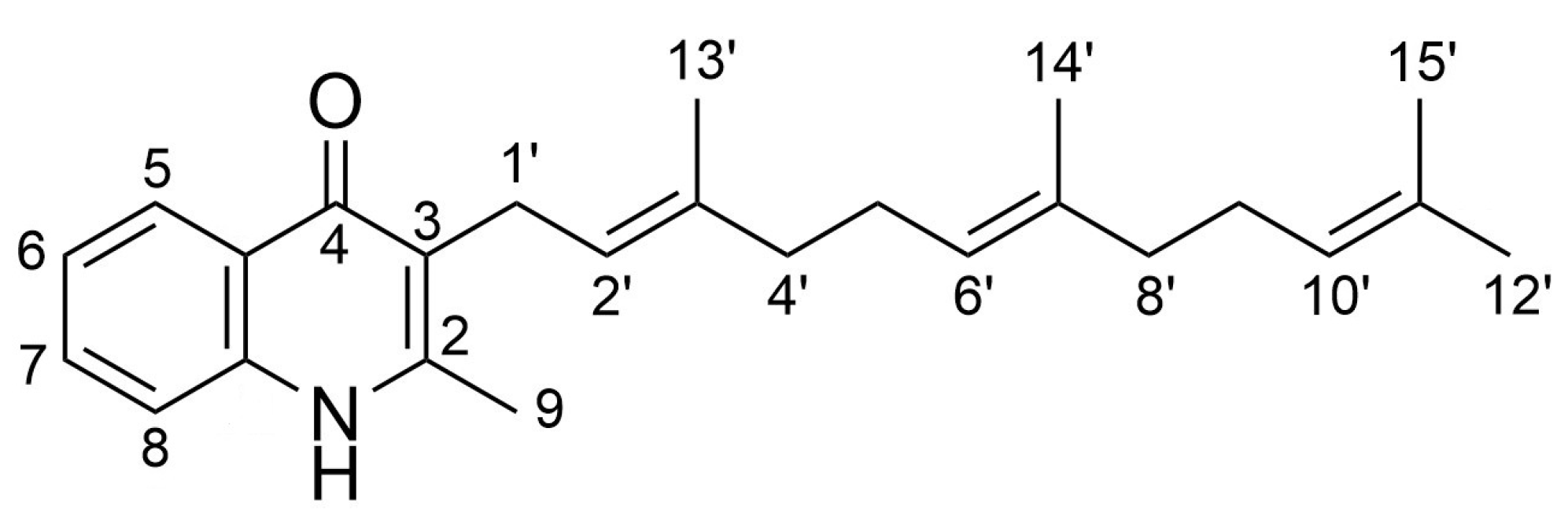
2.2. Spectroscopic Analysis of the Generated Aurachin D Analogs

The identities of the aurachin analogs were confirmed by comparison of their spectroscopic data (Figures S18–S43 in Supplementary Materials) with literature values [12,16,20]. Two of the produced compounds, 4 and 10, have not been reported before and were therefore subjected to a comprehensive characterization.
The high-resolution (HR) ESI-MS spectrum of 4 showed a pseudomolecular ion peak at m/z 378.2729 [M+H]+, which was assigned to an empirical formula of C26H35NO and 10 double bond equivalents. An analysis of the 1H NMR spectrum revealed three resonances in the aromatic region. The splitting of these signals and the concomitant coupling constants were consistent with an ABX spin system, which could be assigned to a 2,3,7-trisubstituted 4-quinolone moiety following the interpretation of 13C NMR, HSQC and HMBC data. The two methyl substituents at C-2 and C-7 were deduced from correlations observed in the HMBC spectrum (Figure 2). The farnesyl residue of 4 was identified on the basis of COSY and HMBC data. Its connection with C-3 concluded the structure determination.
The HR ESI-MS spectrum of compound 10 exhibited two distinct [M+H]+ ion peaks at m/z 442.1668 and m/z 444.1652 with almost equal intensities. This isotopic pattern is characteristic of a compound featuring a single bromine atom. The molecular formula of 10 was established as C25H32NOBr. It accounts for 10 double bond equivalents. The NMR spectra of 10 were very similar to those of 4, except that the resonance for the methyl group at C-7 was missing. Furthermore, several signals in the aromatic region were shifted. Interpretation of HMBC data led to the conclusion that 10 possesses a 2,3,6-trisubstituted 4-quinolone motif. A methyl group is located at C-2 and a farnesyl side chain at C-3 in accordance with 4. The bromine atom, which was deduced from the MS analysis, was assigned to C-6. Its electron-withdrawing, negative-inductive effect explains the downfield shifts of the protons at C-5 and C-7, respectively.

2.3. Evaluation of Antiprotozoal Properties

Serial dilution assays with 11 dilution steps in 96-well plates were used in order to determine the IC50 values of the generated aurachin D analogs against P. falciparum NF54 (erythrocytic stages), T. brucei rhodesiense STIB 900 (bloodstream forms), T. cruzi Tulahuen C4 (intracellular amastigotes) and L. donovani MHOM-ET-67/L82 (axenic amastigotes). Potential cytotoxic effects were evaluated with L6 myoblast cells (Table 2).
The bioactivity testing revealed that the antiprotozoal properties of aurachin D can be significantly altered by furnishing its quinolone backbone with different functional groups. Of particular interest was the observation that these structural modifications have unequal effects on the tested protozoa. An illustrative example is 4, the 7-methyl derivative of 1, which showed comparable antiplasmodial activities to the parental compound (IC50 0.007 μM vs. 0.012 μM). On the other hand, 4 was the least active compound in our assay against the two Trypanosoma species. Another example concerns the 6-fluoro and the 6-chloro aurachin D analog. Both 7 and 8 exhibited comparable bioactivity on T. brucei rhodesiense and also comparable cytotoxicity, as evidenced by their IC50 values against L6 cells. In contrast, their potency against T. cruzi and L. donovani was antipodal. Two conclusions can be drawn from these results: (i) It is possible to tune the antiprotozoal activity range of aurachins through minor structural variations; (ii) The lack of consistent SAR trends suggests that the aurachins exert their effects via different targets in the tested protozoa and also in mammalian cells.
If the IC50 values of 1–10 are compared to reference drugs, it becomes evident that the aurachins exhibit only moderate activities against T. brucei rhodesiense, the causative agent of East African trypanosomiasis (sleeping sickness). Much more interesting are the inhibitory effects on T. cruzi and L. donovani. As stated before, 1 exhibits competitive IC50 values and also a favorable selectivity index. These properties could not be further improved with the structural modifications introduced in this project. A comparison of their IC50 values shows that 2 and 7 were the two most active trypanocidal agents of all aurachin D derivatives tested (Table 2). However, since 2 was found to be about six-fold more toxic for L6 cells, its selectivity index significantly decreased. Further studies are now necessary to identify the target of 1 and 7 in T. cruzi and L. donovani as well as to evaluate their in vivo efficacies. Moreover, the validation of the antileishmanial activity is reasonable in view of the limitations of axenic amastigote models [22].

3. Discussion

NTDs caused by protozoa are affecting millions of people worldwide. Because of the ongoing climate change, which may impact the distribution of NTDs, and the emergence of drug-resistant parasites, new antiprotozoal drugs are urgently needed [7,23]. Natural products are an essential resource for drug development. Previous studies have already unveiled the aurachin scaffold as a suitable starting point for antimalarial drug development [11,12]. Our extended testing shows that aurachin D is also highly active against other pathogenic protozoa like T. cruzi and L. donovani. In order to explore the effects of structural modifications on its bioactivity, we decided to produce aryl-substituted aurachin D analogs by whole-cell biotransformation. This strategy merges the versatility of organic synthesis with the efficiency of biocatalysis for generating natural product analogs from chemically synthesized precursor molecules, as recently demonstrated for the generation of novel chalanilines [24], physostigmines [25] and amicetins [26], respectively. In the present study, a total of nine aryl-substituted aurachin D analogs were produced after feeding a recombinant E. coli strain with various 1H-quinolin-4-ones.
Interestingly, even substrates featuring large substituents like bromine were successfully integrated into aurachin biosynthesis. Except for compounds 2 and 4, which had lower IC50 values against P. falciparum than aurachin D, none of the generated derivatives showed increased activities against the tested protozoa. However, the antiprotozoal activity range of aurachin D could be significantly altered through minor structural variation. We assume that aurachin D can be biotransformed even further by recruiting additional enzymes from aurachin biosynthesis [27]. This will make it possible to access an even larger chemical space.

4. Materials and Methods

4.1. General Experimental Procedures

All chemicals and solvents were obtained from commercial sources and used without further purification, if not otherwise mentioned. Chemical syntheses were performed under inert conditions in a nitrogen atmosphere via Schlenk technique. TLC was performed on TLC-foil Polygram SIL G/UV254 (Macherey-Nagel). For the staining of TLC plates, ceric molybdate solution (12.5 g phosphomolybdic acid hydrate, 5.0 g ceric(IV) sulfate tetrahydrate, 30 mL conc. sulfuric acid, 200 mL demineralized water) and ninhydrin stain (400 mg ninhydrin, 200 mL ethanol) were used followed by heating with a heat gun. NMR spectra of 1H-quinolin-4-ones were recorded on a Bruker Avance/DRX 66. NMR analyses of aurachins were carried out at ambient temperature using either a Bruker AV 700 Avance III HD or a Bruker AV 600 Avance III HD spectrometer, both of which were equipped with a 5 mm helium-cooled inverse quadruple resonance cryoprobe. For NMR measurements, all compounds (except for Yh) were dissolved in deuterated methanol (methanol-d4), which also served as internal standard to calibrate spectra to δH = 3.31 ppm and δC = 49.0 ppm. Compound Yh was dissolved in deuterated dimethyl sulfoxide (DMSO-d6), and the spectra were calibrated to δH = 2.50 ppm and δC = 39.5 ppm. High-resolution ESI-MS spectra were recorded in positive ionization mode on an Agilent 1260 Infinity HPLC system combined with a Bruker Daltonics Compact quadrupole time of flight mass spectrometer. The HPLC was operated with a Nucleoshell RP 18 ec column (100 × 2 mm, 2.7 μm; Macherey-Nagel) at the following conditions: Flow rate: 0.4 mL/min. Column oven: 40 °C. Mobile phases: acetonitrile and water with 0.1% (v/v) formic acid. Gradient: 0–10 min: 2–98% ACN; 10–15 min: 98% ACN; 15–17 min: 98–5% ACN; 17–20 min: 5% ACN; 20–27 min: 5–100% ACN; 27–28 min: 100–30% ACN; 28–32 min: 30% ACN. For the MS analyses, a capillary voltage of 4.5 kV, a desolvation gas (N2) temperature of 220 °C and a dry gas (N2) flow rate of 12 L/min were used.

4.2. Synthesis of 1H-Quinolin-4-One Substrates

For the preparation of 1H-quinolin-4-ones Ya, Yc and Ye-h, a previously described protocol [28] was adapted. The aniline derivative (10 mmol, 1 equiv.) was dissolved in 20 mL ethanol under a nitrogen atmosphere. Ethyl acetoacetate (1.9 mL, 15 mmol, 1.5 equiv.), Zn(ClO4)2·6H2O (373 mg, 1 mmol, 0.1 equiv.) and anhydrous MgSO4 (1.20 g, 10 mmol, 1 equiv.) were added and the reaction mixture was stirred at room temperature for 24–48 h. The reaction was monitored via TLC until equilibrium was reached. The reaction mixture was filtered through a pad of celite, and the filter cake was washed with dichloromethane. The solvent was evaporated under reduced pressure. The formed enamine was rapidly added to 200 mL degassed diphenylether. Activated molecular sieve powder 4Å (1 g) was added. The reaction mixture was stirred at 250 °C for 1 h. The reaction mixture was cooled down to room temperature, and the precipitate formed was recovered by filtration. The filter cake, which consisted of product and molecular sieve, was washed with petroleum ether. Subsequently, methanol was added to the filter cake in order to separate the methanol-soluble product from the molecular sieve by filtration. The solvent was evaporated under reduced pressure. Column chromatography was performed using silica gel (0.040–0.063 mm; Merck) as stationary phase and a mixture of dichloromethane and methanol (9:1) as eluent. For the separation of regioisomers Ye and Ye’, a preparative HPLC was operated with a HyperClone ODS (C18) column (250 × 10 mm, 5 µm) at the following conditions: Flow rate: 5 mL/min. Column oven: 25 °C. Mobile phases: methanol and water. Gradient: 0–2 min: 10% MeOH; 2–3.7 min: 10–20% MeOH; 3.7–25 min: 20% MeOH; 25–26.2 min: 20–40% MeOH; 26.2.-45 min: 40% MeOH; 45–46.2 min: 40–10% MeOH; 46.2–60 min: 10% MeOH.
For the preparation of 1H-quinolin-4-ones Yb and Yd, a previously described procedure was adapted [29,30]. Acetoacetic acid (1 equiv.) was dissolved in anhydrous ethanol. Ceric(IV) ammonium nitrate (0.05 equiv.) and aniline derivative (1 equiv.) were added. After the mixture was stirred for 2–36 h at 25–40 °C, dichloromethane was added. The reaction mixture was washed with demineralized water and brine. The organic layer was dried with MgSO4, and the solvent was evaporated under reduced pressure. A CEM reaction tube with crude enamine dissolved in diphenyl ether was irradiated for 0.75–1.5 h at 190 °C with 280 W. Petroleum ether was added, and the precipitate was filtered and washed with petroleum ether and ethyl acetate. Column chromatography was performed using silica gel (0.040–0.063 mm; Merck) as stationary phase and a mixture of dichloromethane and methanol (9:1) as eluent.

4.3. Analytical Data of 1H-Quinolin-4-One Substrates

2,7-Dimethyl-1H-quinolin-4-one (Ya). 1H NMR (600 MHz, methanol-d4), δH [ppm] (J [Hz]) 2.45 (3 H, s, H-1‘), 2.48 (3 H, s, H-1′’), 6.17 (1 H, s, H-3), 7.22 (1 H, d,J 8.4, H-6), 7.33–7.30 (1 H, m, H-8), 8.08 (1 H, d, J 8.3, H-5). 13C NMR (150 MHz, methanol-d4), δC [ppm] 19.8 (C-1‘), 21.8 (C-1′’), 109.3 (C-3), 118.3 (C-8), 123.3 (C-4a), 125.9 (C-5), 126.9 (C-6), 141.8 (C-8a), 144.6 (C-7), 152.8 (C-2), 180.4 (C-4). HR-ESIMS: m/z 174.0916 [M+H]+ (calcd. for C11H12NO 174.0914; Δppm 1.1).
2,5-Dimethyl-1H-quinolin-4-one (Ya’). 1H NMR (600 MHz, methanol-d4), δH [ppm] (J [Hz]) 2.38 (3 H, s, H-1‘), 2.87 (3 H, s, H-1′’), 6.17 (1 H, s, H-3), 7.05 (1 H, d, J 7.2, H-6), 7.33–7.30 (1 H, m, H-8), 7.45 (1 H, dd, J 8.3, 7.2, H-7). 13C NMR (150 MHz, methanol-d4), δC [ppm] 19.3 (C-1‘), 23.8 (C-1‘‘), 111.2 (C-3), 117.0 (C-8), 124.1 (C-4a), 127.4 (C-6), 132.40 (C-7), 141.2 (C-5), 143.3 (C-8a), 151.0 (C-2), 183.1 (C-4). HR-ESIMS: m/z 174.0916 [M+H]+ (calcd. for C11H12NO 174.0914; Δppm 1.1).
6-Hexyl-2-methyl-1H-quinolin-4-one (Yb). 1H NMR (600 MHz, methanol-d4), δH [ppm] (J [Hz]) 0.92–0.87 (3 H, m, H-6‘‘), 1.40–1.29 (6 H, m, H-3‘‘, H-4‘‘, H-5‘‘), 1.68 (2 H, q, J 7.5, H-2‘‘), 2.46 (3 H, s, H-1‘), 2.75 (2 H, t, J 7.7, H-1‘‘), 6.19 (1 H, s, H-3), 7.47 (1 H, d, J 8.5, H-8), 7.54 (1 H, dd, J 8.5, J 2.0, H-7), 8.01 (1 H, d, J 2.0, H-5). 13C NMR (150 MHz, methanol-d4), δC [ppm] 14.4 (C-6‘‘), 19.8 (C-1‘), 23.7 (C-5), 30.0 (C-3‘‘), 32.6 (C-2‘‘), 32.9 (C-4), 36.6 (C-1‘‘), 109.4 (C-3), 119.0 (C-8), 124.6 (C-5), 125.3 (C-4a), 134.4 (C-7), 139.9 (C-8a), 140.2 (C-6), 152.5 (C-2), 180.4 (C-4). HR-ESIMS: m/z 244.1699 [M+H]+ (calcd. for C16H22NO3 244.1696; Δppm 1.2).
6-Methoxy-2-methyl-1H-quinolin-4-one (Yc). 1H NMR (600 MHz, methanol-d4), δH [ppm] (J [Hz]) 2.46 (3 H, s, 3H, H-1‘), 3.89 (3 H, s, H-1′’), 6.19 (1 H, s, H-3), 7.33–7.30 (1 H, m, H-7), 7.49 (1 H, m, H-8), 7.63–7.61 (1 H, m, H-5). 13C NMR (150 MHz, methanol-d4), δC [ppm] 19.7 (C-1‘), 56.1 (C-1‘‘), 104.8 (C-5), 108.8 (C-3), 120.6 (C-8), 124.4 (C-7), 126.4 (C-4a), 136.3 (C-8a), 151.7 (C-2), 158.1 (C-6), 179.7 (C-4). HR-ESIMS: m/z 190.0866 [M+H]+ (calcd. for C11H12NO2 190.0863; Δppm 1.6). Analytical data in agreement with those previously reported [31].
6,7-Dimethoxy-2-methyl-1H-quinolin-4-one (Yd). 1H NMR (600 MHz, methanol-d4), δH [ppm] (J [Hz]) 2.41 (3 H, s, H-1′), 3.90 (3 H, s, H-1′’), 3.92 (3 H, s, H-1′’’), 6.12 (1 H, s, H-3), 6.86 (1 H, s, H-8), 7.52 (1 H, s, H-5). 13C NMR (150 MHz, methanol-d4): δ [ppm] = 179.0 (C-4), 155.5 (C-7), 150.9 (C-2), 149.0 (C-6), 137.6 (C-8a), 119.2 (C-4a), 108.6 (C-3), 104.9 (C-5), 99.5 (C-8), 56.5 (C-1‘‘‘), 56.4 (C-1‘‘), 19.7 (C-1‘). HR-ESIMS: m/z 220.0971 [M+H]+ (calcd. for C12H14NO3 220.0968; Δppm 1.4). Analytical data in agreement with those previously reported [32].
7-Chloro-2-methyl-1H-quinolin-4-one (Ye). 1H NMR (600 MHz, methanol-d4), δH [ppm] (J [Hz]) 2.44 (3 H, s, H-1‘), 6.17 (1 H, s, H-3), 7.34 (1 H, dd, J 8.7, J 1.9, H-6), 7.52 (1 H, d, J 1.9, H-8), 8.14 (1 H, d, J 8.7, H-5). 13C NMR (150 MHz, methanol-d4), δC [ppm] 19.9 (C-1‘), 110.1 (C-3), 118.3 (C-8), 123.9 (C-4a), 125.5 (C-6), 128.0 (C-5), 139.3 (C-7), 142.2 (C-8a), 153.5 (C-2), 180.0 (C-4). HR-ESIMS: m/z 194.0371 [M+H]+ (calcd. for C10H9ClNO 194.0373; Δppm 1.0). Analytical data in agreement with those previously reported [33].
5-Chloro-2-methyl-1H-quinolin-4-one (Ye’). 1H NMR (600 MHz, methanol-d4), δH [ppm] (J [Hz]) 2.39 (3 H, s, H-1‘), 6.11 (1 H, s, H-3), 7.29 (1 H, dd, J 7.6, J 1.2, H-6), 7.40 (1 H, dd, J 8.4, J 1.2, H-8), 7.49 (1 H, t, J 8.0, H-7). 13C NMR (150 MHz, methanol-d4), δC [ppm] 19.3 (C-1‘), 111.9 (C-3), 118.2 (C-8), 122.0 (C-4a), 127.6 (C-6), 132.7 (C-7), 134.3 (C-5), 144.1 (C-8a), 151.6 (C-2), 180.2 (C-4). HR-ESIMS: m/z 194.0371 [M+H]+ (calcd. for C10H9ClNO 194.0373; Δppm 1.0).
6-Chloro-2-methyl-1H-quinolin-4-one (Yf). 1H NMR (600 MHz, methanol-d4), δH [ppm] (J [Hz]) 2.46 (3 H, s, H-1‘), 6.20 (1 H, s, H-3), 7.53 (1 H, d, J 8.9, H-8), 7.65 (1 H, dd, J 8.9, J 2.3, H-7), 8.15 (1 H, d, J 2.3, H-5). 13C NMR (150 MHz, methanol-d4), δC [ppm] 19.9 (C-1‘), 109.9 (C-3), 121.0 (C-8), 125.2 (C-5), 126.4 (C-4a), 130.8 (C-6), 133.6 (C-7), 140.0 (C-8a), 153.4 (C-2), 179.3 (C-4). HR-ESIMS: m/z 194.0368 [M+H]+ (calcd. for C10H9ClNO 194.0373; Δppm 2.6). Analytical data in agreement with those previously reported [31].
6-Bromo-2-methyl-1H-quinolin-4-one (Yg). 1H NMR (600 MHz, methanol-d4), δH [ppm] (J [Hz]) 2.46 (3 H, s, H-1‘), 6.21 (1 H, s, H-3), 7.47 (1 H, d, J 8.8, H-8), 7.78 (1 H, dd, J 8.8, J 2.3, H-7), 8.32 (1 H, d, J 2.3, H-5). 13C NMR (150 MHz, methanol-d4), δC [ppm] 19.9 (C-1‘), 110.0 (C-3), 118.2 (C-6), 121.2 (C-8), 126.8 (C-4a), 128.5 (C-5), 136.3 (C-7), 140.4 (C-8a), 153.5 (C-2), 179.2 (C-4). HR-ESIMS: m/z 237.9865 [M+H]+ (calcd. for C10H9BrNO 237.9863; Δppm 0.8). Analytical data in agreement with those previously reported [34].
2-Methyl-6-nitro-1H-quinolin-4-one (Yh). 1H NMR (600 MHz, DMSO-d6), δH [ppm] (J [Hz]) 2.38 (3 H, s, H-1‘), 6.08 (2 H, s, H-3), 7.68 (2 H, d, J 9.1, 2H, H-8), 8.40 (2 H, dd, J 9.1, J 2.7, 2H, H-7), 8.80 (1 H, s, H-5). 13C NMR (150 MHz, DMSO-d6), δC [ppm] 19.5 (C-1‘), 109.9 (C-3), 119.6 (C-8), 121.5 (C-5), 123.5 (C-4a), 125.8 (C-7), 142.4 (C-6), 143.9 (C-8a), 151.4 (C-2), 176.2 (C-4). HR-ESIMS: m/z 205.0610 [M+H]+ (calcd. for C10H9N2O3 205.0608; Δppm 1.0). Analytical data in agreement with those previously reported [35].

4.4. Whole-Cell Biotransformation and Product Recovery

The E. coli BL21(DE3) strain for the whole-cell biotransformation was taken from the strain collection of the Technical Biology group at TU Dortmund University and is available upon request. Cultures of this strain, which harbors the two plasmids pSK46 [13] and pJBEI-2997 [36], were grown in terrific broth (TB) medium [37] supplemented with 50 μg/mL kanamycin, 30 μg/mL chloramphenicol and 0.12% (v/v) glycerol. The cultivation was conducted at a 1 L scale in shaken Erlenmeyer flasks at 30 °C and 120 rpm. Substrates were added at the time of inoculation at a final concentration of 10 or 20 mg/L. Once the cultures had reached an optical density at 600 nm (OD600) of 1, they were induced by addition of 0.025 mM isopropyl β-d-1-thiogalactopyranoside (IPTG). The incubation was then continued for 24 h. At the end of fermentation, the adsorber resin XAD7HP was added to the cultures at a concentration of 2% (w/v). After 3 h of shaking at 90 rpm, the resin was separated from the culture broth by filtration and washed with water. Subsequently, the adsorbed compounds were eluted with 1 L methanol. The resulting extracts were dried with a rotary evaporator, resuspended in 60% methanol (100 mL) and extracted three times with dichloromethane (60 mL). The organic phases were collected and concentrated to dryness with a rotary evaporator. After resuspension in 1.5 mL methanol, the chromatographic purification was carried out using a Shimadzu HPLC system (LC-20 AD) equipped with a diode array detector (SPD-M20A) and a Nucleodur C18 Isis column (250 × 10 mm, 5 μm, Macherey-Nagel). For the separation, a linear gradient of acetonitrile in water supplemented with 0.1% (v/v) trifluoroacetic acid from 40% to 90% over 20 min was used. The gradient was kept at 90% for additional 7 min. The flow rate was set to 5.6 mL/min. In the case of 5 and 6, a further purification step with the same column was required. For this, an isocratic separation with 55% acetonitrile in 45% water supplemented with 0.1% (v/v) trifluoroacetic acid was carried out. Except for 2-desmethyl aurachin D (2), which was recovered as a pale brownish oil after chromatography, all products were isolated as beige solids.

4.5. Spectroscopic Data of Aurachin Derivatives

2-Desmethyl aurachin D (2). 1H NMR (700 MHz, methanol-d4), δH [ppm] (J [Hz]) 1.54 (3 H, d, J 1.0, H-15′), 1.60 (3 H, d, J 1.3, H-14′), 1.62 (3 H, d, J 1.3, H-12′), 1.73 (3 H, d, J 1.3, H-13′), 1.94 (2 H, m, H-8′), 2.02 (2 H, m, H-9′), 2.10 (2 H, m, H-4′), 2.15 (2 H, m, H-5′), 3.32 (2 H, not resolved, H-1′), 5.04 (1 H, m, H-10′), 5.13 (1 H, m, H-6′), 5.38 (1 H, m, H-2′), 7.40 (1 H, ddd, J 1.2, 6.9, 8.4, H-6), 7.56 (1 H, dd, J 1.2, 8.4, H-8), 7.68 (1 H, ddd, J 1.5, 6.9, 8.4, H-7), 7.80 (1 H, s, H-2), 8.29 (1 H, dd, J 1.5, 8.4, H-5). 13C NMR (175 MHz, methanol-d4), δC [ppm] 16.1 (C-14′), 16.2 (C-13′), 17.7 (C-15′), 25.9 (C-12′), 26.7 (C-1′), 27.5 (C-5′), 27.7 (C-9′), 40.8 (C-4′), 40.8 (C-8′), 119.2 (C-8), 122.3 (C-3), 122.6 (C-2′), 125.0 (C-6), 125.3 (C-6′), 125.4 (C-10′), 125.6 (C-4a), 126.2 (C-5), 132.0 (C-11′), 133.0 (C-7), 136.1 (C-7′), 138.4 (C-3′), 138.9 (C-2), 141.0 (C-8a), 179.0 (C-4). HR-ESIMS: m/z 350.2438 [M+H]+ (calcd. for C24H32NO 350.2406; Δppm 9.1). Analytical data in agreement with those previously reported [16].
6-Methyl aurachin D (3). 1H NMR (700 MHz, methanol-d4), δH [ppm] (J [Hz]) 1.50 (3 H, d, J 1.0, H-15′), 1.55 (3 H, d, J 1.3, H-14′), 1.61 (3 H, d, J 1.3, H-12′), 1.80 (3 H, d, J 1.3, H-13′), 1.88 (2 H, m, H-8′), 1.95 (2 H, m, H-9′), 2.02 (2 H, m, H-4′), 2.10 (2 H, m, H-5′), 2.45 (3 H, s, H-9), 2.46 (3 H, s, H-10), 3.40 (2 H, d, J 6.8, H-1′), 5.00 (1 H, m, H-10′), 5.05 (1 H, m, H-6′), 5.10 (1 H, m, H-2′), 7.42 (1 H, d, J 8.4, H-8), 7.48 (1 H, dd, J 2.2, 8.4, H-7), 8.02 (1 H, d, J 2.2, H-5). 13C NMR (175 MHz, methanol-d4), δC [ppm] 16.1 (C-14′), 16.3 (C-13′), 17.6 (C-15′), 18.2 (C-9), 21.3 (C-10), 24.8 (C-1′), 25.8 (C-12′), 27.3 (C-5′), 27.8 (C-9′), 40.7 (C-4′), 40.8 (C-8′), 118.6 (C-8), 120.6 (C-3), 124.0 (C-2′), 125.0 (C-4a), 125.3 (C-6′), 125.4 (C-10′), 125.4 (C-5), 132.0 (C-11′), 134.2 (C-7), 134.6 (C-6), 135.8 (C-3′), 135.9 (C-7′), 138.7 (C-8a), 149.4 (C-2), 178.2 (C-4). HR-ESIMS: m/z 378.2739 [M+H]+ (calcd. for C26H36NO 378.2719; Δppm 5.3). Analytical data in agreement with those previously reported [16].
7-Methyl aurachin D (4). 1H NMR (600 MHz, methanol-d4), δH [ppm] (J [Hz]) 1.50 (3 H, d, J 0.9, H-15′), 1.55 (3 H, d, J 1.1, H-14′), 1.61 (3 H, d, J 1.3, H-12′), 1.80 (3 H, d, J 1.2, H-13′), 1.88 (2 H, m, H-8′), 1.94 (2 H, m, H-9′), 2.01 (2 H, m, H-4′), 2.10 (2 H, m, H-5′), 2.45 (3 H, s, H-9), 2.47 (3 H, s, H-10), 3.39 (2 H, d, J 6.9, H-1′), 5.00 (1 H, m, H-10′), 5.05 (1 H, m, H-6′), 5.09 (1 H, m, H-2′), 7.19 (1 H, dd, J 1.7, 8.4, H-6), 7.29 (1 H, d, J 1.7, H-8), 8.11 (1 H, d, J 8.4, H-5). 13C NMR (150 MHz, methanol-d4), δC [ppm] 16.1 (C-14′), 16.3 (C-13′), 17.6 (C-15′), 18.2 (C-9), 21.8 (C-10), 24.7 (C-1′), 25.8 (C-12′), 27.3 (C-5′), 27.8 (C-9′), 40.7 (C-4′), 40.8 (C-8′), 118.0 (C-8), 120.5 (C-3), 123.1 (C-4a), 123.9 (C-2′), 125.3 (C-6′), 125.4 (C-10′), 126.3 (C-5), 126.4 (C-6), 132.0 (C-11′), 135.8 (C-3′), 135.9 (C-7′), 140.8 (C-8a), 143.6 (C-7), 149.4 (C-2), 178.4 (C-4). HR-ESIMS: m/z 378.2729 [M+H]+ (calcd. for C26H36NO 378.2719; Δppm 2.6).
6-Methoxy aurachin D (5). 1H NMR (600 MHz, methanol-d4), δH [ppm] (J [Hz]) 1.50 (3 H, d, J 0.7, H-15′), 1.56 (3 H, d, J 1.0, H-14′), 1.61 (3 H, d, J 1.3, H-12′), 1.81 (3 H, d, J 1.2, H-13′), 1.88 (2 H, m, H-8′), 1.94 (2 H, m, H-9′), 2.03 (2 H, m, H-4′), 2.10 (2 H, m, H-5′), 2.46 (3 H, s, H-9), 3.42 (2 H, d, J 6.7, H-1′), 3.90 (3 H, s, H-10), 5.00 (1 H, m, H-10′), 5.05 (1 H, m, H-6′), 5.11 (1 H, m, H-2′), 7.27 (1 H, dd, J 2.9, 9.0, H-7), 7.46 (1 H, d, J 9.0, H-8), 7.64 (1 H, d, J 2.9, H-5). 13C NMR (150 MHz, methanol-d4), δC [ppm] 16.1 (C-14′), 16.3 (C-13′), 17.6 (C-15′), 18.2 (C-9), 24.9 (C-1′), 25.8 (C-12′), 27.3 (C-5′), 27.8 (C-9′), 40.7 (C-4′), 40.8 (C-8′), 56.0 (C-10), 104.9 (C-5), 120.1 (C-3), 120.4 (C-8), 123.8 (C-7), 123.9 (C-2′), 125.3 (C-6′), 125.4 (C-10′), 126.0 (C-4a), 132.0 (C-11′), 135.4 (C-8a), 135.8 (C-3′), 135.9 (C-7′), 148.8 (C-2), 157.8 (C-6), 177.6 (C-4). HR-ESIMS: m/z 394.2691 [M+H]+ (calcd. for C26H36NO2 394.2668; Δppm 5.8). Analytical data in agreement with those previously reported [12,20].
7-Methoxy aurachin D (6). 1H NMR (600 MHz, methanol-d4), δH [ppm] (J [Hz]) 1.50 (3 H, d, J 1.0, H-15′), 1.56 (3 H, d, J 1.2, H-14′), 1.61 (3 H, d, J 1.3, H-12′), 1.81 (3 H, d, J 1.2, H-13′), 1.88 (2 H, m, H-8′), 1.94 (2 H, m, H-9′), 2.03 (2 H, m, H-4′), 2.11 (2 H, m, H-5′), 2.52 (3 H, s, H-9), 3.43 (2 H, d, J 7.2, H-1′), 3.93 (3 H, s, H-10), 4.99 (1 H, m, H-10′), 5.05 (1 H, m, H-6′), 5.09 (1 H, m, H-2′), 6.98 (1 H, d, J 2.4, H-8), 7.07 (1 H, dd, J 2.4, 9.1, H-6), 8.17 (1 H, d, J 9.1, H-5). 13C NMR (150 MHz, methanol-d4), δC [ppm] 16.1 (C-14′), 16.4 (C-13′), 17.6 (C-15′), 18.4 (C-9), 24.7 (C-1′), 25.8 (C-12′), 27.3 (C-5′), 27.8 (C-9′), 40.7 (C-8′), 40.8 (C-4′), 56.3 (C-10), 99.1 (C-8), 116.9 (C-6), 119.9 (C-3), 123.1 (C-2′), 125.3 (C-10′), 125.3 (C-6′), n.d. (C-4a), 127.5 (C-5), 132.0 (C-11′), 136.0 (C-7′), 136.6 (C-3′), 142.3 (C-8a), 151.2 (C-2), 164.5 (C-7), 175.1 (C-4). HR-ESIMS: m/z 394.2690 [M+H]+ (calcd. for C26H36NO2 394.2668; Δppm 5.6). Analytical data in agreement with those previously reported [20].
6-Fluoro aurachin D (7). 1H NMR (700 MHz, methanol-d4), δH [ppm] (J [Hz]) 1.51 ppm (3H, d, JH,H 1.5, H-15′), 1.55 (3H, d, JH,H 1.3, H-14′), 1.61 (3H, d, JH,H 1.4, H-12′), 1.81 (3H, d, JH,H 1.3, H-13′), 1.88 (2H, m, H-8′), 1.94 (2H, m, H-9′), 2.02 (2H, m, H-4′), 2.09 (2H, m, H-5′), 2.47 (3H, s, H-9), 3.40 (2H, d, JH,H 6.9, H-1′), 4.99 (1H, ddt, JH,H 1.4, 1.5, 7.0, H-10′), 5.05 (1H, dt, JH,H 1.3, 6..9, H-6′), 5.09 (1H, dt, JH,H 1.3, 6.9, H-2′), 7.44 (1H, ddd, JH,H 2.9, 9.1, JH,F 8.1, H-7), 7.56 (1H, dd, JH,H 9.1, JH,F 4.5, H-8), 7.84 (1H, dd, JH,H 2.9, JH,F 9.5, H-5). 13C NMR (175 MHz, methanol-d4), δC [ppm] 16.1 (C-14′), 16.3 (C-13′), 17.6 (C-15′), 18.3 (C-9), 24.8 (C-1′), 25.8 (C-12′), 27.3 (C-5′), 27.8 (C-9′), 40.7 (C-8′), 40.8 (C-4′), 110.2 (d, JC,F 23.1, C-5), 120.4 (C-3), 121.3 (d, JC,F 8.5, C-8), 121.5 (d, JC,F 26.7, C-7), 123.6 (C-2′), 125.3 (C-10′), 125.4 (C-6′), 126.1 (d, JC,F 7.1, C-4a), 132.1 (C-11′), 135.9 (C-7′), 136.1 (C-3′), 137.2 (C-8a), 149.9 (C-2), 160.5 (d, JC,F 241.7, C-6), 177.6 (C-4). HR-ESIMS: m/z 382.2485 [M+H]+ (calcd. for C25H33FNO 382.2468; Δppm 4.4). Analytical data in agreement with those previously reported [14,20].
6-Chloro aurachin D (8). 1H NMR (600 MHz, methanol-d4), δH [ppm] (J [Hz]) 1.50 (3 H, d, J 0.9, H-15′), 1.55 (3 H, d, J 1.0, H-14′), 1.61 (3 H, d, J 1.3, H-12′), 1.80 (3 H, d, J 1.1, H-13′), 1.87 (2 H, m, H-8′), 1.93 (2 H, m, H-9′), 2.02 (2 H, m, H-4′), 2.10 (2 H, m, H-5′), 2.46 (3 H, s, H-9), 3.39 (2 H, d, J 7.0, H-1′), 4.99 (1 H, m, H-10′), 5.04 (1 H, m, H-6′), 5.08 (1 H, m, H-2′), 7.50 (1 H, d, J 8.9, H-8), 7.60 (1 H, dd, J 2.5, 8.9, H-7), 8.18 (1 H, d, J 2.5, H-5). 13C NMR (150 MHz, methanol-d4), δC [ppm] 16.2 (C-14′), 16.3 (C-13′), 17.6 (C-15′), 18.3 (C-9), 24.8 (C-1′), 25.8 (C-12′), 27.3 (C-5′), 27.8 (C-9′), 40.7 (C-4′), 40.8 (C-8′), 120.8 (C-8), 121.3 (C-3), 123.5 (C-2′), 125.3 (C-6′), 125.3 (C-10′), 125.5 (C-5), 125.9 (C-4a), 130.3 (C-6), 132.0 (C-11′), 132.9 (C-7), 135.9 (C-7′), 136.1 (C-3′), 139.0 (C-8a), 150.1 (C-2), 177.3 (C-4). HR-ESIMS: m/z 398.2188 [M+H]+ (calcd. for C25H33ClNO 398.2172; Δppm 4.0). Analytical data in agreement with those previously reported [20].
7-Chloro aurachin D (9). 1H NMR (600 MHz, methanol-d4), δH [ppm] (J [Hz]) 1.49 (3 H, d, J 0.8, H-15′), 1.55 (3 H, d, J 1.0, H-14′), 1.61 (3 H, d, J 1.3, H-12′), 1.80 (3 H, d, J 1.1, H-13′), 1.87 (2 H, m, H-8′), 1.92 (2 H, m, H-9′), 2.02 (2 H, m, H-4′), 2.10 (2 H, m, H-5′), 2.45 (3 H, s, H-9), 3.37 (2 H, d, J 6.8, H-1′), 4.98 (1 H, m, H-10′), 5.04 (1 H, m, H-6′), 5.08 (1 H, m, H-2′), 7.31 (1 H, dd, J 2.0, 8.8, H-6), 7.51 (1 H, d, J 2.0, H-8), 8.18 (1 H, d, J 8.8, H-5). 13C NMR (150 MHz, methanol-d4), δC [ppm] 16.2 (C-14′), 16.3 (C-13′), 17.6 (C-15′), 18.3 (C-9), 24.7 (C-1′), 25.8 (C-12′), 27.2 (C-5′), 27.8 (C-9′), 40.7 (C-4′), 40.8 (C-8′), 118.0 (C-8), 121.4 (C-3), 123.5 (C-2′), 123.6 (C-4a), 125.0 (C-6), 125.3 (C-6′), 125.3 (C-10′), 128.5 (C-5), 132.0 (C-11′), 135.9 (C-7′), 136.0 (C-3′), 138.6 (C-7), 141.2 (C-8a), 150.1 (C-2), 178.0 (C-4). HR-ESIMS: m/z 398.2191 [M+H]+ (calcd. for C25H33ClNO 398.2172; Δppm 4.8). Analytical data in agreement with those previously reported [16,20].
6-Bromo aurachin D (10). 1H NMR (600 MHz, methanol-d4), δH [ppm] (J [Hz]) 1.50 (3 H, d, J 0.8, H-15′), 1.55 (3 H, d, J 1.0, H-14′), 1.61 (3 H, d, J 1.3, H-12′), 1.80 (3 H, d, J 1.1, H-13′), 1.87 (2 H, m, H-8′), 1.93 (2 H, m, H-9′), 2.02 (2 H, m, H-4′), 2.10 (2 H, m, H-5′), 2.46 (3 H, s, H-9), 3.38 (2 H, d, J 6.9, H-1′), 4.99 (1 H, m, H-10′), 5.04 (1 H, m, H-6′), 5.08 (1 H, m, H-2′), 7.44 (1 H, d, J 8.9, H-8), 7.72 (1 H, dd, J 2.2, 8.9, H-7), 8.34 (1 H, d, J 2.2, H-5). 13C NMR (150 MHz, methanol-d4), δC [ppm] 16.2 (C-14′), 16.3 (C-13′), 17.6 (C-15′), 18.3 (C-9), 24.8 (C-1′), 25.8 (C-12′), 27.3 (C-5′), 27.8 (C-9′), 40.7 (C-4′), 40.8 (C-8′), 117.7 (C-4a), 120.9 (C-8), 121.4 (C-3), 123.5 (C-2′), 125.3 (C-6′), 125.3 (C-10′), 126.4 (C-6), 128.8 (C-5), 132.0 (C-11′), 135.5 (C-7), 135.9 (C-7′), 136.1 (C-3′), 139.3 (C-8a), 150.1 (C-2), 177.2 (C-4). HR-ESIMS: m/z 442.1668 [M+H]+ (calcd. for C25H33BrNO 442.1667; Δppm 0.2).

4.6. Biological Tests

4.6.1. Antiplasmodial Assay

Antiplasmodial activity was determined against the NF54 strain of Plasmodium falciparum using a modified 3H-hypoxanthine incorporation assay. Briefly, infected erythrocytes were exposed to serial drug dilutions in microtiter plates for 48 h at 37 °C in a gas mixture with reduced oxygen and elevated carbon dioxide concentrations [38,39]. 3H hypoxanthine was added to each well, and after further incubation for 24 h, the wells were harvested on glass fiber filters and counted in a liquid scintillation counter. From the sigmoidal inhibition curve, the IC50 value was calculated. Chloroquine was used as positive control in each test series.

4.6.2. Antitrypanosomal Assays

Activity against Trypanosoma brucei rhodesiense (strain STIB 900) was evaluated according to a previously established protocol [39,40]. Parasites were grown axenically in culture medium supplemented with horse serum. Following a 3-day exposure to test compounds, the viability of tryptomastigote parasites was quantified using the dye resazurin by monitoring the reductive environment of living cells. Fluorescence development was expressed as percentage of the control, and IC50 values were calculated. Melarsoprol was included as positive control.
Activity against Trypanosoma cruzi was evaluated according to a procedure described by Buckner and coworkers [41]. The strain Tulahuen C4 of T. cruzi, which had been transfected with the β-galactosidase lacZ gene, was cultivated for 4 days on rat skeletal myoblasts (5% CO2, 37 °C) in the presence of drug. For measurement of the IC50, the substrate chlorophenol red-β-d-galactopyranoside was added. The color reaction that developed during the following 2–4 h was quantified photometrically employing an ELISA reader. Benznidazole was included in each test series as positive control.

4.6.3. Antileishmanial Assay

Evaluation of antileishmanial activity was carried out with axenically grown amastigotes of Leishmania donovani MHOM-ET-67/L82, as previously described [39]. For this, the amastigotes were cultured at 37 °C and pH 5.4 in SM medium [42] supplemented with 10% heat-inactivated fetal bovine serum under an atmosphere of 5% CO2 in air. Following a 3-day exposure to test compounds, the viability of amastigote parasites was quantified using the dye resazurin by monitoring the reductive environment of living cells. Fluorescence development was expressed as percentage of the control, and IC50 values were calculated. Miltefosine was used as positive control.

4.6.4. Cytotoxicity Assay

Cytotoxicity was tested with L6 cells (ATCC CRL-1458), a primary cell line derived from rat skeletal myoblasts, as previously described [38]. For this, L6 cells were grown in culture medium supplemented with heat-inactivated fetal bovine serum under an atmosphere of 5% CO2 in air. Following a 3-day exposure to test compounds, the viability of L6 cells was quantified using the dye resazurin by monitoring the reductive environment of living cells. Fluorescence development was expressed as percentage of the control, and IC50 values were calculated. Podophyllotoxin was used as reference.

Supplementary Materials

The following supporting information can be downloaded at: https://www.mdpi.com/article/10.3390/molecules28031066/s1, Figure S1: 1H NMR spectrum (600 MHz, methanol-d4) of a mixture of Ya and Ya’; Figure S2: 1H-decoupled 13C NMR spectrum (150 MHz, methanol-d4) of a mixture of Ya and Ya’; Figure S3: 1H NMR spectrum (600 MHz, methanol-d4) of Ya; Figure S4: 1H NMR spectrum (600 MHz, methanol-d4) of Yb; Figure S5: 1H-decoupled 13C NMR spectrum (150 MHz, methanol-d4) of Yb; Figure S6: 1H NMR spectrum (600 MHz, methanol-d4) of Yc; Figure S7: 1H-decoupled 13C NMR spectrum (150 MHz, methanol-d4) of Yc; Figure S8: 1H NMR spectrum (600 MHz, methanol-d4) of Yd; Figure S9: 1H-decoupled 13C NMR spectrum (150 MHz, methanol-d4) of Yd; Figure S10: 1H NMR spectrum (600 MHz, methanol-d4) of a mixture of Ye and Ye’; Figure S11: 1H-decoupled 13C NMR spectrum (150 MHz, methanol-d4) of a mixture of Ye and Ye’; Figure S12: 1H NMR spectrum (600 MHz, methanol-d4) of Ye; Figure S13: 1H NMR spectrum (600 MHz, methanol-d4) of Ye’; Figure S14: 1H NMR spectrum (600 MHz, methanol-d4) of Yg; Figure S15: 1H-decoupled 13C NMR spectrum (150 MHz, methanol-d4) of Yg; Figure S16: 1H NMR spectrum (600 MHz, DMSO-d6) of Yh; Figure S17: 1H-decoupled 13C NMR spectrum (150 MHz, DMSO-d6) of Yh; Figure S18: 1H NMR spectrum (700 MHz, methanol-d4) of 1; Figure S19: 1H-decoupled 13C NMR spectrum (175 MHz, methanol-d4) of 1; Figure S20: 1H NMR spectrum (700 MHz, methanol-d4) of 2; Figure S21: 1H-decoupled 13C NMR spectrum (175 MHz, methanol-d4) of 2; Figure S22: 1H NMR spectrum (700 MHz, methanol-d4) of 3; Figure S23: 1H-decoupled 13C NMR spectrum (175 MHz, methanol-d4) of 3; Figure S24: 1H NMR spectrum (600 MHz, methanol-d4) of 4; Figure S25: 1H-decoupled 13C NMR spectrum (150 MHz, methanol-d4) of 4; Figure S26: COSY spectrum (methanol-d4) of 4; Figure S27: HSQC spectrum (methanol-d4) of 4; Figure S28: HMBC spectrum (methanol-d4) of 4; Figure S29: 1H NMR spectrum (600 MHz, methanol-d4) of 5; Figure S30: 1H-decoupled 13C NMR spectrum (150 MHz, methanol-d4) of 5; Figure S31: 1H NMR spectrum (600 MHz, methanol-d4) of 6; Figure S32: 1H-decoupled 13C NMR spectrum (150 MHz, methanol-d4) of 6; Figure S33: 1H NMR spectrum (700 MHz, methanol-d4) of 7; Figure S34: 1H-decoupled 13C NMR spectrum (175 MHz, methanol-d4) of 7; Figure S35: 1H NMR spectrum (600 MHz, methanol-d4) of 8; Figure S36: 1H-decoupled 13C NMR spectrum (150 MHz, methanol-d4) of 8; Figure S37: 1H NMR spectrum (600 MHz, methanol-d4) of 9; Figure S38: 1H-decoupled 13C NMR spectrum (150 MHz, methanol-d4) of 9; Figure S39: 1H NMR spectrum (600 MHz, methanol-d4) of 10; Figure S40: 1H-decoupled 13C NMR spectrum (150 MHz, methanol-d4) of 10; Figure S41: COSY spectrum (methanol-d4) of 10; Figure S42: HSQC spectrum (methanol-d4) of 10; Figure S43: HMBC spectrum (methanol-d4) of 10.

Author Contributions

Conceptualization, S.L., J.P., M.K. and M.N.; formal analysis, S.K., C.J.-M.Z., M.K. and M.N.; investigation S.K., C.J.-M.Z., K.K., W.H. and M.K..; writing—original draft preparation, S.K., C.J.-M.Z. and M.N.; writing—review and editing, S.K., C.J.-M.Z., S.L., J.P., M.K. and M.N.; supervision, J.P. and M.N.; project administration, M.N.; funding acquisition, S.L., J.P. and M.N. All authors have read and agreed to the published version of the manuscript.

Funding

This research was funded by the German Research Foundation grant number 445751305 (DFG Priority Program 2240)) as well as by the European Regional Development Fund (CLIB Competence Center Biotechnology; grant numbers EFRE-0300096, EFRE-0300097, EFRE-0300098).

Institutional Review Board Statement

Not applicable.

Informed Consent Statement

Not applicable.

Data Availability Statement

The data presented in this study are available in the Supplementary Materials).

Acknowledgments

We thank Professor Oliver Kayser (Laboratory of Technical Biochemistry, TU Dortmund University) for providing access to his LC/MS facilities, where the mass spectra of the aurachins were recorded. We thank M. Cal, R. Rocchetti and S. Keller (both Parasite Chemotherapy Unit, Swiss Tropical and Public Health Institute) for help with antiparasitic compound testing and Birgit Henßen (Institute of Bioorganic Chemistry, Heinrich Heine University Düsseldorf at Forschungszentrum Jülich) for analytical support.

Conflicts of Interest

The authors declare no conflict of interest.

Sample Availability

The recombinant E. coli strain used in this study is available upon request.

References

  1. Andrews, K.T.; Fisher, G.; Skinner-Adams, T.S. Drug repurposing and human parasitic protozoan diseases. Int. J. Parasitol. Drugs Drug Resist. 2014, 4, 95–111. [Google Scholar] [CrossRef] [PubMed] [Green Version]
  2. Talapko, J.; Škrlec, I.; Alebić, T.; Jukić, M.; Včev, A. Malaria: The past and the present. Microorganisms 2019, 7, 179. [Google Scholar] [CrossRef] [PubMed] [Green Version]
  3. Molyneux, D.H.; Savioli, L.; Engels, D. Neglected tropical diseases: Progress towards addressing the chronic pandemic. Lancet 2017, 389, 312–325. [Google Scholar] [CrossRef] [PubMed]
  4. Franco, J.R.; Simarro, P.P.; Jannin, J.G. Epidemiology of human African trypanosomiasis. Clin. Epidemiol. 2014, 6, 257–275. [Google Scholar] [PubMed]
  5. Lidani, K.C.F.; Andrade, F.A.; Bavia, L.; Damasceno, F.S.; Beltrame, M.H.; Messias-Reason, I.J.; Sandri, T.L. Chagas disease: From discovery to a worldwide health problem. Front. Public Health 2019, 7, 166. [Google Scholar] [CrossRef]
  6. Kumari, D.; Mahajan, S.; Kour, P.; Singh, K. Virulence factors of Leishmania parasite: Their paramount importance in unraveling novel vaccine candidates and therapeutic targets. Life Sci. 2022, 306, 120829. [Google Scholar] [CrossRef]
  7. Varikuti, S.; Jha, B.K.; Volpedo, G.; Ryan, N.M.; Halsey, G.; Hamza, O.M.; McGwire, B.S.; Satoskar, A.R. Host-directed drug therapies for neglected tropical diseases caused by protozoan parasites. Front. Microbiol. 2018, 9, 2655. [Google Scholar] [CrossRef]
  8. Newman, D.J.; Cragg, G.M. Natural products as sources of new drugs over the nearly four decades from 01/1981 to 09/2019. J. Nat. Prod. 2020, 83, 770–803. [Google Scholar] [CrossRef] [Green Version]
  9. Achan, J.; Talisuna, A.O.; Erhart, A.; Yeka, A.; Tibenderana, J.K.; Baliraine, F.N.; Rosenthal, P.J.; D’Alessandro, U. Quinine, an old anti-malarial drug in a modern world: Role in the treatment of malaria. Malar. J. 2011, 10, 144. [Google Scholar] [CrossRef] [Green Version]
  10. Dorababu, A. Quinoline: A promising scaffold in recent antiprotozoal drug discovery. ChemistrySelect 2021, 6, 2164–2177. [Google Scholar] [CrossRef]
  11. Höfle, G.; Böhlendorf, B.; Fecker, T.; Sasse, F.; Kunze, B. Semisynthesis and antiplasmodial activity of the quinoline alkaloid aurachin E. J. Nat. Prod. 2008, 71, 1967–1969. [Google Scholar] [CrossRef] [PubMed]
  12. Li, X.-W.; Herrmann, J.; Zang, Y.; Grellier, P.; Prado, S.; Müller, R.; Nay, B. Synthesis and biological activities of the respiratory chain inhibitor aurachin D and new ring versus chain analogues. Beilstein J. Org. Chem. 2013, 9, 1551–1558. [Google Scholar] [CrossRef] [PubMed]
  13. Kruth, S.; Schibajew, L.; Nett, M. Biocatalytic production of the antibiotic aurachin D in Escherichia coli. AMB Express 2022, 12, 138. [Google Scholar] [CrossRef]
  14. Sester, A.; Stüer-Patowsky, K.; Hiller, W.; Kloss, F.; Lütz, S.; Nett, M. Biosynthetic plasticity enables production of fluorinated aurachins. ChemBioChem 2020, 21, 2268–2273. [Google Scholar] [CrossRef]
  15. Winand, L.; Sester, A.; Nett, M. Bioengineering of anti-inflammatory natural products. ChemMedChem 2021, 16, 767–776. [Google Scholar] [CrossRef] [PubMed]
  16. Stec, E.; Pistorius, D.; Müller, R.; Li, S.-M. AuaA, a membrane-bound farnesyltransferase from Stigmatella aurantiaca catalyzes the prenylation of 2-methyl-4-hydroxyquinoline in the biosynthesis of aurachins. ChemBioChem 2011, 12, 1724–1730. [Google Scholar] [CrossRef] [PubMed]
  17. Kunze, B.; Höfle, G.; Reichenbach, H. The aurachins, new quinoline antibiotics from myxobacteria: Production, physico-chemical and biological properties. J. Antibiot. 1987, 40, 258–265. [Google Scholar] [CrossRef] [Green Version]
  18. Meunier, B.; Madgwick, S.A.; Reil, E.; Oettmeier, W.; Rich, P.R. New inhibitors of the quinol oxidation sites of bacterial cytochromes bo and bd. Biochemistry 1995, 34, 1076–1083. [Google Scholar] [CrossRef]
  19. Radloff, M.; Elamri, I.; Grund, T.N.; Witte, L.F.; Hohmann, K.F.; Nakagaki, S.; Goojani, H.G.; Nasiri, H.; Miyoshi, H.; Bald, D.; et al. Short-chain aurachin D derivatives are selective inhibitors of E. coli cytochrome bd-I and bd-II oxidases. Sci. Rep. 2021, 11, 23852. [Google Scholar] [CrossRef]
  20. Lawer, A.; Tyler, C.; Hards, K.; Keighley, L.M.; Cheung, C.-Y.; Kierek, F.; Su, S.; Matikonda, S.S.; McInnes, T.; Tyndall, J.D.A.; et al. Synthesis and biological evaluation of aurachin D analogues as inhibitors of Mycobacterium tuberculosis cytochrome bd oxidase. ACS Med. Chem. Lett. 2022, 13, 1663–1669. [Google Scholar] [CrossRef]
  21. Vollmann, D.J.; Winand, L.; Nett, M. Emerging concepts in the semisynthetic and mutasynthetic production of natural products. Curr. Opin. Biotechnol. 2022, 77, 102761. [Google Scholar] [CrossRef] [PubMed]
  22. Dias-Lopes, G.; Zabala-Peñafiel, A.; de Albuquerque-Melo, B.C.; Souza-Silva, F.; do Canto, L.M.; Cysne-Finkelstein, L.; Alves, C.R. Axenic amastigotes of Leishmania species as a suitable model for in vitro studies. Acta Trop. 2021, 220, 105956. [Google Scholar] [CrossRef] [PubMed]
  23. Tidman, R.; Abela-Ridder, B.; De Castañeda, R.R. The impact of climate change on neglected tropical diseases: A systematic review. Trans. R. Soc. Trop. Med. Hyg. 2021, 115, 147–168. [Google Scholar] [CrossRef] [PubMed]
  24. Khoshbakht, M.; Srey, J.; Adpressa, D.A.; Jagels, A.; Loesgen, S. Precursor-directed biosynthesis of aminofulvenes: New chalanilines from endophytic fungus Chalara sp. Molecules 2021, 26, 4418. [Google Scholar] [CrossRef]
  25. Winand, L.; Schneider, P.; Kruth, S.; Greven, N.-J.; Hiller, W.; Kaiser, M.; Pietruszka, J.; Nett, M. Mutasynthesis of physostigmines in Myxococcus xanthus. Org. Lett. 2021, 23, 6563–6567. [Google Scholar] [CrossRef]
  26. Aryal, N.; Chen, J.; Bhattarai, K.; Hennrich, O.; Handayani, I.; Kramer, M.; Straetener, J.; Wommer, T.; Berschied, A.; Peter, S.; et al. High plasticity of the amicetin biosynthetic pathway in Streptomyces sp. SHP 22-7 led to the discovery of streptcytosine P and cytosaminomycins F and G and facilitated the production of 12F-plicacetin. J. Nat. Prod. 2022, 85, 530–539. [Google Scholar] [CrossRef]
  27. Pistorius, D.; Li, Y.; Sandmann, A.; Müller, R. Completing the puzzle of aurachin biosynthesis in Stigmatella aurantiaca Sg a15. Mol. Biosyst. 2011, 7, 3308–3315. [Google Scholar] [CrossRef]
  28. Walia, M.; Teijaro, C.N.; Gardner, A.; Tran, T.; Kang, J.; Zhao, S.; O’Connor, S.E.; Courdavault, V.; Andrade, R.B. Synthesis of (-)-melodinine K: A case study of efficiency in natural product synthesis. J. Nat. Prod. 2020, 83, 2425–2433. [Google Scholar] [CrossRef]
  29. Dejon, L.; Speicher, A. Synthesis of aurachin D and isoprenoid analogues from the myxobacterium Stigmatella aurantiaca. Tetrahedron Lett. 2013, 54, 6700–6702. [Google Scholar] [CrossRef]
  30. Cao, X.; You, Q.-D.; Li, Z.-Y.; Yang, Y.; Wang, X.-J. Microwave-assisted simple synthesis of substituted 4-quinolone derivatives. Synth. Commun. 2009, 39, 4375–4383. [Google Scholar] [CrossRef]
  31. Cross, R.M.; Manetsch, R. Divergent route to access structurally diverse 4-quinolones via mono or sequential cross-couplings. J. Org. Chem. 2010, 75, 8654–8657. [Google Scholar] [CrossRef] [PubMed]
  32. Pitta, E.; Rogacki, M.K.; Balabon, O.; Huss, S.; Cunningham, F.; Lopez-Roman, E.M.; Joossens, J.; Augustyns, K.; Ballell, L.; Bates, R.H.; et al. Searching for new leads for tuberculosis: Design, synthesis and biological evaluation of novel 2-quinolin-4-yloxyacetamides. J. Med. Chem. 2016, 59, 6709–6728. [Google Scholar] [CrossRef] [PubMed]
  33. Singh, S.; Roy, K.K.; Khan, S.R.; Kashyap, V.K.; Sharma, A.; Jaiswal, S.; Sharma, S.K.; Krishnan, M.Y.; Chaturvedi, V.; Lal, J.; et al. Novel, potent, orally bioavailable and selective mycobacterial ATP synthase inhibitors that demonstrated activity against both replicating and non-replicating M. tuberculosis. Bioorg. Med. Chem. 2015, 23, 742–752. [Google Scholar] [CrossRef]
  34. Upadhayaya, R.S.; Vandavasi, J.K.; Kardile, R.A.; Lahore, S.V.; Dixit, S.S.; Deokar, H.D.; Shinde, P.D.; Sarmah, M.P.; Chattopadhyaya, J. Novel quinoline and naphthalene derivatives as potent antimycobacterial agents. Eur. J. Med. Chem. 2010, 45, 1854–1867. [Google Scholar] [CrossRef]
  35. Bender, S.L.; Bhumralkar, D.; Collins, M.R.; Cripps, S.J.; Deal, J.G.; Jia, L.; Nambu, M.D.; Palmer, C.L.; Peng, Z.; Varney, M.D. Amide Compounds and Pharmaceutical Compositions for Inhibiting Protein Kinases, and Methods for Their Use. U.S. Patent 6635641B2, 21 October 2003. [Google Scholar]
  36. Peralta-Yahya, P.P.; Ouellet, M.; Chan, R.; Mukhopadhyay, A.; Keasling, J.D.; Lee, T.S. Identification and microbial production of a terpene-based advanced biofuel. Nat. Commun. 2011, 2, 483. [Google Scholar] [CrossRef] [PubMed] [Green Version]
  37. Tartoff, K.D.; Hobbs, C.A. Improved media for growing plasmids and cosmid clones. Bethesda Res. Lab. Focus 1987, 9, 12. [Google Scholar]
  38. Dolensky, J.; Hinteregger, C.; Leitner, A.; Seebacher, W.; Saf, R.; Belai, F.; Mäser, P.; Kaiser, M.; Weis, R. Antiprotozoal activity of azabicyclo-nonanes linked to tetazole or sulfonamide cores. Molecules 2022, 27, 6217. [Google Scholar] [CrossRef]
  39. Mahmoud, A.B.; Danton, O.; Kaiser, M.; Khalid, S.; Hamburger, M.; Mäser, P. HPLC-based activity profiling for antiprotozoal compounds in Croton gratissimus and Cuscuta hyaline. Front. Pharmacol. 2020, 11, 1246. [Google Scholar] [CrossRef]
  40. Räz, B.; Iten, M.; Grether-Bühler, Y.; Kaminsky, R.; Brun, R. The Alamar Blue® assay to determine drug sensitivity of African trypanosomes (T.b. rhodesiense and T.b. gambiense) in vitro. Acta Trop. 1997, 68, 139–147. [Google Scholar] [CrossRef]
  41. Buckner, F.S.; Verlinde, C.L.M.J.; La Flamme, A.C.; Van Voorhis, W.C. Efficient techniques for screening drugs for activity against Trypanosoma cruzi using parasites expressing β-galactosidase. Antimicrob. Agents Chemother. 1996, 40, 2592–2597. [Google Scholar] [CrossRef] [Green Version]
  42. Cunningham, I. New culture medium for maintenance of tsetse tissues and growth of trypanosomatids. J. Protozool. 1977, 24, 325–329. [Google Scholar] [CrossRef] [PubMed]
Figure 1. Chemical structure of aurachin D (1).
Molecules 28 01066 g001
Scheme 1. Reaction conditions: (i) Zn(ClO4)2·6H2O, MgSO4, EtOH, rt, 12–48 h. (ii) Ph2O, 4Å MS, 250 °C, 1 h. [a] Reaction conditions: (i) CAN, EtOH, rt, 36 h. (ii) Ph2O, 190 °C, 280 W, 45 min. [b] Obtained as a mixture of regioisomers. [c] Separated via preparative HPLC. [d] Ratio of regioisomers determined via 1H NMR (Figures S1 and S10).
Molecules 28 01066 sch001
Figure 2. Selected 2D NMR correlations in 4.
Molecules 28 01066 g002
Table 1. Whole-cell biotransformation of 1H-quinolin-4-one substrates into aurachin D analogs. The relative activity was defined as the ratio of the conversion rate of a substrate to that of 2-methyl-1H-quinolin-4-one.
Functional Groups on
1H-Quinolin-4-One Substrate
ProductMolar Yield Coefficient YP/S [%]Relative Activity [%]
2-methylaurachin D (1)11100.0
-2-desmethyl aurachin D (2)27245.5
2,6-dimethyl6-methyl aurachin D (3)545.5
2,7-dimethyl (Ya)7-methyl aurachin D (4)14127.3
2-methyl, 6-hexyl (Yb)-00
2-methyl, 6-methoxy (Yc)6-methoxy aurachin D (5)218.2
2-methyl, 7-methoxy7-methoxy aurachin D (6)19.1
2-methyl, 6,7-dimethoxy (Yd)-00
2-methyl, 6-fluoro6-fluoro aurachin D (7)13118.2
2-methyl, 6-chloro (Yf)6-chloro aurachin D (8)872.7
2-methyl, 7-chloro (Ye)7-chloro aurachin D (9)19172.7
2-methyl, 6-bromo (Yg)6-bromo aurachin D (10)981.8
2-methyl, 6-nitro (Yh)-00
Table 2. Activities of aurachin D and its derivatives against parasitic protozoa and mammalian cells. IC50 values are given in μM. a The antileishmanial selectivity index (S.I.) was determined as IC50 (L6)/IC50 (L. donovani).
Test or Reference CompoundIC50 P. falciparum NF54IC50 T. brucei rhodesiense STIB 900IC50 T. cruzi Tulahuen C4IC50 L. donovani MHOM-ET-67/L82IC50 Rat Myoblast L6 Cells Antileishmanial S.I.
10.0124.51.30.044130.72969.5
20.0069.52.41.520.413.2
30.51443.27.64.318.64.3
40.007163.845.79.165.37.2
50.86640.319.37.3118.316.2
60.08944.821.917.180.84.7
70.06137.21.80.617125.8203.9
80.02137.411.26.2117.518.9
90.070104.941.89.254.75.9
100.11940.222.613.9107.77.7
Chloroquine0.008
Melarsoprol 0.013
Benznidazole 2.5
Miltefosine 0.732
Podophyllotoxin 0.022
a Values represent the average of four determinations (two determinations of two independent experiments).
Disclaimer/Publisher’s Note: The statements, opinions and data contained in all publications are solely those of the individual author(s) and contributor(s) and not of MDPI and/or the editor(s). MDPI and/or the editor(s) disclaim responsibility for any injury to people or property resulting from any ideas, methods, instructions or products referred to in the content.

