Article

Antiplasmodial and Antileishmanial Activities of a New Limonoid and Other Constituents from the Stem Bark of Khaya senegalensis

by
Gabrielle Ange Amang à Ngnoung
1,
Yves Oscar Nganso Ditchou
1,*,
Peron Bosco Leutcha
1,2,
Darline Dize
3,
Simplice Joël Ndendoung Tatsimo
2,
Lauve Rachel Yamthe Tchokouaha
4,
Theodora Kopa Kowa
5,
Babalwa Tembeni
6,
Hamadou Mamoudou
7,
Madan Poka
6,
Patrick Hulisani Demana
6,
Xavier Siwe Noundou
6,*,
Fabrice Fekam Boyom
3,4,8 and
Alain Meli Lannang
2,9
1
Department of Chemistry, Faculty of Science, University of Maroua, Maroua P.O. Box 814, Cameroon
2
Natural Product and Environmental Chemistry Group (NAPEC), Department of Chemistry, Higher Teachers’ Training College, University of Maroua, Maroua P.O. Box 55, Cameroon
3
Antimicrobial and Biocontrol Agents Unit (AmBcAU), Laboratory for Phytobiochemistry and Medicinal Plants Studies, Department of Biochemistry, Faculty of Science, University of Yaoundé I, Yaoundé P.O. Box 812, Cameroon
4
Laboratory of Pharmacology, Centre for Research on Medicinal Plants and Traditional Medicine, Institute of Medical Research and Medicinal Plants Studies, Yaoundé P.O. Box 13033, Cameroon
5
Laboratory of Phytochemistry, Centre for Research on Medicinal Plants and Traditional Medicine, Institute of Medical Research and Medicinal Plants Studies, Yaoundé P.O. Box 13033, Cameroon
6
Department of Pharmaceutical Sciences, School of Pharmacy, Sefako Makgatho Health Sciences University, Pretoria 0204, South Africa
7
Department of Biological Sciences, Faculty of Science, University of Maroua, Maroua P.O. Box 814, Cameroon
8
Advanced Research and Health Innovation Hub (ARHIH), Yaoundé P.O. Box 20133, Cameroon
9
Department of Chemical Engineering, School of Chemical Engineering and Mineral Industries, University of Ngaoundere, Ngaoundere P.O. Box 454, Cameroon
*
Authors to whom correspondence should be addressed.
Molecules 2023, 28(20), 7227; https://doi.org/10.3390/molecules28207227
Submission received: 3 October 2023 / Revised: 19 October 2023 / Accepted: 19 October 2023 / Published: 23 October 2023

Abstract

:
Plasmodium falciparum and Leishmania sp. resistance to antiparasitic drugs has become a major concern in malaria and leishmaniasis control. These diseases are public health problems with significant socioeconomic impacts, and mostly affect disadvantaged populations living in remote tropical areas. This challenge emphasizes the need to search for new chemical scaffolds that preferably possess novel modes of action to contribute to antimalarial and antileishmanial research programs. This study aimed to investigate the antimalarial and antileishmanial properties of a methanol extract (KS-MeOH) of the stem bark of the Cameroonian medicinal plant Khaya senegalensis and its isolated compounds. The purification of KS-MeOH led to the isolation of a new ordered limonoid derivative, 21β-hydroxybourjotinolone A (1a), together with 15 known compounds (1bc14) using a repeated column chromatography. Compound 1a was obtained in an epimeric mixture of 21α-melianodiol (1b) and 21β-melianodiol (1c). Structural characterization of the isolated compounds was achieved with HRMS, and 1D- and 2D-NMR analyses. The extracts and compounds were screened using pre-established in vitro methods against synchronized ring stage cultures of the multidrug-resistant Dd2 and chloroquine-sensitive/sulfadoxine-resistant 3D7 strains of Plasmodium falciparum and the promastigote form of Leishmania donovani (1S(MHOM/SD/62/1S). In addition, the samples were tested for cytotoxicity against RAW 264.7 macrophages. Positive controls consisted of artemisinin and chloroquine for P. falciparum, amphotericin B for L. donovani, and podophyllotoxin for cytotoxicity against RAW 264.7 cells. The extract and fractions exhibited moderate to potent antileishmanial activity with 50% inhibitory concentrations (IC50) ranging from 5.99 ± 0.77 to 2.68 ± 0.42 μg/mL, while compounds displayed IC50 values ranging from 81.73 ± 0.12 to 6.43 ± 0.06 μg/mL. They were weakly active against the chloroquine-sensitive/sulfadoxine-resistant Pf3D7 strain but highly potent toward the multidrug-resistant PfDd2 (extracts, IC50 2.50 ± 0.12 to 4.78 ± 0.36 μg/mL; compounds IC50 2.93 ± 0.02 to 50.97 ± 0.37 μg/mL) with selectivity indices greater than 10 (SIDd2 > 10) for the extract and fractions and most of the derived compounds. Of note, the limonoid mixture [21β-hydroxylbourjotinolone A (1a) + 21α-melianodiol (1b) + 21β-melianodiol (1c)] exhibited moderate activity against P. falciparum and L. donovani. This novel antiplasmodial and antileishmanial chemical scaffold qualifies as a promising starting point for further medicinal chemistry-driven development of a dually active agent against two major infectious diseases affecting humans in Africa.

1. Introduction

Secondary metabolites play an important role in the identification of plants through chemotaxonomy, and exhibit a panel of bioactivities. Secondary metabolites are reported from soil, animals, fungi, bacteria, and green plants, and they are grouped into several classes and subclasses, such as terpenoids, flavonoids, and polyketides. The presence, type, and abundance of these compounds are usually related to the origin, species, and environment where the studied samples were collected. The search for secondary metabolites began when scientists examined the factors responsible for the sweetness or bitterness of fruit [1,2]. Limonoids are a subclass of well-known triterpenoids in the plant kingdom, and are among the secondary metabolites responsible for the bitter taste of fruit. The occurrence of limonoids in the plant kingdom is limited to plants of the Rutales order, and they are most abundant in the Rutaceae and Meliaceae families [1,3]. They are less abundant in the Simaroubaceae and Cneoraceae families [2]. The structural modification of Rutaceae limonoids is usually restricted to the modification of A and B rings, while Meliaceae limonoids are the most complex limonoids of the Rutale order with a high oxidation degree [2,3]. Limonoids are usually divided into two main groups, of which the first group contains a moiety of a well-known triterpenoid called an ordered limonoid, while the second group is called the degraded (modified) triterpenoid without a fixed skeleton. Both groups of limonoids are highly oxygenated with a skeleton of 4,4,8-trimethyl-17-furanylsteroid or a protolimonoid as a prototypical precursor [2,4]. Almost all naturally reported limonoids contain either a furan or a pyran ring linked to the D-ring at C-17, and the preferable oxygenated carbons are C23, C21, C-17, C-16, C-7, C-4, C-3, and C1 [2,5,6,7,8]. Limonoids exhibit a panel of biological activities, such as insect antifeedant, and insecticidal, antimalarial, antimicrobial, antiviral, and anticancer activities [2,3,9].
Khaya senegalensis (Desr.) A. Juss. (Meliaceae) is a plant species known as Caïlcédrat (French). K. senegalensis is a tree of approximately 25 m in height whose leaves, fruit, and stems are largely used by many African tribes in Cameroon and Nigeria for the treatment of malaria, headache, and yellow fever [9,10,11,12]. Previous chemical and biological studies of different parts of K. senegalensis have revealed the occurrence of flavonoids, alkaloids, triterpenoids, steroids, and limonoids with cytotoxic, antimicrobial, hepatoprotective, antiviral, and antioxidant activities [2,9,11,12,13]. Limonoids are reported to be the most abundant and chemophenetic taxon of K. senegalensis as well as the Khaya genus [3,13]. In our search for bioactive compounds from medicinal plants growing in Cameroon [11,14,15], we have undertaken a chemical investigation of the methanol extract of the stem bark of K. senegalensis. This study aimed to investigate the antimalarial and antileishmanial properties of the extract and fractions of the stem bark of the Cameroonian medicinal plant Khaya senegalensis as well as its isolated compounds. This investigation led to the isolation of 16 secondary metabolites (1abc14). All of the isolated compounds were characterized by NMR and MS techniques, and compound 1a was characterized as an unprecedentedly ordered limonoid-type bourjotinolone. The chemophenetic aspect was also discussed. In addition, the methanolic crude extract, the EtOAc and n-hexane fractions and the isolated compounds (1abc14) were subjected to antiplasmodial, antileishmanial, and cytotoxicity testing.

2. Results

A mass of 275.0 g of the MeOH extract of K. senegalensis was partitioned into n-hexane (15.6 g) and EtOAc (160.8 g) fractions. Repeated column chromatography with Sephadex as well as silica gel as the stationary phase of the EtOAc (160.0 g) and n-hexane (15.0 g) extracts led to the isolation of 16 secondary metabolites, including nine triterpenoids (1abc7), four steroids (1114), two flavonoids (89) and one ceramide (10) (Figure 1).

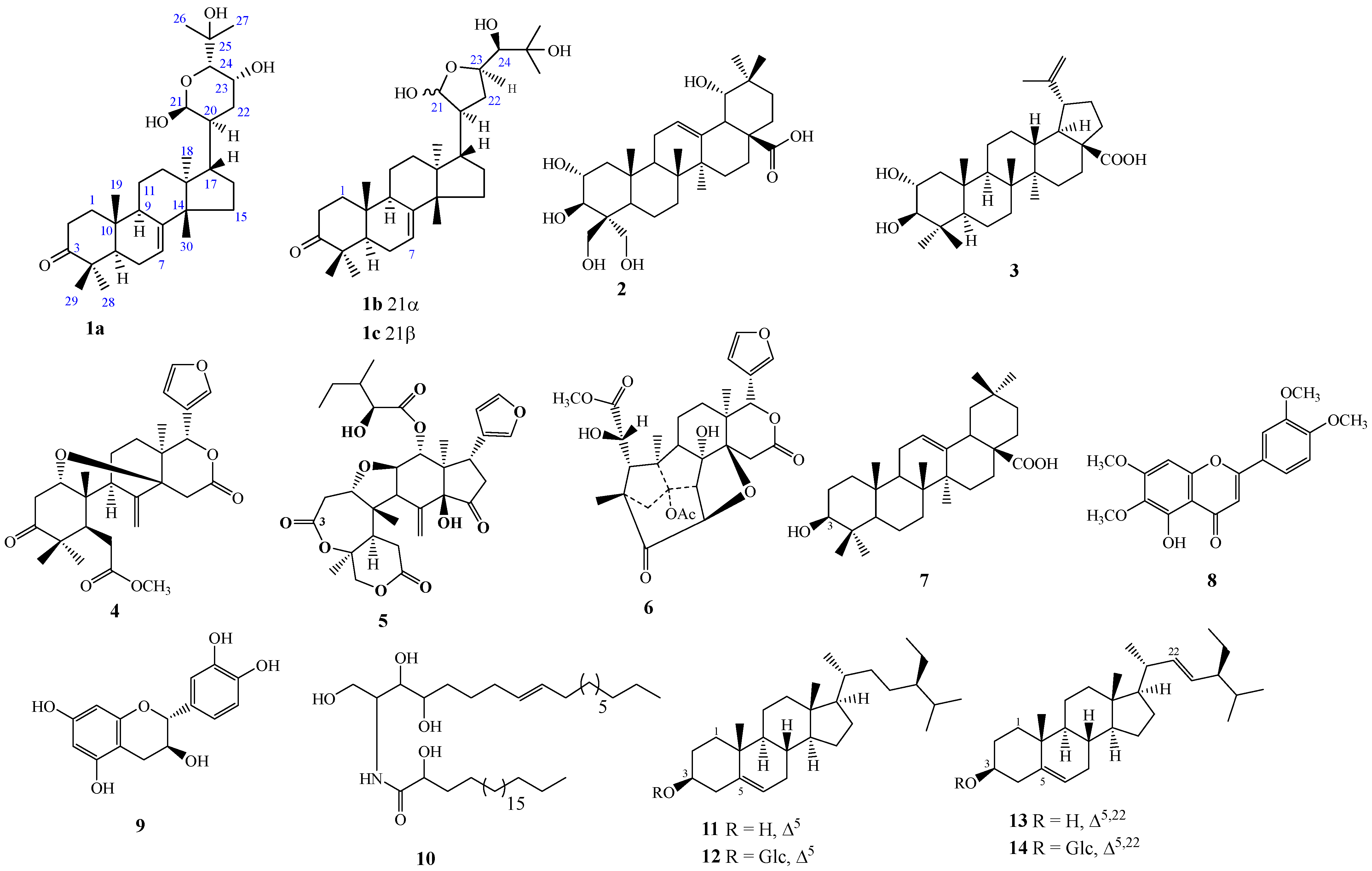
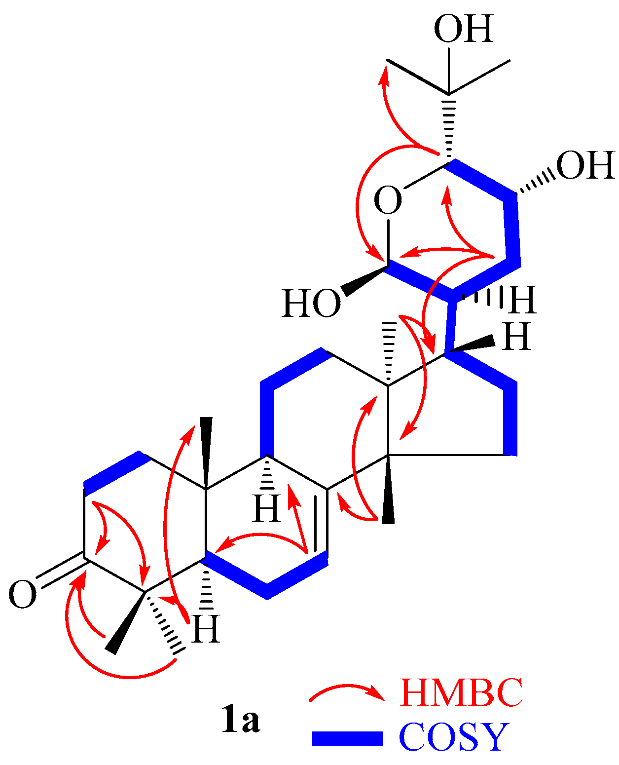
2.1. Structure Elucidation of Compound 1

Compound 1 was isolated as a shiny white powder whose molecular formula was established as C30H48O5; HR-ESI-MS (positive) m/z 511.3394 [M + Na]+; calcd. m/z 511.3399 (for C30H48O5Na+), indicating seven double bond equivalents (Figure S1). The UV spectrum (Figure S2) of compound 1 showed absorption maxima at 268 and 365 nm, while its IR spectrum (Figure S3) revealed a broad absorption band ranging from 3669 to 3200 cm−1 attributable to hydroxyl groups and other important absorption bands at 1697 and 1488 cm−1 assigned to carboxyl and olefinic functions, respectively.
The 1H-NMR spectrum of compound 1 exhibited the profile of a triterpenoid derivative. The combination of the proton and carbon-13 NMR spectra of compound 1 led to its identification as an inseparable mixture of isomers. The 1H-NMR data of compound 1 were identical to those of melianodiol (1b) and 21-epi-melianodiol (1c) as well as the bourjotinolone A skeleton, with few differences, such as the appearance of signals at δH 5.26, 4.10 and 3.60 [16,17,18,19]. In addition, the signals of three olefinic protons were observed at δH 5.35 (3H, brq, J = 3.1 Hz, H-7abc) as well as typical oxymethine protons at δH 5.26 (1H, brd, J = 2.8 Hz, H-21a), 5.21 (2H, brdd, J = 4.0, 1.8 Hz, H-21bc), 4.46 (1H, brddd, J = 9.3, 6.7, 1.8 Hz, H-23c), 4.34 (1H, brddd, J = 10.8, 5.0, 2.3 Hz, H-23b), 4.10 (1H, brt, J = 3.6 Hz, H-23a), 3.60 (1H, brd, J = 1.5 Hz, H-24a), 3.20 (1H, brd, J = 2.3 Hz, H-24b), and 3.14 (1H, brd, J = 1.8 Hz, H-24c), which were in agreement with the three characteristic signals integrating for 21 angular methyl groups at δH [0.89 (3H, s, Me-18c), 0.91 (3H, s, Me-18a), 0.92 (3H, s, Me-18b)], [1.03 (9H, s), Me-28abc], [1.05 (9H, s, Me-19abc)], [1.06 (9H, s, Me-30abc)], [1.13 (9H, s, Me-29abc)], [1.24 (3H, s, Me-26a), 1.21 (3H, s, Me-26b), 1.19 (3H, s, Me-26c)], and [1.27 (3H, s, Me-27a). In addition, 1.26 (3H, s, Me-27b) and 1.23 (3H, s, Me-27c)] were also observed on 1H-NMR (Table 1, Figure S4), confirming the presence of three triterpenoid moieties. The 13C-NMR (Table 1, Figure S6a,b) spectrum of compound 1 showed overlapping carbon signals from C1 to C16 for the three triterpenoids due to the similar chemical environment.
The HSQC spectrum (Figure S7a,b) revealed characteristic correlations of an olefinic carbon at δC 119.2 (C-7)/δH 5.35 (H-7) and furanic carbons at δC 103.2 (C-21α)/δH 5.21 (H-21α), 94.1 (C-21β)/δH 5.21 (H-21β), 77.0 (C-23α)/δH 4.34 (H-23α), and 78.3 (C-23β)/δH 4.46 (H-23β) assigned to the furan moieties of 21α/β-melianodiol. The existence of 21α/β-melianodiol might be due to mutarotation with the formation of an equilibrium mixture of C-21 epimers [17,18,20]. In addition, the characteristic signals of a pyranic ring at δC 94.1 (C-21)/δH 5.26 (H-21) and 73.6 (C-24)/δH 3.60 (H-24) were also noticed and attributable to the bourjotinolone derivative with little difference, such as the appearance of the signal at δC 67.1 (C-23)/δH 4.10 (H-23) [4,16]. Moreover, the oxymethine carbons observed at δC 78.5 (C-24α)/δH 3.20 (H-24α) and 78.5 (C-24β)/δH 3.14 (H-24β) were also in agreement with the limonoid-type bourjotinolone and melianodiol derivatives [4,17,18,19].
The HMBC (Table 1, Figure 2 and Figure S8a,b) spectrum revealed some important heteronuclear correlations that led to the construction of the skeleton of compound (1abc). Therefore, the correlation between the methyls Me-28/Me-29 (δC/δH 25.1/1.03)/(δC/δH 22.0/1.13) and the carbons at δC 219.2 (C-3), 53.9 (C-5) and 49.1 (C-4) led to the position of the ketone group and supported the triterpernoid skeleton [16,19,21]. However, the corrections between methyl Me-30 (δC/δH 28.1/1.06) with carbons at δC147.2 (C-8), 52.9 (C-14), and 35.9 (C-15) as well as methyl Me-18 (δC/δH 28.1/1.06) with carbons at δC 31.1 (C-13), 44.9 (C-13), 48.1 (C-17), and 52.5 (C-14) led to positioning of the double bond and supported the tirucallan moiety of the compound (1abc) epimeric mixture [16,18,19,21]. The oxymethine protons H-21α/H-21β (δC/δH 103.2/5.21)/(δC/δH 94.1/5.21) correlating with carbons at δC 52.0 (C-17), 50.2/47.9 (C-20), 32.9 (C-22), and 78.3/77.0 (C-23) supported the presence of the 21α/β-melianodiol (1b/1c) epimer in the mixture [16,17,18,22]. Finally, the free oxygen bearing methine H-21 (δC/δH 94.1/5.26) correlating with carbons at δC 73.6 (C-24) and 32.3 (C-22) agrees with the existence of free hydroxylation of the pyran ring of bourjotinolone A, which has not been reported previously. This pyranic ring is also supported by the COSY cross peak correlations between H-21 (δH 5.26), H-20 (δH 2.12), H-17 (δH 1.85), H-22 (δH 1.70/1.65), H-23 (δH 4.10), and H-24 (δH 3.60) [16]. The relative configurations of the chiral centers were deduced from the NOESY spectrum (Figure 2 and Figure S9) through homonuclear correlations between H-21 (δH 5.26) and H-20 (δH 2.12) and H-18 (δH 0.91), showing their alpha-orientation, while the correlation between H-22 (δH 4.11) and H-24 (δH 3.60) was beta-oriented. These special orientations were in agreement with the reported data of Xu et al. [16]. Therefore, compound 1 was established as an isomeric mixture including a stable new 21β-hydroxybourjotinolone A (1a), together with a mutarotant of 21α-melianodiol or 21,23-epoxy-21α,24,25-trihydroxytirucall-7-en-3-one (1b) [16,17,22] and 21β-melianodiol or 21,23-epoxy-21β,24,25-trihydroxytirucall-7-en-3- one (1c) [16,17,18] in the ratio of 3:1:1. Compounds 21α-melianodiol (1b) and 21β-melianodiol (1c) usually occur as an epimeric mixture due to the mutarotation formation at C-21, while the mixture of 21α/β-melianone and bourjotinolone A also occurs as a mixture of position isomers when they are not abundant in the plant species [17,19,23].
The remaining 13 known compounds were identified as follows: bellericagenin B (2) [24,25], alphitolic acid (3) [26,27], methyl angolensate (4) [28], rohituka-3 (5) [11,29], khayanolide E (6) [30,31], oleanolic acid (7) [32], Belamcanidin (8) [25,33], catechin (9) [34], gynuramide IV (10) [35], a mixture of β-sitosterol (11) and stigmasterol (13) [36], and a mixture of β-sitosterol glycoside (12) and stigmasterol glycoside (14) [37,38].

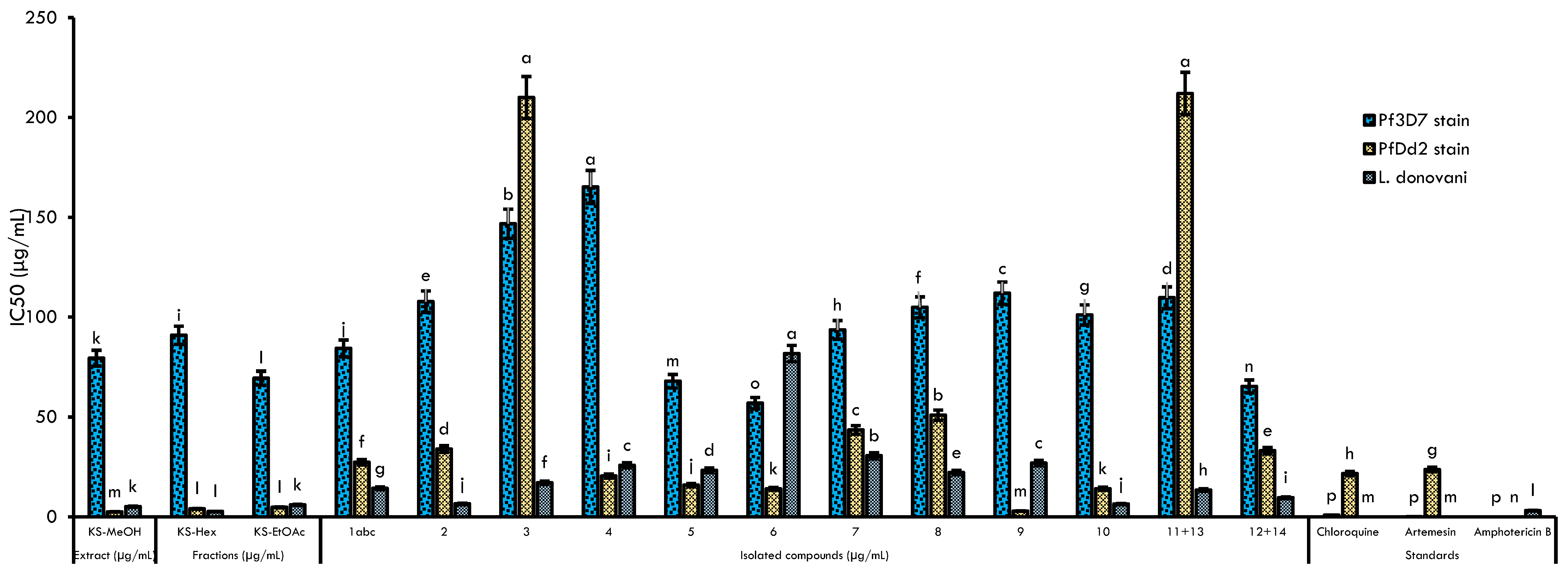
2.2. Antileishmanial, Antiplasmodial, and Cytotoxic Activities

The MeOH crude extract from Khaya senegalensis stem bark, the fractions, and the isolated compounds were evaluated for their antiplasmodial activity against chloroquine-resistant Dd2 and chloroquine-sensitive 3D7 strains of Plasmodium falciparum as well as antileishmanial and cytotoxicity activities against the Leishmania donovani strain and RAW 264.7 cancer cell lines, respectively (Figure 3, Table 2). According to a previously published report, the antiplasmodial activity of these samples was categorized as follows: high (IC50 < 5 μg/mL), promising (5 ≤ IC50 < 15 μg/mL), moderate (15 ≤ IC50 < 50 μg/mL), and weak or inactive (IC50 ≥ 50 μg/mL) [39]. The crude extract of K. senegalensis (KS-MeOH) exhibited high antiplasmodial activity against PfDd2 coupled with a good selectivity index (SI) toward RAW cells (IC50 = 2.5 ± 0.12 μg/mL, SI 19) and was inactive against Pf3D7 (IC50 = 79.4 ± 0.33 μg/mL, SI 0.59). Consequently, the crude extract was found to be highly active against Leishmania donovani promastigotes (SI value of 9.28) with a moderate SI in RAW cells (IC50 = 5.12 ± 0.7 μg/mL, SI 0.59). Liquid-liquid partitioning of KS-MeOH afforded two fractions (KS-Hex and KS-EtOAc) that displayed equipotent antiplasmodial (Pf3D7: IC50 69.7–90.87 μg/mL; PfDd2: IC50 4.05–4.78 μg/mL), antileishmanial (IC50 2.68–5.99 μg/mL) and cytotoxic activities (SI_Pf3D7 0.72–0.88, SI_Pf3D7 10.59–10.93, SI Leish 8.45–30.12).
The purification of KS-Hex and KS-EtOAc led to the isolation of diverse classes of secondary metabolites. Table 2 also reports the biological potencies of the isolated compounds. The criteria for antiplasmodial activity were focused on the established limits [40], where IC50 < 1 μg/mL indicates that the compound has excellent activity; IC50 of 1–20 μg/mL indicates good activity; IC50 of 20–100 μg/mL indicates moderate activity; IC50 of 100–200 μg/mL indicates low activity; and IC50 > 200 μg/mL indicates inactivity. Overall, compounds 1abc14 showed antiplasmodial activity with IC50 values ranging from 56.98 μg/mL (khayanolide E) (6) to 165.19 μg/mL (methylangolensate) (4) on Pf3D7 and 2.95 μg/mL (catechin) (9) to 50.97 μg/mL (belamcanidin) (8) on PfDd2 with catechin (9), khayanolide E (6), gyrunamide IV (10), rhohituka-3 (5) and methylangolensate (4) displaying the highest activity against PfDd2 (IC50 ˂ 20 μg/mL) with IC50 values of 2.95, 14.03, 14.13, 15.92, and 20.51 μg/mL, respectively. Alphitolic acid (3) and the mixture of β-sitosterol (11) and stigmasterol (13) were inactive against the multidrug-resistant PfDd2 strain (IC50 ˃ 200 μg/mL). The active compounds (410) showed resistance indices of less than 1, indicating a more pronounced potency against the resistant strain PfDd2 than against the sensitive strain. Concurrently, they were also found to be more selective toward PfDd2 (13.73 to 239.05) than the chloroquine-sensitive Pf3D7 strain (SI 0.24 to 10.71). These compounds (1abc14) were also tested against the promastigote form of L. donovani (Table 2). Globally, the isolated compounds revealed moderate to high inhibition of L. donovani promastigotes (IC50: 6.5–81.73 μg/mL), and except for khayanolide E (6) (SI 0.99), all of the samples were selective toward RAW 264.7 cells.

3. Discussion

Parasitic diseases, including malaria and leishmaniasis, represent a public health challenge, particularly for poor populations living in developing countries. Although significant progress has been made through the development of control methods such as chemotherapy, the impact of these diseases remains in the countries concerned, causing enormous socioeconomic losses [41,42]. This is due in particular to the drawbacks associated with approved drugs, including toxicity, duration of treatment regimens, and the spreading resistance of parasites to first-line drugs [42,43]. For several decades, medicinal plants and natural products have played a key role in the discovery of lead compounds, which have been used to advance the discovery and development of drugs against infectious diseases. In fact, 20 antiparasitic agents were approved by the Food and Drug Administration between 1981 and 2019, among which nine (45%) were derived from natural products [44]. Moreover, in the case of malaria, the two famous compounds that have positively revolutionized the management of this disease are quinine and artemisinin isolated from Cinchona sp. stem bark and Artemisia annua, respectively [45,46]. This supports the hypothesis that remarkable discoveries can be made from medicinal plants for the identification of new scaffolds against infectious diseases, including malaria and leishmaniasis.
The goal of this study was to search for bioactive secondary metabolites from the stem bark of K. senegalensis (Meliaceae) that are endowed with antiplasmodial and antileishmanial activities. The methanol extract (KS-MeOH) of K. senegalensis showed moderate antiplasmodial activity against Pf3D7, a chloroquine-sensitive strain, but exhibited a more promising effect against multidrug-resistant PfDd2 and Leishmania donovani promastigotes (Table 2, Figure 3). A similar trend was observed with the Sudanese methanolic extract from K. senegalensis stem bark against Pf3D7 and PfDd2, with IC50 values of 25 μg/mL and 150 μg/mL, respectively [47]. Moreover, two other distinct studies have previously demonstrated the antiplasmodial activity of extracts from this plant species against Pf3D7 and PfW2; however, the bioactive compounds were not reported [48,49]. With regard to its antileishmanial properties, very limited studies have reported the activity of K. senegalensis against Leishmania sp. parasites [50,51]. In addition, the extract KS-MeOH was shown to be selective against PfDd2 (SI > 19) and L. donovani (SI > 9.28) but not against Pf3D7 compared to the cytotoxic effect on RAW 264.7 cell lines. Climate change, geographical location, and harvesting period can modify the qualitative and quantitative content of secondary metabolites in medicinal plants and consequently their biological activities [52]. This further suggests that KS-MeOH preferentially inhibits PfDd2 and Leishmania parasites rather than mammalian cells. Recently, plants from the Meliaceae family have been recognized as a vast source of lead candidates for the development of antiplasmodial and insecticidal drugs [53]. Therefore, our phytochemical investigation of the antiprotozoal activity of Khaya senegalensis confirmed the activities previously reported [52]. The subsequently afforded hexane (KS-Hex) and ethyl acetate (KS-EtOAc) fractions from the KS-MeOH extract displayed the same antileishmanial, antiplasmodial, and selectivity profiles as the crude KS-MeOH extract. It can be alluded that the potent activity of KS-MeOH crude extract and fractions KS-Hex and KS-EtOAc (PfDd2 IC50 2.5–4.78 μg/mL, SI 10.59–19.93; L. donovani IC50 2.68–5.99 μg/mL, SI 8.45–30.12) is consequential of synergistic interactions between limonoids and eventually other secondary metabolites that are present in the extracts.
In further detail, a total of 16 compounds (1abc14) were isolated from both KS-Hex and KS-EtOAc using several column chromatographic techniques, including a new ordered limonoid-type bourjotinolone (1a) along with triterpenoid-type limonoids (1bc, 46). The protolimonoid compound (1abc) was found to be a mixture of a new limonoid 21β-hydroxybourjotinolone A (1a) together with an epimeric mixture of 21α-melianodiol (1b) and 21β-melianodiol (1c). However, degraded triterpenoid-type limonoids (compounds 46), lupanes (3), oleanane (7) and steroids (1114), two flavonoids (89) and one ceramide (10) were also identified here. Most of these compounds (2, 39 and 1114) have been previously reported from K. senegalensis [8,11]. Regarding their antiparasitic activities, as far as we know, the current study reveals for the first time the antiplasmodial and antileishmanial activities of alphitolic acid (3), rohituka-3 (5), belamcanidin (8), bellericagenin B (2), and gynuramide IV (10). Alphitolic acid (3) and Rohituka-3 (5) isolated from the same plant have been reported as antibacterial agents with cidal effects against Escherichia coli and Pseudomonas aeruginosa [11]. We have shown that, in addition to antibacterial activity, Rohituka-3 (5) has broad antiparasitic activity, while alphitolic acid (3) is mainly active against Leishmania donovani. For the other compounds, belamcanidin (8), bellericagenin B (2), and gynuramide IV (10), very little biological data are available in the literature. Our findings on the antiplasmodial activity of khayanolide (6) corroborate previous studies on this compound. Indeed, while investigating the antibacterial and antiplasmodial activity of constituents from the stem bark of Trichilia monadelpha (Thonn.), a plant from the Meliaceae family, Djoumessi and collaborators found that khayanolide (6) has a potent effect in vitro with an IC50 value of 1.68 μg/mL against the chloroquine-sensitive/sulfadoxine-resistant Pf3D7 strain [54].
Previously identified as an antibacterial agent [11], methylangolensate (4) was also identified here as an antileishmanial and antiplasmodial natural product. Oleanolic acid is well reputed for its antiplasmodial and antileishmanial activities. In fact, Cimanga et al. [55] and Peixoto et al. [56] have shown that it inhibits the growth of a clinical isolate of P. falciparum and Leishmania amazonensis with IC50 values of 15.2 μg/mL and 439.5 μg/mL, respectively [55,56]. Most importantly, Torres-Santos et al. [57] demonstrated that in addition to its antipromastigote effect (IC50 of 10 μg/mL), oleanolic acid from Pourouma guianensis also possesses antiamastigote inhibitory activity (IC50 of 27 μg/mL) [57]. Catechin (9) is a phenolic compound also known for its antiparasitic properties. Mogana et al. [58] investigated the antipromastigote activity of isolates from Canarium patentinervium and found an IC50 value of 478.93 ± 0.28 μg/mL for catechin [58]. Interestingly, two dimeric proanthocyanidins, namely, catechin-(4a,6)-catechin and catechin-(4a,8)-catechin), isolated from Khaya senegalensis, exhibited significant effects when tested against the amastigote forms of Leishmania donovani, with IC50 values of 3.85 and 3.98 μg/mL, respectively [51]. Moreover, catechin isolated from Garcinia celebica leaves was found to inhibit Plasmodium falciparum growth through the induction of oxidative stress [59]. Another study [60] demonstrated that at a concentration of 400 mg/kg, catechin from the leaf extract of Osyris quadripartita displayed an antimalarial effect with a suppression value of 64.26% [61].
Compounds 1b, 1c, and 10 are reported for the first time from the Khaya genus, while compound 1a is an unprecedented compound and reported for the first time from the Meliaceae family. Compound 1 (1abc), a mixture of three limonoids (hydroxybourjotinolone A, 21α-melianodiol, and 21β-melianodiol), displayed moderate activity against Pf3D7 (IC50 84.3 μg/mL), promising activity against both PfDd2 (IC50 27.38 μg/mL), high activity against Leishmania donovani (IC50 14.31 μg/mL), and high selectivity (SI > 10) for all strains. This is particularly relevant, as previous research has revealed antiplasmodial and antileishmanial activities for compound 7α-obacunyl acetate (IC50 5.14 μg/mL) and trigilgianin (IC50 6.044 μg/mL), which are two limonoids isolated from Trichilia gilgiana (Meliaceae) [62] and Entandrophragma angolense (Meliaceae), respectively [62]. Previous studies have shown that melanodiol is an anticancer [62], antioxidant, anti-inflammatory [63], and antibacterial agent [64]. The presence of such compounds with different properties could therefore explain the good activity profile obtained with this mixture. The results once again highlight the extensive antiparasitic properties of secondary metabolites belonging to the limonoid class. It is also important to point out that limonoids are reported to be the most abundant and best chemophenetic markers of the Meliaceae family as well as the K. senegalensis species [1,3,8,11,65]. This study therefore agrees with the classification of this plant species and improves knowledge of the classes of secondary metabolites found in K. senegalensis as well as their biological properties.

4. Materials and Methods

4.1. General Experimental Procedures

An alpha spectrometer (Brüker) spectrophotometer was used to record the IR spectrum in KBr. For NMR analysis, samples were first dissolved (85 μL) in their dissolving solvent before introduction into Eurisotop tubes (ref. D048T) and then analysis with Brüker Avance II+ at 500 MHz and 600 MHz equipped with a TCI Cryoprobe working under TopSpin 3.2.5. The mass spectrometry (ESI-MS, positive mode) of the samples was performed on a pneumatically assisted API (Atmospheric Pressure Ionization) mass spectrometer (Waters) type SYNAPT G2 HDMS. HRESIMS were measured with a TOF (Time-of-flight) analyzer. Silica gel (0.063–0.200 mm) was used as the stationary phase for CC (chromatographic column) for the fractionation and purification of compounds. TLC and UV (CN-6 UV, 254 nm & 365 nm) were used to check the purity of the isolated compounds. Ceric sulfate and 20% sulfuric acid were used to visualize the spotted samples on TLC after heating at approximately 90 °C. Iodine vapor was also used for visualization.

4.2. Plant Material

The stem bark of Khaya senegalensis was harvested on 15 September 2019, at Tchatibali Village (14°5′0″ E) in the Mayo-Danay Division of the Far-North Region of Cameroon. The plant material was taken to the National Herbarium of Cameroon, where it was identified in comparison with an available voucher specimen deposited under registration number 56853/HNC.

4.3. Extraction and Isolation

The fresh stem bark of K. senegalensis was collected and allowed to dry at room temperature, then powdered (7.5 kg) and extracted for 3 days (72 h) with 22 L MeOH, leading to 500.7 g of MeOH crude extract. A total of 275.0 g of the MeOH crude extract was subjected to liquid-liquid extraction to afford 15.6 g of n-hexane (KS-Hex), 160.8 g of EtOAc (KS-EtOAc), and 10.1 g of MeOH residue.
The n-hexane fraction (KS-Hex) (15.0 g) was fractionated by open CC (silica gel, gradient system n-hexane/EtOAc, 150 mL), leading to 195 fractions that were grouped into 5 principal fractions (A, B, C, D, and E) based on their TLC profile. Fraction A (5.2 g) was purified using over CC (silica gel, n-hexane/EtOAc, isocratic, 50 mL) to afford a mixture of compounds 11 and 13 (1.3 g, 9:1, v:v). Fraction C (0.5 g) was first fractionated using open CC (silica gel, n-hexane/EtOAc 7:3, isocratic, v:v, 50 mL), which led to two subfractions indexed C1 and C2. Subfraction C2 (100.4 mg) was further purified over Sephadex LH-20 (CH2Cl2/MeOH 1:1, isocratic, v:v, 5 mL), leading to a mixture of 1abc (5.1 mg, 7:3, v:v). The main fractions D (3.0 g) and E (0.9 g) were also purified using open CC (silica gel, gradient system n-hexane/EtOAc, 50 mL) to afford compounds 5 (7 mg, 3:7, v:v) and 10 (2 mg, 0:10, v:v), respectively (Figure 1).
The EtOAc fraction (KS-EtOAc) (150.0 g) was fractionated by open CC (silica gel, n-hexane/EtOAc and EtOAc/MeOH, gradient, 150 mL), leading to 219 fractions that were grouped into 6 major fractions (F, G, H, I, J and K) based on their TLC profile. Fraction F (20.1 g) was further purified using open CC (silica gel, n-hexane/EtOAc, gradient, 50 mL) to afford compounds 8 (109.8 mg, 8:2, v:v) and 7 (2.1 g, 8.5:2.5, v:v). Fraction G (10.3 g) was also purified by open CC (silica gel, n-hexane/EtOAc, gradient, 50 mL), leading to 3 (72.1 mg, 6:4, v:v). Fraction H (40.5 g) was purified using open CC (silica gel, n-hexane/EtOAc, gradient, 50 mL) to afford compounds 4 (41.1 mg, 1:1, v:v) and 6 (82.3 mg, 1:1, v:v). Fraction I (30.7 g) was purified using open CC (silica gel, n-hexane/EtOAc, gradient, 50 mL) to afford compounds 9 (4.1 mg, 3:7, v:v) and 2 (6.1 mg, 3:7, v:v). Fraction J (10.3 g) was finally purified by open CC (silica gel, EtOAc/MeOH, isocratic, 50 mL) to afford a mixture of compounds 12 and 14 (1.3 g, 9:1, v:v).

4.4. Physicochemical Characteristics of Compounds 1abc

  • 21β-hydroxybourjotinolone A (1a), white powder (MeOH); UV: 268, 365, 405, 666 nm; IR: 3 669–3 200, 2 964–2 869, 1 697, 1 488, 1 468, 1 444, 1 270, 1 070, 970 cm−1; HR-ESI-MS [M + Na]+ m/z 511.3394 (calculated for C30H48NaO5+, 511.3399); 1H NMR (CD3OD, 600 MHz) δ 2.04 (1H, m, H1)/1.47 (1H, m, H1), 2.84 (1H, brtd, J = 14.6, 5.6 Hz, H2)/2.19 (1H, brd, J = 14.6 Hz, H2), 1.75 (1H, brdd, J = 11.5, 6.0 Hz, H5), 2.13 (1H, m, H6)/1.28 (1H, m, H6), 5.35 (1H, brq, J = 3.1 Hz, H7), 2.38 (1H, m, H9), 1.62 (2H, m, H11), 1.99 (1H, m, H12)/1.81 (1H, m, H12), 2.20 (1H, m, H15)/2.17 (1H, m, H15), 1.87 (1H, m, H16)/1.31 (1H, m, H16), 1.85 (1H, m, H17), 0.91 (3H, s, H19), 1.05 (3H, s, H19), 2.12 (1H, m, H2O), 5.26 (1H, brd, J = 2.8 Hz, H21), 1.70 (1H, m, H22)/1.65 (1H, m, H22), 4.10 (1H, brt, J = 3.6 Hz, H23), 3.60 (1H, brd, J = 1.5 Hz, H24), 1.24 (3H, s, H26), 1.27 (3H, s, H27), 1.03 (3H, s, H28), 1.13 (3H, s, H29), 1.06 (3H, s, H30); 13C NMR (CD3OD, 150 MHz) δ 39.7 (C1), 35.9 (C2), 219.2 (C3), 49.1 (C4), 53.9 (C5), 25.1 (C6), 119.2 (C7), 147.2 (C8), 49.9 (C9), 36.2 (C10), 19.3 (C11), 31.1 (C12), 44.9 (C13), 52.4 (C14), 35.9 (C15), 27.7 (C16), 48.1 (C17), 23.2 (C18), 13.1 (C19), 40.0 (C20), 94.1 (C21), 32.3 (C22), 67.1 (C23), 73.6 (C24), 73.9 (C25), 26.8 (C26), 27.3 (C27), 25.1 (C28), 22.0 (C29), 28.1 (C30).
  • 21α-Melianodiol (1b) and 21β-melianodiol (1c); UV: 268, 365, 405, 666 nm; IR: 3669–3200, 2964–2869, 1697, 1488, 1468, 1444, 1270, 1070, 970 cm−1; HR-ESI-MS [M + Na]+ m/z 511.3394 (calculated for C30H48NaO5+, 511.3399); 1H- and 13C-NMR: see Table 1.

4.5. Biological Activities

4.5.1. Parasite and Cell Culture

The parasite strains used in this study are continuously cultured at the Antimicrobial and Biocontrol Agents Unit of the University of Yaoundé I, and were graciously provided by BEI Resources (https://www.beiresources.org (accessed on 16 February 2019)). They comprise the Leishmania donovani strain (1S(MHOM/SD/62/1S), the multiresistant (PfDd2), and the chloroquine-sensitive (Pf3D7-(MRA-102)) strains of Plasmodium falciparum. RPMI 1640 media supplemented with 0.2% sodium bicarbonate (Sigma Aldrich, Billings, MT, USA), 0.5% Albumax II (Gibco), 1% hypoxanthine 100X (Gibco, Billings, MT, USA), 25 mM HEPES, and 0.04% gentamicin (Sigma Aldrich) was used to sustain P. falciparum strains in sterile petri dishes. Fresh red blood cells from healthy O+ human volunteers were also added to the medium. These cells were suspended at 4% hematocrit (v/v) and incubated at 37 °C in a humidified environment with 5% CO2. To regularly maintain the culture, fresh medium was added to the culture every day. Thin blood smears stained with 10% Giemsa were used to microscopically monitor cell-cycle transition and parasitemia under immersion oil.
A T-75 cm2 cell culture flask was filled with complete Medium 199 [M199 supplemented with 10% heat-inactivated fetal bovine serum (HIFBS) and 100 IU/mL penicillin/streptomycin (Sigma, Darmstadt, Germany)] and used to grow the promastigote forms of Leishmania donovani. Every 72 h, flasks were subcultured and incubated at 28 °C.
The murine macrophage RAW 264.7 cell line was grown in DMEM (Dulbecco’s modified Eagle’s medium) containing 1% penicillin-streptomycin, 1% nonessential amino acids (NEA), and 10% HIFBS at 37 °C, and 5% CO2 in a humidified environment. A reverse Etaluma fluorescence microscope was used to view the cells every day, and the growth media was replaced every 72 h. At 80% confluence, cells were isolated using 0.25% Trypsine-EDTA (Gibco, MT, USA).

4.5.2. Sample Preparation for Biological Assays

In 100% dimethyl sulfoxide (DMSO, Sigma Aldrich), stock solutions of crude extracts, fractions, and compounds were prepared for final concentrations of 100 mg/mL, 50 mg/mL, and 20 mg/mL, respectively. Chloroquine phosphate (CQ) (Sigma Aldrich) and artemisinin (Art) (Sigma Aldrich) were used as antiplasmodial reference medicines, and were prepared at 10 mM in sterile distilled water and 100% DMSO, respectively. For antileishmanial tests, amphotericin B (10 mM in 100% DMSO, Sigma Aldrich) was included in the experiment as a positive control.

4.5.3. Antiplasmodial Activity of Compounds, Fractions and Extracts

The antiplasmodial efficacy of the samples was evaluated using the SYBR Green I-based fluorescence assay, as previously reported by Smilkstein et al. [66], on a sorbitol-synchronized ring stage culture of Plasmodium falciparum (PfDd2 and Pf3D7 strains). The test relies on the development of intense fluorescence following SYBR green binding to the exposed DNA found in wells containing healthy cells. Ninety microliters of parasites prepared with 1% hematocrit and 2% parasitemia and 10 μL of test sample extracts, fractions, isolated chemicals, chloroquine, and artemisinin were each added in triplicate into wells of a flat-bottomed microplate. The final test concentration values for the samples varied from 50 μg/mL-0.08 μg/mL and 100 μg/mL-0.16 μg/mL for extract/fractions. Following a 72 h incubation at 37 °C, 100 μL of SYBR Green I solution prepared with lysis buffer (0.2 μL of 10,000 SYBR Green I per mL of lysis buffer) made of EDTA (5 mM), Tris (20 mM; pH 7.5), Triton X-100 (0.08%; v/v), and saponin (0.008%; m/v) was added to each well. Fluorescence measurements were taken using an Infinite M200 microplate reader (Tecan, MT, USA) at emission and excitation wavelengths of 538 and 485 nm, respectively.

4.5.4. Antileishmanial Potency of Compounds, Fractions, and Extracts

The resazurin-based assay was used to evaluate the antileishmanial activity of the extract (100 μg/mL–0.16 μg/mL), major fractions (100 μg/mL–0.16 μg/mL), and isolated compounds (50 μg/mL–0.08 μg/mL) against the promastigote form of Leishmania donovani [67]. Briefly, 10 μL of inhibitors at various triplicate doses (as prescribed for the antiplasmodial assay) were added to 90 μL of promastigotes from a logarithmic phase culture at a density of 4.105 parasites/mL. The plates were incubated for 28 h at 28 °C, and 10 μL of resazurin solution (Sigma, Darmstadt, Germany) was added. Amphotericin B (Sigma, Darmstadt, Germany) (10 μg–0.016 μg/mL) and 0.1% DMSO-treated wells were used as the negative and positive controls, respectively. After a further 44 h of incubation, plates were read using a Magelan Infinite M200 fluorescence plate reader (Tecan, Männedorf, Switzerland) at excitation and emission wavelengths of 530 and 590 nm, respectively.

4.5.5. Cytotoxicity Assay

The murine macrophages, RAW 264.7 cells, were plated into a 96-well cell culture treated flat-bottomed plate (SARSTEDT, Inc., Newton, NC 28658, USA) at a density of 10 × 104 cells/100 μL/well and incubated overnight to enable cell adhesion. A fresh complete medium containing serially diluted concentrations of crude extract, fractions, and compounds was used to replace the previous medium. The final assay concentration values for extracts/fractions and compounds varied from 400 μg/mL to 0.8 μg/mL and 100–0.16 μg/mL, respectively. Podophyllotoxin (Sigma-Aldrich, Munich, Germany) was used as a positive control at a maximum dose of 10 μM. After 48 h of incubation, 10 μL of resazurin solution (0.15 mg/mL dissolved in PBS) was added to each well, and the plates were incubated for an additional 4 h [68]. Fluorescence data were finally recorded with a Magellan Infinite M200 plate reader (Tecan, Germany) at excitation and emission wavelengths of 530 nm and 590 nm, respectively.

4.5.6. Data Analysis for the Performed Assays

The resulting fluorescence readings were used to calculate the growth inhibition percentages relative to the negative control for each sample and for each of the tested doses. GraphPad Prism 8.0 software was used to generate concentration-response curves from the nonlinear regression fit to determine the median inhibitory or cytotoxic concentration (IC50/CC50). The resistance index (RI) for antiplasmodial tests was calculated as the ratio between the samples’ IC50 values for the chloroquine-sensitive strain 3D7 and the multiresistant strain Dd2 [69]. Inhibitors with an RI value below 1 were considered to act preferentially on the resistant strain. Based on each inhibitor’s antiparasitic efficacy (IC50) and cell toxicity (CC50), selectivity indices (SI) were calculated [SI = CC50 (RAW)/IC50 (Parasites)]. Samples were classified as poorly toxic to RAW cells if their SI values were greater than 10 [70].

5. Conclusions

The chemical investigation undertaken on the stem bark of K. senegalensis led to the isolation of a new limonoid named 21β-hydroxybourjotinolone A (1a) in a C-21 epimeric mixture of 21α-melianodiol (1b) and 21β-melianodiol (1c), together with 13 known compounds. Compounds 2, 39, and 1114 have previously been reported from K. senegalensis; while 1b, 1c, and 10 are reported for the first time from the Khaya genus, and 1a is reported for the first time from the Meliaceae family. The mixture of bourjotinolone (1a) and 21α/β-melianodiol (1b/1c) was found to possess cross activity against L. donovani and P. falciparum, and a good selectivity profile toward the parasitic strains. Other compounds displayed moderate to high antiparasitic activity and selectivity, qualifying them as interesting starting points for novel antiprotozoal agent discovery. Concurrently, it would be useful to investigate the mechanisms through which these natural products exert their antiprotozoal effects as well as the correlations between structural activity and pharmacokinetic properties.

Supplementary Materials

The following supporting information can be downloaded at: https://www.mdpi.com/article/10.3390/molecules28207227/s1. Figure S1: HR-ESI-MS (+), [M + Na]+ of compound 1abc; Figure S2: UV spectrum of compound 1abc; Figure S3: IR spectrum (KBr) of compound 1abc; Figure S4: 1H-NMR (CD3OD, 600 MHz) spectrum of compound 1abc; Figure S5a: COSY (CD3OD) expanded spectrum of compound 1abc; Figure S5b: COSY (CD3OD) full spectrum of compound 1abc; Figure S6a: 13C-NMR (CD3OD, 150 MHz) expanded spectrum of compound 1abc; Figure S6b: 13C-NMR (CD3OD, 150 MHz) full spectrum of compound 1abc; Figure S7a: HSQC (CD3OD) expanded spectrum of compound 1abc; Figure S7b: HSQC (CD3OD) full spectrum of compound 1abc; Figure S8a: HMBC (CD3OD) expanded spectrum of compound 1abc; Figure S8b: HMBC (CD3OD) expanded spectrum of compound 1abc; Figure S9: NOESY (CD3OD) full spectrum of compound 1abc; Figure S10: 1H-NMR (600 MHz, DMSO-d6) spectrum of compound 2; Figure S11: 13C-NMR (150 MHz, DMSO-d6) spectrum of compound 2; Figure S12: 1H-NMR (500 MHz, CD3OD) spectrum of compound 3; Figure S13: 13C-NMR (125 MHz, CD3OD) spectrum of compound 3; Figure S14: 1H-NMR (500 MHz, CDCl3) spectrum of compound 4; Figure S15: 13C-NMR (125 MHz, CDCl3) spectrum of compound 4; Figure S16: 1H-NMR (500 MHz, CDCl3) spectrum of compound 5; Figure S17: 13C-NMR (125 MHz, CDCl3) spectrum of compound 5; Figure S18: 1H-NMR (500 MHz, CDCl3) spectrum of compound 6; Figure S19: 13C-NMR (125 MHz, CDCl3) spectrum of compound 6; Figure S20: 1H-NMR (600 MHz, CD3OD) spectrum of compound 7; Figure S21: 13C-NMR (150 MHz, CD3OD) spectrum of compound 7; Figure S22: 1H-NMR (600 MHz, CDCl3) spectrum of compound 8; Figure S23: 13C-NMR (150 MHz, CDCl3) spectrum of compound 8; Figure S24: 1H-NMR (600 MHz, CDCl3) spectrum of compound 9; Figure S25: 13C-NMR (150 MHz, CDCl3) spectrum of compound 9; Figure S26: 1H-NMR (600 MHz, Pyridine-d5) spectrum of compound 10; Figure S27: 13C-NMR (150 MHz, Pyridine-d5) spectrum of compound 10; Figure S28: 1H-NMR (CDCl3, 500 MHz) spectrum of the mixture of compounds 11 and 13 and Figure S29: 1H-NMR (CDCl3, 500 MHz) spectrum of the mixture of compounds 12 and 14.

Author Contributions

Conceptualization, G.A.A.à.N. and Y.O.N.D.; Methodology, D.D., B.T., G.A.A.à.N., S.J.N.T., P.B.L. and Y.O.N.D.; Validation, A.M.L., F.F.B. and Y.O.N.D.; Isolation, G.A.A.à.N.; Formal analysis, P.H.D., L.R.Y.T., T.K.K., M.P., X.S.N., B.T., P.B.L., H.M. and S.J.N.T.; Investigation, G.A.A.à.N. and Y.O.N.D.; Bioassays, D.D., L.R.Y.T. and F.F.B.; Writing—original draft preparation, P.B.L., G.A.A.à.N., D.D., S.J.N.T., Y.O.N.D. and X.S.N.; Writing—review and editing, Y.O.N.D., X.S.N. and A.M.L.; Visualization, S.J.N.T., A.M.L. and Y.O.N.D.; Supervision, F.F.B., A.M.L. and Y.O.N.D. All authors have read and agreed to the published version of the manuscript.

Funding

The present research was not financially funded by any organization.

Institutional Review Board Statement

Not applicable.

Informed Consent Statement

Not applicable.

Data Availability Statement

Data are contained within the article and Supplementary Materials.

Acknowledgments

The Institute for Medical Research and Medicinal Plants Studies (IMPM), the University of Maroua, the Laboratory of Phytobiochemistry and Medicinal Plant Studies, the Antimicrobial and Biocontrol Agents Unit at the Department of Biochemistry of the University of Yaoundé I, Cameroon are gratefully acknowledged by the authors. The Yaoundé-Bielefield Graduate School through YaBiNaPA and the Department of Pharmaceutical Sciences, School of Pharmacy, Sefako Makgatho Health Sciences University, Pretoria, 0204, South Africa are also acknowledged for their facilities. X.S.N. kindly acknowledges a National Research Foundation (NRF) CSUR grant, Number: SRUG2203291031. P.B.L. gratefully thanks ICCBS (University of Karachi, Pakistan) and TWAS-ICCBS for support through project N° 3240311211. The Grand Challenges Africa (GCA) program [GCA/DD/rnd3/006] is gratefully thanked by FFB for supporting the biological activities. For this work, GCA is supported by the AAS, Bill & Melinda Gates Foundation (BMGF), Medicines for Malaria Venture (MMV), and Drug Discovery and Development Centre of University of Cape Town (H3D). FFB.

Conflicts of Interest

The authors declare no conflict of interest.

Sample Availability

Samples of the compounds are not available from the authors.

References

  1. Siva, B.; Suresh, G.; Poornima, B.; Venkanna, A.; Babu, K.S.; Prasad, K.R.; Rao, C.V. Cipadessin-type limonoids from the leaves of Cipadessa baccifera. Tetrahedron Lett. 2013, 54, 2934–2937. [Google Scholar] [CrossRef]
  2. Roy, A.; Saraf, S. Limonoids: Overview of significant bioactive triterpenes distributed in plants kingdom. Biol. Pharm. Bull. 2006, 29, 191–201. [Google Scholar] [CrossRef] [PubMed]
  3. Jimenez, A.; Villarreal, C.; Toscano, R.A.; Cook, M.; Arnason, J.T.; Bye, R.; Mata, R. Limonoids from Swietenia humilis and Guarea grandiflora (Meliaceae) Taken in part from the PhD and MS theses of C. Villarreal and MA Jiménez, respectively. Phytochemistry 1998, 49, 1981–1988. [Google Scholar] [CrossRef]
  4. Yang, M.H.; Wang, J.S.; Luo, J.G.; Wang, X.B.; Kong, L.Y. Chisopanins A–K, 11 new protolimonoids from Chisocheton paniculatus and their anti-inflammatory activities. Bioorg. Med. Chem. 2011, 19, 1409–1417. [Google Scholar] [CrossRef] [PubMed]
  5. Li, Y.; Lu, Q.; Luo, J.; Wang, J.; Wang, X.; Zhu, M.; Kong, L. Limonoids from the stem bark of Khaya senegalensis. Chem. Pharm. Bull. 2015, 63, 305–310. [Google Scholar] [CrossRef]
  6. Yuan, C.M.; Tang, G.H.; Wang, X.Y.; Zhang, Y.; Guo, F.; Liao, J.H.; Zou, T.; Zuo, G.Y.; Hua, H.M.; He, H.P.; et al. Two new compounds from Khaya senegalensis. J. Asian Nat. Prod. Res. 2013, 15, 638–643. [Google Scholar] [CrossRef] [PubMed]
  7. Yuan, C.M.; Zhang, Y.; Tang, G.H.; Li, S.L.; Di, Y.T.; Hou, L.; Hao, X.J. Senegalensions A–C, three limonoids from Khaya senegalensis. Chem. Asian J. 2012, 7, 2024–2027. [Google Scholar] [CrossRef]
  8. Yuan, T.; Zhang, C.R.; Yang, S.P.; Yue, J.M. Limonoids and triterpenoids from Khaya senegalensis. J. Nat. Prod. 2010, 73, 669–674. [Google Scholar] [CrossRef]
  9. Tyoapine, D.A.; Victory, P.C.; Yusuf, A.; Badamasi, A.M. A Review of the Phytochemistry and Pharmacology of the Medicinal Plant: Khaya Senegalensis (Desr.) A. Juss. Int. J. Res. Publ. Rev. 2022, 3, 1239–1246. [Google Scholar] [CrossRef]
  10. Gouissi, D.H.A.; Nzangue, R.T.; Kalaza, J.H.; Pabo, W.; Chegaing, S.P.F. Medicinal Plants Used for Malaria Treatment in Gamba Village, North Region of Cameroon: Ethnopharmacological Survey; In Vivo Antimalarial Activity of Aqueous Extracts of Khaya Senegalensis Bark. Res. Sq. 2021, 1–17. [Google Scholar] [CrossRef]
  11. Nganso, Y.O.; Tchana, E.M.; Kahouo, A.D.; Abah, K.; Fomena, H.; Mamoudou, H. Inhibitory effect and antimicrobial activity of secondary metabolites of Khaya senegalensis (Desr.) A. Juss. (Meliaceae). Sci. J. Chem. 2020, 8, 92–105. [Google Scholar] [CrossRef]
  12. Idu, M.; Erhabor, J.O.; Oghale, O.; Obayagbona, N.O. Antimicrobial qualities, phytochemistry and micronutritional content of Khaya senegalensis (Desr.) A. Juss. Seed oil. J. Phytopharm. 2014, 3, 95–101. [Google Scholar] [CrossRef]
  13. Hayasida, W.; Oliveira, L.M.; Ferreira, A.G.; Lima, M.P. Ergostane steroids, tirucallane and apotirucallane triterpenes from Guarea convergens. Chem. Nat. Compd. 2017, 53, 312–317. [Google Scholar] [CrossRef]
  14. Leutcha, B.P.; Dzoyem, J.P.; Jouda, J.B.; Sema, D.K.; Tsague, T.V.F.; Bitchagno, G.T.M.; Sewald, N. Antimicrobial and Cytotoxic Activities of Constituents from the Fruit of Albizia lebbeck L. Benth (Fabaceae). Molecules 2022, 27, 4823. [Google Scholar] [CrossRef] [PubMed]
  15. Magnibou, L.M.; Leutcha, P.B.; Tchegnitegni, B.T.; Wouamba, S.C.; Magne, C.Y.; Yaya, A.J.; Talla, E. A new phenanthrene derivative from Entada abyssinica with antimicrobial and antioxidant properties. Z Fur Naturforsch. B 2022, 77, 1–7. [Google Scholar] [CrossRef]
  16. Xu, J.; Xiao, D.; Lin, Q.H.; He, J.F.; Liu, W.Y.; Xie, N.; Qu, W. Cytotoxic tirucallane and apotirucallane triterpenoids from the stems of Picrasma quassioides. J. Nat. Prod. 2016, 79, 1899–1910. [Google Scholar] [CrossRef]
  17. Arriaga, A.; Mesquita, A.C.; Pouliquen, Y.; Lima, R.A.; Cavalcante, S.H.; Carvalho, M.G.; Braz-Filho, R. Chemical constituents of Simarouba versicolor. An. Acad. Bras. Cienc. 2002, 74, 415–424. [Google Scholar] [CrossRef] [PubMed]
  18. Puripattanavong, J.; Weber, S.; Brecht, V.; Frahm, A.W. Phytochemical investigation of Aglaia andamanica. Planta Med. 2000, 66, 740–745. [Google Scholar] [CrossRef]
  19. Cortez, D.A.; Vieira, P.C.; Fernandes, J.B.; da Silva, G.F.G.; Ferreira, A.G. Limonoids from Trichilia hirta. Phytochemistry 1992, 31, 625–628. [Google Scholar] [CrossRef]
  20. Salam, S.; Harneti, D.; Maharani, R.; Nurlelasari; Safari, A.; Hidayat, A.T.; Lesmana, R.; Nafiah, M.A.; Supratman, U.; Kyle, P.T.A.; et al. Cytotoxic triterpenoids from Chisocheton pentandrus. Phytochemistry 2021, 187, 112759. [Google Scholar] [CrossRef]
  21. Leutcha, B.P.; Sema, D.K.; Dzoyem, J.P.; Ayimele, G.A.; Nyongbela, K.D.; Delie, F.; Alléman, É.; Sewald, N.; Meli, L.A. Cytotoxicity of a new tirucallane derivative isolated from Stereospermum acuminatissimum K. Schum stem bark. Nat. Prod. Res. 2021, 35, 4417–4422. [Google Scholar] [CrossRef] [PubMed]
  22. Polonsky, J.; Varon, Z.; Rabanal, R.M.; Jacquemin, H. 21,20-anhydromelianone and melianone from Simarouba amara (Simaroubaceae); carbon-13 NMR spectral analysis of Δ7-tirucallol-type triterpenes. Isr. J. Chem. 1977, 16, 16–19. [Google Scholar] [CrossRef]
  23. Biavatti, M.W.; Vieira, P.C.; Da Silva, M.F.; Fernandes, J.B.; Albuquerque, S. Triterpenoid constituents of Raulinoa echinata. J. Nat. Prod. 2002, 65, 562–565. [Google Scholar] [CrossRef]
  24. Mahato, S.B.; Nandy, A.K.; Kundu, A.P. Pentacyclic triterpenoid sapogenols and their glycosides from Terminalia bellerica. Tetrahedron 1992, 48, 2483–2494. [Google Scholar] [CrossRef]
  25. Gade, I.S.; Chadeneau, C.; Simo, R.T.; Talla, E.; Atchade, A.D.T.; Seité, P.; Vannier, B.; Laurent, S.; Henoumont, C.; Kamdje, A.H.N.; et al. A new phenyl alkyl ester and a new combretin triterpene derivative from Combretum fragrans F. Hoffm (Combretaceae) and antiproliferative activity. Open Chem. 2020, 18, 1523–1531. [Google Scholar] [CrossRef]
  26. Chae, H.J.; Kim, G.J.; Deshar, B.; Kim, H.J.; Shin, M.J.; Kwon, H.; Suh, S.S. Anticancer activity of 2-O-caffeoyl alphitolic acid extracted from the Lichen, Usnea barbata 2017-KL-10. Molecules 2021, 26, 3937. [Google Scholar] [CrossRef]
  27. Bae, G.H.; Lee, S.M.; Lee, E.S.; Lee, J.S.; Gang, J.S. Isolation and quantitative analysis of betulinic acid and alphitolic acid from Zyziphi fructus. Yakhak Hoeji. 1996, 40, 558–562. [Google Scholar]
  28. Abdelgaleil, S.A.; Hashinaga, F.; Nakatani, M. Antifungal activity of limonoids from Khaya ivorensis. Pest. Manag. Sci. 2005, 61, 186–190. [Google Scholar] [CrossRef]
  29. Gunatilaka, A.A.; Bolzani, V.S.; Dagne, E.; Hofmann, G.A.; Johnson, R.K.; McCabe, F.L.; Mattern, M.R.; Kingston, D.G. Limonoids showing selective toxicity to DNA repair-deficient yeast and other constituents of Trichilia emetica. J. Nat. Prod. 1998, 61, 179–184. [Google Scholar] [CrossRef]
  30. Chun-Mao, Y.; Yu, Z.; Gui-Hua, T.; Ying-Tong, D.; Ming-Ming, C.; Xiao-Ying, W.; Guo-Ying, Z.; Shun-Lin, L.; Hui-Ming, H.; Hong-Ping, H.; et al. Khayseneganins A–H, Limonoids from Khaya senegalensis. J. Nat. Prod. 2012, 76, 327–333. [Google Scholar] [CrossRef]
  31. Nakatani, M.; Abdelgaleil, S.A.; Kassem, S.M.; Takezaki, K.; Okamura, H.; Iwagawa, T.; Doe, M. Three new modified limonoids from Khaya senegalensis. J. Nat. Prod. 2002, 65, 1219–1221. [Google Scholar] [CrossRef] [PubMed]
  32. Nwodo, N.J.; Ibezim, A.; Ntie-Kang, F.; Adikwu, M.U.; Mbah, C.J. Anti-trypanosomal activity of Nigerian plants and their constituents. Molecules 2015, 28, 7750–7771. [Google Scholar] [CrossRef] [PubMed]
  33. Dawé, A.; Mbiantcha, M.; Yakai, F.; Jabeen, A.; Ali, M.S.; Lateef, M.; Ngadjui, B.T. Flavonoids and triterpenes from Combretum fragrans with anti-inflammatory, antioxidant and antidiabetic potential. Z. Naturforsch. C 2018, 73, 211–219. [Google Scholar] [CrossRef] [PubMed]
  34. Cren-Olivé, C.; Wieruszeski, J.M.; Maes, E.; Rolando, C. Catechin and epicatechin deprotonation followed by 13C NMR. Tetrahedron Lett. 2002, 43, 4545–4549. [Google Scholar] [CrossRef]
  35. Lin, W.Y.; Yen, M.H.; Teng, C.M.; Tsai, I.L.; Chen, I.S. Cerebrosides from the rhizomes of Gynura japonica. J. Chin. Chem. Soc. 2004, 51, 1429–1434. [Google Scholar] [CrossRef]
  36. Kamboj, A.; Saluja, A.K. Isolation of stigmasterol and β-sitosterol from petroleum ether extract of aerial parts of Ageratum conyzoides (Asteraceae). Int. J. Pharm. Pharm. Sci. 2011, 3, 94–96. [Google Scholar]
  37. Ramiarantsoa, H.; Attioua, B.K.; Kouamé, M.A.; Djakouré, L.A. Le O-β-D-glucoside du β-sitostérol Isolé des Feuilles de Ravenala madagascariensis. J. Soc. Ouest-Afr. Chim. 2008, 26, 99–103. [Google Scholar]
  38. Khan, N.M.U.; Hossain, M.S. Scopoletin and β-sitosterol glucoside from roots of Ipomoea digitata. J. Pharmacogn. Phytochem. 2015, 4, 5–7. [Google Scholar]
  39. Jonville, M.C.; Kodja, H.; Humeau, L.; Fournel, J.; De Mol, P.; Cao, M.; Angenot, L.; Frédérich, M. Screening of medicinal plants from Reunion Island for antimalarial and cytotoxic activity. J. Ethnopharmacol. 2008, 120, 382–386. [Google Scholar] [CrossRef]
  40. Batista, R.; Silva Ade, J.J.; de Oliveira, A.B. Plant-derived antimalarial agents: New leads and efficient phytomedicines. Part II. Nonalkaloidal natural products. Molecules 2009, 14, 3037–3072. [Google Scholar] [CrossRef]
  41. WHO. Leishmaniasis. 2023. Available online: https://www.who.int/news-room/fact-sheets/detail/leishmaniasis (accessed on 27 September 2023).
  42. WHO. Malaria. 2023. Available online: https://www.who.int/news-room/fact-sheets/detail/malaria (accessed on 27 September 2023).
  43. Ponte-Sucre, A.; Gamarro, F.; Dujardin, J.C.; Barrett, M.P.; López-Vélez, R.; García-Hernández, R.; Pountain, A.W.; Mwenechanya, R.; Papadopoulou, B. Drug resistance and treatment failure in leishmaniasis: A 21st century challenge. PLoS Negl. Trop. Dis. 2017, 11, e0006052. [Google Scholar] [CrossRef] [PubMed]
  44. Newman, D.J.; Cragg, G.M. Natural Products as Sources of New Drugs over the Nearly Four Decades from 01/1981 to 09/2019. J. Nat. Prod. 2020, 83, 770–803. [Google Scholar] [CrossRef] [PubMed]
  45. Van Agtmael, M.A.; Eggelte, T.A.; Van Boxtel, C.J. Artemisinin drugs in the treatment of malaria: From medicinal herb to registered medication. Trends Pharmacol. Sci. 1999, 20, 199–205. [Google Scholar] [CrossRef] [PubMed]
  46. Schwikkard, S.; van Heerden, F.R. Antimalarial activity of plant metabolites. Nat. Prod. Rep. 2002, 19, 675–692. [Google Scholar] [CrossRef]
  47. Tahir, A.E.; Satti, G.M.H.; Khalid, S.A. Antiplasmodial activity of selected Sudanese medicinal plants with emphasis on Maytenus senegalensis (Lam.). J. Ethnopharmacol. 1999, 64, 227–233. [Google Scholar] [CrossRef]
  48. Manga, A.; Gassama, A.; Diatta, K.; Bassène, E.; Cojean, S.; Cavé, C. Antiplasmodial activity of extracts of Khaya senegalensis (DERS.) A. Jus (Meliaceae) and Melia azedarach L.; Plants of Senegalese Traditional Medicine. Int. J. Pharm. Sci. Res. 2018, 9, 4659–4665. [Google Scholar] [CrossRef]
  49. Shayoub, M.E.A.; Kabbashi, A.S.; Osman, H.M.; Mahmoud, A.N.; Elhassan, A.M.; Dawoud, A.D.H.; Jawad, A.S.A. Anti-malarial activity of Khaya senegalensis. Indo Am. J. Pharm. Res. 2016, 6. [Google Scholar]
  50. Ahua, K.M.; Ioset, J.R.; Ioset, K.N.; Diallo, D.; Mauël, J.; Hostettmann, K. Antileishmanial activities associated with plants used in the Malian traditional medicine. J. Ethnopharmacol. 2007, 110, 99–104. [Google Scholar] [CrossRef]
  51. Kayser, O.; Abreu, P.M. Antileishmania and immunostimulating activities of two dimeric proanthocyanidins from Khaya senegalensis. Pharm. Biol. 2001, 39, 284–288. [Google Scholar] [CrossRef]
  52. Palit, P.; Mandal, S.C. Evidence Based Validation of Traditional Medicines: A Comprehensive Approach; Mandal, S.C., Chakraborty, R., Sen, S., Eds.; Springer: Berlin/Heidelberg, Germany, 2021; p. 359. [Google Scholar]
  53. Happi, G.; Nangmo, P.; Dzouemo, L.; Kache, S.; Kouam, A.; Wansi, J. Contribution of Meliaceous plants in furnishing lead compounds for antiplasmodial and insecticidal drug development. J. Ethnopharmacol. 2022, 285, 114906. [Google Scholar] [CrossRef]
  54. Djoumessi, A.K.; Nono, R.N.; Neumann, B.; Stammler, H.G.; Bitchagno, G.T.M.; Efange, N.M.; Nkenfou, C.N.; Ayong, L.; Lenta, B.N.; Sewald, N.; et al. Constituents of the Stem Bark of Trichilia monadelpha (Thonn.) J. J. De Wilde (Meliaceae) and Their Antibacterial and Antiplasmodial Activities. Metabolites 2023, 13, 298. [Google Scholar] [CrossRef]
  55. Cimanga, R.K.; Tona, G.L.; Mesia, G.K.; Kambu, O.K.; Bakana, D.P.; Kalenda, P.D.T.; Penge, A.O.; Muyembe, J.J.T.; Totté, J.; Pieters, L.; et al. Bioassay-guided isolation of antimalarial triterpenoid acids from the leaves of Morinda lucida. Pharm. Biol. 2006, 44, 677–681. [Google Scholar] [CrossRef]
  56. Peixoto, J.A.; Silva, M.L.A.E.; Crotti, A.E.M.; Veneziani, R.C.S.; Gimenez, V.M.M.; Januário, A.H.; Groppo, M.; Magalhaes, L.G.; Dos Santos, F.F.; Albuquerque, S.; et al. Antileishmanial activity of the hydroalcoholic extract of miconia langsdorffii, isolated compounds, and semisynthetic derivatives. Molecules 2011, 16, 1825–1833. [Google Scholar] [CrossRef] [PubMed]
  57. Torres-Santos, E.C.; Lopes, D.; Rodrigues Oliveira, R.; Carauta, J.P.P.; Bandeira Falcao, C.A.; Kaplan, M.A.C.; Rossi-Bergmann, B. Antileishmanial activity of isolated triterpenoids from Pourouma guianensis. Phytomedicine 2004, 11, 114–120. [Google Scholar] [CrossRef] [PubMed]
  58. Mogana, R.; Adhikari, A.; Debnath, S.; Hazra, S.; Hazra, B.; Teng-Jin, K.; Wiart, C. The antiacetylcholinesterase and antileishmanial activities of Canarium patentinervium Miq. Biomed Res. Int. 2014, 2014, 903529. [Google Scholar] [CrossRef] [PubMed]
  59. Rizky, A.; Eka, W.S.; Anas, S.; Unang, S.; Milyadi, S.; Ajeng, D.; Keri, L.; Melisa, I.B.; Shinichiro, K.; Hiroshi, K. Catechin Isolated from Garcinia celebica Leaves Inhibit Plasmodium falciparum Growth through the Induction of Oxidative Stress. Pharmacogn. Mag. 2017, 13, 301–305. [Google Scholar] [CrossRef]
  60. Kemal, T.; Feyisa, K.; Bisrat, D.; Asres, K. In Vivo Antimalarial Activity of the Leaf Extract of Osyris quadripartita Salzm. ex Decne and Its Major Compound (-) Catechin. J. Trop. Med. 2022, 2022, 3391216. [Google Scholar] [CrossRef]
  61. Bickii, J.; Tchouya, G.R.F.; Tchouankeu, J.C.; Tsamo, E. Antimalarial activity in crude extracts of some Cameroonian medicinal plants. Afr. J. Trad. Compl. Alt. Med. 2007, 4, 107–111. [Google Scholar] [CrossRef]
  62. Kowa, T.K.; Tchokouaha, L.R.; Cieckiewicz, E.; Philips, T.J.; Dotse, E.; Wabo, H.K.; Frédérich, M. Antileishmanial and cytotoxic activities of a new limonoid and a new phenyl alkene from the stem bark of Trichilia gilgiana (Meliaceae). Nat. Prod. Res. 2020, 34, 3182–3188. [Google Scholar] [CrossRef]
  63. Kurimoto, S.I.; Takaishi, Y.; Ahmed, F.A.; Kashiwada, Y. Triterpenoids from the fruits of Azadirachta indica (Meliaceae). Fitoterapia 2014, 92, 200–205. [Google Scholar] [CrossRef]
  64. Shen, J.; Ma, X.; He, Y.; Wang, Y.; Zhong, T.; Zhang, Y. Anti-inflammatory and antioxidant properties of Melianodiol on DSS-induced ulcerative colitis in mice. PeerJ 2022, 10, e14209. [Google Scholar] [CrossRef] [PubMed]
  65. Zhang, H.; Tan, J.; Vanderveer, D.; Wang, X.; Wargovich, M.J.; Chen, F. Khayanolides from African mahogany Khaya senegalensis (Meliaceae): A revision. Phytochemistry 2009, 70, 294–299. [Google Scholar] [CrossRef]
  66. Smilkstein, M.; Sriwilaijaroen, N.; Kelly, J.X.; Wilairat, P.; Riscoe, M. Simple and inexpensive fluorescence-based technique for high-throughput antimalarial drug screening. Antimicrob. Agents Chemother. 2004, 48, 1803–1806. [Google Scholar] [CrossRef] [PubMed]
  67. Siqueira-Neto, J.L.; Song, O.R.; Oh, H.; Sohn, J.H.; Yang, G.; Nam, J.; Jang, J.; Cechetto, J.; Lee, C.B.; Moon, S.; et al. Antileishmanial high-throughput drug screening reveals drug candidates with new scaffolds. PLoS Negl. Trop. Dis. 2010, 4, e675. [Google Scholar] [CrossRef] [PubMed]
  68. Bowling, T.; Mercer, L.; Don, R.; Jacobs, R.; Nare, B. Application of a resazurin-based high-throughput screening assay for the identification and progression of new treatments for human African trypanosomiasis. Int. J. Parasitol. Drugs Drug Resist. 2012, 2, 262–270. [Google Scholar] [CrossRef] [PubMed]
  69. Douanla, P.D.; Tabopda, T.K.; Tchinda, A.T.; Cieckiewicz, E.; Frédérich, M.; Boyom, F.F.; Tsabang, N.; Yeboah, S.; Nkengfack, A.E.; Tchuendem, M.H.K. Antrocarines A-F, antiplasmodial ergostane steroids from the stem bark of Antrocaryon klaineanum. Phytochemistry 2015, 117, 521–526. [Google Scholar] [CrossRef] [PubMed]
  70. Mosmann, T. Rapid colorimetric assay for cellular growth and survival: Application to proliferation and cytotoxicity assays. J. Immunol. Methods 1983, 65, 55–63. [Google Scholar] [CrossRef]
Figure 1. Compounds isolated from the stem bark of K. senegalensis 1abc14.
Molecules 28 07227 g001
Figure 2. Important COSY and HMBC correlations of compound 1a.
Molecules 28 07227 g002
Figure 3. Column diagram summarizing the biological activities of the compounds, fractions, and extract from Khaya senegalensis. The results with the same letters are not significantly different (p < 0.05). The error bars represent the standard deviation of measurement of samples from duplicate runs.
Molecules 28 07227 g003
Table 1. 13C, 1H and HMBC NMR data (1H 600 MHz, 13C 150 MHz, CD3OD) of compound 1abc.
21β Hydroxybourjotinolone A
1a
21α-Melianodiol
1b
21β-Melianodiol
1c
δCδHHMBCδCδHδCδH
139.72.04 (1H, m)
1.47 (1H, m)
39.72.04 (1H, m)
1.47 (1H, m)
39.72.04 (1H, m)
1.47 (1H, m)
235.92.84 (1H, brtd, 14.6, 5.6)
2.19 (1H, brd, 14.6)
1, 3, 4, 1035.92.84 (1H, brtd, 14.6, 5.6)
2.19 (1H, brd, 14.6)
35.92.84 (1H, brtd, 14.6, 5.6)
2.19 (1H, brd, 14.6)
3219.2- 219.2-219.2-
449.1- 49.1-49.1-
553.91.75 (1H, brdd, 11.5, 6.0)4, 8, 1953.91.75 (1H, brdd, 11.5, 6.0)53.91.75 (1H, brdd, 11.5, 6.0)
625.12.13 (1H, m)
1.28 (1H, m)
4, 8, 1025.12.13 (1H, m)
1.28 (1H, m)
25.12.13 (1H, m)
1.28 (1H, m)
7119.25.35 (1H, brq, 3.1)5, 6, 9119.25.35 (1H, brq, 3.1)119.25.35 (1H, brq, 3.1)
8147.2- 147.2-147.2-
949.92.38 (1H, m) 49.92.38 (1H, m)49.92.38 (1H, m)
1036.2- 36.2-36.2-
1119.31.62 (2H, m) 19.31.62 (2H, m)19.31.62 (2H, m)
1231.11.99 (1H, m)
1.81 (1H, m)
31.11.99 (1H, m)
1.81 (1H, m)
31.11.99 (1H, m)
1.81 (1H, m)
1344.9- 44.9-44.9-
1452.4- 52.4-52.4-
1535.92.20 (1H, m)
2.17 (1H, m)
1735.92.20 (1H, m)
2.17 (1H, m)
35.92.20 (1H, m)
2.17 (1H, m)
1627.71.87 (1H, m)
1.31 (1H, m)
27.71.87 (1H, m)
1.31 (1H, m)
27.71.87 (1H, m)
1.31 (1H, m)
1748.11.85 (1H, m) 52.01.85 (1H, m)52.01.85 (1H, m)
1823.20.91 (3H, s)12, 13, 14, 17 0.92 (3H, s) 0.89 (3H, s)
1913.11.05 (3H, s)1, 5, 9, 1013.11.05 (3H, s)13.11.05 (3H, s)
2040.02.12 (1H, m) 50.22.11 (1H, m)47.92.00 (1H, m)
2194.15.26 (1H, brd, 2.8)22, 24103.25.21 (1H, brdd, 4.0, 1.8)94.15.21 (1H, brdd, 4.0, 1.8)
2232.31.70 (1H, m)
1.65 (1H, m)
17, 21, 2432.91.99 (1H, m)
1.82 (1H, m)
32.91.99 (1H, m)
1.82 (1H, m)
2367.14.10 (1H, brt, 3.6)2077.04.34 (1H, brddd, 10.8, 5.0, 2.3)78.34.46 (1H, brddd, 9.3, 6.7, 1.8)
2473.63.60 (1H, brd, 1.5)21, 23, 25, 2778.53.20 (1H, brd, 2.3)78.53.14 (1H, brd, 1.8)
2573.9- 74.5-73.6-
2626.81.24 (3H, s)24, 25, 2725.61.21 (3H, s)25.61.19 (3H, s)
2727.31.27 (3H, s)24, 25, 2627.51.26 (3H, s)27.51.23 (3H, s)
2825.11.03 (3H, s)3, 4, 5, 2925.11.03 (3H, s)25.11.03 (3H, s)
2922.01.13 (3H, s)3, 4, 5, 2822.01.13 (3H, s)22.01.13 (3H, s)
3028.11.06 (3H, s)8, 13, 1528.11.06 (3H, s)28.11.06 (3H, s)
δ (ppm), (br: broad, s: singlet, d: doublet, t: triplet, q: quadruplet, m: multiplet, J (Hz)), blue: for differences.
Table 2. Antileishmanial and antiplasmodial activities and selectivity of compounds, fractions, and crude extract from K. senegalensis.
SamplesCytotoxicity on RAW 264.7 CellsAntiplasmodial Activity (μg/mL), Selectivity (SI) and Resistance Index (RI)Antileishmanial Activity (μg/mL) and Selectivity (SI)
CC50 ± SDPf3D7 IC50 ± SDSI_Pf3D7PfDd2 IC50 ± SDSI_PfDd2RIIC50 ± SDSI
Extract (μg/mL)
KS-MeOH47.5279.40 ± 0.330.592.50 ± 0.12190.035.12 ± 0.709.28
Fractions (μg/mL)
KS-Hex80.7390.87 ± 0.080.884.05 ± 0.0019.930.042.68 ± 0.4230.12
KS-EtOAc50.6469.52 ± 0.330.724.78 ± 0.3610.590.065.99 ± 0.778.45
Isolated compounds (μg/mL)
21β-hydroxylbourjotinolone A (1a) + 21α-melianodiol (1b) + 21β-melianodiol (1c)>40084.30 ± 0.11>4.7427.38 ± 0.18>14.60.3214.31 ± 0.87>27.94
Bellericagenin B (2)>400107.72 ± 0.37>3.8134.02 ± 0.09>11.750.316.5 ± 0.01>61.54
Alphitolic acid (3)35.76146.77 ± 0.130.24>200NDND17.09 ± 0.902.09
Methylangolensate (4)72.74165.19 ± 0.060.4420.51 ± 0.4534.130.1225.83 ± 0.272.81
Rohituka-3 (5)>40068 ± 0.02>5.8815.92 ± 0.33>25.130.2323.35 ± 0.21>17.13
Khayanolide E (6)81.6156.98 ± 0.081.4314.03 ± 0.0649.880.2481.73 ± 0.120.99
Oleanolic acid (7)>40093.58 ± 0.92˃4.2743.58 ± 0.73˃9.170.4730.63 ± 0.67˃13.06
Belamcanidin (8)>400104.92 ± 0.39>3.8250.97 ± 0.37>7.840.4822.21 ± 0.08>18.013
Catechin (9)>400111.96 ± 0.04>3.572.93 ± 0.02>136.60.0226.97 ± 0.70>14.83
Gynuramide IV (10)>400101.07 ± 0.01>3.9614.13 ± 0.03>28.30.146.43 ± 0.06>62.22
β-sitosterol (11) + stigmasterol (13)>400109.69± 1.66>6.12>200ND>1.113.47 ± 0.74>29.69
β-sitosterol glycoside (12) + stigmasterol glycoside (14)>40065.34 ± 0.89˃3.6533.16 ± 0.43˃120.519.60 ± 0.64˃41.65
Reference drugs (μg/mL)
Chloroquine-0.022 ± 0.25-0.085 ± 0.01---ND
Artemesin-0.02 ± 0.00-0.024 ± 0.24----
Amphotericin B------3.14 ± 0.46-
Podophyllotoxin0.71 ± 0.20-------
KS-MeOH: methanol extract of K. senegalensis, KS-Hex: n-hexane fraction of Khaya senegalensis extract, KS-EtOAc: ethyl acetate fraction of K. senegalensis extract, SI: selectivity index, RI: reactivity index, ND: not determined, NC: not calculated, P. falciparum: IC50: 50% inhibitory concentration in μg/mL; CC50: 50% cytotoxic concentration in μg/mL. The results are expressed as the mean ± standard deviation; the SI (selective index) of bioactive compounds was determined as a measure of their toxicity against RAW cell line macrophages. SI = CC50 against macrophages/IC50 against promastigotes.
Disclaimer/Publisher’s Note: The statements, opinions and data contained in all publications are solely those of the individual author(s) and contributor(s) and not of MDPI and/or the editor(s). MDPI and/or the editor(s) disclaim responsibility for any injury to people or property resulting from any ideas, methods, instructions or products referred to in the content.

