Article

Crystal Structure of Allantoinase from Escherichia coli BL21: A Molecular Insight into a Role of the Active Site Loops in Catalysis

1
Department of Biomedical Sciences, Chung Shan Medical University, Taichung City 402, Taiwan
2
Department of Beauty Science, National Taichung University of Science and Technology, Taichung City 403, Taiwan
3
Department of Medicine, College of Medicine, Chung Shan Medical University, Taichung City 402, Taiwan
4
Department of Pediatrics, National Taiwan University Children’s Hospital, Taipei 100, Taiwan
5
Department of Medical Research, Chung Shan Medical University Hospital, Taichung City 402, Taiwan
*
Author to whom correspondence should be addressed.
Molecules 2023, 28(2), 827; https://doi.org/10.3390/molecules28020827
Submission received: 18 December 2022 / Revised: 10 January 2023 / Accepted: 11 January 2023 / Published: 13 January 2023

Abstract

:
Allantoinase (ALLase; EC 3.5.2.5) possesses a binuclear metal center in which two metal ions are bridged by a posttranslationally carbamylated lysine. ALLase acts as a key enzyme for the biogenesis and degradation of ureides by catalyzing the conversion of allantoin into allantoate. Biochemically, ALLase belongs to the cyclic amidohydrolase family, which also includes dihydropyrimidinase, dihydroorotase, hydantoinase (HYDase), and imidase. Previously, the crystal structure of ALLase from Escherichia coli K-12 (EcALLase-K12) was reported; however, the two active site loops crucial for substrate binding were not determined. This situation would limit further docking and protein engineering experiments. Here, we solved the crystal structure of E. coli BL21 ALLase (EcALLase-BL21) at a resolution of 2.07 Å (PDB ID 8HFD) to obtain more information for structural analyses. The structure has a classic TIM barrel fold. As compared with the previous work, the two missed active site loops in EcALLase-K12 were clearly determined in our structure of EcALLase-BL21. EcALLase-BL21 shared active site similarity with HYDase, an important biocatalyst for industrial production of semisynthetic penicillin and cephalosporins. Based on this structural comparison, we discussed the functional role of the two active site loops in EcALLase-BL21 to better understand the substrate/inhibitor binding mechanism for further biotechnological and pharmaceutical applications.

1. Introduction

Allantoinase (ALLase; EC 3.5.2.5) plays an essential role in the catabolism pathway for purine degradation [1]. ALLase occurs in a wide variety of organisms, including bacteria, fungi, and plants, and a few animals. ALLase catalyzes the reversible conversion of allantoin to allantoic acid by hydrolytic cleavage of the five-member hydantoin ring (Figure 1). This ALLase-catalyzed reaction is a key process in the biosynthesis and degradation of ureide required for the utilization of nitrogen in purine-derived compounds [2]. ALLase is a homotetrameric dinuclear metalloenzyme [3,4], but some ALLases initially annotated as polysaccharide deacetylases are metal independent [5]. Thus, even without the ALLase gene, some bacteria can still use allantoin to utilize nitrogen.
Allantoin in plants participates in many positively signaling regulations and is also a signaling chemical that conveys information on local conditions in plant–plant interactions [6]. For example, allantoin enhances drought tolerance in rice [7]. In addition, allantoin also positively affects salt tolerance by increasing the reactive oxygen species scavenging capacity and maintaining sodium and potassium homeostasis [8]. ALLase is required for flux of fixed nitrogen to ureides in bean nodules [9]. Allantoin is abundant in many medical plants and has the pharmaceutical potential to be used in IgE-independent anti-allergic and anti-inflammatory therapies [10,11,12]. Allantoin is one of the most important botanicals pertaining to dermatologic uses [13]; therefore, understanding the enzymatic reaction of ALLase is of considerable interest for further biotechnological applications.
ALLase [14] is a member of the cyclic amidohydrolase family [15,16], which also includes dihydroorotase (DHOase) [17,18,19,20,21,22,23], dihydropyrimidinase (DHPase) [24,25,26,27,28,29,30,31], hydantoinase (HYDase) [32,33,34], and imidase [35,36,37]. Almost all these enzymes contain the binuclear metal center that consists of four His, one Asp, and one posttranslational carbamylated Lys (Kcx) residues [15]. Some of these amidohydrolases have been suggested as chemotherapeutic targets for anticancer, antimicrobial, and antimalarial drug development because of their involvement in the key reactions of nucleotide biosynthesis [15]. Thus, it is worth exploiting the new inhibitors against these targets for drug development.
Recently, we found that the extract of Nepenthes miranda was capable of inhibiting ALLase [38], DHOase [39], and DHPase [40] activities. Through gas chromatography–mass spectrometry analysis (GC–MS), the active ingredients abundant in this extract were detected and identified [38]. Given that the crystal structure of ALLase from Escherichia coli K-12 (EcALLase-K12) [41] is available, the top six compounds in this N. miranda extract, namely, plumbagin, lupenone, palmitic acid, stigmast-5-en-3-ol, neophytadiene, and citraconic anhydride, were docked to EcALLase-K12 [38]. These compounds might be useful alone or in combination for further inhibitor optimization and structure-based drug design for anti-ALLase activity.
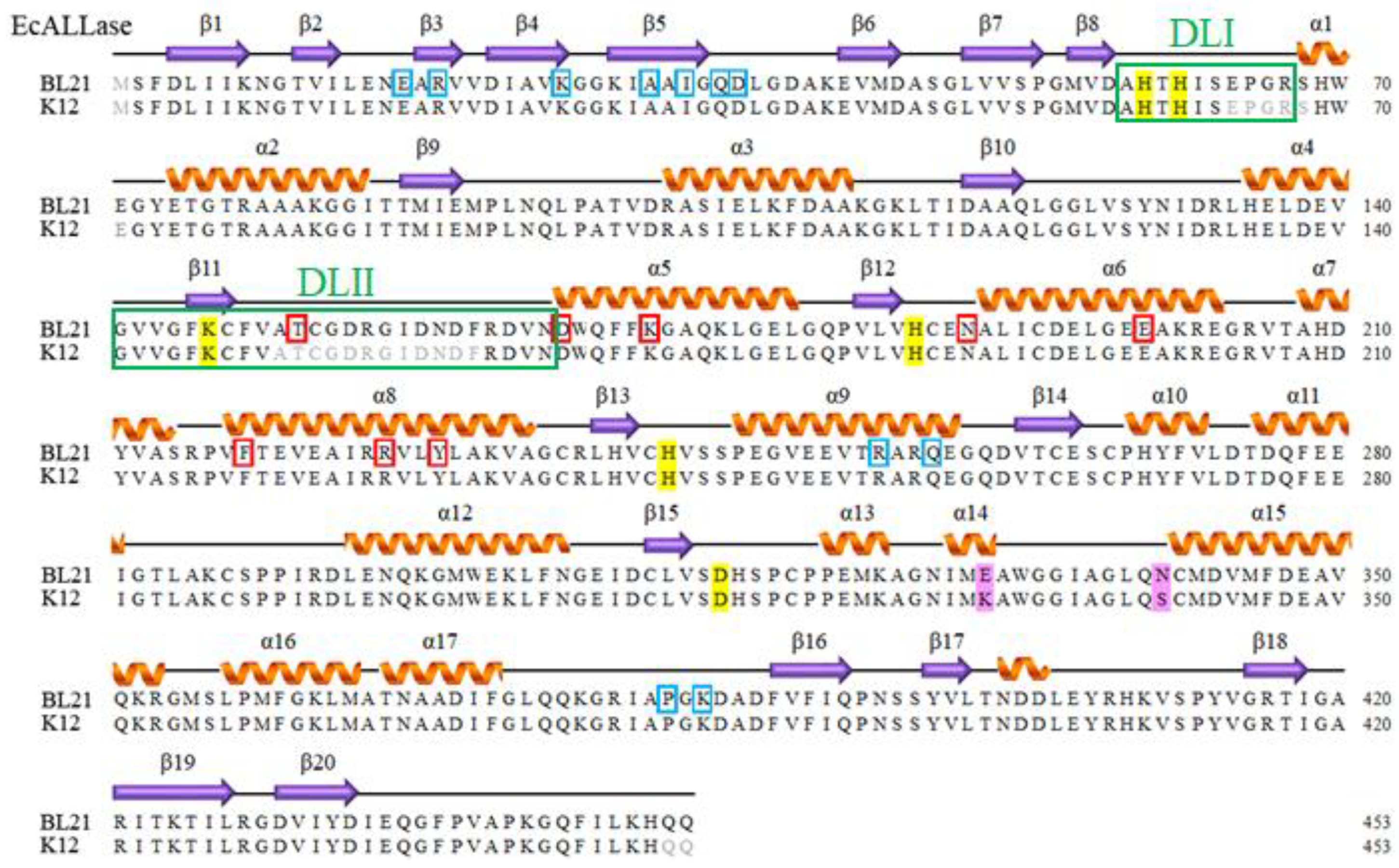
It is well established that the two dynamic loops in the active site of DHPase [15], HYDase [33], and DHOase [42] are crucial for substrate entrance and product release. HYDase is an important enzyme used for hydrolysis of chemically synthesized cyclic hydantoins for industrial production of antibiotics such as semisynthetic penicillin and cephalosporins [43,44]. The two active site loops important for substrate recognition, the segments of amino acid residues (aa) D62–D72 and M153–D162 in Bacillus stearothermophilus HYDase (BsHYDase), correspond to segments aa A58–R67 and G141–N165 in EcALLase-K12 (boxed in green in Figure 2). However, these two dynamic loops were not observed in the ternary structure of EcALLase-K12. To better study the substrate binding, substrate specificity, and catalytic mechanism of ALLase, knowing the structure of these two important loops is highly desirable for further drug design and pharmaceutical applications of ALLase. In addition, these missed loops in the structure of EcALLase-K12 will significantly affect the docking results and protein engineering strategy.
E. coli K-12 and BL21 have been the subjects of classical experiments from which much of our understanding of molecular genetics has emerged [45]. In this study, we solved the crystal structure of E. coli BL21 ALLase (EcALLase-BL21) at a resolution of 2.07 Å (PDB ID 8HFD) to obtain more information for structural analyses. The two missed active site loops in EcALLase-K12 were clearly determined in our structure of EcALLase-BL21. This EcALLase-BL21 structure gives full structural information and therefore might be more suitable for further docking and protein engineering experiments.

2. Results

2.1. Sequence Analysis of EcALLase-BL21

E. coli BL21(DE3) is widely used to express recombinant proteins. This study aimed to investigate the structure of EcALLase-BL21 for further pharmaceutical applications such as drug design and biotechnology development. The gene ECD_00462, encoding EcALLase-BL21, was initially found in a database of the National Center for Biotechnology Information (NCBI) [46]. Based on the known nucleotide sequence, the predicted EcALLase-BL21 monomer has a length of 453 amino acid residues and a molecular mass of 49.6 kDa. EcALLase-BL21 has a calculated pI value of 5.12 via the amino acid sequence. Six essential residues, H59, H61, Kcx146, H186, H242, and D315, participate in the assembly of a binuclear metal center within the active site of EcALLase-K12 [41], and are also found in EcALLase-BL21 (Figure 2). In comparison, only two residues of EcALLase-K12, i.e., K330 and S340, are different from and replaced with E330 and N34 in EcALLase-BL21. Although the crystal structure of EcALLase-K12 was solved, many regions, especially several active site residues, were not determined. Due to its sequence similarity, EcALLase-BL21 was selected to obtain the whole structural information of ALLase.

2.2. Crystallization and Data Collection of EcALLase-BL21

Prior to this study, the crystal structure of the EcALLase from subspecies K-12 was available for computational use (PDB ID 3E74) [41]. However, three segments, i.e., aa 64–68, 150–161, and 452–453, in the ternary structure of EcALLase-K12 were undetermined [41]. The region aa 150–161 may contain several important residues for substrate binding (Figure 2). Therefore, this situation certainly limited binding model prediction of the position the inhibitor of ALLase occupies as the molecular docking is performed. Given that this active site loop is unobserved in the structure of EcALLase-K12, we attempted to determine the structure of another EcALLase, namely EcALLase-BL21, for inhibitor docking experiments. Crystals of EcALLase-BL21 were grown at room temperature by hanging drop vapor diffusion in 20% PEG8000 and 100 mM CHES at pH 9.5. These crystals reached full size in 7–13 days. Data were collected using an EIGER2 X 16M Detector at an SPXF beamline TPS 07A of the National Synchrotron Radiation Research Center (NSRRC). The data completeness was over 99% (Table 1).

2.3. Overall Structure of EcALLase-BL21

We crystallized EcALLase-BL21 and determined its structure at a resolution of 2.07 Å (Table 1). The crystal of EcALLase-BL21 belonged to space group C121, with cell dimensions of a = 203.16, b = 77.14, and c = 144.85 Å. Four monomers of EcALLase-BL21 per asymmetric unit were present (Figure 3A). Most of the electron density for EcALLase-BL21 was of good quality and no discontinuity was observed. The global architecture of EcALLase-BL21 revealed a TIM-barrel structure that consisted of 17 α-helices, 20 β-sheets, and 2 Zn ions. The electrostatic potential surface of EcALLase-BL21 (Figure 3B) shows that the subunit–subunit interactions are stabilized by the charge–charge interactions at each subunit interface. Tetramerization of EcALLase-BL21 monomers generated a central cavity, but it is unclear what function, if any, it was created for (Figure 3B). The active site of EcALLase-BL21 containing H59, H61, Kcx146, H186, H242, and D315, which were required for the metal binding, was similar to that of EcALLase-K12 and other members of the cyclic amidohydrolase family (Figure 3C), such as Pseudomonas aeruginosa DHPase (PaDHPase) and the dihydroorotase domain of human CAD protein (huDHOase). Accordingly, these cyclic amidohydrolases might use a similar mechanism for catalysis (Figure 3D) [15].

2.4. Dynamic Loops of EcALLase-BL21

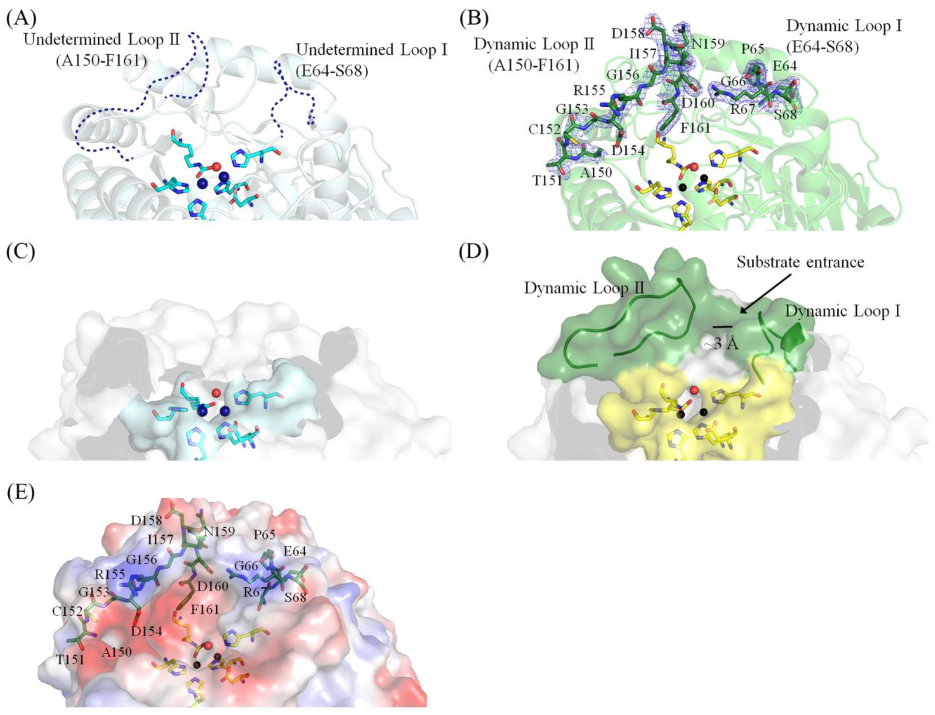
Based on their similar binuclear metal active site architecture and post-carbamylation, ALLase, DHOase, DHPase, and HYDase share several enzymatic properties and may use a similar mechanism for catalysis [47,48]. HYDase is an industrial enzyme [44,49], which has been highlighted for its capacity for biotechnological and pharmaceutical applications in the production of unnatural amino acids. It is well established that DHOase [39,42,50], DHPase [30,31,51], and HYDase [33,43,52] have flexible dynamic loops directly involved in substrate binding, product release, and stabilization of the transition state. This type of dynamic loop has not yet been mentioned and identified in ALLase. Correspondingly, the two dynamic loops in EcALLase-K12 should be A58–R67 (loop I) and G141–N165 (loop II). When we used the structure of EcALLase-K12 for inhibitor docking and sketching the chemical mechanism, we found that it was difficult to draw a correct conclusion, probably due to the lack of structure for these two active site loops (Figure 4A). Therefore, we solved the crystal structure of EcALLase-BL21 to better understand the structure–function relationship of ALLase. This structural information of EcALLase may also be useful for production of other unnatural amino acids when manipulating and engineering these two active site loops to modulate and change the substrate specificity for obtaining a new product [52]. As compared to the crystal structure of EcALLase-K12, these active loops in our structure, as solved in this study, were fully determined (Figure 4B). Accordingly, the position information of these active site residues can be used for further docking and protein engineering experiments.
These two dynamic loops that remain unsolved in the structure of EcALLase-K12 (Figure 4C) but appear in the current EcALLase-BL21 structure (Figure 4D,E) may be used for determining the catalytic state of this enzyme. It is known that the cross-section bottleneck size of the tunnel in DHPases can determine the open (for substrate binding) or closed (product release) form of the enzyme. For example, the tunnel bottlenecks created by these two dynamic loops in the open and closed forms of the DHPase are approximately 10 and 3 Å, respectively [31]. Based on our structure, the bottleneck size of the active site pocket of EcALLase-BL21 was approximately 3 Å (Figure 4D). Given that the tunnel bottleneck (~3 Å) is narrower than the size of substrate allantoin (6–9 Å), this structure may be the closed form of EcALLase-BL21. In addition, the basically charged residues on these loops may mediate the substrate entrance (Figure 4E). However, this speculation must be further elucidated biochemically and structurally.

2.5. Molecular Docking

ALLase-catalyzed reaction is a key process in the biosynthesis of ureide [53], which is required for the utilization of nitrogen in purine-derived compounds, and, thus, ALLase may be a cytotoxic target. Recently, we have identified that the leaf extract of N. miranda possesses anti-ALLase activity [38]. Through GC–MS, the 16 most abundant ingredients in this extract were identified. The top six compounds [38] were plumbagin, lupenone, palmitic acid, stigmast-5-en-3-ol, neophytadiene, and citraconic anhydride. Given that a certain abundant compound in N. miranda may be responsible for inhibiting the activity of ALLase, their binding modes should be elucidated (Figure 5A). Along with the binding poses of plumbagin (Figure 5B), lupenone (Figure 5C), palmitic acid (Figure 5D), stigmast-5-en-3-ol (Figure 5E), neophytadiene (Figure 5F), and citraconic anhydride (Figure 5G) to EcALLase-BL21, the binding capacities of EcALLase-BL21 were also calculated via the MOE (molecular operating environment)-Dock tool [54]. Through MOE-Dock, receptor–ligand binding affinities with all possible binding geometries could be predicted on the basis of the docking score (the S score). These docking results using the structure of EcALLase-BL21, however, significantly differed from those obtained when the structure of EcALLase-K12 was used [38]. 8-Hydroxyquinoline-5-sulfonic acid (8-HQSA), an inhibitor of EcALLase [46], was also docked to EcALLase-BL21 (Figure 5H and Table 2). The docking result showed that, except for citraconic anhydride (Figure 5A), all other compounds could be docked into the active site pocket of EcALLase-BL21. In comparison with previous work, in that study only plumbagin could be docked into the active site of EcALLase-K12 [38]. These obvious differences may have resulted from the incomplete active site loops in the structure of EcALLase-K12 [41]. Based on the S scores, the binding capacity of these compounds was in the following order: stigmast-5-en-3-ol > lupenone > 8-HQSA > palmitic acid > plumbagin > neophytadiene > citraconic anhydride (Table 2). Accordingly, stigmast-5-en-3-ol, possessing the highest S score, might exhibit the greatest binding affinity to ALLase-BL21 among these selected compounds. To further elucidate how ALLase-BL21 can be suppressed by these possible inhibitors, the complexed crystal structure of ALLase-BL21 is highly desired.

2.6. Analysis of Substrate Binding Pockets of HYDase and ALLase

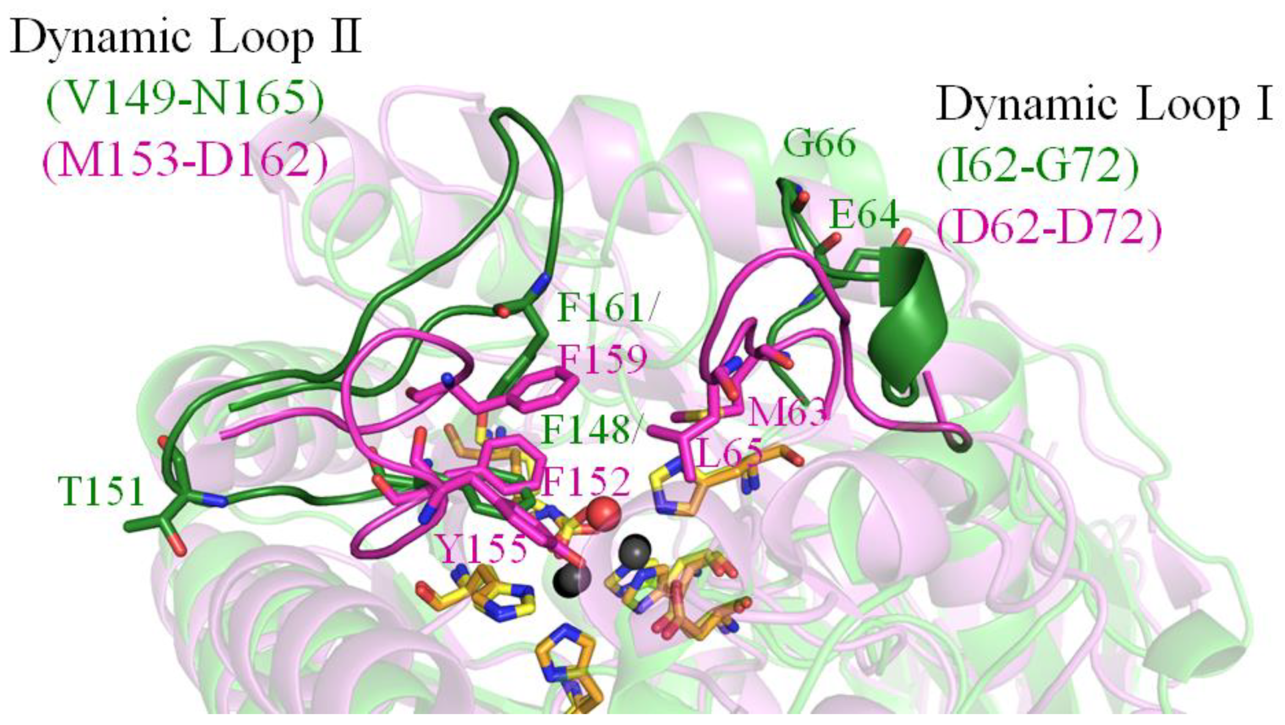
HYDase catalyzing the cleavage of the cyclic amide bond of 5′-monosubstituted hydantoins is widely employed for the commercial production of optically pure D- and L-amino acids for pharmaceutical applications [44]. Although HYDase and ALLase are similar enzymes (Supplementary Figure S1) and may use a similar mechanism for catalysis, their substrate specificities do not overlap [14]. Like the EcALLase-BL21 examined in this study, HYDase also has two dynamic loops as crucial determinants of the substrate specificities [52]. Structural analysis indicated that the active site loop residues M63, L65, F152, Y155, and F159 in BsHYDase (PDB ID 1K1D) constitute a hydrophobic lid to interact with the exocyclic substituent of the substrate hydroxyphenylhydantoin. The corresponding residues in EcALLase-BL21 are E64, G66, F148, T151, and F161 (Figure 6). Given that the substrate specificity of HYDase can be rationally modulated by manipulating those residues on these two dynamic loops [52], changing the corresponding residues in EcALLase-BL21 may create a new ALLase for producing a series of unnatural amino acids for pharmaceutical applications. Further studies can directly focus on this issue for ALLase applications.

3. Discussion

The goal of the present work was to acquire a crystal structure of EcALLase for further drug design, docking and binding analyses, and substrate specificity studies. The EcALLase-K12 structure had been solved, but three segments, including the two active site loops, namely aa 64–68 and 150–161, in the ternary structure, were undetermined [41]. This situation therefore significantly affected the docking results and protein engineering strategy (Figure 5). This is why we tried to obtain a crystal of EcALLase-BL21 and determine its crystal structure (Figure 3). In this study, these two active site loops of EcALLase-BL21 were fully solved structurally (Figure 4). Thus, this current EcALLase structure may be more suitable for use in further docking and product design experiments (Figure 6).
Multidrug-resistant pathogenic bacteria are spreading rapidly worldwide and could become untreatable [55]. Given that the catabolism of allantoin (purine) is a nitrogen source in E. coli [56] and other microorganisms [57], and the utilization of allantoin as a nitrogen source is recognized as very important for bacterial virulence [58], blocking the activity of ALLase would be detrimental to bacterial survival. In addition, ALLase is not found in humans. Thus, ALLase may be a promising therapeutic target for developing antibiotics. Although 8-HQSA is obviously capable of inhibiting the activity of EcALLase-BL21 [46], this chelator may be harmful to human health. Recently, we found that extracts of N. miranda exhibit antibacterial and anti-ALLase activities [38]. More plant-derived products acting as active antibacterial and anti-ALLase agents for human health care should be worth further determination.
Optically pure D-p-hydroxylphenylglycine and its derivatives as side-chain precursors are required for industrial production of semisynthetic penicillin and cephalosporins [43,44]. To obtain these precursors, HYDase is used for hydrolysis of chemically synthesized cyclic hydantoins [44]. In this study, we determined the crystal structure of EcALLase-BL21 at a resolution of 2.07 Å (Table 1). Despite an undetectable similarity in the amino acid sequence, EcALLase-BL21 and HYDase share a structural resemblance. Although the catalytic chemistry is conserved [15], their substrate specificity does not overlap [14]. Since allantoin is a kind of hydantoin, we thought that ALLase might also be a useful enzyme for production of optically pure D-amino acids for medical applications. The structure of the two active site loops solved in this study may provide molecular insight into how allantoin, as well as other hydantoin with novel exocyclic substituents, can be recognized and hydrolyzed by EcALLase-BL21 and its engineered enzymes (Figure 4).
On the two dynamic loops of BsHYDase, the residues M63, L65, F152, Y155, and F159 interact with the exocyclic substituent of hydantoin [33]. Protein engineering of these residues had a significant effect on the substrate specificity of HYDase [52]. For example, the substrate specificity of the mutant F159A toward aromatic substrate hydroxyphenylhydantoin was enhanced by approximately 200-fold compared to that of the wild-type HYDase. In addition, saturation mutagenesis at this position (F159) revealed that the catalytic constant of the HYDase for aromatic substrates increased gradually as the size of the amino acid side chain decreased [52]. Correspondingly, these residues in EcALLase-BL21 are E64 (loop I), G66 (loop I), F148 (loop II), T151 (loop II), and F161 (loop II). Accordingly, these amino acid residues on the dynamic loops may be also critical and can be modulated by rational design for the substrate specificity of EcALLase-BL21. However, this speculation must be investigated and confirmed experimentally and structurally.
Previously, we solved complexed crystal structures of DHPase and DHOase with the anticancer drug 5-fluorouracil (5-FU) [20,24,26]. 5-FU is an FDA-approved drug with a remarkable therapeutic effect for the systemic treatment of cancers of the gastrointestinal tract, breast, head, and neck in the clinic [59]. 5-FU can bind to the active site of DHPase and DHOase. Based on the active sites’ similarity among ALLase, DHPase, and DHOase, we may therefore tentatively speculate about the binding capacity of ALLase to 5-FU. Currently, our laboratory is attempting to obtain crystals of the ALLase-5-FU complex for this investigation.
The crystal structures of HYDase [33], DHPase [24,27,28,29,31], DHOase [50,60,61], and ALLase (this study) revealed that the two active site dynamic loops are crucial for catalysis. The movement of the dynamic loop (corresponding to loop II of EcALLase-BL21) in DHOase was proposed as a part of the catalytic cycle [61]. Crystal structures of ligand-bound E. coli DHOase (EcDHOase) show that the flexible loop extends toward the active site when the substrate is bound (loop-in mode) or moves away from the active site, facilitating the product release (loop-out mode). EcDHOase also binds non-substrate ligands such as the product-like inhibitor 5-fluoroorotate through loop-out mode [61]. However, several recent structures of ScDHOase in complexes with different non-substrate ligands all indicated the loop-in mode, suggesting that this previous conclusion reached with EcDHOase may be species-dependent [18,19,20,39]. Regardless of their different binding modes, this flexible loop in DHPases and DHOases, perhaps including ALLase now, was crucial for the catalysis [15]. Thus, this dynamic loop in these amidohydrolases should be suitable drug targets for inhibiting the pyrimidine and purine metabolism selectively [40]. Accordingly, we propose that a compound that could partially occupy the active site and stably lock off the loop movement of EcALLase-BL21 would be a good candidate as an antibacterial drug lead for developing a potent inhibitor toward ALLase. The complexed structures of ALLase still need to be solved and obtained to better understand the substrate/inhibitor binding mechanism for further biotechnological uses.

4. Materials and Methods

4.1. Protein Expression and Purification

Based on the complete genome sequence of E. coli BL21 [45], the plasmid for EcALLase-BL21 (allB; ECD_00462) expression was constructed [46]. The expression vector pET21b-EcALLase-BL21 [46] was transformed into E. coli BL21 (DE3) cells and grown in LB medium at 37 °C. The overexpression was induced by incubating with 1 mM isopropyl thiogalactopyranoside at 25 °C for 9 h. Recombinant zinc-amended EcALLase-BL21 [3] was purified from the soluble supernatant by using Ni2+-affinity chromatography. The recombinant protein was eluted with a linear imidazole gradient and dialyzed against a dialysis buffer (20 mM Tris-HCl and 0.1 M NaCl, pH 7.9). Approximately 12 mg of purified EcALLase-BL21 was obtained from 1 L of an E. coli cell culture. The protein purity remained at >97% as determined using SDS–PAGE.

4.2. Crystallization Experiments

Before crystallization, the purified EcALLase-BL21 was concentrated to 30 mg/mL. Crystals of EcALLase-BL21 were grown at room temperature by hanging drop vapor diffusion in 20% PEG8000 and 100 mM CHES at pH 9.5. These crystals reached full size in 7–13 days. The crystals were transferred from a crystallization drop into the cryoprotectant solution (2 μL) with precipitant solution containing glycerol (25–30%) for a few seconds, mounted on a synthetic nylon loop (0.1–0.2 mm), flash cooled in liquid N2, and validated in an SPXF beamline TPS 07A of the NSRRC (Hinchu, Taiwan).

4.3. X-Ray Diffraction Data and Structure Determination

Data were collected using an EIGER2 X 16M Detector at an SPXF beamline TPS 07A at the NSRRC. Data sets were indexed, integrated, and scaled by HKL-2000 [62] and XDS [63]. The initial phase, density modification, and model building were performed using the AutoSol program [64] in the PHENIX. The iterative model building and structure refinement were performed using Refmac in the CCP4 software suite [65] and phenix.refine in the PHENIX software suite [66]. The initial phase of EcALLase-BL21 was determined through the molecular replacement software Phaser MR [67] by using EcALLase-K12 [41] as a search model. The correctness of the stereochemistry of the models was verified using MolProbity [68]. Atomic coordinates and related structure factors were deposited in the PDB with accession code 8HFD.

4.4. Molecular Docking

Using MOE-Dock [54], 8-HQSA, plumbagin, lupenone, palmitic acid, stig-mast-5-en-3-ol, neophytadiene, and citraconic anhydride were docked to EcALLase-BL21 (PDB ID 8HFD) for their binding capacity. Their S scores and binding modes were compared. The top-ranked confirmations were analyzed.

5. Conclusions

Here, we solved the crystal structure of EcALLase-BL21 at a resolution of 2.07 Å (PDB ID 8HFD) to obtain more information for structural analyses. As compared with the previous work, the two missed active site loops in EcALLase-K12 were clearly determined in our structure of EcALLase-BL21. Thus, this current EcALLase structure may be more suitable for use in further docking and product design experiments. In addition, EcALLase-BL21 shared active site similarity with HYDase, an important biocatalyst for industrial production of semisynthetic penicillin and cephalosporins. Based on this structural comparison, the crucial amino acid residues on the dynamic loops were suggested and might be modulated by rational design for the substrate specificity of EcALLase-BL21 for further biotechnological and pharmaceutical applications. The structure of these two dynamic loops solved in this study will significantly affect the docking results, protein engineering strategy, and further understanding of the substrate/inhibitor binding mechanism of ALLase.

Supplementary Materials

The following supporting information can be downloaded at: https://www.mdpi.com/article/10.3390/molecules28020827/s1. Figure S1: Structural comparison of EcALLase-BL21 and BsHYDase.

Author Contributions

Y.-H.H., P.-C.Y., E.-S.L., Y.-Y.H., H.-P.L., and W.-F.P. performed the experiments; Y.-H.H., P.-C.Y., E.-S.L., C.-C.H., and C.-Y.H. analyzed the data; Y.-H.H. and C.-Y.H. contributed to the study design and manuscript writing. All authors reviewed the results, contributed to the data interpretation, and approved the final version of the manuscript. All authors have read and agreed to the published version of the manuscript.

Funding

This research received no external funding.

Institutional Review Board Statement

Not applicable.

Informed Consent Statement

Not applicable.

Data Availability Statement

Atomic coordinates and related structure factors were deposited in the PDB with accession code 8HFD.

Acknowledgments

We thank the experimental facility and the technical services provided by the Synchrotron Radiation Protein Crystallography Facility of the National Core Facility Program for Biotechnology, National Science and Technology Council, Taiwan.

Conflicts of Interest

The authors declare no conflict of interest.

References

  1. Werner, A.K.; Witte, C.P. The biochemistry of nitrogen mobilization: Purine ring catabolism. Trends Plant Sci. 2011, 16, 381–387. [Google Scholar] [CrossRef]
  2. Hayashi, S.; Fujiwara, S.; Noguchi, T. Evolution of urate-degrading enzymes in animal peroxisomes. Cell Biochem. Biophys. 2000, 32, 123–129. [Google Scholar] [CrossRef] [PubMed]
  3. Ho, Y.Y.; Huang, Y.H.; Huang, C.Y. Chemical rescue of the post-translationally carboxylated lysine mutant of allantoinase and dihydroorotase by metal ions and short-chain carboxylic acids. Amino Acids 2013, 44, 1181–1191. [Google Scholar] [CrossRef] [PubMed]
  4. Mulrooney, S.B.; Hausinger, R.P. Metal ion dependence of recombinant Escherichia coli allantoinase. J. Bacteriol. 2003, 185, 126–134. [Google Scholar] [CrossRef] [Green Version]
  5. Ramazzina, I.; Cendron, L.; Folli, C.; Berni, R.; Monteverdi, D.; Zanotti, G.; Percudani, R. Logical identification of an allantoinase analog (puuE) recruited from polysaccharide deacetylases. J. Biol. Chem. 2008, 283, 23295–23304. [Google Scholar] [CrossRef] [PubMed] [Green Version]
  6. Wang, N.Q.; Kong, C.H.; Wang, P.; Meiners, S.J. Root exudate signals in plant-plant interactions. Plant Cell Environ. 2021, 44, 1044–1058. [Google Scholar] [CrossRef]
  7. Lu, S.; Jia, Z.; Meng, X.; Chen, Y.; Wang, S.; Fu, C.; Yang, L.; Zhou, R.; Wang, B.; Cao, Y. Combined Metabolomic and Transcriptomic Analysis Reveals Allantoin Enhances Drought Tolerance in Rice. Int. J. Mol. Sci. 2022, 23, 14172. [Google Scholar] [CrossRef]
  8. Wang, J.; Li, Y.; Wang, Y.; Du, F.; Zhang, Y.; Yin, M.; Zhao, X.; Xu, J.; Yang, Y.; Wang, W.; et al. Transcriptome and Metabolome Analyses Reveal Complex Molecular Mechanisms Involved in the Salt Tolerance of Rice Induced by Exogenous Allantoin. Antioxidants 2022, 11, 2045. [Google Scholar] [CrossRef]
  9. Voß, L.; Heinemann, K.J.; Herde, M.; Medina-Escobar, N.; Witte, C.P. Enzymes and cellular interplay required for flux of fixed nitrogen to ureides in bean nodules. Nat. Commun. 2022, 13, 5331. [Google Scholar] [CrossRef]
  10. Zhang, P.; Wang, Y.; Zhang, J.; Hong, T. Allantoin Inhibits Compound 48/80-Induced Pseudoallergic Reactions In Vitro and In Vivo. Molecules 2022, 27, 3473. [Google Scholar] [CrossRef]
  11. Seo, C.S.; Lee, M.Y. Simultaneous Determination of Fourteen Marker Compounds in the Traditional Herbal Prescription, Geumgwesingihwan, Using Ultra-Performance Liquid Chromatography-Tandem Mass Spectrometry. Molecules 2022, 27, 3890. [Google Scholar] [CrossRef]
  12. Aguilar-Guadarrama, A.B.; Yáñez-Ibarra, G.; Cancino-Marentes, M.E.; González-Ibarra, P.; Ortiz-Andrade, R.; Sánchez-Recillas, A.; Rodríguez-Carpena, J.G.; Aguirre-Vidal, Y.; Medina-Diaz, I.M.; Ávila-Villarreal, G. Chromatographic Techniques and Pharmacological Analysis as a Quality Control Strategy for Serjania triquetra a Traditional Medicinal Plant. Pharmaceuticals 2022, 15, 1289. [Google Scholar] [CrossRef]
  13. Thornfeldt, C. Cosmeceuticals containing herbs: Fact, fiction, and future. Dermatol. Surg. 2005, 31, 873–880. [Google Scholar] [CrossRef]
  14. Peng, W.F.; Huang, C.Y. Allantoinase and dihydroorotase binding and inhibition by flavonols and the substrates of cyclic amidohydrolases. Biochimie 2014, 101, 113–122. [Google Scholar] [CrossRef] [PubMed]
  15. Huang, C.Y. Structure, catalytic mechanism, posttranslational lysine carbamylation, and inhibition of dihydropyrimidinases. Adv. Protein Chem. Struct. Biol. 2020, 122, 63–96. [Google Scholar]
  16. Gerlt, J.A.; Babbitt, P.C. Divergent evolution of enzymatic function: Mechanistically diverse superfamilies and functionally distinct suprafamilies. Annu. Rev. Biochem. 2001, 70, 209–246. [Google Scholar] [CrossRef]
  17. Rice, A.J.; Pesavento, R.P.; Ren, J.; Youn, I.; Kwon, Y.; Ellepola, K.; Che, C.T.; Johnson, M.E.; Lee, H. Identification of Small Molecule Inhibitors against Staphylococcus aureus Dihydroorotase via HTS. Int. J. Mol. Sci. 2021, 22, 9984. [Google Scholar] [CrossRef]
  18. Guan, H.H.; Huang, Y.H.; Lin, E.S.; Chen, C.J.; Huang, C.Y. Structural Analysis of Saccharomyces cerevisiae Dihydroorotase Reveals Molecular Insights into the Tetramerization Mechanism. Molecules 2021, 26, 7249. [Google Scholar] [CrossRef]
  19. Guan, H.H.; Huang, Y.H.; Lin, E.S.; Chen, C.J.; Huang, C.Y. Complexed Crystal Structure of Saccharomyces cerevisiae Dihydroorotase with Inhibitor 5-Fluoroorotate Reveals a New Binding Mode. Bioinorg. Chem. Appl. 2021, 2021, 2572844. [Google Scholar] [CrossRef]
  20. Guan, H.H.; Huang, Y.H.; Lin, E.S.; Chen, C.J.; Huang, C.Y. Structural basis for the interaction modes of dihydroorotase with the anticancer drugs 5-fluorouracil and 5-aminouracil. Biochem. Biophys. Res. Commun. 2021, 551, 33–37. [Google Scholar] [CrossRef]
  21. Lipowska, J.; Miks, C.D.; Kwon, K.; Shuvalova, L.; Zheng, H.; Lewinski, K.; Cooper, D.R.; Shabalin, I.G.; Minor, W. Pyrimidine biosynthesis in pathogens—Structures and analysis of dihydroorotases from Yersinia pestis and Vibrio cholerae. Int. J. Biol. Macromol. 2019, 136, 1176–1187. [Google Scholar] [CrossRef]
  22. Del Cano-Ochoa, F.; Moreno-Morcillo, M.; Ramon-Maiques, S. CAD, A Multienzymatic Protein at the Head of de Novo Pyrimidine Biosynthesis. Subcell Biochem. 2019, 93, 505–538. [Google Scholar]
  23. Rice, A.J.; Lei, H.; Santarsiero, B.D.; Lee, H.; Johnson, M.E. Ca-asp bound X-ray structure and inhibition of Bacillus anthracis dihydroorotase (DHOase). Bioorg. Med. Chem. 2016, 24, 4536–4543. [Google Scholar] [CrossRef] [PubMed] [Green Version]
  24. Lin, E.S.; Luo, R.H.; Yang, Y.C.; Huang, C.Y. Molecular Insights into How the Dimetal Center in Dihydropyrimidinase Can Bind the Thymine Antagonist 5-Aminouracil: A Different Binding Mode from the Anticancer Drug 5-Fluorouracil. Bioinorg. Chem. Appl. 2022, 2022, 1817745. [Google Scholar] [CrossRef] [PubMed]
  25. Basbous, J.; Aze, A.; Chaloin, L.; Lebdy, R.; Hodroj, D.; Ribeyre, C.; Larroque, M.; Shepard, C.; Kim, B.; Pruvost, A.; et al. Dihydropyrimidinase protects from DNA replication stress caused by cytotoxic metabolites. Nucleic Acids Res. 2020, 48, 1886–1904. [Google Scholar] [CrossRef] [Green Version]
  26. Huang, Y.H.; Ning, Z.J.; Huang, C.Y. Crystal structure of dihydropyrimidinase in complex with anticancer drug 5-fluorouracil. Biochem. Biophys. Res. Commun. 2019, 519, 160–165. [Google Scholar] [CrossRef]
  27. Cheng, J.H.; Huang, Y.H.; Lin, J.J.; Huang, C.Y. Crystal structures of monometallic dihydropyrimidinase and the human dihydroorotase domain K1556A mutant reveal no lysine carbamylation within the active site. Biochem. Biophys. Res. Commun. 2018, 505, 439–444. [Google Scholar] [CrossRef] [PubMed]
  28. Cheng, J.H.; Huang, C.C.; Huang, Y.H.; Huang, C.Y. Structural Basis for pH-Dependent Oligomerization of Dihydropyrimidinase from Pseudomonas aeruginosa PAO1. Bioinorg. Chem. Appl. 2018, 2018, 9564391. [Google Scholar] [CrossRef] [PubMed] [Green Version]
  29. Tzeng, C.T.; Huang, Y.H.; Huang, C.Y. Crystal structure of dihydropyrimidinase from Pseudomonas aeruginosa PAO1: Insights into the molecular basis of formation of a dimer. Biochem. Biophys. Res. Commun. 2016, 478, 1449–1455. [Google Scholar] [CrossRef]
  30. Huang, C.Y. Inhibition of a putative dihydropyrimidinase from Pseudomonas aeruginosa PAO1 by flavonoids and substrates of cyclic amidohydrolases. PLoS ONE 2015, 10, e0127634. [Google Scholar] [CrossRef] [Green Version]
  31. Hsieh, Y.C.; Chen, M.C.; Hsu, C.C.; Chan, S.I.; Yang, Y.S.; Chen, C.J. Crystal structures of vertebrate dihydropyrimidinase and complexes from Tetraodon nigroviridis with lysine carbamylation: Metal and structural requirements for post-translational modification and function. J. Biol. Chem. 2013, 288, 30645–30658. [Google Scholar] [CrossRef] [PubMed] [Green Version]
  32. Huang, C.Y.; Hsu, C.C.; Chen, M.C.; Yang, Y.S. Effect of metal binding and posttranslational lysine carboxylation on the activity of recombinant hydantoinase. J. Biol. Inorg. Chem. 2009, 14, 111–121. [Google Scholar] [CrossRef] [PubMed]
  33. Cheon, Y.H.; Kim, H.S.; Han, K.H.; Abendroth, J.; Niefind, K.; Schomburg, D.; Wang, J.; Kim, Y. Crystal structure of D-hydantoinase from Bacillus stearothermophilus: Insight into the stereochemistry of enantioselectivity. Biochemistry 2002, 41, 9410–9417. [Google Scholar] [CrossRef]
  34. Soong, C.L.; Ogawa, J.; Honda, M.; Shimizu, S. Cyclic-imide-hydrolyzing activity of D-hydantoinase from Blastobacter sp. strain A17p-4. Appl. Environ. Microbiol. 1999, 65, 1459–1462. [Google Scholar] [CrossRef] [PubMed] [Green Version]
  35. Huang, C.Y.; Yang, Y.S. A novel cold-adapted imidase from fish Oreochromis niloticus that catalyzes hydrolysis of maleimide. Biochem. Biophys. Res. Commun. 2003, 312, 467–472. [Google Scholar] [CrossRef]
  36. Huang, C.Y.; Yang, Y.S. The role of metal on imide hydrolysis: Metal content and pH profiles of metal ion-replaced mammalian imidase. Biochem. Biophys. Res. Commun. 2002, 297, 1027–1032. [Google Scholar] [CrossRef]
  37. Ogawa, J.; Soong, C.L.; Honda, M.; Shimizu, S. Imidase, a dihydropyrimidinase-like enzyme involved in the metabolism of cyclic imides. Eur. J. Biochem 1997, 243, 322–327. [Google Scholar] [CrossRef]
  38. Lin, E.S.; Huang, C.Y. Cytotoxic Activities and the Allantoinase Inhibitory Effect of the Leaf Extract of the Carnivorous Pitcher Plant Nepenthes miranda. Plants 2022, 11, 2265. [Google Scholar] [CrossRef]
  39. Guan, H.H.; Huang, Y.H.; Lin, E.S.; Chen, C.J.; Huang, C.Y. Plumbagin, a Natural Product with Potent Anticancer Activities, Binds to and Inhibits Dihydroorotase, a Key Enzyme in Pyrimidine Biosynthesis. Int. J. Mol. Sci. 2021, 22, 6861. [Google Scholar] [CrossRef]
  40. Huang, Y.H.; Lien, Y.; Chen, J.H.; Lin, E.S.; Huang, C.Y. Identification and characterization of dihydropyrimidinase inhibited by plumbagin isolated from Nepenthes miranda extract. Biochimie 2020, 171–172, 124–135. [Google Scholar] [CrossRef]
  41. Kim, K.; Kim, M.I.; Chung, J.; Ahn, J.H.; Rhee, S. Crystal structure of metal-dependent allantoinase from Escherichia coli. J. Mol. Biol. 2009, 387, 1067–1074. [Google Scholar] [CrossRef] [PubMed]
  42. Del Cano-Ochoa, F.; Grande-Garcia, A.; Reverte-Lopez, M.; D’Abramo, M.; Ramon-Maiques, S. Characterization of the catalytic flexible loop in the dihydroorotase domain of the human multi-enzymatic protein CAD. J. Biol. Chem. 2018, 293, 18903–18913. [Google Scholar] [CrossRef] [PubMed]
  43. Nam, S.H.; Park, H.S.; Kim, H.S. Evolutionary relationship and application of a superfamily of cyclic amidohydrolase enzymes. Chem. Rec. 2005, 5, 298–307. [Google Scholar] [CrossRef] [PubMed]
  44. Schoemaker, H.E.; Mink, D.; Wubbolts, M.G. Dispelling the myths--biocatalysis in industrial synthesis. Science 2003, 299, 1694–1697. [Google Scholar] [CrossRef] [PubMed]
  45. Jeong, H.; Barbe, V.; Lee, C.H.; Vallenet, D.; Yu, D.S.; Choi, S.H.; Couloux, A.; Lee, S.W.; Yoon, S.H.; Cattolico, L.; et al. Genome sequences of Escherichia coli B strains REL606 and BL21(DE3). J. Mol. Biol. 2009, 394, 644–652. [Google Scholar] [CrossRef]
  46. Ho, Y.Y.; Hsieh, H.C.; Huang, C.Y. Biochemical characterization of allantoinase from Escherichia coli BL21. Protein J. 2011, 30, 384–394. [Google Scholar] [CrossRef]
  47. Gojkovic, Z.; Rislund, L.; Andersen, B.; Sandrini, M.P.; Cook, P.F.; Schnackerz, K.D.; Piskur, J. Dihydropyrimidine amidohydrolases and dihydroorotases share the same origin and several enzymatic properties. Nucleic Acids Res. 2003, 31, 1683–1692. [Google Scholar] [CrossRef] [Green Version]
  48. Kim, G.J.; Lee, D.E.; Kim, H.S. Functional expression and characterization of the two cyclic amidohydrolase enzymes, allantoinase and a novel phenylhydantoinase, from Escherichia coli. J. Bacteriol. 2000, 182, 7021–7028. [Google Scholar] [CrossRef] [Green Version]
  49. Clemente-Jimenez, J.M.; Martinez-Rodriguez, S.; Rodriguez-Vico, F.; Heras-Vazquez, F.J. Optically pure alpha-amino acids production by the “Hydantoinase Process”. Recent Pat. Biotechnol. 2008, 2, 35–46. [Google Scholar]
  50. Thoden, J.B.; Phillips, G.N., Jr.; Neal, T.M.; Raushel, F.M.; Holden, H.M. Molecular structure of dihydroorotase: A paradigm for catalysis through the use of a binuclear metal center. Biochemistry 2001, 40, 6989–6997. [Google Scholar] [CrossRef]
  51. Lohkamp, B.; Andersen, B.; Piskur, J.; Dobritzsch, D. The crystal structures of dihydropyrimidinases reaffirm the close relationship between cyclic amidohydrolases and explain their substrate specificity. J. Biol. Chem. 2006, 281, 13762–13776. [Google Scholar] [CrossRef] [PubMed] [Green Version]
  52. Cheon, Y.H.; Park, H.S.; Kim, J.H.; Kim, Y.; Kim, H.S. Manipulation of the active site loops of D-hydantoinase, a (beta/alpha)8-barrel protein, for modulation of the substrate specificity. Biochemistry 2004, 43, 7413–7420. [Google Scholar] [CrossRef] [PubMed]
  53. Nyhan, W.L. Disorders of purine and pyrimidine metabolism. Mol. Genet. Metab. 2005, 86, 25–33. [Google Scholar] [CrossRef] [PubMed]
  54. Scholz, C.; Knorr, S.; Hamacher, K.; Schmidt, B. DOCKTITE-a highly versatile step-by-step workflow for covalent docking and virtual screening in the molecular operating environment. J. Chem. Inf. Model. 2015, 55, 398–406. [Google Scholar] [CrossRef] [PubMed]
  55. Tommasi, R.; Brown, D.G.; Walkup, G.K.; Manchester, J.I.; Miller, A.A. ESKAPEing the labyrinth of antibacterial discovery. Nat. Rev. Drug Discov. 2015, 14, 529–542. [Google Scholar] [CrossRef]
  56. Vogels, G.D.; Van der Drift, C. Degradation of purines and pyrimidines by microorganisms. Bacteriol. Rev. 1976, 40, 403–468. [Google Scholar] [CrossRef]
  57. Marzluf, G.A. Genetic regulation of nitrogen metabolism in the fungi. Microbiol. Mol. Biol. Rev. 1997, 61, 17–32. [Google Scholar]
  58. Chou, H.C.; Lee, C.Z.; Ma, L.C.; Fang, C.T.; Chang, S.C.; Wang, J.T. Isolation of a chromosomal region of Klebsiella pneumoniae associated with allantoin metabolism and liver infection. Infect. Immun. 2004, 72, 3783–3792. [Google Scholar] [CrossRef] [Green Version]
  59. Longley, D.B.; Harkin, D.P.; Johnston, P.G. 5-fluorouracil: Mechanisms of action and clinical strategies. Nat. Rev. Cancer 2003, 3, 330–338. [Google Scholar] [CrossRef] [PubMed]
  60. Grande-Garcia, A.; Lallous, N.; Diaz-Tejada, C.; Ramon-Maiques, S. Structure, functional characterization, and evolution of the dihydroorotase domain of human CAD. Structure 2014, 22, 185–198. [Google Scholar] [CrossRef] [Green Version]
  61. Lee, M.; Chan, C.W.; Graham, S.C.; Christopherson, R.I.; Guss, J.M.; Maher, M.J. Structures of ligand-free and inhibitor complexes of dihydroorotase from Escherichia coli: Implications for loop movement in inhibitor design. J. Mol. Biol. 2007, 370, 812–825. [Google Scholar] [CrossRef] [PubMed]
  62. Otwinowski, Z.; Minor, W. Processing of X-ray Diffraction Data Collected in Oscillation Mode. Methods Enzymol. 1997, 276, 307–326. [Google Scholar] [PubMed]
  63. Kabsch, W. XDS. Acta Crystallogr. D Biol. Crystallogr. 2010, 66, 125–132. [Google Scholar] [CrossRef] [PubMed] [Green Version]
  64. Terwilliger, T.C.; Adams, P.D.; Read, R.J.; McCoy, A.J.; Moriarty, N.W.; Grosse-Kunstleve, R.W.; Afonine, P.V.; Zwart, P.H.; Hung, L.W. Decision-making in structure solution using Bayesian estimates of map quality: The PHENIX AutoSol wizard. Acta Crystallogr. D Biol. Crystallogr. 2009, 65, 582–601. [Google Scholar] [CrossRef]
  65. Lebedev, A.A.; Young, P.; Isupov, M.N.; Moroz, O.V.; Vagin, A.A.; Murshudov, G.N. JLigand: A graphical tool for the CCP4 template-restraint library. Acta Crystallogr. D Biol. Crystallogr. 2012, 68, 431–440. [Google Scholar] [CrossRef] [PubMed] [Green Version]
  66. Terwilliger, T.C.; Grosse-Kunstleve, R.W.; Afonine, P.V.; Moriarty, N.W.; Zwart, P.H.; Hung, L.W.; Read, R.J.; Adams, P.D. Iterative model building, structure refinement and density modification with the PHENIX AutoBuild wizard. Acta Crystallogr. D Biol. Crystallogr. 2008, 64, 61–69. [Google Scholar] [CrossRef] [Green Version]
  67. Winn, M.D.; Ballard, C.C.; Cowtan, K.D.; Dodson, E.J.; Emsley, P.; Evans, P.R.; Keegan, R.M.; Krissinel, E.B.; Leslie, A.G.; McCoy, A.; et al. Overview of the CCP4 suite and current developments. Acta Crystallogr. D Biol. Crystallogr. 2011, 67, 235–242. [Google Scholar] [CrossRef] [Green Version]
  68. Chen, V.B.; Arendall, W.B., 3rd; Headd, J.J.; Keedy, D.A.; Immormino, R.M.; Kapral, G.J.; Murray, L.W.; Richardson, J.S.; Richardson, D.C. MolProbity: All-atom structure validation for macromolecular crystallography. Acta Crystallogr. D Biol. Crystallogr. 2010, 66, 12–21. [Google Scholar] [CrossRef]
Figure 1. The pathways of purine nucleotide degradation. The chemical reaction catalyzed by allantoinase is highlighted. Allantoinase is a key enzyme for allantoin degradation. In the present study, we solved the crystal structure of E. coli BL21 allantoinase (EcALLase-BL21) at a resolution of 2.07 Å (PDB ID 8HFD) to obtain more information for structural analyses.
Molecules 28 00827 g001
Figure 2. Sequence alignment of EcALLase-BL21 and EcALLase-K12. The secondary structural elements of EcALLase-BL21 are shown with the sequence. The active site dynamic loop I (DLI; A58–R67) and dynamic loop II (DLII; G141–N165) are boxed in green. The unobserved residues 64–68, 150–161, and 452–453 in EcALLase-K12 ternary structure were colored in grey. In comparison, these residues in the structure of EcALLase-BL21 were clearly found. The metal-binding sites are shaded in yellow. The amino acids that are involved in monomer–monomer interface via hydrogen bonding are boxed in red. The amino acids that are involved in dimer–dimer interface via hydrogen bonding are boxed in blue. In comparison, only two residues of EcALLase-K12, i.e., K330 and S340, are different from and replaced with E330 and N34 in EcALLase-BL21 (shaded in pink).
Molecules 28 00827 g002
Figure 3. Structure of EcALLase-BL21. (A) Ribbon diagram of EcALLase-BL21 tetramer. Each ScDHOase monomer is color coded. Two zinc ions in the active site are presented as black spheres. (B) The electrostatic potential surface of EcALLase-BL21. Tetramerization of EcALLase-BL21 monomers generated a central cavity but it is unclear what function, if any, it was created for. (C) Superposition of the active site of members of the cyclic amidohydrolase family. Their active sites contain four His, one Asp, and one Kcx, which are required for di-metal binding and catalytic activity. EcALLase-BL21 (PDB ID 8HFD; green), PaDHPase (PDB ID 5E5C; light orange), and huDHOase (PDB ID 8GW0; slate) are shown. The architecture of these active sites is similar. (D) The proposed mechanism of EcALLase-BL21 for catalysis. The hydrolysis of allantoin likely undergoes three steps: the hydrolytic water molecule must be activated for nucleophilic attack, the amide bond of the substrate must be made more electrophilic by polarization of the carbonyl–oxygen bond, and then the leaving-group nitrogen must be protonated as the carbon–nitrogen bond is cleaved. The metal ions are shown as green circles.
Molecules 28 00827 g003
Figure 4. Dynamic loops and the tunnel bottleneck of EcALLase-BL21. (A) The active site of EcALLase-K12. The two active site loops crucial for substrate binding were not determined. These two dynamic loops in EcALLase-K12 are segments A58–R67 (loop I) and G141–N165 (loop II). When we used the structure of EcALLase-K12 for inhibitor docking and sketching the chemical mechanism, we found that it was difficult to draw a correct conclusion, probably due to the lack of structure for these two active site loops. (B) The active site of EcALLase-BL21. The electron density of the residues on the two dynamic loops in our structure of EcALLase-BL21 was well defined. (C) The tunnel bottleneck of EcALLase-K12. Because the structure of the two loops was not solved, the cross-section bottleneck size for substrate entrance could not be determined. (D) The tunnel bottleneck of EcALLase-BL21. The bottleneck size of the active site pocket of EcALLase-BL21 was approximately 3 Å. Given that the tunnel bottleneck (~3 Å) is narrower than the size of substrate allantoin (6–9 Å), this structure may be the closed form of EcALLase-BL21. (E) The electrostatic potential surface of the structure of EcALLase-BL21. Residues on the dynamic loops are indicated.
Molecules 28 00827 g004
Figure 5. Molecular docking. (A) Docking results. Plumbagin, lupenone, palmitic acid, stigmast-5-en-3-ol, neophytadiene, citraconic anhydride, and 8-HQSA were docked to EcALLase-BL21 (PDB ID 8HFD) via the MOE-Dock tool. The superimposed structures of these EcALLase-BL21 complexes based on docking experiments are shown. As indicated, only citraconic anhydride could not be docked into the active site of EcALLase-BL21. The binding mode of (B) plumbagin, (C) lupenone, (D) palmitic acid, (E) stigmast-5-en-3-ol, (F) neophytadiene, (G) citraconic anhydride, and (H) 8-HQSA to EcALLase-BL21 is shown.
Molecules 28 00827 g005
Figure 6. Analysis of substrate binding pockets of HYDase and ALLase. HYDase catalyzing the cleavage of the cyclic amide bond of 5′-monosubstituted hydantoins is widely employed for the commercial production of optically pure D- and L-amino acids for pharmaceutical applications. Like ALLase, HYDase also has two dynamic active site loops crucial for catalysis. The dynamic loop I and II (peach) are segments D62–D72 and M153–D162 in BsHYDase, respectively. Although HYDase and ALLase are similar enzymes and may use a similar mechanism for catalysis, their substrate specificities do not overlap. The active site loop residues M63, L65, F152, Y155, and F159 in BsHYDase constitute a hydrophobic lid to interact with the exocyclic substituent of the substrate hydroxyphenylhydantoin. The corresponding residues in EcALLase-BL21 are E64, G66, F148, T151, and F161. Given that the substrate specificity of HYDase can be rationally modulated by manipulating those residues on these two dynamic loops, changing the corresponding residues in EcALLase-BL21 may create a new ALLase for producing a series of unnatural amino acids for pharmaceutical applications.
Molecules 28 00827 g006
Table 1. Data collection and refinement statistics.
Data Collection
    CrystalEcALLase-BL21
    Wavelength (Å)1
    Resolution (Å)30–2.07
    Space groupC121
    Cell dimension (Å)a = 203.16   α = 90°
b = 77.14   β = 100.8°
c = 144.85   γ = 90°
Completeness (%)99.02 (97.21) *
<I/σI>7.39 (2.14)
CC1/20.992 (0.855)
Redundancy3.5 (3.2)
Refinement
    Resolution (Å)29.92–2.07
    No. reflections132,804
    Rwork/Rfree0.208/0.248
    No. atoms
      Protein14,759
      Ligands110
      Water926
R.m.s deviation
    Bond lengths (Å)0.009
    Bond angles (°)1.10
Ramachandran Plot
    In preferred regions96.89%
    In allowed regions2.66%
    Outliers0.45%
PDB entry8HFD
* Values in parentheses are for the highest resolution shell. CC1/2 is the percentage of correlation between intensities of random half-data sets.
Table 2. Results of the docking studies against EcALLase-BL21.
CompoundS ScoreResidueInteractionReceptor–Ligand Distance (Å)E (kcal/mol)
8-HQSA−5.6100Asn94H-donor2.82−7.4
Plumbagin−5.2950Asp315H-donor2.94−3.1
Asp315H-donor3.34−0.7
Lupenone−5.7230No important residue
Palmitic acid−5.4744Ser317H-donor3.13−0.7
Zn-βMetal1.98−2.8
Stigmast-5-en-3-ol−6.4260Asp315H-donor3.05−0.6
Zn-αMetal2.43−0.9
Trp332H-pi4.50−1.9
Neophytadiene−4.6771No important residue
Citraconic anhydride−1.3467Gln168pi-H 4.32−0.6
Disclaimer/Publisher’s Note: The statements, opinions and data contained in all publications are solely those of the individual author(s) and contributor(s) and not of MDPI and/or the editor(s). MDPI and/or the editor(s) disclaim responsibility for any injury to people or property resulting from any ideas, methods, instructions or products referred to in the content.