Review

Recent Advances in N-Heterocyclic Small Molecules for Synthesis and Application in Direct Fluorescence Cell Imaging

1
School of Biomedical Engineering, Anhui Medical University, Hefei 230032, China
2
School of Chemical Engineering, Anhui University of Science and Technology, Huainan 232001, China
*
Author to whom correspondence should be addressed.
Molecules 2023, 28(2), 733; https://doi.org/10.3390/molecules28020733
Submission received: 7 December 2022 / Revised: 6 January 2023 / Accepted: 8 January 2023 / Published: 11 January 2023

Abstract

:
Nitrogen-containing heterocycles are ubiquitous in natural products and drugs. Various organic small molecules with nitrogen-containing heterocycles, such as nitrogen-containing boron compounds, cyanine, pyridine derivatives, indole derivatives, quinoline derivatives, maleimide derivatives, etc., have unique biological features, which could be applied in various biological fields, including biological imaging. Fluorescence cell imaging is a significant and effective imaging modality in biological imaging. This review focuses on the synthesis and applications in direct fluorescence cell imaging of N-heterocyclic organic small molecules in the last five years, to provide useful information and enlightenment for researchers in this field.

1. Introduction

Nitrogen-containing heterocycles are cyclic molecules with one or more nitrogen atoms in the cyclic scaffold, which are important and unique classes among heterocycles [1]. Nitrogen-based heterocyclic structures are ubiquitous in biologically active natural products, pharmaceutical drugs, and agrochemicals [2,3,4]. According to the statistics, more than 59% of the FDA-approved small-molecule drugs bear at least one nitrogen heterocycle [5]. Moreover, nitrogen-containing heterocycles possess various physiological and pharmacological properties [6,7].
Magnetic resonance imaging (MRI), positron emission tomography (PET), ultrasound imaging (US), and radionuclides have been studied and applied for biological diagnoses [8]. These imaging techniques have great significance in the bioimaging field; however, they still have some shortcomings, such as: poor accuracy, low spatiotemporal resolution, and high radiation risk [9,10]. Fluorescence imaging (FI), as an attractive imaging technique, has many advantages, including high selectivity, high sensitivity, low cost, in situ real-time detections, non-invasive, and non-radiative characteristics [11,12]. It is a simple but effective imaging approach that can be applied to the in-depth study of the physiological and pathological processes at molecular, cellular, and tissue levels in multiple dimensions and in real time [13]. Several N-heterocyclic fluorophores such as indocyanine green and methylene blue have been approved by the FDA for clinical use and have been utilized for fluorescence-guided tumor resection during clinical surgery [14].
Organic small molecular dyes have many advantages, such as easy modification and functionalization, easy metabolism, low toxicity, good biocompatibility, and tailored optical properties [15]. The previous reviews mainly focused on the drug treatment of N-heterocyclic organic small molecules, and the reviews on imaging are few. Therefore, the fluorescence characteristics of N-heterocyclic organic small molecules have received our interest.
In this review, we mainly introduce recent advances since 2018 in direct fluorescence cell imaging of nitrogen-containing heterocyclic organic small molecules, including nitrogen-containing boron compounds, cyanine, pyridine derivatives, indole derivatives, quinoline derivatives, maleimide derivatives, and others (Figure 1). This review is divided into seven sections according to the different scaffolds of nitrogen-containing heterocycles, selected representative examples are described schematically, and finally, the challenge of direct fluorescence cell imaging of N-heterocyclic small molecules is discussed.

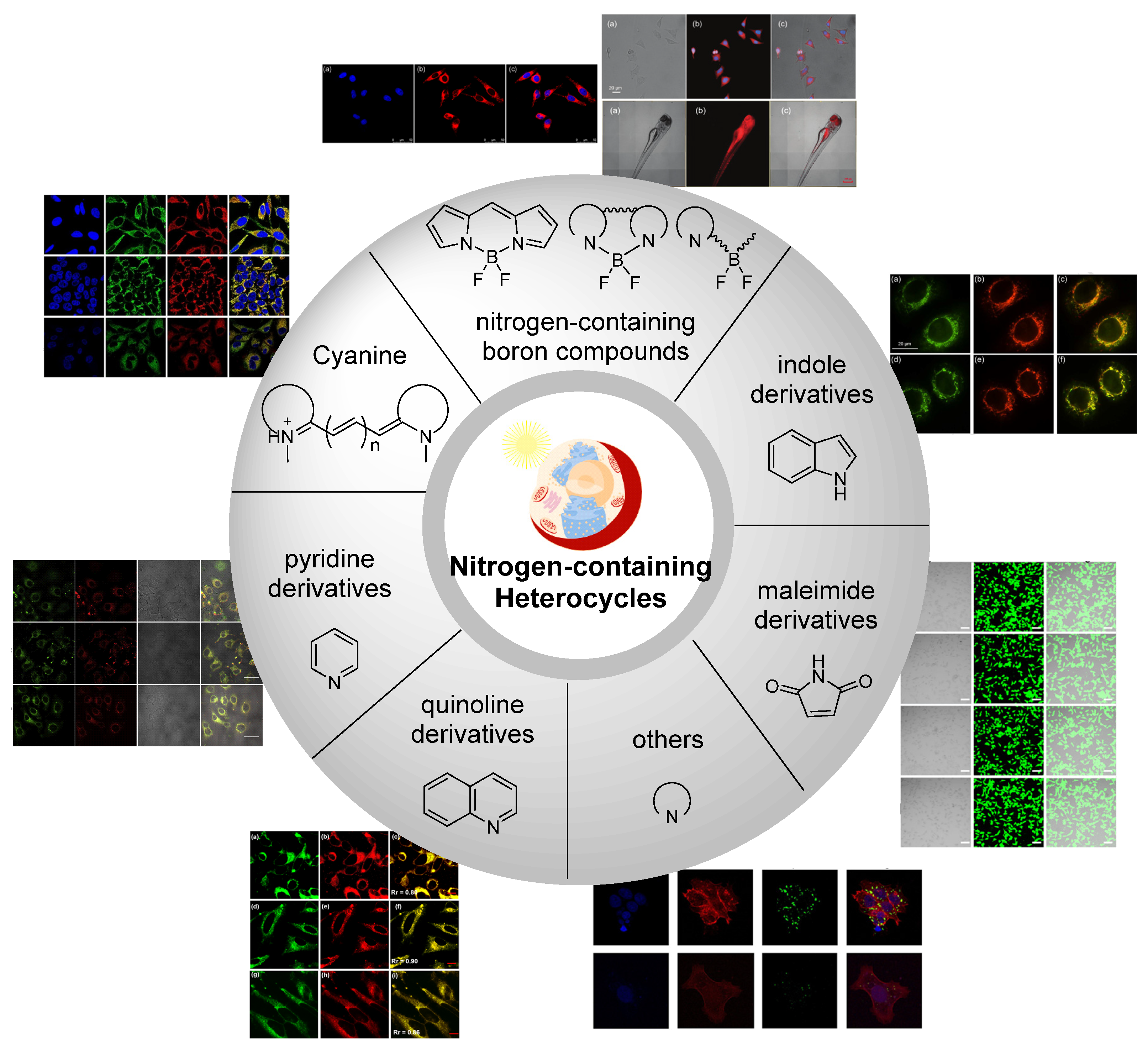
2. Nitrogen-Containing Boron Compounds

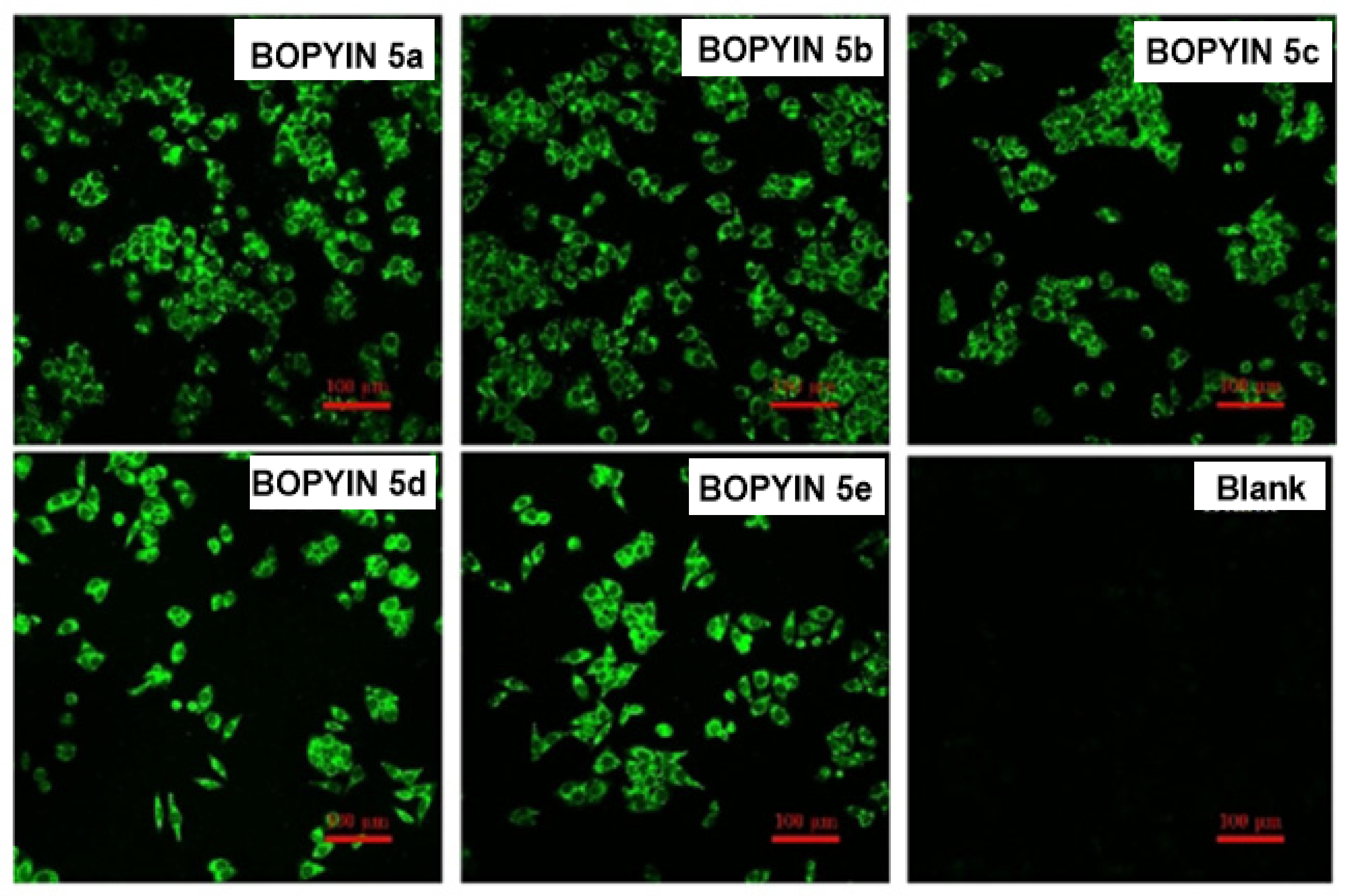
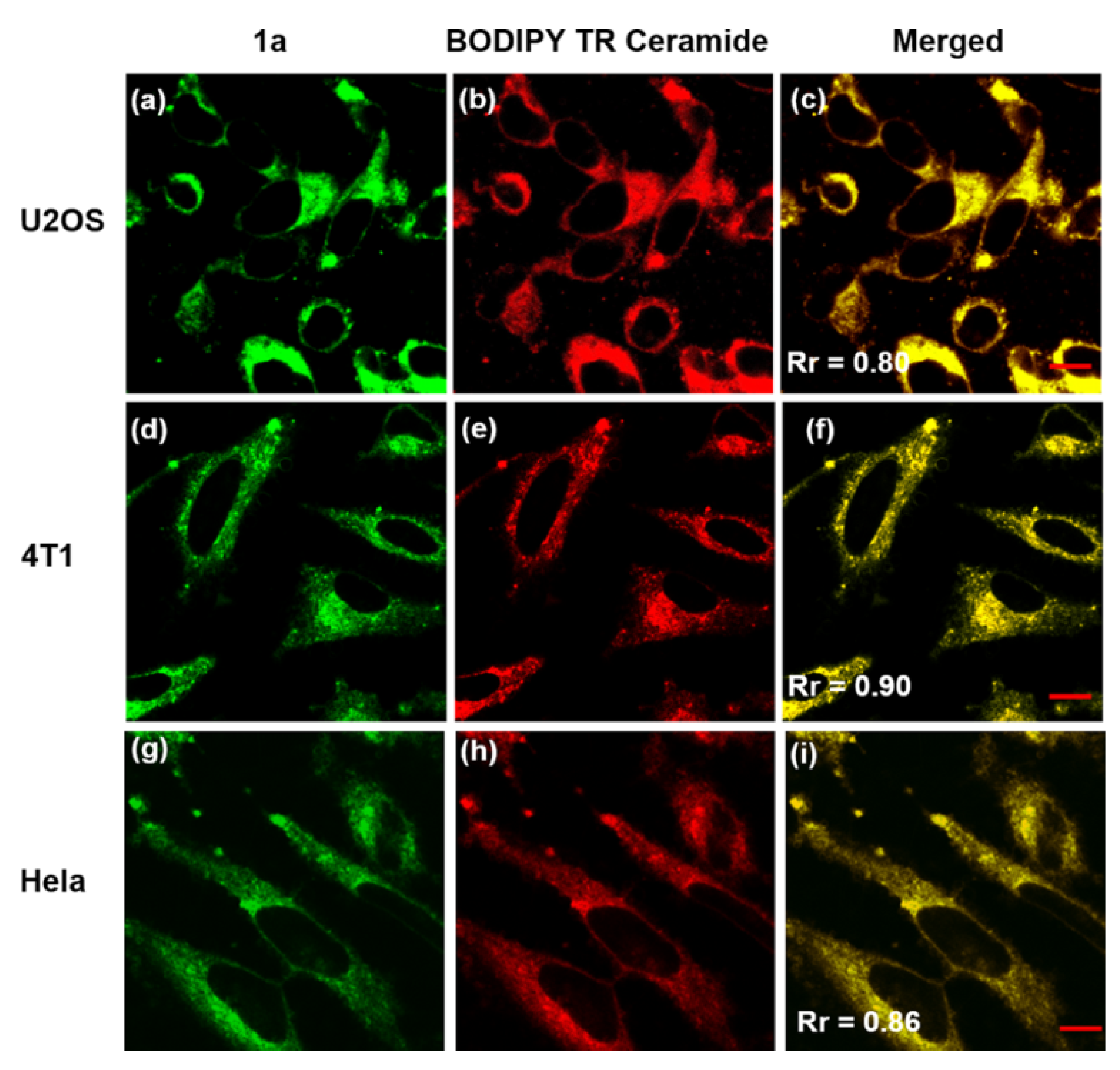
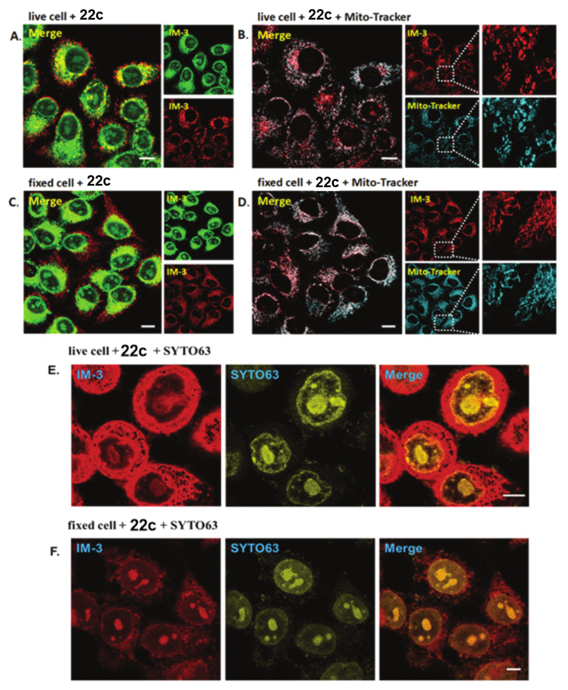
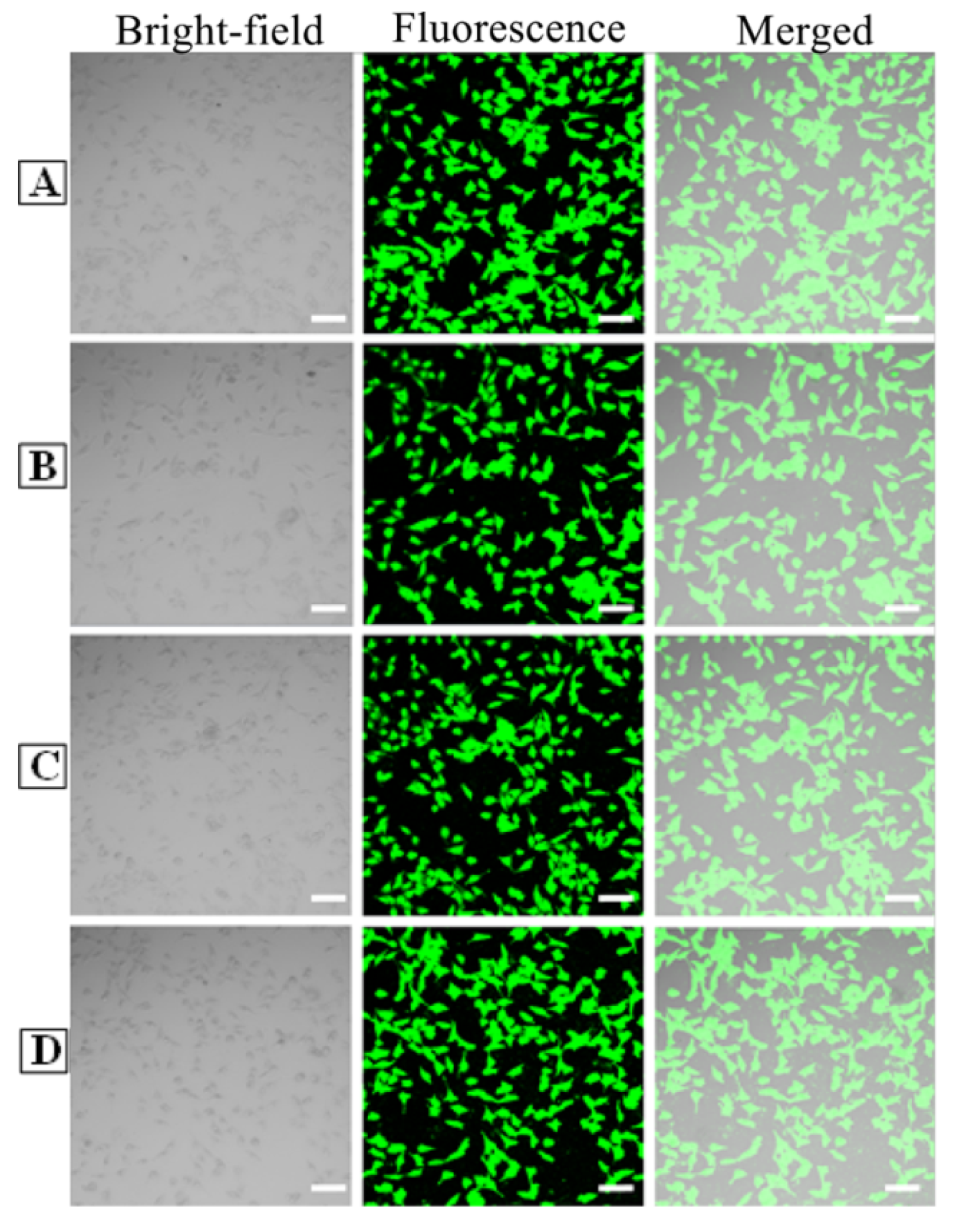
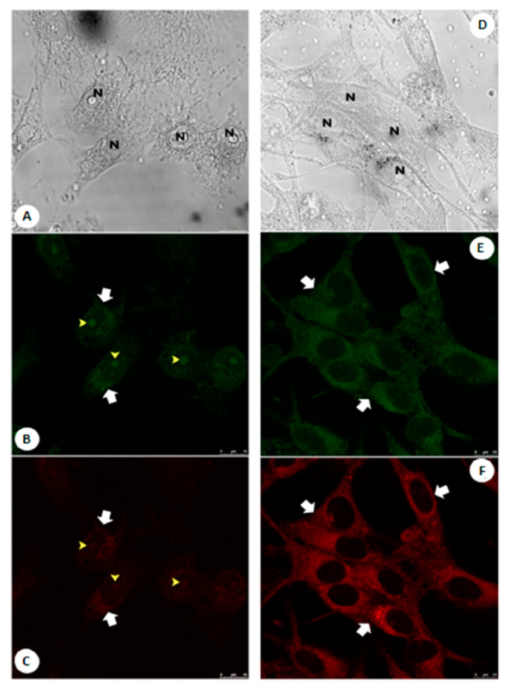
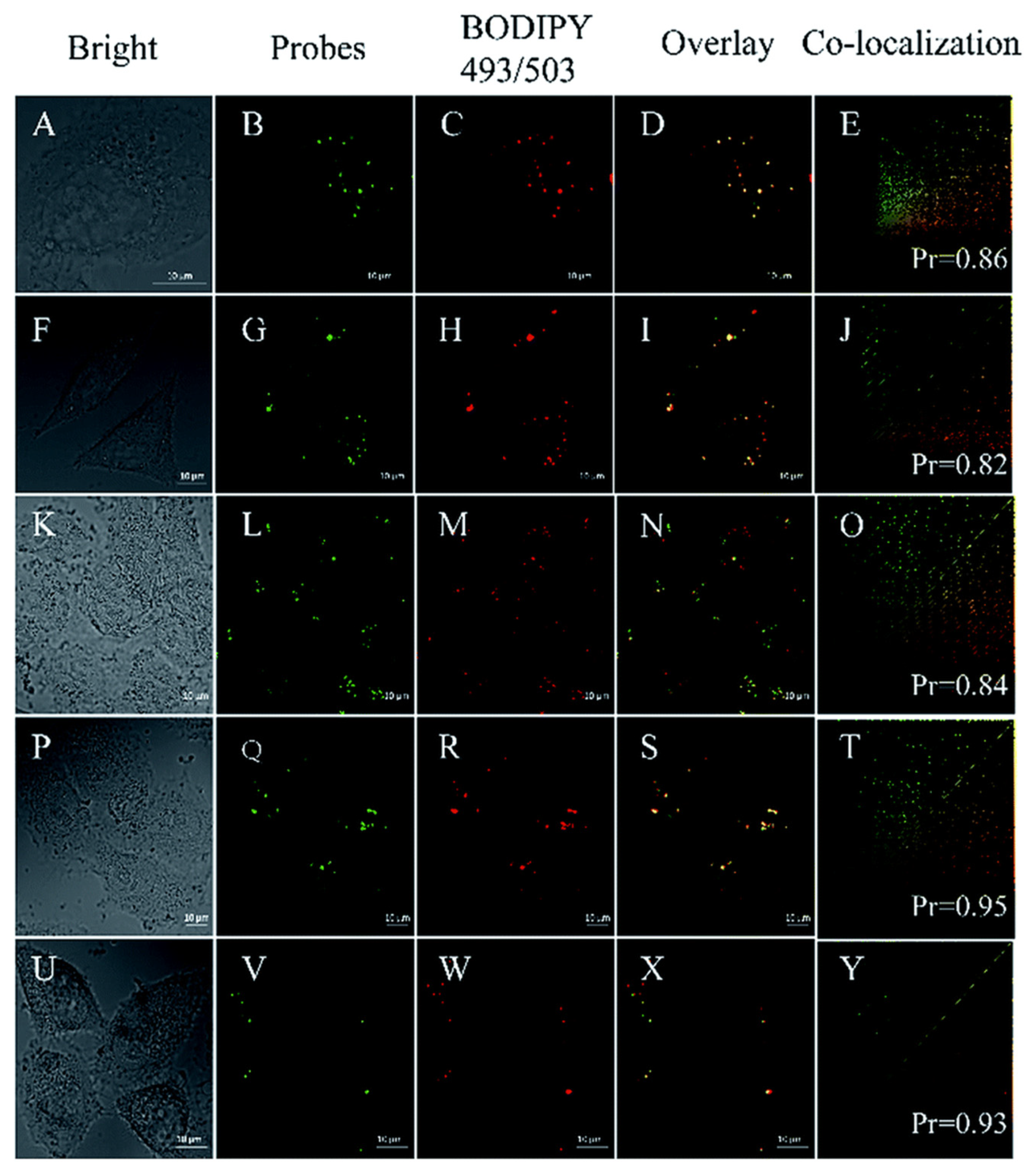
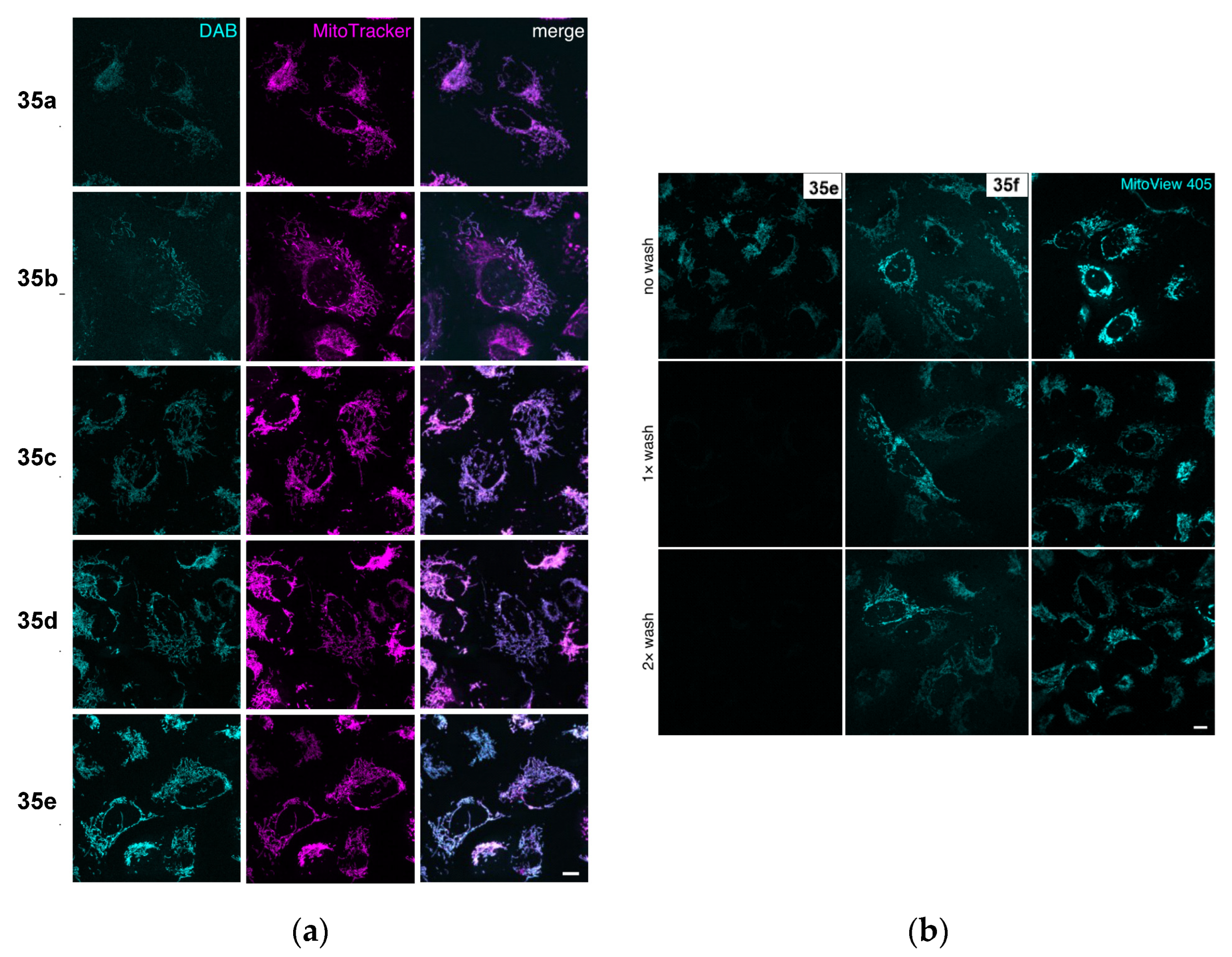
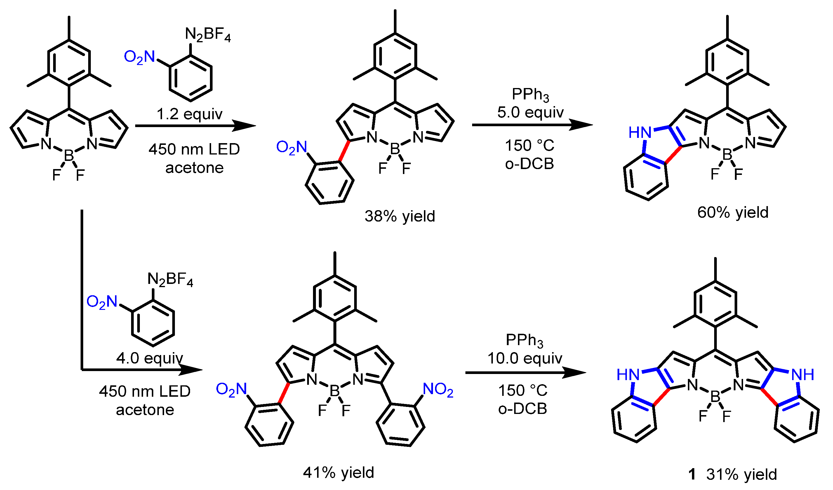
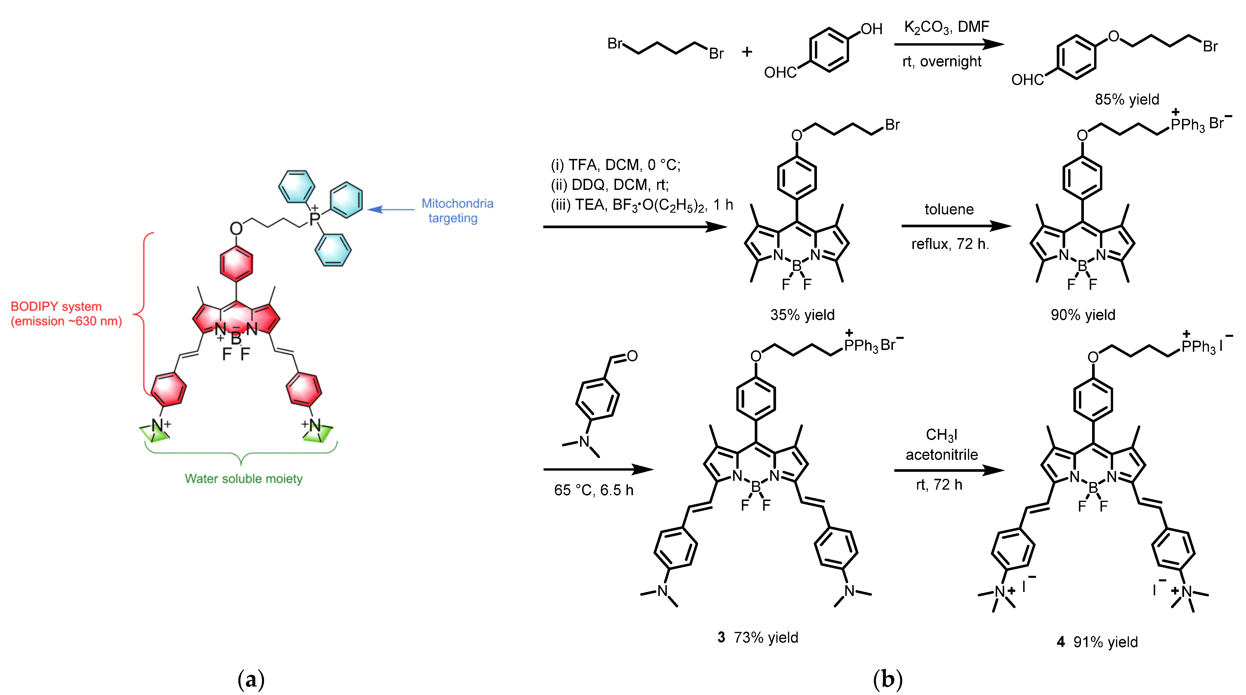
2.1. Boron Dipyrromethene (BODIPY) Derivates

The 4,4′-difluoro-4-bora-3a,4a-diaza-s-indacene dyes, usually abbreviated as BODIPYs, are one of the most significant fluorescent dyes [16]. The first BODIPY dye was synthesized by chemists Treibs and Kreuzer in 1968 [17]. Since BODIPYs offer unique properties, including high photostability, high fluorescence quantum yields, easy modification, and other advantages, the BODIPY derivates have attracted the attention of chemical and biological researchers in recent years [18]. Here, several representative BODIPY derivates applied for direct live-cell fluorescence imaging were introduced.
In 2019, Hao’s group developed a novel BODIPY with near-infrared (NIR) absorption and bright fluorescence [19]. Various BODIPYs were synthesized through a metal- and additive-free direct C−H α-arylation of easily accessible BODIPYs with aryl diazonium salts in the presence of visible light (Scheme 1). Representative diindole-annulated BODIPY 1 shows a maximum absorption peak and a maximum emission peak at the NIR region, respectively. The CCK-8 assay indicated that BODIPY 1 has good biocompatibility with cells. Fluorescence images stained with BODIPY 1 revealed bright red fluorescence in the cytoplasm of HeLa cells (Figure 2). The BODIPY core was expanded with nearly planar annulate indoles, thus resulting in a well-extended π-conjugation and a red shift of the absorption and emission.
In 2020, Tang et al. synthesized a new quinoline-fused BODIPY 2 [20]. This quinoline-fused BODIPY 2 was obtained in moderate yield by oxidation of precursory BODIPY with FeCl3 (Scheme 2). Due to the fusion of the quinoline group, the expansion of electron distribution, the reduction of molecular symmetry, and the change of the charge transfer direction, 2 emitted strong NIR fluorescence. With the advantages of a good imaging effect, high photostability, and low cytotoxicity, BODIPY 2 could be applied for biological imaging. Fluorescence images of stained HeLa cells and human dental pulp cells showed that BODIPY 2 generally aggregated in the cytoplasm (red region), not in the nucleus (black region) (Figure 3).
A year later, Jiang and co-workers reported two novel fluorescent BODIPY dyes, 3 and 4, for mitochondrial imaging (Scheme 3a) [21]. Lipophilic BODIPY 3 was afforded through a Knoevenagel condensation of 4-dimethylaminobenzaldehyde with the corresponding BODIPY. Water-soluble BODIPY 4 was afforded through methylation of 3 with methyl iodide (Scheme 3b). MTT experiments showed that the 3 and 4 have good biocompatibility. Confocal images indicated that 4 could target the mitochondrial region in HeLa cells specifically by introducing the cationic moiety, the TPP+ moiety, whereas weak fluorescence of 3 in HeLa cells was observed, which suggested that 3 was not suitable for targeting the mitochondrial region, due to its aggregation in the aqueous media (Figure 4). Both dyes inherited the good optical properties of the BODIPY core, including high photostability and fluorescence quantum yield.

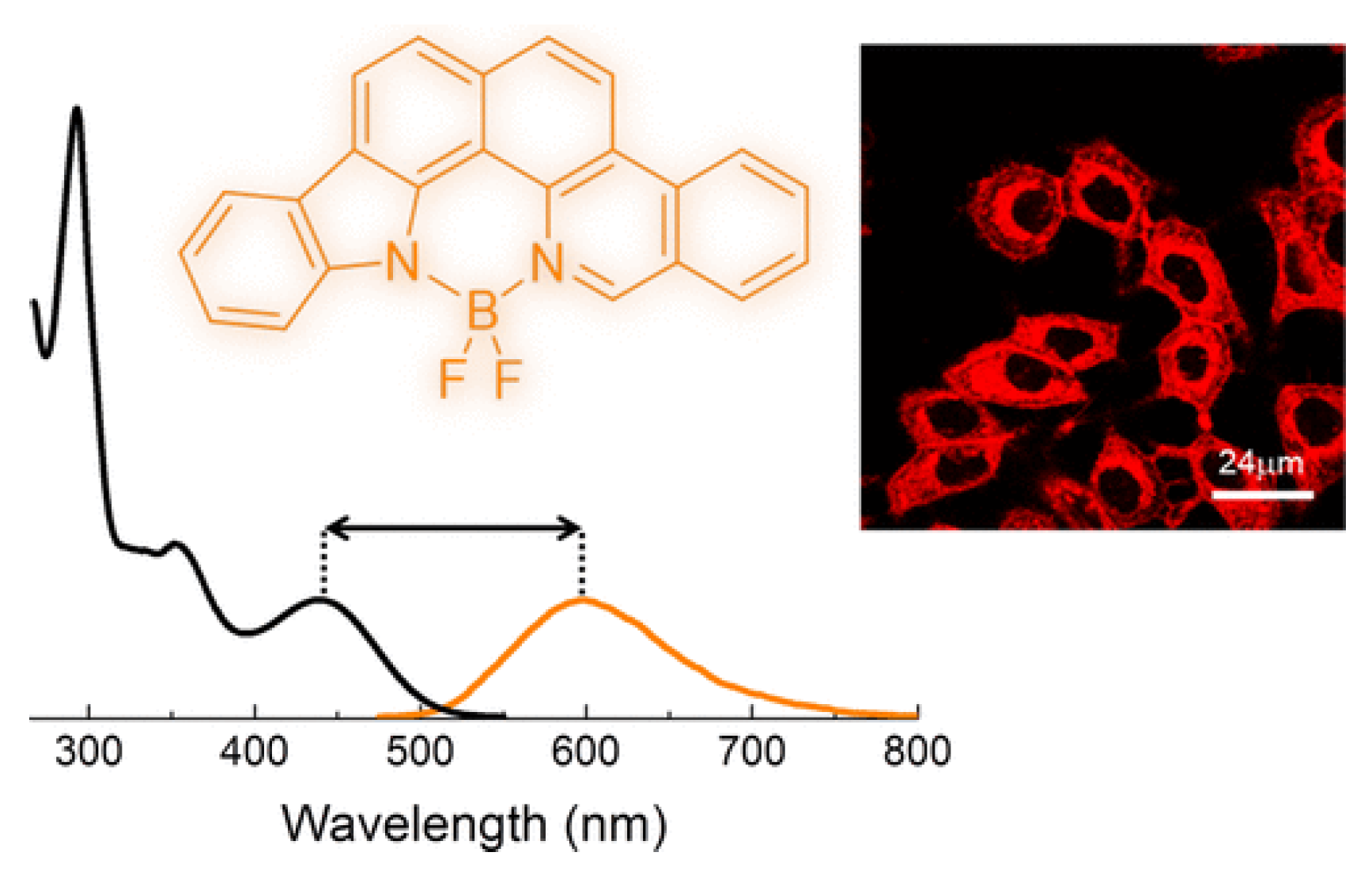
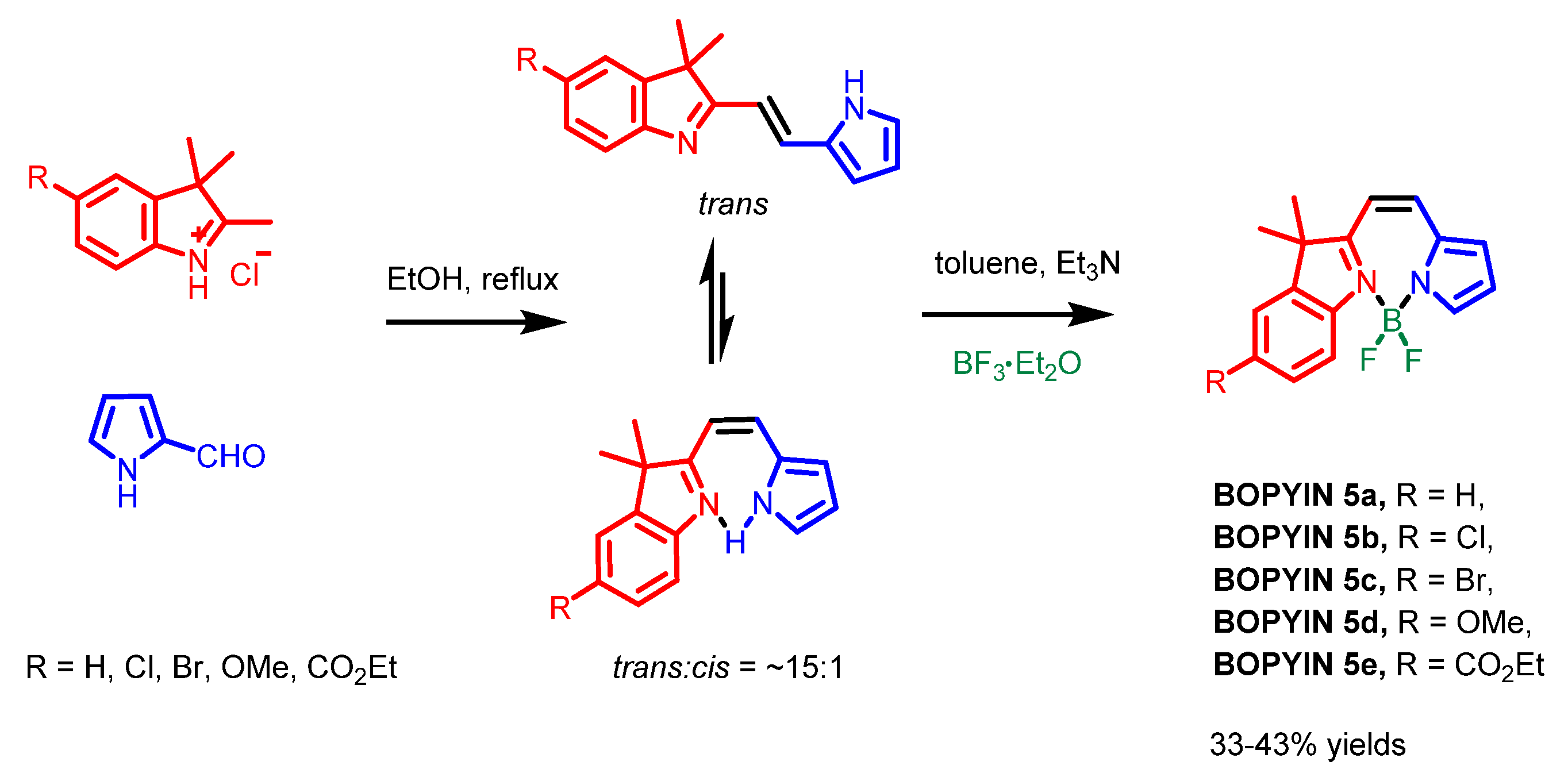
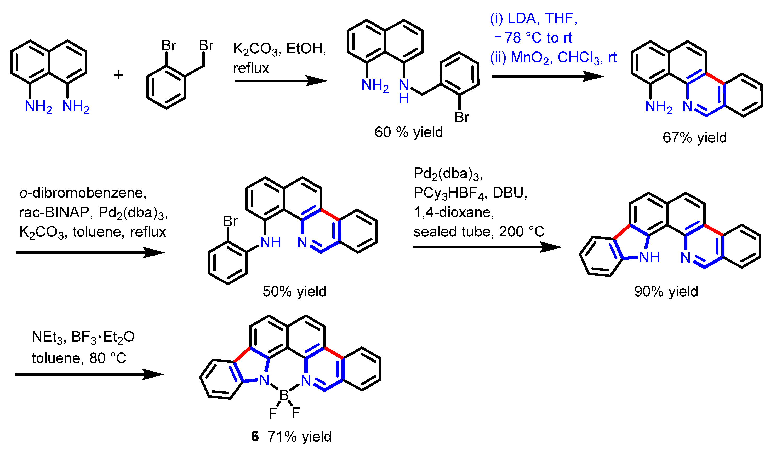
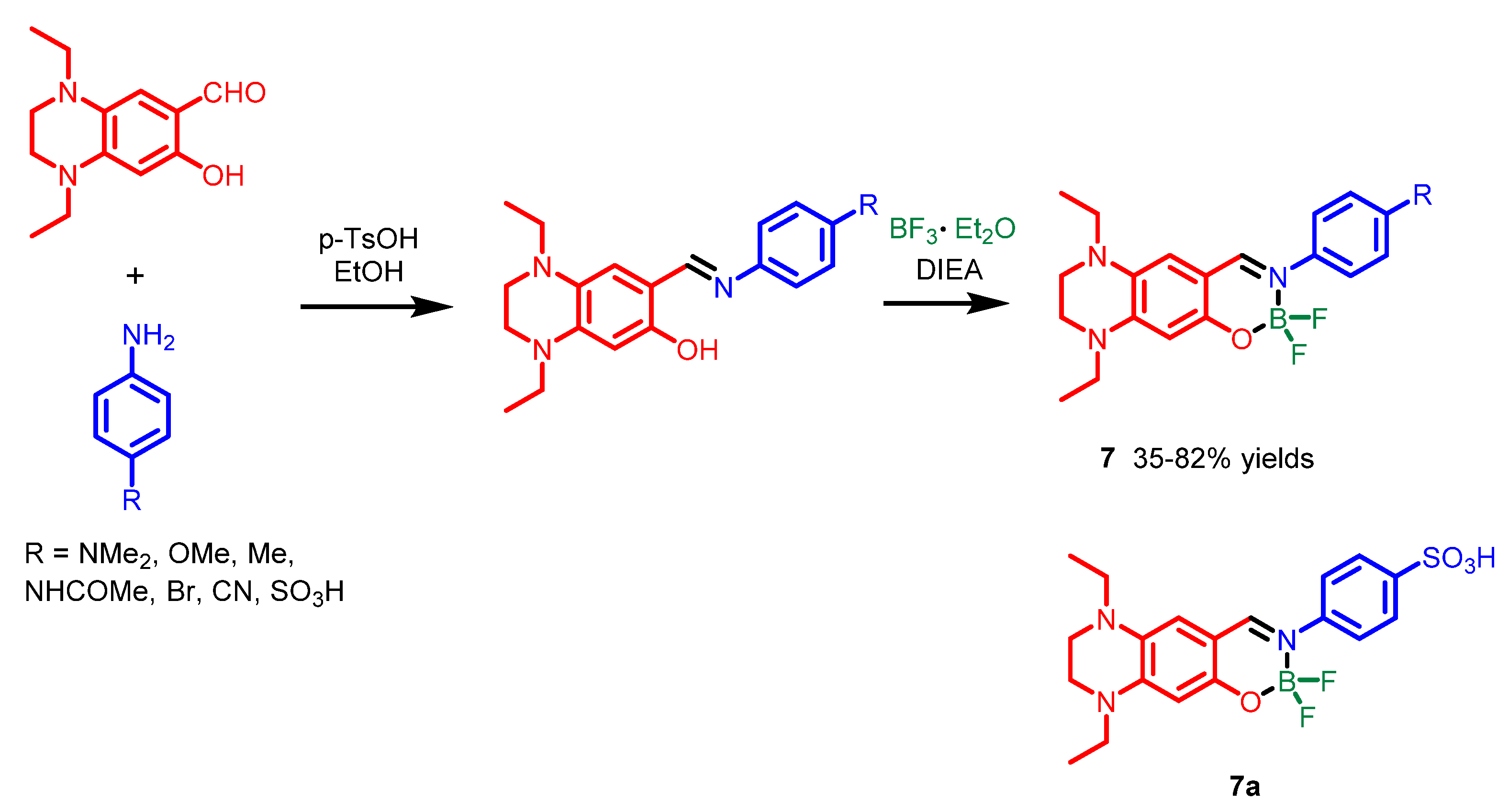
2.2. Other Nitrogen-Containing Boron Compounds
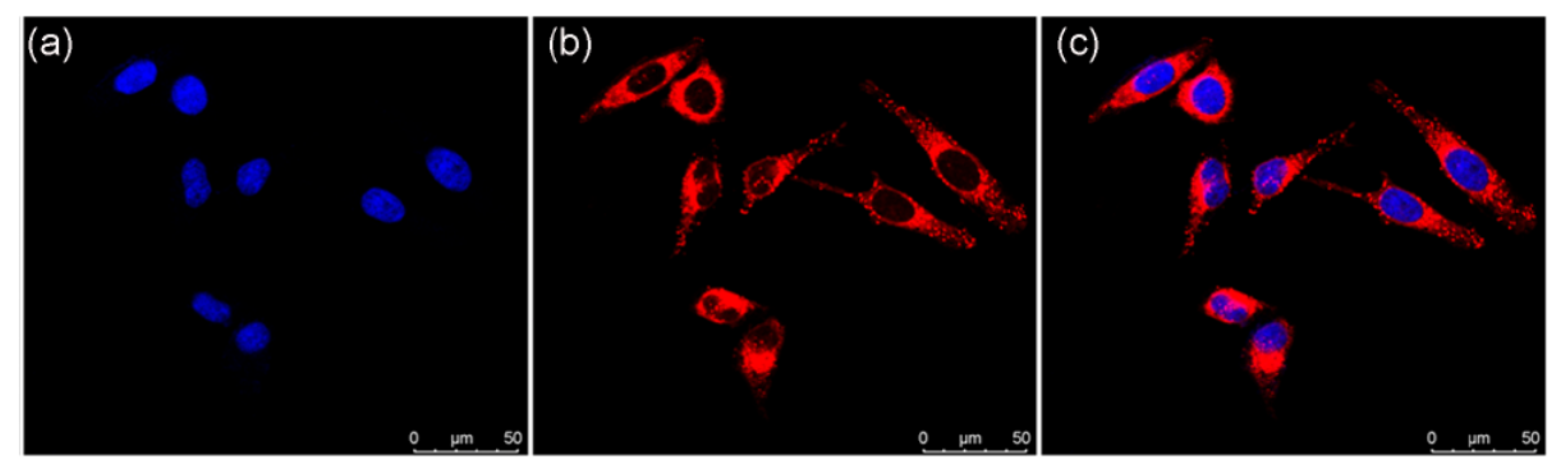
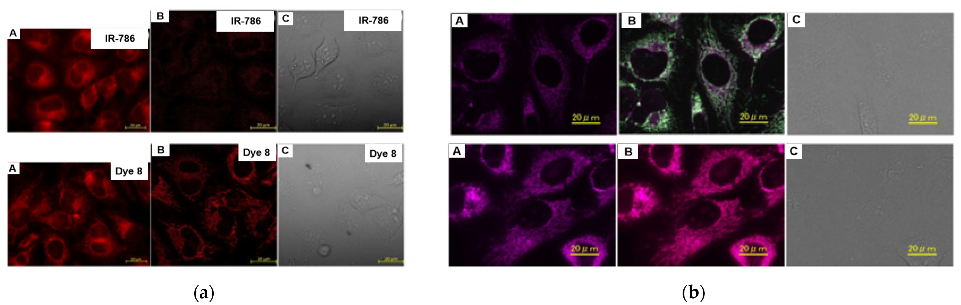
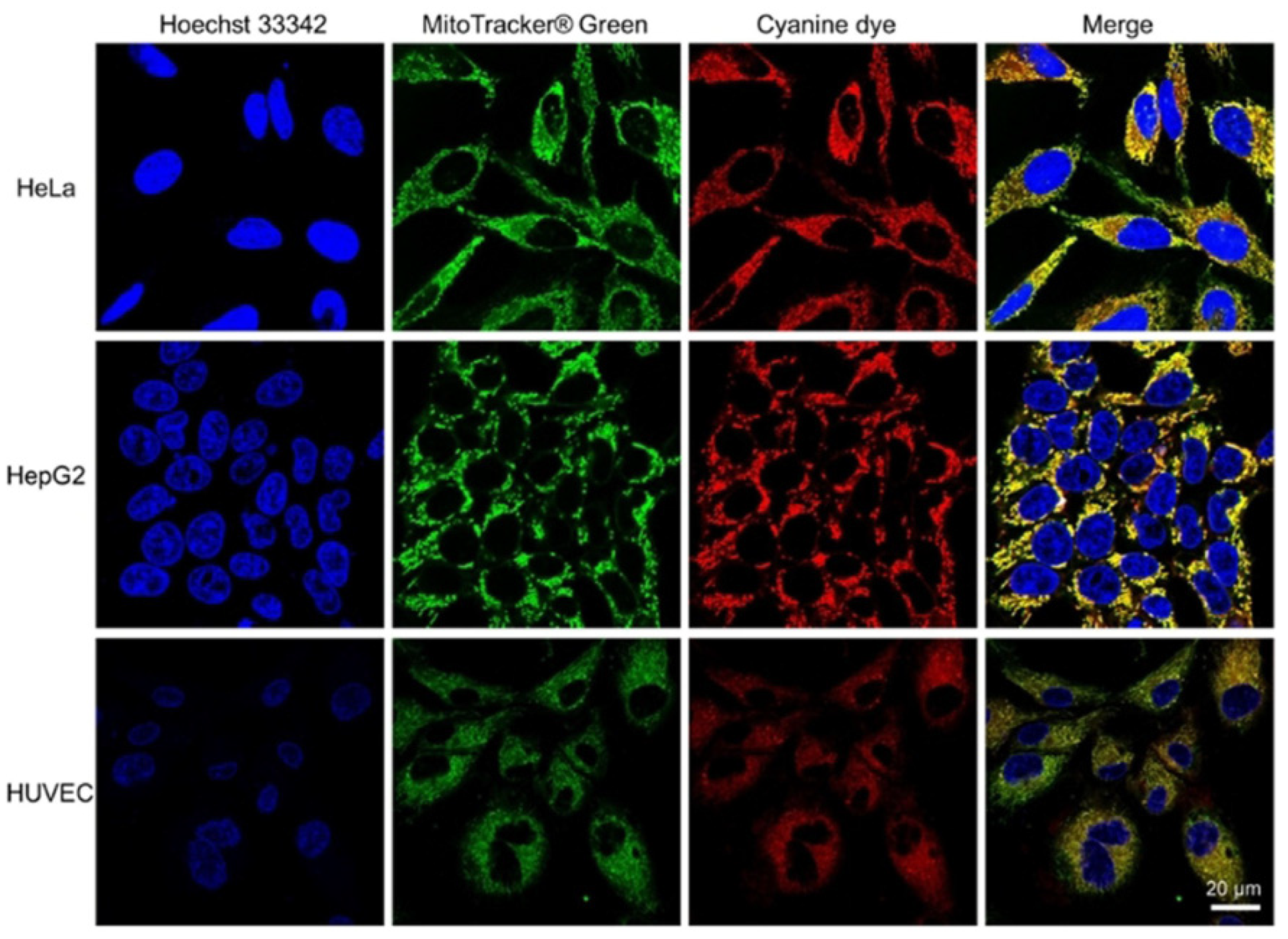
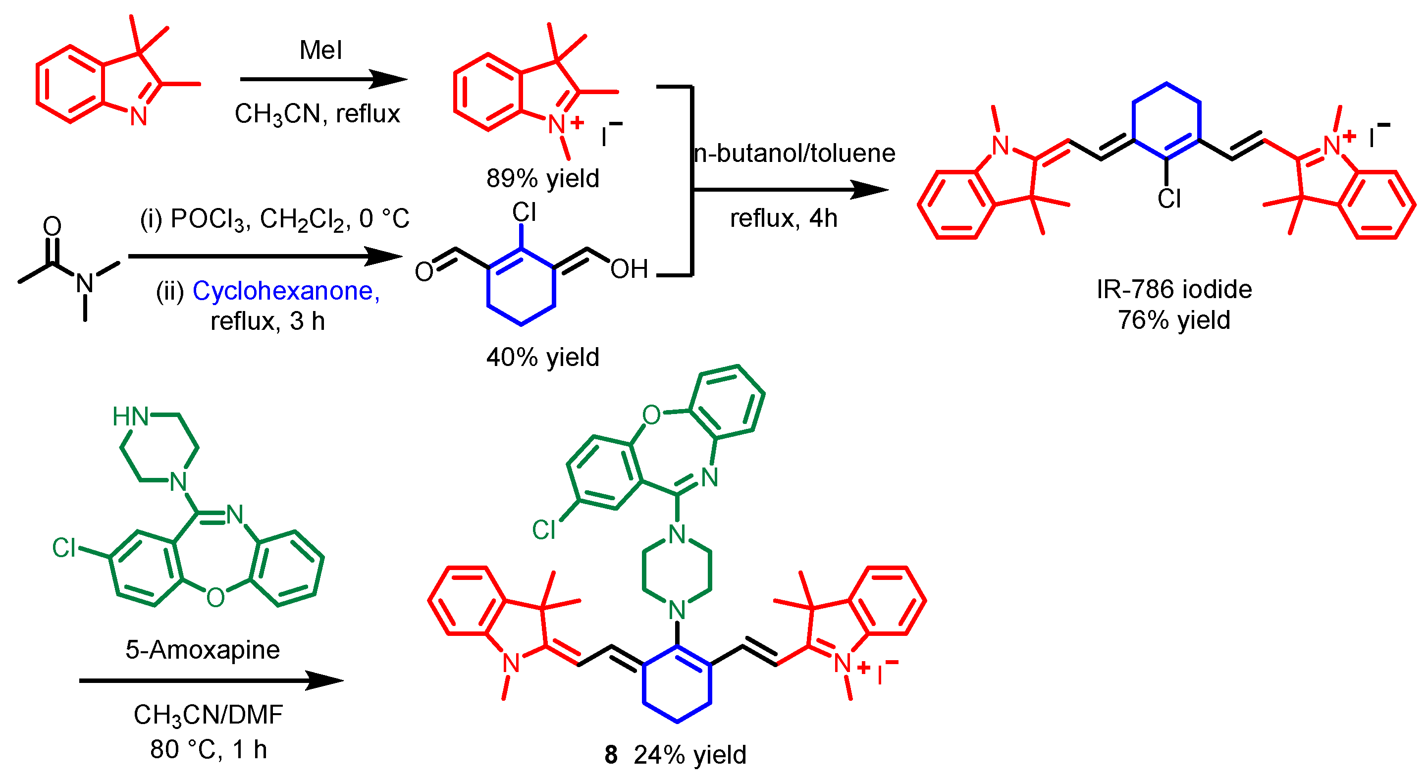
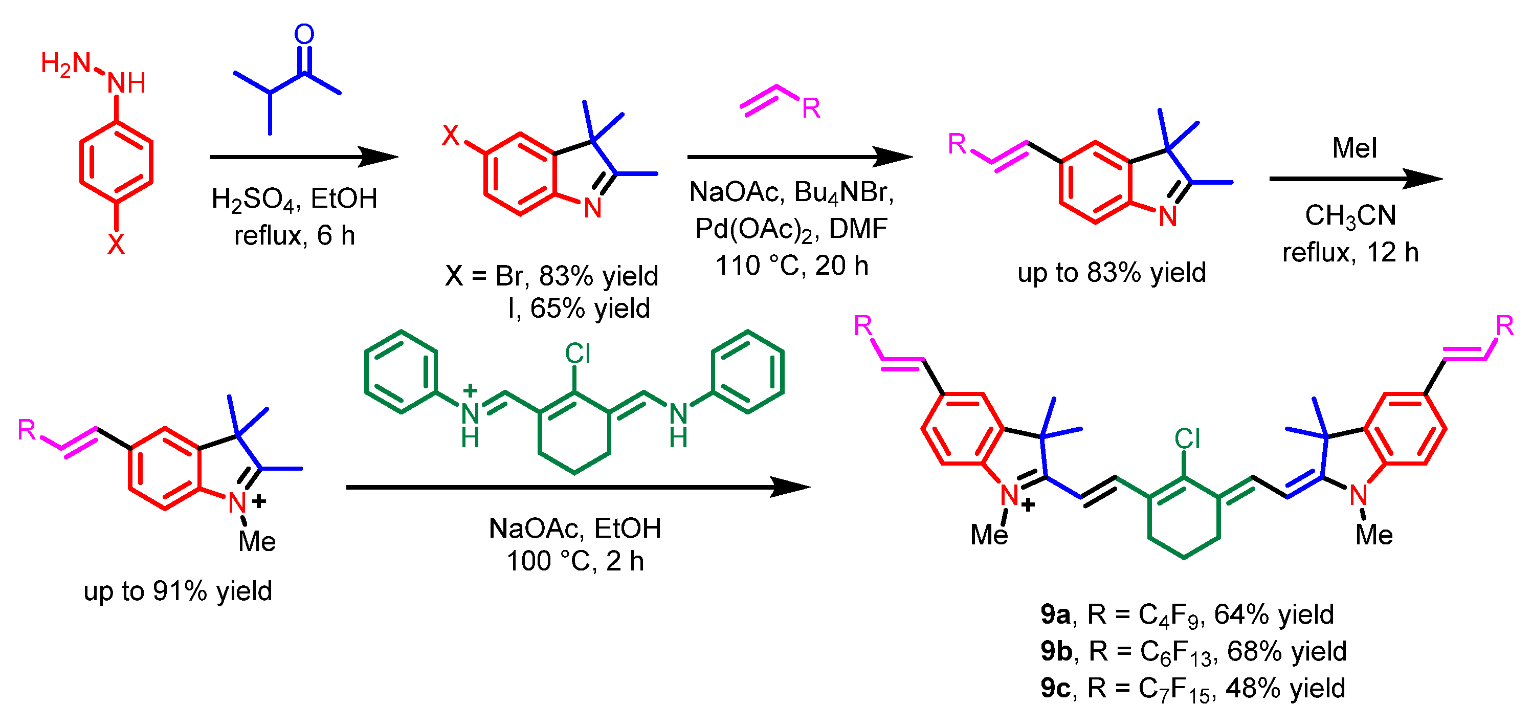
The N, N-chelate boron compounds contain BOIMPY, (aza)BODIPY, BOPHY, diazaborepin, BOPYIN, etc., according to the different numbers of membered rings [22]. Meanwhile, there are some other nitrogen-containing boron compounds that could be applied for cell imaging besides BODIPY.
In 2019, Zhang et al. reported various BOPYIN derivatives, 5ae, that have been synthesized via a one-pot method [22]. Various 3,3-dimethyl-2-[2-(2-pyr-rolyl)ethenyl] indoles were formed by different 2,3,3-trimethylindole hydrochlorides with pyrrole-2-carboxaldehyde. The N, N-ligand compounds then reacted with BF3·OEt2 to form the BOPYIN derivatives 5ae (Scheme 4). These BOPYIN derivatives were applied as biocompatible fluorophores in cell bioimaging. HeLa cells were stained with BOPYINsxxx 5ae, and these dyes rapidly passed through cell membranes, located in the cytoplasm predominantly afterwards, and showed bright green fluorescence. Confocal fluorescence images suggested that these BOPYIN were located in the perinuclear region, probably due to endocytic vesicles (Figure 5).
In 2020, Curiel et al. reported a novel four-coordinate N, N-difluoroboryl complex 6 (Scheme 5) [23]. The compound 6 was synthesized through a substitution reaction, a Buchwald−Hartwig reaction, an intramolecular cyclization, and a B-N bond coupling, respectively. This N, N-difluoroboryl complex 6 has a large Stokes shift due to its unique structural features, including desymmetrization, rigidification of the ligand, and π-expansion of the conjugated system, which could be applied as a fluorescent probe for cancer cells’ imaging (Figure 6).
In 2020, Chen’s group reported various novel N, N-difluoroboryl complexes with a tetrahydro-quinoxaline moiety donor, 7 [24]. For the synthesis of N, N-difluoroboryl complexes 7 (Scheme 6), a condensation of 1,4-diethyl-7-hydroxy-1,2,3,4-tetrahydroquinoxaline-6-carbaldehyde with various substituted anilines was carried out to afford the intermediates. The intermediates were subsequently treated with BF3·Et2O to obtain the corresponding products. MTT assays indicated that compound 7a was less toxic. The HeLa cells were stained with 7a with commercial Hoechst 33342 as a nuclear stain and bright red fluorescence could be observed from the cytoplasm (Figure 7). A 3-day-old zebrafish was fed with 7a, and remarkable red fluorescence could be observed (Figure 7). The tetrahydro-quinoxaline donor was introduced into these D–A-type fluorescent dyes, which enhanced the intramolecular change transfer (ICT) effect, thus resulting in red shifts of the emissions and large Stokes shifts. Meanwhile, a reasonable fluorescence quantum yield was retained due to the rigidity of the tetrahydro-quinoxaline moiety as the electron donor.

3. Cyanine

Cyanine is a conjugated system between two nitrogen atoms; in each resonance structure, exactly one nitrogen atom is oxidized to an iminium. Cyanine is one of the most significant fluorescent dyes due to its unique optical properties, including a high molar extinction coefficient, high fluorescence quantum yield, narrow absorption/emission band, and readily tunable fluorescence profile from the UV-vis to near-infrared (NIR) range [25,26].
In 2018, Jose et al. synthesized a novel dye, 8 [27], which is a combination of heptamethine cyanine dye IR-786 [28] and an FDA-approved drug, Amoxapine [29]. IR-786 iodide 4 was generated through a methylation, a cyclization, and a dehydrative condensation, respectively [30]. Fluorescent dye 8 was synthesized through an amination reaction of IR-786 iodide with Amoxapine (Scheme 7). Dye 8 could selectively stain the mitochondria, whereas IR-786 iodide does not show specificity for any organelles (Figure 8a). Colocalization experiments using different commercial mitochondrial stains incubated with dye 8 further confirmed its mitochondrial selectivity (Figure 8b). The lipophilic nature of the dye 8 could assist with penetrating the cell membrane and the delocalized positive charge helped the dye 8 to target the negatively charged mitochondria.
In 2019, Bräse’s group reported three novel polyfluorinated cyanine dyes, 9ac, and discussed their photophysical properties [31]. These cyanine probes were synthesized through a cyclization, a Heck reaction, a methylation, and a dehydrative condensation, respectively (Scheme 8). All these dyes could penetrate the cell membrane and selectively accumulate in the mitochondria (Figure 9), due to the lipophilicity of cyanine dyes and the positive charge of these dyes [32]. Moreover, the addition of fluorous side chains resulted in red shifts of the absorption and emission of these cyanine dyes.
In 2022, Ge’s group reported two hemi-cyanine probes, 10a and 10b, and a neutral probe 10c based on a quinoxaline skeleton [33]. The three probes were generated through a Knoevenagel condensation of 1,3-dimethylquinoxalinium iodide or 3-methylquinoxalinium with different aldehydes (Scheme 9). Fluorescence imaging indicated that all these dyes could target the mitochondria of HeLa cells. Probes 10a and 10b stained the nucleic acid in the mitochondria with excellent selectivity, while dye 10c neutrally stained the mitochondrial region (Figure 10). This research suggested that bioactive quinoxaline derivatives could be effectively applied in the field of fluorescence images.
Additionally, in 2022, Gallavardin and co-workers replaced the classical electron-donating group indole with indazole in merocyanines to study the substitution effect [34]. The indazole and indole merocyanines 12 and 13 were generally synthesized through a key-step Knoevenagel condensation of ethyl-3,3-dimethyl-3H-indolium iodide 11 with various corresponding aldehydes (Scheme 10). These merocyanines were used to treat tumoral PC12 cells. Confocal fluorescence images indicated that indazole-based merocyanines 12ac selectively stained the mitochondria. Indole derivatives 13a,b did not stain the same cell compartment: 13a did not target the mitochondria and stained in the liposome, whereas 13b selectively stained in the mitochondria, similar to indazole compounds (Figure 11). Indazole merocyanines 12 have an electron-donating indazole ring and a charged electron-accepting indolinium moiety to target the mitochondria, while the absorption and emission spectra of indole merocyanines 13 are slightly red-shifted because indoles are more electron-rich.

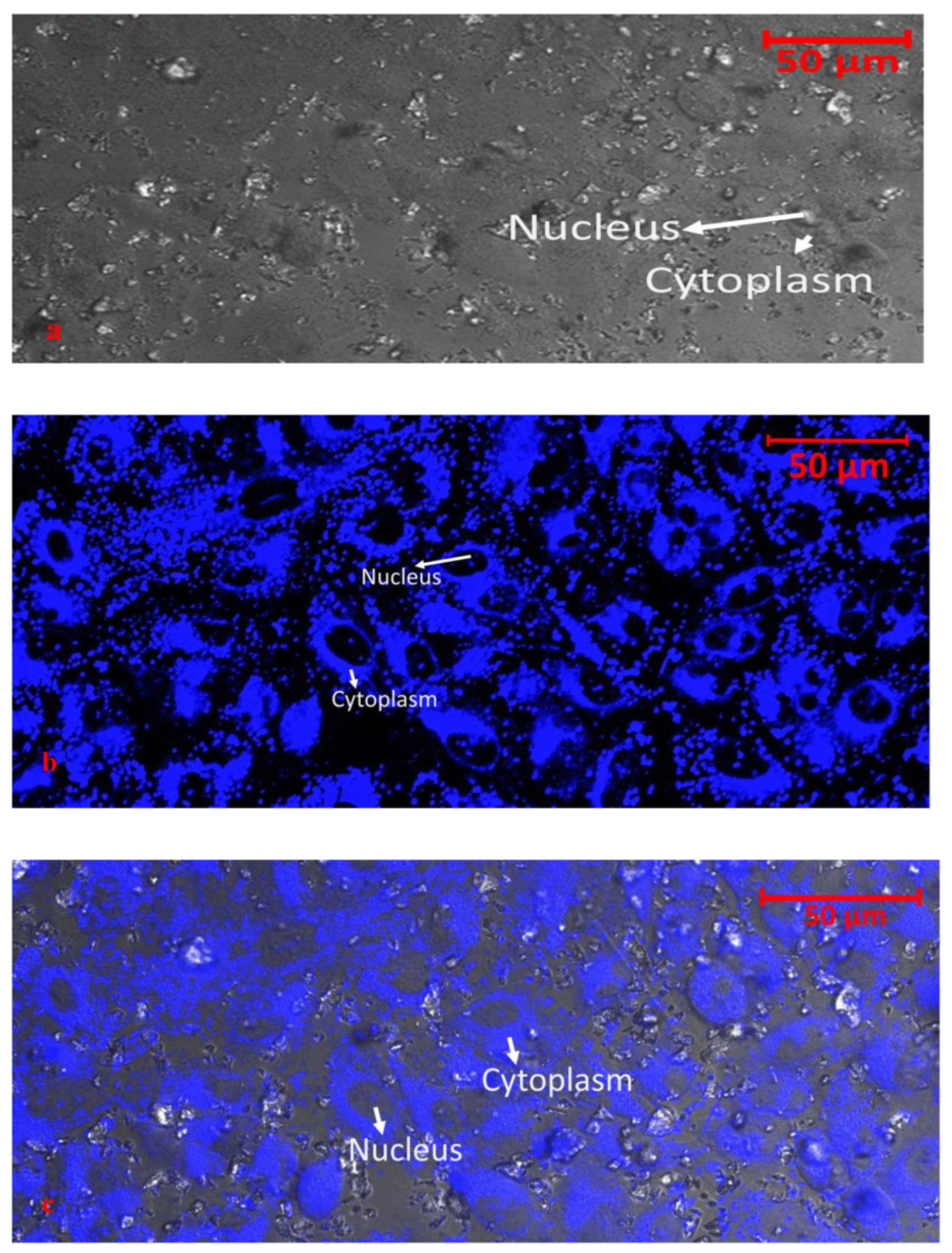
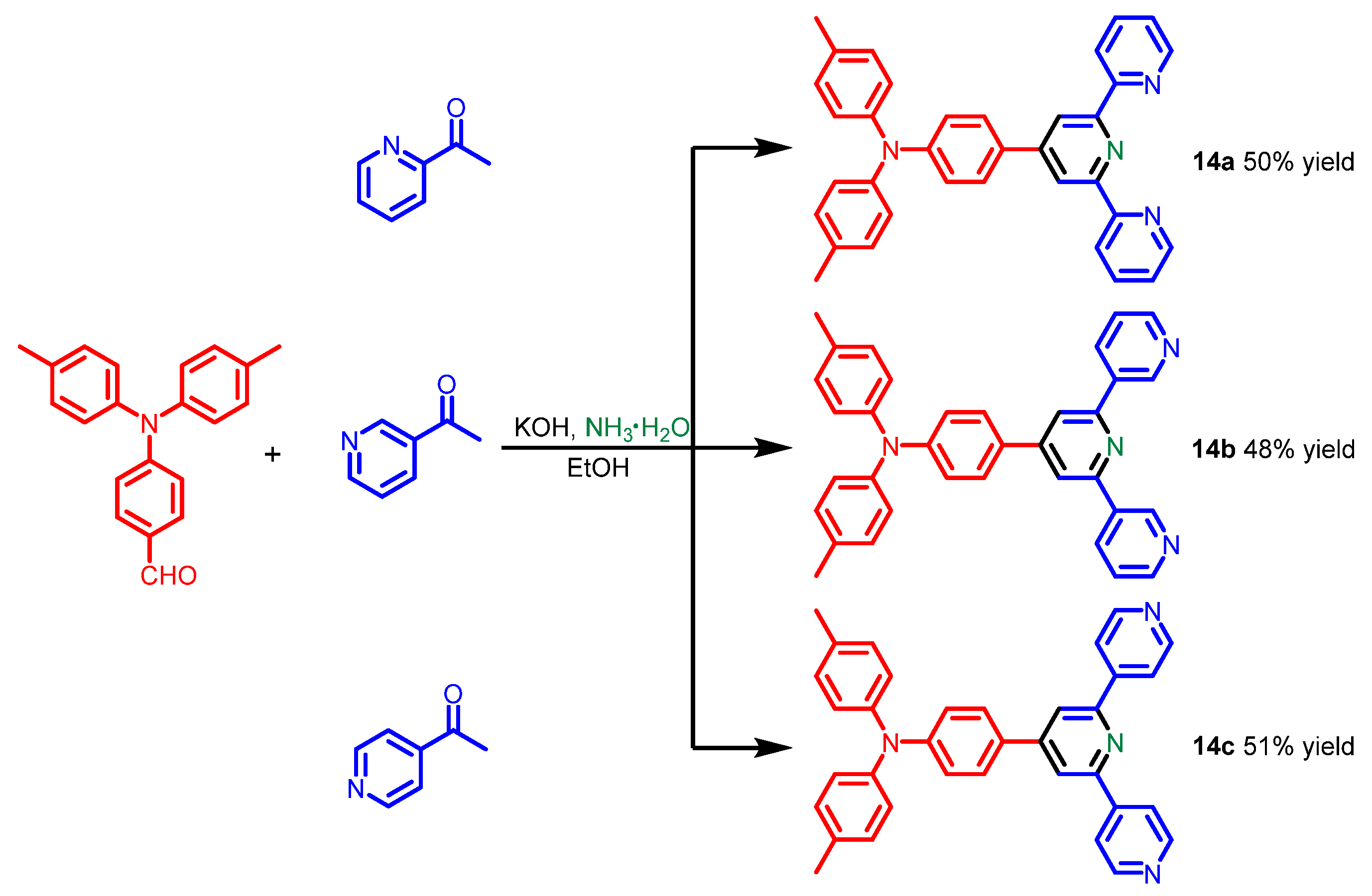
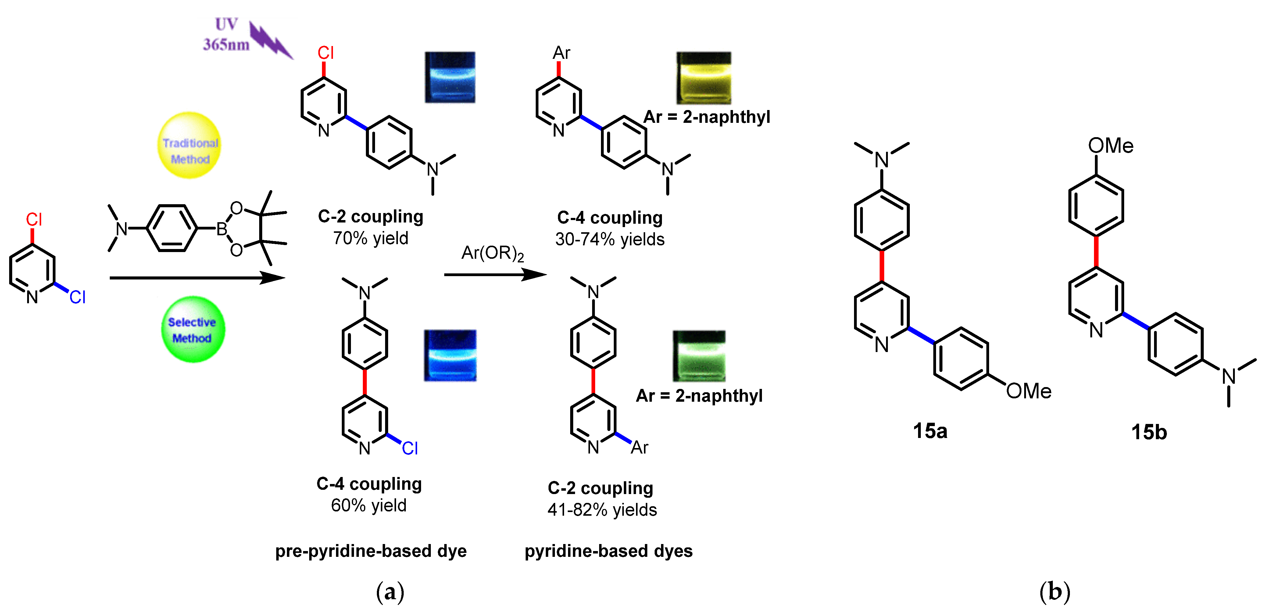
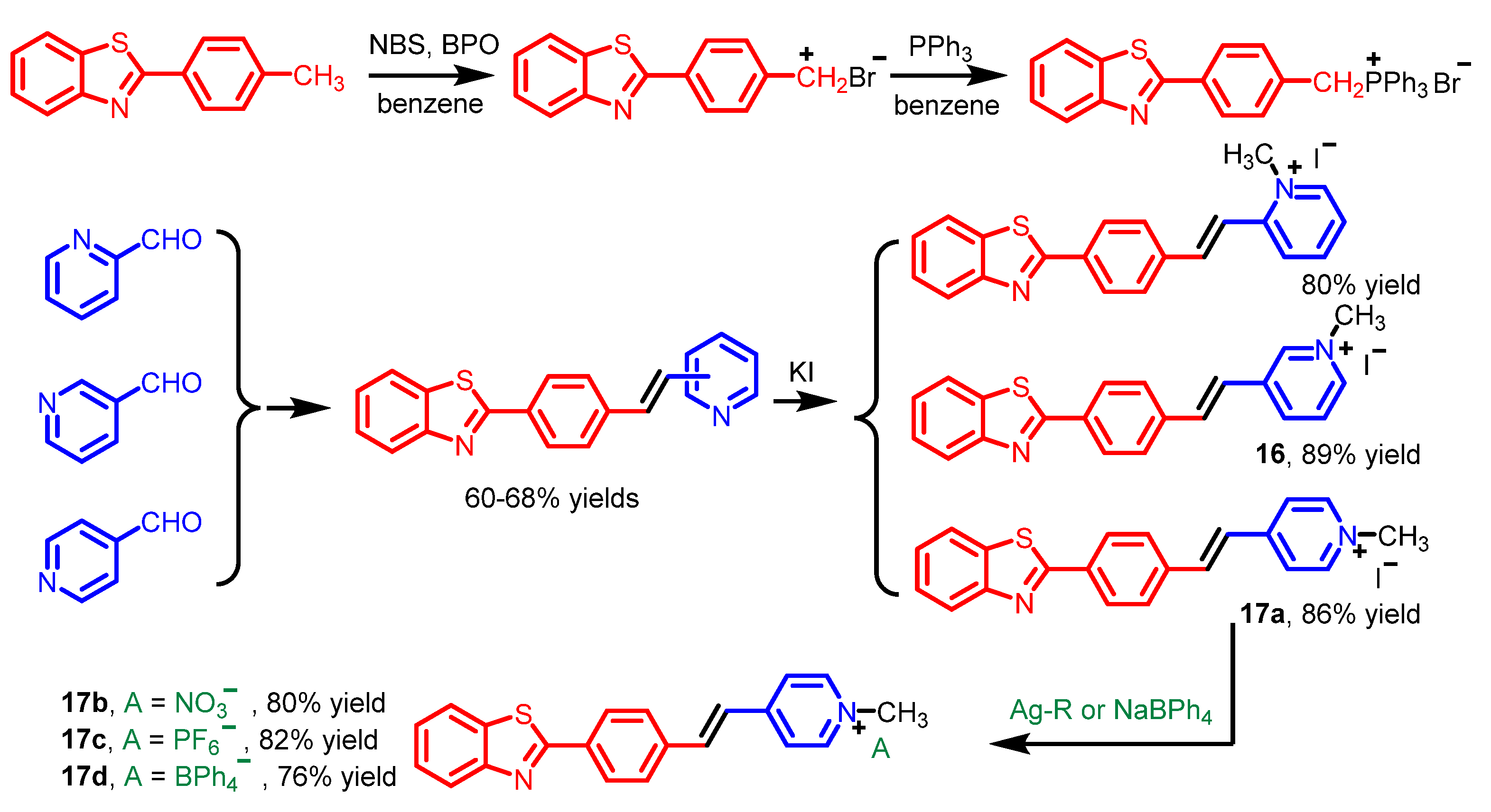
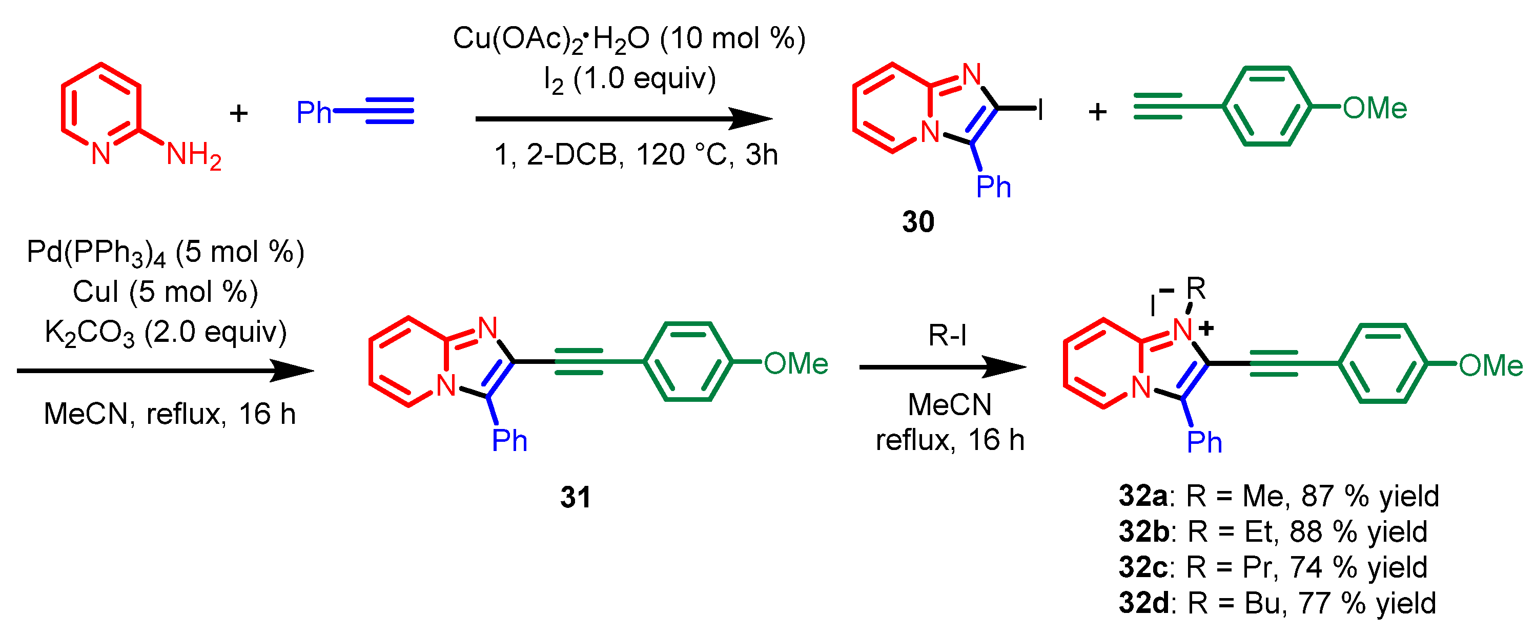
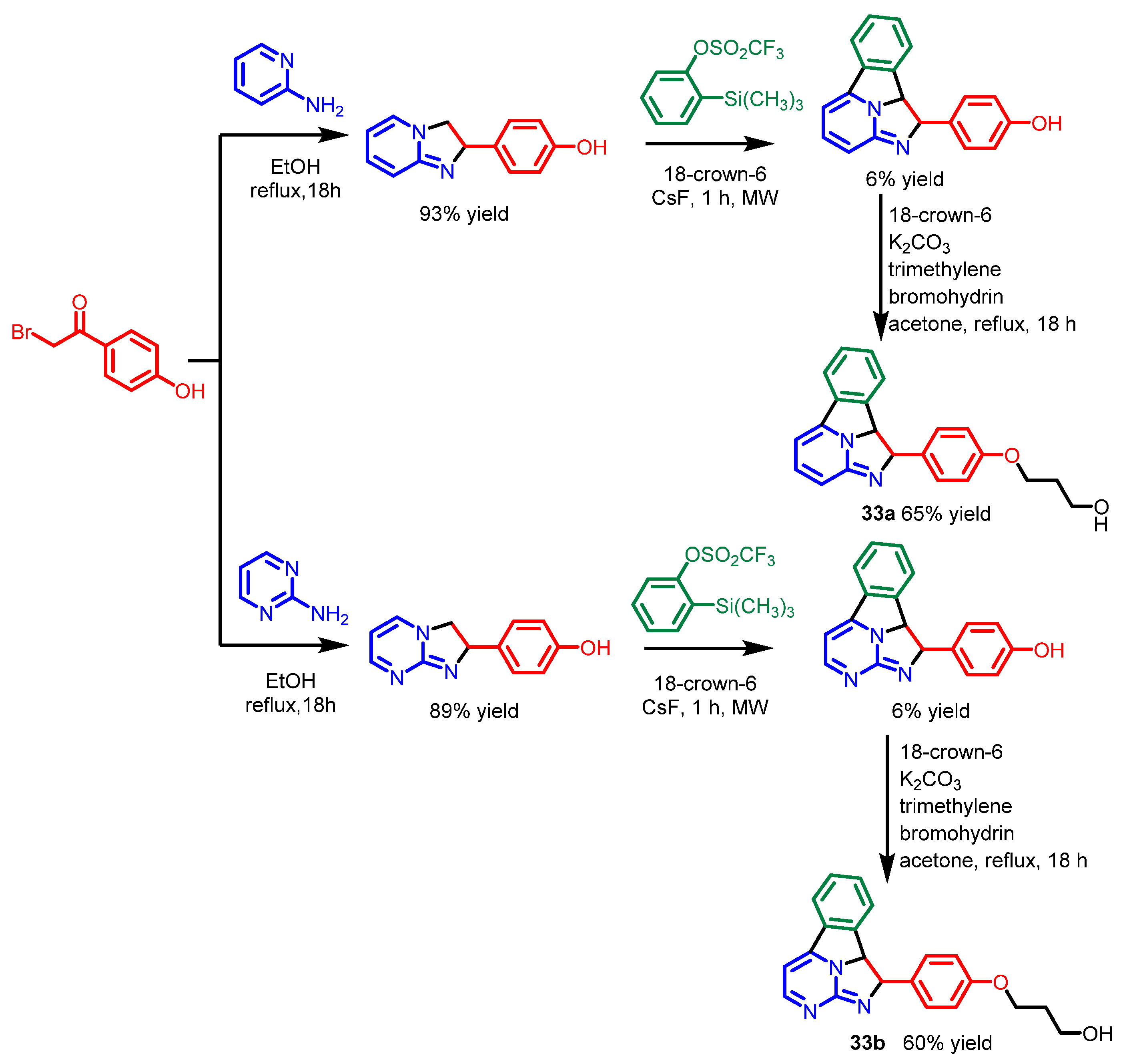
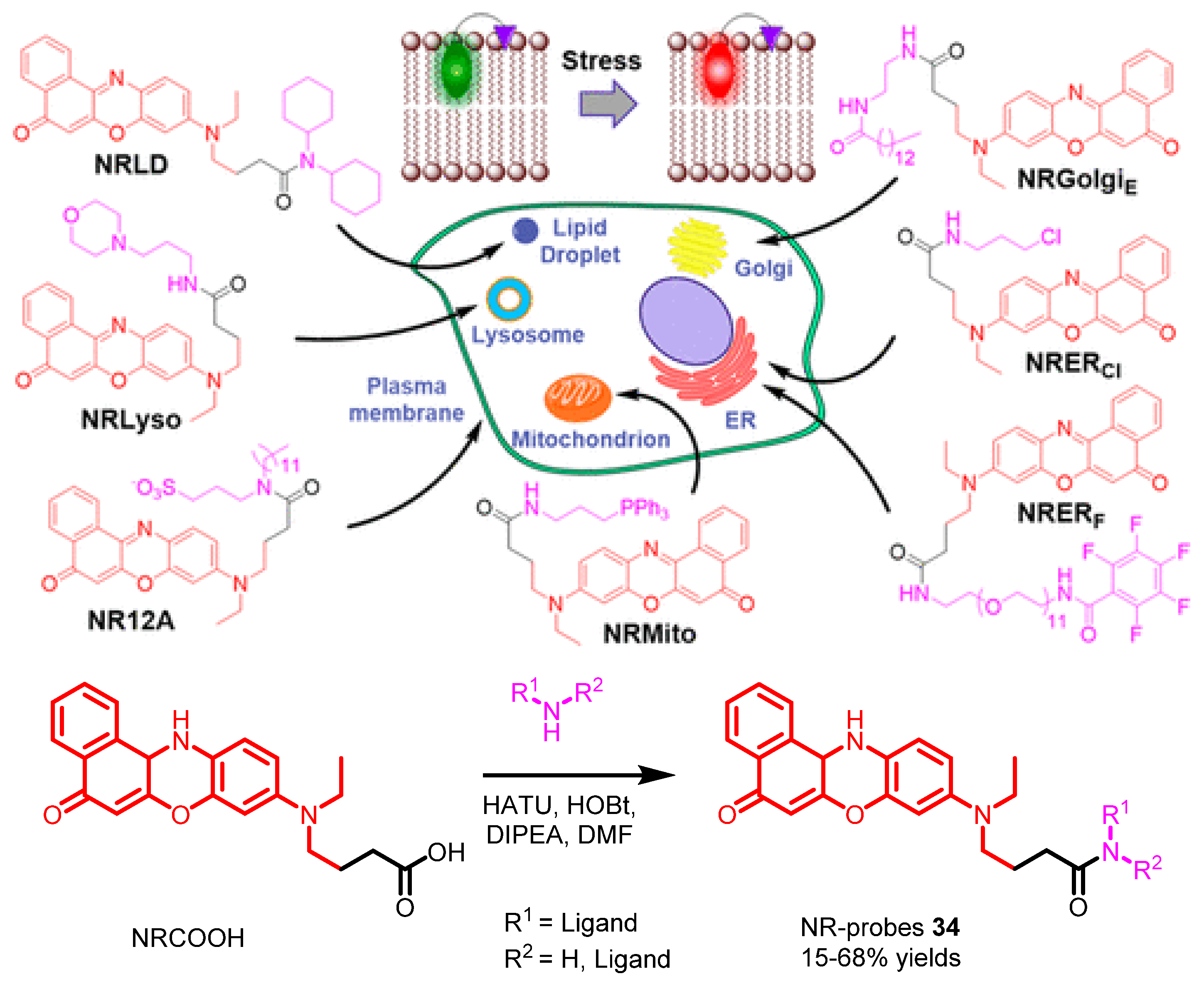
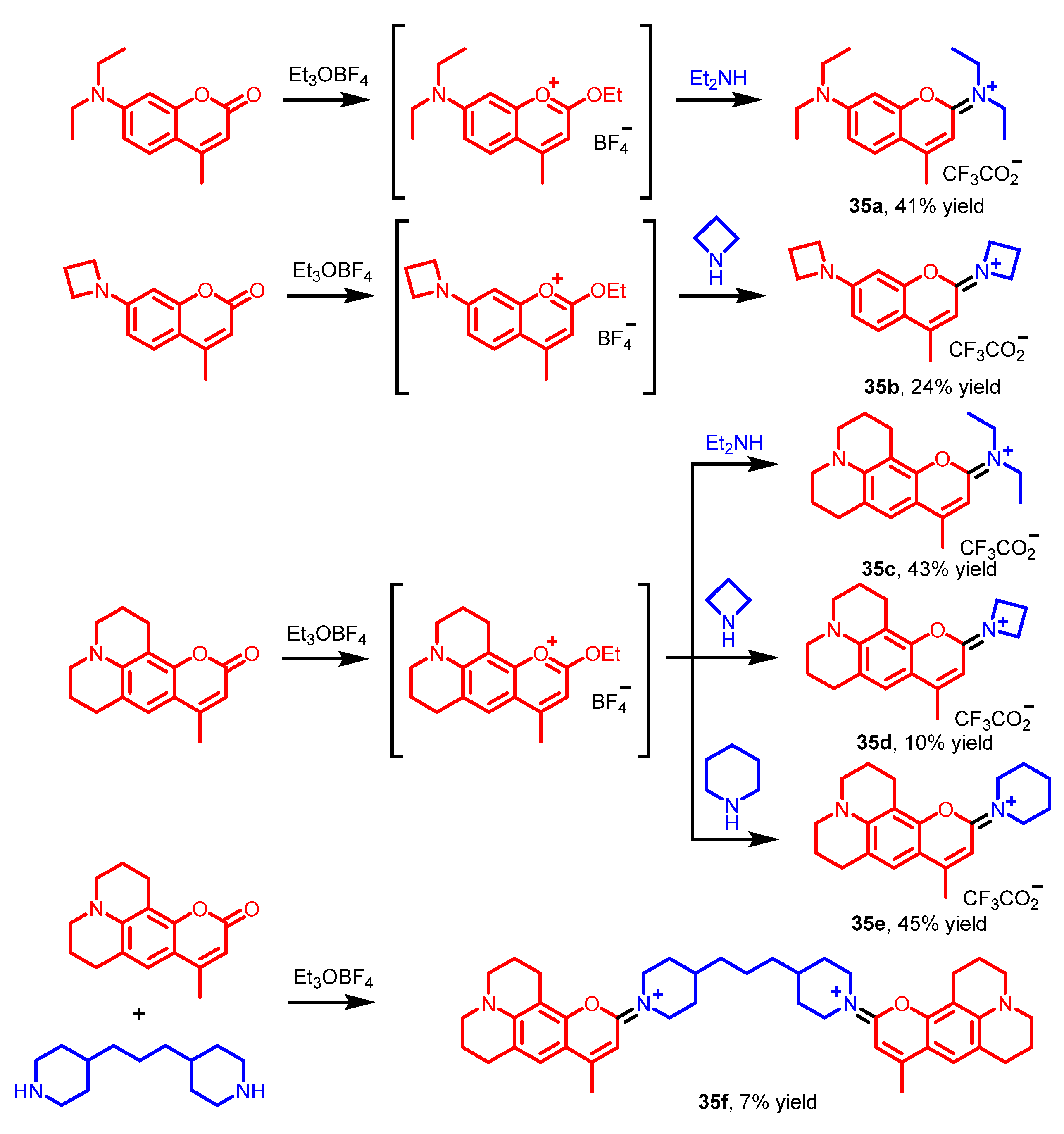
4. Pyridine Derivatives

Pyridine is an aromatic heterocycle composed of a six-membered ring with one nitrogen. The first pyridine was reported through heating animal bones by Scottish chemist Thomas Anderson in 1849 [35]. Due to the mitochondrial-targeting ability of the pyridinium group and other biological features of pyridine derivatives, pyridine derivatives have been generally utilized as fluorescent dyes in cellular imaging [36,37,38,39]. Herein, we mainly introduce the direct fluorescence cell imaging of pyridine derivatives.
In 2018, Zhang and Wang reported three isomers of triphenylamine-based terpyridine derivatives, 14ac [40]. These terpyridine derivatives were synthesized through an aldol reaction and a subsequent cyclization (Scheme 11). The terpyridine derivatives 14ac could stain similarly in cytosolic space of the HepG2 cells instead of the nuclear region. The bright field images revealed good cell morphologies, which showed the low toxicity of 14ac (Figure 12). The D–A structure was constructed to build the optically active probes. The triphenylamine unit owned the forceful electron donor and highly efficient p-electron bridge, while the terpyridine unit was applied as the electron acceptor. Moreover, the methyl group attached to the triphenylamine unit was applied to improve the electron-donating ability and the lipophilicity.
In 2020, Huang et al. developed a novel approach to obtain C-4 arylated pyridine derivatives through a C−C coupling reaction of 2,4-dichloropyridines with boronic esters catalyzed by Palladium (Scheme 12a) [41]. Here, 15a and 15b were chosen as representative compounds to identify their good applicability in cell imaging (Scheme 12b). Blue fluorescence (Figure 13A) and green fluorescence (Figure 13B) could be observed by exciting at 405 and 488 nm, respectively, while bright cyan fluorescence was shown in merged images (Figure 13D). Inspection of the photophysical properties showed that the stronger the electron-absorbing ability of C-2 or C-4 substitution, the greater the red-shifted emission of the pyridine derivatives, indicating the formation of a D−π-A system.
In 2021, Zhou’s group synthesized various water-soluble benzoxazole-pyridinium salt derivatives [42]. These probes were generally synthesized through a bromination, a Wittig reaction, a methylation, and an ion-exchanging reaction, respectively (Scheme 13). Cell imaging revealed that 3-pyridinium salt 16 crossed the nuclear membrane and selectively stained the nucleus, whereas 4-pyridinium salt derivatives 17ad stained in the nuclear membrane, which clearly showed the morphology of the nucleus (Figure 14). The plausible reason for the different staining ability may be that the nature of the pyridinium salt isomer influences the particle shape and size in aggregates in the cells.

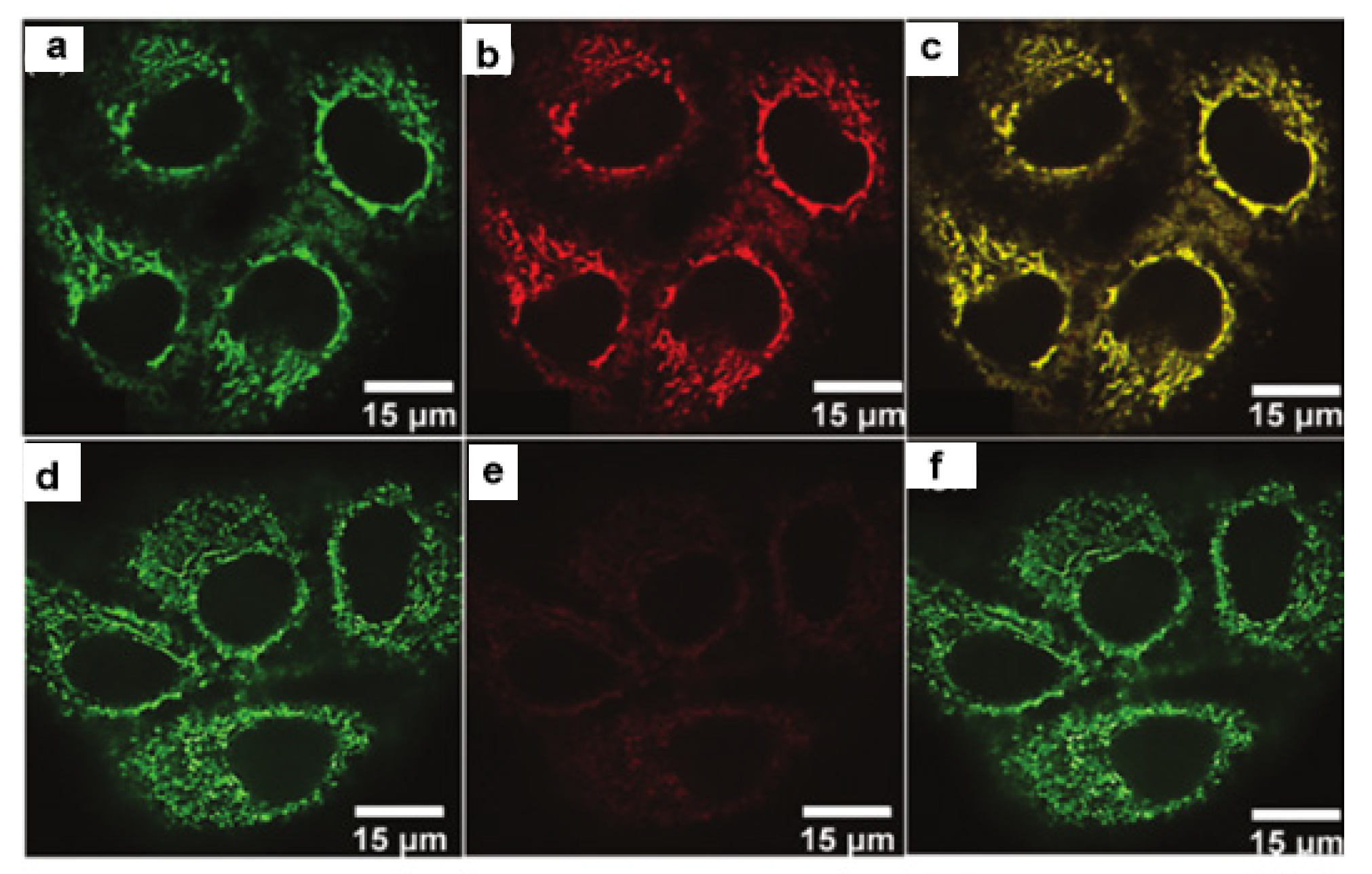
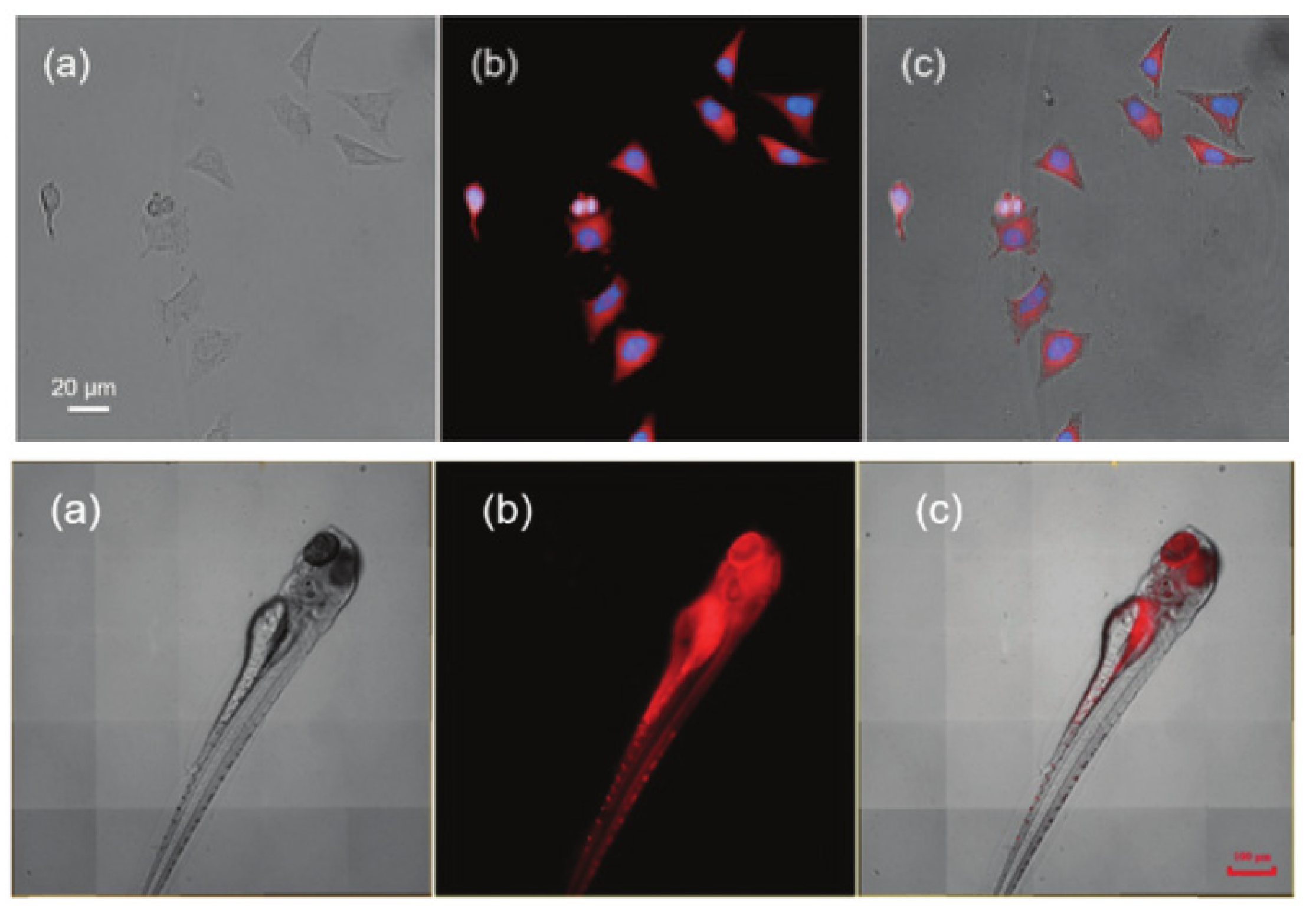
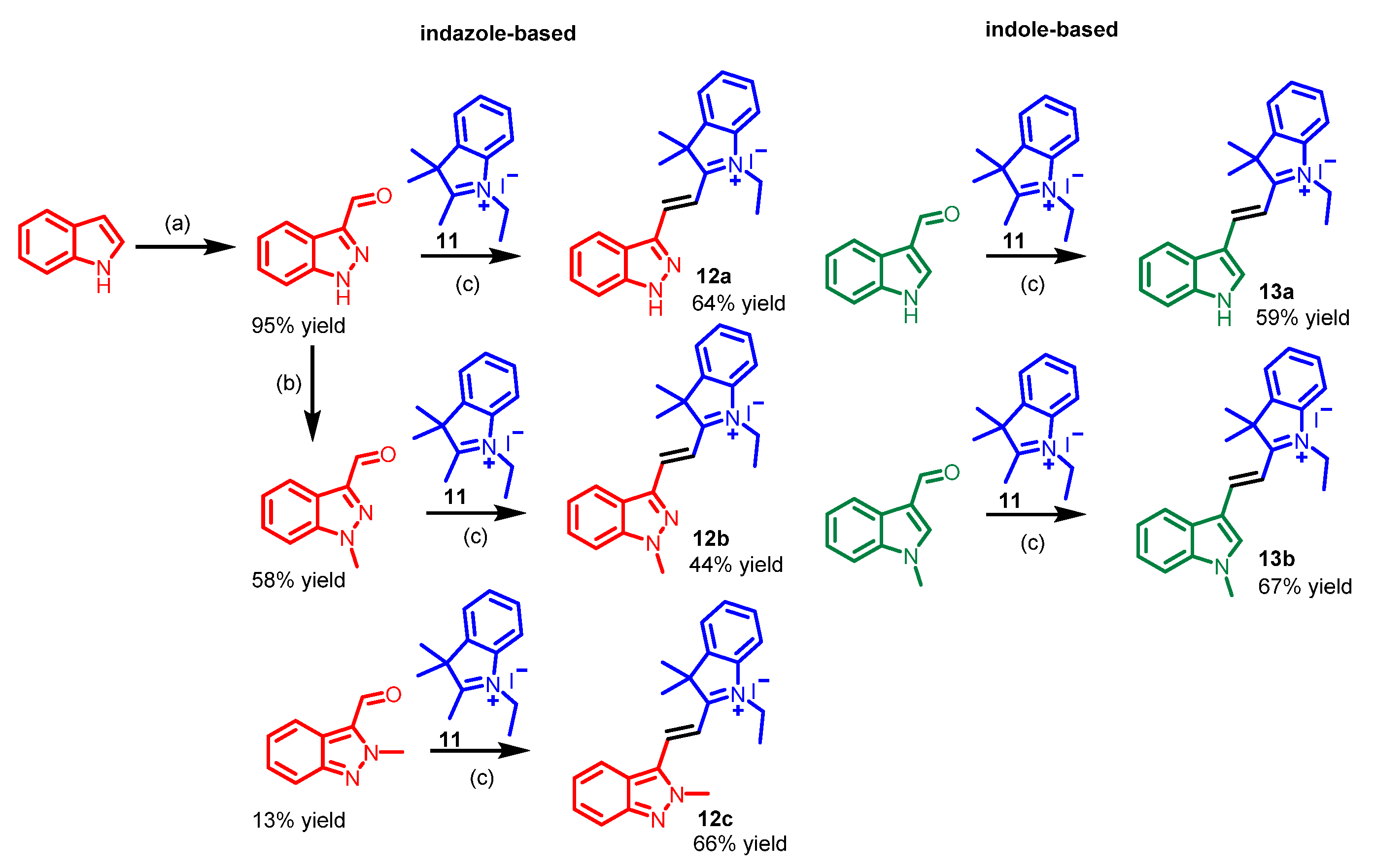
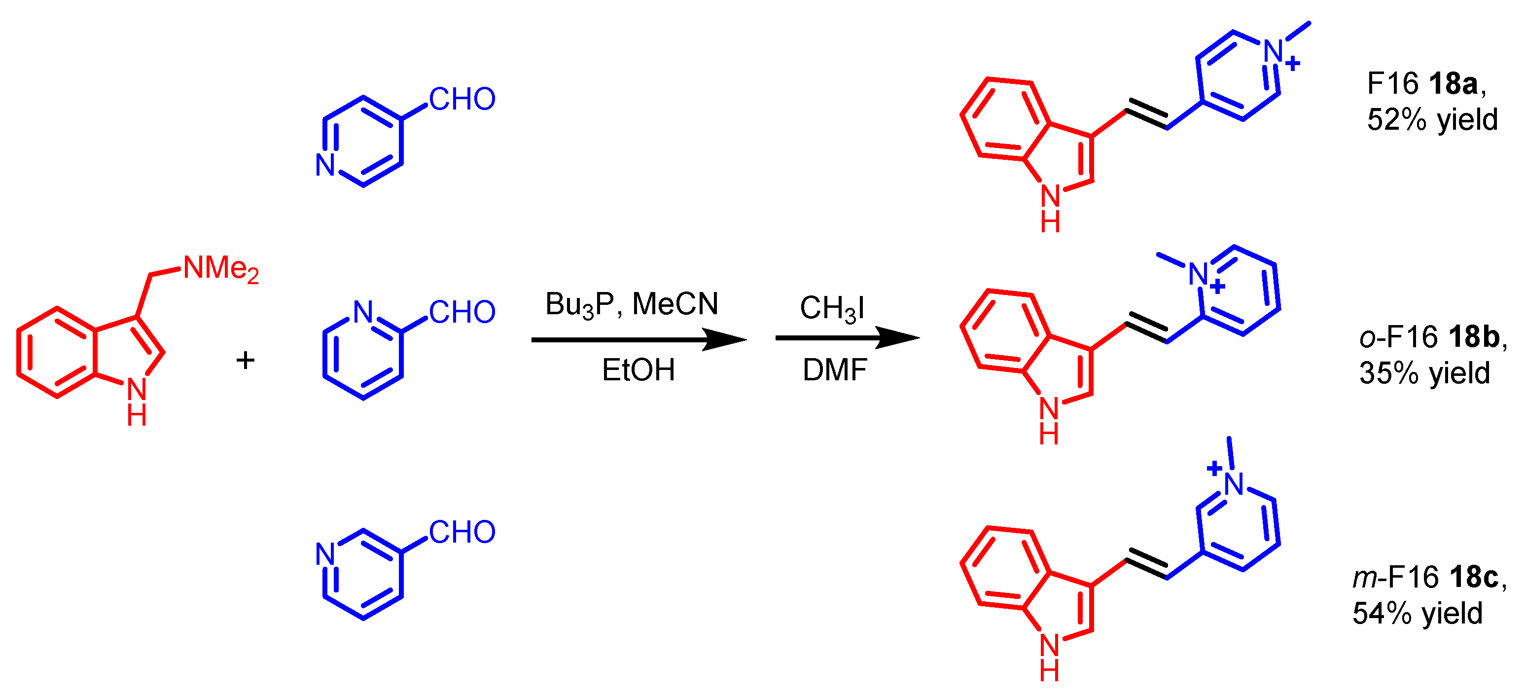
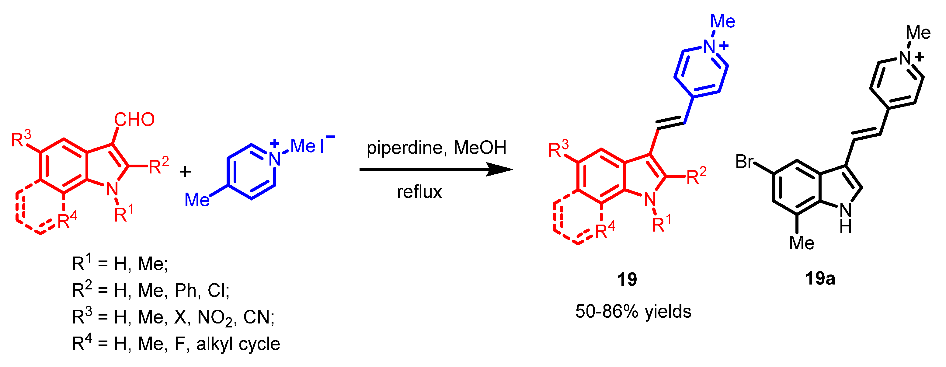
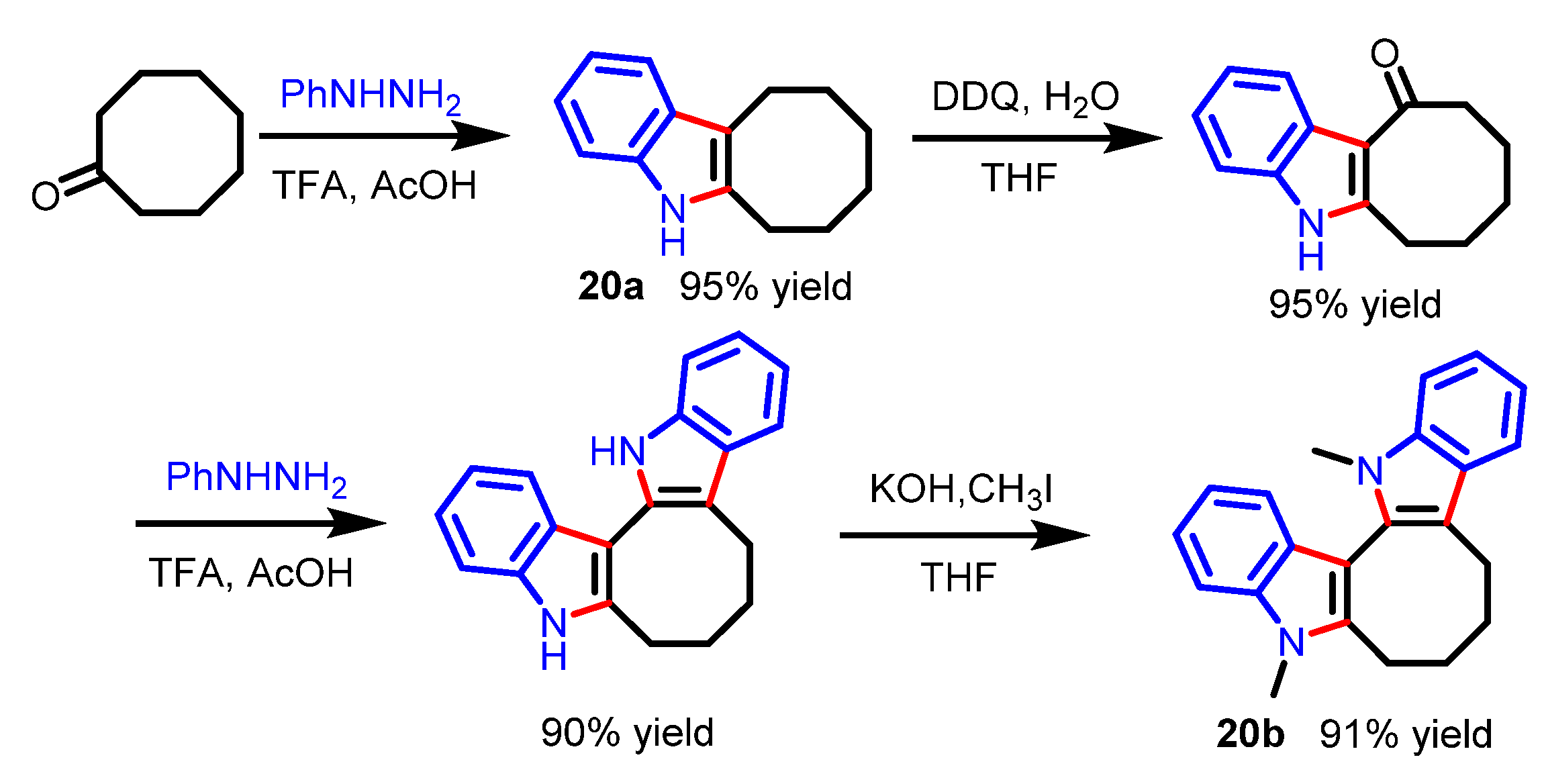
5. Indole Derivatives

Indole is an aromatic heterocyclic organic compound made of benzene and a fused pyrrole ring. The first indole was generated through Fischer indole synthesis, reported in 1883 [43]. Indole derivatives have been included in the synthesis of essential FDA-approved drugs [5]. To our knowledge, the scientific researchers generally focused on indole derivatives applied as pharmaceutical drugs [44,45]. However, only a few indole derivatives applied in cell imaging systems were reported. In this section, indole derivatives for direct cell imagining were investigated.
F16 is an indole-containing, mitochondria-targeted, broad-spectrum anticancer drug. In 2018, Liu et al. reported two isomers of F16 (o-F16 18b and m-F16 18c) with different fluorescence [46]. The F16s 18ac were synthesized through a Wittig-like reaction and a methylation of gramine with the corresponding pyridine carboxaldehyde, respectively (Scheme 14). Fluorescent images pointed out that the two isomers could selectively accumulate in the live SGC-7901 cells (Figure 15).
In 2019, Cheng et al. synthesized a series of representative F16 derivatives, 19, which could selectively accumulate in the mitochondria [47]. These F16 derivatives 19 with different isomers were prepared through a Knoevenagel condensation of 1,4-dimethylpyridin-1-ium iodide with the corresponding indole-3-carboxaldehydes (Scheme 15). Among them, compound 19a showed good anti-tumor activity in various cancer cell lines. Meanwhile, 19a could also be applied as a fluorescence probe for cancer cell imaging (Figure 16). The cell experiments revealed that the mitochondrial selectivity of F16s is driven by the negative transmembrane potential, such as other DLCs.
In 2022, Beşer et al. investigated the fluorescent probe properties of a Caulerpin derivative, 20b, as a member of the indole family [48]. For the synthesis of this bis indole compound 20b (Scheme 16), the corresponding indole 20a was synthesized by Fisher indolization reaction of cyclooctanone with phenylhydrazine. Afterwards, the DDQ/H2O/THF system selectively oxidized benzylic CH3 groups at the 3-position of the indole 20a to corresponding ketone. Indolization of this ketone by another Fisher indolization reaction and a subsequent methylation furnished the bis indole 20b in a high yield. Cytotoxic activity tests indicated the low toxicity of 20b on various cancer and healthy cell lines. Confocal fluorescence imaging showed that the probe 20b selectively stained the cytoplasm of MCF-7 cells (Figure 17). This research suggested that this molecule can be applied as a fluorescence probe for biological cell imaging.

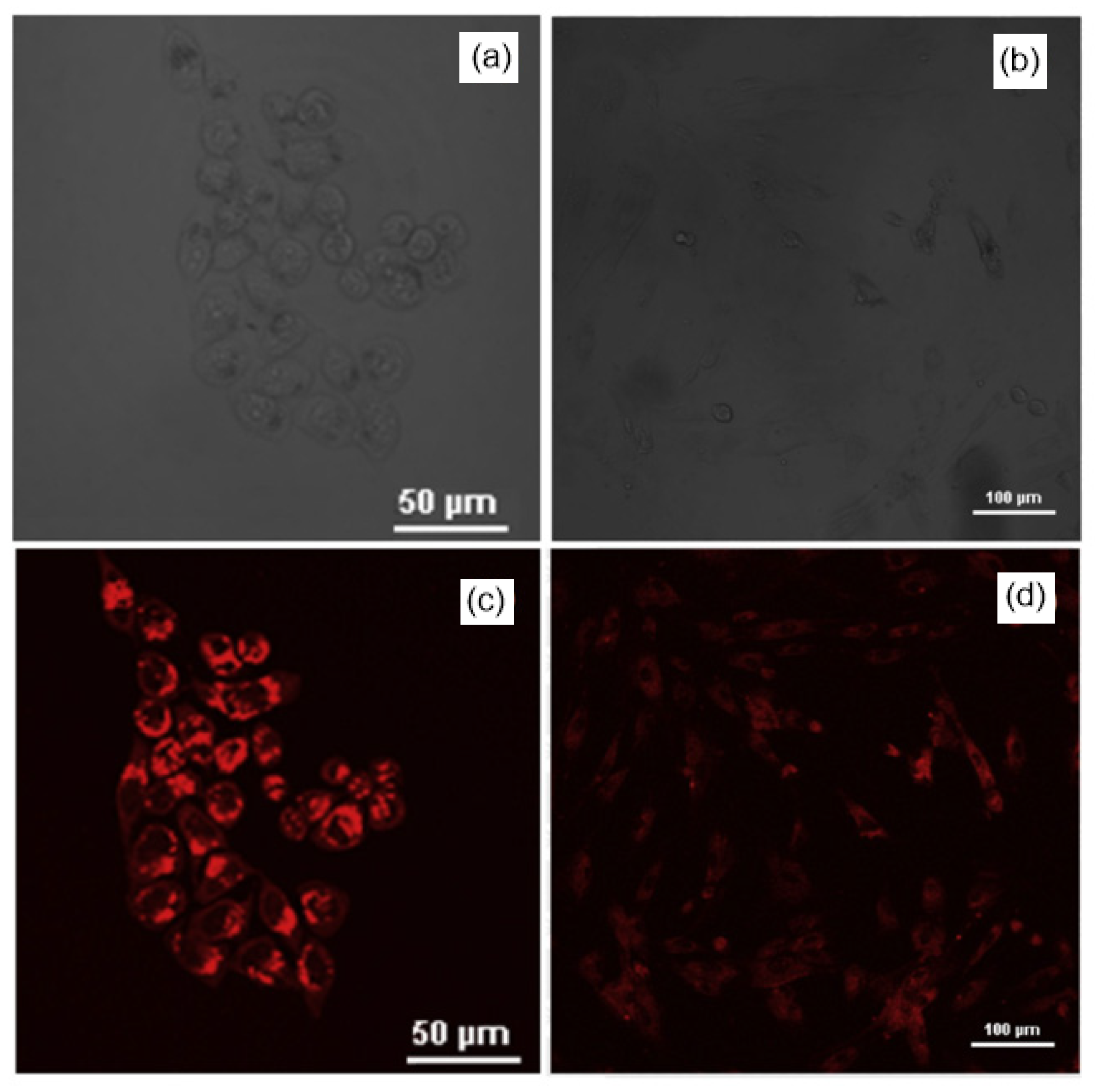
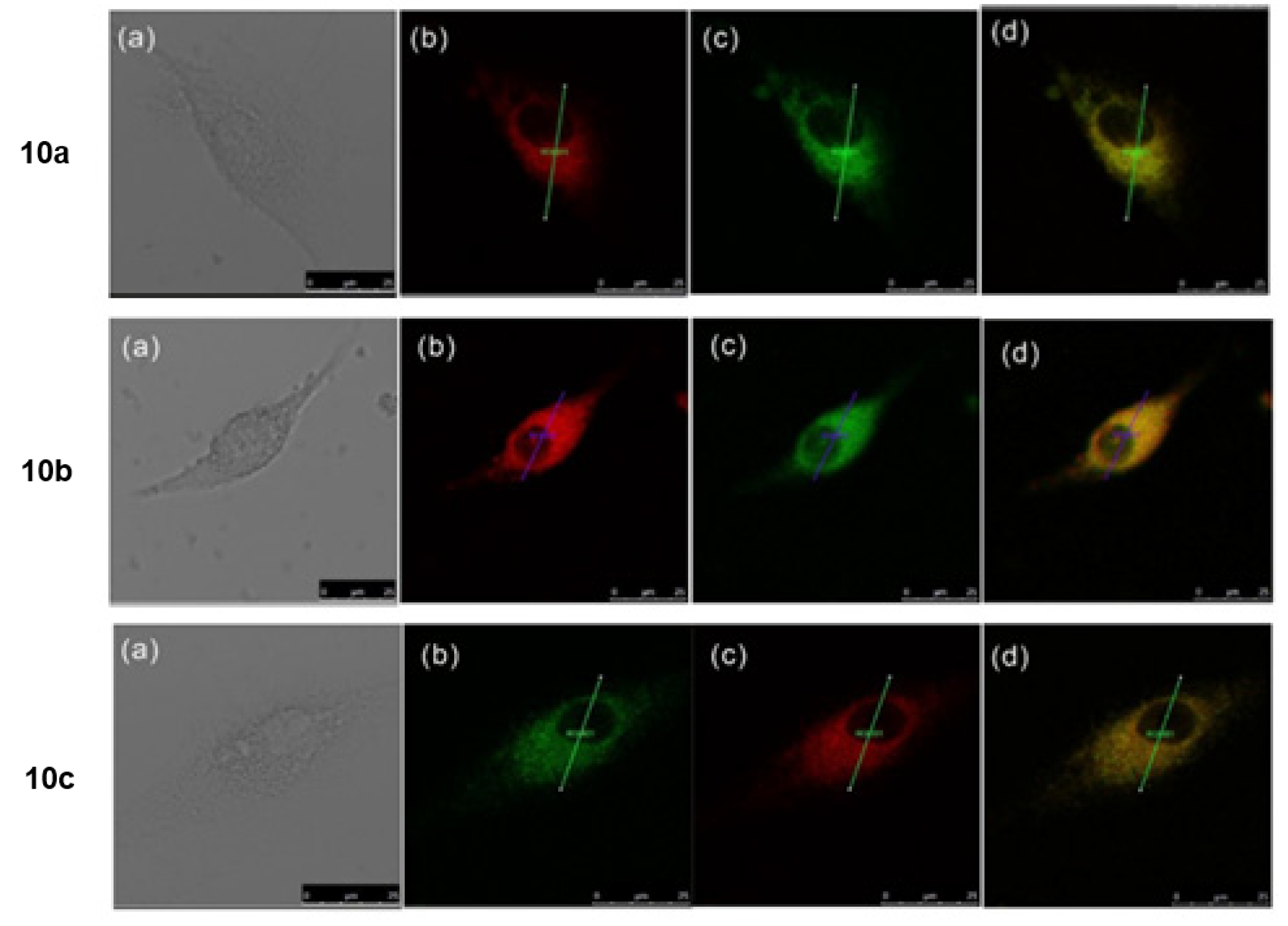
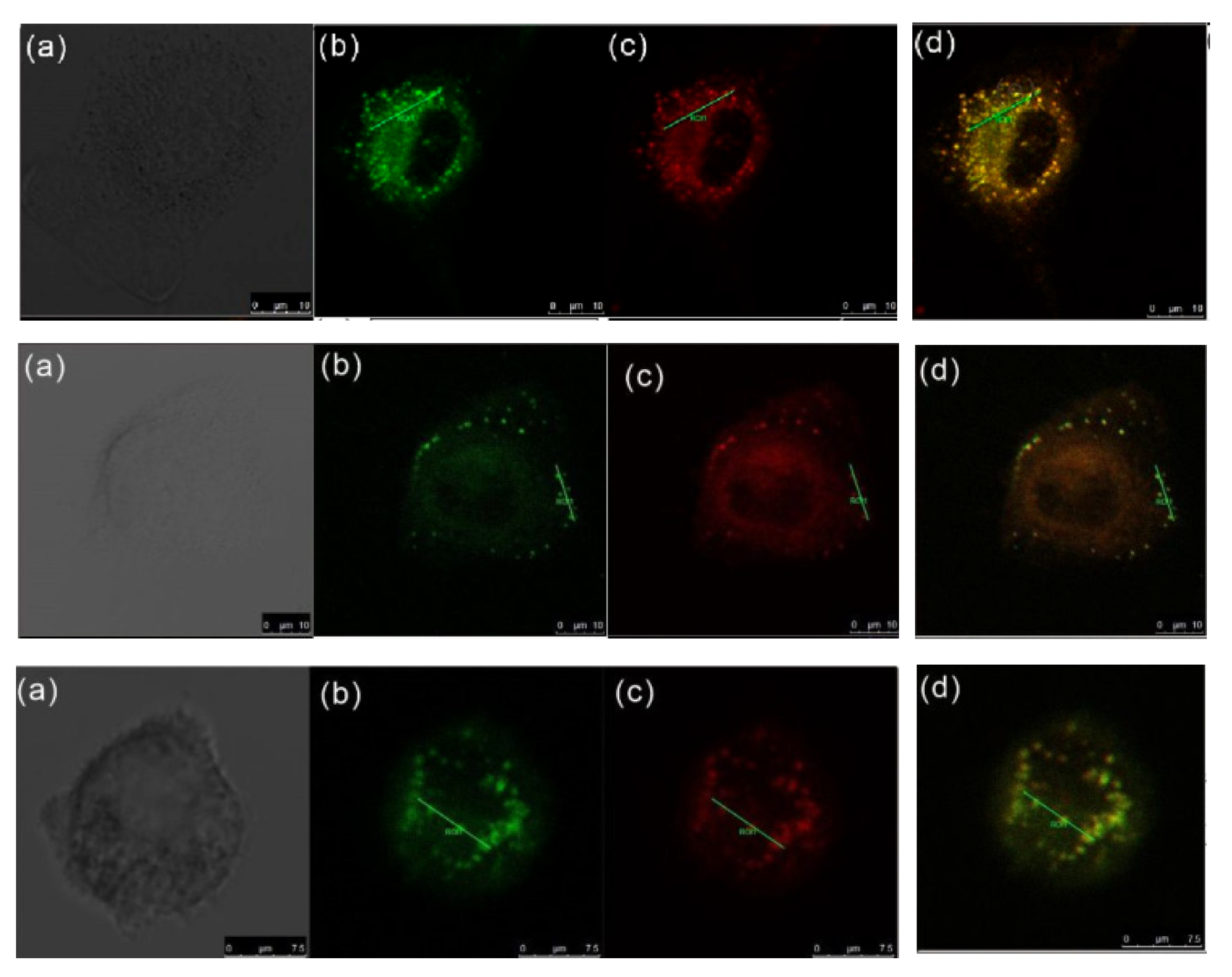
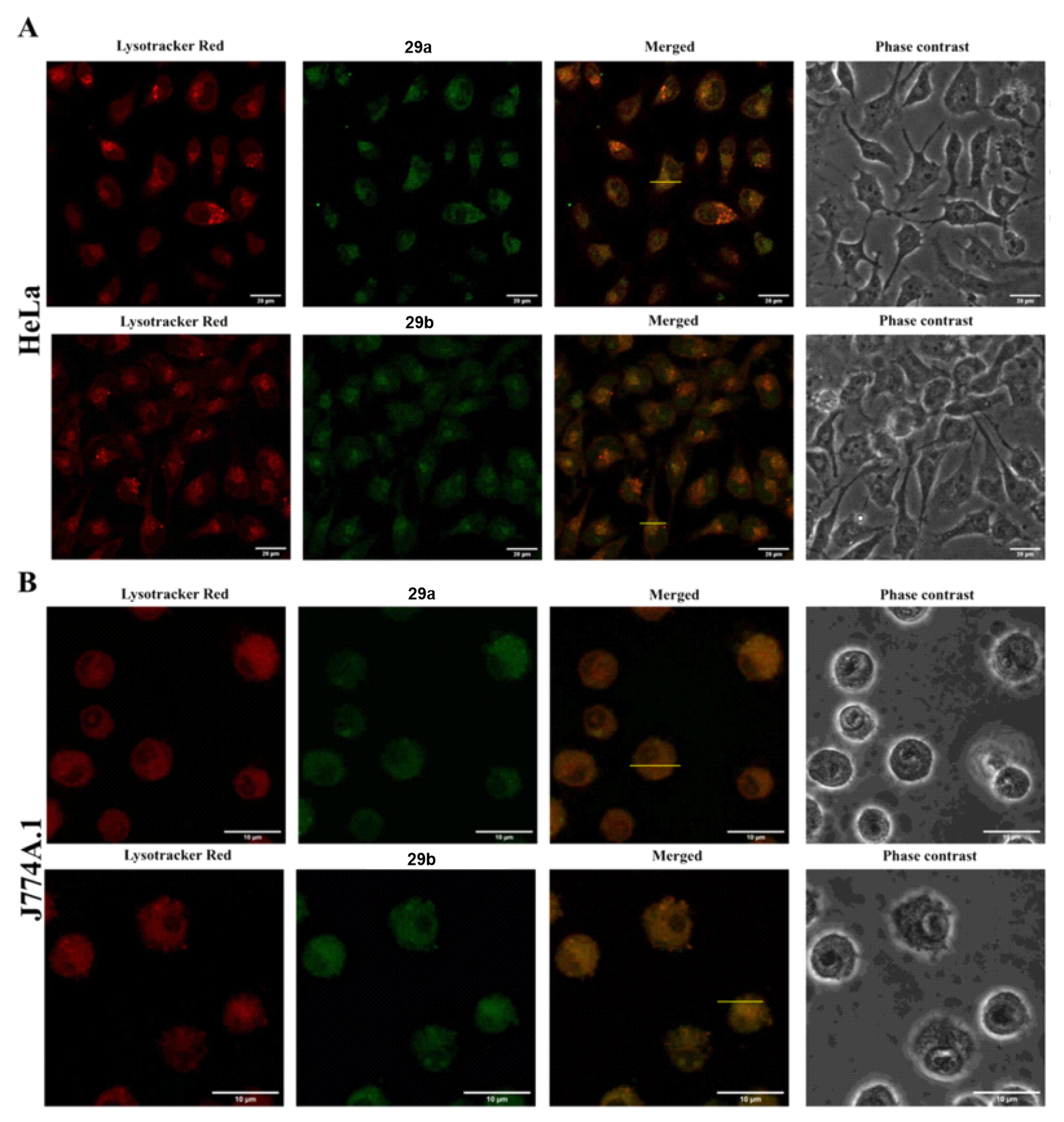
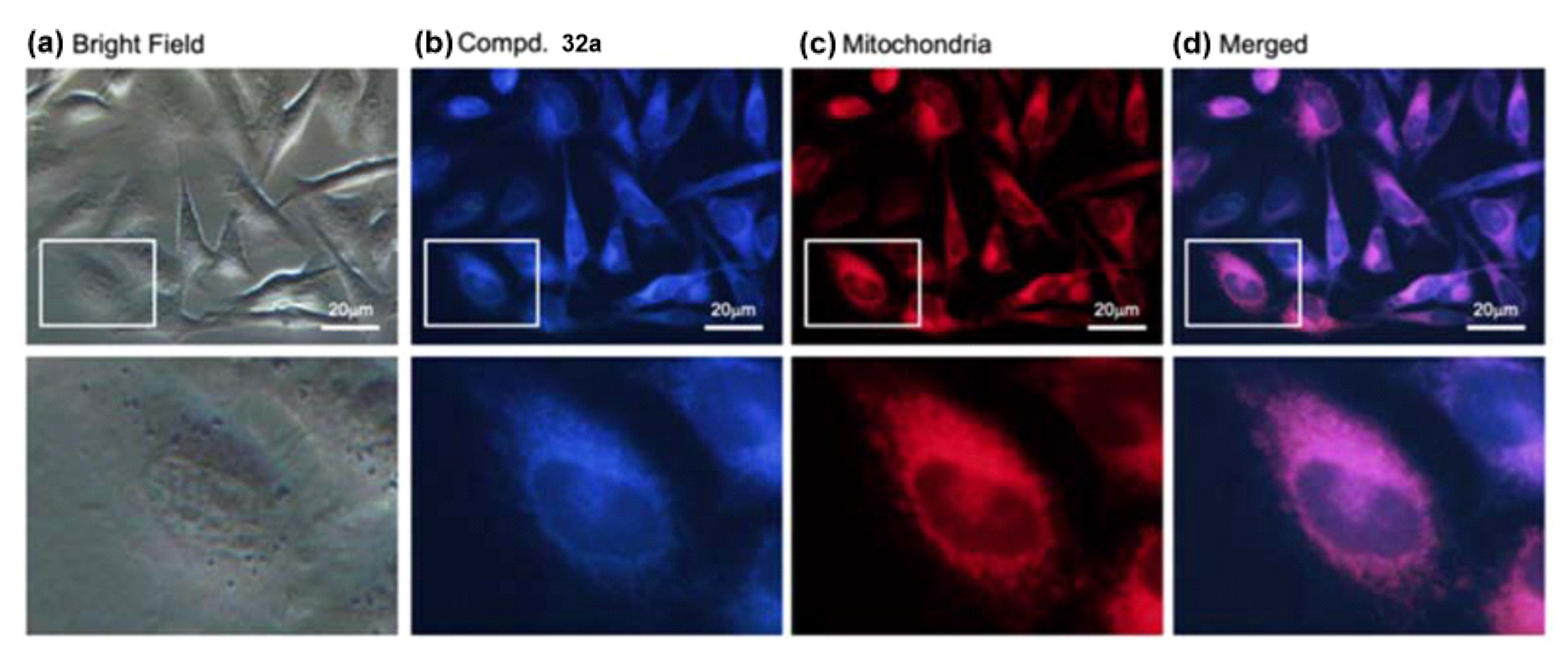
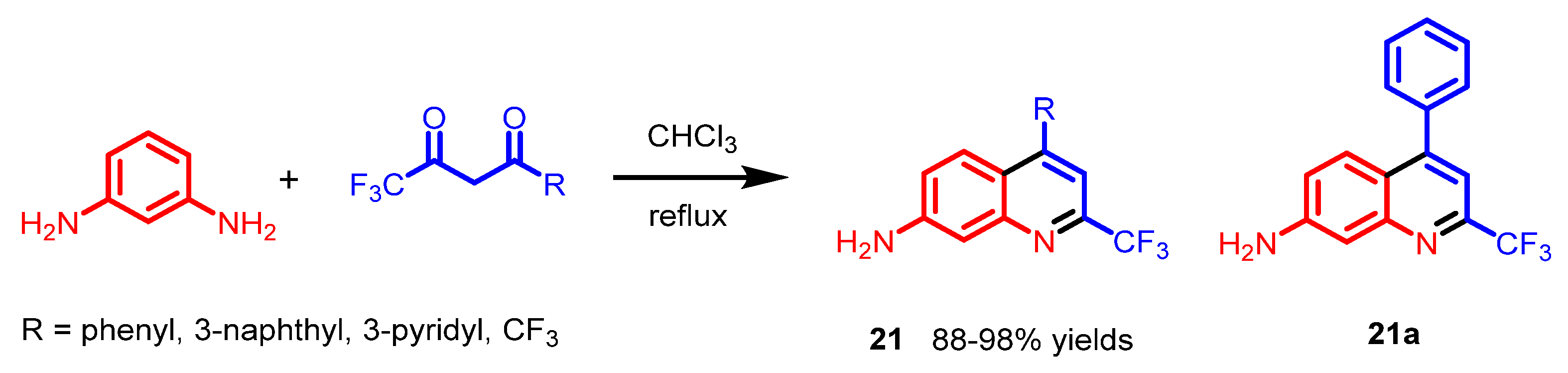
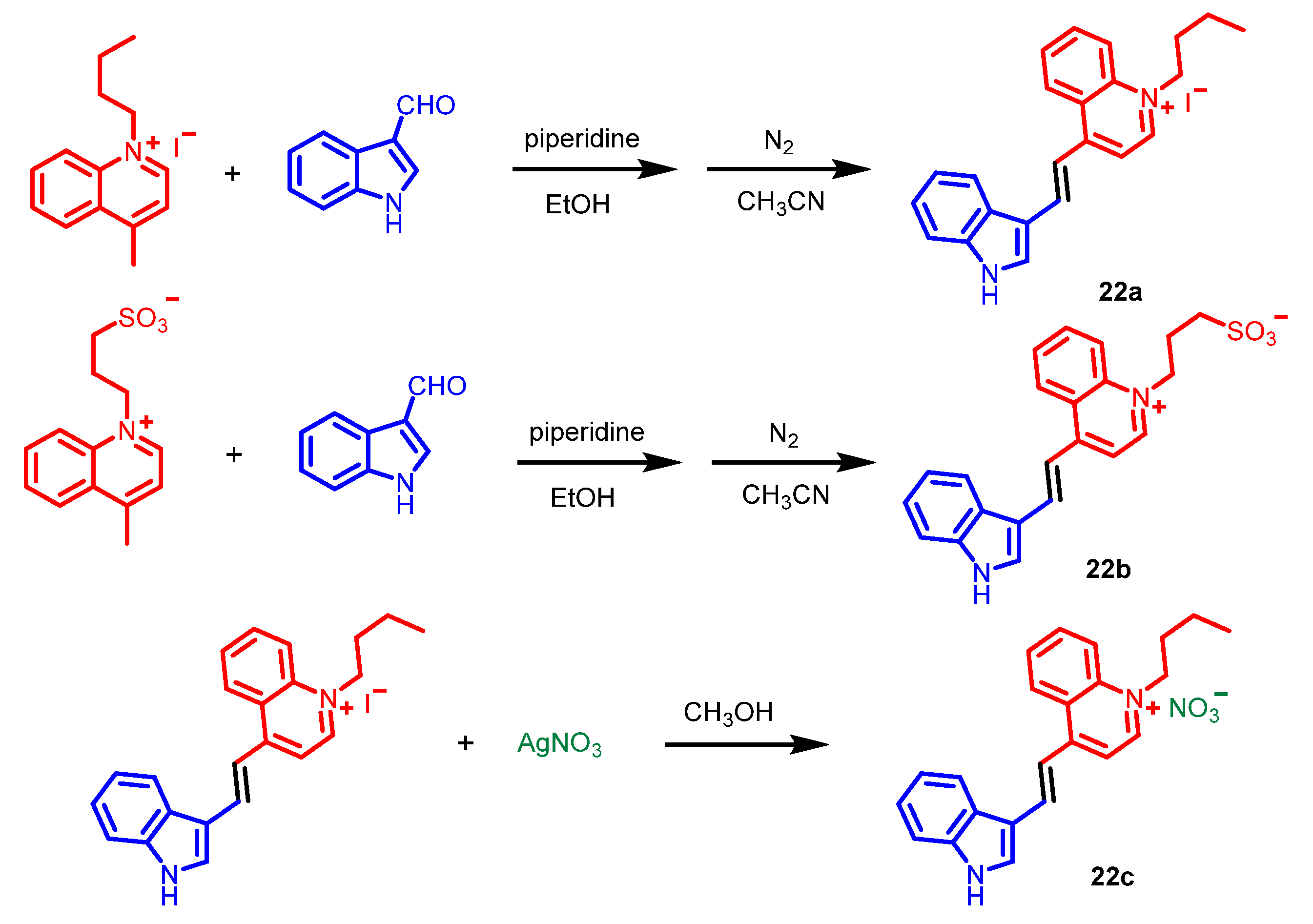
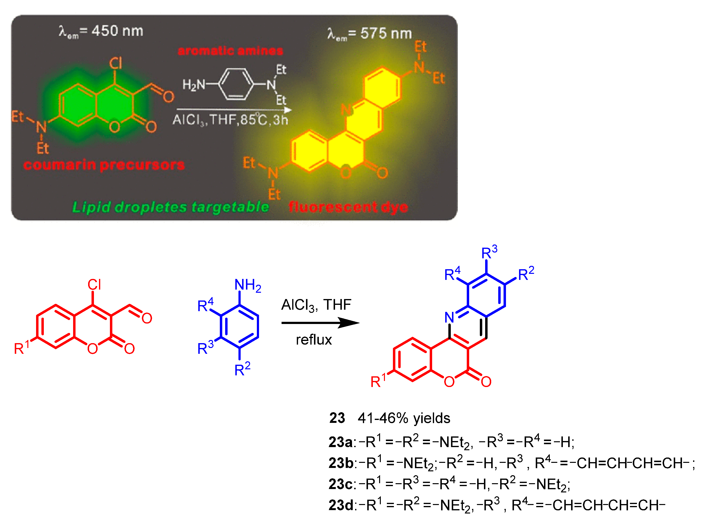
6. Quinoline Derivatives

Quinoline is an aromatic heterocycle made of benzene and a fused pyridine ring. The first quinoline was obtained through coal tar extraction in 1834 by chemist Friedlieb Ferdinand Runge [49]. Quinoline derivatives have been widely used in various fields, including pharmaceutical, biological, and industrial chemical fields [50]. In the biological field, the quinoline derivatives have been demonstrated as star molecular probes, because of their excellent biological activities [51,52]. Herein, we focus on the synthesis and applications in direct cell imaging of quinoline derivatives.
In 2019, Chen and co-workers synthesized various novel 7-aminoquinolines 21 that exhibit high selectivity towards the Golgi apparatus [53]. These 7-aminoquinoline derivatives were synthesized through a selective condensation of m-phenylenebenzene with various 1,3-diaketones (Scheme 17). Confocal images revealed that the compound 21a chosen as a representative was located in the Golgi apparatus of various cell lines (HeLa, U2OS, and 4T1 cells), specifically (Figure 18). The strong electron-withdrawing trifluoromethyl group potentially enhances the intramolecular change transfer (ICT) state of the 7-aminoquolines between the strong electron-donating amine group and the trifluoromethyl group, thus resulting in red shifts of the absorption and emission of the compounds.
In 2020, Tian et al. synthesized various water-soluble quinolineindole-based derivatives 22ac [54]. These probes were generally synthesized through a Knoevenagel condensation of 4-methylquinolinium salt derivatives with 1H-indole-3-carbaldehyde (Scheme 18). Confocal images suggested that 22c as a representative could target the nucleus and mitochondria in live and fixed cells (Figure 19A,C). Colocalization experiments using different commercial nuclear stains incubated with 22c further confirmed the nuclear and mitochondrial selectivity of 22c (Figure 19B,D–F). I or SO3 replaced by NO3 on 22c enhanced the water solubility and biocompatibility compared to 22a,b.
In 2022, Ge’s group synthesized various functional dyes 23ad with a chromeno[b]quinoline skeleton through a cyclization of coumarin derivatives with aromatic amines in the presence of the catalyst anhydrous AlCl3 (Scheme 19) [55]. Moreover, the optical performance, toxicity, cell imaging, and calculations of these dyes were comprehensively evaluated. Among these functional dyes, probes 23ac, which possess a diethylamine group as a chromophore, exhibited ideal fluorescence performance, and could be applied as fluorescent markers to lipid droplets in HeLa cells (Figure 20). Compared with probes 23ac, the structure of probe 23d may lack the diethylamino group to reduce its lipophilicity, thus resulting in the inability to penetrate the organelle membrane.

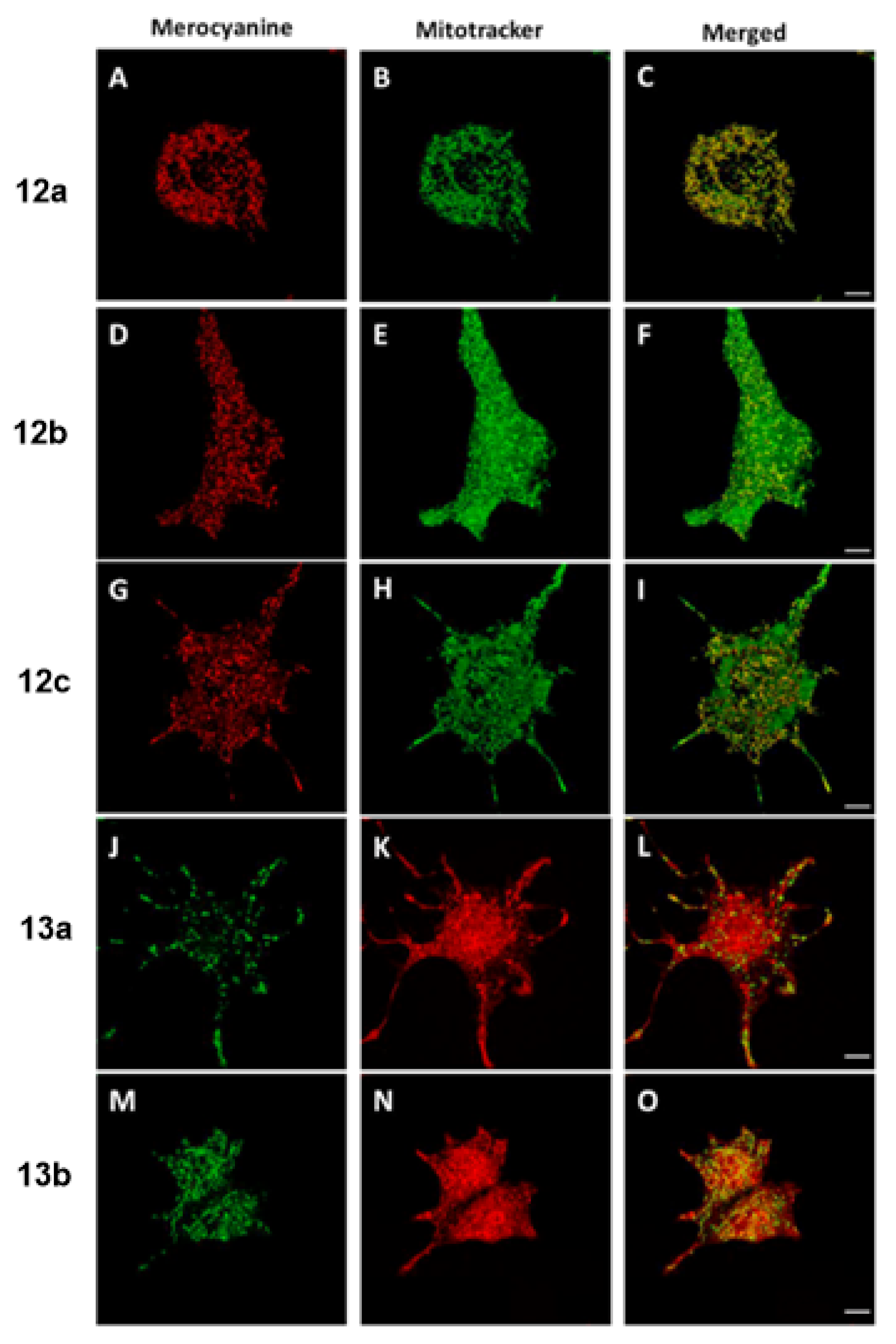
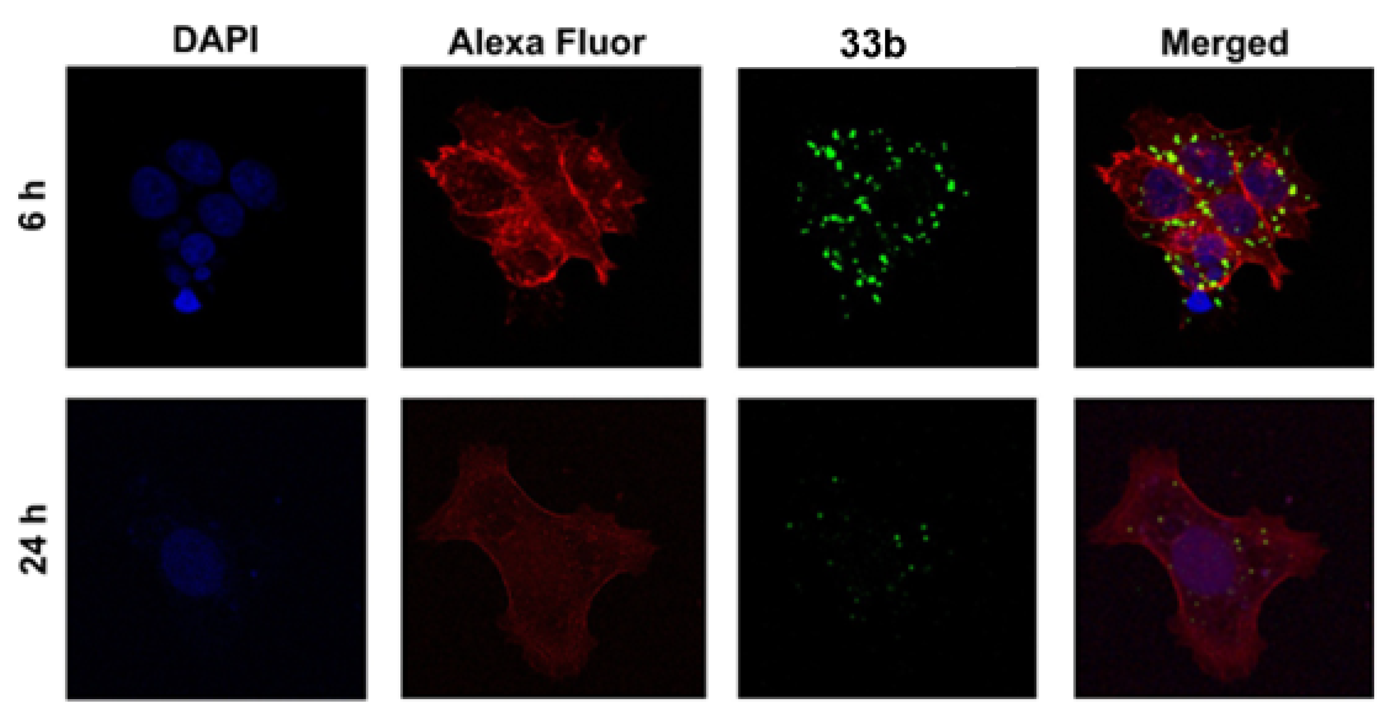
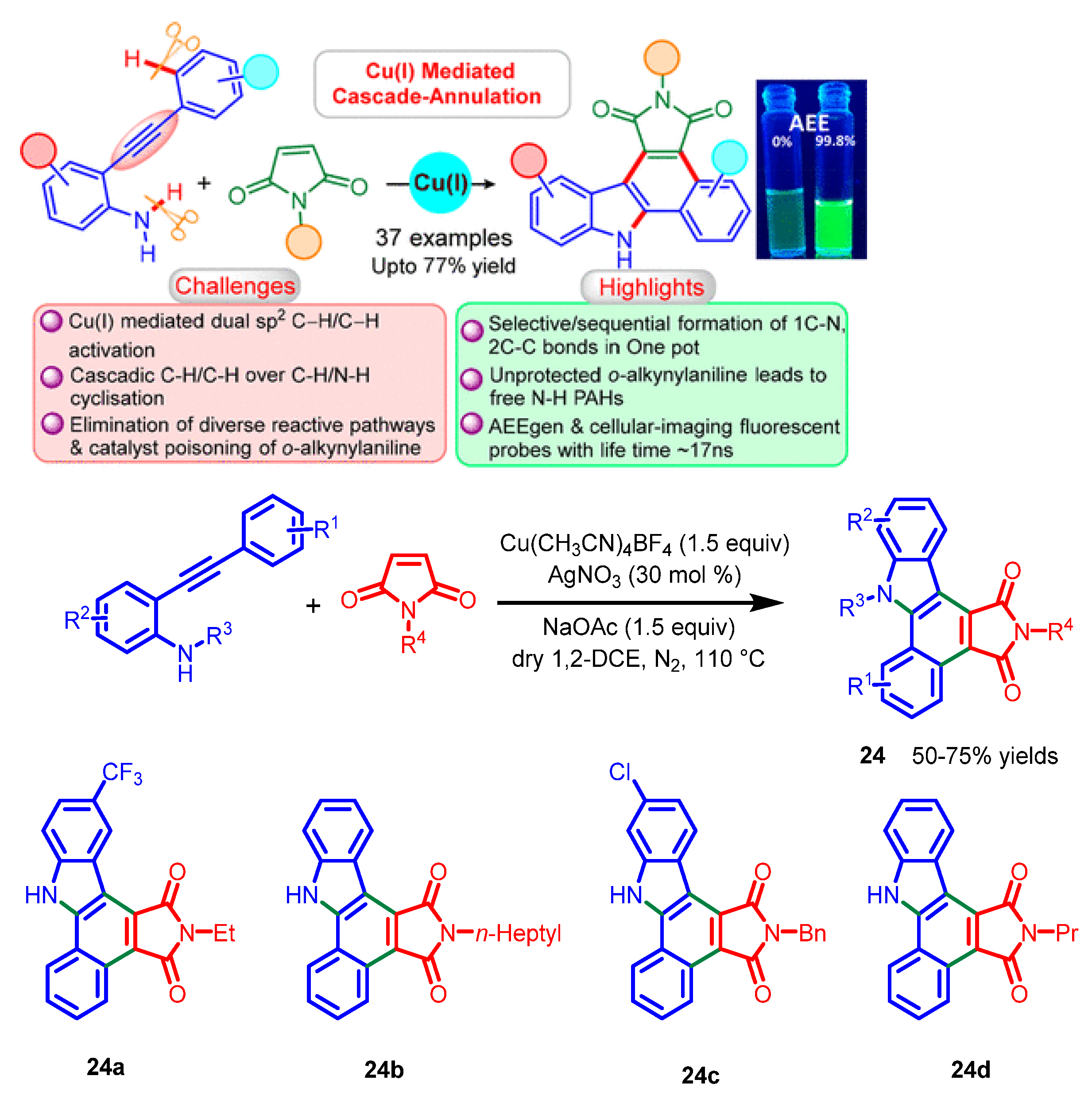
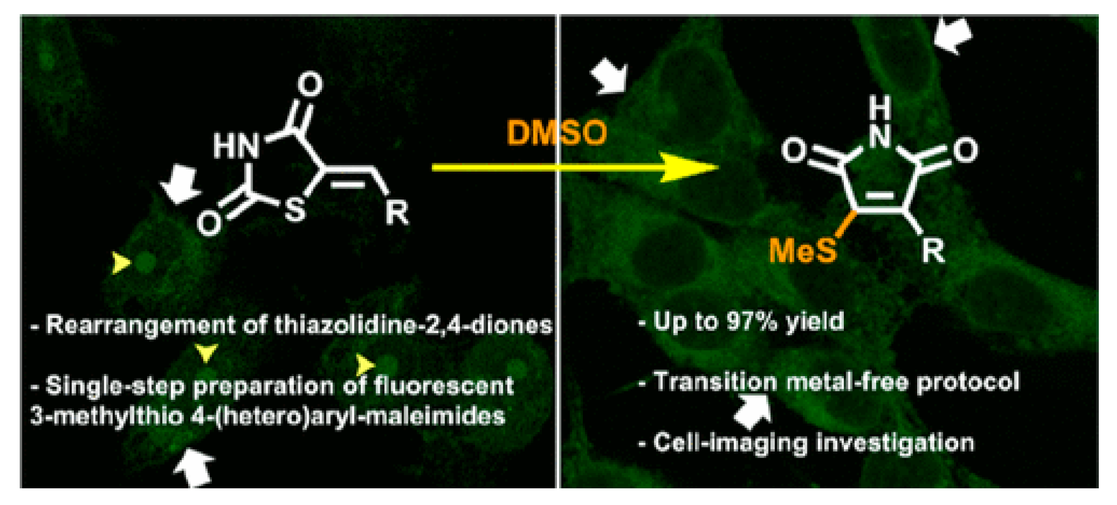
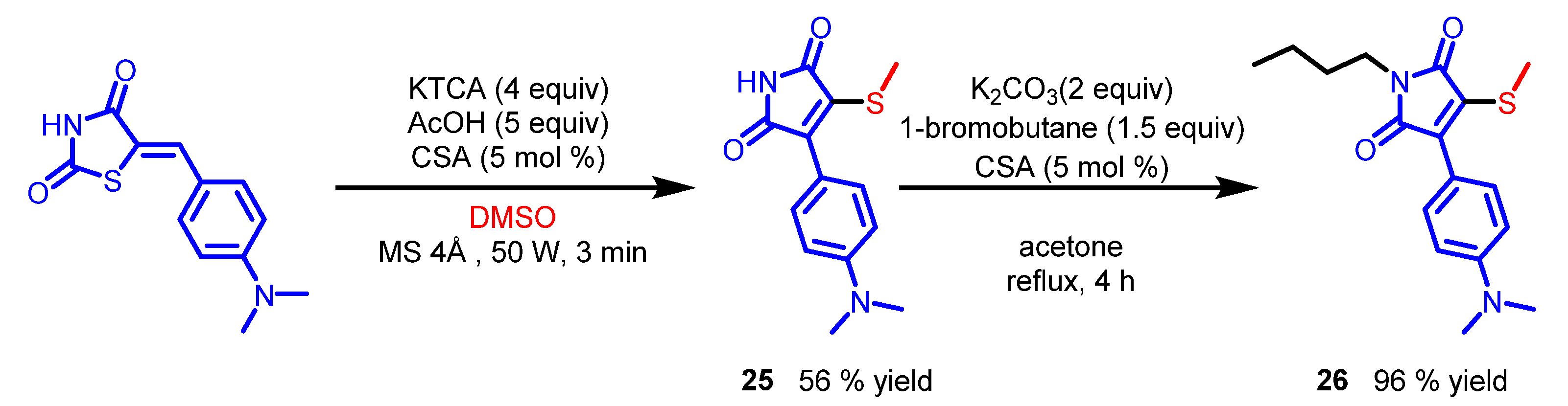
7. Maleimide Derivatives
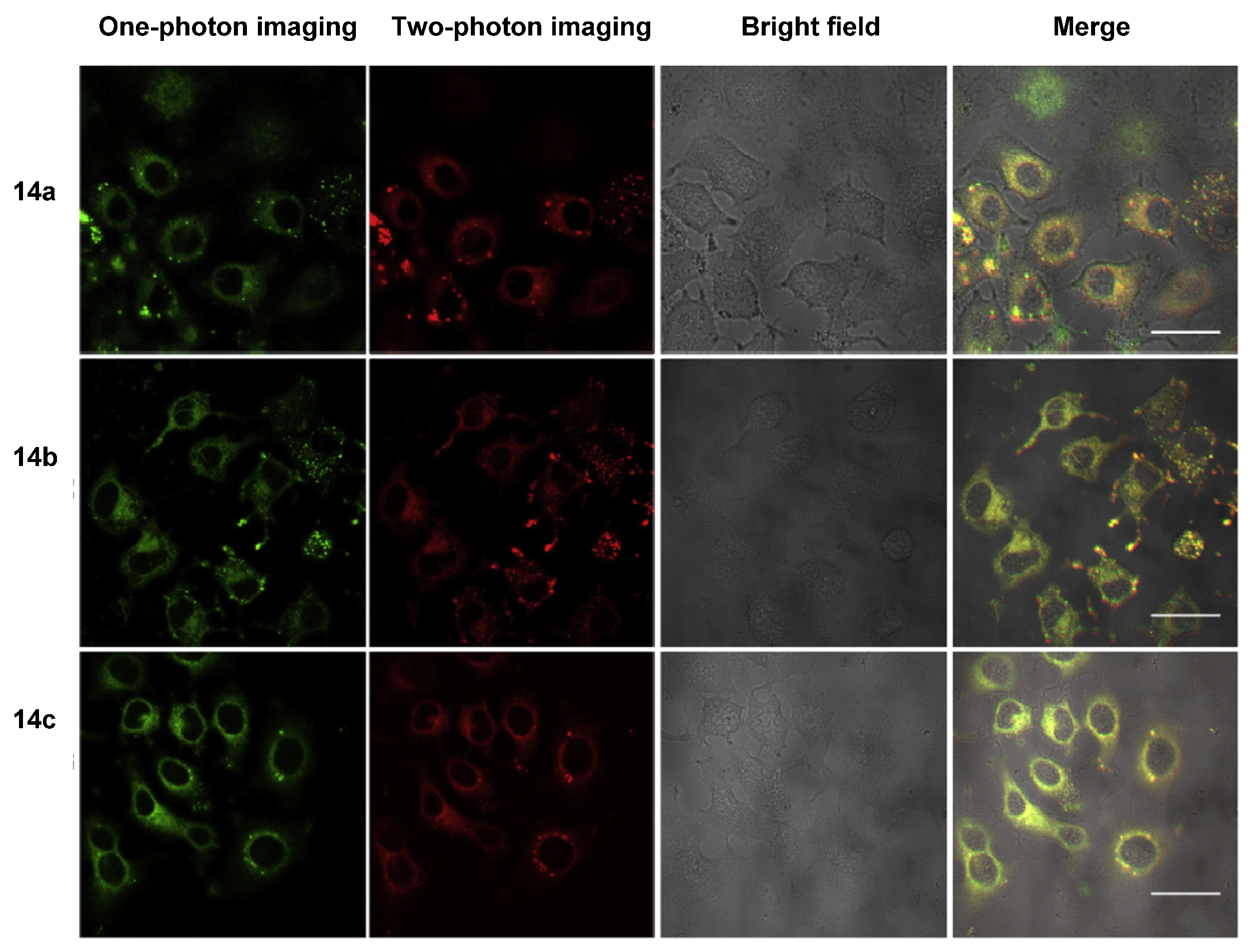
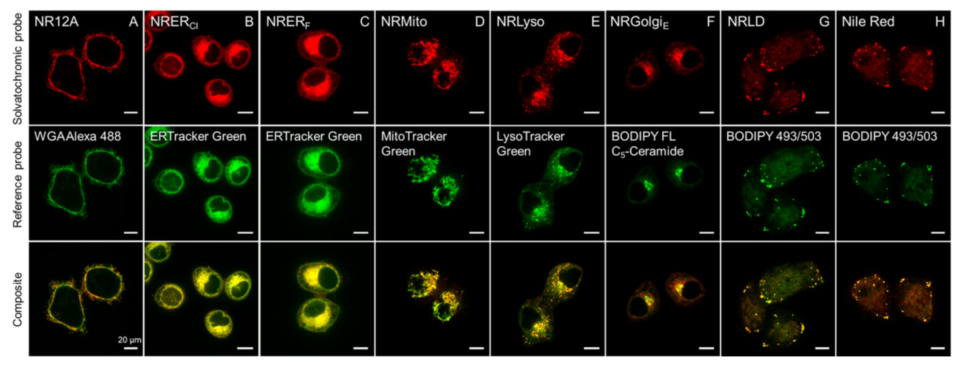
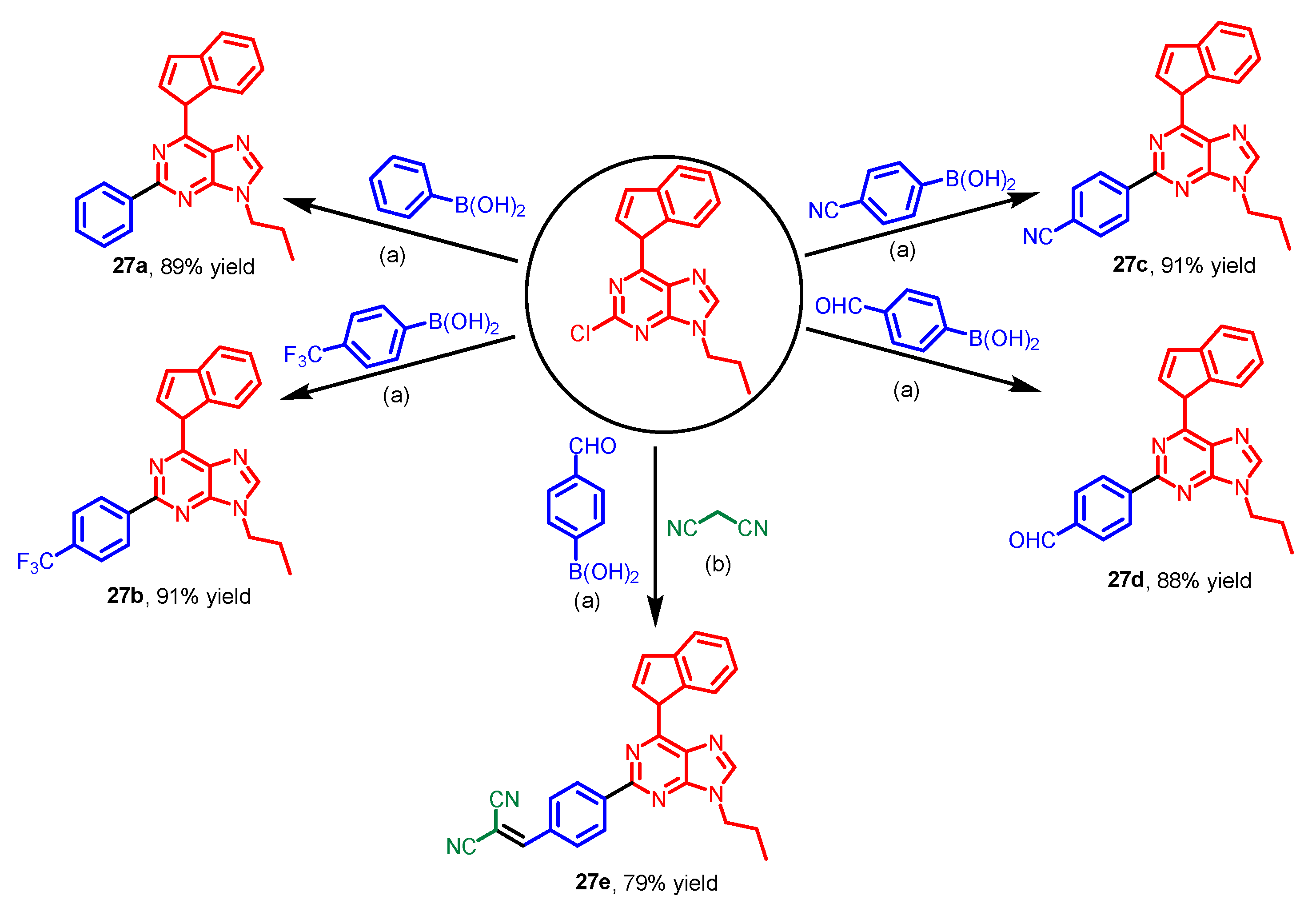
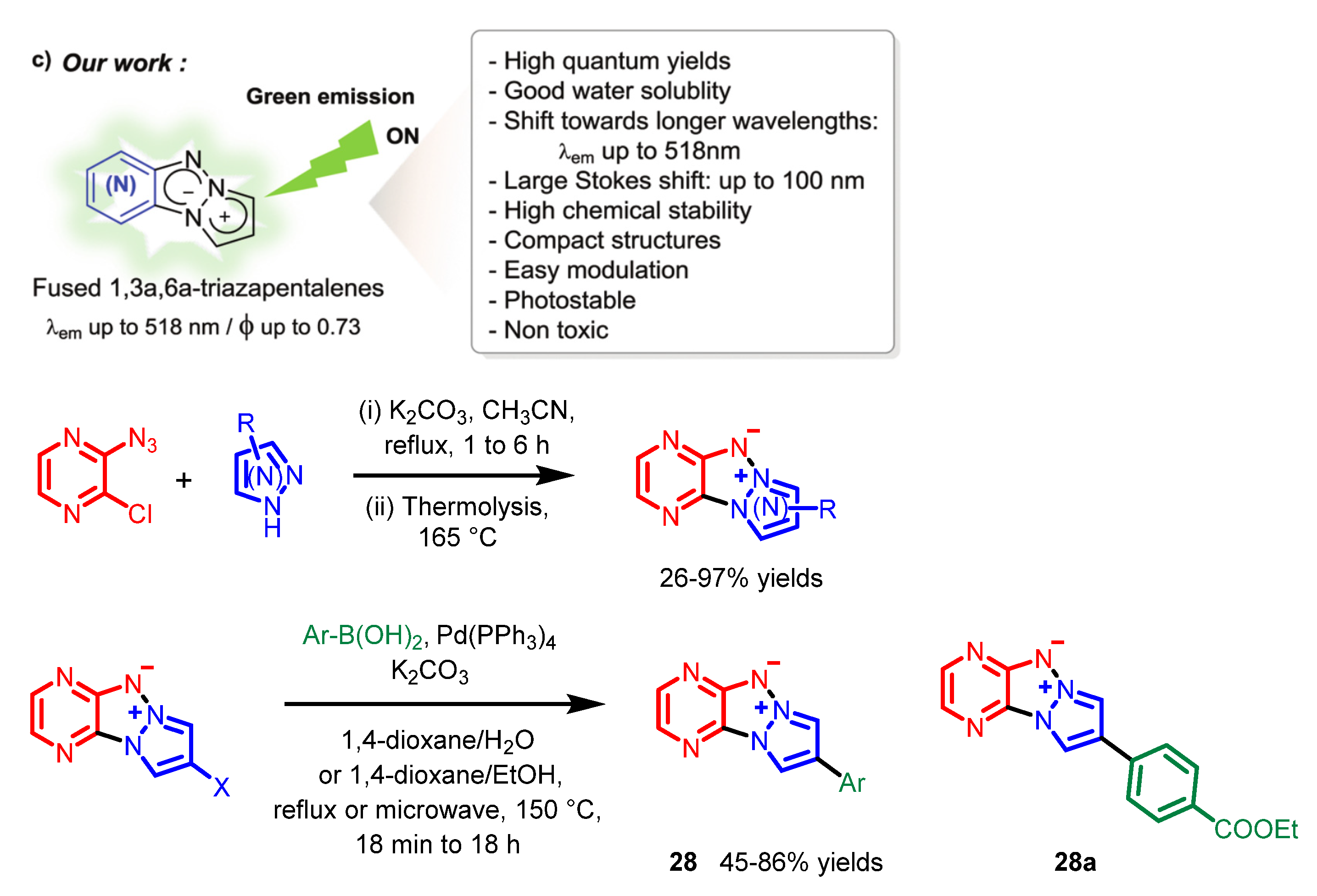
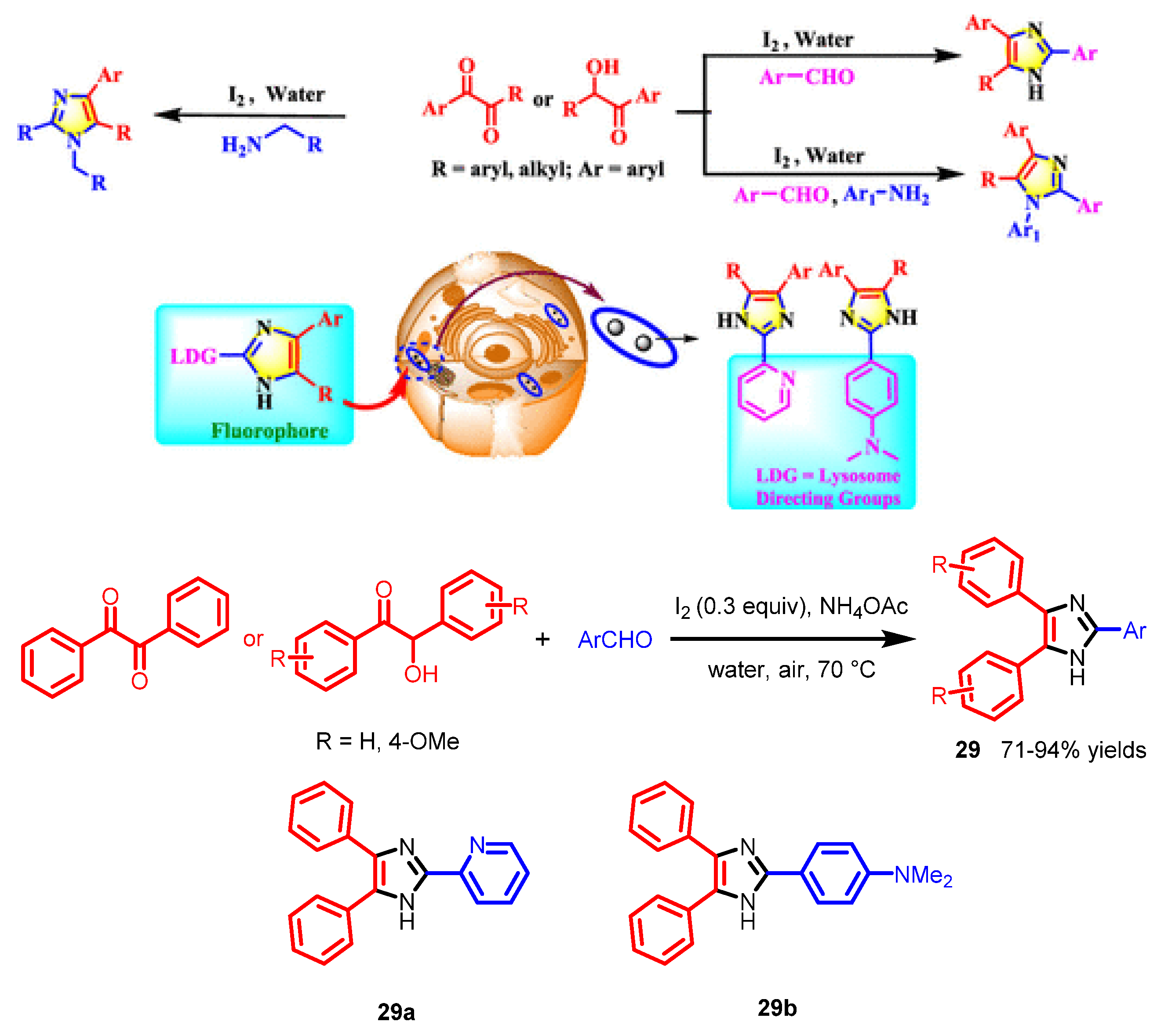
Maleimides are generally synthesized by dehydration of maleic anhydride with amines [56]. Maleimide derivatives have exhibited various biological properties which can be applied in medicinal and natural product chemical fields [57,58]. In the bioimaging field, due to the fluorescence properties of the maleimide group, maleimide derivatives have been employed as fluorescence probes for biological cell imaging [59,60]. Here, the direct cellular fluorescence imaging of maleimide derivatives is discussed.
In 2021, Patel et al. synthesized a series of maleimide derivatives with bright fluorescence [61]. These compounds were obtained through the tandem intra- and inter-molecular cyclization of o-alkynylanilines with maleimides (Scheme 20). Confocal images revealed bright fluorescence of HeLa cells stained with the representative compounds 24ad (Figure 21).
Recently, Amarante and co-workers reported a transition metal-free approach to obtain maleimide derivative 25 through a novel rearrangement from thiazolidine-2,4-diones in one step [62]. N-butylmaleimide 26 was obtained through a substitution reaction of compound 25 and 1-bromobutane (Scheme 21). Compounds 25 and 26, which emitted green and red fluorescence, were chosen as respective compounds to study. Confocal images indicated that compound 25 could stain the cytoplasm in live and fixed cells. A mild staining in the cell nucleus could be observed (Figure 22A−C). Compound 26 stained the perinuclear region preferentially, perhaps due to its accumulation in the mitochondria (Figure 22D−F).

8. Others

There have been some other nitrogen-containing heterocycles with bioimaging functions reported in recent years. These N-heterocycles containing different amounts of nitrogen have various biological features. However, in recent years, they have not been systematically studied. In this section, we summarize the direct cell imaging of these nitrogen-containing heterocycles based on the amount of nitrogen for reference.
In 2018, Yu’s group reported various novel purine-based AIEgens 27ae, which were generated via the Suzuki coupling reaction (Scheme 22) [63]. The compounds have the advantages of high selectivity, low background, and good biocompatibility. Cell imaging experiments showed that these probes could selectively stain lipid droplets and have good photostability, similar to the commercial dyes (Figure 23). A D–p-A structure was constructed to build the tunable emission AIE fluorophores. Purine was chosen as the core structure, indole was chosen as the electron donor, and the electron acceptor was changed to regulate the emission. Meanwhile, an n-propyl group was introduced to improve the lipophilicity.
In 2019, based on Namba’s group’s work [64], Suzenet et al. reported substituted triazapentalenes 28 which exhibited good fluorescent properties [65]. These novel triazapentalene compounds were synthesized through a one-step cyclization and a Suzuki cross-coupling reaction (Scheme 23). These fluorescent probes have the advantages of high quantum yields, good photostability, and large Stokes shifts, which is suitable for optical imaging applications. Photobleaching experiments suggested that representative compound 28a shows good photostability (Figure 24). The fused diazine on the triazapentalene ring induced strong red shifts of the emission and increased quantum yields due to a plausible ICT process.
Imidazoles are ubiquitous N-containing heterocyclic molecules in natural products and drugs [66,67]. In 2020, Banerji and co-workers synthesized substituted imidazoles 29 through an oxidative cyclization with the diketone or α-hydroxy ketone, aromatic aldehyde, and amine source catalyzed by iodine (Scheme 24) [68]. This methodology has advantages of being peroxide-, transition metal-, and organic solvent-free. It could be employed at the gram-scale level. Due to the excellent fluorescence properties of these molecules, two of the derivatized imidazoles, 29a and 29b, were modified with lysosome-directing groups. These two molecules showed bright blue fluorescence of lysosomes in human and murine cells, which could be applied as lysosome-targeted probes (Figure 25).
In 2020, Yagishita et al. synthesized various novel quaternized imidazo[1,2-a]pyridine dyes 32ad, excited with blue light [69]. For the synthesis of these compounds (Scheme 25), 2-iodinated imidazo[1,2-a]pyridine 30 was synthesized through a copper-catalyzed oxidative coupling of 2-aminopyridine with phenyl acetylene in the presence of I2 [70]. Afterwards, compound 31 was generated through a Sonogashira coupling reaction of compound 30 with 1-ethynyl-4-methoxybenzene. The alkylation of 31 with different alkyl iodides yielded various corresponding products, 32ad. The HeLa cells were stained with the compound 35a, and colocalization experiments using a commercial mitochondrial marker, further confirming that representative salt 32a could target the mitochondria (Figure 26). The D−π-A structure plays a key role for the two- and three-photon fluorescence imaging. The p-methoxyphenyl ring was chosen as the electron donor and the cationic imidazo[1,2-a]pyridine was chosen as the electron acceptor.
In 2021, Ornelas and co-workers reported the imidazo[1,2-a]pyrimidine compounds which could be applied for fluorescence imaging and PDT [71]. The alkoxylated alcohols 33a and 33b were obtained through a condensation, a one-step cyclization, and a Williamson reaction, respectively (Scheme 26). Fluorescence images indicated the low toxicity of representative compound 33a in the dark and showed bright fluorescence in the intracellular medium of MCF-7 cells (Figure 27). Compared with the imidazopyridine 33a, the third nitrogen atom in the imidazopyrimidine 33b is important for the solubility in the cell culture medium as well as the cell penetration process.
Additionally, in 2021, Klymchenko et al. reported a series of fluorescent probes 34 based on Nile Red for specific targeting of different organelle, including the endoplasmic reticulum, Golgi apparatus, lysosomes, lipid droplets, mitochondria, and plasma membranes [72]. These organelle markers were generally synthesized through a one-step amidation reaction (Scheme 27). These probes were incubated with live KB cells with different commercial markers and could show significant colocalization with the corresponding markers (Figure 28).
Recently, Lavis et al. reported a series of novel mitochondrial stains 35af based on 2,7-diaminobenzopyrylium (DAB) dyes excited with violet light [73]. To obtain the corresponding probes (Scheme 28), different known 2,7-diaminobenzopyrylium (DAB) dyes were treated with Et3OBF4 to generate 2-ethoxychromenylium intermediates, and the intermediates were reacted with diethylamine afterwards. These probes could stain in mitochondria because of their positively charged scaffolds, while the julolidine-based derivatives 35ce showed brighter fluorescence due to increased lipophilicity of the compact cationic structure. DAB 35e and diDAB 35f showed excellent mitochondrial-targeting ability (Figure 29a). After media exchange washes, the DAB 31e signal rapidly decreased, whereas the diDAB 35f was retained (Figure 29b).

9. Conclusions

The small molecules applied for direct fluorescence cell imaging are summarized in Table 1 according to different scaffolds of nitrogen-containing heterocycles for clarity.
In summary, nitrogen-containing heterocycles play an important role in chemical and biological fields, which has attracted scientific researchers to develop new approaches for their synthesis. In this review, the synthesis and application in direct fluorescence cell imaging of N-heterocyclic organic small molecules were described. In organic synthesis, many classical reactions to obtain N-heterocycles were applied, and meanwhile, various novel approaches were developed. In biological imaging, fluorescence imaging for direct staining of the live cells, cytoplasm, or various organelles was described.
Looking forward, there remain some challenges to the direct fluorescence cell imaging of nitrogen-containing heterocycles, including: (1) How to develop more efficient and green approaches to synthesize desired nitrogen-containing heterocycles in organic synthesis. (2) It is still a challenge to develop N-heterocyclic dyes with high photostability, high-fluorescence quantum yields, good water-solubility, and good biocompatibility through molecular structural modification. (3) The live-cell fluorescence imaging of N-heterocyclic organic small molecules in the NIR region is rarely developed. Regarding these challenges, we hope that the summarization of live-cell fluorescence imaging of N-heterocyclic organic small molecules in this review will provide useful guidance and enlightenment for further development of fluorescence cell imaging.

Author Contributions

J.S. wrote the abstract, introduction, nitrogen-containing boron compounds, cyanine, pyridine derivatives, indole derivatives, quinoline derivatives, others, and conclusion sections. Y.L. wrote the maleimide derivatives section and revised the manuscript. T.L. revised the final version of the manuscript. All authors have read and agreed to the published version of the manuscript.

Funding

This research was funded by Anhui Provincial Department of Education, Natural Science Key Project, grant number KJ2021A0250, and Anhui Medical University Youth Fund, grant number 2021xkj019.

Institutional Review Board Statement

Not applicable.

Informed Consent Statement

Not applicable.

Data Availability Statement

Not applicable.

Conflicts of Interest

The authors declare no conflict of interest.

References

  1. Medway, A.M.; Sperry, J. Heterocycle construction using the biomass-derived building block itaconic acid. Green Chem. 2014, 16, 2084–2101. [Google Scholar] [CrossRef]
  2. Leeson, P.D.; Springthorpe, B. The influence of drug-like concepts on decision-making in medicinal chemistry. Nat. Rev. Drug Discov. 2007, 6, 881–890. [Google Scholar] [CrossRef] [PubMed]
  3. Ju, Y.; Varma, R.S. Aqueous N-heterocyclization of primary amines and hydrazines with dihalides: Microwave-assisted syntheses of N-azacycloalkanes, isoindole, pyrazole, pyrazolidine, and phthalazine derivatives. J. Org. Chem. 2006, 71, 135–141. [Google Scholar] [CrossRef] [PubMed]
  4. Kerru, N.; Maddila, S.; Jonnalagadda, S.B. Design of carbon–carbon and carbon–heteroatom bond formation reactions under green conditions. Curr. Org. Chem. 2019, 23, 3156–3192. [Google Scholar] [CrossRef]
  5. Vitaku, E.; Smith, D.T.; Njardarson, J.T. Analysis of the structural diversity, substitution patterns, and frequency of nitrogen heterocycles among U.S. FDA approved pharmaceuticals. J. Med. Chem. 2014, 57, 10257–10274. [Google Scholar] [CrossRef] [PubMed]
  6. Kerru, N.; Gummidi, L.; Maddila, S.; Gangu, K.K.; Jonnalagadda, S.B. A review on recent advances in nitrogen-containing molecules and their biological applications. Molecules 2020, 25, 1909. [Google Scholar] [CrossRef] [Green Version]
  7. Tran, T.N.; Henary, M. Synthesis and applications of nitrogen-containing heterocycles as antiviral agents. Molecules 2022, 27, 2700. [Google Scholar] [CrossRef]
  8. Cecchin, D.; Motta, R.; Zucchetta, P.; Bui, F.; Basso, S.M.M.; Lumachi, F. Imaging studies in hypercalcemia. Curr. Med. Chem. 2011, 18, 3485–3493. [Google Scholar] [CrossRef] [PubMed]
  9. Hong, G.S.; Antaris, A.L.; Dai, H.J. Near-infrared fluorophores for biomedical imaging. Nat. Biomed. Eng. 2017, 1, 10. [Google Scholar] [CrossRef]
  10. Sun, Y.; Liu, J.; Li, L.; Gong, C.; Wang, S.; Yang, F.; Hua, H.; Lin, H. New butanolide derivatives from the marine sponge-derived fungus aspergillus terreus. Bioorg. Med. Chem. Lett. 2018, 28, 315–318. [Google Scholar] [CrossRef]
  11. Gao, M.; Yu, F.; Lv, C.; Choo, J.; Chen, L. Fluorescent chemical probes for accurate tumor diagnosis and targeting therapy. Chem. Soc. Rev. 2017, 46, 2237–2271. [Google Scholar] [CrossRef] [PubMed]
  12. Li, Y.; Gao, J.; Wang, S.; Du, M.; Hou, X.; Tian, T.; Qiao, X.; Tian, Z.; Stang, P.J.; Li, S.; et al. Self-assembled NIR-II fluorophores with ultralong blood circulation for cancer imaging and image-guided surgery. J. Med. Chem. 2022, 65, 2078–2090. [Google Scholar] [CrossRef] [PubMed]
  13. Yin, J.; Huang, L.; Wu, L.; Li, J.; James, T.D.; Lin, W. Small molecule based fluorescent chemosensors for imaging the microenvironment within specific cellular regions. Chem. Soc. Rev. 2021, 50, 12098–12150. [Google Scholar] [CrossRef] [PubMed]
  14. Troyan, S.L.; Kianzad, V.; Gibbs-Strauss, S.L.; Gioux, S.; Matsui, A.; Oketokoun, R.; Ngo, L.; Khamene, A.; Azer, F.; Frangioni, J.V. The flare intraoperative near-infrared fluorescence imaging system: A first-in-human clinical trial in breast cancer sentinel lymph node mapping. Ann. Surg. Oncol. 2009, 16, 2943–2952. [Google Scholar] [CrossRef] [Green Version]
  15. Liu, Z.; Xiao, W.; Wang, R. Organic molecules: Desirable candidates for NIR-II window bioimaging. J. Phys. Conf. Ser. 2021, 1885, 032070. [Google Scholar] [CrossRef]
  16. Jean-Gérard, L.; Vasseur, W.; Scherninski, F.; Andrioletti, B. Recent advances in the synthesis of [a]-benzo-fused BODIPY fluorophores. Chem. Commun. 2018, 54, 12914–12929. [Google Scholar] [CrossRef]
  17. Treibs, A.; Kreuzer, F.H. Difluorboryl-komplexe von di-und tripyrrylmethenen. Justus Liebigs Ann. Chem. 1968, 718, 208–223. [Google Scholar] [CrossRef]
  18. Kaur, P.; Singh, K. Recent advances in the application of BODIPY in bioimaging and chemosensing. J. Mater. Chem. C 2019, 7, 11361–11405. [Google Scholar] [CrossRef]
  19. Wang, D.; Cheng, C.; Wu, Q.; Wang, J.; Kang, Z.; Guo, X.; Wu, H.; Hao, E.; Jiao, L. Visible-light excitation of BODIPYs enables self-promoted radical arylation at their 3,5-positions with diazonium salts. Org. Lett. 2019, 21, 5121–5125. [Google Scholar] [CrossRef]
  20. Liu, Y.; Yang, L.; Ma, C.; Tang, A. Quinoline-fused BODIPY with large stokes shift as near-infrared dye for cell imaging. Dyes Pigm. 2020, 173, 107981. [Google Scholar] [CrossRef]
  21. Wang, J.; Zhang, L.; Gao, L.; Chen, J.; Zhou, T.; Liu, Y.; Jiang, F. A bright, red-emitting water-soluble BODIPY fluorophore as an alternative to the commercial mito tracker red for high-resolution mitochondrial imaging. J. Mater. Chem. B 2021, 9, 8639–8645. [Google Scholar] [CrossRef] [PubMed]
  22. Zhang, T.; Yan, J.; Hu, Y.; Liu, X.; Wen, L.; Zheng, K.; Zhang, N. A simple central seven-membered BOPYIN: Synthesis, structural, spectroscopic properties, and cellular imaging application. Chem. Eur. J. 2019, 25, 9266–9271. [Google Scholar] [CrossRef]
  23. Más-Montoya, M.; Montenegro, M.F.; Ferao, A.E.; Tárraga, A.; Rodríguez-López, J.N.; Curiel, D. Rigid π-extended boron difluoride complex with mega-stokes shift for bioimaging. Org. Lett. 2020, 22, 3356–3360. [Google Scholar] [CrossRef] [PubMed]
  24. Ren, X.; Zhang, F.; Luo, H.; Liao, L.; Song, X.; Chen, W. Red-emitting boron difluoride complexes with a mega-large stokes shift and unexpectedly high fluorescence quantum yield. Chem. Commun. 2020, 56, 2159–2162. [Google Scholar] [CrossRef]
  25. Fang, M.; Xia, S.; Bi, J.; Wigstrom, T.; Valenzano, L.; Wang, J.; Mazi, W.; Tanasova, M.; Luo, F.; Liu, H. A cyanine-based fluorescent cassette with aggregation-induced emission for sensitive detection of pH changes in live cells. Chem. Commun. 2018, 54, 1133–1136. [Google Scholar] [CrossRef]
  26. Gui, L.; Yuan, Z.; Kassaye, H.; Zheng, J.; Yao, Y.; Wang, F.; He, Q.; Shen, Y.; Liang, L.; Chen, H. A tumor-targeting probe based on a mitophagy process for live imaging. Chem. Commun. 2018, 54, 9675–9678. [Google Scholar] [CrossRef] [PubMed]
  27. Choi, P.; Noguchi, K.; Ishiyama, M.; Denny, W.A.; Jose, J. A mitochondria-selective near-infrared-emitting fluorescent dye for cellular imaging studies. Bioorg. Med. Chem. Lett. 2018, 28, 2013–2017. [Google Scholar] [CrossRef]
  28. Zhang, C.; Wang, S.; Xiao, J.; Tan, X.; Zhu, Y.; Su, Y.; Cheng, T.; Shi, C. Sentinel lymph node mapping by a near-infrared fluorescent heptamethine dye. Biomaterials 2010, 31, 1911–1917. [Google Scholar] [CrossRef]
  29. Lydiard, R.B.; Gelenberg, A.J. Amoxapine—An antidepressant with some neuroleptic properties?: A review of its chemistry, animal pharmacology and toxicology, human pharmacology, and clinical efficacy. Pharmacotherapy 1981, 1, 163–178. [Google Scholar] [CrossRef] [PubMed]
  30. Gallaher, D.L.; Johnson, M.E. Development of near-infrared fluorophoric labels for the determination of fatty acids separated by capillary electrophoresis with diode laser induced fluorescence detection. Analyst 1999, 124, 1541–1546. [Google Scholar] [CrossRef]
  31. Braun, A.B.; Wehl, I.; Kolmel, D.K.; Schepers, U.; Brase, S. New polyfluorinated cyanine dyes for selective NIR staining of mitochondria. Chem. Eur. J. 2019, 25, 7998–8002. [Google Scholar] [CrossRef] [PubMed]
  32. Johnson, L.V.; Walsh, M.L.; Chen, L.B. Localization of mitochondria in living cells with rhodamine 123. Proc. Natl. Acad. Sci. USA 1980, 77, 990–994. [Google Scholar] [CrossRef] [PubMed] [Green Version]
  33. Peng, H.; Zhang, G.; Xu, Y.; Sun, R.; Ge, J. Near-infraredfluorescent probes based on a quinoxaline skeleton for imaging nucleic acids in mitochondria. Org. Biomol. Chem. 2022, 20, 5558–5565. [Google Scholar] [CrossRef] [PubMed]
  34. Boujut, M.; Chevalier, A.; Schapman, D.; Bénard, M.; Galas, L.; Gallavardin, T.; Franck, X. Indazole versus indole-based cationic merocyanines with red shifted in-cellulo emission for selective mitochondria imaging. Dyes Pigm. 2022, 198, 109988. [Google Scholar] [CrossRef]
  35. Anderson, T. Producte der trocknen Destillation thierischer Materien. Ann. Chem. Pharm. 1849, 70, 32–38. [Google Scholar] [CrossRef] [Green Version]
  36. Seferoglu, Z.; Ihmels, H.; Şahin, E. Synthesis and photophysical properties of fluorescent arylstyrylimidazo [1,2-a] pyridine-based donor-acceptor chromophores. Dyes Pigm. 2015, 113, 465–473. [Google Scholar] [CrossRef]
  37. Golden, J.H.; Facendola, J.W.D.; Sylvinson, M.R.; Baez, C.Q.; Djurovich, P.I.; Thompson, M.E. Boron dipyridylmethene (DIPYR) dyes: Shedding light on pyridine-based chromophores. J. Org. Chem. 2017, 82, 7215–7222. [Google Scholar] [CrossRef]
  38. Gong, J.; Li, Y.H.; Zhang, C.J.; Huang, J.; Sun, Q. A thiazolo [4, 5-b] pyridine-based fluorescent probe for detection of zinc ions and application for in vitro and in vivo bioimaging. Talanta 2018, 185, 396–404. [Google Scholar] [CrossRef]
  39. Xiang, M.H.; Huang, H.; Liu, X.J.; Tong, Z.X.; Zhang, C.X.; Wang, F.; Yu, R.Q.; Jiang, J.H. Mitochondrion-targeting fluorescence probe via reduction induced charge transfer for fast methionine sulfoxide reductases imaging. Anal. Chem. 2019, 91, 5489–5493. [Google Scholar] [CrossRef]
  40. Zhang, J.; Wang, H. Photophysical investigaions and the bioimaings of α-, β-, γ-pyridine-based terpyridine derivatives. J. Mol. Struct. 2018, 1157, 457. [Google Scholar] [CrossRef]
  41. Yang, M.; Chen, J.; He, C.; Hu, X.; Ding, Y.; Kuang, Y.; Liu, J.; Huang, Q. Palladium-catalyzed C-4 selective coupling of 2,4-dichloropyridines and synthesis of pyridine-based dyes for live-cell imaging. J. Org. Chem. 2020, 85, 6498–6508. [Google Scholar] [CrossRef] [PubMed]
  42. Shu, D.; Wang, D.; Li, M.; Cai, H.; Kong, L.; Tian, Y.; Gan, X.; Zhou, H. Small molecules based benzothiazole-pyridinium salts with different anions: Two-photon fluorescence regulation and difference in cell imaging application. Dyes Pigm. 2021, 194, 109639. [Google Scholar] [CrossRef]
  43. Fischer, E.; Jourdan, F. Ueber die hydrazine der brenztraubensäure. Ber. Dtsch. Chem. Ges. 1883, 16, 2241–2245. [Google Scholar] [CrossRef] [Green Version]
  44. Kumari, A.; Singh, R.K. Medicinal chemistry of indole derivatives: Current to future therapeutic prospectives. Bioorg. Chem. 2019, 89, 10302. [Google Scholar] [CrossRef]
  45. Zhang, M.; Chen, Q.; Yang, G. A review on recent developments of indole-containing antiviral agents. Eur. J. Med. Chem. 2015, 89, 421–441. [Google Scholar] [CrossRef] [PubMed]
  46. Xu, J.; He, H.; Zhou, L.J.; Liu, Y.Z.; Li, D.W.; Jiang, F.L.; Liu, Y. Pyridinium and indole orientation determines the mitochondrial uncoupling and anti-cancer efficiency of F16. Eur. J. Med. Chem. 2018, 154, 305–313. [Google Scholar] [CrossRef] [PubMed]
  47. Chen, H.; Wang, J.; Feng, X.; Zhu, M.; Hoffmann, S.; Hsu, A.; Qian, K.; Huang, D.; Zhao, F.; Liu, W.; et al. Mitochondria-targetingfluorescent molecules for high efficiency cancer growth inhibition and imaging. Chem. Sci. 2019, 10, 7946–7951. [Google Scholar] [CrossRef] [Green Version]
  48. Beşer, B.M.; Altay, A.; Talaz, O.; Türkmenoğlu, B. A bis indole analog molecule fluorescent probe for living cell staining. J. Photoch. Photobio. A 2022, 424, 113613. [Google Scholar] [CrossRef]
  49. Runge, F.F. Ueber einige produkte der steinkohlendestillation. Ann. Phys. Chem. 1834, 31, 65–78. [Google Scholar] [CrossRef] [Green Version]
  50. Prajapati, S.M.; Patel, K.D.; Vekariya, R.H.; Panchal, S.N.; Patel, H.D. Recent advances in the synthesis of quinolines: A review. RSC Adv. 2014, 4, 24463–24476. [Google Scholar] [CrossRef]
  51. Niu, W.; Fan, L.; Nan, M.; Li, Z.; Lu, D.; Wong, M.; Shuang, S.; Dong, C. Ratiometric emission fluorescent PH probe for imaging of living cells in extreme acidity. Anal. Chem. 2015, 87, 2788–2793. [Google Scholar] [CrossRef] [PubMed]
  52. Jun, J.V.; Petersson, E.J.; Chenoweth, D.M. Rational design and facile synthesis of a highly tunable quinoline-based fluorescent small-molecule scaffold for live cell imaging. J. Am. Chem. Soc. 2018, 140, 9486–9493. [Google Scholar] [CrossRef]
  53. Chen, J.; Liu, H.; Yang, L.; Jiang, J.; Bi, G.; Zhang, G.; Li, G.; Chen, X. Highly selective and efficient synthesis of 7-aminoquinolines and their applications as Golgi-localized probes. ACS Med. Chem. Lett. 2019, 10, 954–959. [Google Scholar] [CrossRef]
  54. Elhussin, I.E.H.; Zhang, S.; Liu, J.; Li, D.; Zhang, Q.; Li, S.; Tian, X.; Wu, J.; Tian, Y. A novel water-soluble quinoline-indole derivative as three-photon fluorescent probe for identifying nucleolus RNA and mitochondrial DNA. Chem. Commun. 2020, 56, 1859–1862. [Google Scholar] [CrossRef] [PubMed]
  55. Zhu, M.; Zhang, X.; Wang, Y.; Xu, Y.; Sun, R.; Ge, J. Preparation of chromeno[b]quinoline derivatives and their application for lipid droplets markers. J. Org. Chem. 2022, 87, 10385–10389. [Google Scholar] [CrossRef]
  56. Cava, M.P.; Deana, A.A.; Muth, K.; Mitchell, M.J. N-Phenylmaleimide. Org. Synth. 1961, 41, 93. [Google Scholar]
  57. Wu, M.; Cheng, M.; Wang, B.; Yech, Y.; Lai, J.; Kuo, Y.; Yuan, G.; Chen, I. Maleimide and maleic anhydride derivatives from the mycelia of antrodia cinnamomea and their nitric oxide inhibitory activities in macrophages. J. Nat. Prod. 2008, 71, 1258–1261. [Google Scholar] [CrossRef]
  58. Ghosh, A.K.; Samanta, S.; Ghosh, P.; Neogi, S.; Hajra, A. Regioselective hydroarylation and arylation of maleimides with indazoles via a Rh(iii)-catalyzed C−H activation. Org. Biomol. Chem. 2020, 18, 3093–3097. [Google Scholar] [CrossRef] [PubMed]
  59. Qu, L.; Yin, C.; Huo, F.; Li, J.; Chao, J.; Zhang, Y. A maleimide-based thiol fluorescent probe and its application for bioimaging. Sens. Actuators B Chem. 2014, 195, 246–251. [Google Scholar] [CrossRef]
  60. Zhang, S.; Liu, M.; Tan, L.Y.F.; Hong, Q.; Pow, Z.L.; Owyong, T.C.; Ding, S.; Wong, W.W.H.; Hong, Y. A maleimide-functionalized tetraphenylethene for measuring and imaging unfolded proteins in cells. Chem. Asian J. 2019, 14, 904–909. [Google Scholar] [CrossRef] [PubMed]
  61. Khandelia, T.; Ghosh, S.; Panigrahi, P.; Shome, R.; Ghosh, S.S.; Patel, B.K. Copper(I)-mediated cascade annulation via dual C−H/C−H activation: Access to benzo[a]carbazolic AEEgens. J. Org. Chem. 2021, 86, 16948–16964. [Google Scholar] [CrossRef] [PubMed]
  62. Meirelles, L.; de Castro, P.P.V.; Passos, S.T.A.; Carvalho, B.B.P.P.; Franco, C.H.J.; Correa, J.R.; Neto, B.A.D.; Amarante, G.W. Diverse 3-methylthio-4-substituted maleimides through a novel rearrangement reaction: Synthesis and selective cell imaging. J. Org. Chem. 2022, 87, 2809–2820. [Google Scholar] [CrossRef] [PubMed]
  63. Shi, L.; Li, K.; Li, L.; Chen, S.; Li, M.; Zhou, Q.; Wang, N.; Yu, X. Novel easily available purine-based AIEgens with colour tunability and applications in lipid droplet imaging. Chem. Sci. 2018, 9, 8969–8974. [Google Scholar] [CrossRef] [Green Version]
  64. Namba, K.; Osawa, A.; Ishizaka, S.; Kitamura, N.; Tanino, K. Direct synthesis of fluorescent 1,3a,6a-triazapentalene derivatives via click–cyclization–aromatization cascade reaction. J. Am. Chem. Soc. 2011, 133, 11466–11469. [Google Scholar] [CrossRef] [PubMed]
  65. Sirbu, D.; Diharce, J.; Martinić, I.; Chopin, N.; Eliseeva, S.V.; Guillaumet, G.; Petoud, S.; Bonneta, P.; Suzenet, F. An original class of small sized molecules as versatile fluorescent probes for cellular imaging. Chem. Commun. 2019, 55, 7776–7779. [Google Scholar] [CrossRef] [PubMed]
  66. Girish, K.G.; Kumar, V.; Kaur, K. Imidazole containing natural products as antimicrobial agents: A review. J. Nat. Prod. 2014, 4, 73–81. [Google Scholar]
  67. Zhang, L.; Peng, X.M.; Damu, G.L.V.; Geng, R.X.; Zhou, C.H. Comprehensive review in current developments of imidazole-based medicinal chemistry. Med. Res. Rev. 2014, 34, 340–437. [Google Scholar] [CrossRef] [PubMed]
  68. Adhikary, S.; Majumder, L.; Pakrashy, S.; Srinath, R.; Mukherjee, K.; Mandal, C.; Banerji, B. Polysubstituted imidazoles as lysotracker molecules: Their synthesis via iodine/H2O and cell-imaging studies. ACS Omega 2020, 5, 14394–14407. [Google Scholar] [CrossRef]
  69. Hase, E.; Takanari, H.; Hoshi, K.; Okamoto, M.; Tabata, A.; Nagamune, H.; Minamikawa, T.; Yasui, T.; Yoshida, Y.; Minagawa, K.; et al. Two- and three-photon excitable quaternized imidazo[1,2-a]pyridines as mitochondrial imaging and potent cancer therapy agents. Org. Biomol. Chem. 2020, 18, 7571–7576. [Google Scholar] [CrossRef]
  70. Samanta, S.; Jana, S.; Mondal, S.; Monir, K.; Chandra, S.K.; Hajra, A. Switching the regioselectivity in the copper-catalyzed synthesis of iodoimidazo[1,2-a]pyridines. Org. Biomol. Chem. 2016, 14, 5073–5078. [Google Scholar] [CrossRef]
  71. Lima, M.L.S.O.; Braga, C.B.; Becher, T.B.; Odriozola-Gimeno, M.; Torrent-Sucarrat, M.; Rivilla, I.; Cossio, F.P.; Marsaioli, A.J.; Ornelas, C. Fluorescent imidazo[1,2-a]pyrimidine compounds as biocompatible organic photosensitizers that generate singlet oxygen: A potential tool for phototheranostics. Chem. Eur. J. 2021, 27, 6213–6222. [Google Scholar] [CrossRef] [PubMed]
  72. Danylchuk, D.I.; Jouard, P.H.; Klymchenko, A.S. Targeted solvatochromic fluorescent probes for imaging lipid order in organelles under oxidative and mechanical stress. J. Am. Chem. Soc. 2021, 143, 912–924. [Google Scholar] [CrossRef] [PubMed]
  73. Banala, S.; Tkachuk, A.N.; Patel, R.; Kumar, P.; Brown, T.A.; Lavis, L.D. 2,7-diaminobenzopyrylium dyes are live-cell mitochondrial stains. ACS Bio. Med. Chem. Au 2022, 2, 307–312. [Google Scholar] [CrossRef] [PubMed]
Figure 1. N-heterocyclic small molecules for synthesis and applications in direct fluorescence cell imaging.
Molecules 28 00733 g001
Scheme 1. Synthesis of indole-fused BODIPYs.
Molecules 28 00733 sch001
Figure 2. Confocal fluorescence images of HeLa cells stained with BODIPY 1 and DAPI. (a) DAPI fluorescence, (b) BODIPY 1 fluorescence, and (c) merged image. Adapted from [19].
Molecules 28 00733 g002
Scheme 2. Synthesis of quinoline-fused BODIPY 2.
Molecules 28 00733 sch002
Figure 3. Confocal fluorescence images of HeLa cells and human dental pulp cells stained with BODIPY 2. (a,b) Bright field image, (c,d) fluorescence images under a red channel (650 nm). Adapted from [20].
Molecules 28 00733 g003
Figure 4. Confocal microscopy colocalization images of HeLa cells stained with BODIPYs 3 and 4, and MTG. (a,d) MTG under a green channel (488 nm), (b,e) BODIPYs 4 and 3 under a red channel, and (c,f) merged images. Adapted from [21].
Molecules 28 00733 g004
Scheme 3. (a) Chemical structure of 4 and the role of each functional moiety. (b) Synthesis of BODIPYs 3 and 4. Adapted from [21].
Molecules 28 00733 sch003
Scheme 4. Synthesis of BOPYINs 5ae.
Molecules 28 00733 sch004
Figure 5. Confocal fluorescence images of HeLa cells stained with BOPYINs 5ae under a green channel (488 nm) and blank. Adapted from [22].
Molecules 28 00733 g005
Scheme 5. Synthesis of N, N-difluoroboryl complex 6.
Molecules 28 00733 sch005
Figure 6. Left: Normalized absorption and fluorescence emission spectra of complex 6. Right: Confocal fluorescence images of HeLa cells stained with compound 6 under a red channel. Adapted from [23].
Molecules 28 00733 g006
Scheme 6. Synthesis of N, N-difluoroboryl complexes 7 and representative compound 7a.
Molecules 28 00733 sch006
Figure 7. Confocal fluorescence images of HeLa cells and a zebrafish stained with 7a and Hoechst 33342. (a) Bright field image, (b) fluorescence image, (c) merged images. Top: High content screening images of HeLa cells stained with 7a and Hoechst 33342. Red channel: 7a (490–515 nm); blue channel: Hoechst 33342 (350 nm). Bottom: High content screening images of zebrafish stained with 7a. Red channel (490–515 nm). Adapted from [24].
Molecules 28 00733 g007
Scheme 7. Synthesis of dye 8.
Molecules 28 00733 sch007
Figure 8. (a) (A) Epifluorescence, (B) confocal, and (C) differential interference contrast images of HeLa cells stained with (row 1) IR-786 iodide and (row 2) Dye 8. (b) (A) Colocalization images of (A) Dye 8 and (B) MitoTracker Green (top)/MitoTracker Red (bottom) in mitochondria, and (C) differential interference contrast images. Adapted from [27].
Molecules 28 00733 g008
Scheme 8. Synthesis of cyanine dyes 9ac.
Molecules 28 00733 sch008
Figure 9. Confocal fluorescence images of various cell lines (HeLa, HepG2, and HUVEC cells) stained with cyanine dye 9b, the mitochondria indicator MitoTracker Green, and the nucleus indicator Hoechst 33,342. Adapted from [31].
Molecules 28 00733 g009
Scheme 9. The synthetic route to probes 10ac.
Molecules 28 00733 sch009
Figure 10. Confocal fluorescence images of HeLa cells stained with probes 10ac. (a) Bright field images, (b) rows 1 and 2: probe 10ab fluorescence under a red channel, row 3: probe 10c fluorescence under a green channel, (c) rows 1 and 2: MitoTracker Green FM fluorescence under a green channel, row 3: MitoTracker Red CMXRos fluorescence under a red channel. (d) Merged images. Adapted from [33].
Molecules 28 00733 g010
Scheme 10. Synthesis of merocyanines 12ac, 13a, and 13b. (a) NaNO2, DMF, H2O, HCl, 0 °C, then rt, 3 h. (b) MeI, K2CO3, MeCN, 80 °C, 16 h. (c) Piperidine, MeCN, 80 °C, 16 h.
Molecules 28 00733 sch010
Figure 11. Confocal fluorescence images of PC12 cells stained with merocyanines 12ac, 13a, 13b, MitoTracker™ Red, and MitoTracker™ Green. (A,D,G,J,M) Merocyanines 12ac, 13a, and 13b fluorescence, (B,E,H) mitochondria marker MitoTracker™ Green fluorescence, (K,N) MitoTracker™ Red fluorescence, (C,F,I) merged images of 12ac with MitoTracker™ Green, and (L,O) merged images of 13a or 13b with MitoTracker™ Red. Adapted from [34].
Molecules 28 00733 g011
Scheme 11. Synthesis of terpyridine derivatives 14ac.
Molecules 28 00733 sch011
Figure 12. One- and two-photon imaging of HepG2 cells stained with terpyridine derivatives 14ac. Adapted from [40].
Molecules 28 00733 g012
Scheme 12. (a) Synthesis of various pyridine derivatives. (b) Representative compounds 15a and 15b. Adapted from [41].
Molecules 28 00733 sch012
Figure 13. Confocal fluorescence images of HeLa cells stained with 15a and 15b. (A) DAPI channel fluorescence (405 nm), (B) FITC channel fluorescence (488 nm), (C) bright field image, and (D) merged images. Adapted from [41].
Molecules 28 00733 g013
Scheme 13. Synthesis of compounds 16 and 17ad.
Molecules 28 00733 sch013
Figure 14. One- and two-photon fluorescence images of Hela cells stained with compounds 16 and 17ad. Adapted from [42].
Molecules 28 00733 g014
Scheme 14. Synthesis of F16s 18ac.
Molecules 28 00733 sch014
Figure 15. Confocal fluorescence imaging of SGC-7901 cells stained with 18b and 18c. (a) 18b fluorescence (488 nm), (b) MitoTracker Red fluorescence (561 nm), and (c) merged images. (d) MitoTracker Green fluorescence (488 nm), (e) 18c fluorescence (561 nm), and (f) merged images. Adapted from [46].
Molecules 28 00733 g015
Scheme 15. Synthesis of F16s 19 and representative compound 19a.
Molecules 28 00733 sch015
Figure 16. Confocal microscopy colocalization images of various cell lines (3T3, H838, T24 cells) of 19a and mitochondrial-specific probe MitoTracker® Red. Column 1: 19a fluorescence under a GFP channel, column 2: MitoTracker® Red fluorescence under an RFP channel, and column 3: merged images. Adapted from [47].
Molecules 28 00733 g016
Scheme 16. Synthesis of compound 20b.
Molecules 28 00733 sch016
Figure 17. Confocal fluorescence images of MCF-7 cells stained with 20b. (a) Bright field image, (b) fluorescence image, and (c) merged image. Adapted from [48].
Molecules 28 00733 g017
Scheme 17. Synthesis of 7-aminoquinolines 21 and representative compound 21a.
Molecules 28 00733 sch017
Figure 18. Confocal fluorescence images of Golgi apparatuses in various cell lines (HeLa, U2OS, and 4T1 cells) stained with 21a and BODIPY TR Ceramide. (a,d,g) 21a fluorescence (488 nm), (b,e,h) BODIPY TR Ceramide fluorescence (633 nm), and (c,f,i) merged images. Scale bar: 10 μm. Adapted from [53].
Molecules 28 00733 g018
Scheme 18. Synthesis of quinolineindole-based derivatives 22ac.
Molecules 28 00733 sch018
Figure 19. Colocalization experiments of HepG2 cells stained with 22ac. (A) 22c fluorescence (485 nm), (B) 22c fluorescence and MitoTracker® (633 nm), (C) fixed HepG2 cells stained with 22c, (D) fixed HepG2 cells stained with 22c and MitoTracker®, and (E,F) live and fixed HepG2 cells incubated with 22c and SYTO63® (657 nm). Scale bar = 8 μm. Adapted from [54].
Molecules 28 00733 g019
Scheme 19. Synthesis of dyes 23ad. Adapted from [55].
Molecules 28 00733 sch019
Figure 20. Confocal fluorescence images of HeLa cells with probes 23ac and LDs Tracker Nile Red. (a) Bright field image, (b) probes 23ac fluorescence under a green channel (row 1: 23a, row 2: 23b, row 3: 23c), (c) LDs Tracker Nile Red fluorescence under a red channel, and (d) merged image. Adapted from [55].
Molecules 28 00733 g020
Scheme 20. Synthesis of maleimide derivatives 24 and representative compounds 24ad. Adapted from [61].
Molecules 28 00733 sch020
Figure 21. Confocal fluorescence images of HeLa cells stained with 24ad. (A) 24a fluorescence, (B) 24b fluorescence, (C) 24c fluorescence, and (D) 24d fluorescence. λex = 405 nm. λem = 460−550 nm. Scale bar: 100 μm. Adapted from [61].
Molecules 28 00733 g021
Scheme 21. Top: Summary of 3-methylthio-4-substituted maleimides. Bottom: Synthesis of 3-methylthio-4-substituted maleimides 25 and 26. Adapted from [62].
Molecules 28 00733 sch021aMolecules 28 00733 sch021b
Figure 22. (AC) Confocal fluorescence images of live MDA-MB-231 cells stained with compound 25 in the cytoplasm, preferentially, and mildly in the nucleus region, as indicated by yellow arrows. (DF) Confocal fluorescence images of live MeWo cells stained with compound 26 in the perinuclear region, as indicated by white arrows. Scale bar: 10 μm. Adapted from [62].
Molecules 28 00733 g022
Scheme 22. Synthesis of 27ae. Reagents and conditions: (a) Pd(PPh3)4, K2CO3, 2,6-dioxane, H2O, 80 °C, 8 h. (b) Malononitrile, piperidine, DMF, rt, 2 h.
Molecules 28 00733 sch022
Figure 23. Confocal fluorescence images of HeLa cells stained with BODIPY 493/503 and (AE) 27a, (FJ) 27b, (KO) 27c, (PT) 27d, and (UY) 27e. Probe Channel: λex = 420 nm, λem = 420–480 nm. BODIPY 493/503 channel: λex = 488 nm, λem = 530–560 nm. Adapted from [63].
Molecules 28 00733 g023
Scheme 23. Top: Summary of triazapentalenes. Bottom: Synthesis of various substituted triazapentalenes 28 and representative 28a. Adapted from [65].
Molecules 28 00733 sch023
Figure 24. Photobleaching experiments of HeLa cells incubated with compound 28a (top) or LysoTracker Green DND-26 (bottom) after exposure to the green light (λex = 414 nm), continuously, during: (a) 0 s, (b) 30 s, and (c) 100 s. Adapted from [65].
Molecules 28 00733 g024
Scheme 24. Top: Summary of substituted imidazoles. Bottom: Synthesis of substituted imidazoles 27 and representative compounds 29a and 29b. Adapted from [68].
Molecules 28 00733 sch024
Figure 25. Confocal fluorescence images of (A) HeLa cells and (B) J774A.1 cells stained with LysoTracker Red and compounds 29a and 29b. Adapted from [68].
Molecules 28 00733 g025
Scheme 25. Synthesis of quaternized imidazo[1,2-a]pyridines 32ad.
Molecules 28 00733 sch025
Figure 26. Confocal fluorescence images of mitochondria in HeLa cells stained with 32a and mitochondrial marker. (a) Bright field image, (b) blue florescence of 32a, (c) red fluorescence of mitochondrial marker, and (d) merged images. Adapted from [69].
Molecules 28 00733 g026
Scheme 26. Synthesis of imidazo[1,2-a]pyridine derivative 33a and imidazo[1,2-a]pyrimidine derivative 33b.
Molecules 28 00733 sch026
Figure 27. Confocal fluorescence images of MCF-7 cells incubated with DAPI, Alexa FluorS 555-Phalloidin, and imidazopyrimidine compound 33b after 6 and 24 h. Adapted from [71].
Molecules 28 00733 g027
Scheme 27. Top: Organelle-targeted probes based on Nile Red. Bottom: Synthesis of probes 34. Adapted from [72].
Molecules 28 00733 sch027
Figure 28. Confocal microscopy colocalization images of live KB cells stained with probes 32 and commercial organelle probes. Row 1: Probes 34 (NR12A, NRMito, NRLyso, NRLD, NRERCl, NRERF, NRGolgiE) fluorescence under a red channel, row 2: commercial organelle probes under a green channel, and row 3: merged images. Adapted from [72].
Molecules 28 00733 g028
Scheme 28. Synthesis of dyes 35af.
Molecules 28 00733 sch028
Figure 29. (a) Confocal fluorescence images of U2OS cells stained with DAB dyes 35ae and MitoTracker Deep Red. (b) Confocal fluorescence images of live U2OS cells stained with DAB 35e, diDAB 35f, and MitoView 405 after 0, 1, or 2 dye-free media exchange washes. Scale bar: 10 μm. Adapted from [73].
Molecules 28 00733 g029
Table 1. Summary of the N-heterocyclic small molecules.
ScaffoldsCompounds
Boron dipyrromethene (BODIPY) derivatesMolecules 28 00733 i001
[19]
Molecules 28 00733 i002
[20]
Molecules 28 00733 i003
[21]
Molecules 28 00733 i004
[21]
Other nitrogen-containing boron compoundsMolecules 28 00733 i005
[22]
Molecules 28 00733 i006
[23]
Molecules 28 00733 i007
[24]
CyanineMolecules 28 00733 i008
[27]
Molecules 28 00733 i009
[31]
Molecules 28 00733 i010
[33]
Molecules 28 00733 i011
[33]
Molecules 28 00733 i012
[33]
Molecules 28 00733 i013
[34]
Molecules 28 00733 i014
[34]
Molecules 28 00733 i015
[34]
Molecules 28 00733 i016
[34]
Molecules 28 00733 i017
[34]
Pyridine derivativesMolecules 28 00733 i018
[40]
Molecules 28 00733 i019
[40]
Molecules 28 00733 i020
[40]
Molecules 28 00733 i021
[41]
Molecules 28 00733 i022
[41]
Molecules 28 00733 i023
[42]
Molecules 28 00733 i024
[42]
Indole derivativesMolecules 28 00733 i025
[46]
Molecules 28 00733 i026
[46]
Molecules 28 00733 i027
[46]
Molecules 28 00733 i028
[47]
Molecules 28 00733 i029
[48]
Quinoline derivativesMolecules 28 00733 i030
[53]
Molecules 28 00733 i031
[54]
Molecules 28 00733 i032
[54]
Molecules 28 00733 i033
[54]
Molecules 28 00733 i034
[55]
Maleimide derivativesMolecules 28 00733 i035
[61]
Molecules 28 00733 i036
[62]
Molecules 28 00733 i037
[62]
OthersMolecules 28 00733 i038
[63]
Molecules 28 00733 i039
[65]
Molecules 28 00733 i040
[68]
Molecules 28 00733 i041
[69]
Molecules 28 00733 i042
[71]
Molecules 28 00733 i043
[71]
Molecules 28 00733 i044
[72]
Molecules 28 00733 i045
[73]
Molecules 28 00733 i046
[73]
Molecules 28 00733 i047
[73]
Molecules 28 00733 i048
[73]
Molecules 28 00733 i049
[73]
Molecules 28 00733 i050
[73]
Disclaimer/Publisher’s Note: The statements, opinions and data contained in all publications are solely those of the individual author(s) and contributor(s) and not of MDPI and/or the editor(s). MDPI and/or the editor(s) disclaim responsibility for any injury to people or property resulting from any ideas, methods, instructions or products referred to in the content.

