Review

Functionalized Calixarenes as Promising Antibacterial Drugs to Face Antimicrobial Resistance

by
Maxime Mourer
1,*,
Jean-Bernard Regnouf-de-Vains
1 and
Raphaël E. Duval
1,2,*
1
Université de Lorraine, CNRS, L2CM, F-54000 Nancy, France
2
ABC Platform®, F-54505 Vandœuvre-lès-Nancy, France
*
Authors to whom correspondence should be addressed.
Molecules 2023, 28(19), 6954; https://doi.org/10.3390/molecules28196954
Submission received: 31 August 2023 / Revised: 25 September 2023 / Accepted: 3 October 2023 / Published: 6 October 2023

Abstract

:
Since the discovery of polyphenolic resins 150 years ago, the study of polymeric compounds named calix[n]arene has continued to progress, and those skilled in the art perfectly know now how to modulate this phenolic ring. Consequently, calix[n]arenes are now used in a large range of applications and notably in therapeutic fields. In particular, the calix[4]arene exhibits multiple possibilities for regioselective polyfunctionalization on both of its rims and offers researchers the possibility of precisely tuning the geometry of their structures. Thus, in the crucial research of new antibacterial active ingredients, the design of calixarenes finds its place perfectly. This review provides an overview of the work carried out in this aim towards the development of intrinsically active prodrogues or metallic calixarene complexes. Out of all the work of the community, there are some excellent activities emerging that could potentially place these original structures in a very good position for the development of new active ingredients.

Graphical Abstract

1. Introduction/Context

The search for innovative treatments against infectious diseases is an important topical issue. With the emergence of new viral pathologies without any available treatment and the dissemination of antimicrobial resistance, both community- and hospital-acquired infections generate a major public health problem, the management of which represents a real challenge and a significant cost for society [1,2,3]. To deal with this situation, it is therefore crucial to develop new—if possible, innovative—anti-infective therapies [4,5]. Although the pathogens responsible for infectious diseases can be viruses or fungi, antibiotic-resistant infections are increasingly common; thus, bacterial resistance being, according to the WHO, are one of the “biggest threats to global health today” [6].
Bacterial resistance to antibiotics (i.e., natural resistance) is an old and natural phenomenon and is the basis of the spectrum of antibacterial compounds. However, the repeated or prolonged exposure of bacteria to the same active compound(s) most often results in the appearance of acquired resistance. This phenomenon, called selection (or selective) pressure, leads to the selection of resistant bacteria, and this resistance phenotype can be transmitted either horizontally or vertically to subsequent generations, depending on the nature of the mechanism of resistance [7,8]. The consequence is that, for several years, we have observed a dramatic increase (i) in the number of bacteria resistant to antibiotics, but also (ii) in the number of resistance mechanisms, which ultimately leads to the appearance and the spread of multidrug-resistant (MDR) bacteria. There is currently no evidence that these MDRs are more virulent than wild-type bacteria of the same species, but the small therapeutic arsenal remaining available can sometimes prove ineffective and cause great difficulties in patient care; this raises the risk of therapeutic impasse [9].
The extremely low number of new antibiotics developed and validated by the health authorities in recent years may also play an important role in the emergence of antimicrobial resistance; the “renewed interest” in colistin over the last ten years is the best example [10]. In September 2019, a total of 50 antibiotics (synthetic or biological (mono- or polyclonal antibodies)), combinations included, were in development, 32 of which target pathogenic organisms prioritized by the WHO. The WHO estimates that barely 10 of them could be on the market within the next 5 to 10 years. Unfortunately, only eight new antibacterial agents, with limited clinical benefits, have been approved since July 2017 [11]. These new treatments bring little to the existing situation and fail to cope with antimicrobial resistance [12].
In addition, the misuse of available antibiotics leads to a vicious circle which also contributes to the dissemination of both antimicrobial resistance and bacteria resistant to almost all available antibiotics all around the world. Parallelly, global antibiotic consumption increased by 65% between 2000 and 2015, with an estimated global consumption of 42.3 billion daily doses [13]. On this path, it is estimated that in 2030, overall consumption will be up to 200% higher than the 42 billion daily doses estimated in 2015.
Finally, it is quite easy to conclude that the fight against antibiotic resistance is a complex, multifactorial problem; however, at the same time, there is an urgent need to (re)act. Indeed, according to projections and if nothing is done, antibiotic resistance could cause 10 million deaths per year by 2050 [14]. If the “appropriate” use of antibiotics should allow us to save time, it has become clear that it is essential to develop new active ingredients, if possible with original mechanisms of action, in order to face antimicrobial resistance and bypass the current bacterial-resistance mechanisms.
For two decades, calixarenes have demonstrated remarkable activities in various biological fields—in particular, for the research and development of antimicrobial compounds such as antivirals, antibacterials, or antifungals. Indeed, the very wide possibilities for molecular design offered by the calixarenic platform place it in a good position for the development of new drugs with original modes of action.
In this review, we focus on the role of calixarenes in antibacterial applications and concentrate the bibliography only on discrete molecules, i.e., the largest molecular structures incorporating calixarenes are not considered (for example, polymers, dendrimers, metal nanoparticles, vesicles, etc.)

2. Objectives of this Review

Basket-type molecular architectures have found a place in the therapeutic field and more particularly in the development of active-ingredient-administration systems (increased solubility of active substances, stability, or bioavailability), for example, [15,16,17,18,19,20,21,22,23,24,25,26,27,28,29,30,31]. These systems are based on molecular recognition between the host macrocycle and the guest molecule forming a host-guest complex via non-covalent interactions. Among the best known of these macrocycles, capable of forming inclusion with various active molecules, are: the cyclodextrins, for example, [15,16,17]; the crown ethers, for example, [18,19,20]; the resorcinarenes, for example, [21,22,23]; the calix[n]arenes, for example, [24,25,26,27,28]; or the cucurbit[n]uriles, for example, [29,30,31].
Considered to be the third generation of host-supramolecules after the cyclodextrins and ether-crowns, the calix[4]arenes are becoming one of the most studied macromolecules, finding utility in a wide variety of fields, including: separative sciences, for example, [32,33,34]; drug delivery, for example, [35,36,37]; catalysis, for example, [38,39,40,41]; the nuclear industry, for example, [42,43,44]; sensor development, for example, [45,46,47]; or recovery of metals, for example, [48,49,50]. Moreover, the calix[4]arene derivatives take an important place in a large range of subjects associated with therapeutic fields, particularly due to their potential antibacterial properties. Thus, the growing number of studies dealing with the antibacterial properties of calix[n]arenes published in recent years has pushed us to propose the present review as a first assessment, which we hope is exhaustive.

3. Bibliographic Search

The bibliographic search was performed electronically using SciFinder, (CAPLUS and PUBMED database) as the search engine, until 2 September 2022. SciFinder search terms were as follows: “calix” AND (“bacterial” OR “microbial” OR “drugs”). The publication years of this subject matter ranged from 1955 to 2022.

4. Summary Description of the Structure of Calixarenes

This structure was discovered in the 19th century when A. von Baeyer [51,52] explored the base-catalysed condensation of phenol with formaldehyde. At the beginning of the 20th century, the commercialization of the phenolic resin “bakelite”, resulting from this type of condensation, started the studies on these polymeric compounds. The works of Niederl and Vogel in 1940 [53], and then Zinke and Ziegler in 1942 [54], allowed the proposition of a cyclic tetrameric structure, notably by their studies on condensation using para-substituted phenol. C. D. Gutsche confirmed, in the 1980s, the existence of cyclic oligomers of various sizes (4, 6, or 8 phenolic units). The denomination “calixarene” was used for the first time in 1978 by Gutsche [55]. This name was chosen for the similarity between this shape and a kind of Greek vase called a calyx krater; the suffix “arene” refers to the aromatic building blocks (Figure 1). Thus “calix[n]arene” corresponds to a macrocycle constituted of n phenolic units.
This name used initially by Gutsche for the macrocycles obtained by base-catalysed condensation of phenol with formaldehyde was then more widely used in a generic way for various cyclic structures (calixresorcinarene, thiacalixarene, azacalixarene…).
The most popular and widely studied of these cyclic oligomers is the calix[4]arene, constituted by four phenolic units linked by methylene bridges at the 2,6-positions and exhibiting an upper and lower rim from either side of a central annulus (Figure 2).
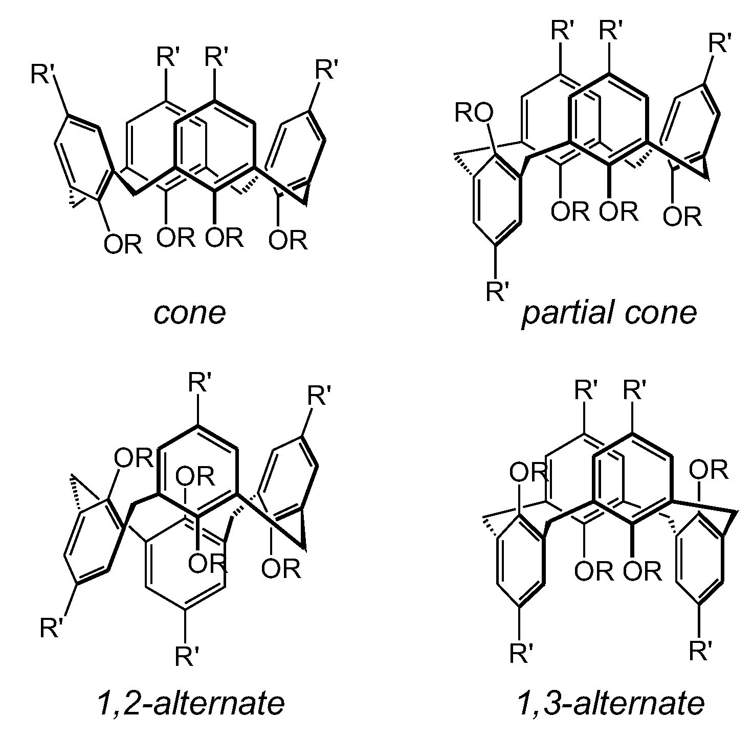
Moreover, this calixarenic skeleton displays the possibility to existing under four conformational isomeric structures: cone, partial cone, 1,2-alternate, and 1,3-alternate (Figure 3). Thus, the calixarene offers great possibility for structural modifications by the functionalization on the upper and/or lower rim with many kinds of functional groups. This versatility enables scientists to customize, to their taste, the properties of this molecular ring to provide the different chemical and physical properties required.
However, native calixarenes are not water-soluble; thus, exploration of their potential biological applications was quickly trained towards the development of water-soluble structures [56]. The first water-soluble calixarene appeared in 1984 with Arduini et al. and the introduction of four acetate groups on the lower rim of para-tert-butylcalix[4]arene [57]. However, the solubility of alkali and ammonium salts of the acid in water is only between 5 × 10⁻⁴ and 5 × 10⁻³ M, depending on the cation used (Na+ < K+ < Cs+ < NH4+ < Li+). Then, in 1988, Gutsche introduced the carboxylate functions on the upper rim [58]. In 1984, Shinkai et al. described the synthesis and application of sulfonated calixarenes, for example, [59,60,61], obtained by electrophilic substitution of previously detertiobutylated calixarene. The emergence of phosphonate derivatives in 1989 [62] was followed by ammonium via introduction of NH2 groups in the para position on a phenolic nucleus [63,64] or by aminodehydroxylation [65,66] of phenolic functions.
The addition of hydrophilic properties has also been envisaged by the introduction of sugar groups [67,68], polyhydroxy-amines [69], or PEG arms [70]. More recently, the development of water-soluble guanidinium calixarenes was introduced by Dudic et al. [71], and then by Mourer et al. [72].

5. Overview of Functionalization of Calix[4]arene

The different nature of the two rims available on the calixarene skeleton allows for selective functionalization on the upper rim (functionalization of the para carbon of the aromatic nucleus) or on the lower rim (functionalization of the phenolic hydroxyl) (Figure 2). Concerning the upper rim, it can be modified by aromatic nucleophilic substitution, via halogenation [73,74], nitration [75,76], sulfonation [59,61,77], acylation [78,79], or other means. In addition, on the native, para-tert-butylated calixarene, it is possible to completely or selectively detertiobutylate by first passing through the protection of the OH of the phenolic rings on which it is desired to retain a para-substitution [80]. On the other hand, the lower rim allows, via the reactive hydroxyl groups, the introduction of various arms by formation of ether or ester bonds [81].
In the case of calix[4]arene, the lower rim has four hydroxyl groups engaged in a network of intramolecular hydrogen bonds favoring the conical conformation (Figure 3). These strong hydrogen bonds lead to different pKa values for the hydroxyl groups involved in this ring [82]. This allows the establishment of various regio- and stereo-selective functionalization procedures on the lower crown. It has been shown that by implementing a carrier ion effect (“template effect”) relating to the nature and strength of the base used, it is possible to stereospecifically incorporate different substituents on the lower part of the calixarene. It is thus possible to substitute in the lower rim a calix[4]arene with 1 to 4 groups and, moreover, for bis-functionalization, a possibility of regioselectivity in the 1,2-adjacent or 1,3-alternated position [83,84,85,86,87,88,89,90,91,92].

6. Cytotoxicity of Calix[n]arenes

To develop biomedical applications, research into strong, possibly deleterious, interactions between the design molecular object and the biological target (cell, protein…) must examine the toxicity of various biological entities that these objects could potentially encounter before reaching their target. This is the case for the calixarene derivatives, more particularly the water-soluble ones, with the increase in interest which devoted to them for various biological applications and potential clinical studies. Relatively rare some twenty years ago, these studies are beginning to appear, and almost all converge towards the fact that this structure seems to have no toxicity or immunogenicity [93,94].
For example, the para-sulfonatocalix[n]arenes, widely used for numerous biomedical applications (anti-viral, anti-thrombotic, protein complexation, anti-coagulant…) exhibit an innocuous character with regard to studies conducted until this date [35].
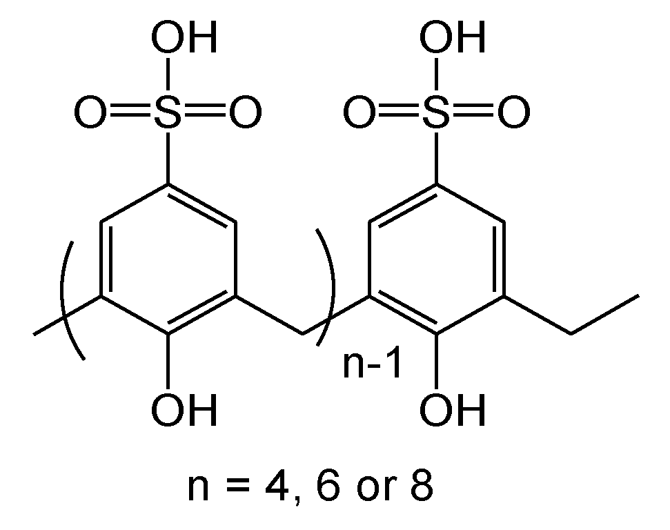
Therefore, for the first time, Da Silva et al. studied the toxicity of some sulfonated calixarenes (Figure 4) on erythrocytes for their haemolytic properties [95]. These, functionalized at the lower rim by the 2-carboxy methoxy group, 2-amido methoxy group, or 2-amino ethoxy group, and designed for some complexation properties of bovine serum albumin (circulating physiological protein) [96], present a maximum of 30% of haemolysis (evaluated by release of haemoglobin from lysed cells) for the para-sulfonatocalix[8]arene at the high concentration of 200 mM (about 300 g/L of blood). The corresponding calix[4 and 6]arenes are much less toxic, with 0.5 and 8.0% of haemolysis, respectively. These results show a very low toxicity on red blood cells of sulfonated calixarenes.
The same para-sulfonatocalix[n]arenic structures were tested for the viability and stimulation of neutrophils involved in immune response. Cell viability was not affected and the calix[n]arenes studied did not induce activation of NADPH oxidase in neutrophils, and did not reduce their responses to an external stimulus [97].
The first in vivo study of the distribution and pharmacokinetics of 35S-labelling para-sulfonatocalix[4]arene was conducted on mice by Coleman et al. in 2008 [98]. The experiment showed no acute toxicity for single injected doses (equivalent to 2–5 g in humans), no crossing of the blood–brain barrier, nor passing into muscles, and the molecule is quickly eliminated in mouse urine. It is unlikely that the 35S-para-sulfonatocalix[4]arene is metabolized, considering the lack of accumulation in the liver.
A study for using it as an anticancer drug delivery vehicle showed that this same structure presents no cytotoxicity against the human ovarian carcinoma cell line A2780 and its equivalent cisplatin-resistant cell line A2780cis [99].
In 2010, Geller et al. proposed a study on the antiseptic properties of para-sulfonatocalix[4]arene and its bis-bithiazolyl derivative against human coronavirus 229 E [100]. These two calixarenes did not show any cytotoxicity on the L-132 epithelial cell line, contrary to chlorhexidine and hexamidine, used as antiseptic references.
The toxicity of a fluorescent probe derived from an aminocalix[4]arene was studied on Chinese hamster ovary cells (CHO) and HL50 cells (i.e., human leukemia cells) [101]. The fluorescent water-soluble calix[4]arene and its unlabeled parent did not show more toxicity than phosphate-buffered saline (PBS) over a range of concentrations with IC50 of 82 and 81 mM, respectively.
A wide series of anionic calixarenes—para-methylcarboxylate, para-sulphonate, and para-methylphosphonate and their bis-bithiazolyl derivatives—were evaluated as anti-HIV compounds by Mourer et al. [102]. All of the nine water-soluble calixarene derivatives evaluated on the two cell lines MT4 (HTLV-I-transformed T-cell line) and CEM-SS (T-cell line expressing the CXCR4 co-receptor) display very weak or no toxicity, as the CC50 was not reached at 100 µM. A similarly very low toxicity level was also detected on PBMC (T-cell line expressing the CCR5 co-receptor) for the majority of substances tested.
Since 2006, Mourer et al. have examined systematically the cellular toxicity of calixarenes designed for the research on biological applications. They showed that most calixarene structures are often devoid of in vitro cellular toxicity, particularly on HaCaT cells (spontaneous transformed aneuploid immortal keratinocyte cell line) and/or on MRC-5 cells (fibroblast-like human embryonic lung cells) [72,103].

7. Antibacterial Activities of Calix[n]arenes

The calixarenic structure is endowed with great versatility, and this is why it is used in a wide variety of fields. The most interesting features are the multiple functionalizable positions and the access to multivalence. Multivalency is, for example, a key element in generating the strongest and most specific interactions possible [104,105,106].
This last is widely used for new drug research, for example: simple use as a carrier of multiple active pharmaceutical ingredients (API) (identical or different) in the case of development of prodrugs; the genesis of an environment conducive to the complexation of metals in the search for compounds capable of disrupting the activity of metalloenzymes or the formation of toxic metal complexes; and research on therapeutic activities related to strong interactions with proteins, enzymes, or nucleic acids [107].
As the versatility of calixarenes is very wide, it is possible to optimize the design of a multivalent ligand to improve the desired activity. For example, the length of the bond between the calixarene and the group of interest will often be a very important and critical point: a calix[4]arene could lead to spatial constraints and greater interactions than a calix[8]arene; however, this last, more fluxional, could allow more adaptability face to a given target. Thus, using this central skeleton, researchers can very precisely tune the nature, number, distance, or orientation in space of any type of functionality in such a way as to best match a biological target as finely as possible.

7.1. Intrinsically Active Calixarenes

Since the 1950s, researchers have sought to functionalize calixarene crowns with various types of functional groups, sometimes known to already possess a biological activity. The objective is often to take advantage of the multivalence provided by the macrocycle and to envisage a synergy of action. It appears very clearly in certain studies, by comparison with the constitutive monomers of calixarene, that the latter allows a marked improvement in the activities sought, fully justifying its use.
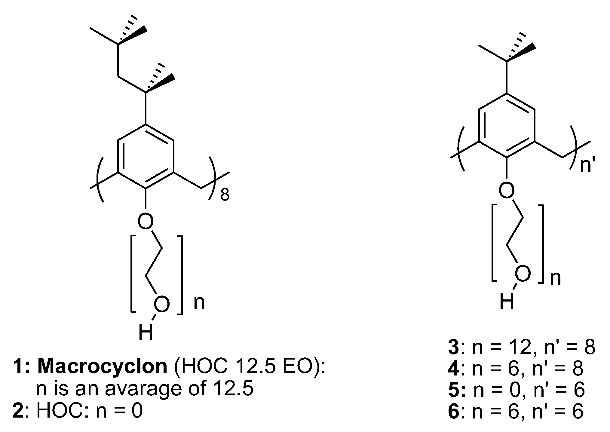
The first direct antibacterial activity of calixarene was reported in 1955 by Cornforth et al. [108,109]. An important study was carried out on macrocyclon, alternatively named HOC 12.5 EO (1 Figure 5), constituted from macrocyclic phenol (calix[8]arene), which for brevity is termed HOC, and which was prepared from tert-octylphenol and formaldehyde by a modified Zinke–Ziegler procedure, and by reacting under basic conditions with ethylene oxide (EO). The four resulting polyoxyethylene ether chains have an average chain length of 11–12.5 units (HOC 12.5 EO) [109,110].
The macrocyclon enters macrophages via endocytosis and disrupts their lipid metabolism by inhibiting triglyceride lipase and phospholipase, blocking bacterial growth. While the growth of Mycobacterium tuberculosis (H37Rv strain) in infected macrophages is inhibited by macrocyclon (bacteriostatic), this is stimulated by HOC-60 (derived from the macrocyclon with a much longer polyoxyethylene chain) [111,112].
The study of compounds 2 to 6 has shown that a polyethylene glycol (PEG) chain with 6 units (4 and 6 Figure 5) is sufficient to provide a high antimycobacterial activity and that the change to 12 units (3 Figure 5) does not bring significant improvement. Likewise, hexamer 6 and octamer 4 exhibit similar activities, suggesting that the size of the calixarenic crown is not critical. However, in the case of non-functionalized compounds 2 and 5, the size of the cavity seems to intervene, 2 having a slight antimycobacterial activity while 5 is inactive. The authors showed additionally that macrocyclon significantly affected mycobacterial growth in murine macrophages by a mechanism involving L-arginine metabolism and iNOS activity [113].
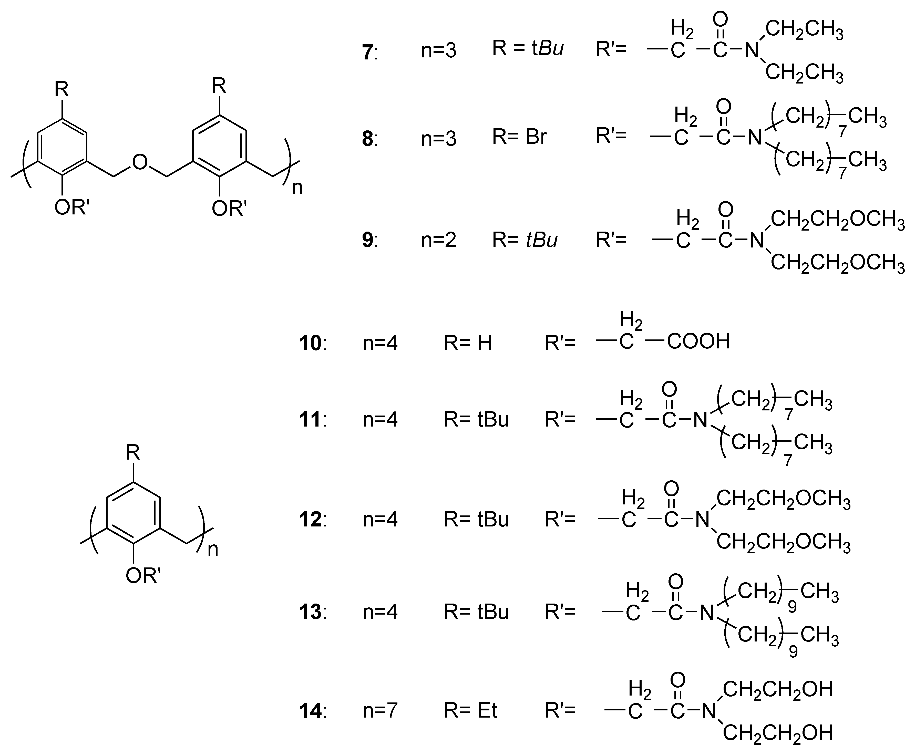
In 1995, S. J. Harris patented a large screening of more than 200 molecules (calixarenes, oxacalixarenes, cyclotriveratrylene derivatives…) for their anti-HIV, anti-cancer, antibacterial, and antifungal activities [114]. Only some antibacterial activities were reported for a series of calixarenes and oxacalixarenes having amide functional groups in the lower rim (Figure 6).
The bacteria chosen are those usually employed in routine screening for potential antibacterial activity in the 1990s: Escherichia coli, Pseudomonas aeruginosa, Klebsiella pneumoniae (lactamase producing strain), Bacillus subtilis, and Staphylococcus aureus (Table 1).
The structures tested do not show good activities, i.e., from 1 to 10 mg/mL, corresponding to MICs 50 to 100 times greater than that obtained for the different references used in the study (ampicillin, tetracycline, or erythromycin MIC values are ≤0.1 mg/mL). There does not seem to be any selectivity towards the structure of the bacteria (Gram-positive or -negative). Compounds 7, 11, 12, and 13 display, for example, an activity on both Gram types. Similarly, the results do not allow clear determination of a structure–activity relationship. Whether it is a calixarene crown (4 or 7 phenolic units) or oxacalixarene, the activities seem more or less random. Functional groups N,N-bis-alkyl- (7, 8, 11, 13), N,N-bis(2-hydroxyethyl)- (14), or N,N-bis(2-methoxyethyl)-ethanamide (9, 12) in the lower rim do not appear to lead to any particular activities.
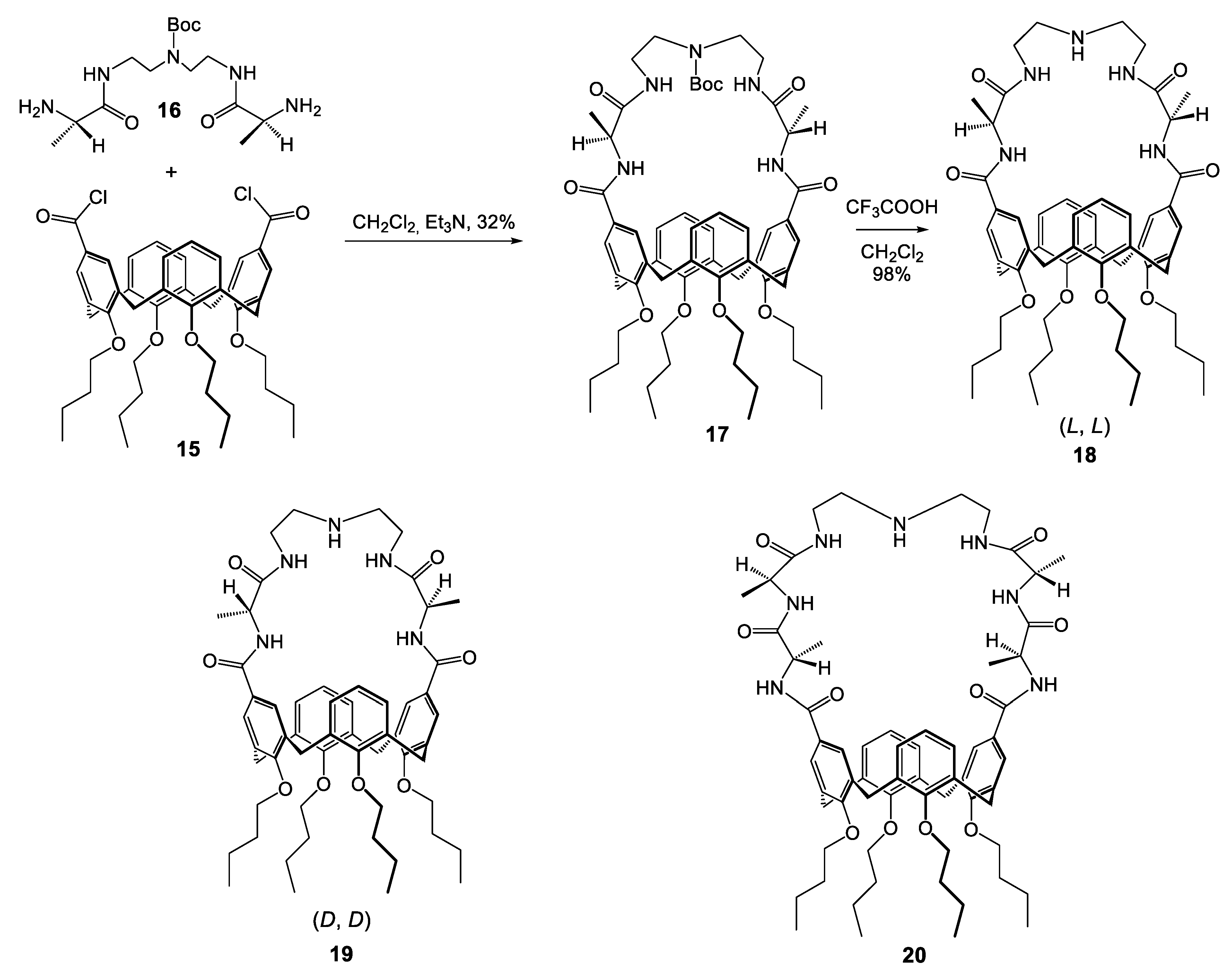
Casnati and coworkers, in 1996, used the calixarenic platform bridged in the upper rim by D- or L-alanine units and the diethylentriamine segment as vancomycin antibiotic mimics [115]. Isolated in 1950, the glycopeptide vancomycin was clinically used in 1959 against S. aureus both sensitive and resistant to penicillin or methicillin. Some studies determined its mode of action via interaction between his peptide moieties and the cell wall mucopeptide precursors terminating in the sequence –D-alanyl-D-alanine. This hydrophobic interaction inhibits the growing of the cell wall and thus the bacterial wall.
An example of the synthesis of the short-bridged (L, L) compound 6 is presented in Scheme 1. Condensation of calix[4]arene diacylchloride 1 and precursor 4 in high-dilution conditions gave the N-Boc protected macrobicyclic compound 5, which was deprotected in acidic conditions to obtain the expected peptido-calixarene 6. A similar procedure was used for the development of compounds 7 and 9 (Scheme 1).
These four new calixarenic structures (Scheme 1), and vancomycin taken as reference, were tested against various Gram-positive strains such as S. aureus (strains 663 (penicillin-sensitive), 853 (penicillin-resistant), and 1131 (methicillin-resistant)), Staphylococcus epidermidis, and Bacillus cereus. Although slightly inferior to vancomycin (2 µg/mL), the calixarenes studied, notably compounds 18 et 19, show good activity against the MRSA (methicillin-resistant Staphylococcus aureus) strains (4 to 8 µg/mL) (Table 2).
The two enantiomers 18 and 19 present the same range of activities. Moreover, the Boc-protected compound 17 and the calixarene with a largest peptide bridge 20 display a reduction in antibacterial activity. In parallel, the peptide precursors used for obtaining calixarenes 18, 19, and 20 are inactive, confirming the importance of the calixarenic skeleton to maximize the interactions with the sequence –D-alanyl-D-alanine. These results were confirmed by binding studies with the classical model N-acetyl-D-alanyl-D-alanine for cell wall peptidoglycan termini via NMR analysis [115,116,117].
In 2002, Lamartine et al. published a study of 57 calixarenes having different side chains, moieties, and/or substitution groups against a diverse set of plant pathogen bacteria and fungi [118]. Firstly, antibacterial activity was performed against the specific Gram-positive genus Corynebacterium to select the calixarenic active structures according to the inhibition diameter. From this first screening, only seven structures exhibited antibacterial activity. Then a second antibacterial test was performed to extend the activity as a function of calixarene concentration (0.001 to 1%, i.e., 10 to 10,000 ppm (part per million)). Finally, only sulfonated (basic form) compounds 21 to 24 (Figure 7) exhibited slight antibacterial activities. These sulfonate-calixarenes 21, 22, and 23 were easily obtained by ipso-sulfonation [77] and by diazo-coupling for the 24 derivative [119]. Unfortunately, these structures are active above 1% concentration (21, 22, 23) and at best at 0.1% (1000 ppm) for the derivative 24. These activities seem not effective enough for pesticide development (considered effective at less than 100 ppm).
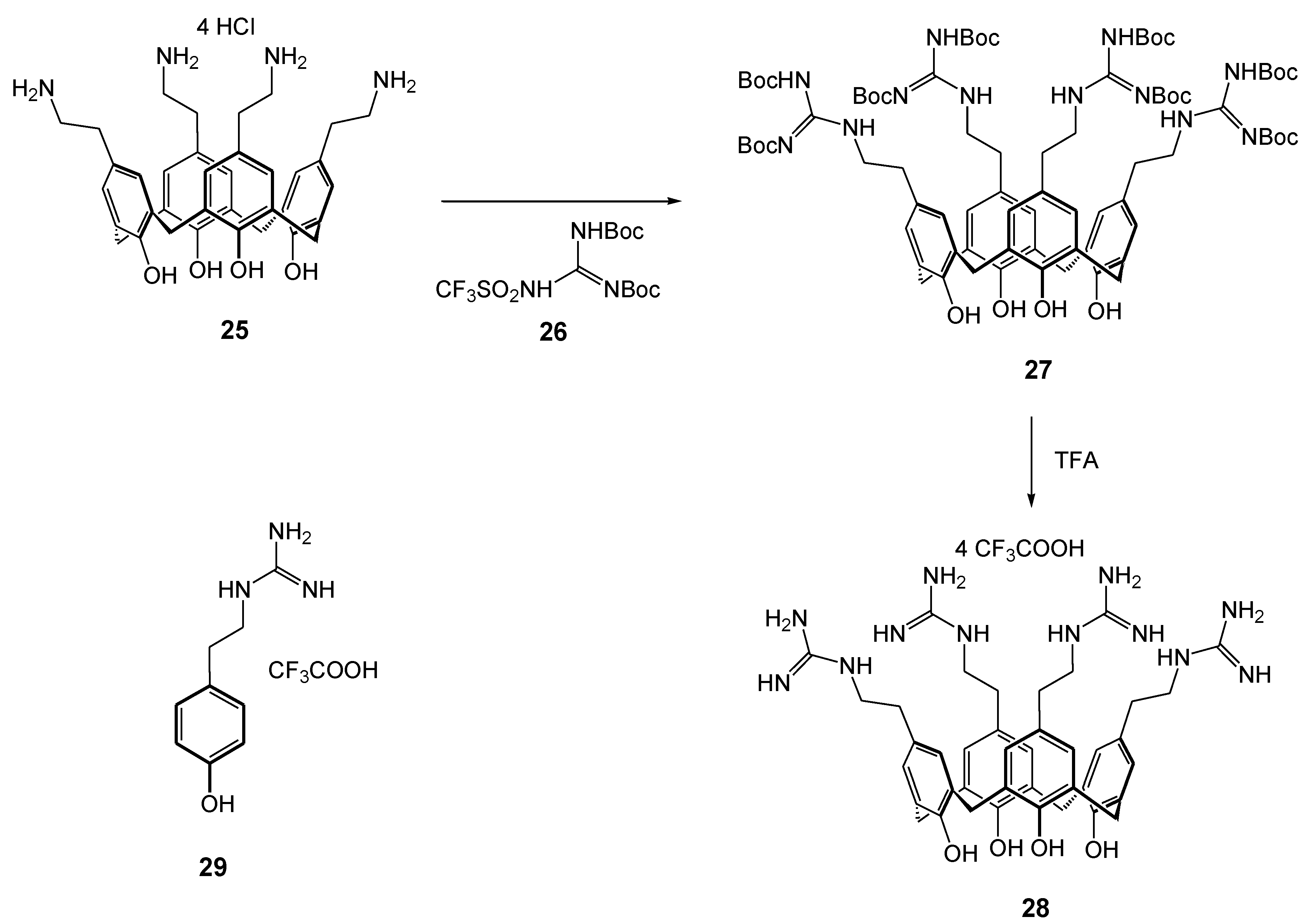
In 2006, Mourer and co-authors began a large series of studies on the antibacterial activities of calixarenes variously substituted in the upper and/or lower rim. The object of the first study was the tetra-para-guanidino-ethylcalix[4]arene 28 (Scheme 2), which exhibits an organization of positive guanidinium charges on the upper rim and displays a significant antibacterial activity (MIC <8 µg/mL) against the bacterial reference strains E. coli (ATCC 25922), S. aureus (ATCC 25923 and ATCC 29213), Enterococcus faecalis (ATCC 29212), and P. aeruginosa (ATCC 27853) (Table 3) [72], but also on the clinical isolates penicillinase-producing E. coli (EcR1), methicillin-resistant S. aureus (MRSA), vancomycin-resistant E. faecium (EfR1), vancomycin- and teicoplanin-resistant E. faecalis (EfR2), and P. aeruginosa overexpressing efflux pumps (PaR1) (Table 3). [120]. The initial hypothesis was based on the fact that most bacteria are negatively charged, and that introduction of positive charges in a constrained manner using the organization capacity of calixarene could lead, via spatially organized electrostatic interactions, to a modification of the ionic surface properties, inducing a disorganization of it and thus to a deleterious effect for bacterial cells. In this study, the authors propose to compare the activity of the calixarene structure with its constitutive monomer 29 (Scheme 2) on the assumption that the high organization of calixarene derivatives should lead, with regard to the monomeric analogue, to an efficient synergistic effect in ionic interaction with the bacterial surface, resulting in an antibacterial behavior.
Calixarene 28 was obtained according to Scheme 2, with tetra-para-ethylaminocalix[4]arene 25, which was synthesized by a modified Gutsche procedure [64]. Addition of di-Boc-triflylguanidine 26 allows the octa-Boc species 27 formation, which was finally treated with trifluoroacetic acid to give the tetra-guanidinium salt 28 with overall yield of 52%.
The authors thus show the importance of the calixarenic skeleton: its ability to organize in space and to constrain cationic functions. Switching from the monomer 29 (inactive, MIC > 512 µg/mL) to cyclotetramer results in a very strong gain of activity on all the strains (4 to 32 µg/mL on E. coli and E. faecalis respectively). The very good activity of 28 on Gram-positive and Gram-negative bacteria, suggests a broad antibacterial spectrum.
Additional tests showed that 28 exhibits a rapid concentration-dependent bactericide activity (in 2 to 4 h of contact). A minimal bactericidal concentration against E. coli of 8 × MIC, and 4 × MIC on P. aeruginosa and S. aureus, was observed [121]. Tetra-para-guanidino-ethylcalix[4]arene 28 presented an antibacterial spectrum similar to hexamidine HX or chlorhexidine CHX, well-known antibacterial agents used as references [120]. Both tetra-para-guanidino-ethylcalix[4]arene 28 and its constitutive monomer 29 showed no apparent cytotoxic effect against eukaryotic cells (human keratinocytes (HaCaT) and human pulmonary embryonic fibroblasts (MRC-5)) (>256 µg/mL) even up to 168 h of cell exposure, contrary to hexamidine, with loss of cell viability seen after 24 h of cell exposure (IC50 = 36–37 µg/mL) for both HaCaT and MRC-5 cell lines [72].
A complementary study on this cationic calix[4]arene 28 was carried out on a large series of multiresistant bacteria from clinical isolates: 39 MDR Gram-positive bacteria (15 S. aureus, methicillin-resistant (MRSA) or methicillin-sensitive (MSSA), 12 coagulase-negative staphylococci (CoNS), resistant or susceptible to methicillin, 14 Enterococcus spp., with or without van genes) and 30 MDR Gram-negative bacteria (20 Enterobacteriaceae, with or without Extended Spectrum Beta Lactamase (ESBL) and 10 non-fermenting bacilli). The results showed a maintained activity on almost all of the strains and the authors advance a probable action of 28 at the level of the bacterial cell wall [122].
Faced with the excellent activity of this cationic derivative and the fact that it is not stopped by the resistance mechanisms commonly used by bacteria, the authors tried to understand the mechanism of action with a first study via microelectrophoresis [123] in order to obtain a global evaluation of the interfacial properties of bacteria with or without drug treatment. These experiments carried out on P. aeruginosa after 24 h of exposure to the drug showed a significant decrease in electrophoretic mobility, reflecting a strong increase in the permeability of the walls and/or a specific adsorption of calixarenes on the surface justifying a wall effect. In order to confirm this impact on the cell wall, they carried out a study via Atomic Force Microscopy (AFM) in order to visualize any alterations in the bacterial cell wall. The first images obtained on P. aeruginosa effectively show alterations in the wall after 24 h of contact with calixarene as well as very significant cell fragility upon simple contact with the AFM tip compared with untreated cells [123]. The same study showed that the constitutive monomer 29 does not cause any alterations.
In a second step on P. aeruginosa ATCC 27853 and PaR3 (i.e., a MDR strain), a comparative study was carried out between ticarcillin, tobramycin (to which PaR3 is resistant), and calixarene 28 [124]. If the general appearance of sensitive bacteria is well modified in the presence of ticarcillin and tobramycin, the resistant strain is not impacted. On the other hand, treated by 28, the two strains present alterations of the wall, appearance of perforations, and surface inequalities (from 0.2 ± to 0.04 nm for untreated bacteria to 1.0 ± 0.2 nm for cells treated with 28).
The results also showed that on the resistant strain of P. aeruginosa (i.e., PaR3), 28 had a radical effect on the elasticity of the wall not encountered with the reference antibiotics. For example, the PaR3 strain saw its modulus of elasticity drop from 520 ± 100 kPa to 300 ± 66 and 252 ± 61 kPa after treatment with ticarcillin and tobramycin, respectively, but fell to 76 ± 28 kPa during treatment by 28.
Other experiments conducted on phospholipid bilayers showed that 28 created perforations in the latter (0.5 to 1 mm in diameter), agreeing with the observations made on the cells treated with 28.
In 2014, Grare and Duval patents the use of calixarene tetra-guanidinium salt 28 in the treatment of bacterial infections in combination with some known antibiotics [125]. Considered as possible way to bypass the antibiotic resistances, the bi-therapy could ideally generate a synergistic effect between two drugs. The objective of the study is to determine, in vitro, the possible benefit of the association of 28 with various antibiotics conventionally used in practice, with different targets of action. They performed an important screening against sensitive E. coli ATCC 25922, S. aureus ATCC 29213, and P. aeruginosa ATCC 27853 reference strains, three clinical isolates by species presenting all various resistance profiles to antibiotics conventionally used in practice. Calixarene 28 was evaluated in association with 12, 11, and 10 antibiotics against E. coli, S. aureus and P. aeruginosa, respectively. Initially, the results allowed the absence of antagonism on all the associations evaluated and the activity of some antibiotics seems restored for all clinical isolates tested. An important impact on P. aeruginosa clearly appears, as it exhibits a greater sensitivity than E. coli or S. aureus to the association of drugs. Indeed, it is the strain for which the most synergistic associations have been recorded, even against clinical isolates displaying carbapenem resistance, strains classified as a priority pathogen by the WHO in the search of new antibiotics [126]. The derivative 28 makes it possible to confer de novo a certain level of susceptibility to antibiotic on bacterial strains having a natural or acquired resistance to said antibiotic.
More recently, Korchowiec et al. have studied the behavior of calix[4]arene guanidinium salt 28 and its constitutive monomer 29 against models of lipid membranes (monolayers) by simulation of molecular dynamics and measurement of surface pressure. The studies were carried out on a zwitterionic monolayer (1,2-dimyritoyl-sn-glycero-3-phosphocholine: DMPC) as a eukaryotic cell membrane model and negatively charged (1,2-dimyritoyl-sn-glycero-3-phospho -L-serine: DMPS) as a bacterial membrane model [127]. Although no effect has been demonstrated on the (zwitterionic) eukaryotic cell membrane model, the (anionic) bacterial cell membrane model is significantly affected in the presence of 28.
Beyond the fact that the charge–charge interactions play an important role in the adsorption on the negatively charged phospholipid heads, leading to an accumulation of molecules at this level, it would seem that 28 also penetrates into the hydrophobic part of the monolayer, in particular via its hydrophobic lower rim. A reversal of the orientation of tetra-guanidinium upon penetration into the lipid layer, minimizing harmful interactions, could also play a role in the demonstrated antibacterial activity. Under the same study conditions, the monomer 29 appears to be rapidly removed from the monolayer.
In order to increase the antibacterial activities of tetra-guanidino calix[4]arene 28, Mourer et al. developed, in 2009, some derivative carriers of bis-heterocyclic arms in the 1,3-alternating position of the lower rim [128]. In addition to the electrostatic interactions between the positive guanidinium charges and the negatively charged wall of the bacteria, they hope, by introducing an amphiphilic character to their structure, to generate a disorganizing insertion in the bacterial wall. In addition, the complexing capacity of bis-heterocycles could allow the capture of metal ions necessary for bacterial multiplication (metalloenzymes) or the genesis of toxic metal complexes for the bacterial cell. These structures were obtained according to Scheme 3. The tetra-salt of ammonium 25, once protected under carbamate form, undergoes the introduction of bis-heterocyclic units in the 1,3-alternate positions in acetonitrile and K2CO3 as base (compounds 30, 31, 32; Scheme 3). The bis-heterocyclic compounds obtained were deprotected (33, 34, 35; Scheme 3) and the introduction of guanidine moieties was performed in the same way as described above using the di-Boc-triflylguanidine 26, followed by a final elimination of the Boc protective groups in TFA conditions to give the expected tetra-guanidinium salts 36, 37, and 38.
The microbiological study consisted of the determination of active concentrations on Gram-positive and Gram-negative reference strains as well as the in vitro cellular toxicity on eukaryotic cells of the MRC-5 type (Table 4). Unfortunately, even if the activities of bis-heterocyclic derivatives are preserved with MICs of 10 to 20 µg/mL close to the reference derivative 28, the introduction of bipyridyl or bithiazolyl arms does not appear to bring about a significant modification of the antibacterial behavior. In addition, a much greater toxicity appears on the eukaryotic cell, in particular for the derivatives 36 and 38 with IC50s of 16 and 64 µg/mL, close to the antibacterial values, leading to weak selectivity indexes.
In the same way as with the introduction of positive charges, negative charges also provide hydrophilicity to the calixarenic platform. Thus, the same team published in 2012 the synthesis and antibacterial evaluation of water-soluble derivatives incorporating carboxylate groups in the upper rim and bis-heterocycles in the 1,3-alternate positions on the lower rim of calix[4]arene [129].
The tetra acetic acid derivative 39 and its tetraethylester 40 were prepared according to the procedures of Gutsche and co-workers [64,130]. Introduction of the heterocyclic subunits in alternate positions on the lower rim of 40 was carried out by the method of Mourer et al. [102] by reaction of bromomethyl derivatives 42 to 45 in MeCN and K2CO3 as base (Scheme 4). The sodium salts were obtained under a previously described procedure [131], namely that the tetra-esters 46, 47 and octa-esters 48, 49 were saponified with NaOH in hydro-alcoholic medium and precipitated under acidic form with aqueous HCl. Then, a titration with NaOH gave the expected sodium salt derivatives 50 to 53.
Conducting against Gram-negative (E. coli ATCC 25922 and P. aeruginosa ATCC 27853) and Gram-positive (S. aureus ATCC 25923 and ATCC 29213, and E. faecalis ATCC 29212) reference strains, the MIC determination showed that these carboxylate water-soluble calixarenes display no antibacterial activity under 256 µg/mL (128 µg/mL for 53 on S. aureus ATCC 25923).
The authors report that they observe, at this stage of their studies, an effective discrimination between the antiviral [102] and antibacterial activities of these compounds, directing the negatively charged derivatives towards antiviral activities and the positively charged derivatives towards antibacterial activities.
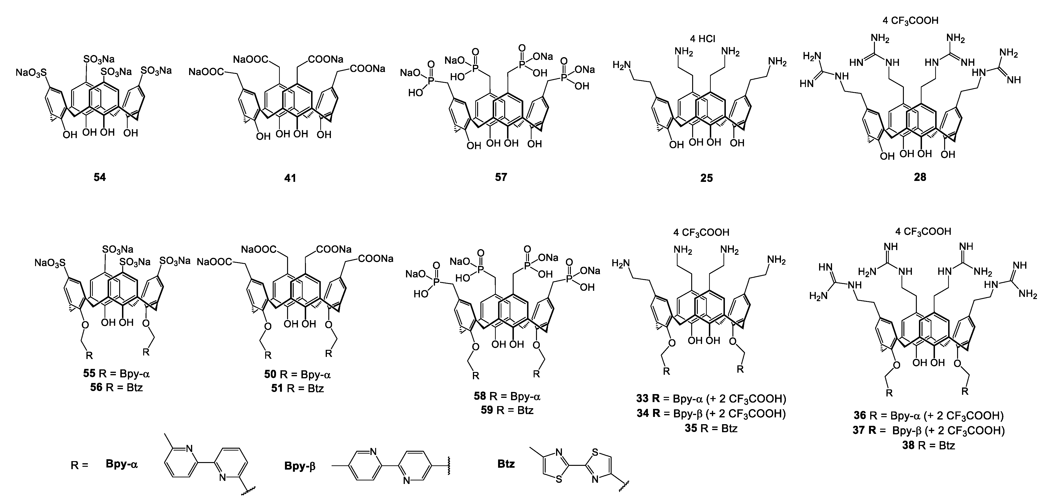
On the strength of their results, Mourer and co-authors started a study of some of their calixarene derivatives (Figure 8) on a particular strain: Mycobacterium tuberculosis, with the resurgence of tuberculosis in sight, placing this bacillus at the foreground of the infections to fight [132].
A first screening at 10 and 1 μM was carried out against the isonicotinic acid hydrazide (INH)-sensitive M. tuberculosis H37Rv strain, with some of the anionic (sulfonates 54, 55, and 55; carboxylates 41, 50, and 51, phosphonates 57, 58, and 59), and all cationic derivatives (ammoniums 25, 33, 34, and 35; guanidiniums 28, 36, 37, and 38). The authors confirmed their previous results [129], namely that anionic compounds were inactive. On the other hand, the results are more encouraging with cationic species, but variable, depending on the type of structure. In the amino family, the compounds α-bipyridyl 33 and bithiazolyl 35 are non-active to weakly active with 0 to 19% inhibition at 10 µM, respectively. The unsubstituted ammonium 25 and β-bipyridyl 34 derivatives displayed a better inhibitory property, evolving respectively from 76% and 81% at 10 µM to 27% and 63% at 1 µM. A similar behavior was observed for the guanidinium family. Exhibiting an inhibition of 10% at 10 µM, the α-bipyridyl 36 derivative is non-active. The unsubstituted guanidinium derivative 28, the bis-(bithiazolyl) 38 and the bis-(β-bipyridyl) 37 display a high activity with respectively 97%, 90%, and 98% inhibitory concentration at 10 µM, but also at 1 µM concentration with 73%, 54%, and 62% inhibition.
After this first selection, the authors determined more precisely the active concentration via MIC determination against INH-sensitive (H37Rv) and -resistant (MYC5165) M. tuberculosis strains (Table 5).
Ammonium derivatives, unsubstituted and bipyridylated, display interesting activities with MIC values of 2.58 and 1.34 µg/mL, respectively, for 25 and 34 on H37Rv. Introduction of the guanidinium groups decreases the MICs with values of 1.0, 1.51, and 2.69 µg/mL, respectively, for 28, 37, and 38 on an INH-sensitive strain. In comparison, commercial antimycobacterial compounds INH, ethambutol, and streptomycin show MICs of 0.08, 2.0, and 0.4 µg/mL respectively.
In parallel against INH-resistant strain MYC5165, the same compounds exhibit very low MIC values, mostly sub-micromolar, while INH is active at 1.7 µg/mL. Indeed, the unsubstituted guanidinium 28, β-bipyridyl 37, and bithiazolyl 38 guanidinium compounds are responsible for remarkable MICs of 1.0, 0.75, and 0.17 µg/mL respectively.
The introduction of the guanidine groups at the upper rim in derivatives 28, 37, and 38 resulted, particularly for 38, in more interesting activities compared with ammonium derivatives.
In the same way as on the standard strains [128], the contribution of the bis-heterocyclic functions does not lead to a significant gain in activity compared to the unsubstituted platforms, although the β-bipyridine and the bithiazole slightly increase the MIC values, particularly on INH-resistant strains. However, depending on the type of bis-heterocycle, the results differ, with an extinction of activity in the case of α-bipyridine: at a concentration of 10 µM, the inhibition goes from 76% to 0% for the ammonium derivatives and from 97 to 10% for the guanidiniums, while the inhibition activity remains the same with β-bipyridine.
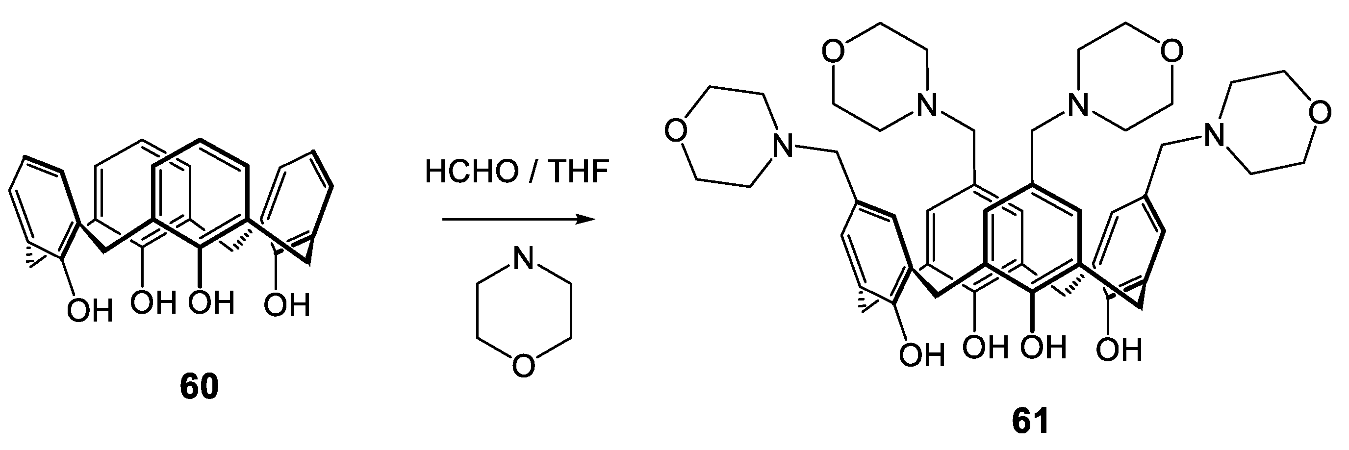
A water-soluble macromolecule based on calix[4]arene and morpholine units (61 Scheme 5) was developed by Memon et al. in 2012 [133], which is easily obtained according to the Gutsche procedure [64] using detertiobutylated calix[4]arene, morpholine and formaldehyde via the Mannich reaction (Scheme 5). The antibacterial tests were carried out against two Gram-positive species: Staphylococcus albus and Streptococcus viridians, as well as five Gram-negative ones: Bacillus procynous, Enterobacter aerogenes, Klebsiella aerogenous, E. coli, and Salmonella enterica subsp. enterica. There appears to be an excellent activity profile for this tetra-morpholinocalix[4]arene. The inhibitions of growth were measured at 4 µg/mL against all strains, except for Staphylococcus albus and Enterobacter aerogenes, at 16 and 8 µg/mL, respectively.
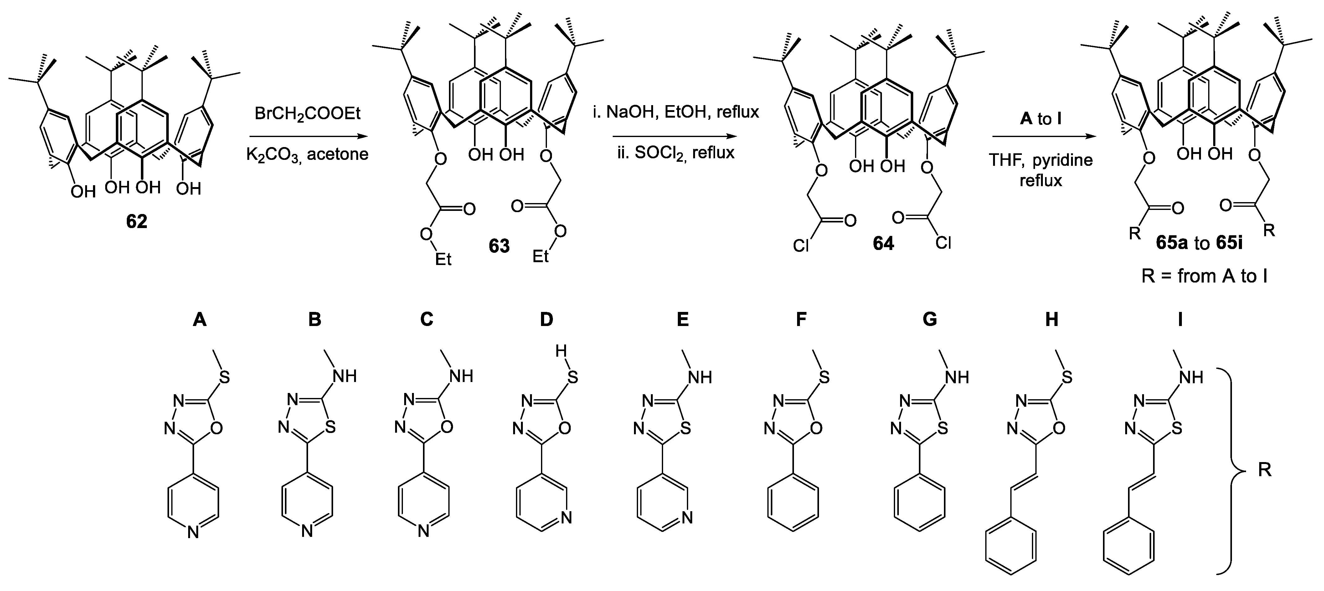
A series of nine new calixarenes based on 1,3,4-oxadiazole and thiadiazole were synthesized and have been subjected to antibacterial screening [134]. Based on the fact that the first-line anti-mycobacterium drug isoniazid (isonicotinic acid hydrazide: INH) is metabolized in the liver by acetylation and causes hepatotoxicity effects, the authors considered the replacement of hydrazide moieties of INH with 1,3,4-thiadiazole and 1,3,4-oxadiazole heterocycles, hoping to eliminate the in vivo acylation step. Therefore, they synthesized nine oxadiazole and thiazole derivatives of isoniazid, nicotinic, benzoic, and cis-cinnamic acid hydrazide A to I (Scheme 6). In parallel, the preparation of functionalized calixarene in the 1,3-alternate position on the lower rim of bearing two ethanoyl chloride arms was performed from tetra-tert-butylcalix[4]arene 62, via grafting of ester functions (63 Scheme 6), saponification and final chlorination (64 Scheme 6). Then oxa- and thia-diazole were introduced on 64 in THF in the presence of pyridine to give the expected derivatives 65a to 65i (Scheme 6).
Determination of MICs of the all calixarene derivatives 65a to 65i and the isoniazid derivatives with 1,3,4-thiadiazole and 1,3,4-oxadiazole heterocycles A to H were carried out against various Gram-negative (E. coli MTCC 443 and P. aeruginosa MTCC 1688) and Gram-positive (S. aureus MTCC 96 and Streptococcus pyogenes MTCC 442) bacteria strains, as well as against isoniazid-sensitive Mycobacterium tuberculosis (H37Rv). With regard to the other calixarene derivatives already described in literature, the antibacterial activities of these new compounds are moderate on Gram-positive and -negative strains. The MIC values fluctuate between 100 and 500 µg/mL. Also carried out on tubercular bacillus, this study gives a good to moderate bioactivity on the isoniazid-sensitive strain used (MIC values from 50 to 250 µg/mL). Better activities were measured for calixarenes 65a, 65c, and 62h with MIC at 50, 62.5, and 62.5 µg/mL, respectively, compared with 0.2 µg/mL for isoniazid used as reference. All the values recorded do not make it possible to determine a notable difference between the different heterocycles introduced. However, in general, the substituted calixarenes seem to have an activity slightly greater than unsubstituted oxadiazole and thiadiazole (A to I); calixarene 65h, for example, exhibits MIC at 100 µg/mL on E. coli, P. aeruginosa, and S. aureus compared with 250 µg/mL for the oxadiazole H subunit alone on the same strains. The authors justified these differences by increasing of the lipophilic character introduced via oxa- or thia-diaozole and calixarenic core and facilitating the crossing through of the biological membrane. A cooperative effect of the pharmacophore is also advanced to justify the enhancement of activity of substituted calixarenes (65a65i) compared to unsubstituted oxadiazole and thiadiazole (AI).
In 2015, Melezhyk et al. published the antibacterial properties of tetra-alkylammonium and imidazolium calix[4]arene derivatives [135] compared to macrobicyclic peptidocalix[4]arenes 18 and 19 (Scheme 1) [115], trifluoroacetate of tetra-guanidinium calix[4]arene 28 (Scheme 2) [72], and morpholinocalix[4]arene 61 (Scheme 5) [133]. These new cationic structures were obtained from tetra-propyloxy- or tetra-octyloxy-calix[4]arene (66 or 67) chloromethylated with couple methyl chloromethyl ether/tin tetra-chloride (intermediates 68 and 69 Scheme 7), then chlorine displacement by N-methylimidazole or N,N-dimethylethanolamine (choline) to give 7071 and 7273, respectively, or by trifluoroacetamide of N,N-dimethylethylendiamine followed by treatment in ammonia solution, leading to 7475 (Scheme 7).
The antibacterial activities were determined by MIC measurement on P. aeruginosa ATCC 9027, S. aureus ATCC 6538, K. pneumoniae ATCC 10031, and E. coli ATCC 25922 strains by the broth microdilution method. The first point is the significant decrease in activity with the switch from the propyl derivative (10 to 1000 µg/mL) to the octyl derivative (500 to 1000 µg/mL), i.e., 5 to >100 times less activity for the tetra-octylcalixarenes (Table 6). The authors propose a possible explanation for the lower antibacterial activity of long-chain derivatives based on their stronger self-assembly ability in water solution (Critical micellar concentrations at 3 × 10⁻⁶ M and 8 × 10⁻⁵ M for octyl 73 and propyl 72, respectively) [136].
In addition, there is clearly a difference in activity between the cations studied, with a better activity of the imidazole derivative against all strains tested (MIC < 10 µg/mL on E. coli, S. aureus and K. pneumoniae, and 50 µg/mL on P. aeruginosa). The choline and ethylenediamine exhibit higher MICs between 10 and 1000 µg/mL according to the strains. The differences observed were attributed to the mode of interaction with bacterial membrane structures according to the nature of cationic centers, and were also potentially linked to the distance between two opposite-site nitrogen atoms, an important value to optimize the interactions with the negatively charged groups of the bacterial membrane. Moreover, The MIC values are close to those of antibiotics and better or equal to those of quaternary ammonium salt used as reference. For example, the imidazole derivative 70 exhibits a MIC <10 µg/mL against K. pneumoniae, while tetracycline, vancomycin, and didecyldimethylammonium chloride are measured at 16, 64, and 16 µg/mL respectively.
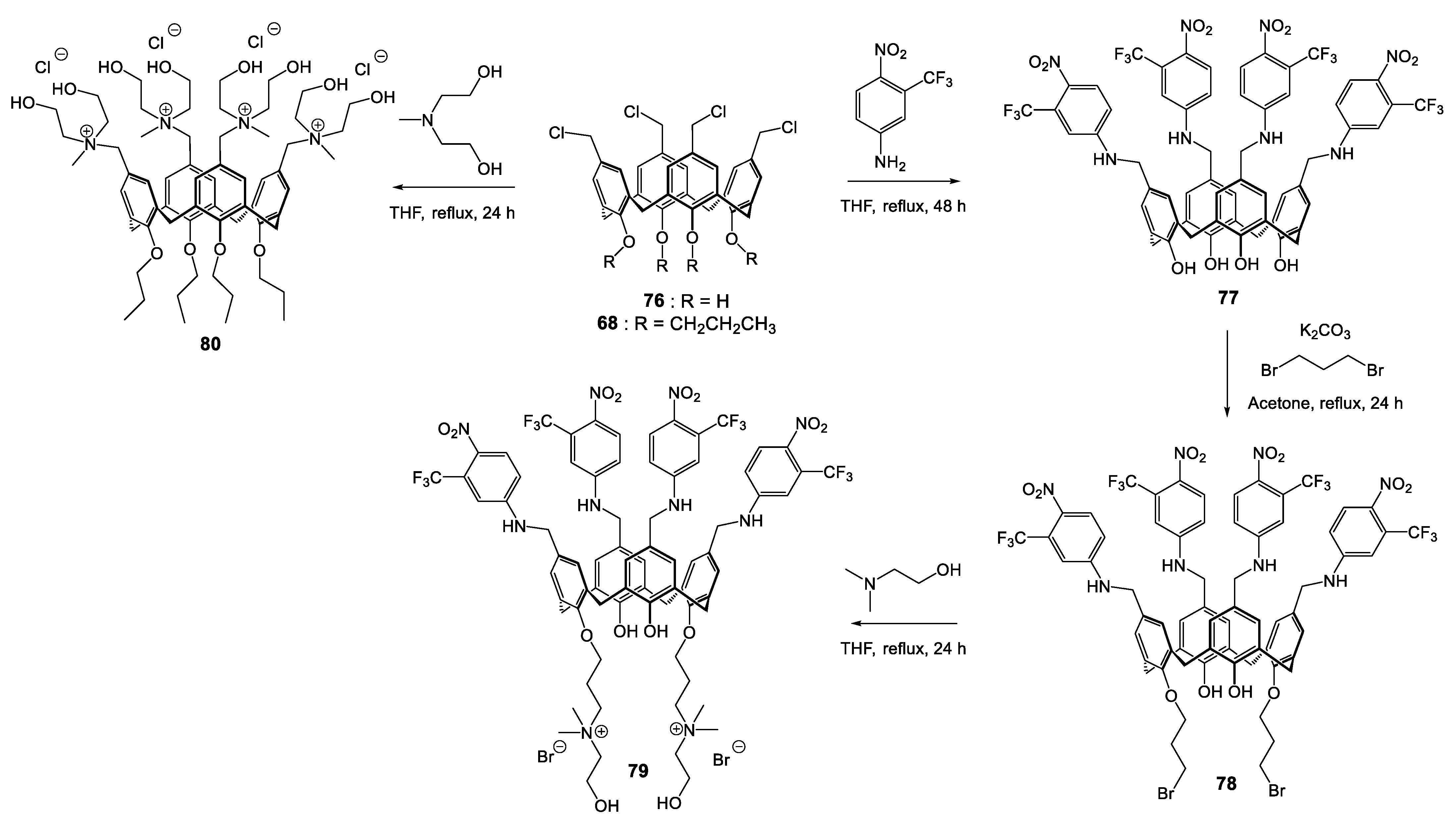
In 2017, Consoli et al. [137] used the calixarene platform to cluster multiple NO photodonor units, associated on the other side of platform at two quaternary ammonium groups to introduce a solubility in hydro-alcoholic solvent and allow an electrostatic interaction with the bacteria wall according to Scheme 8. Treatment of chloromethylated calix[4]arene (76 Scheme 8) by 3-(trifluoromethyl)-4-nitrobenzenamine gave the intermediate 77, on which the introduction of quaternary ammonium moieties was carried out in two steps in the 1,3-alternate positions of the lower rim: addition of dibromopropane in K2CO3 medium (intermediate 78) followed by substitution of residual halogens by N,N-dimethyl-ethanol amine to give the expected ammonium 79. The investigations of antibacterial effects were conducted on S. aureus ATCC 6538 and E. coli ATCC 10536 strains. For the NO-donor derivative 79, a reduction of 98.9% of bacterial load appears after 30 min in dark conditions (reduction of 1.96-log in CFU/mL), and under irradiation, the effect was greater, with a reduction of 99.2% (−2.1-log) at 10 min and up to 99.95% (−3.31-log) after 20 min.
On the other hand, the impact on E. coli is different, with non-activity detected in dark conditions and a reduction of bacterial load of only 93.5% (minus 93.5-log) after 30 min of irradiation. The understanding of these activities comes, according to the authors, firstly by the presence of quaternary ammonium functions, which may bind the negatively charged bacterial wall by electrostatic interactions, and also by the hydroxylamines grafted on the polar head, known for their capacity to penetrate the bacterial membrane [138]. This first point explains the activity observed in dark conditions for S. aureus, the Gram-negative strain (E. coli) being more resistant to quaternary ammonium salts [139]. The increase in bacteria load under irradiation is, meanwhile, associated with the light-controlled NO-releasing.
Scheme 8. Synthetic route for the preparation of polycationic calixarenes 79 and 80 [137,140].
Molecules 28 06954 sch008
In a second study, the same authors described the synthesis of an another polycationic calix[4]arene bearing four ethanol ammonium groups in the upper rim (80 Scheme 8) [140].
Treatment of the tetra-propoxy-chloromethylcalix[4]arene derivative 68 with N-methyldiethanolamine allows the formation of tetra quaternary ammonium salt 80 (Scheme 8). In the same way as derivative 79 (Scheme 8), this new cationic compound exhibits an excellent activity at 4.0 µg/mL against Gram-positive S. aureus reference strain ATCC 6538 and methicillin-resistant strain (MRSA 15) as well as on S. epidermidis (reference ATCC 35984 and methicillin-resistant MRSE 57 strains). The Gram-negative strain is less sensitive to the ammonium salt, where the active concentration reaches 128 and 256 µg/mL on P. aeruginosa reference strain ATCC 9027 and the antibiotic-resistant clinical isolate, respectively. Analogously to the cationic calixarenes previously described [72,120] the authors advance a perturbation of the cell membrane via electrostatic interactions associated with hydrogen bonds and hydrophobic interactions in which the OH groups and the alkylated aromatic core could be involved. Moreover, the derivative 80 was much more efficient on MRSA 15 and MRSE 57 than ofloxacin or chloramphenicol, with MICs 32-fold to 128-fold lower.
This Italian team also started, through this study, an evaluation of the antibacterial activity of this calixarene in synergy with various known antibacterial compounds and with the objective of detecting additivity or synergy. Thus, the strains presented above made it possible to evaluate in vitro the additive, synergistic, or antagonistic capacities of calixarene 80 in association with ofloxacin, chloramphenicol, or tetracycline.
The results showed that although none of the calixarene 80/antibiotic combinations bring a synergistic effect, there is also no antagonism. If the effects were mostly indifferent, it appears an interesting additivity against P. aeruginosa isolate 1, allowing to record a reduction of initial MICs by a factor 2, 8, 32, and 16 of calixarene 80, ofloxacin, chloramphenicol, and tetracycline, respectively. The authors consider this additive effect by a possible enhancement of membrane permeability generated by the cationic calixarene, allowing a better uptake of antibiotic through the bacterial wall, as described by Formosa et al. [124]. These results, in accordance with the observations of Grare and Duval [125], and the synergistic effect on P. aeruginosa via association with a cationic calixarene, are very encouraging for the development of new strategies to fight the bacterial resistance of this priority pathogenic strain.
While a number of azo groups exhibit various therapeutic activities, their introduction could increase the antibacterial activities of certain molecules; their use in this sense, via the calixarenic core; has not been evaluated. On these observations, Ali and co-workers synthesize a series of mono- and tetra-azo calixarene derivatives [141]. These new structures presented in Figure 9 were synthesized according to the reported procedure [142,143] with slight modifications, such as the molar ratio of reactants and reaction times for obtaining notably mono-azo derivatives.
The synthesized compounds were screened against five Gram-positive bacterial strains (B. subtilis, S. aureus, methicillin-resistant Staphylococcus aureus (MRSA), S. epidermidis, and E. faecalis), and two Gram-negative bacterial strains (E. coli and P. aeruginosa). The results, shown in Table 7, are clearly in favor of an activity on Gram-positive strains. Azo-based calixarene 82 showed an excellent activity, the best among the compounds tested. It exhibits a range of MICs between 0.97 μg/mL on MRSA and B. subtilis and 7.8 μg/mL on E. faecalis regarding Gram-positive bacteria. These values are equivalent to or better than those measured on Gentamycin used as the reference drug. The activity of 82 may be attributed to the presence of sulfonamide units known for their antibacterial action. The derivative sulfaguanidine-based 81 is also very active on Gram-positive strains, with MIC values at 7.8 µg/mL on S. aureus and S. epidermidis or 15.6 µg/mL on MRSA and B. subtilis, but contrary to the 82 analogue, it is also active against P. aeruginosa (15.6 µg/mL). Activities of derivative 81 were compared to those of its constitutive monomer 86, which is inactive on all the strains tested, indicating that the activity of azo-based calixarene could be due to the combined effect of a guanidinium unit macrocyclic ring, similar to that reported by Mourer et al. [72] for antibacterial evaluations, but also by de Fátima and co-authors [144] in research on the antifungal properties of functionalized calixarene. The benzoic acid derivative 83 also has a remarkable activity close to those determined with the 82 analogue, although the authors expected a better activity due to its resemblance with p-aminobenzoic acid, which is produced by bacterial cells for biosynthesis of folic acid, essential to its growth. The tetra-substituted calixarenes 84 and 85 are, respectively, non- to moderately active, greater than 126 μg/mL for 84 and at best 15.6 and 31.25 μg/mL for 85 against E. faecalis and MRSA, respectively. Moreover, molecular docking was performed to see the interaction of the designed compounds with penicillin-binding protein receptors (extracellular membrane-bound enzymes); inhibition of it led to cell death. It appears that the results obtained are correlated with the inhibitory capacity measured in vitro. The most affinity was measured for the two derivatives 81 and 82, followed by 83, 86, 85, and 84.
In 2019, Mourer et al., still based on a hypothesis of a mechanism of action involving deleterious electrostatic interactions between negative bacterial surface charges and the charges introduced on a spatial organizer such as calixarene, described the first study comparing the antibacterial activity of the different conformers of a cationic calixarene derivative [103].
The four conformers were obtained in four steps from para-tert-butylcalix[4]arene 62 according to a procedure adapted from Böhmer and coworkers [145] (Scheme 9). Treatment of 62 with N-(3-bromopropyl)phthalimide in presence of cesium carbonate led, after treatment by crystallization and chromatography, to the three conformers partial cone 89, 1,2-alternate 90, and 1,3-alternate 91. The cone conformer analogue 88 was obtained in two steps with a first bis-alkylation by N-(3-bromopropyl)phthalimide of 62 in K2CO3 medium (87 Scheme 9), following by alkylation at the residual OH functions by treatment with N-(3-bromopropyl)phthalimide in the presence of NaH as base. The phthalimide cleavage process by a standard procedure with hydrazine was followed by introduction of guanidine moieties with the N1,N2-(di-Boc)-N3-triflylguanidine 26. Then, a final deprotection in acidic medium afforded the expected tetra-guanidinium conformers 92 to 95.
These four calixarenic conformers 92 to 95 and the constitutive monomer 96 were evaluated for their in vitro antibacterial activities against Gram-positive (E. faecalis ATCC 29212 and S. aureus ATCC 25923 and ATCC 29213) and Gram-negative (E. coli ATCC 25922 and P. aeruginosa ATCC 27853) bacteria, as well as INH-sensitive (H37Rv) and -resistant (MYC5165) Mycobacterium tuberculosis (Table 8).
The amphiphilic conical compound 92 displays the lowest activity, with a MIC of 8 to 128 µg/mL on standard strains and 78 µg/mL on M. tuberculosis, but nevertheless exhibits low toxicity (approximately 140 µg/mL on the noncancerous human pulmonary embryonic fibroblast (MRC-5) cell line). A significant increase in activity on all the strains appears from the reversal of a single cycle (partial cone 93) of the calixarenic crown, for example, MICs of 128 to 8 µg/mL on E. coli, respectively, for the cone 92 and partial cone 93, but also a MIC divided by 4 on INH-resistant M. tuberculosis for the same compounds. The switch to the 1,3-alternate bolaamphiphile conformation 95 generated the most enhancement of antibacterial activities of the series, in particular on M. tuberculosis, with 5 and 0.8 µg/mL on H37Rv and MYC5165, respectively.
In terms of toxicity, it appears that the cycle reversal in the case of the partial cone 93 and 1,2-alternate 94 derivatives results in the appearance of a certain toxicity 2 to 2.5 times greater respectively than in the conical derivative. On the other hand, the 1,3-alternate derivative 95 appears to be the least toxic of the series (>256 µg/mL) for excellent selectivity indexes.
The authors attribute these differences in behavior to the orientations of the different hydrophilic (guanidine part) and lipophilic (aromatic nuclei and propyl ether components) regions, modifying the amphiphilic natures of these calixarene species, from normal amphiphilicity for 92 to bolaamphiphilicity, partial for 93, and full for 94 and 95. The variation of the hydrophilic/hydrophobic balance of various functional groups grafted on the calixarenic core and their organization as bolaamphiphiles appears to modify the in vitro activity. Moreover, the 1,3-alternate conformer, with the highest symmetry and alternating guanidinium para-tert-butyl groups on each rim of the calixarene crown, seems to represent the best compromise for good activity and low toxicity.
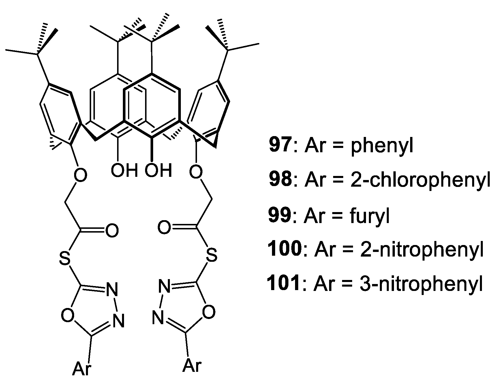
1,3,4-oxadiazole nucleus and its derivatives have attracted much attention because of their pharmaceutical applications, such as antimicrobial, antituberculostatic, or fungicidal agents. In 2020, Gezelbash and Dilmaghani used the oxadiazole core, as bioisosteres of amides and esters, and coupled them to the calixarene skeleton to generate synergy in the therapeutic effect [146].
The new oxadiazole-based calixarenes 97 to 101 presented in Figure 10 were obtained by reaction of para-tert-butylcalix[4]arene 62 with the corresponding 1,3,4-oxadiazole acyl chloride derivatives under carbonate medium in DMF. The antibacterial evaluation was carried out against the E. coli strain. Unfortunately, the results do not seem convincing for a synergistic effect provided by the calixarenic platform. No activities were recorded for calixarenes 97 and 98 compared with the corresponding 1,3,4-oxadiazole acyl chlorides, with MICs at 10 and 15 µg/mL, respectively. Only 97 and 101 presented an antibacterial activity where the initial oxadiazole acyl chloride was inactive. However, the authors provide no explanation for the measurements acquired.
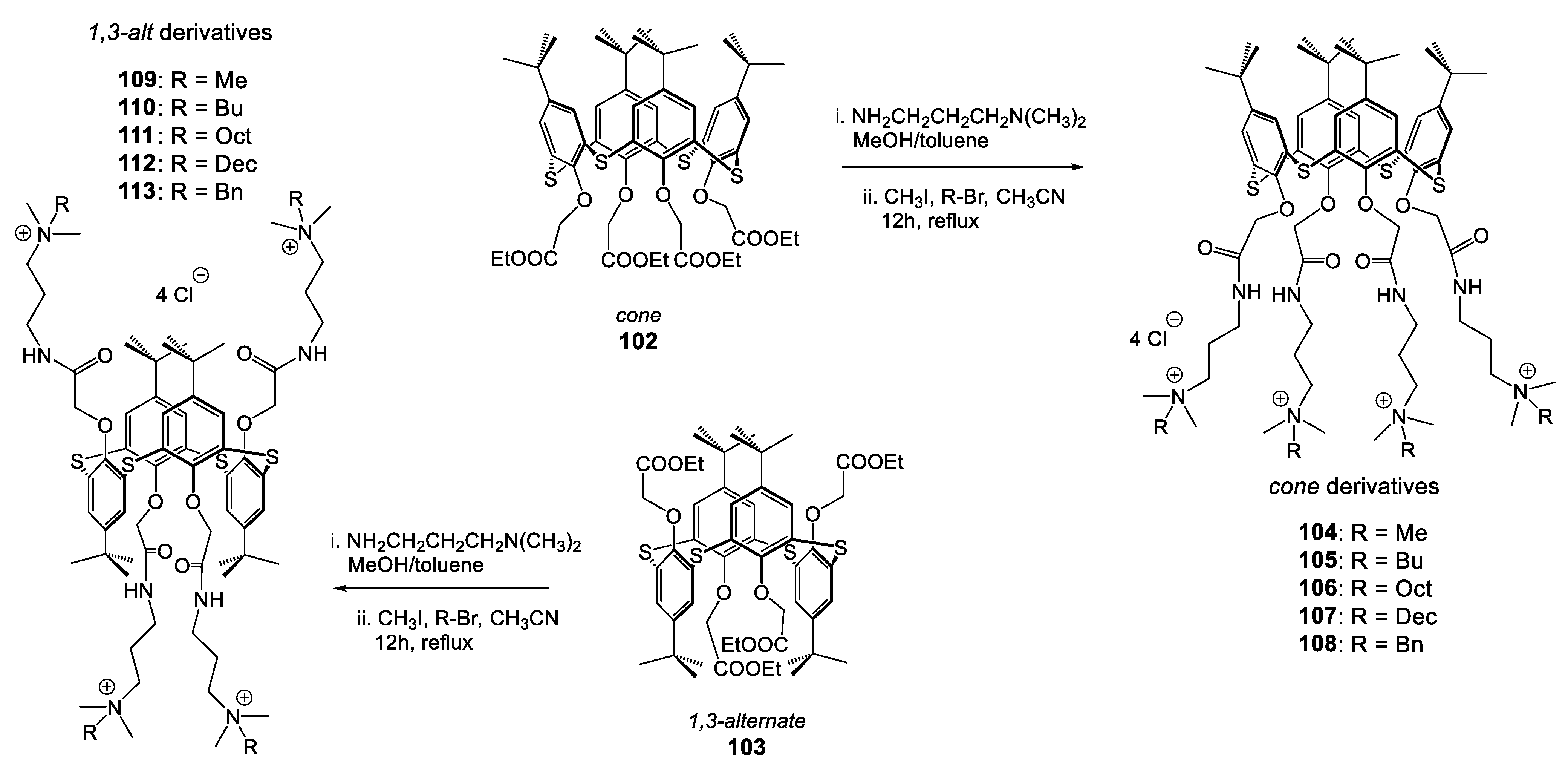
A very recent study complements previous work on positively charged antibacterial structures (ammonium, imidazolium, guanidinium). In 2021, Padnya et al. have developed a series of thiacalix[4]arene-based quaternary ammonium functions under both cone and 1,3-alternate conformations [147].
From 102 in cone conformation, or from its 1,3-alternate analogue 103 and according to the same operating conditions, they obtain, in two steps, the ammonium compounds 104 to 108 (conical derivatives) and 109 to 113 (1,3-alt derivatives) with excellent yields (>90%). Ester derivatives are easily converted to amides by the introduction of N,N-dimethylaminopropylamine. In a second step, the tertiary amines are alkylated by various halogenated derivatives (bromide or iodide). Finally, the passage on an ion exchange chromatography makes it possible to obtain the 10 new quaternary ammonium compounds in the form of chloride salts (Scheme 10).
MIC and MBC measurements were conducted on three Gram-positive strains (S. aureus ATCC 29213, a clinical isolate of S. epidermidis, and B. subtilis 168) as well as three Gram-negative strains (E. coli ATCC 25922, K. pneumoniae 1813, and P. aeruginosa ATCC 27853). The results are presented in Table 9 and have been compared, among others, with the activity of chlorhexidine used as a reference. Except for the 109 derivative, all the compounds studied have a very good antibacterial activity, with MIC values often below 16 μg/mL, and close to the activities measured with reference compounds. As previously observed [103], the results show that the derivatives in the 1,3-alt conformation exhibit higher activities compared with the conical homologues. The 1,3-alt octyl thiacalix[4]arene 111 produces the lowest MIC values of 1 to 2 µg/mL and 8 to 32 µg/mL, respectively, against Gram-positive and Gram-negative strains.
The authors also measured the cytotoxic activities of these thiacalixarene-based quaternary ammonium salts. The cell viability measured on human skin fibroblast cells allows the calculation of the selectivity index. In agreement with the work of Mourer et al. [103], there clearly appears to be lower toxicity for 1,3-alternated conformers, with CC50 values close to 1000 µg/mL for 109, 111, and 113. Based on the selectivity index values, the study shows that the most promising antibacterial candidate is the 1,3-alt-Oct derivative 111 against both Gram-positive and Gram-negative bacteria strains. To complete the study, the authors tested three most active candidates against 16 Gram-positive and Gram-negative strains from clinical isolates. In the same way as on standard strains, the compound 111 exhibits the weakest MIC values, more particularly on Gram-positive strains.
Finally, to complete the study and try to understand the activities measured, the authors evaluated the different types of interactions of the cone-Oct 106 and 1,3-alt-Oct 111 derivatives with two lipid membrane models: DPPC (1,2-dihexadecanoyl-sn-glycero-3-phosphocoline) and POPG (1-palmitoyl-2-oleyl-sn-glycro-3-phospho-(1′-rac-glycerol). The evolution of the hydrodynamic diameters, of the polydispersity indexes, and of the zeta potentials of the vesicles in the presence of thiacalixarene indicates different modes of interaction. The conical compound is adsorbed on the surface of the vesicles, while the 1,3-alt derivative leads to clumping of these. These two different interaction mechanisms could explain the variation in activity of the two conformers. As proposed in previous studies of other groups [62,115,119], the authors suggest a mechanism of antibacterial action, based initially on an approach of thiacalixarene generated by electrostatic interactions (negatively charged bacterial membrane), followed by a membrane integration of the hydrophobic parts.
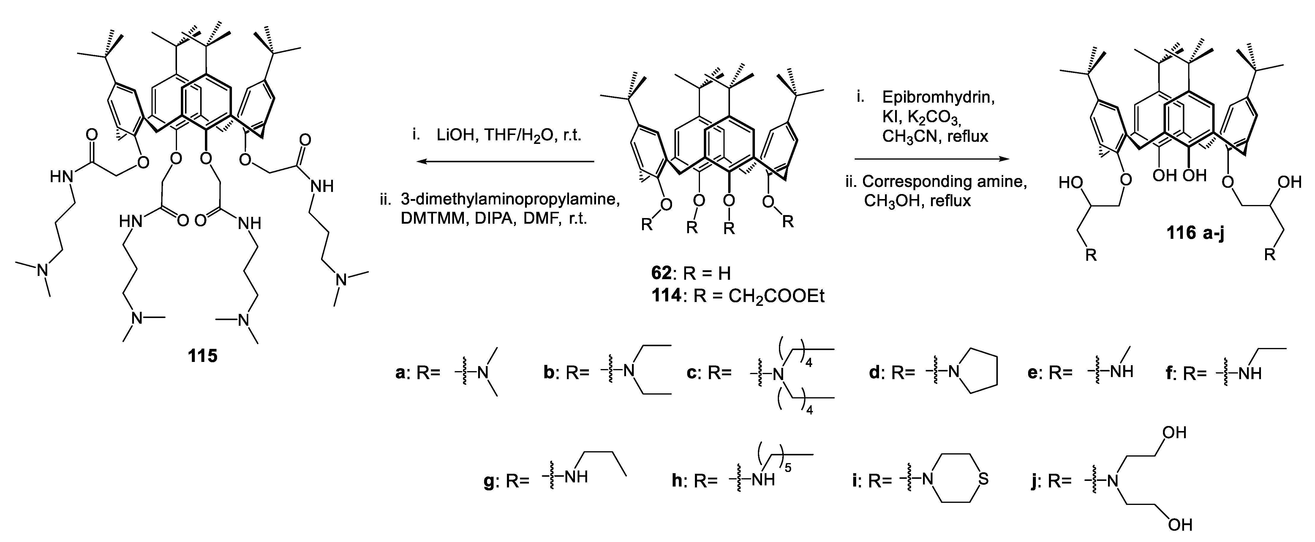
In 2022, Fang et al. designed and synthesized a series of bis- and tetra-functionalized calix[4]arenes as membrane-active antibacterial agents. Synthesized from a tertiobutylated calix[4]arene platform (from 62 or 114), they introduce either two or four amino functions into the lower rim (Scheme 11) [148]. The latter become potentially cationic at physiological pH and can thus exert their action. As hypothesized in previous studies [62,115,119], these cationic amphiphilic structures will again be able to interact electrostatically with the negatively charged bacterial membrane. Here again, the authors point out that the lipophilic charge on the upper rim (tertio-butyl group) could facilitate the insertion of the structure into the phospholipid bilayer of the membrane, altering the permeability and causing a deleterious effect on the cell.
Tested on the Gram-positive reference strain S. aureus ATCC 29213 and two resistant strains MRSA N315 and MRSA NCTC10442, the results are very promising, especially for the tetra-functionalized compound 115 incorporating 3-dimethylaminopropylamine functions. Indeed, it shows minimal inhibitory concentrations of 1.6, 1.6, and 3.1 µg/mL, respectively, on the three strains mentioned above. In addition, the authors showed a rapid action of the compound 115, where the time–kill kinetics curves against MRSA NCTC10442 show a decrease of 4.3 to 5.6 log of the bacterial population in 0.5 h to, respectively, 4 and 8 times the MIC. In comparison, vancomycin, used as a reference, only leads to a reduction of 2 log to 8 times its CMI in 4 h.
The membrane target and increased permeability were supported by the use of the SYTOX Green dye, which easily penetrates bacteria with damaged membranes and for which the fluorescence intensity increases when bound to intracellular nucleic acids. The increase in 115-concentration on MRSA NCTC10442 is indeed accompanied by a very significant increase in fluorescence intensity.

7.2. Molecular Carrier/Drug Delivery

Widely used in the search for new active compounds, the principle of carriers is part of the strategies for the development of new active ingredients. In the case of calixarenes, the authors generally seek to use the platform in order to modify the bioavailability (increase the hydro- or the lipo-solubility for examples), increasing the activity of the active ingredient by generating a greater density of antibiotic per unit surface (cluster effect), and this via strong covalent bonds or physiologically hydrolyzable bonds.
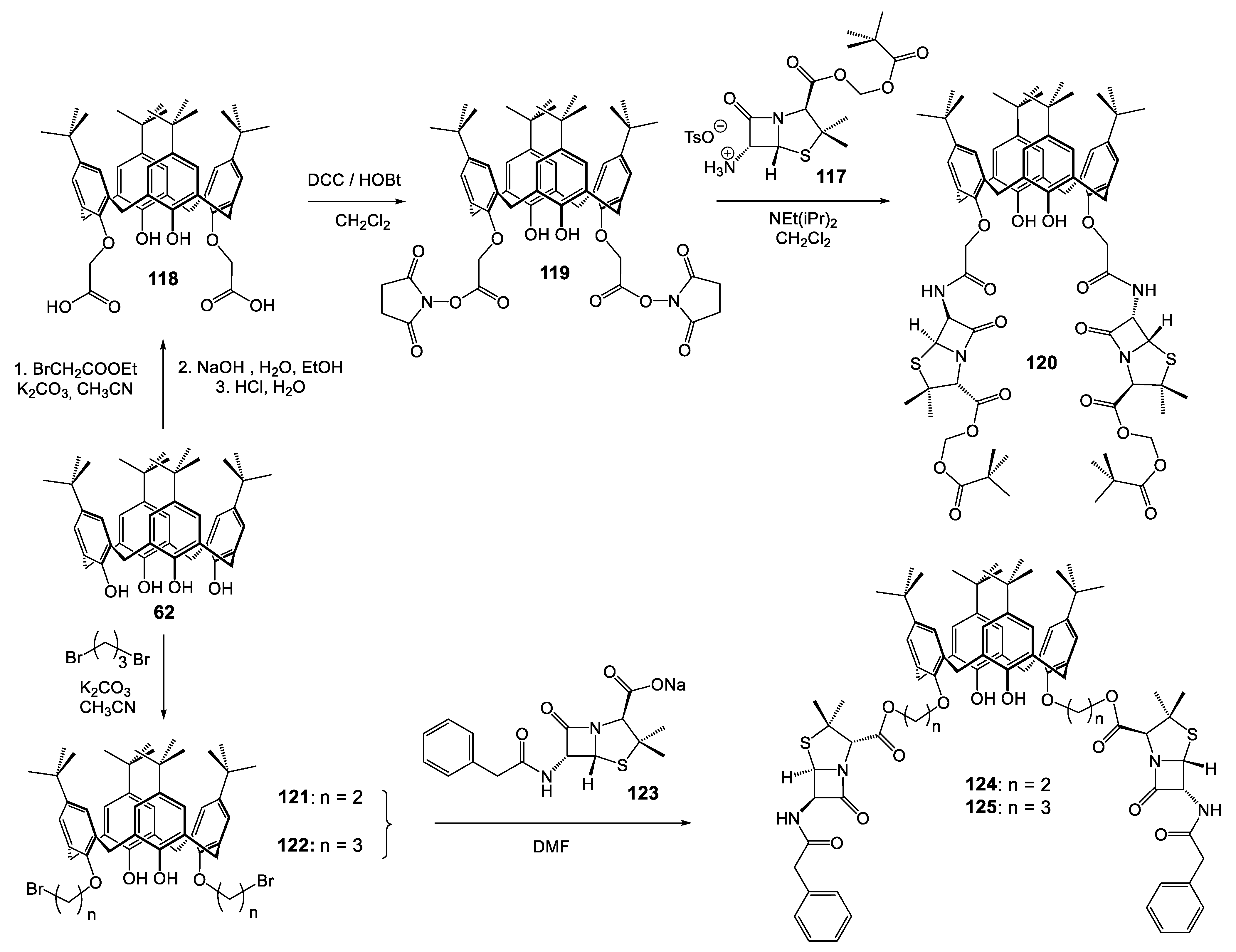
In 2001, Regnouf-de-Vains and Ben Salem initiated a study to develop calixarene-based podands shaped as potent drug dispensers, first with antibiotic penicillin grafted in alternate positions of the cone conformer of the para-tert-butylcalix[4]arene [149]. The synthetic pathway described in Scheme 12 involved the formation of hemi-synthetic penicillin via the formation of an amide bond. Penicillin was firstly protected at the carboxy group by a pivaloyloxymethyl ester function (117 Scheme 12), thus giving a lipophilic prodrug which should release the biologically active free acid after hydrolysis by esterases, a peptide coupling via the formation of bis-activated ester 119 between the diacid calixarene 118, N-hydroxysuccinimide and dicyclohexylcarbodiimide (Scheme 12). Reaction between the penicillin salt derivatives 117 and 119 in mild conditions gave the expected bis-penicillin podand 120.
The authors also developed a para-tert-butylcalix[4]arene grafted by two penicillin G arms at the lower rim via ester linkage [150]. The choice of para-tert-butylcalixarene was directly related to a possible application of the molecules studied as prodrugs. An oral administration would lead to the release of the soluble antibacterial agent in the intestinal compartment; the insoluble, nontoxic calixarene molecule could be thus easily eliminated from the organism. The 1,3-bis(bromoethyl)- 121 or 1,3-bis(bromopropyl)-calix[4]arene 122 react in dry DMF with penicillin G sodium salt 113 to give 124 (47%) and 125 (32%), respectively (Scheme 12).
The amphiphilic structures 6-pivAPA 120, and two benzylpenicillin derivatives 124 and 125, were the subject of preliminary study of the possibility of incorporation and/or translocation across a model bacterial membrane lipid, using lipid monolayers prepared from 1,2-dimyristoyl-sn-glycero-3-phosphoethanolamine (DMPE) [150]. The investigations, assessed by surface pressure and surface potential measurements, as well as Brewster angle microscopy, show a possible incorporation of benzylpenicillin derivatives 124 and 125 into the biological membrane whereas the 6-pivAPA 120 could be more easily translocated across the membrane, probably due to the aliphatic pivaloyl terminal moieties decreasing molecule packing in the monolayer compared to the benzyl moieties.
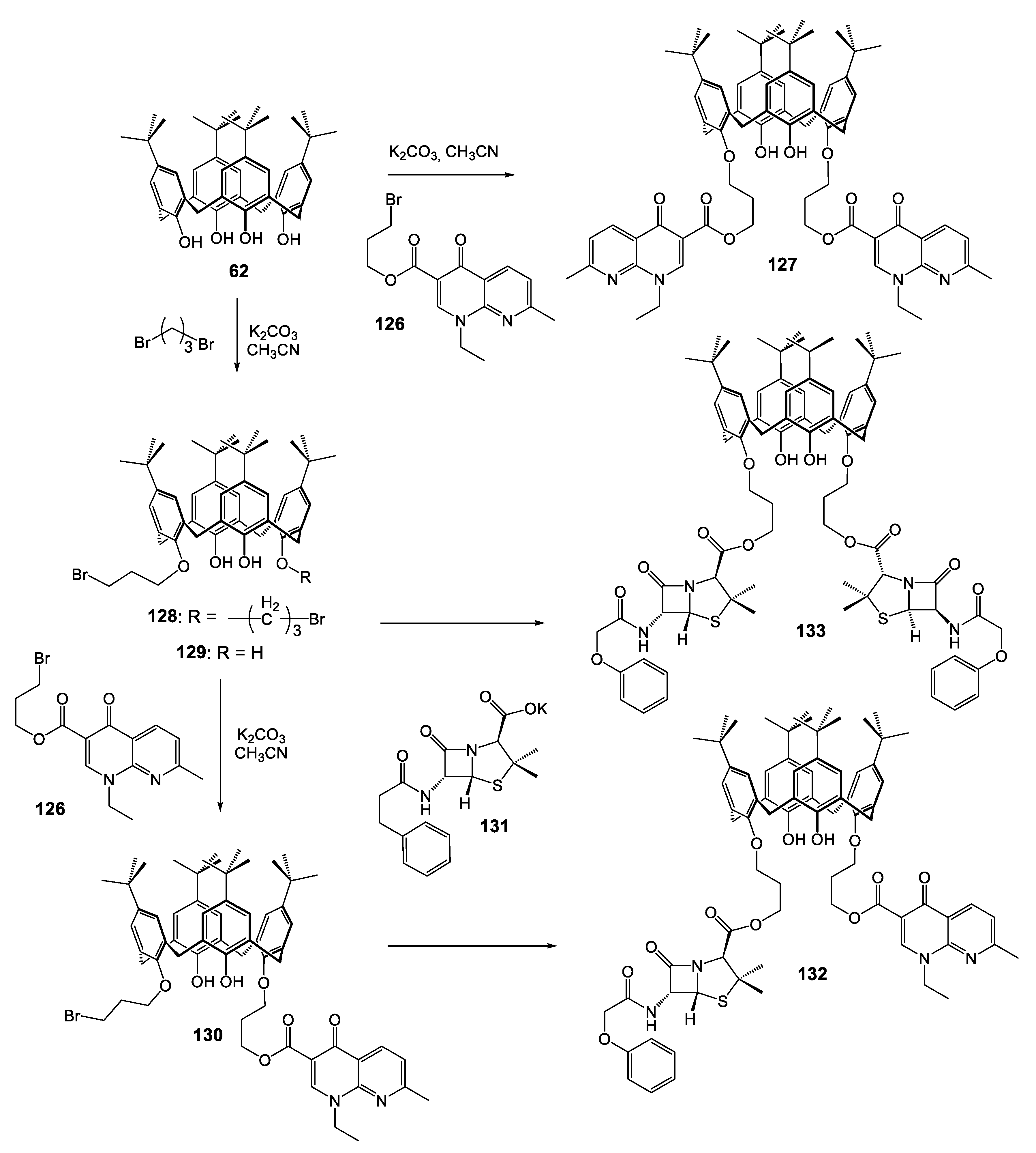
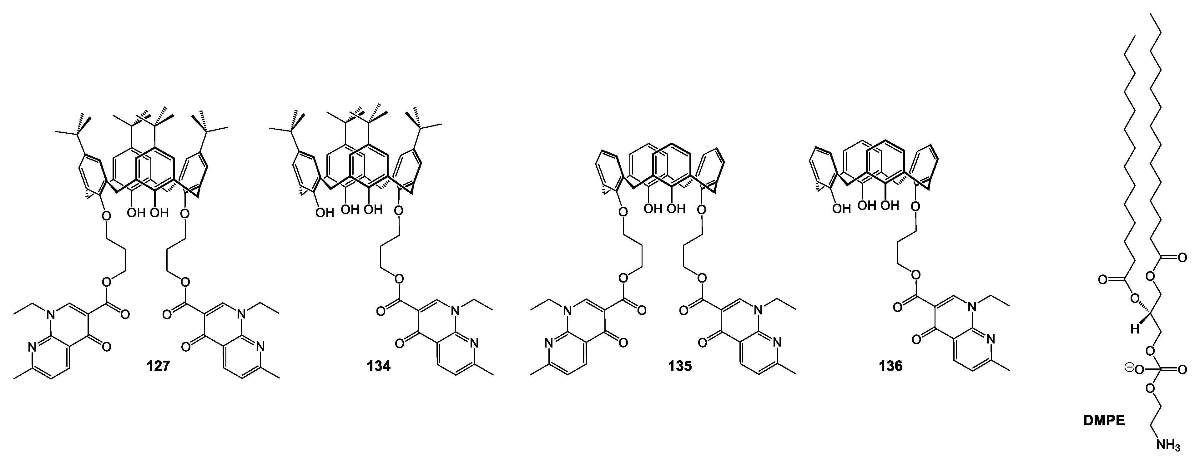
The same strategy involving ester linkage allows the synthesis of bis-quinolone derivative 127 [151,152]. The bromopropyl ester of nalidixic acid 126 was initially prepared by reaction in DMF of an excess of 1,3-dibromopropane with sodium salt of nalidixic acid (Scheme 13). Then the reaction with para-tert-butylcalix[4]arene 62 was performed in basic conditions using K2CO3 as base to finally afford the bis-nalidixic podand 127 with an overall yield of 35%.
Using the monobromopropylcalix[4]arene 129 as starting material and bromopropyl ester of nalidixic acid 127 in the presence of 0.6 equivalent of K2CO3 led the authors to obtain the mononalidixic derivative 130. Incorporation of penicillin V onto the residual brominated arm was performed in dry DMF at 35 °C, under an inert atmosphere, by reacting 130 with an excess of potassium salt of penicillin V (PVK) 131 to give the nalidixic acid/penicillin V calix[4]arene podand 132 (Scheme 13) [153] that could mix synergistically two antiviral strategies, inhibition of AND-gyrase with quinolone and inhibition of peptidoglycan reticulation with penicillin. The 1,3-bis-propylpenicillinate 133 was prepared in 32% yield by direct reaction of 131 on the 1,3-bis-(3-bromopropyl)-calixarene 128 in dry DMF (Scheme 13).
The first antibacterial evaluations were performed with disk diffusion assays in DMSO solution of the dissymmetric podand (penicillin V and nalidixic acid) 132, the corresponding bis-nalidixic 127, and the bis-penicillin V 133 [153], against three Gram-positive reference strains, S. aureus ATCC 25923, S. aureus ATCC 29213, and E. faecalis ATCC 29212, as well as against two Gram-negative reference strains E. coli ATCC 25922 and P. aeruginosa ATCC 27853. The results showed that only the dissymmetric compound 132 exhibits an important growth inhibition against S. aureus ATCC 25923, with an inhibition diameter of 31.5 mm.
To confirm the possible prodrug behavior of these compounds, Regnouf-de-Vains et al. reinvestigated the amphiphilic calixarenes bearing one or two nalidixic moieties attached as esters to the lower rim of the calixarene platform via a propyl linker, and H- or tert-butyl groups in the para-position at the upper rim [154]. The previously described compounds 127 [150] and 134 [151] were obtained in a better yield from bromopropylnalidixate in basic conditions (K2CO3 and NaHCO3 respectively) with KI as catalyst. The same conditions applied to detertiobutylated calix[4]arene gave the bis- and mono-nalidixate 118 and 119 (Figure 11).
The monolayers (monomolecular film) of amphiphilic calixarenes 127, 134, 135, and 136 have been studied for the first time at the air–water interface on a Langmuir balance, showing a better stability of monolayers for the para-tert-butylated derivatives 127 and 134. The films formed with derivatives 135 and 136 cannot be used in hydrolysis studies, as they are not stable enough. For a second time, the study at the air-carbonate buffer (pH 10) interface for 127 and 134 confirmed the initial hypothesis with a nalidixate salt release (HPLC monitoring).
The same nalidixic calixarenes 127 and 134 were studied upon interaction with model bacterial membranes via molecular dynamics simulation, as well as surface pressure, surface potential, or Brewster angle microscopy, for example, [155]. The results obtained show that mixed films formed with DMPE and calixarenes 127 or 134 have a more liquid-like character (more marked for 134) compared with pure DMPE, and that interaction between the calixarene derivatives and DMPE occurs via the phosphate and carbonyl groups present in the lipid. Thus, diffusion of 134 in the monolayer would be easier compared with 127, and this one can be useful for developing new calixarene based drug carriers.
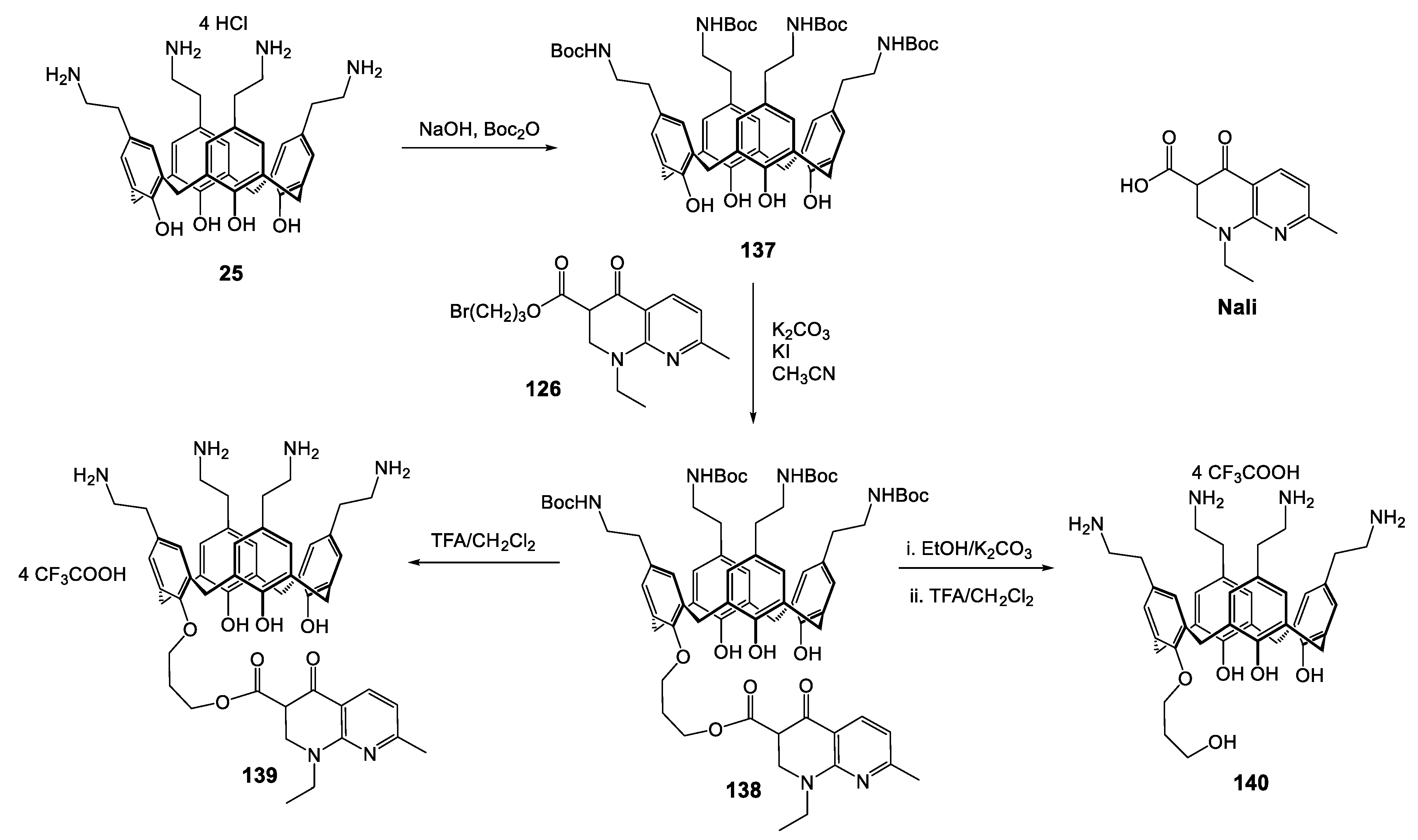
Designed as hydrophobic compounds, these structures are not really easy for in vitro standard evaluation as antimicrobial agents. Thus, in 2019, Massimba Dibama and co-authors developed a synthetic strategy leading to a water-soluble analogue of the previous compounds [156]. They described the synthesis of a prodrug-like structure that involves one nalidixic acid moiety tethered via a propyl linker to the lower rim of the tetra-para-aminoethylcalix[4]arene. Obtained according to Gutsche’s procedure [64], the tetra-para-aminoethylcalix[4]arene 25 was protected as Boc-derivative (137 Scheme 14), and the introduction of bromopropylnalidixate 126 was performed under basic conditions in the presence of a catalytic amount of KI. Then the mononalidixic podand 138 was unprotected in TFA conditions to give the expected compound 139 (Scheme 14).
The antibacterial activities (MIC) of prodrug 139, of its two expected metabolites nalidixic acid Nali, and 3-hydroxypropyl calix[4]arene 140 (Scheme 12) were evaluated in liquid phase against various Gram-negative (E. coli ATCC 25922, P. aeruginosa ATCC 27853) and Gram-positive (S. aureus ATCC 25923 and ATCC 29213, and E. faecalis ATCC 29212) bacteria. The calixarene species 139 displays MIC values of 35 µg/mL for E. coli, 70 µg/mL for the two S. aureus strains, and 140 µg/mL for E. faecalis and P. aeruginosa, while the mono-alcohol 140 needs from 2 (E. faecalis, P. aeruginosa) to 4 (E. coli, S. aureus) times higher concentrations, in ranges considered as non-active. Nali exhibits a MIC close to 32 µg/mL in all strains. The authors concluded that even with the ideal hypothesis of 100% hydrolysis, the MIC values obtained for the mononalidixate 139 are not consistent with the simple release of nalidixic acid, leading to a potential specific activity of 139 or an additive or a multiplicative effect between 139 or 140 and Nali.
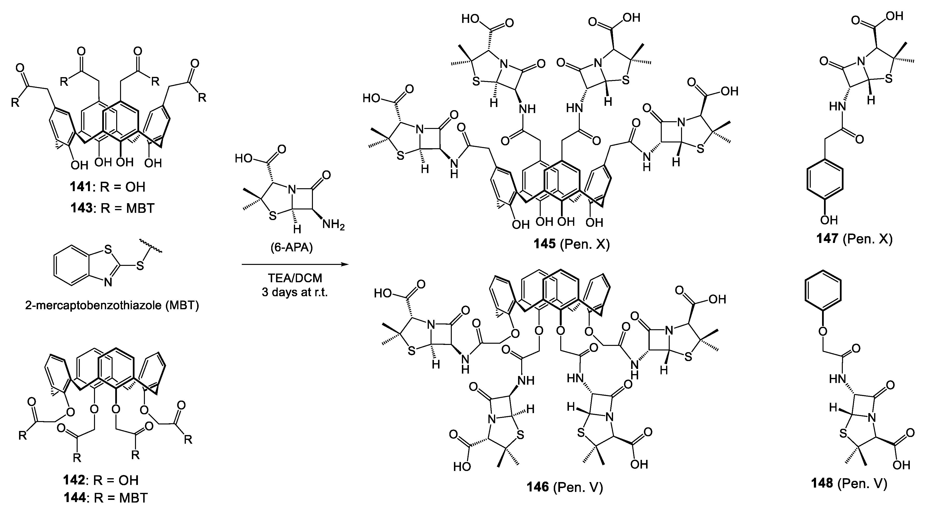
On the same principle, Pur and Dilmaghani published, in 2014, the synthesis and biological evaluation of penicillins V and X clustered by a calixarene scaffold (named calixpenams) [157]. In this work, calixarene is not used as an active principle carrier; it becomes an integral part of it. They used the phenol moieties of calixarene to acylate the amino group of 6-aminopenicillanic acid (6-APA) and conduct the formation of cluster (cyclotetramer) of penicillin X (6-APA grafted on the upper rim) or penicillin V (6-APA grafted on the lower rim).
Previously prepared according the method of Gutsche et al. and McKuvey et al., respectively, the calixarenes 141 and 142 react in soft conditions via an active thioester formation process that involves the use of 2,2′dibenzothiazole disulfide (DBTDS) as carboxylic acid activator (143 and 144 Scheme 15). Then the aminolysis reaction occurs in the presence of 6-APA and trimethylamine in CH2Cl2 to give the corresponding calixpenams 145 (58%) and 146 (49%), tetramers of penicillin X or penicillin V, respectively.
For completeness, the authors conversed the calixpenams 145 and 146 to the corresponding calixcephems 151 and 152, respectively (Scheme 16) [158]. This conversion was first through oxidation of sulfur atoms of penam moieties in the presence of peracetic acid (149 and 150 Scheme 16), followed by ring expansion employing the N,N′-bis(trimethylsilyl)urea (BSU) and pyridine-hydrogen bromide (Py-HBr) to produce calixcephens 151 (Ceph. X) and 152 (Ceph. V) Scheme 16.
The antibacterial effect was initially measured against three Gram-positive non-penicillinase-producing strains of Streptococcus including S. pyogenes ATCC 19615, S. agalactiae ATCC 12386, and Streptococcus pneumoniae ATCC 49619 (Table 10) [157]. The impact of cyclotetramerization was demonstrated by a noteworthy increase in activity on all the strains of penicillin cyclotetramer (145 and 146) compared to the monomers 147 and 148, respectively, for example, 0.002 mg/mL against S. pyogenes for 145 (Pen. X) and 0.012 mg/mL for its constitutive monomer 147 (Pen. X). It appears globally for both penams (X and V) a ratio of 6 between the calixarene’s MIC and the monomer’s MIC in favor of macrocycle. The authors attributed the slight difference of activity between 145 (Pen. X) (more active) and 146 (Pen. V) to a larger contact surface with the bacterial membrane offered by the upper rim of calixarene in the first case compared with that of the lower rim in the second.
In the second evaluation [158], the antibacterial susceptibility testing was assessed against five S. aureus strains: two β-lactamase-producing methicillin-resistant (MRSA) strains ATCC 43300 and ATCC 33591 and three methicillin-sensitive (MSSA) strains, including one non-β-lactamase producing strain ATCC 25923 and two β-lactamase-producing strains ATCC 29213 and 11632 (penicillin-resistant strain). The results also showed a great activity of calixarene structures versus the monomers. Calixcephens were about 10-fold more active than the corresponding monomer (1.550 µg/mL for 151 (Ceph. X) versus 16.325 µg/mL for constitutive monomer 153 (Ceph. X) on ATCC 3359). On the other hand, the comparison in the calixarene family demonstrates a considerable gain of activity for the calixcephems: 0.925, 1.225, and 1.635 µg/mL, respectively, on three β-lactamase-producing strains ATCC 11632, 43300 and 33591 versus the calixpenams >64 µg/mL for the same strains. These last results are probably due, according to the authors, to the fact that compounds including cephalosporanic acid cores have greater activity against β-lactamase-producing bacterial strains in comparison to compounds exhibiting penicillanic acid cores.
Nowadays, many advances have been made in the search for new agents for nucleic acid (NA) delivery, notably through the use of pre-constituted building blocks and more particularly through the calixarene platform, due to its three-dimensional architecture and its possible multifunctionality increasing the interaction with NAs, as illustrated by the important work carried out by the Ungaro group’s examples [159,160,161]. Candiani and co-workers use this working basis for the development of new multivalent calixarenes and the study of their transfection efficiency [162]. The high affinity of aminoglycosides (AGs) for NAs, associated with their known antibacterial capabilities encourage the authors to cluster some AG derivatives on a calixarenic platform with the aim to obtain a new class of multivalent gene delivery vectors with inherent antibacterial properties (Scheme 17).
The starting material, the para-tetra-amino-tetra-hexyloxycalix[4]arene 155, reacts with the isothiocyanate-functionalized aminoglycosides (AGs) derived from neomycin B, neamine and paromomycin in DMSO medium, giving rise to the formation of calix-AG derivatives 156, 157, and 158, respectively (Scheme 17). If these three derivatives did induce very effective DNA complexation, they also have some antibacterial properties. These last were evaluated against Gram-negative E. coli and Gram-positive Staphylococcus lutea bacteria.
The starting calixarene 155 is ineffective, and the introduction of AGs results in the development of an antibacterial effect. In solution, derivatives 1156 and 1158 seem to be the most active and effectively inhibit the growth of E. coli compared with the inactive derivative 157. For example, the paromomycin derivative 158 has a MIC90 around 28.2 µg/mL. However, it turns out that the calixarene-AG derivatives are still slightly less active than the free AGs used as references (neomycin and paromomycin show a MIC90 at 4 µg/mL). Surprisingly, the calixarene neamine derivative 157 is inactive, while its free AG displayed a MIC90 at 32 µg/mL. The authors attribute this lack of activity to the conjugation to the calixarenic skeleton, which, in this particular case, would produce an antagonistic effect to neighboring AGs. On the other hand, the same derivatives 156 to 158 displayed a weak antibacterial effect on Gram-positive strains.

7.3. Complexes

The ion complexation by calixarene derivatives has been the subject of numerous studies and is widely used in the separation and extraction of metal ions, sensors material or catalysts. They are good carriers for cations due to their cyclic structure with different sizes of cavities and the many possibilities of functionalization. These functional arms, endowed of electron donating groups, coupled to the organizational capacity of calixarene, allow generating a conducive environment to the ion’s reception.
Through their research about DNA cleavage activity, Özkan et al. published, in 2015, a synthesis of the antimicrobial and DNA cleavage activities of para-tert-butylcalix[4]arene diamide derivatives and some of their copper(II)complexes [163]. After synthesising the diethyl-ester derivative 159 of para-tert-butylcalix[4]arene 62 (Scheme 18), hydrolysis process affords the di-acid compound 160, which undergoes the introduction of amines to give the diamide derivatives 161 to 164 (Scheme 18). The capacities of complexation were carried out against some transition metal ions [Cu(II), Co(II), Cd(II), Zn(II), Ni(II), Mn(II), Pb(II)] and the best results are in favor of calixarene 163 and 164 for capture of Cu(II) giving Cu(II) complexes 165 and 166 (copper(II) chloride and copper(II) nitrate, respectively). These last two complexes were studied with ligands 161 to 164 for their antibacterial activities against Gram-positive (B. subtilis ATCC 6633, B. cereus NRRI-B-3711, E. faecalis (clinic sample), S. aureus NTCT 8325) and Gram-negative bacteria (E. coli ATCC 25922, E. coli ATCC 35128, P. aeruginosa ATCC 27853, Proteus vulgaris ATCC 8427) (Table 11).
The result of DNA interaction showed that of all the compounds tested, calixarene derivatives (159164) and metal complexes (165, 166) were able to cause damage to the plasmid DNA, and that copper(II) complexes are very effective for DNA cleavage. In parallel, the results of antibacterial susceptibility are less striking. The complexes are not active on the strains tested. The other structures 160, 162, 163, and 164 showed no activity (MIC > 625 mg/mL); only the bi-ester intermediate 159 and the bis-furane derivative 161 give an inhibition of bacteria cell growth. B. cereus, E. faecalis, and S. aureus E. coli (ATCC 35128) are sensitive to compound 161 (MIC values 39, 39, 39, and 78 µg/mL, respectively). Surprisingly, it appears that compound 159 is the most active of the series and exhibits an activity on almost all strains, with MICs at 78 µg/mL on B. subtilis, 39 µg/mL on B. cereus and two strains of E. coli, and 156 µg/mL for E. faecalis, S. aureus, and P. vulgaris. The authors do not expose a reason for the activities detected for these two structures. They consider the non-activity of the copper(II) complexes by the largest molecular structures compared to compound 161, for example, preventing the passage thought the cell bacterial membrane.
Based on a similar structure, Akkuş et al. published, the same year, a study on selective extraction of toxic heavy metals by a pyrimidylthioamide calixarene derivative and the corresponding antibacterial activities [164]. Para-tert-butylcalix[4]arene 62 was grafted in the 1,3-alternate position via ether bond with benzyl moieties, then on the other phenolic positions by para-nitrobenzyl groups, in the presence of NaH as base (Scheme 18). The obtained compound 167 is placed in the presence of Raney-Ni, and the reduced derivative 168 which emerges reacts with 2-(pyrimidin-2-ylthio)acetyl chloride to afford the pyrimidylthioamide calixarene derivative 169 (Scheme 18). This last is a good extractant and has a good affinity for Hg and Pb ions with respect to the intermediates 167 and 168. These mercury(II) or lead(II) complexes were not evaluated in antibacterial tests. On the other hand, the ligand 169 was tested for its antibacterial effect against the E. coli ATCC 25922, K. pneumoniae, P. aeruginosa, S. aureus NRRL-B 767, Salmonella typhimurium, B. cereus ATCC 11778, and B. subtilis NRS 744 bacterial strains. Measured by inhibition zones on an impregnated disc (5 mm) with 10 μg of drugs, an activity emerges against S. typhimurium, B. cereus, B. subtilis, and S. aureus (inhibition zone at 10, 10, 11, and 11 respectively) compared to 15, 8, 8, and 30 mm of inhibition obtained with Ceftriaxone used as reference. This activity of pyrimidylthioamide calixarene is associated by the authors with the presence of nitrogen and oxygen donor atoms and amide groups, which might have an activity against the enzyme production, in agreement with the literature [165].
On the same principle, Memon et al. have synthesized a diamide derivative of para-tert-butylcalix[4]arene and its metal complex, and exhibit the antibacterial activities [166]. The 1,3-alternate bis-amide ligand 171 was obtained by action of ethanolamine and methanol on the intermediate bis-ester 170; then, the copper-complex 172 was formed via addition of stoichiometric amount of cuprique nitrate salt (Scheme 19).
Evaluation of bacterial growth was carried out Gram-positive Staphylococcus albus and Gram-negative E. coli ATCC strains with inhibition zone and MIC determinations on the ligand 171 and its copper complex 172 (Table 12).
The results show that these two calix[4]arene derivatives are very active against two bacteria strains tested. The organosoluble ligand 171 is active up to 3 µg/mL, while its binuclear Cu(II)-complex 172 generates an antibacterial activity eight times greater, with an active concentration at 0.37 µg/mL. The authors explain this variation of activity in favor of the complex by the fact that the complexation of the two copper atoms by the ligand makes it possible to increase the lipophilic nature of the structure via a significant delocalization and a sharing of electrons between the copper ions and the donor atoms. A greater lipophilicity thus allows easier passage of the lipid membrane, the complexes being able to cause problems in the process of respiration by blocking protein synthesis and therefore bacterial growth.
Multi-dentate chelating para-tert-butyl-calix[4]arene, functionalized at the 1,3-alternate positions of the lower rim with two thiosemicarbazide or thiosemicarbazone moieties, was reported by Bahojb Noruzi et al. in 2019 and 2020, respectively, with the aim to develop a new class of antimicrobial compound [167,168]. The thiosemicarbazide moieties and their thiosemicarbazone derivatives are known for their ability to coordinate with transition metals by bonding with sulfur and nitrogen atoms. They also studied, alone or in metal complex form, their biological properties, including antibacterial, antiviral, or antitumor activities. Weakly studied in literature, the calixarene-based thiosemicarbazone compounds could be an interesting candidate for antibacterial compounds. Therefore, the authors developed the ligand L following the synthetic way presented in Scheme 20. The para-tert-butylcalix[4]arene 62 functionalized in the 1,3-alternate position by cyanoethyl arms followed by reduction step allows the bis-amino intermediate 173 formation. This last has been derived in isothiocyanate (compound 174), then transformed in thiosemicarbazide using hydrazine (compound 175). The expected thiosemicarbazone equivalent 176 was obtained by treatment of 175 with salicylaldehyde.
Eight divalent metal complexes were obtained by bringing 175 or 176 into contact with one equivalent of cobalt (II), nickel (II), copper (II), or zinc (II) salts to afford the metal derivatives 175-Co, 175-Ni, 175-Cu, 175-Zn, 176-Co, 176-Ni, 176-Cu, and 176-Zn. All of the synthesized compounds were evaluated with regard to their antibacterial capacity against Gram-positive bacteria (S. aureus and B. subtilis) and Gram-negative bacteria (E. coli and P. aeruginosa) and the MIC values are presented in Table 13.
The first observation is the fact that the thiosemicarbazide ligand 175 is almost inactive against the tested strains, while the thiosemicarbazone 176 analogue exhibits a stronger antibacterial effect with an increase in MIC values of 2-fold and 8-fold against P. aeruginosa and E. coli, respectively, i.e., 31.25 µg/mL on these strains. No decrease in bacterial growth is observed for S. aureus. In the case of 175, all of the metal derivatives show an enhancement of the antibacterial activity against Gram-negative bacteria (except for the Co(II) and Cu(II) derivatives against P. aeruginosa), with a more significant improvement for the Ni(II) and Zn(II) complexes. In parallel, the 176 complexes are all very active on the all strains tested in the study, with MICs of 31.25 µg/mL, and are thus found to be much more interesting in terms of activity than their thiosemicabazide-based analogues.

8. Outlook

Several important points emerge from this review. First, it is undeniable that the organizational capacity of the calixarene plays a pivotal role in the antimicrobial activities observed. Second, numerous works propose decorating the calixarene structure with positively charged functions (e.g., ammonium, guanidinium, imidazolium…) to most likely target the bacterial cell wall. Third, the antibacterial activities measured are very promising: broad spectrum activity, against both wild-type bacteria or antibiotic-resistant bacteria. Fourth, even if the majority of the reported work is based on calixarenes with conical conformation, others are beginning to focus on other conformations, and the authors reach identical conclusions: significant differences in terms of activity in function of conformation. Thus, we can admit that the stereochemical orientation of the functional groups present on the surface of the calixarenic skeleton plays on the antibacterial activity. Fifth, several studies also show that the multivalence offered by calixarenes seems very favorable to the development of prodrugs (e.g., modification of bioavailability, cluster effect, hydrolyzable link, etc.). Finally, all of these studies should lead us to more in-depth research on structure-activity relationships and the design of new functionalized calixarenes with enhanced antibacterial properties.
Nonetheless, the use of calix[n]arenes in the therapeutic fields, particularly for infectious diseases, is still recent, which, despite their numerous advantages, results (still) in an undervaluation of their biological properties, or even a lack of interest. However, the almost unlimited possibilities for functionalization of these structures, associated with their multivalence and their potential for spatial organization, offer a very wide range of possibilities for the development of promising candidates usable in different medical fields, and more precisely, bacteriology. However, if the antibacterial activities are very promising, the compounds currently studied will still require more in-depth studies to possibly become potential drug candidates.

9. Conclusions

Calixarenes have already come a long way since A. von Baeyer in 1872 and have found their place in many fields of study and applications. Thanks to their great flexibility (functionalization and spatial organization), the Man of Art knew how to modulate them in order to confer remarkable activities. If we only consider the discrete calixarene structures, the subject of this review, they have found a place in the first stage of the therapeutic field, and more particularly, the search of new antibacterial agents. The crucial lack of new antibiotics associated with the alarming increase in bacterial resistance requires a major effort in the search for new therapeutic strategies, and it appears that the calixarene structure finds its place there. The researchers have thus designed many structures carrying intrinsic activity, activity linked to the possibility of releasing active principles or to the formation of metal ion complexes. From average to very good, these activities make it possible to determine structure–activity relationships and to direct researchers towards structures with higher potential. For example, many works report the development of cationic calixarenes with an original mechanism of action based on electrostatic interactions with the bacterial cell wall. The very interesting results highlighted could lead to improvement in future work. The exceptional modularity (regioselective polyfunctionalization and multiple conformations) of the calixarene structure can still allow researchers to shape their structures in order to further increase the affinities with the targets of action sought, to broaden the spectrum of action, or, on the contrary, to target more specific strains.

Author Contributions

Writing—original draft preparation, M.M.; writing—review and editing, M.M. and R.E.D.; review, J.-B.R.-d.-V. All authors have read and agreed to the published version of the manuscript.

Funding

This work was supported by French Ministry of Higher Education & Research, CNRS, Region Grand Est.

Institutional Review Board Statement

Not applicable.

Informed Consent Statement

Not applicable.

Data Availability Statement

Not applicable.

Conflicts of Interest

The authors declare no conflict of interest.

References

  1. Chinemerem Nwobodo, D.; Ugwu, M.C.; Oliseloke Anie, C.; Al-Ouqaili, M.T.S.; Chinedu Ikem, J.; Victor Chigozie, U.; Saki, M. Antibiotic Resistance: The Challenges and Some Emerging Strategies for Tackling a Global Menace. J. Clin. Lab. Anal. 2022, 36, e24655. [Google Scholar] [CrossRef] [PubMed]
  2. Coque, T.M.; Cantón, R.; Pérez-Cobas, A.E.; Fernández-de-Bobadilla, M.D.; Baquero, F. Antimicrobial Resistance in the Global Health Network: Known Unknowns and Challenges for Efficient Responses in the 21st Century. Microorganisms 2023, 11, 1050. [Google Scholar] [CrossRef] [PubMed]
  3. Ayukekbong, J.A.; Ntemgwa, M.; Atabe, A.N. The Threat of Antimicrobial Resistance in Developing Countries: Causes and Control Strategies. Antimicrob. Resist. Infect. Control 2017, 6, 47. [Google Scholar] [CrossRef]
  4. Dutescu, I.A.; Hillier, S.A. Encouraging the Development of New Antibiotics: Are Financial Incentives the Right Way Forward? A Systematic Review and Case Study. Infect Drug Resist. 2021, 14, 415–434. [Google Scholar] [CrossRef] [PubMed]
  5. Miethke, M.; Pieroni, M.; Weber, T.; Brönstrup, M.; Hammann, P.; Halby, L.; Arimondo, P.B.; Glaser, P.; Aigle, B.; Bode, H.B.; et al. Towards the Sustainable Discovery and Development of New Antibiotics. Nat Rev Chem 2021, 5, 726–749. [Google Scholar] [CrossRef]
  6. WHO | Worldwide Country Situation Analysis: Response to Antimicrobial Resistance. WHO. Available online: http://www.who.int/drugresistance/documents/situationanalysis/en/ (accessed on 23 September 2020).
  7. Salam, M.A.; Al-Amin, M.Y.; Salam, M.T.; Pawar, J.S.; Akhter, N.; Rabaan, A.A.; Alqumber, M.A.A. Antimicrobial Resistance: A Growing Serious Threat for Global Public Health. Healthcare 2023, 11, 1946. [Google Scholar] [CrossRef]
  8. Reygaert, W.C. An Overview of the Antimicrobial Resistance Mechanisms of Bacteria. AIMS Microbiol 2018, 4, 482–501. [Google Scholar] [CrossRef]
  9. Chaïbi, K.; Jaureguy, F.; Do Rego, H.; Ruiz, P.; Mory, C.; El Helali, N.; Mrabet, S.; Mizrahi, A.; Zahar, J.-R.; Pilmis, B. What to Do with the New Antibiotics? Antibiotics 2023, 12, 654. [Google Scholar] [CrossRef]
  10. El-Sayed Ahmed, M.A.E.-G.; Zhong, L.-L.; Shen, C.; Yang, Y.; Doi, Y.; Tian, G.-B. Colistin and Its Role in the Era of Antibiotic Resistance: An Extended Review (2000–2019). Emerg. Microbes Infect. 2020, 9, 868–885. [Google Scholar] [CrossRef]
  11. Antibacterial Agents in Clinical Development: An Analysis of the Antibacterial Clinical Development Pipeline. WHO. 2019. Available online: https://www.who.int/publications-detail-redirect/9789240000193 (accessed on 23 September 2020).
  12. Duval, R.E.; Grare, M.; Demoré, B. Fight Against Antimicrobial Resistance: We Always Need New Antibacterials but for Right Bacteria. Molecules 2019, 24, 3152. [Google Scholar] [CrossRef]
  13. Klein, E.Y.; Van Boeckel, T.P.; Martinez, E.M.; Pant, S.; Gandra, S.; Levin, S.A.; Goossens, H.; Laxminarayan, R. Global Increase and Geographic Convergence in Antibiotic Consumption between 2000 and 2015. Proc. Natl. Acad. Sci. USA 2018, 115, E3463. [Google Scholar] [CrossRef] [PubMed]
  14. O’Neill, J. Publications | AMR Review. Available online: http://amr-review.org/sites/default/files/160518_Final%20paper_with%20cover.pdf (accessed on 23 September 2020).
  15. Azzi, J.; Jraij, A.; Auezova, L.; Fourmentin, S.; Greige-Gerges, H. Novel Findings for Quercetin Encapsulation and Preservation with Cyclodextrins, Liposomes, and Drug-in-Cyclodextrin-in-Liposomes. Food Hydrocoll. 2018, 81, 328–340. [Google Scholar] [CrossRef]
  16. Lima, P.S.S.; Lucchese, A.M.; Araújo-Filho, H.G.; Menezes, P.P.; Araújo, A.A.S.; Quintans-Júnior, L.J.; Quintans, J.S.S. Inclusion of Terpenes in Cyclodextrins: Preparation, Characterization and Pharmacological Approaches. Carbohydr. Polym. 2016, 151, 965–987. [Google Scholar] [CrossRef] [PubMed]
  17. Liu, D.-D.; Guo, Y.-F.; Zhang, J.-Q.; Yang, Z.-K.; Li, X.; Yang, B.; Yang, R. Inclusion of Lycorine with Natural Cyclodextrins (α-, β- and γ-CD): Experimental and in Vitro Evaluation. J. Mol. Struct. 2017, 1130, 669–676. [Google Scholar] [CrossRef]
  18. Avilés-Moreno, J.R.; Berden, G.; Oomens, J.; Martínez-Haya, B. Complexes of Crown Ether Macrocycles with Methyl Guanidinium: Insights into the Capture of Charge in Peptides. ChemPhysChem 2018, 19, 2169–2175. [Google Scholar] [CrossRef]
  19. Dutta, A.; Barman, B.K.; Mahato, B.; Rahaman, H.; Roy, M.N. Study to Explore Complexation of Crown Ether with Antidepressant Drug Prevalent in Aqueous System by Physicochemical Contrivance. Indian J. Adv. Chem. Sci. 2018, 6, 171–177. [Google Scholar] [CrossRef]
  20. Barman, S.; Yasmin, A.; Roy, M.N. Interactions of an Antifungal Sulfa Drug with Diverse Macrocyclic Polyethers Explaining Mechanism, Performance and Physiognomies Leading to Formation of Stable Complexes. J. Mol. Liq. 2017, 243, 420–430. [Google Scholar] [CrossRef]
  21. Menon, S.K.; Modi, N.R.; Mistry, B.; Joshi, K. Improvement of Some Pharmaceutical Properties of Mycophenolate Mofetil (MMF) by Para Sulphonatocalix[4]Resorcinarene Inclusion Complex. J. Incl. Phenom. Macrocycl. Chem. 2011, 70, 121–128. [Google Scholar] [CrossRef]
  22. Syakaev, V.V.; Morozova, J.E.; Bogdanov, A.V.; Shalaeva, Y.V.; Ermakova, A.M.; Voloshina, A.D.; Zobov, V.V.; Nizameev, I.R.; Kadirov, M.K.; Mironov, V.F.; et al. Solubilization of Azo-Dye-Modified Isatin Derivative by Amphiphilic Carboxyresorcinarenes: The Effect of Macrocycle Structure on the Supramolecular Association. Colloids Surf. A Physicochem. Eng. Asp. 2018, 553, 368–377. [Google Scholar] [CrossRef]
  23. Botta, B.; Fraschetti, C.; Novara, F.R.; Tafi, A.; Sacco, F.; Mannina, L.; Sobolev, A.P.; Mattay, J.; Letzel, M.C.; Speranza, M. Interactions of Vinca Alkaloid Subunits with Chiral Amido[4]Resorcinarenes: A Dynamic, Kinetic, and Spectroscopic Study. Org. Biomol. Chem. 2009, 7, 1798–1806. [Google Scholar] [CrossRef]
  24. Chao, J.; Song, K.; Wang, H.; Guo, Z.; Zhang, B.; Zhang, T. Study on the Inclusion Interaction of P-Sulfonatocalix[n]Arenes with Norfloxacin. Phys. Chem. Liq. 2017, 55, 579–588. [Google Scholar] [CrossRef]
  25. Seridi, L.; Boufelfel, A.; Soltani, S. Structural, Electronic and QTAIM Analysis of Host-Guest Interaction of Warfarin with β-Cyclodextrin and Calix[4]Arene. J. Mol. Liq. 2016, 221, 885–895. [Google Scholar] [CrossRef]
  26. Meenakshi, C.; Jayabal, P.; Ramakrishnan, V. Molecular Recognition Study of Carbamazepine, Antiseizure Drug, by p-t-Butyl Calix(8)Arene. Spectrochim. Acta Part A Mol. Biomol. Spectrosc. 2014, 122, 447–450. [Google Scholar] [CrossRef] [PubMed]
  27. Khan, K.; Badshah, S.L.; Ahmad, N.; Rashid, H.U.; Mabkhot, Y. Inclusion Complexes of a New Family of Non-Ionic Amphiphilic Dendrocalix[4]Arene and Poorly Water-Soluble Drugs Naproxen and Ibuprofen. Molecules 2017, 22, 783. [Google Scholar] [CrossRef]
  28. Mehra, C.; Gala, R.; Kakatkar, A.; Kumar, V.; Khurana, R.; Chatterjee, S.; Kumar, N.N.; Barooah, N.; Bhasikuttan, A.C.; Mohanty, J. Cooperative Enhancement of Antibacterial Activity of Sanguinarine Drug through p-Sulfonatocalix[6]Arene Functionalized Silver Nanoparticles. Chem. Commun. 2019, 55, 14275–14278. [Google Scholar] [CrossRef]
  29. Lee, M.J.; Shee, N.K.; Son, J.-I.; Karthikeyan, S.; Jhee, K.-H.; Lee, J.Y.; Kim, H.-J. Supramolecular Complexation of Homocysteine and Cysteine with Cucurbit[7]Uril. Supramol. Chem. 2019, 31, 369–376. [Google Scholar] [CrossRef]
  30. Pandey, S.; Soni, V.K.; Choudhary, G.; Sharma, P.R.; Sharma, R.K. Understanding Behaviour of Vitamin-C Guest Binding with the Cucurbit[6]Uril Host. Supramol. Chem. 2017, 29, 387–394. [Google Scholar] [CrossRef]
  31. Li, S.; Yin, H.; Martinz, G.; Wyman, I.W.; Bardelang, D.; Macartney, D.H.; Wang, R. Supramolecular Encapsulation of Benzocaine and Its Metabolite Para-Aminobenzoic Acid by Cucurbit[7]Uril. New J. Chem. 2016, 40, 3484–3490. [Google Scholar] [CrossRef]
  32. Sun, T.; Shuai, X.; Bin, L.; Ren, K.; Jiang, X.; Chen, H.; Hu, S.; Cai, Z. P-Nitro-Tetradecyloxy-Calix[4]Arene as a Highly Selective Stationary Phase for Gas Chromatographic Separations. New J. Chem. 2019, 43, 16960–16967. [Google Scholar] [CrossRef]
  33. Sun, T.; Qi, L.; Li, W.; Li, Y.; Shuai, X.; Cai, Z.; Chen, H.; Qiao, X.; Ma, L. Amphiphilic Calix[4]Arenes as a Highly Selective Gas Chromatographic Stationary Phase for Aromatic Amine Isomers. J. Chromatogr. A 2019, 1601, 310–318. [Google Scholar] [CrossRef]
  34. Hu, K.; Zhang, W.; Yang, H.; Cui, Y.; Zhang, J.; Zhao, W.; Yu, A.; Zhang, S. Calixarene Ionic Liquid Modified Silica Gel: A Novel Stationary Phase for Mixed-Mode Chromatography. Talanta 2016, 152, 392–400. [Google Scholar] [CrossRef] [PubMed]
  35. Bahojb Noruzi, E.; Molaparast, M.; Zarei, M.; Shaabani, B.; Kariminezhad, Z.; Ebadi, B.; Shafiei-Irannejad, V.; Rahimi, M.; Pietrasik, J. Para-Sulfonatocalix[n]Arene-Based Biomaterials: Recent Progress in Pharmaceutical and Biological Applications. Eur. J. Med. Chem. 2020, 190, 112121. [Google Scholar] [CrossRef] [PubMed]
  36. Rahimi, M.; Karimian, R.; Noruzi, E.B.; Ganbarov, K.; Zarei, M.; Kamounah, F.S.; Yousefi, B.; Bastami, M.; Yousefi, M.; Kafil, H.S. Needle-Shaped Amphoteric Calix[4]Arene as a Magnetic Nanocarrier for Simultaneous Delivery of Anticancer Drugs to the Breast Cancer Cells. Int. J. Nanomed. 2019, 14, 2619–2636. [Google Scholar] [CrossRef] [PubMed]
  37. Fahmy, S.A.; Ponte, F.; Abd El-Rahman, M.K.; Russo, N.; Sicilia, E.; Shoeib, T. Investigation of the Host-Guest Complexation between 4-Sulfocalix[4]Arene and Nedaplatin for Potential Use in Drug Delivery. Spectrochim. Acta Part A Mol. Biomol. Spectrosc. 2018, 193, 528–536. [Google Scholar] [CrossRef]
  38. Kauerhof, D.; Niemeyer, J. Functionalized Macrocycles in Supramolecular Organocatalysis. ChemPlusChem 2020, 85, 889–899. [Google Scholar] [CrossRef]
  39. Peramo, A.; Abdellah, I.; Pecnard, S.; Mougin, J.; Martini, C.; Couvreur, P.; Huc, V.; Desmaële, D. A Self-Assembling NHC-Pd-Loaded Calixarene as a Potent Catalyst for the Suzuki-Miyaura Cross-Coupling Reaction in Water. Molecules 2020, 25, 1459. [Google Scholar] [CrossRef]
  40. Wang, X.-X.; Yang, J.; Xu, X.; Ma, J.-F. Highly Stable Copper(I)–Thiacalix[4]Arene-Based Frameworks for Highly Efficient Catalysis of Click Reactions in Water. Chem. A Eur. J. 2019, 25, 16660–16667. [Google Scholar] [CrossRef]
  41. Yue, L.-J.; Liu, Y.-Y.; Xu, G.-H.; Ma, J.-F. Calix[4]Arene-Based Polyoxometalate Organic–Inorganic Hybrid and Coordination Polymer as Heterogeneous Catalysts for Azide–Alkyne Cycloaddition and Knoevenagel Condensation Reaction. New J. Chem. 2019, 43, 15871–15878. [Google Scholar] [CrossRef]
  42. Wang, J.; Zhuang, S. Cesium Separation from Radioactive Waste by Extraction and Adsorption Based on Crown Ethers and Calixarenes. Nucl. Eng. Technol. 2020, 52, 328–336. [Google Scholar] [CrossRef]
  43. Mokhtari, B.; Pourabdollah, K.; Dallali, N. A Review of Calixarene Applications in Nuclear Industries. J. Radioanal. Nucl. Chem. 2011, 287, 921–934. [Google Scholar] [CrossRef]
  44. Perrin, R.; Lamartine, R.; Perrin, M. The Potential Industrial Applications of Calixarenes. Pure Appl. Chem. 1993, 65, 1549–1559. [Google Scholar] [CrossRef]
  45. Sarkar, T.; Ashraf, P.M.; Srinives, S.; Mulchandani, A. Calixarene-Functionalized Single-Walled Carbon Nanotubes for Sensitive Detection of Volatile Amines. Sens. Actuators B Chem. 2018, 268, 115–122. [Google Scholar] [CrossRef]
  46. Erdemir, S.; Tabakci, B.; Tabakci, M. A Highly Selective Fluorescent Sensor Based on Calix[4]Arene Appended Benzothiazole Units for Cu2+, S2− and HSO4− Ions in Aqueous Solution. Sens. Actuators B Chem. 2016, 228, 109–116. [Google Scholar] [CrossRef]
  47. Jiang, X.-K.; Ikejiri, Y.; Jin, C.-C.; Wu, C.; Zhao, J.-L.; Ni, X.-L.; Zeng, X.; Redshaw, C.; Yamato, T. Synthesis and Evaluation of a Novel Fluorescent Sensor Based on Hexahomotrioxacalix[3]Arene for Zn2+ and Cd2+. Tetrahedron 2016, 72, 4854–4858. [Google Scholar] [CrossRef]
  48. Ohto, K.; Fuchiwaki, N.; Yoshihara, T.; Chetry, A.B.; Morisada, S.; Kawakita, H. Extraction of Scandium and Other Rare Earth Elements with a Tricarboxylic Acid Derivative of Tripodal Pseudcalix[3]Arene Prepared from a New Phenolic Tripodal Framework. Sep. Purif. Technol. 2019, 226, 259–266. [Google Scholar] [CrossRef]
  49. Morohashi, N.; Iijima, S.; Akasaka, K.; Hattori, T. Selective Extraction of Pd(II) by p-Tert-Butylcalix[4]Arenedicarboxylic Acid. New J. Chem. 2017, 41, 2231–2234. [Google Scholar] [CrossRef]
  50. Konczyk, J.; Nowik-Zajac, A.; Kozlowski, C.A. Calixarene-Based Extractants for Heavy Metal Ions Removal from Aqueous Solutions. Sep. Sci. Technol. 2016, 51, 2394–2410. [Google Scholar] [CrossRef]
  51. Baeyer, A. Ueber Die Verbindungen Der Aldehyde Mit Den Phenolen. Berichte Der Dtsch. Chem. Ges. 1872, 5, 280–282. [Google Scholar] [CrossRef]
  52. Baeyer, A. Ueber Die Verbindungen Der Aldehyde Mit Den Phenolen Und Aromatischen Kohlenwasserstoffen. Berichte Der Dtsch. Chem. Ges. 1872, 5, 1094–1100. [Google Scholar] [CrossRef]
  53. Niederl, J.B.; Vogel, H.J. Aldehyde—Resorcinol Condensations1. J. Am. Chem. Soc. 1940, 62, 2512–2514. [Google Scholar] [CrossRef]
  54. Zinke, A.; Ziegler, E. Zur Kenntnis Des Härtungsprozesses von Phenol-Formaldehyd-Harzen. V. Mitteilung. Berichte Der Dtsch. Chem. Ges. (A B Ser.) 1941, 74, 541–545. [Google Scholar] [CrossRef]
  55. Gutsche, C.D.; Muthukrishnan, R. Calixarenes. 1. Analysis of the Product Mixtures Produced by the Base-Catalyzed Condensation of Formaldehyde with Para-Substituted Phenols. J. Org. Chem. 1978, 43, 4905–4906. [Google Scholar] [CrossRef]
  56. Gutsche, C.D. Calixarenes Revisited; Royal Society of Chemistry: Cambridge, UK, 1998. [Google Scholar] [CrossRef]
  57. Arduini, A.; Pochini, A.; Raverberi, S.; Ungaro, R. P-t-Butyl-Calix[4]Arene Tetracarboxylic Acid. A Water Soluble Calixarene in a Cone Structure. J. Chem. Soc. Chem. Commun. 1984, 15, 981–982. [Google Scholar] [CrossRef]
  58. Gutsche, C.D.; Alam, I. Calixarenes. 23. the Complexation and Catalytic Properties of Water Soluble Calixarenes. Tetrahedron 1988, 44, 4689–4694. [Google Scholar] [CrossRef]
  59. Shinkai, S.; Mori, S.; Tsubaki, T.; Sone, T.; Manabe, O. New Water-Soluble Host Molecules Derived from Calix[6]Arene. Tetrahedron Lett. 1984, 25, 5315–5318. [Google Scholar] [CrossRef]
  60. Shinkai, S.; Araki, K.; Tsubaki, T.; Arimura, T.; Manabe, O. New Syntheses of Calixarene-p-Sulphonates and p-Nitrocalixarenes. J. Chem. Soc. Perkin Trans. 1987, 1, 2297–2299. [Google Scholar] [CrossRef]
  61. Seiji, S.; Seiichi, M.; Hiroshi, K.; Takayuki, T.; Osamu, M. Hexasulfonated Calix[6]Arene Derivatives: A New Class of Catalysts, Surfactants, and Host Molecules. J. Am. Chem. Soc. 1986, 108, 2409–2416. [Google Scholar] [CrossRef]
  62. Shinkai, S.; Mori, S.; Koreishi, H.; Tsubaki, T.; Manabe, O. Chloromethylation of Calixarenes and Synthesis of New Water Soluble Macrocyclic Hosts. Tetrahedron 1989, 45, 2177–2182. [Google Scholar] [CrossRef]
  63. Gutsche, C.D.; Iqbal, M.; Nam, K.S.; See, K.; Alam, I. Conformational and Complexational Characteristics of Calixarenes. Pure Appl. Chem. 1988, 60, 483–488. [Google Scholar] [CrossRef]
  64. Gutsche, C.D.; Nam, K. Chun. Calixarenes. 22. Synthesis, Properties, and Metal Complexation of Aminocalixarenes. J. Am. Chem. Soc. 1988, 110, 6153–6162. [Google Scholar] [CrossRef]
  65. Ohseto, F.; Murakami, H.; Araki, K.; Shinkai, S. Substitution of OH with NH2 Calix[4]Arenes: An Approach to the Synthesis of Aminocalixarenes. Tetrahedron Lett. 1992, 33, 1217–1220. [Google Scholar] [CrossRef]
  66. Aleksiuk, O.; Grynszpan, F.; Biali, S.E. Spirodienone Route for Aminodehydroxylation: Monoaminotrihydroxy-p-Tert-Butylcalix[4]Arene. J. Org. Chem. 1993, 58, 1994–1996. [Google Scholar] [CrossRef]
  67. Marra, A.; Dondoni, A.; Sansone, F. Calixsugars:† Preparation of Upper Rim O-Ketopyranosyl Calix[4]Arenes. J. Org. Chem. 1996, 61, 5155–5158. [Google Scholar] [CrossRef]
  68. Dondoni, A.; Kleban, M.; Marra, A. The Assembly of Carbon-Linked Calixarene-Carbohydrate Structures (C-Calixsugars) by Multiple Wittig Olefination. Tetrahedron Lett. 1997, 38, 7801–7804. [Google Scholar] [CrossRef]
  69. Newkome, G.R.; Hu, Y.; Saunders, M.J.; Fronczek, F.R. Silvanols: Water-Soluble Calixarenes. Tetrahedron Lett. 1991, 32, 1133–1136. [Google Scholar] [CrossRef]
  70. Shen, J.; Koch, H.F.; Roundhill, D.M. Synthesis and Characterization of Calix[4]Arene Functionalized Poly(Ethylene Glycol) Derivatives. J. Incl. Phenom. 2000, 38, 57–67. [Google Scholar] [CrossRef]
  71. Dudic, M.; Colombo, A.; Sansone, F.; Casnati, A.; Donofrio, G.; Ungaro, R. A General Synthesis of Water Soluble Upper Rim Calix[n]Arene Guanidinium Derivatives Which Bind to Plasmid DNA. Tetrahedron 2004, 60, 11613–11618. [Google Scholar] [CrossRef]
  72. Mourer, M.; Duval, R.E.; Finance, C.; Regnouf-de-Vains, J.-B. Functional Organisation and Gain of Activity: The Case of the Antibacterial Tetra-Para-Guanidinoethyl-Calix[4]Arene. Bioorganic Med. Chem. Lett. 2006, 16, 2960–2963. [Google Scholar] [CrossRef]
  73. Gutsche, C.D.; Pagoria, P.F. Calixarenes. 16. Functionalized Calixarenes: The Direct Substitution Route. J. Org. Chem. 1985, 50, 5795–5802. [Google Scholar] [CrossRef]
  74. Conner, M.; Janout, V.; Regen, S.L. Synthesis and Alkali Metal Binding Properties of “Upper Rim” Functionalized Calix[4]Arenes. J. Org. Chem. 1992, 57, 3744–3746. [Google Scholar] [CrossRef]
  75. Verboom, W.; Durie, A.; Egberink, R.J.M.; Asfari, Z.; Reinhoudt, D.N. Ipso Nitration of P-Tert-Butylcalix[4]Arenes. J. Org. Chem. 1992, 57, 1313–1316. [Google Scholar] [CrossRef]
  76. Redon, S.; Li, Y.; Reinaud, O. Unprecedented Selective Ipso-Nitration of Calixarenes Monitored by the O-Substituents. J. Org. Chem. 2003, 68, 7004–7008. [Google Scholar] [CrossRef] [PubMed]
  77. Marcillac, A.; Choquard, P.; Vains, J.-B.R.D.; Lamartine, R. Process for the Dealkylating Sulfonation of P-Alkyl Calixarenes. CA2258904A1, 31 December 1997. Available online: https://patents.google.com/patent/CA2258904A1/en (accessed on 21 September 2023).
  78. Arduini, A.; Manfredi, G.; Pochini, A.; Sicuri, A.R.; Ungaro, R. Selective Formylation of Calix[4]Arenes at the ‘Upper Rim’ and Synthesis of New Cavitands. J. Chem. Soc. Chem. Commun. 1991, 14, 936–937. [Google Scholar] [CrossRef]
  79. Van Loon, J.D.; Arduini, A.; Coppi, L.; Verboom, W.; Pochini, A.; Ungaro, R.; Harkema, S.; Reinhoudt, D.N. Selective Functionalization of Calix[4]Arenes at the Upper Rim. J. Org. Chem. 1990, 55, 5639–5646. [Google Scholar] [CrossRef]
  80. Van Loon, J.-D.; Arduini, A.; Verboom, W.; Ungaro, R.; van Hummel, G.J.; Harkema, S.; Reinhoudt, D.N. Selective Functionalization of Calix[4]Arenes at the Upper Rim. Tetrahedron Lett. 1989, 30, 2681–2684. [Google Scholar] [CrossRef]
  81. Gutsche, C.D.; Dhawan, B.; Levine, J.A.; Hyun No, K.; Bauer, L.J. Calixarenes 9: Conformational Isomers of the Ethers and Esters of Calix[4]Arenes. Tetrahedron 1983, 39, 409–426. [Google Scholar] [CrossRef]
  82. Araki, K.; Iwamoto, K.; Shinkai, S.; Matsuda, T. “pKa” of Calixarenes and Analogs in Nonaqueous Solvents. BCSJ 1990, 63, 3480–3485. [Google Scholar] [CrossRef]
  83. Van Loon, J.D.; Kraft, D.; Ankone, M.J.K.; Verboom, W.; Harkema, S.; Vogt, W.; Boehmer, V.; Reinhoudt, D.N. Calix[4]Arenes Bridged at the Lower Rim. J. Org. Chem. 1990, 55, 5176–5179. [Google Scholar] [CrossRef]
  84. Arduini, A.; Casnati, A.; Dodi, L.; Pochini, A.; Ungaro, R. Selective 1,2-Functionalization of Calix[4]Arenes at the Lower Rim. Synthesis of a New Type of Bis-Calixcrown Ether. J. Chem. Soc., Chem. Commun. 1990, 22, 1597–1598. [Google Scholar] [CrossRef]
  85. Groenen, L.C.; Ruël, B.H.M.; Casnati, A.; Timmerman, P.; Verboom, W.; Harkema, S.; Pochini, A.; Ungaro, R.; Reinhoudt, D.N. Syn-1,2-Dialkylated Calix[4]Arenes: General Intermediates in the NaH/DMF Tetraalkylation of Calix[4]Arenes. Tetrahedron Lett. 1991, 32, 2675–2678. [Google Scholar] [CrossRef]
  86. See, K.A.; Fronczek, F.R.; Watson, W.H.; Kashyap, R.P.; Gutsche, C.D. Calixarenes. 26. Selective Esterification and Selective Ester Cleavage of Calix[4]Arenes. J. Org. Chem. 1991, 56, 7256–7268. [Google Scholar] [CrossRef]
  87. Iwamoto, K.; Araki, K.; Shinkai, S. Syntheses of All Possible Conformational Isomers of O-Alkyl-p-t-Butylcalix[4]Arenes. Tetrahedron 1991, 47, 4325–4342. [Google Scholar] [CrossRef]
  88. Brunink, J.A.J.; Verboom, W.; Engbersen, J.F.J.; Reinhoudt, D.N.; Harkema, S. Synthesis and Cation Complexation Selectivity of Bis(Syn-Proximally) Functionalized Calix[4]Arenes. Recl. Des Trav. Chim. Des Pays Bas 1992, 111, 511–516. [Google Scholar] [CrossRef]
  89. Iqbal, M.; Mangiafico, T.; Gutsche, C.D. Calixarenes 21: The Conformations and Structures of the Products of Aroylation of the, Calix[4]Arenes. Tetrahedron 1987, 43, 4917–4930. [Google Scholar] [CrossRef]
  90. Bottino, F.; Giunta, L.; Pappalardo, S. Calix[4]Arenes with Pyridine Pendant Groups. Regioselective Proximal Alkylation at the “Lower Rim”. J. Org. Chem. 1989, 54, 5407–5409. [Google Scholar] [CrossRef]
  91. Iwamoto, K.; Fujimoto, K.; Matsuda, T.; Shinkai, S. Remarkable Metal Template Effects on Selective Syntheses of P-t-Butylcalix[4]Arene Conformers. Tetrahedron Lett. 1990, 31, 7169–7172. [Google Scholar] [CrossRef]
  92. Gutsche, C.D.; Reddy, P.A. Calixarenes. 25. Conformations and Structures of the Products of Arylmethylation of Calix[4]Arenes. J. Org. Chem. 1991, 56, 4783–4791. [Google Scholar] [CrossRef]
  93. Perret, F.; Lazar, A.N.; Coleman, A.W. Biochemistry of the Para-Sulfonato-Calix[n]Arenes. Chem. Commun. 2006, 23, 2425–2438. [Google Scholar] [CrossRef]
  94. Perret, F.; Coleman, A.W. Biochemistry of Anionic Calix[n]Arenes. Chem. Commun. 2011, 47, 7303–7319. [Google Scholar] [CrossRef]
  95. Da Silva, E.; Shahgaldian, P.; Coleman, A.W. Haemolytic Properties of Some Water-Soluble Para-Sulphonato-Calix-[n]-Arenes. Int. J. Pharm. 2004, 273, 57–62. [Google Scholar] [CrossRef]
  96. Memmi, L.; Lazar, A.; Brioude, A.; Ball, V.; Coleman, A.W. Protein–Calixarene Interactions: Complexation of Bovine Serum Albumin by Sulfonatocalix[n]Arenes. Chem. Commun. 2001, 23, 2474–2475. [Google Scholar] [CrossRef] [PubMed]
  97. Paclet, M.-H.; Rousseau, C.F.; Yannick, C.; Morel, F.; Coleman, A.W. An Absence of Non-Specific Immune Response towards Para-Sulphonato-Calix[n]Arenes. J. Incl. Phenom. Macrocycl. Chem. 2006, 55, 353–357. [Google Scholar] [CrossRef]
  98. Coleman, A.W.; Jebors, S.; Cecillon, S.; Perret, P.; Garin, D.; Marti-Battle, D.; Moulin, M. Toxicity and Biodistribution of Para-Sulfonato-Calix[4]Arene in Mice. New J. Chem. 2008, 32, 780–782. [Google Scholar] [CrossRef]
  99. Wheate, N.J.; Abbott, G.M.; Tate, R.J.; Clements, C.J.; Edrada-Ebel, R.; Johnston, B.F. Side-on Binding of p-Sulphonatocalix[4]Arene to the Dinuclear Platinum Complex Trans-[{PtCl(NH3)2}2μ-Dpzm]2+ and Its Implications for Anticancer Drug Delivery. J. Inorg. Biochem. 2009, 103, 448–454. [Google Scholar] [CrossRef]
  100. Geller, C.; Fontanay, S.; Mourer, M.; Dibama, H.M.; Regnouf-de-Vains, J.-B.; Finance, C.; Duval, R.E. Antiseptic Properties of Two Calix[4]Arenes Derivatives on the Human Coronavirus 229E. Antivir. Res. 2010, 88, 343–346. [Google Scholar] [CrossRef] [PubMed]
  101. Lalor, R.; Baillie-Johnson, H.; Redshaw, C.; Matthews, S.E.; Mueller, A. Cellular Uptake of a Fluorescent Calix[4]Arene Derivative. J. Am. Chem. Soc. 2008, 130, 2892–2893. [Google Scholar] [CrossRef]
  102. Mourer, M.; Psychogios, N.; Laumond, G.; Aubertin, A.-M.; Regnouf-de-Vains, J.-B. Synthesis and Anti-HIV Evaluation of Water-Soluble Calixarene-Based Bithiazolyl Podands. Bioorganic Med. Chem. 2010, 18, 36–45. [Google Scholar] [CrossRef]
  103. Mourer, M.; Duval, R.E.; Constant, P.; Daffé, M.; Regnouf-de-Vains, J.-B. Impact of Tetracationic Calix[4]Arene Conformation—From Conic Structure to Expanded Bolaform—On Their Antibacterial and Antimycobacterial Activities. ChemBioChem 2019, 20, 911–921. [Google Scholar] [CrossRef]
  104. Mammen, M.; Choi, S.-K.; Whitesides, G.M. Polyvalent Interactions in Biological Systems: Implications for Design and Use of Multivalent Ligands and Inhibitors. Angew. Chem. Int. Ed. 1998, 37, 2754–2794. [Google Scholar] [CrossRef]
  105. Fasting, C.; Schalley, C.A.; Weber, M.; Seitz, O.; Hecht, S.; Koksch, B.; Dernedde, J.; Graf, C.; Knapp, E.-W.; Haag, R. Multivalency as a Chemical Organization and Action Principle. Angew. Chem. Int. Ed. 2012, 51, 10472–10498. [Google Scholar] [CrossRef]
  106. Haag, R. Multivalency as a Chemical Organization and Action Principle. Beilstein J. Org. Chem. 2015, 11, 848–849. [Google Scholar] [CrossRef] [PubMed]
  107. Giuliani, M.; Morbioli, I.; Sansone, F.; Casnati, A. Moulding Calixarenes for Biomacromolecule Targeting. Chem. Commun. 2015, 51, 14140–14159. [Google Scholar] [CrossRef] [PubMed]
  108. Cornforth, J.W.; Hart, P.D.; Nicholls, G.A.; Rees, R.J.W.; Stock, J.A. Antituberculous Effects of Certain Surface-Active Polyoxyethylene Ethers. Br. J. Pharmacol. Chemother. 1955, 10, 73–86. [Google Scholar] [CrossRef]
  109. Cornforth, J.W.; Morgan, E.D.; Potts, K.T.; Rees, R.J.W. Preparation of Antituberculous Polyoxyethylene Ethers of Homogeneous Structure. Tetrahedron 1973, 29, 1659–1667. [Google Scholar] [CrossRef]
  110. Loiseau, F.A.; Hill, A.M.; (Mimi) Hii, K.K. Preparation of Macrocyclon Analogues: Calix[8]Arenes with Extended Polyethylene Glycol Chains. Tetrahedron 2007, 63, 9947–9959. [Google Scholar] [CrossRef]
  111. Jain, M.K.; Jahagirdar, D.V. Effect of Antituberculous Calixarenes on Phospholipase A2 Susceptibility and on Fusion of Phospholipid Bilayers. Biochem. J. 1985, 227, 789–794. [Google Scholar] [CrossRef]
  112. Hart, P.D.; Armstrong, J.A.; Brodaty, E. Calixarenes with Host-Mediated Potency in Experimental Tuberculosis: Further Evidence That Macrophage Lipids Are Involved in Their Mechanism of Action. Infect. Immun. 1996, 64, 1491–1493. [Google Scholar] [CrossRef]
  113. Colston, M.J.; Hailes, H.C.; Stavropoulos, E.; Hervé, A.C.; Hervé, G.; Goodworth, K.J.; Hill, A.M.; Jenner, P.; Hart, P.D.; Tascon, R.E. Antimycobacterial Calixarenes Enhance Innate Defense Mechanisms in Murine Macrophages and Induce Control of Mycobacterium Tuberculosis Infection in Mice. Infect. Immun. 2004, 72, 6318–6323. [Google Scholar] [CrossRef]
  114. Harris, S.J. Calixarene-Based Compounds Having Antibacterial, Antifungal, Anticancer-Hiv Activity. WO1995019974A2, 27 July 1995. Available online: https://patents.google.com/patent/WO1995019974A2/da (accessed on 23 September 2020).
  115. Casnati, A.; Fabbi, M.; Pelizzi, N.; Pochini, A.; Sansone, F.; Ungaro, R.; Di Modugno, E.; Tarzia, G. Synthesis, Antimicrobial Activity and Binding Properties of Calix[4]Arene Based Vancomycin Mimics. Bioorganic Med. Chem. Lett. 1996, 6, 2699–2704. [Google Scholar] [CrossRef]
  116. Frish, L.; Sansone, F.; Casnati, A.; Ungaro, R.; Cohen, Y. Complexation of a Peptidocalix[4]Arene, a Vancomycin Mimic, with Alanine-Containing Guests by NMR Diffusion Measurements. J. Org. Chem. 2000, 65, 5026–5030. [Google Scholar] [CrossRef]
  117. Casnati, A.; Sansone, F.; Ungaro, R. Peptido- and Glycocalixarenes: Playing with Hydrogen Bonds around Hydrophobic Cavities. Acc. Chem. Res. 2003, 36, 246–254. [Google Scholar] [CrossRef] [PubMed]
  118. Lamartine, R.; Tsukada, M.; Wilson, D.; Shirata, A. Antimicrobial Activity of Calixarenes. Comptes Rendus Chim. 2002, 5, 163–169. [Google Scholar] [CrossRef]
  119. Bouoit-Montésinos, S.; Vocanson, F.; Bassus, J.; Lamartine, R. Synthesis of New Calix[9]Arenes. Synth. Commun. 2000, 30, 911–915. [Google Scholar] [CrossRef]
  120. Grare, M.; Mourer, M.; Fontanay, S.; Regnouf-de-Vains, J.-B.; Finance, C.; Duval, R.E. In Vitro Activity of Para-Guanidinoethylcalix[4]Arene against Susceptible and Antibiotic-Resistant Gram-Negative and Gram-Positive Bacteria. J. Antimicrob. Chemother. 2007, 60, 575–581. [Google Scholar] [CrossRef] [PubMed]
  121. Grare, M.; Mourer, M.; Regnouf de Vains, J.-B.; Finance, C.; Duval, R.-E. Vers de nouvelles molécules antibactériennes. Intérêt du para-guanidinoéthylcalix[4]arène. Pathol. Biol. 2006, 54, 470–476. [Google Scholar] [CrossRef]
  122. Grare, M.; Dibama, H.M.; Lafosse, S.; Ribon, A.; Mourer, M.; Regnouf-de-Vains, J.-B.; Finance, C.; Duval, R.E. Cationic Compounds with Activity against Multidrug-Resistant Bacteria: Interest of a New Compound Compared with Two Older Antiseptics, Hexamidine and Chlorhexidine. Clin. Microbiol. Infect. 2010, 16, 432–438. [Google Scholar] [CrossRef]
  123. Grare, M.; Dague, E.; Mourer, M.; Regnouf-de-Vains, J.-B.; Finance, C.; Duval, J.-F.L.; Duval, R.-E.; Gaboriaud, F. Microélectrophorèse et microscopie à force atomique: Deux nouveaux outils d’évaluation de l’effet pariétal d’antibactériens. Pathol. Biol. 2007, 55, 465–471. [Google Scholar] [CrossRef]
  124. Formosa, C.; Grare, M.; Jauvert, E.; Coutable, A.; Regnouf-de-Vains, J.B.; Mourer, M.; Duval, R.E.; Dague, E. Nanoscale Analysis of the Effects of Antibiotics and CX1 on a Pseudomonas Aeruginosa Multidrug-Resistant Strain. Sci. Rep. 2012, 2, 575. [Google Scholar] [CrossRef]
  125. Grare, M.; Duval, R.-E. Use of Calixarenes Associated with an Antibiotic in the Treatment of Bacterial Infections. US2014066365, 6 March 2014. [Google Scholar]
  126. L’OMS Publie une Liste de Bactéries Contre Lesquelles il est Urgent D’avoir de Nouveaux Antibiotiques. WHO. Available online: https://www.who.int/fr/news-room/detail/27-02-2017-who-publishes-list-of-bacteria-for-which-new-antibiotics-are-urgently-needed (accessed on 23 September 2020).
  127. Korchowiec, B.; Gorczyca, M.; Rogalska, E.; Regnouf-de-Vains, J.-B.; Mourer, M.; Korchowiec, J. The Selective Interactions of Cationic Tetra-p-Guanidinoethylcalix[4]Arene with Lipid Membranes: Theoretical and Experimental Model Studies. Soft Matter. 2015, 12, 181–190. [Google Scholar] [CrossRef]
  128. Mourer, M.; Dibama, H.M.; Fontanay, S.; Grare, M.; Duval, R.E.; Finance, C.; Regnouf-de-Vains, J.-B. P-Guanidinoethyl Calixarene and Parent Phenol Derivatives Exhibiting Antibacterial Activities. Synthesis and Biological Evaluation. Bioorganic Med. Chem. 2009, 17, 5496–5509. [Google Scholar] [CrossRef]
  129. Mourer, M.; Fontanay, S.; Duval, R.E.; Regnouf-de-Vains, J.-B. Synthesis, Characterization, and Biological Evaluation as Antibacterial Agents of Water-Soluble Calix[4]Arenes and Phenol Derivatives Incorporating Carboxylate Groups. Helv. Chim. Acta 2012, 95, 1373–1386. [Google Scholar] [CrossRef]
  130. Sharma, S.K.; Kanamathareddy, S.; Gutsche, C.D. Upper Rim Substitution of Calixarenes: Carboxylic Acids. Synthesis 1997, 1997, 1268–1272. [Google Scholar] [CrossRef]
  131. Mourer, M.; Regnouf-de-Vains, J.-B. New Water-Soluble Calix[4]Arene-Based Bipyridyl Podands Incorporating Carboxylate Groups. Tetrahedron Lett. 2006, 47, 1895–1899. [Google Scholar] [CrossRef]
  132. Mourer, M.; Massimba Dibama, H.; Constant, P.; Daffé, M.; Regnouf-de-Vains, J.-B. Anti-Mycobacterial Activities of Some Cationic and Anionic Calix[4]Arene Derivatives. Bioorganic Med. Chem. 2012, 20, 2035–2041. [Google Scholar] [CrossRef]
  133. Memon, S.; Soomro, A.M.; Oad, R.K.; Qureshi, I. Bioactivity Assessment of Water Soluble Calix[4]Arene Derivative. Pak. J. Anal. Environ. Chem. 2012, 13, 39. [Google Scholar]
  134. Patel, M.B.; Modi, N.R.; Raval, J.P.; Menon, S.K. Calix[4]Arene Based 1,3,4-Oxadiazole and Thiadiazole Derivatives: Design, Synthesis, and Biological Evaluation. Org. Biomol. Chem. 2012, 10, 1785–1794. [Google Scholar] [CrossRef]
  135. Melezhyk, O.I.; Rodik, V.R.; Iavorska, V.N.; Klymchenko, S.A.; Mely, Y.; Shepelevych, V.V.; Skivka, M.L.; Kalchenko, I.V. Antibacterial Properties of Tetraalkylammonium and Imidazolium Tetraalkoxycalix[4]Arene Derivatives. AIA 2015, 13, 87–94. [Google Scholar] [CrossRef]
  136. Rodik, R.V.; Klymchenko, A.S.; Jain, N.; Miroshnichenko, S.I.; Richert, L.; Kalchenko, V.I.; Mély, Y. Virus-Sized DNA Nanoparticles for Gene Delivery Based on Micelles of Cationic Calixarenes. Chem. A Eur. J. 2011, 17, 5526–5538. [Google Scholar] [CrossRef] [PubMed]
  137. Consoli, G.M.L.; Di Bari, I.; Blanco, A.R.; Nostro, A.; D’Arrigo, M.; Pistarà, V.; Sortino, S. Design, Synthesis, and Antibacterial Activity of a Multivalent Polycationic Calix[4]Arene–NO Photodonor Conjugate. ACS Med. Chem. Lett. 2017, 8, 881–885. [Google Scholar] [CrossRef]
  138. Julián, E.; Baelo, A.; Gavaldà, J.; Torrents, E. Methyl-Hydroxylamine as an Efficacious Antibacterial Agent That Targets the Ribonucleotide Reductase Enzyme. PLoS ONE 2015, 10, e0122049. [Google Scholar] [CrossRef]
  139. Bragg, R.; Jansen, A.; Coetzee, M.; van der Westhuizen, W.; Boucher, C. Bacterial Resistance to Quaternary Ammonium Compounds (QAC) Disinfectants. In Infectious Diseases and Nanomedicine II; Adhikari, R., Thapa, S., Eds.; Advances in Experimental Medicine and Biology; Springer: New Delhi, India, 2014; pp. 1–13. [Google Scholar] [CrossRef]
  140. Consoli, G.M.L.; Granata, G.; Picciotto, R.; Blanco, A.R.; Geraci, C.; Marino, A.; Nostro, A. Design, Synthesis and Antibacterial Evaluation of a Polycationic Calix[4]Arene Derivative Alone and in Combination with Antibiotics. Med. Chem. Commun. 2018, 9, 160–164. [Google Scholar] [CrossRef] [PubMed]
  141. Ali, Y.; Muhamad Bunnori, N.; Susanti, D.; Muhammad Alhassan, A.; Abd Hamid, S. Synthesis, in-Vitro and in Silico Studies of Azo-Based Calix[4]Arenes as Antibacterial Agent and Neuraminidase Inhibitor: A New Look Into an Old Scaffold. Front. Chem. 2018, 6, 210. [Google Scholar] [CrossRef] [PubMed]
  142. Elçin, S.; İlhan, M.M.; Deligöz, H. Synthesis and Spectral Characterization of Azo Dyes Derived from Calix[4]Arene and Their Application in Dyeing of Fibers. J. Incl. Phenom. Macrocycl. Chem. 2013, 77, 259–267. [Google Scholar] [CrossRef]
  143. Morita, Y.; Agawa, T.; Nomura, E.; Taniguchi, H. Syntheses and NMR Behavior of Calix[4]Quinone and Calix[4]Hydroquinone. J. Org. Chem. 1992, 57, 3658–3662. [Google Scholar] [CrossRef]
  144. Da Silva, C.M.; da Silva, D.L.; Magalhães, T.F.F.; Alves, R.B.; de Resende-Stoianoff, M.A.; Martins, F.T.; de Fátima, Â. Iminecalix[4]Arenes: Microwave-Assisted Synthesis, X-Ray Crystal Structures, and Anticandidal Activity. Arab. J. Chem. 2019, 12, 4365–4376. [Google Scholar] [CrossRef]
  145. Danila, C.; Bolte, M.; Böhmer, V. 1,3-Alternate Calix[4]Arenes, Selectively Functionalized by Amino Groups. Org. Biomol. Chem. 2005, 3, 172–184. [Google Scholar] [CrossRef]
  146. Gezelbash, Z.D.; Dilmaghani, K.A. Synthesis, Antifungal and Antibacterial Activity of Calix[4]Arene-Based 1,3,4-Oxadiazole Derivatives. J. Chin. Chem. Soc. 2020, 67, 1446–1452. [Google Scholar] [CrossRef]
  147. Padnya, P.L.; Terenteva, O.S.; Akhmedov, A.A.; Iksanova, A.G.; Shtyrlin, N.V.; Nikitina, E.V.; Krylova, E.S.; Shtyrlin, Y.G.; Stoikov, I.I. Thiacalixarene Based Quaternary Ammonium Salts as Promising Antibacterial Agents. Bioorganic Med. Chem. 2021, 29, 115905. [Google Scholar] [CrossRef]
  148. Fang, S.; Dang, Y.-Y.; Li, H.; Li, H.; Liu, J.; Zhong, R.; Chen, Y.; Liu, S.; Lin, S. Membrane-Active Antibacterial Agents Based on Calix[4]Arene Derivatives: Synthesis and Biological Evaluation. Front. Chem. 2022, 10, 816741. [Google Scholar] [CrossRef]
  149. Ben Salem, A.; Regnouf-de-Vains, J.-B. Synthesis and Characterisation of a New Podand Based on a Calixarene and a β-Lactam. Tetrahedron Lett. 2001, 42, 7033–7036. [Google Scholar] [CrossRef]
  150. Korchowiec, B.; Salem, A.B.; Corvis, Y.; Regnouf-de-Vains, J.-B.; Korchowiec, J.; Rogalska, E. Calixarenes in a Membrane Environment:  A Monolayer Study on the Miscibility of Three p-Tert-Butylcalix[4]Arene β-Lactam Derivatives with 1,2-Dimyristoyl-Sn-Glycero-3-Phosphoethanolamine. J. Phys. Chem. B 2007, 111, 13231–13242. [Google Scholar] [CrossRef] [PubMed]
  151. Ben Salem, A.; Regnouf-de-Vains, J.-B. Towards a New Family of Calixarene-Based Podands Incorporating Quinolone Arms. An Example Using Nalidixic Acid. Tetrahedron Lett. 2003, 44, 6769–6771. [Google Scholar] [CrossRef]
  152. Korchowiec, B.; Orlof, M.; Sautrey, G.; Ben Salem, A.; Korchowiec, J.; Regnouf-de-Vains, J.-B.; Rogalska, E. The Mechanism of Metal Cation Binding in Two Nalidixate Calixarene Conjugates. A Langmuir Film and Molecular Modeling Study. J. Phys. Chem. B 2010, 114, 10427–10435. [Google Scholar] [CrossRef] [PubMed]
  153. Ben Salem, A.; Sautrey, G.; Fontanay, S.; Duval, R.E.; Regnouf-de-Vains, J.-B. Molecular Drug-Organiser: Synthesis, Characterization and Biological Evaluation of Penicillin V and/or Nalidixic Acid Calixarene-Based Podands. Bioorganic Med. Chem. 2011, 19, 7534–7540. [Google Scholar] [CrossRef]
  154. Sautrey, G.; Clarot, I.; Salem, A.B.; Rogalska, E.; Regnouf-de-Vains, J.-B. Organosoluble Calixarene-Based Quinolone Carriers: Syntheses, Evaluation and Model Hydrolytic Studies at the Air–Water Interface. New J. Chem. 2011, 36, 78–85. [Google Scholar] [CrossRef]
  155. Korchowiec, B.; Korchowiec, J.; Gorczyca, M.; Regnouf de Vains, J.-B.; Rogalska, E. Molecular Organization of Nalidixate Conjugated Calixarenes in Bacterial Model Membranes Probed by Molecular Dynamics Simulation and Langmuir Monolayer Studies. J. Phys. Chem. B 2015, 119, 2990–3000. [Google Scholar] [CrossRef]
  156. Dibama, H.M.; Clarot, I.; Fontanay, S.; Salem, A.B.; Mourer, M.; Finance, C.; Duval, R.E.; Regnouf-de-Vains, J.-B. Towards Calixarene-Based Prodrugs: Drug Release and Antibacterial Behaviour of a Water-Soluble Nalidixic Acid/Calix[4]Arene Ester Adduct. Bioorganic Med. Chem. Lett. 2009, 19, 2679–2682. [Google Scholar] [CrossRef]
  157. Pur, F.N.; Dilmaghani, K.A. Calixpenams: Synthesis, Characterization, and Biological Evaluation of Penicillins V and X Clustered by Calixarene Scaffold. Turk. J. Chem. 2014, 38, 288–296. [Google Scholar] [CrossRef]
  158. Pur, F.N.; Dilmaghani, K.A. Calixcephems: Clustered cephalosporins analogous to calixpenams as novel potential anti-MRSA agents. Turk. J. Chem. 2014, 38, 850–858. [Google Scholar] [CrossRef]
  159. Bagnacani, V.; Franceschi, V.; Bassi, M.; Lomazzi, M.; Donofrio, G.; Sansone, F.; Casnati, A.; Ungaro, R. Arginine Clustering on Calix[4]Arene Macrocycles for Improved Cell Penetration and DNA Delivery. Nat. Commun. 2013, 4, 1721. [Google Scholar] [CrossRef]
  160. Bagnacani, V.; Franceschi, V.; Fantuzzi, L.; Casnati, A.; Donofrio, G.; Sansone, F.; Ungaro, R. Lower Rim Guanidinocalix[4]Arenes: Macrocyclic Nonviral Vectors for Cell Transfection. Bioconjugate Chem. 2012, 23, 993–1002. [Google Scholar] [CrossRef] [PubMed]
  161. Bagnacani, V.; Sansone, F.; Donofrio, G.; Baldini, L.; Casnati, A.; Ungaro, R. Macrocyclic Nonviral Vectors: High Cell Transfection Efficiency and Low Toxicity in a Lower Rim Guanidinium Calix[4]Arene. Org. Lett. 2008, 10, 3953–3956. [Google Scholar] [CrossRef] [PubMed]
  162. Bono, N.; Pennetta, C.; Sganappa, A.; Giupponi, E.; Sansone, F.; Volonterio, A.; Candiani, G. Design and Synthesis of Biologically Active Cationic Amphiphiles Built on the Calix[4]Arene Scaffold. Int. J. Pharm. 2018, 549, 436–445. [Google Scholar] [CrossRef] [PubMed]
  163. Özkan, Ş.Ç.; Yilmaz, A.; Arslan, E.; Açık, L.; Sayın, Ü.; Mutlu, E.G. Novel Copper(II) Complexes of p-Tert-Butylcalix[4]Arene Diamide Derivatives: Synthesis, Antimicrobial and DNA Cleavage Activities. Supramol. Chem. 2015, 27, 255–267. [Google Scholar] [CrossRef]
  164. Akkuş, G.U.; Al, E.; Korcan, S.E. Selective Extraction of Toxic Heavy Metals and Biological Activity Studies Using Pyrimidylthioamide Functionalised Calix[4]Arene. Supramol. Chem. 2015, 27, 522–526. [Google Scholar] [CrossRef]
  165. Koroglu, M.A.; Erol, I.; Korcan, E.; Konuk, M. Synthesis and Characterization of Novel Methacrylate Monomers Having Pendant Oxime Esters and Their Copolymerization with Styrene. J. Macromol. Sci. Part A 2007, 44, 817–830. [Google Scholar] [CrossRef]
  166. Memon, S.; Chandio, A.A.; Memon, A.A.; Panhwar, Q.K.; Nizamani, S.M.; Bhatti, A.A.; Brohi, N.A. Synthesis, Characterization, and Exploration of Antimicrobial Activity of Copper Complex of Diamide Derivative of p-Tert-Butylcalix[4]Arene. Polycycl. Aromat. Compd. 2017, 37, 362–374. [Google Scholar] [CrossRef]
  167. Bahojb Noruzi, E.; Shaabani, B.; Geremia, S.; Hickey, N.; Nitti, P.; Kafil, H.S. Synthesis, Crystal Structure, and Biological Activity of a Multidentate Calix[4]Arene Ligand Doubly Functionalized by 2-Hydroxybenzeledene-Thiosemicarbazone. Molecules 2020, 25, 370. [Google Scholar] [CrossRef]
  168. Bahojb Noruzi, E.; Kheirkhahi, M.; Shaabani, B.; Geremia, S.; Hickey, N.; Asaro, F.; Nitti, P.; Kafil, H.S. Design of a Thiosemicarbazide-Functionalized Calix[4]Arene Ligand and Related Transition Metal Complexes: Synthesis, Characterization, and Biological Studies. Front. Chem. 2019, 7, 663. [Google Scholar] [CrossRef]
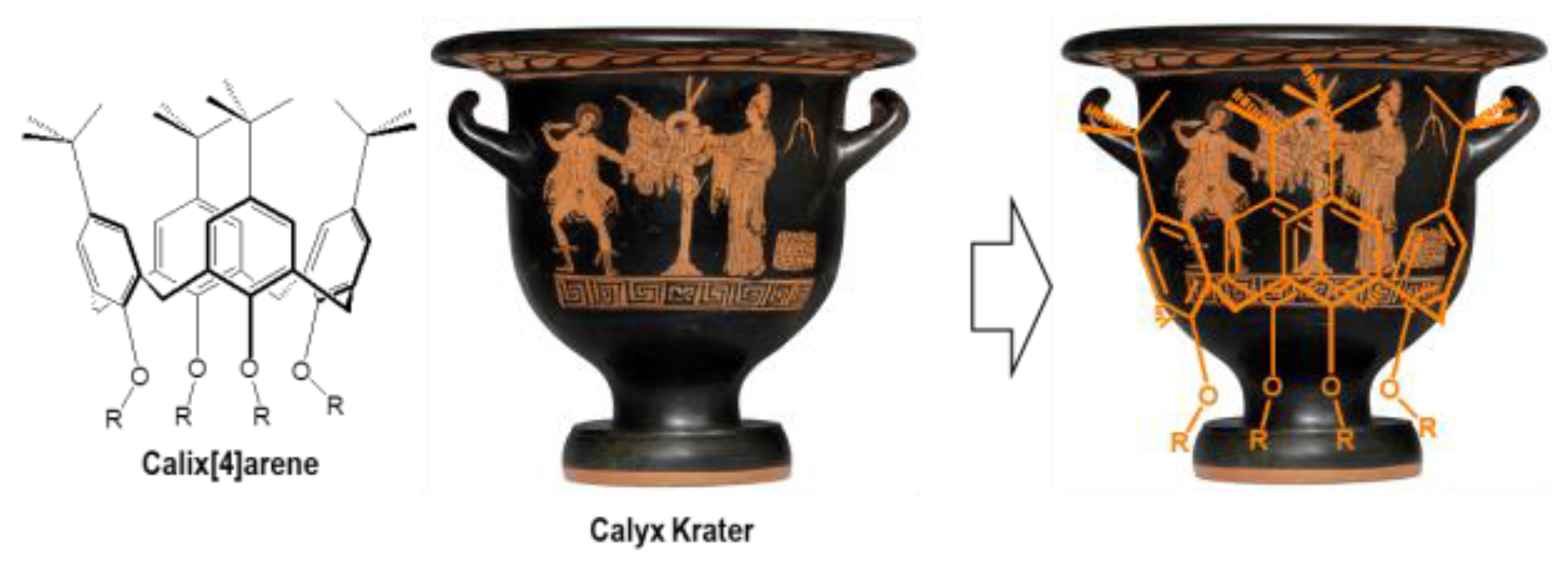
Figure 1. Analogy between calix[n]arene and a calyx krater.
Molecules 28 06954 g001
Figure 2. General structure of calix[n]arene. X = CH2 or S and n = 4, 6, or 8 for the most representative of them.
Molecules 28 06954 g002
Figure 3. The four possible conformations of substituted calix[4]arene.
Molecules 28 06954 g003
Figure 4. Structure of para-sulfonated calix[4, 6, or 8]arenes [95].
Molecules 28 06954 g004
Figure 5. Schematic structures of the new antimycobacterial calixarenes [111,112].
Molecules 28 06954 g005
Figure 6. Structure of some calixarenes and oxacalixarenes presenting antibacterial activities [114].
Molecules 28 06954 g006
Scheme 1. Synthetic way leading to the peptidocalix[4]arene 18 and structures of its analogues 19 and 20 [115].
Molecules 28 06954 sch001
Figure 7. Calixarenic structures 21 to 24 [77,109].
Molecules 28 06954 g007
Scheme 2. Synthetic pathway to compound 28, and representation of its monomer analogue 29 [72]. Boc: tert-Butoxycarbonyl.
Molecules 28 06954 sch002
Scheme 3. Synthetic pathway to tetra-guanidinium bis-heterocyclic compounds 36 to 38 [128]. Boc: tert-Butoxycarbonyl, Bpy-α: 6-[6′methyl-2,2′-bipyridine], Bpy-β: 5-[5′-methyl-2,2′-bipyridine], Btz: 4-[4′-methyl-2,2′-bithiazole], TFA: trifluoroacetic acid.
Molecules 28 06954 sch003
Scheme 4. Synthetic pathway to water-soluble calix[4]arenes incorporating carboxylate groups 50 to 53 [129].
Molecules 28 06954 sch004
Figure 8. Negatively and positively charged calix[4]arenes tested on M. tuberculosis [132]. Boc: tert-Butoxycarbonyl, Bpy-α: 6-[6′methyl-2,2′-bipyridine], Bpy-β: 5-[5′-methyl-2,2′-bipyridine], Btz: 4-[4′-methyl-2,2′-bithiazole].
Molecules 28 06954 g008
Scheme 5. Synthetic pathway of tetra-morpholinocalix[4]arene 61 [133].
Molecules 28 06954 sch005
Scheme 6. Synthetic pathway to compounds 65a to 65i, and representation of subunits 1,3,4-thiadiazole and 1,3,4-oxadiazole heterocycles A to I [134].
Molecules 28 06954 sch006
Scheme 7. Synthetic pathway to cationic calixarenes modified with methylimidazolium (70, 71), choline (72, 73) and ethylenediamine (74, 75) groups [135]. prop: propyl, oct: octyl.
Molecules 28 06954 sch007
Figure 9. Mono-azo 81, 82, 83, tetra-azo 84, 85 calixarene derivatives and monomer 86 [141].
Molecules 28 06954 g009
Scheme 9. Synthetic pathway to four conformer guanidinium derivatives 92, 93, 94, 95 and structure of monomeric analogue 96 [103].
Molecules 28 06954 sch009
Figure 10. Structure of oxadiazole-based calixarenes 97 to 101 [146].
Molecules 28 06954 g010
Scheme 10. Synthetic route for the preparation of quaternary ammonium salts of thiacalix[4]arene 104 to 113 [147]. Me: methyl, Bu: butyl, Oct: octyl, Dec: decyl, Bn: benzyl.
Molecules 28 06954 sch010
Scheme 11. Synthetic way to obtain new cationic amphiphilic calix[4]arene derivatives 115 and 116a-j [148]. DMTMM: 4-(4,6-dimethoxy-1,3,5-triazin-2-yl)-4-methyl-morpholinium chloride, DIPA: diisopropylamine.
Molecules 28 06954 sch011
Scheme 12. Synthetic way to obtain the penicillinic podands 120, 124, and 125 [149,150].
Molecules 28 06954 sch012
Scheme 13. Synthetic pathway leading to the dissymmetric podand (penicillin V and nalidixic acid) 132, the corresponding bis-nalidixic 127, and bis-penicillin V 133 [151,152,153].
Molecules 28 06954 sch013
Figure 11. Schematic structures of the nalidixate podands 127, 134, 135, 136, and 1,2-dimyristoyl-sn-glycero-3-phosphoethanolamine (DMPE) [154].
Molecules 28 06954 g011
Scheme 14. Synthetic pathway to podands 139 and 140 [156].
Molecules 28 06954 sch014
Scheme 15. Synthetic pathway of calixpenams 145 (Pen. X), 146 (Pen. V) and their corresponding single penams 147 (Pen. X) and 148 (Pen. V) [157].
Molecules 28 06954 sch015
Scheme 16. Synthetic pathway of calixcephems 151 (Ceph. X) and 152 (Ceph. V) and their corresponding single cephems 153 (Ceph. X) and 154 (Ceph. V) [158]. BSU: N,N′-bis(trimethylsilyl)urea, Py: pyridine.
Molecules 28 06954 sch016
Scheme 17. Synthetic pathway to calixarene-derivative- aminoglycoside-based 156, 157, and 158 [162].
Molecules 28 06954 sch017
Scheme 18. Synthetic route for obtaining ligand 161, 162, 163, 165, and 169 [163,164].
Molecules 28 06954 sch018
Scheme 19. Synthetic pathway of binuclear copper complex 172 [166].
Molecules 28 06954 sch019
Scheme 20. Synthetic route for obtaining of ligands 175 and 176 [167].
Molecules 28 06954 sch020
Table 1. Antibacterial activities of compounds 7 to 14; zone of inhibition corresponds to the total diameter zone [114].
Compounds
Bacterial Strains 7891011121314
E. coli10 mg/mL-++++------
5 mg/mL-++++------
S. aureus10 mg/mL-++++++++-+++++++++-
5 mg/mL-++++++++-+++++++-
1 mg/mL-+++++-++-+-
B. subtilis10 mg/mL+++-++++++++--++++-
5 mg/mL+-+++---++++-
1 mg/mL--++---++-
K. pneumoniae10 mg/mL++++---+++++++-++++
5 mg/mL+++---+++++-+++
P. aeruginosa10 mg/mL++++---+++---
5 mg/mL+++---++---
Zone of inhibition: -: none; +: 6 mm; ++: 7 mm; +++: 10 mm; ++++: 14 mm.
Table 2. MICs (Minimum Inhibitory Concentrations in µg/mL) of peptidocalixarene and vancomycin against several bacterial strains of interest [115].
Compounds
Reference StrainsVancomycin17181920
S. aureus 6632648832
S. aureus 8532>6416832
S. aureus 11312644432
S. epidermidis2324832
B. cereus2nt16ntnt
S. aureus 663 = penicillin-sensitive, S. aureus 853 = penicillin-resistant, S. aureus 1131 = methicillin-resistant, nt = not tested.
Table 3. MICs (in µg/mL) of 28 and its constitutive monomer 29 against ATCC reference strains and clinical isolates, respectively. HX (hexamidine) and CHX (chlorhexidine) were used as reference antibacterial agent [72].
Compounds
Strains2829HXCHX
Reference strains
E. coli ATCC 25922451280.25
P. aeruginosa ATCC 2785332>512328
S. aureus ATCC 25923851240.5
S. aureus ATCC 292138512<10.5
E. faecalis ATCC 2921232>51222
Clinical isolates
Penicillinase-producing E. coli25128nd
MRSA (mecA gene)85122nd
E. faecium (vanB gene)8>5122nd
E. faecalis (vanA gene)64>5124nd
P. aeruginosa (overexpression of efflux pumps)64>51264nd
nd: not determined.
Table 4. CI50 and MICs (in µg/mL) of 28 and its bis-heterocycle derivatives 36 to 38 on MRC-5 cells and against reference bacterial strains, respectively [128].
Compounds
28363738
CI50>25616–32>12864–128
Reference strains
E. coli ATCC 259224163216
P. aeruginosa ATCC 2785332163216
S. aureus ATCC 2592381688
S. aureus ATCC 29213816816
E. faecalis ATCC 29212321688
Table 5. MICs (in µg/mL) of 25, 28, 34, 37, 38 against reference strains H37Rv and MYC5165. Isoniazid (INH) was used as reference [132].
Compounds
2528343738INH
Strains
M. tuberculosis H37Rv2.581.01.341.512.690.08
M. tuberculosis MYC51652.61.02.70.750.171.7
Table 6. MICs (in µg/mL) of cationic calixarenes modified with methylimidazolium (70, 71), choline (72, 73) and ethylenediamine (74, 75) groups against ATCC reference strains [135].
Compounds
Reference Strains70
(prop)
72
(prop)
74
(prop)
71
(oct)
73
(oct)
75
(oct)
18/19 a28 a61 a
S. aureus
ATCC 6538
<10<10<10>1000>1000>10008816
P. aeruginosa
ATCC 9027
50100050>1000500>1000nd b32nd b
K. pneumoniae
ATCC 10031
<10100500500>1000>1000nd b84
E. coli
ATCC 25922
<101005001000500>10003.244
a no data for these specific strains, MICs for species is shown. b nd: no data available.
Table 7. MIC values (in µg/mL) of azo-compounds 81 to 86 against several bacterial strains [133].
Compounds
Bacterial Strains818283848586
S. aureus7.83.97.8>125>125>125
MRSA15.60.977.8>12531.25>125
B. subtilis15.60.970.97>125125>125
S. epidermidis7.81.91.9>12562.5>125
E. faecalis31.257.87.8>12515.6>125
E. coli>125>125125>125>125>125
P. aeruginosa15.6>12562.5>12562.5>125
Table 8. MICs (in µg/mL) of guanidinium derivatives 92, 93, 94, 95, and 96 against the various bacteria used in the study [103].
Compounds
Reference Strains92
cone
93
paco
94
1,2-alt
95
1,3-alt
96
E. coli ATCC 259226488464
P. aeruginosa ATCC 2785312881616128
S. aureus ATCC 259233224<132
S. aureus ATCC 292133248832
E. faecalis ATCC 292128488128
M. tuberculosis H37Rv783839510
M. tuberculosis MYC51659.52.43.81.237.2
Note: MICs of INH: 0.16 and 20 μg/mL against H37Rv- and INH-resistant MYC5165, respectively.
Table 9. MICs (in µg/mL) of ammonium derivatives 104 to 113 against the various bacteria used in the study [147].
Compounds
104109108113105110106111107112CHX
R=MeBnBuOctDec
Reference Strains cone1,3-altcone1,3-altcone1,3-altcone1,3-altcone1,3-alt
S. aureus ATCC 29213 8>64441166413242
S. epidermis a 4>6424116641841
B. subtilis 168 164412842442
E. coli ATCC 25922 >64>64>6432>6464>648>64161
K. pneumoniae 1813 >6416>6432>6418324322
P. aeruginosa ATCC 27853 >64>64>64>64>64>64>6416>64644
a: clinical isolate.
Table 10. MICs (in µg/mL) (by broth microdilution) of calixpenams (145 (Pen. X), 146 (Pen. V)), calixcephems (151 (Ceph. X), 152 (Ceph. V)) and their corresponding single penams (147 (Pen. X), 148 (Pen. V)) and cephems (153 (Ceph. X), 154 (Ceph. V)), respectively [157].
Compounds
Reference
Strains
145
(Pen. X)
146
(Pen. V)
147
(Pen. X)
148
(Pen. V)
151
(Ceph. X)
152
(Ceph. V)
153
(Ceph. X)
154
(Ceph. V)
S. pyogenes
ATCC 19615
0.0020.0030.0120.016ntntntnt
S. agalactiae
ATCC 12386
0.0040.0060.0250.032ntntntnt
S. pneumoniae
ATCC 49619
0.0220.0240.1250.125ntntntnt
S. aureus
ATCC 25923
0.0250.032ntnt0.2250.251.7751.550
S. aureus
ATCC 29213
0.240.256ntnt0.9921.0125.3255.150
S. aureus
ATCC 11632
>64>64ntnt0.9120.9258.7758.350
S. aureus
ATCC 43300
>64>64ntnt1.1501.22511.77510.950
S. aureus
ATCC 33591
>64>64ntnt1.5501.63516.65016.325
nt: not tested.
Table 11. MICs (in µg/mL) of calixarene derivatives 159 to 166 against ATCC reference strains [163].
Compounds
Reference Strains159160161162163164165166
B. subtilis ATCC 66337850001250125010,00010,000ndnd
B. cereus NRRI-B-37113910,00039250010,00010,000ndnd
E. faecalis (clinical isolate)15610,00039125010,0001250ndnd
S. aureus NTCT 832515610,00039125010,000625ndnd
E. coli ATCC 259223910,00062510,00010,.0001250ndnd
E. coli ATCC 3512839500078500050002500ndnd
P. aeruginosa ATCC 27853125050005000500050005000ndnd
P. vulgaris ATCC 842715610,0005000500050002500ndnd
nd: No MICs determined in the range of tested concentrations (≥10,000 μg/mL).
Table 12. Zones of inhibition (in mm) and MICs (in μg/mL) of compound 171 and its Cu(II)-complex 172 [166].
Compounds
171172
Conc. in μg/mL631.50.750.37 631.50.750.37
Reference StrainsInhibition ZoneMICInhibition ZoneMIC
S. albus62---311853-0.75
E. coli83---312107520.37
Table 13. MICs (in µg/mL) of synthesized calix[4]arene-based ligands 175 and 176 as well as their metal complexes against bacterial reference strains [167].
Compounds
Reference StrainsThiosemicarbazideThiosemicarbazone
175175-Co175-Ni175-Cu175-Zn176176-Co176-Ni176-Cu176-Zn
S. aureus ATCC 29213-250-250500---31.25-
B. subtilis ATCC 663331.25-31.2531.2512531.2531.2531.2531.2531.25
E. coli ATCC 2592225012562.512531.2531.2531.2531.2531.2531.25
P. aeruginosa ATCC 2785362.512531.2562.531.2531.2531.2531.2531.2531.25
Disclaimer/Publisher’s Note: The statements, opinions and data contained in all publications are solely those of the individual author(s) and contributor(s) and not of MDPI and/or the editor(s). MDPI and/or the editor(s) disclaim responsibility for any injury to people or property resulting from any ideas, methods, instructions or products referred to in the content.