Review

Sulforaphane’s Multifaceted Potential: From Neuroprotection to Anticancer Action

by
Raymond A. Otoo
1,2,3 and
Antiño R. Allen
1,2,3,*
1
Division of Radiation Health, University of Arkansas for Medical Sciences, 4301 West Markham, Suite 441B-2, Little Rock, AR 72205, USA
2
Department of Pharmaceutical Sciences, University of Arkansas for Medical Sciences, 4301 West Markham, Suite 441B-2, Little Rock, AR 72205, USA
3
Neurobiology & Developmental Sciences, University of Arkansas for Medical Sciences, 4301 West Markham, Suite 441B-2, Little Rock, AR 72205, USA
*
Author to whom correspondence should be addressed.
Molecules 2023, 28(19), 6902; https://doi.org/10.3390/molecules28196902
Submission received: 3 July 2023 / Revised: 16 August 2023 / Accepted: 30 August 2023 / Published: 1 October 2023
(This article belongs to the Section Natural Products Chemistry)

Abstract

:
Sulforaphane (SFN) is a naturally occurring compound found in cruciferous vegetables such as broccoli and cauliflower. It has been widely studied for its potential as a neuroprotective and anticancer agent. This review aims to critically evaluate the current evidence supporting the neuroprotective and anticancer effects of SFN and the potential mechanisms through which it exerts these effects. SFN has been shown to exert neuroprotective effects through the activation of the Nrf2 pathway, the modulation of neuroinflammation, and epigenetic mechanisms. In cancer treatment, SFN has demonstrated the ability to selectively induce cell death in cancer cells, inhibit histone deacetylase, and sensitize cancer cells to chemotherapy. SFN has also shown chemoprotective properties through inhibiting phase I metabolizing enzymes, modulating phase II xenobiotic-metabolizing enzymes, and targeting cancer stem cells. In addition to its potential as a therapeutic agent for neurological disorders and cancer treatment, SFN has shown promise as a potential treatment for cerebral ischemic injury and intracranial hemorrhage. Finally, the ongoing and completed clinical trials on SFN suggest potential therapeutic benefits, but more research is needed to establish its effectiveness. Overall, SFN holds significant promise as a natural compound with diverse therapeutic applications.

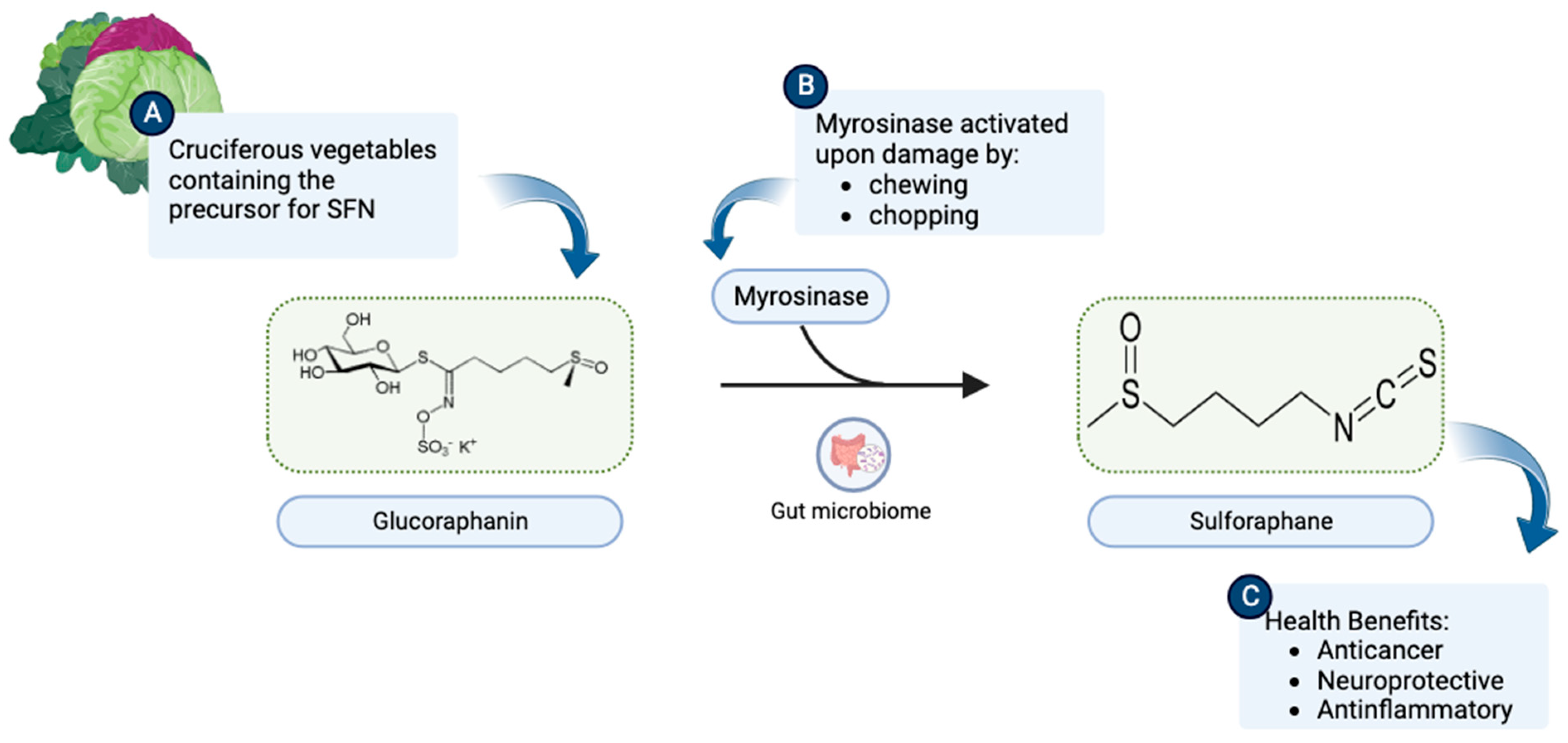
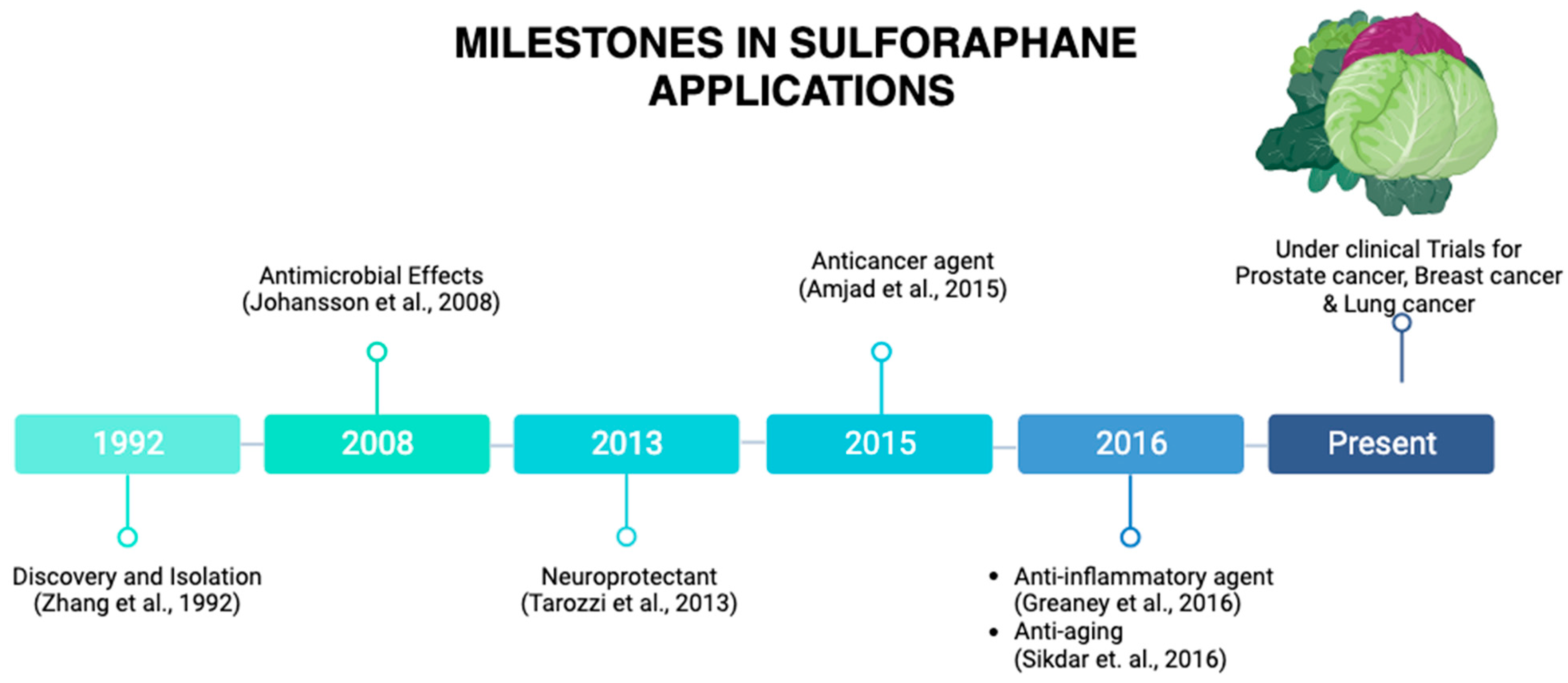
1. Origin and Discovery

In the middle of the last century, sulforaphane (SFN; sulphoraphane in British English) was described as an antibiotic and was isolated from red cabbage and from hoary cress, a weed in rangelands of the western US [1]. It was first synthesized by Talalay and Zhang, who were the first to isolate it from broccoli [2]. SFN is a compound within the isothiocyanate (ITC) group of organosulfur compounds. ITCs are hydrolysis products of glucosinolates, secondary plant metabolites that are found in high concentrations in Brassica vegetables [3]. ITCs are known to be synthesized and stored as glucosinolates in plants and are released when damage to plant tissues occurs [4]. The most characterized ITC compound is SFN, the hydrolysis product of glucoraphanin, and it generally is found in high concentrations in broccoli (Figure 1) [5]. SFN occurs in broccoli sprouts, which have been shown to be 20–50 times more effective in chemoprevention than mature heads [6]. Among cruciferous vegetables, broccoli sprouts have the highest concentration the SFN precursor [7], hence broccoli sprouts are preferred over other crucifers as a chemoprotective agent. For this review, we will explore the multifaceted role of SFN including evidence that supports SFN’s neuroprotective effects, its potential as an anticancer agent, and its ability to act as a chemoprotective agent. Additionally, we will discuss the current state of clinical trials with SFN on some selected types of cancer and its potential to enhance the effectiveness of chemotherapy and radiation therapy. Figure 2 illustrates the journey of SFN from a promising naturally occurring compound to its status as a subject of ongoing research.

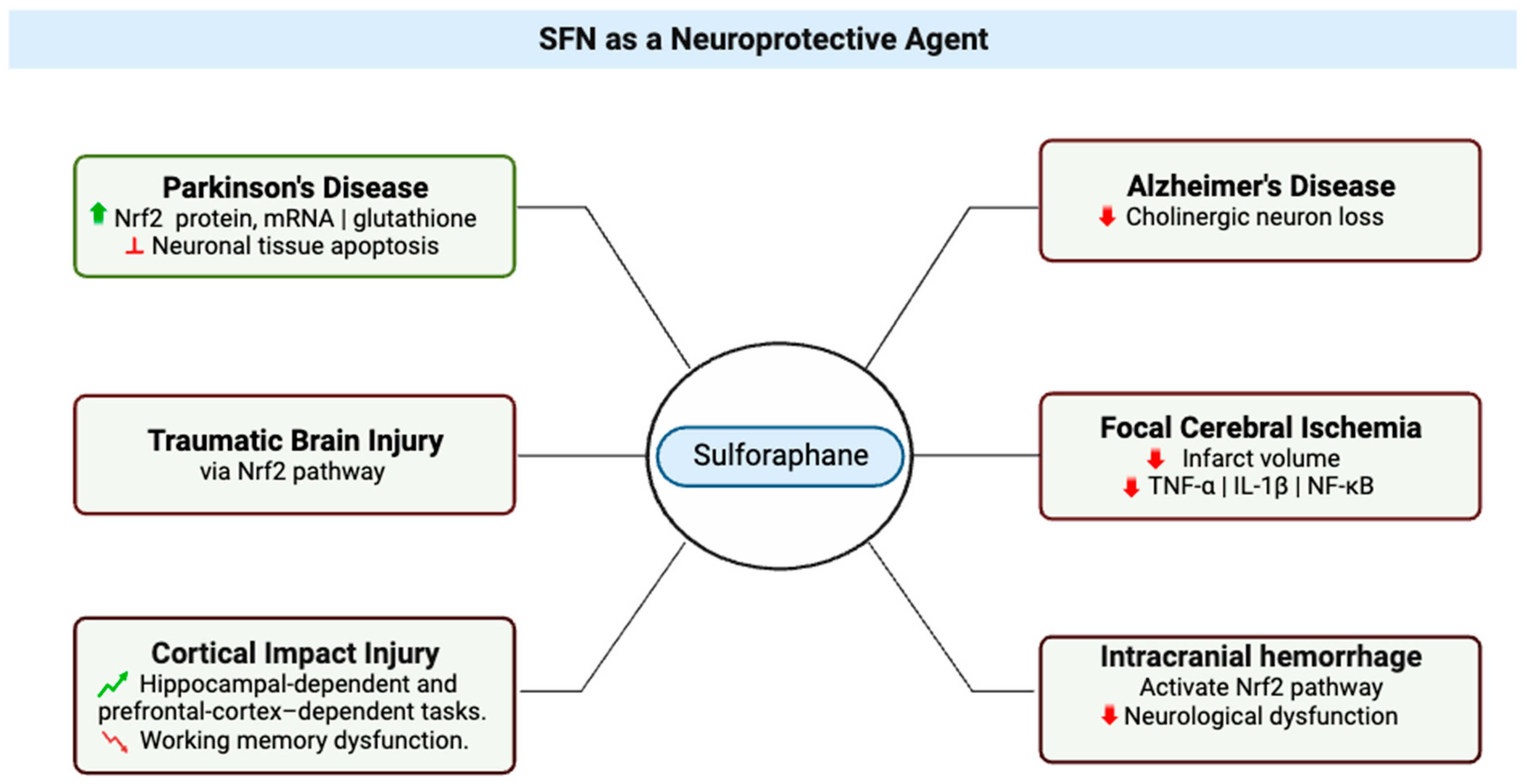
2. SFN as a Neuroprotective Agent

Neuroprotection refers to the mechanisms and strategies used to defend the central nervous system (CNS) (Figure 3) against injury due to both acute (e.g., trauma or stroke) and chronic neurodegenerative disorders (e.g., dementia, Parkinson’s, Alzheimer’s, epilepsy) [13]; by extension, neuroprotective agents comprise a category of agents that generally are used to protect neuronal structure and/or function. Research on the neuroprotective effects of SFN began in 2004 with studies that showed its effects protecting neurons [14] and microglia [15] against oxidative stress via activation of nuclear factor erythroid 2-related factor 2 (Nrf2). The research literature is replete with studies that support the vital role played by the Nrf2 pathway in the neuroprotective effects of SFN [14,16,17,18,19,20,21], evidenced by lack of neuroprotection from toxins in Nrf2-knockout mice treated with SFN [22,23]. In a study of Parkinson’s disease that used a 6-hydroxydopamine-Parkinson’s disease mouse model, treatment of SH-SY5Y cells with SFN was found to have a protective effect on the neurons, which was attributed to the observed increases in active nuclear Nrf2 protein, Nrf2 mRNA, and total glutathione levels and inhibition of neuronal tissue apoptosis [24]. A group studying SFN effects in traumatic brain injury confirmed that SFN showed neuroprotection in spinal cord injury, and it may be an emerging therapeutic agent in this setting [25]. In a study that examined whether administration of SFN after cortical impact injury could improve the performance of rats in hippocampal-dependent and prefrontal-cortex-dependent tasks, SFN treatment was reported to improve performance in the Morris Water Maze task (i.e., decreased latencies during learning and platform localization during a probe trial) and to reduce dysfunction in working memory dysfunction (tested with the delayed match-to-place task) [26].
SFN also has been shown to exert neuroprotective effects in Alzheimer’s disease (AD) (Figure 3); in brains of mice with Alzheimer’s disease-like lesions, SFN ameliorated neurobehavioral deficits by reducing cholinergic neuron loss. SFN is a potent inducer of the Nrf2 antioxidant response element (ARE) pathway, which plays a major role in upregulating cellular defenses to oxidative stress [27]. In 2016, Zhou et al. reported that SFN exerted its neuroprotective effect in several ways, such as mTOR-dependent prevention of neuronal apoptosis, Nrf2-dependent reductions in oxidative stress, and restoration of normal autophagy [17]. Park et al. reported that with an in vitro model, SFN protected neuronal cells from Aβ42-mediated cytotoxicity and ameliorated proteasome activities [32]. A key factor in SFN’s neuroprotective effects is the compound’s effect on neuroinflammation [33,34]. Hernández-Rabaza et al. assessed whether treatment with SFN reduces neuroinflammation; they reported that treatment with SFN promoted differentiation of microglia from the pro-inflammatory M1 phenotype to the anti-inflammatory M2 phenotype and reduced activation of astrocytes in hyperammonemic rats to reduce neuroinflammation [35].
Epigenetics emerges as a pivotal mechanism underlying the neuroprotective potential of SFN. In a study involving mouse neuroblastoma N2a cells expressing human Swedish mutant Aβ precursor protein (N2a/APPswe cells), SFN was observed to induce Nrf2 expression by reducing DNA methylation levels at the Nrf2 promoter [20,36]. The activation of the Nrf2 ARE pathway, subsequently leading to the upregulation of key downstream elements such as NAD(P)H quinone oxidoreductase 1, heme oxygenase 1, and glutathione peroxidase 1, plays a pivotal role in countering oxidative stress [37]. Collectively, SFN’s ability to modify genetic expression, influencing a spectrum of detrimental or protective agents, translates into reduced cellular damage and the attenuation of harmful protein accumulation. This culminates in comprehensive neurological enhancements across various disease states and toxin exposures [38].
Additionally, a noteworthy aspect of SFN’s mechanism involves its capacity as a histone deacetylase (HDAC) inhibitor, warranting attention. Notably, the intricate interplay between biological mechanisms governing cancer transcends genetics and prominently involves epigenetics [39]. As an increasingly explored avenue, epigenetic modifications have garnered attention as a promising strategy for cancer prevention [40]. Central to this landscape are histone deacetylases (HDACs), pivotal orchestrators of epigenetic restructuring [41], which bear relevance in SFN’s anticancer effect [42]. SFN’s HDAC inhibition has been discerned across various cancer types including breast [42,43,44], colorectal [45], and prostate cancer [46,47]. Reinforcing this notion, Hossain et al. (2020) conducted a study elucidating the significance of HDAC inhibition in the context of SFN treatment for breast cancer. Through examination of MCF-7 cells, the investigation delineated the concerted influence of SFN, derived from cruciferous vegetables, and HDAC inhibitors like Trichostatin A (TSA) on gene expression patterns associated with 1,25(OH)2D3 activity. In this context, HDAC inhibition was identified as a critical enhancer of SFN’s impact, with distinct histone acetylation responses differentiating SFN and TSA treatments. The study thereby illuminates the intricate interplay between SFN’s diverse biochemical effects and its ability to modulate HDAC activity, ultimately elucidating the multifaceted molecular underpinnings of their synergistic anticancer effects [42]. In sum, the evolving understanding of SFN’s intricate interplay with epigenetic mechanisms, particularly HDAC inhibition, unveils its promising potential as an agent for combating cancer.
Furthermore, SFN’s potential as a neuroprotective agent extends to various neurological conditions, including focal cerebral ischemia (Figure 3), neuroinflammation, and intracranial hemorrhage. Focal cerebral ischemia arises from reduced blood flow to a specific brain region, resulting in changes in cerebral function [48,49]. Recent studies have probed the neuroprotective effects of SFN in the management of stroke [50,51]. Zhao et al. utilized a rodent common carotid artery/middle cerebral artery model and demonstrated that SFN reduced infarct volume after focal cerebral ischemia [28]. Additionally, SFN exhibited anti-inflammatory effects in this context, reducing pro-inflammatory cytokines and suppressing the expression of phospho-nuclear factor kappa-light-chain-enhancer of activated B cells (NF-κB) p65 [29]. These findings collectively underscore SFN’s potential as a therapeutic agent against cerebral ischemic injury.
Neuroinflammation, a pivotal factor in several neurological disorders, has also been a target of SFN’s neuroprotective effects [35,52,53,54,55]. Microglia, the resident immune cells of the brain [56], play a key role in neuroinflammation by secreting pro-inflammatory and anti-inflammatory mediators [57]. Studies in hyperammonemic rat models revealed that SFN promotes microglia differentiation from pro-inflammatory M1 to anti-inflammatory M2 phenotypes, mitigating neuroinflammation [35]. Furthermore, SFN’s anti-inflammatory effects in microglia were demonstrated by attenuated expression of neuroinflammatory proteins and reduced mitogen-activated protein kinase (MAPK) effector signaling [35]. SFN’s role in countering neuroinflammation was also evident in a study by Subedi et al., where it inhibited nitrite production and decreased the translocation of NF-κB and production of proinflammatory cytokines in microglial cells [53]. These cumulative findings highlight SFN’s potential as a therapeutic tool in managing neuroinflammatory diseases.
Intracranial hemorrhage (ICH), characterized by bleeding within the intracranial vault, presents a significant challenge in medical management [30,31,58,59]. Recent studies have illuminated the potential role of SFN in ICH treatment. SFN’s activation of the Nrf2-ARE signaling pathway was found to improve neurological dysfunction after ICH [30]. Additionally, SFN demonstrated efficacy in reducing oxidative stress and inflammation in ICH, opening avenues for further research [30,31].
Collectively, the extensive body of research underscores SFN’s multifaceted neuroprotective potential, ranging from its impact on oxidative stress, inflammation, epigenetic regulation, and beyond. As scientific understanding evolves, SFN continues to emerge as a promising candidate for the treatment and management of various neurological conditions, offering a beacon of hope in the realm of neuroprotection and therapeutic intervention.

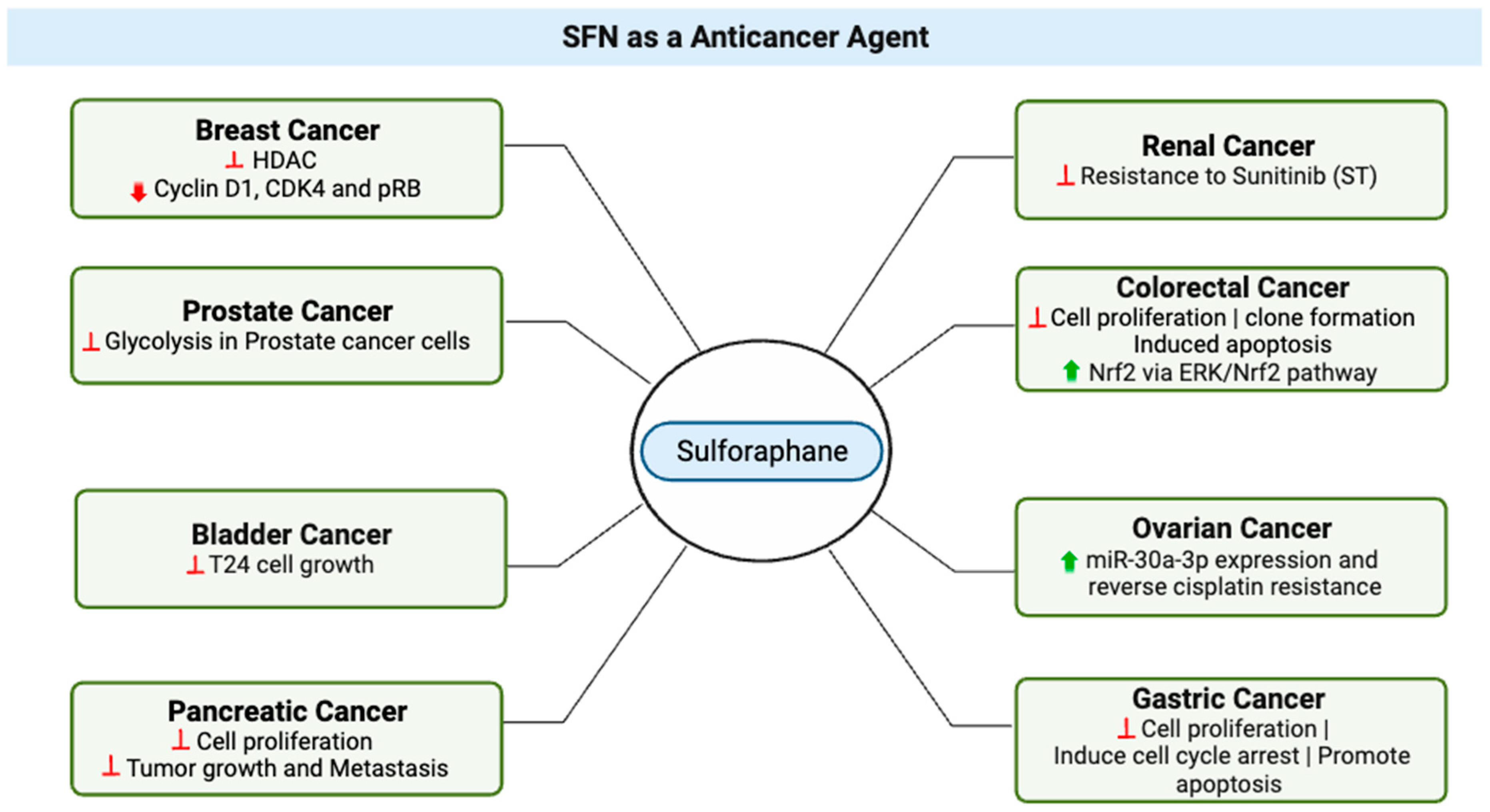
3. SFN as an Anticancer Agent

In recent years, SFN has gained attention for its potential use as an anticancer agent. In a study in 2015, Ullah proposed three major factors that enhance the plausibility of clinical applications and the translational value. First, normal cells are relatively resistant to SFN-induced cell death [60], an important feature for potential anticancer agents, and recent in vitro work demonstrated that SFN suppressed metastasis of triple-negative breast cancer cells by targeting the RAF/MEK/ERK pathway [61]. Second, SFN has good bioavailability; it can reach high intracellular and plasma concentrations, and it has been detected in breast tissues after a single oral administration [62,63]. Finally, a study by Myzak et al. in 2007 provided evidence that histone deacetylase was inhibited after human subjects ingested 68 g of broccoli sprouts, indicating that SFN provides anticancer pharmacological effects at levels that humans can readily ingest.
Ullah proposed a mechanism by which SFN exerts its chemopreventive effects. In this model, low to moderate levels of reactive oxygen species (ROS) are active participants in cellular functions and act as signaling molecules that sustain cellular proliferation and differentiation and that activate responses to oxidative stress [64]. Under normal, unstressed conditions, the cellular NRF2 level is very low [65], but it significantly increases upon exposure to electrophilic chemicals or ROS [66]. In response to oxidative or electrophilic stress, Nrf2 stimulates antistress signaling and consequently inhibits carcinogenesis [67]. Under conditions of stress, Kelch-like ECH-Associated Protein 1 (KEAP1), which is a cytosolic inhibitor of Nrf2, is oxidized, leading to stabilization and translocation of Nrf2 into the nucleus, where the transcription factor activates expression of genes crucial to antioxidant defense [68].
Expanding the horizon of SFN’s impact reveals its significant potential across various cancer types. Studies by Livingstone et al. (2022) uncover SFN’s potential in prostate health, as elevated SFN levels were observed in men receiving glucoraphanin supplements [69]. Supporting this, Singh et al. (2019) demonstrated SFN’s ability to inhibit glycolysis in prostate cancer cells, suggesting therapeutic implications [70].
In bladder cancer (Figure 4), an in vitro study highlighted SFN’s dose-dependent effects on cell growth, presenting opportunities for targeted therapeutic strategies [71]. In this study, at higher concentrations, ranging from 10–160 μM and after 24 to 48 h of treatment, SFN demonstrated a significant inhibitory effect on T24 cell growth. However, it is important to consider that at lower doses, specifically 2.5 μM, SFN resulted in a slight increase in cell proliferation by 5.18–11.84% within a 6 to 48 h treatment window [71]. These results suggest that SFN’s effects on cell growth are dose-dependent, with potential implications for further research and development of targeted therapeutic strategies in the context of bladder cancer.
Interesting, insights from breast cancer cell research revealed that SFN orchestrates DNA methylation through the modulation of DNA methyltransferase and histone deacetylase levels, coupled with the downregulation of cyclin D1, CDK4, and pRB, thereby promoting breast cancer cell apoptosis [72].
Chen et al.’s comprehensive investigation (2018) showcased SFN’s efficacy in inhibiting pancreatic cancer cell proliferation, sensitizing cells to treatment, and affecting multiple cancer control hallmarks [73]. It inhibited clone formation and pancreatic cancer cell migration, induced apoptosis, and disrupted cell invasion in both low- and high-glucose environments, underscoring its multifaceted role. In the same study, in vivo experimentation using a transgenic mouse model further demonstrated SFN’s robust influence by significantly inhibiting tumor growth and metastasis.
Sunitinib (ST), an established therapy for renal cell carcinoma (RCC), faces limitations as a standalone treatment due to tumor reactivation and resistance [78]. To address this, an in vitro study investigated the combination of ST with sulforaphane (SFN). SFN emerged as a critical enhancer of ST’s efficacy by suppressing resistance in RCC cells, offering a potent approach to overcome ST monotherapy limitations. Short-term SFN application reduced cell numbers across diverse lines, sensitizing RCC cells to ST. Long-term SFN use exhibited greater effectiveness, particularly in 786O cells, where the ST-SFN combination outperformed SFN alone [74]. These findings underscore SFN’s significant anticancer potential in countering tumor reactivation and resistance, propelling further clinical research. Continued exploration of this dual therapy holds the promise of revolutionizing RCC management and advancing kidney cancer treatment.
A study aimed at investigating SFN’s capacity to counter resistance to cisplatin, a widely used ovarian carcinoma treatment, employed ovarian cancer cells. In this context, the investigation employed A2780 and IGROV1 cells, along with their cisplatin-resistant counterparts, A2780/CP70 and IGROV1-R10, to explore SFN’s potential in overcoming cisplatin resistance. The study unveiled SFN’s ability to effectively reverse cisplatin resistance by inducing DNA damage and enhancing intracellular cisplatin accumulation. Notably, SFN treatment resulted in a substantial elevation in miR-30a-3p expression, a microRNA that exhibited reduced levels in cisplatin-resistant cells [75]. The combined outcomes of this investigation strongly imply that SFN could heighten the efficacy of cisplatin against ovarian cancer cells. This enhancement is achieved through the upregulation of miR-30a-3p expression, triggering escalated DNA damage and heightened cisplatin accumulation within the cells.
For colorectal cancer, Hao et al.’s study (2020) demonstrated SFN’s potential as a chemopreventive agent (Figure 4), acting through the modulation of the ERK/Nrf2 pathway and impacting cell proliferation, apoptosis, and migration [76]. Additionally, SFN hindered the motility and migration of colorectal cancer cells. Mechanistically, SFN led to dose-dependent upregulation of nuclear factor, erythroid 2 like 2 (Nrf2) and UDP glucuronosyltransferase 1A (UGT1A) expression. This effect was mediated through the ERK/Nrf2 signaling pathway, as ERK inhibition attenuated SFN-induced upregulation of Nrf2 and UGT1A, along with mitigating increased intracellular ROS levels [13]. In sum, SFN emerges as a promising agent for colorectal cancer chemoprevention, acting through modulation of Nrf2-mediated detoxification and anti-proliferative pathways.
In the context of gastric cancer (GC), the intricate mechanisms of action of sulforaphane (SFN) have been explored to shed light on its anticancer properties (Figure 4). A recent study by Wang et al. (2021) has revealed that SFN can impede cell proliferation, induce cell cycle arrest, and promote apoptosis in GC cells [77]. Specifically, SFN treatment led to a pronounced reduction in cell viability, as evidenced by decreased colony-forming efficiency in BGC-823 and MGC-803 cell lines. Furthermore, SFN demonstrated its potential as an inducer of S phase cell cycle arrest, a critical regulator of cell proliferation, and showed promising apoptotic-inducing activity. Mechanistically, SFN achieved S phase arrest through the modulation of the p53-dependent p21-CDK2 axis, effectively inhibiting CDK2 expression while upregulating p53 and p21 levels. Additionally, SFN’s influence on the mitochondrial pathway emerged as a pivotal factor in its apoptotic effect, involving upregulation of Bax and cleaved-caspase-3 expression. Importantly, these findings, as documented by Wang et al. (2021) [77], provide valuable insights into SFN’s multifaceted role in suppressing GC cell growth and triggering apoptosis, thus highlighting its potential as a novel therapeutic agent for the treatment of gastric cancer.
These findings collectively highlight SFN’s diverse mechanisms of action, underscoring its potential as a versatile and potent anticancer agent across various malignancies. Its demonstrated impact on cancer cell proliferation, migration, and resistance presents promising opportunities for innovative therapeutic strategies and improved patient outcomes (Figure 4). Further research and clinical exploration are warranted to fully harness SFN’s potential in the realm of cancer therapy.

4. Chemoprotectant Properties of SFN

Cancer chemoprevention is defined as the use of dietary or pharmacological agents to prevent, block, or reverse the process of tumor development before clinical manifestation of the disease [79]. Chemoprotectants are natural or synthetic chemical compounds that can ameliorate, mimic, or inhibit the toxic or adverse effects of structurally different chemotherapeutic agents, radiation therapy, cytotoxic drugs, or naturally occurring toxins, without compromising the anticancer or antitumor potential of the chemotherapeutic drugs [80]. In vitro, SFN has been shown to be a potent chemopreventive agent and has been demonstrated to target multiple cellular mechanisms [81]. An in vivo study with an animal model (BALBc male mice) has shown that SFN prevented chemically induced cancers and inhibits tumor growth [82]. In a study of prostate cancer, SFN did not reduce the cytotoxic effects of drugs but, rather, strongly increased their anticancer efficacy against prostate cancer stem cells. In nude mice, combination treatment with SFN and a cytotoxic drug efficiently induced apoptosis and inhibited self-renewing potential, ALDH1 activity, clonogenicity, xenograft growth, and relapse of gemcitabine-treated tumor cells [83].
Inhibition of phase I metabolizing enzymes was reported by Langouet et al. as the primary mechanism of chemoprotection by SFN. A secondary mechanism has been proposed, in which phase II xenobiotic-metabolizing enzymes are modulated, and binding of carcinogens to DNA is directly inhibited [84]. As a result, cellular pro-inflammatory responses are suppressed, which inhibits formation of DNA adducts and reduces the mutation rate [84]. A tertiary chemoprevention mechanism also has been proposed, in which SFN abrogates tumorigenesis and progression of metastasis by targeting cancer stem cells in pancreatic and prostate cancer [83]. Royston et al. [72] reported that the combination of SFN with Withaferin A epigenetically reactivated tumor suppressor gene p21 (also known as cyclin-dependent kinase inhibitor 1A).

5. Effects of SFN on Tumors, Chemotherapy, Radiation Therapy, and Cardiotoxicity

Laboratory and animal studies have provided evidence that SFN has anticancer properties, but more research is needed to understand its effects on humans. Studies have proposed that SFN may prevent the growth of certain types of tumors, such as breast and prostate tumors, by causing cell death and stopping the formation of new blood vessels, which are required for tumor growth. A study demonstrated that ROS activation of the p62-KEAP1-Nrf2 signaling pathway has a tumor-suppressive effect [85]. In stark contrast, Li et al. reported a study on bladder cancer cells that showed p62 promoted tumor growth by triggering the KEAP1-Nrf2 signaling pathway [86].
Research supports the proposition that SFN may improve the effectiveness of chemotherapy by increasing cancer cell sensitivity to the drugs used to treat them [87], which is known as “chemo-sensitization”. One way in which SFN may sensitize cancer cells to chemotherapy is by inhibiting the Nrf2 pathway [87]. Several studies have proposed that SFN can promote chemo-sensitization through the Nrf2 pathway [88,89,90]. Preclinical investigations have shown that SFN prevented mice from forming carcinogen-mediated mammary carcinogenesis, lung and gastric cancer, and colonic crypt foci [91]. The activation of Nrf2 leads to its binding to the ARE, this in turn results in the increased expression of antioxidant enzymes and detoxifying enzymes, consequently protecting normal cells from the toxic effects of chemotherapy while making cancer cells more susceptible to the drugs [92]. Another way that SFN may enhance the effectiveness of chemotherapy is by inducing cell death in cancer cells [93,94,95]. SFN can induce apoptosis (programmed cell death) in cancer cells and can inhibit the formation of new blood vessels that tumors need to grow, indirectly inducing cell death [96].
Some studies have suggested that SFN may have beneficial effects on the body after radiation therapy, although more research is needed to confirm these findings. Radiation therapy is a common treatment for cancer that uses high-energy radiation to kill cancer cells, but it can also damage healthy cells and tissues in the area being treated, leading to side effects such as skin irritation, fatigue, and a risk of developing secondary tumors. Preliminary studies have suggested that SFN may help protect healthy cells and tissues from the harmful effects of radiation [97]. For example, one study showed that SFN helped reduce inflammation and DNA damage in cells exposed to radiation, and results suggested that SFN may decrease the extent of radiation-induced skin damage in mice [98].
Cardiotoxicity is the occurrence of heart dysfunction as electric or muscle damage, resulting in heart toxicity [92]. Several studies have demonstrated the protective role of SFN in cardiotoxicity. For example, in a study by Bose et al. [99] with a breast cancer model in rats, they found that SFN reduced cardiac oxidative stress (a contributing factor to cardiotoxicity) induced by doxorubicin (DOX). When DOX was administered alone, only an 11% survival rate was observed as compared to a 62% survival rate observed when DOX was combined with SFN. This study also showed that combining SFN with DOX allowed for a 50% reduction in DOX dosage while maintaining its anticancer effects. In another study by Bai et al. [100], which monitored serum myocardial levels to assess cardiac injury markers in a treatment group receiving DOX, upon treatment with SFN, DOX-induced myocardial injury and inflammation was significantly reduced [99,100,101,102,103]. In sum, although studies have shown SFN’s protective role against cardiotoxicity induced by chemotherapy drugs such as DOX, further research is needed to fully understand the potential for SFN as a therapeutic agent for cancer treatment while mitigating its effects on heart function.

6. SFN on Metastasis

SFN exhibits anticancer properties at various stages of carcinogenesis in prostate, lung, colon, and breast cancers [104,105,106]. Early research has focused on the ability of SFN to activate nuclear factor (erythroid-derived 2)-like 2 (Nrf2). SFN was shown to be effective in preventing breast cancer at different stages of carcinogenesis by increasing the levels of antioxidants and phase II detoxifying enzymes via the activation of the nuclear factor erythroid 2-related factor 2 [107]. SFN has been shown to alter key mechanisms in vivo and in vitro which impact induction of cell cycle arrest and apoptosis and inhibition of histone deacetylase. SFN inhibits transforming growth factor-β1 (TGF-β1)-induced migration and invasion in human triple negative breast cancer (TNBC) cells [61]. Sulforaphane (SFE), an SFN derivative, has been shown to reduce TNBC proliferation by mediating ERG1/PTEN axis [107]. Sulforaphane exerted its anti-metastatic effects on non-small cell lung cancer through down-regulation of miR-616-5p, which was identified as a marker associated with risk of relapse and metastasis in patients [108]. SFN has been reported to inhibit histone deacetylase (HDAC) enzymes, alter histone acetylation, and affect gene regulation. Natural inhibitors of HDAC have received considerable interest as anticancer agents because of their ability to induce p21Cip1/Waf1, leading to cell cycle arrest and apoptosis [62]. SFN inhibits HDAC activity in prostate cancer cells, in mouse xenografts, and in human peripheral blood mononuclear cells [109]. In colon cancers, SFN blocks cells’ progression and angiogenesis by inhibiting HIF-1α and VEGF expression [110].

7. SFN Bioavailability and Pharmacokinetics

In mammals, SFN is metabolized rapidly via a conjugation reaction with glutathione. SFN is metabolized through the mercapturic acid pathway, starting with GSH conjugation by glutathione S-transferase and subsequently generating SFN-cysteine followed by SFN-N-acetylcysteine [111]. Pharmacokinetic studies in rodents have focused on either free SFN or its metabolite SFN-glutathione. Following consumption of broccoli, sulforaphane is excreted in urine predominantly as a conjugate with N-acetyl cysteine. In plasma, it has been found that approximately 50% of sulforaphane is found unconjugated with other thiols [104]. In rats, after an oral dose of 50 μmol of SFN, the plasma concentration of SFN can peak at 20 μM at 4 h and decline with a half-life of about 2.2 h [112]. SFN is well absorbed in the intestine, with an absolute bioavailability of approximately 82%. Elimination of was characterized by a long terminal phase; no major difference was evident in plasma concentrations between 6 and 24 h following intravenous administration or oral administration [113]. In mice fed with diets supplemented with 5 μmol/day and 10 μmol/day of SFN for 3 weeks, the steady-state levels of SFN in plasma and intestine reached 124–254 nM and 3–13 nmol/g of tissue [114]. In humans, given an oral capsule of 200 µmol, the peak plasma was reported to have a Cmax of 0.7 ± 0.2 µM at 3 h, with a half-life of 1.9 ± 0.4 h for elimination [115].

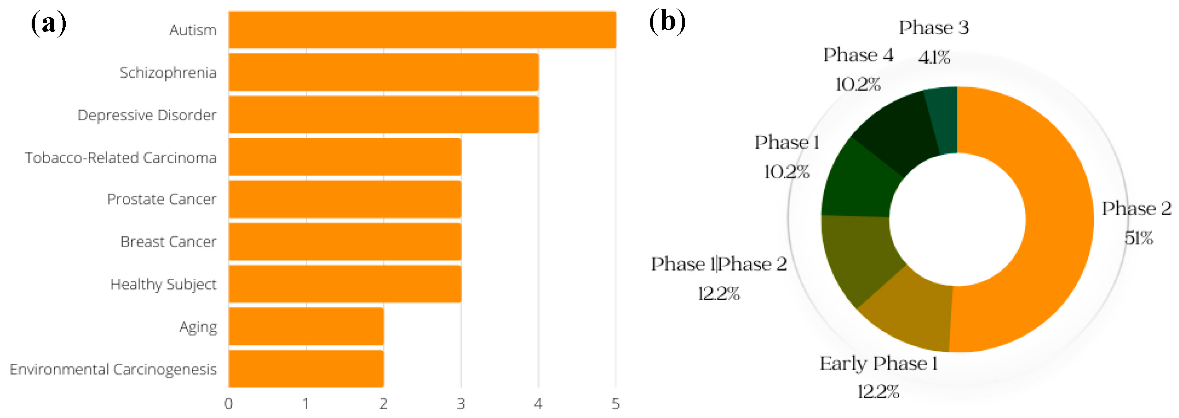
8. Ongoing and Completed Clinical Trials on SFN

Clinical trials are an important part of the research process, and drug development benefits from both favorable and unfavorable results. Even when studies do not yield the predicted outcomes, trial results can help point scientists in the correct direction [72]. (Clinical trials of SFN that were withdrawn are not included in the data presented here.) The conditions with the greatest number of ongoing or previous clinical trials on SFN, including breast cancer and prostate cancer, are shown in Figure 5a; the general phases of clinical trials on SFN (both past and current) are presented in Figure 5b. According to data obtained from ClinicalTrials.gov, a resource provided by the US National Library of Medicine, most clinical trials of SFN are in phase 2. A phase 1 trial (synonymous with “dose-escalation” or “human pharmacology” studies) is the first instance in which a new investigational agent is studied in humans. They are usually performed open-label and in a small number of “healthy” and/or “diseased” volunteers [83]. Phase 2 trials, also referred to as “therapeutic exploratory” trials, are usually larger than phase 1 studies and are conducted with a small number of volunteers who have the disease of interest [83].
Of the four trials involving schizophrenia, only one (NCT02810964) had results to report. The goal of this study was to determine whether symptoms of schizophrenia are reduced when standard antipsychotic medications are combined with SFN nutraceutical versus placebo. The primary outcome was a change (comparing beginning to end of treatment) in scores for the Positive and Negative Syndrome Scale [84]. Additionally, two phase 2 clinical trials (NCT00982319, NCT00843167) have examined effects of SFN in the setting of breast cancer. One of these trials (NCT00982319) revealed that SFN in broccoli-sprout extract resulted in an absolute change in the mean cellular proliferative rate, measured by Ki67 (a marker of active cell proliferation in the normal and tumor cell populations), from baseline to 14 days post-intervention [85]. The other trial (NCT00843167) investigated how treatment with broccoli-sprout extract affects women who have diagnoses of breast cancer, ductal carcinoma in situ, and/or atypical ductal hyperplasia. The results showed changes (comparing baseline to post-therapy) in ITC levels in urine samples, Ki67, and histone deacetylase activity in peripheral blood mononuclear cells [86].

9. Conclusions

In conclusion, this review stands as an invaluable resource, providing researchers with up-to-date insights into the latest advancements and developments in the multifaceted realm of Sulforaphane. By meticulously unraveling its historical journey, discovery, and expansive effects, we offer a comprehensive platform for researchers to grasp the current landscape of this remarkable natural compound abundant in Brassica vegetables, particularly broccoli sprouts. Across a diverse spectrum (Table 1), SFN showcases its potential for neuroprotection in neurological disorders, such as traumatic brain injury, Parkinson’s disease, Alzheimer’s disease, and epilepsy, while also unveiling its promising anticancer attributes, including potential chemoprotective and chemotherapeutic applications. Additionally, SFN’s exploration in managing conditions like intracranial hemorrhage and cerebral ischemic injury adds to its multifaceted profile. As clinical trials hint at the therapeutic prospects of SFN, ongoing research remains vital to solidify its efficacy. Thus, this mini review serves as a dynamic compass to guide researchers through the latest developments to expand the domain of Sulforaphane’s potential applications.

Author Contributions

Conceptualization, R.A.O. and A.R.A.; methodology, R.A.O. and A.R.A.; software, R.A.O.; investigation, R.A.O.; resources, A.R.A.; data curation, R.A.O.; original draft preparation, R.A.O.; review and editing, A.R.A.; visualization, R.A.O.; supervision, A.R.A.; funding acquisition, A.R.A. All authors have read and agreed to the published version of the manuscript.

Funding

This work was supported by NIH funding (R01CA258673) (ARA).

Institutional Review Board Statement

Not applicable.

Informed Consent Statement

Not applicable.

Data Availability Statement

Not applicable.

Acknowledgments

We thank Kerry Evans and the UAMS Science Communication Group for substantively editing this manuscript.

Conflicts of Interest

The authors declare no conflict of interest. The funders had no role in the design of the study; in the collection, analyses, or interpretation of data; in the writing of the manuscript; or in the decision to publish the results.

Sample Availability

Not applicable.

References

  1. Fahey, J.W.; Talalay, P. Antioxidant functions of sulforaphane: A potent inducer of Phase II detoxication enzymes. Food Chem. Toxicol. 1999, 37, 973–979. [Google Scholar] [CrossRef] [PubMed]
  2. Zhang, Y.; Talalay, P.; Cho, C.G.; Posner, G.H. A major inducer of anticarcinogenic protective enzymes from broccoli: Isolation and elucidation of structure. Proc. Natl. Acad. Sci. USA 1992, 89, 2399–2403. [Google Scholar] [CrossRef] [PubMed]
  3. Parker, J.K.; Elmore, S.; Methven, L. Flavour Development, Analysis and Perception in Food and Beverages; Elsevier: Amsterdam, The Netherlands, 2014. [Google Scholar]
  4. Zhang, Y.; Tang, L. Discovery and development of sulforaphane as a cancer chemopreventive phytochemical. Acta Pharmacol. Sin. 2007, 28, 1343–1354. [Google Scholar] [CrossRef] [PubMed]
  5. Kushad, M.M.; Brown, A.F.; Kurilich, A.C.; Juvik, J.A.; Klein, B.P.; Wallig, M.A.; Jeffery, E.H. Variation of glucosinolates in vegetable crops of Brassica oleracea. J. Agric. Food Chem. 1999, 47, 1541–1548. [Google Scholar] [CrossRef]
  6. Shapiro, T.A.; Fahey, J.W.; Wade, K.L.; Stephenson, K.K.; Talalay, P. Chemoprotective glucosinolates and isothiocyanates of broccoli sprouts: Metabolism and excretion in humans. Cancer Epidemiol. Biomark. Prev. 2001, 10, 501–508. [Google Scholar]
  7. Houghton, C.A.; Fassett, R.G.; Coombes, J.S. Sulforaphane: Translational research from laboratory bench to clinic. Nutr. Rev. 2013, 71, 709–726. [Google Scholar] [CrossRef]
  8. Johansson, N.L.; Pavia, C.S.; Chiao, J.W. Growth inhibition of a spectrum of bacterial and fungal pathogens by sulforaphane, an isothiocyanate product found in broccoli and other cruciferous vegetables. Planta Med. 2008, 74, 747–750. [Google Scholar] [CrossRef]
  9. Tarozzi, A.; Angeloni, C.; Malaguti, M.; Morroni, F.; Hrelia, S.; Hrelia, P. Sulforaphane as a Potential Protective Phytochemical against Neurodegenerative Diseases. Oxidative Med. Cell. Longev. 2013, 415078. [Google Scholar] [CrossRef]
  10. Amjad, A.I.; Parikh, R.A.; Appleman, L.J.; Hahm, E.R.; Singh, K.; Singh, S.V. Broccoli-Derived Sulforaphane and Chemoprevention of Prostate Cancer: From Bench to Bedside. Curr. Pharmacol. Rep. 2015, 1, 382–390. [Google Scholar] [CrossRef]
  11. Greaney, A.J.; Maier, N.K.; Leppla, S.H.; Moayeri, M. Sulforaphane inhibits multiple inflammasomes through an Nrf2-independent mechanism. J. Leukoc. Biol. 2016, 99, 189–199. [Google Scholar] [CrossRef]
  12. Sikdar, S.; Papadopoulou, M.; Dubois, J. What Do We Know About Sulforaphane Protection against Photoaging? J. Cosmet. Dermatol. 2016, 15, 72–77. [Google Scholar] [CrossRef]
  13. Bhat, S.A.; Kamal, M.A.; Yarla, N.S.; Ashraf, G.M. Synopsis on Managment Strategies for Neurodegenerative Disorders: Challenges from Bench to Bedside in Successful Drug Discovery and Development. Curr. Top. Med. Chem. 2017, 17, 1371–1378. [Google Scholar] [CrossRef] [PubMed]
  14. Ladak, Z.; Garcia, E.; Yoon, J.; Landry, T.; Armstrong, E.A.; Yager, J.Y.; Persad, S. Sulforaphane (SFA) protects neuronal cells from oxygen & glucose deprivation (OGD). PLoS ONE 2021, 16, e0248777. [Google Scholar] [CrossRef]
  15. Kraft, A.D.; Johnson, D.A.; Johnson, J.A. Nuclear factor E2-related factor 2-dependent antioxidant response element activation by tert-butylhydroquinone and sulforaphane occurring preferentially in astrocytes conditions neurons against oxidative insult. J. Neurosci. 2004, 24, 1101–1112. [Google Scholar] [CrossRef] [PubMed]
  16. Sandouka, S.; Shekh-Ahmad, T. Induction of the Nrf2 Pathway by Sulforaphane Is Neuroprotective in a Rat Temporal Lobe Epilepsy Model. Antioxidants 2021, 10, 1702. [Google Scholar] [CrossRef] [PubMed]
  17. Zhou, Q.; Chen, B.; Wang, X.; Wu, L.; Yang, Y.; Cheng, X.; Hu, Z.; Cai, X.; Yang, J.; Sun, X.; et al. Sulforaphane protects against rotenone-induced neurotoxicity in vivo: Involvement of the mTOR, Nrf2, and autophagy pathways. Sci. Rep. 2016, 6, 32206. [Google Scholar] [CrossRef]
  18. Zhao, J.; Liu, L.; Li, X.; Zhang, L.; Lv, J.; Guo, X.; Chen, H.; Zhao, T. Neuroprotective effects of an Nrf2 agonist on high glucose-induced damage in HT22 cells. Biol. Res. 2019, 52, 53. [Google Scholar] [CrossRef]
  19. Mizuno, K.; Kume, T.; Muto, C.; Takada-Takatori, Y.; Izumi, Y.; Sugimoto, H.; Akaike, A. Glutathione biosynthesis via activation of the nuclear factor E2-related factor 2 (Nrf2)—Antioxidant-response element (ARE) pathway is essential for neuroprotective effects of sulforaphane and 6-(methylsulfinyl) hexyl isothiocyanate. J. Pharmacol. Sci. 2011, 115, 320–328. [Google Scholar] [CrossRef]
  20. Zhao, F.; Zhang, J.; Chang, N. Epigenetic modification of Nrf2 by sulforaphane increases the antioxidative and anti-inflammatory capacity in a cellular model of Alzheimer’s disease. Eur. J. Pharmacol. 2018, 824, 1–10. [Google Scholar] [CrossRef]
  21. Zhao, X.; Wen, L.; Dong, M.; Lu, X. Sulforaphane activates the cerebral vascular Nrf2-ARE pathway and suppresses inflammation to attenuate cerebral vasospasm in rat with subarachnoid hemorrhage. Brain Res. 2016, 1653, 1–7. [Google Scholar] [CrossRef]
  22. Jazwa, A.; Rojo, A.I.; Innamorato, N.G.; Hesse, M.; Fernández-Ruiz, J.; Cuadrado, A. Pharmacological targeting of the transcription factor Nrf2 at the basal ganglia provides disease modifying therapy for experimental parkinsonism. Antioxid. Redox Signal. 2011, 14, 2347–2360. [Google Scholar] [CrossRef] [PubMed]
  23. Hong, Y.; Yan, W.; Chen, S.; Sun, C.R.; Zhang, J.M. The role of Nrf2 signaling in the regulation of antioxidants and detoxifying enzymes after traumatic brain injury in rats and mice. Acta Pharmacol. Sin. 2010, 31, 1421–1430. [Google Scholar] [CrossRef] [PubMed]
  24. Morroni, F.; Sita, G.; Djemil, A.; D’Amico, M.; Pruccoli, L.; Cantelli-Forti, G.; Hrelia, P.; Tarozzi, A. Comparison of Adaptive Neuroprotective Mechanisms of Sulforaphane and its Interconversion Product Erucin in in Vitro and in Vivo Models of Parkinson’s Disease. J. Agric. Food Chem. 2018, 66, 856–865. [Google Scholar] [CrossRef] [PubMed]
  25. Benedict, A.L.; Mountney, A.; Hurtado, A.; Bryan, K.E.; Schnaar, R.L.; Dinkova-Kostova, A.T.; Talalay, P. Neuroprotective effects of sulforaphane after contusive spinal cord injury. J. Neurotrauma 2012, 29, 2576–2586. [Google Scholar] [CrossRef]
  26. Dash, P.K.; Zhao, J.; Orsi, S.A.; Zhang, M.; Moore, A.N. Sulforaphane improves cognitive function administered following traumatic brain injury. Neurosci. Lett. 2009, 460, 103–107. [Google Scholar] [CrossRef] [PubMed]
  27. Trio, P.Z.; Fujisaki, S.; Tanigawa, S.; Hisanaga, A.; Sakao, K.; Hou, D.X. DNA Microarray Highlights Nrf2-Mediated Neuron Protection Targeted by Wasabi-Derived Isothiocyanates in IMR-32 Cells. Gene Regul. Syst. Biol. 2016, 10, 73–83. [Google Scholar] [CrossRef] [PubMed]
  28. Zhao, J.; Kobori, N.; Aronowski, J.; Dash, P.K. Sulforaphane reduces infarct volume following focal cerebral ischemia in rodents. Neurosci. Lett. 2006, 393, 108–112. [Google Scholar] [CrossRef]
  29. Ma, L.L.; Xing, G.P.; Yu, Y.; Liang, H.; Yu, T.X.; Zheng, W.H.; Lai, T.B. Sulforaphane exerts neuroprotective effects via suppression of the inflammatory response in a rat model of focal cerebral ischemia. Int. J. Clin. Exp. Med. 2015, 8, 17811–17817. [Google Scholar]
  30. Imai, T.; Matsubara, H.; Hara, H. Potential therapeutic effects of Nrf2 activators on intracranial hemorrhage. J. Cereb. Blood Flow. Metab. 2021, 41, 1483–1500. [Google Scholar] [CrossRef]
  31. Pan, J.; Wang, R.; Pei, Y.; Wang, D.; Wu, N.; Ji, Y.; Tang, Q.; Liu, L.; Cheng, K.; Liu, Q.; et al. Sulforaphane alleviated vascular remodeling in hypoxic pulmonary hypertension via inhibiting inflammation and oxidative stress. J. Nutr. Biochem. 2023, 111, 109182. [Google Scholar] [CrossRef]
  32. Park, H.M.; Kim, J.A.; Kwak, M.K. Protection against amyloid beta cytotoxicity by sulforaphane: Role of the proteasome. Arch. Pharm. Res. 2009, 32, 109–115. [Google Scholar] [CrossRef] [PubMed]
  33. Angeloni, C.; Malaguti, M.; Rizzo, B.; Barbalace, M.C.; Fabbri, D.; Hrelia, S. Neuroprotective effect of sulforaphane against methylglyoxal cytotoxicity. Chem. Res. Toxicol. 2015, 28, 1234–1245. [Google Scholar] [CrossRef]
  34. Qin, S.; Yang, C.; Huang, W.; Du, S.; Mai, H.; Xiao, J.; Lü, T. Sulforaphane attenuates microglia-mediated neuronal necroptosis through down-regulation of MAPK/NF-κB signaling pathways in LPS-activated BV-2 microglia. Pharmacol. Res. 2018, 133, 218–235. [Google Scholar] [CrossRef] [PubMed]
  35. Hernandez-Rabaza, V.; Cabrera-Pastor, A.; Taoro-Gonzalez, L.; Gonzalez-Usano, A.; Agusti, A.; Balzano, T.; Llansola, M.; Felipo, V. Neuroinflammation increases GABAergic tone and impairs cognitive and motor function in hyperammonemia by increasing GAT-3 membrane expression. Reversal by sulforaphane by promoting M2 polarization of microglia. J. Neuroinflammation 2016, 13, 83. [Google Scholar] [CrossRef] [PubMed]
  36. Cao, H.; Wang, L.; Chen, B.; Zheng, P.; He, Y.; Ding, Y.; Deng, Y.; Lu, X.; Guo, X.; Zhang, Y.; et al. DNA Demethylation Upregulated Nrf2 Expression in Alzheimer’s Disease Cellular Model. Front. Aging Neurosci. 2015, 7, 244. [Google Scholar] [CrossRef] [PubMed]
  37. Schachtele, S.J.; Hu, S.; Lokensgard, J.R. Modulation of experimental herpes encephalitis-associated neurotoxicity through sulforaphane treatment. PLoS ONE 2012, 7, e36216. [Google Scholar] [CrossRef] [PubMed]
  38. Klomparens, E.A.; Ding, Y. The neuroprotective mechanisms and effects of sulforaphane. Brain Circ. 2019, 5, 74–83. [Google Scholar] [CrossRef]
  39. Hanahan, D.; Weinberg, R.A. Hallmarks of cancer: The next generation. Cell 2011, 144, 646–674. [Google Scholar] [CrossRef]
  40. Wu, Y.; Sarkissyan, M.; Vadgama, J.V. Epigenetics in breast and prostate cancer. Methods Mol. Biol. 2015, 1238, 425–466. [Google Scholar] [CrossRef]
  41. Chu, D.T.; Ngo, A.D.; Wu, C.C. Epigenetics in cancer development, diagnosis and therapy. Prog. Mol. Biol. Transl. Sci. 2023, 198, 73–92. [Google Scholar] [CrossRef]
  42. Meeran, S.M.; Ahmed, A.; Tollefsbol, T.O. Epigenetic targets of bioactive dietary components for cancer prevention and therapy. Clin. Epigenetics 2010, 1, 101–116. [Google Scholar] [CrossRef]
  43. Paul, B.; Li, Y.; Tollefsbol, T.O. The Effects of Combinatorial Genistein and Sulforaphane in Breast Tumor Inhibition: Role in Epigenetic Regulation. Int. J. Mol. Sci. 2018, 19, 1754. [Google Scholar] [CrossRef] [PubMed]
  44. Cao, C. HDAC5-LSD1 axis regulates antineoplastic effect of natural HDAC inhibitor sulforaphane in human breast cancer cells. Int. J. Cancer 2018, 143, 1388–1401. [Google Scholar] [CrossRef] [PubMed]
  45. Hossain, S.; Liu, Z.; Wood, R.J. Association between histone deacetylase activity and vitamin D-dependent gene expressions in relation to sulforaphane in human colorectal cancer cells. J. Sci. Food Agric. 2021, 101, 1833–1843. [Google Scholar] [CrossRef] [PubMed]
  46. Clarke, J.D.; Hsu, A.; Yu, Z.; Dashwood, R.H.; Ho, E. Differential effects of sulforaphane on histone deacetylases, cell cycle arrest and apoptosis in normal prostate cells versus hyperplastic and cancerous prostate cells. Mol. Nutr. Food Res. 2011, 55, 999–1009. [Google Scholar] [CrossRef] [PubMed]
  47. Hossain, S.; Liu, Z.; Wood, R.J. Histone deacetylase activity and vitamin D-dependent gene expressions in relation to sulforaphane in human breast cancer cells. J. Food Biochem. 2020, 44, e13114. [Google Scholar] [CrossRef]
  48. Smith, W.S. Pathophysiology of focal cerebral ischemia: A therapeutic perspective. J. Vasc. Interv. Radiol. 2004, 15, S3–S12. [Google Scholar] [CrossRef]
  49. Traystman, R.J. Animal models of focal and global cerebral ischemia. ILAR J. 2003, 44, 85–95. [Google Scholar] [CrossRef]
  50. Li, Q.; Fadoul, G.; Ikonomovic, M.; Yang, T.; Zhang, F. Sulforaphane promotes white matter plasticity and improves long-term neurological outcomes after ischemic stroke via the Nrf2 pathway. Free Radic. Biol. Med. 2022, 193, 292–303. [Google Scholar] [CrossRef]
  51. Franke, M.; Bieber, M.; Kraft, P.; Weber, A.N.R.; Stoll, G.; Schuhmann, M.K. The NLRP3 inflammasome drives inflammation in ischemia/reperfusion injury after transient middle cerebral artery occlusion in mice. Brain Behav. Immun. 2021, 92, 223–233. [Google Scholar] [CrossRef]
  52. Kwon, H.S.; Koh, S.H. Neuroinflammation in neurodegenerative disorders: The roles of microglia and astrocytes. Transl. Neurodegener. 2020, 9, 42. [Google Scholar] [CrossRef] [PubMed]
  53. Subedi, L.; Lee, J.H.; Yumnam, S.; Ji, E.; Kim, S.Y. Anti-Inflammatory Effect of Sulforaphane on LPS-Activated Microglia Potentially through JNK/AP-1/NF-κB Inhibition and Nrf2/HO-1 Activation. Cells 2019, 8, 194. [Google Scholar] [CrossRef] [PubMed]
  54. Subedi, L.; Lee, J.H.; Gaire, B.P.; Kim, S.Y. Sulforaphane Inhibits MGO-AGE-Mediated Neuroinflammation by Suppressing NF-κB, MAPK, and AGE-RAGE Signaling Pathways in Microglial Cells. Antioxidants 2020, 9, 792. [Google Scholar] [CrossRef]
  55. Wang, Z.C.; Chen, Q.; Wang, J.; Yu, L.S.; Chen, L.W. Sulforaphane mitigates LPS-induced neuroinflammation through modulation of Cezanne/NF-κB signalling. Life Sci. 2020, 262, 118519. [Google Scholar] [CrossRef]
  56. Kapoor, K.; Bhandare, A.M.; Farnham, M.M.; Pilowsky, P.M. Alerted microglia and the sympathetic nervous system: A novel form of microglia in the development of hypertension. Respir. Physiol. Neurobiol. 2016, 226, 51–62. [Google Scholar] [CrossRef]
  57. von Bernhardi, R.; Heredia, F.; Salgado, N.; Muñoz, P. Microglia Function in the Normal Brain. Adv. Exp. Med. Biol. 2016, 949, 67–92. [Google Scholar] [CrossRef]
  58. Caceres, J.A.; Goldstein, J.N. Intracranial hemorrhage. Emerg. Med. Clin. N. Am. 2012, 30, 771–794. [Google Scholar] [CrossRef]
  59. Yin, X.P.; Chen, Z.Y.; Zhou, J.; Wu, D.; Bao, B. Mechanisms underlying the perifocal neuroprotective effect of the Nrf2-ARE signaling pathway after intracranial hemorrhage. Drug Des. Devel Ther. 2015, 9, 5973–5986. [Google Scholar] [CrossRef]
  60. Zeng, H.; Trujillo, O.N.; Moyer, M.P.; Botnen, J.H. Prolonged sulforaphane treatment activates survival signaling in nontumorigenic NCM460 colon cells but apoptotic signaling in tumorigenic HCT116 colon cells. Nutr. Cancer 2011, 63, 248–255. [Google Scholar] [CrossRef]
  61. Zhang, Y.; Lu, Q.; Li, N.; Xu, M.; Miyamoto, T.; Liu, J. Sulforaphane suppresses metastasis of triple-negative breast cancer cells by targeting the RAF/MEK/ERK pathway. NPJ Breast Cancer 2022, 8, 40. [Google Scholar] [CrossRef]
  62. Myzak, M.C.; Karplus, P.A.; Chung, F.L.; Dashwood, R.H. A novel mechanism of chemoprotection by sulforaphane: Inhibition of histone deacetylase. Cancer Res. 2004, 64, 5767–5774. [Google Scholar] [CrossRef] [PubMed]
  63. Cornblatt, B.S.; Ye, L.; Dinkova-Kostova, A.T.; Erb, M.; Fahey, J.W.; Singh, N.K.; Chen, M.S.; Stierer, T.; Garrett-Mayer, E.; Argani, P.; et al. Preclinical and clinical evaluation of sulforaphane for chemoprevention in the breast. Carcinogenesis 2007, 28, 1485–1490. [Google Scholar] [CrossRef]
  64. Janssen-Heininger, Y.M.; Mossman, B.T.; Heintz, N.H.; Forman, H.J.; Kalyanaraman, B.; Finkel, T.; Stamler, J.S.; Rhee, S.G.; van der Vliet, A. Redox-based regulation of signal transduction: Principles, pitfalls, and promises. Free Radic. Biol. Med. 2008, 45, 1–17. [Google Scholar] [CrossRef]
  65. Mimura, J.; Ema, M.; Sogawa, K.; Fujii-Kuriyama, Y. Identification of a novel mechanism of regulation of Ah (dioxin) receptor function. Genes. Dev. 1999, 13, 20–25. [Google Scholar] [CrossRef]
  66. Itoh, K.; Chiba, T.; Takahashi, S.; Ishii, T.; Igarashi, K.; Katoh, Y.; Oyake, T.; Hayashi, N.; Satoh, K.; Hatayama, I.; et al. An Nrf2/small Maf heterodimer mediates the induction of phase II detoxifying enzyme genes through antioxidant response elements. Biochem. Biophys. Res. Commun. 1997, 236, 313–322. [Google Scholar] [CrossRef]
  67. Zhang, R.; Miao, Q.W.; Zhu, C.X.; Zhao, Y.; Liu, L.; Yang, J.; An, L. Sulforaphane ameliorates neurobehavioral deficits and protects the brain from amyloid β deposits and peroxidation in mice with Alzheimer-like lesions. Am. J. Alzheimers Dis. Other Demen 2015, 30, 183–191. [Google Scholar] [CrossRef]
  68. Lee, S.; Choi, B.R.; Kim, J.; LaFerla, F.M.; Park, J.H.Y.; Han, J.S.; Lee, K.W.; Kim, J. Sulforaphane Upregulates the Heat Shock Protein Co-Chaperone CHIP and Clears Amyloid-β and Tau in a Mouse Model. of Alzheimer’s Disease. Mol. Nutr. Food Res. 2018, 62, e1800240. [Google Scholar] [CrossRef]
  69. Livingstone, T.L.; Saha, S.; Bernuzzi, F.; Savva, G.M.; Troncoso-Rey, P.; Traka, M.H.; Mills, R.D.; Ball, R.Y.; Mithen, R.F. Accumulation of Sulforaphane and Alliin in Human Prostate Tissue. Nutrients 2022, 14, 3263. [Google Scholar] [CrossRef]
  70. Singh, K.B.; Hahm, E.R.; Alumkal, J.J.; Foley, L.M.; Hitchens, T.K.; Shiva, S.S.; Parikh, R.A.; Jacobs, B.L.; Singh, S.V. Reversal of the Warburg phenomenon in chemoprevention of prostate cancer by sulforaphane. Carcinogenesis 2019, 40, 1545–1556. [Google Scholar] [CrossRef]
  71. He, C.; Buongiorno, L.P.; Wang, W.; Tang, J.C.Y.; Miceli, N.; Taviano, M.F.; Shan, Y.; Bao, Y. The Inhibitory Effect of Sulforaphane on Bladder Cancer Cell Depends on GSH Depletion-Induced by Nrf2 Translocation. Molecules 2021, 26, 4919. [Google Scholar] [CrossRef]
  72. Royston, K.J.; Paul, B.; Nozell, S.; Rajbhandari, R.; Tollefsbol, T.O. Withaferin A and sulforaphane regulate breast cancer cell cycle progression through epigenetic mechanisms. Exp. Cell Res. 2018, 368, 67–74. [Google Scholar] [CrossRef] [PubMed]
  73. Chen, X.; Jiang, Z.; Zhou, C.; Chen, K.; Li, X.; Wang, Z.; Wu, Z.; Ma, J.; Ma, Q.; Duan, W. Activation of Nrf2 by Sulforaphane Inhibits High Glucose-Induced Progression of Pancreatic Cancer via AMPK Dependent Signaling. Cell Physiol. Biochem. 2018, 50, 1201–1215. [Google Scholar] [CrossRef] [PubMed]
  74. Tsaur, I.; Thomas, A.; Taskiran, E.; Rutz, J.; Chun, F.K.; Haferkamp, A.; Juengel, E.; Blaheta, R.A. Concomitant Use of Sulforaphane Enhances Antitumor Efficacy of Sunitinib in Renal Cell Carcinoma In Vitro. Cancers 2022, 14, 4643. [Google Scholar] [CrossRef] [PubMed]
  75. Gong, T.T.; Liu, X.D.; Zhan, Z.P.; Wu, Q.J. Sulforaphane enhances the cisplatin sensitivity through regulating DNA repair and accumulation of intracellular cisplatin in ovarian cancer cells. Exp. Cell Res. 2020, 393, 112061. [Google Scholar] [CrossRef]
  76. Hao, Q.; Wang, M.; Sun, N.X.; Zhu, C.; Lin, Y.M.; Li, C.; Liu, F.; Zhu, W.W. Sulforaphane suppresses carcinogenesis of colorectal cancer through the ERK/Nrf2-UDP glucuronosyltransferase 1A metabolic axis activation. Oncol. Rep. 2020, 43, 1067–1080. [Google Scholar] [CrossRef]
  77. Wang, Y.; Wu, H.; Dong, N.; Su, X.; Duan, M.; Wei, Y.; Wei, J.; Liu, G.; Peng, Q.; Zhao, Y. Sulforaphane induces S-phase arrest and apoptosis via p53-dependent manner in gastric cancer cells. Sci. Rep. 2021, 11, 2504. [Google Scholar] [CrossRef]
  78. Jin, J.; Xie, Y.; Zhang, J.S.; Wang, J.Q.; Dai, S.J.; He, W.F.; Li, S.Y.; Ashby, C.R., Jr.; Chen, Z.S.; He, Q. Sunitinib resistance in renal cell carcinoma: From molecular mechanisms to predictive biomarkers. Drug Resist. Updat. 2023, 67, 100929. [Google Scholar] [CrossRef]
  79. Linsalata, M.; Orlando, A.; Russo, F. Pharmacological and dietary agents for colorectal cancer chemoprevention: Effects on polyamine metabolism (review). Int. J. Oncol. 2014, 45, 1802–1812. [Google Scholar] [CrossRef]
  80. Chain, N.G. N-and O-Glycosylation; Springer: Berlin/Heidelberg, Germany, 2011. [Google Scholar] [CrossRef]
  81. Tsai, J.Y.; Tsai, S.H.; Wu, C.C. The chemopreventive isothiocyanate sulforaphane reduces anoikis resistance and anchorage-independent growth in non-small cell human lung cancer cells. Toxicol. Appl. Pharmacol. 2019, 362, 116–124. [Google Scholar] [CrossRef]
  82. Kallifatidis, G.; Labsch, S.; Rausch, V.; Mattern, J.; Gladkich, J.; Moldenhauer, G.; Büchler, M.W.; Salnikov, A.V.; Herr, I. Sulforaphane increases drug-mediated cytotoxicity toward cancer stem-like cells of pancreas and prostate. Mol. Ther. 2011, 19, 188–195. [Google Scholar] [CrossRef]
  83. Jiang, X.; Liu, Y.; Ma, L.; Ji, R.; Qu, Y.; Xin, Y.; Lv, G. Chemopreventive activity of sulforaphane. Drug Des. Devel Ther. 2018, 12, 2905–2913. [Google Scholar] [CrossRef] [PubMed]
  84. Tsai, T.F.; Chen, P.C.; Lin, Y.C.; Chou, K.Y.; Chen, H.E.; Ho, C.Y.; Lin, J.F.; Hwang, T.I. Miconazole Contributes to NRF2 Activation by Noncanonical P62-KEAP1 Pathway in Bladder Cancer Cells. Drug Des. Devel Ther. 2020, 14, 1209–1218. [Google Scholar] [CrossRef] [PubMed]
  85. Li, T.; Jiang, D.; Wu, K. p62 promotes bladder cancer cell growth by activating KEAP1/NRF2-dependent antioxidative response. Cancer Sci. 2020, 111, 1156–1164. [Google Scholar] [CrossRef]
  86. Sompakdee, V.; Prawan, A.; Senggunprai, L.; Kukongviriyapan, U.; Samathiwat, P.; Wandee, J.; Kukongviriyapan, V. Suppression of Nrf2 confers chemosensitizing effect through enhanced oxidant-mediated mitochondrial dysfunction. Biomed. Pharmacother. 2018, 101, 627–634. [Google Scholar] [CrossRef] [PubMed]
  87. Račkauskas, R.; Zhou, D.; Ūselis, S.; Strupas, K.; Herr, I.; Schemmer, P. Sulforaphane sensitizes human cholangiocarcinoma to cisplatin via the downregulation of anti-apoptotic proteins. Oncol. Rep. 2017, 37, 3660–3666. [Google Scholar] [CrossRef]
  88. Dinkova-Kostova, A.T.; Fahey, J.W.; Kostov, R.V.; Kensler, T.W. KEAP1 and Done? Targeting the NRF2 Pathway with Sulforaphane. Trends Food Sci. Technol. 2017, 69, 257–269. [Google Scholar] [CrossRef]
  89. Su, X.; Jiang, X.; Meng, L.; Dong, X.; Shen, Y.; Xin, Y. Anticancer Activity of Sulforaphane: The Epigenetic Mechanisms and the Nrf2 Signaling Pathway. Oxid. Med. Cell Longev. 2018, 2018, 5438179. [Google Scholar] [CrossRef]
  90. Pouremamali, F.; Pouremamali, A.; Dadashpour, M.; Soozangar, N.; Jeddi, F. An update of Nrf2 activators and inhibitors in cancer prevention/promotion. Cell. Commun. Signal 2022, 20, 100. [Google Scholar] [CrossRef]
  91. Kaspar, J.W.; Niture, S.K.; Jaiswal, A.K. Nrf2:INrf2 (Keap1) signaling in oxidative stress. Free Radic. Biol. Med. 2009, 47, 1304–1309. [Google Scholar] [CrossRef]
  92. Sishi, B.J.N. Autophagy Upregulation Reduces Doxorubicin-Induced Cardiotoxicity. In Autophagy: Cancer, Other Pathologies, Inflammation, Immunity, Infection, and Aging; Academic Press: Cambridge, MA, USA, 2015; pp. 157–173. [Google Scholar]
  93. Choi, S.; Lew, K.L.; Xiao, H.; Herman-Antosiewicz, A.; Xiao, D.; Brown, C.K.; Singh, S.V. D,L-Sulforaphane-induced cell death in human prostate cancer cells is regulated by inhibitor of apoptosis family proteins and Apaf-1. Carcinogenesis 2007, 28, 151–162. [Google Scholar] [CrossRef]
  94. Jo, G.H.; Kim, G.Y.; Kim, W.J.; Park, K.Y.; Choi, Y.H. Sulforaphane induces apoptosis in T24 human urinary bladder cancer cells through a reactive oxygen species-mediated mitochondrial pathway: The involvement of endoplasmic reticulum stress and the Nrf2 signaling pathway. Int. J. Oncol. 2014, 45, 1497–1506. [Google Scholar] [CrossRef] [PubMed]
  95. Soundararajan, P.; Kim, J.S. Anti-Carcinogenic Glucosinolates in Cruciferous Vegetables and Their Antagonistic Effects on Prevention of Cancers. Molecules 2018, 23, 2983. [Google Scholar] [CrossRef] [PubMed]
  96. Talalay, P.; Fahey, J.W.; Healy, Z.R.; Wehage, S.L.; Benedict, A.L.; Min, C.; Dinkova-Kostova, A.T. Sulforaphane mobilizes cellular defenses that protect skin against damage by UV radiation. Proc. Natl. Acad. Sci. USA 2007, 104, 17500–17505. [Google Scholar] [CrossRef] [PubMed]
  97. Wei, J.; Zhao, Q.; Zhang, Y.; Shi, W.; Wang, H.; Zheng, Z.; Meng, L.; Xin, Y.; Jiang, X. Sulforaphane-Mediated Nrf2 Activation Prevents Radiation-Induced Skin Injury through Inhibiting the Oxidative-Stress-Activated DNA Damage and NLRP3 Inflammasome. Antioxidants 2021, 10, 1850. [Google Scholar] [CrossRef]
  98. Ullah, M.F. Sulforaphane (SFN): An Isothiocyanate in a Cancer Chemoprevention Paradigm. Medicines 2015, 2, 141–156. [Google Scholar] [CrossRef]
  99. Bose, C.; Awasthi, S.; Sharma, R.; Beneš, H.; Hauer-Jensen, M.; Boerma, M.; Singh, S.P. Sulforaphane potentiates anticancer effects of doxorubicin and attenuates its cardiotoxicity in a breast cancer model. PLoS ONE 2018, 13, e0193918. [Google Scholar] [CrossRef]
  100. Bai, Y.; Chen, Q.; Sun, Y.P.; Wang, X.; Lv, L.; Zhang, L.P.; Liu, J.S.; Zhao, S.; Wang, X.L. Sulforaphane protection against the development of doxorubicin-induced chronic heart failure is associated with Nrf2 Upregulation. Cardiovasc. Ther. 2017, 35, e12277. [Google Scholar] [CrossRef]
  101. Singh, P.; Sharma, R.; McElhanon, K.; Allen, C.D.; Megyesi, J.K.; Beneš, H.; Singh, S.P. Sulforaphane protects the heart from doxorubicin-induced toxicity. Free Radic. Biol. Med. 2015, 86, 90–101. [Google Scholar] [CrossRef]
  102. Li, B.; Kim, D.S.; Yadav, R.K.; Kim, H.R.; Chae, H.J. Sulforaphane prevents doxorubicin-induced oxidative stress and cell death in rat H9c2 cells. Int. J. Mol. Med. 2015, 36, 53–64. [Google Scholar] [CrossRef]
  103. Tomlinson, L.; Lu, Z.Q.; Bentley, R.A.; Colley, H.E.; Murdoch, C.; Webb, S.D.; Cross, M.J.; Copple, I.M.; Sharma, P. Attenuation of doxorubicin-induced cardiotoxicity in a human in vitro cardiac model by the induction of the NRF-2 pathway. Biomed. Pharmacother. 2019, 112, 108637. [Google Scholar] [CrossRef]
  104. Traka, M.H.; Melchini, A.; Mithen, R.F. Sulforaphane and prostate cancer interception. Drug Discov. Today 2014, 19, 1488–1492. [Google Scholar] [CrossRef]
  105. Jiang, L.L.; Zhou, S.J.; Zhang, X.M.; Chen, H.Q.; Liu, W. Sulforaphane suppresses in vitro and in vivo lung tumorigenesis through downregulation of HDAC activity. Biomed. Pharmacother. 2016, 78, 74–80. [Google Scholar] [CrossRef]
  106. Atwell, L.L.; Beaver, L.M.; Shannon, J.; Williams, D.E.; Dashwood, R.H.; Ho, E. Epigenetic Regulation by Sulforaphane: Opportunities for Breast and Prostate Cancer Chemoprevention. Curr. Pharmacol. Rep. 2015, 1, 102–111. [Google Scholar] [CrossRef]
  107. Jabbarzadeh Kaboli, P.; Afzalipour Khoshkbejari, M.; Mohammadi, M.; Abiri, A.; Mokhtarian, R.; Vazifemand, R.; Amanollahi, S.; Yazdi Sani, S.; Li, M.; Zhao, Y.; et al. Targets and mechanisms of sulforaphane derivatives obtained from cruciferous plants with special focus on breast cancer—Contradictory effects and future perspectives. Biomed. Pharmacother. 2020, 121, 109635. [Google Scholar] [CrossRef] [PubMed]
  108. Wang, D.X.; Zou, Y.J.; Zhuang, X.B.; Chen, S.X.; Lin, Y.; Li, W.L.; Lin, J.J.; Lin, Z.Q. Sulforaphane suppresses EMT and metastasis in human lung cancer through miR-616-5p-mediated GSK3beta/beta-catenin signaling pathways. Acta Pharmacol. Sin. 2017, 38, 241–251. [Google Scholar] [CrossRef] [PubMed]
  109. Ho, E.; Clarke, J.D.; Dashwood, R.H. Dietary sulforaphane, a histone deacetylase inhibitor for cancer prevention. J. Nutr. 2009, 139, 2393–2396. [Google Scholar] [CrossRef]
  110. Kim, D.H.; Sung, B.; Kang, Y.J.; Hwang, S.Y.; Kim, M.J.; Yoon, J.H.; Im, E.; Kim, N.D. Sulforaphane inhibits hypoxia-induced HIF-1alpha and VEGF expression and migration of human colon cancer cells. Int. J. Oncol. 2015, 47, 2226–2232. [Google Scholar] [CrossRef] [PubMed]
  111. Fahey, J.W.; Zalcmann, A.T.; Talalay, P. The chemical diversity and distribution of glucosinolates and isothiocyanates among plants. Phytochemistry 2001, 56, 5–51. [Google Scholar] [CrossRef]
  112. Hu, R.; Hebbar, V.; Kim, B.R.; Chen, C.; Winnik, B.; Buckley, B.; Soteropoulos, P.; Tolias, P.; Hart, R.P.; Kong, A.N. In vivo pharmacokinetics and regulation of gene expression profiles by isothiocyanate sulforaphane in the rat. J. Pharmacol. Exp. Ther. 2004, 310, 263–271. [Google Scholar] [CrossRef]
  113. Hanlon, N.; Coldham, N.; Gielbert, A.; Kuhnert, N.; Sauer, M.J.; King, L.J.; Ioannides, C. Absolute bioavailability and dose-dependent pharmacokinetic behaviour of dietary doses of the chemopreventive isothiocyanate sulforaphane in rat. Br. J. Nutr. 2008, 99, 559–564. [Google Scholar] [CrossRef]
  114. Hu, R.; Khor, T.O.; Shen, G.; Jeong, W.S.; Hebbar, V.; Chen, C.; Xu, C.; Reddy, B.; Chada, K.; Kong, A.N. Cancer chemoprevention of intestinal polyposis in ApcMin/+ mice by sulforaphane, a natural product derived from cruciferous vegetable. Carcinogenesis 2006, 27, 2038–2046. [Google Scholar] [CrossRef] [PubMed]
  115. Atwell, L.L.; Hsu, A.; Wong, C.P.; Stevens, J.F.; Bella, D.; Yu, T.W.; Pereira, C.B.; Löhr, C.V.; Christensen, J.M.; Dashwood, R.H.; et al. Absorption and chemopreventive targets of sulforaphane in humans following consumption of broccoli sprouts or a myrosinase-treated broccoli sprout extract. Mol. Nutr. Food Res. 2015, 59, 424–433. [Google Scholar] [CrossRef] [PubMed]
Figure 1. (A) Cruciferous vegetables are a rich source of glucoraphanin. (B) Upon chewing or chopping, the myrosinase enzyme present in plant tissues or intestinal flora catalyzes the breakdown of glucoraphanin to SFN (C6H11NOS2). (C) SFN consequently becomes available to exert health benefits. (Chemical structures of SFN and Glucoraphanin were sourced from their respective Wikipedia pages: https://en.wikipedia.org/wiki/Sulforaphane and https://en.wikipedia.org/wiki/Glucoraphanin.) This illustration was made with Biorender.com (accessed on 8 August 2023).
Molecules 28 06902 g001
Figure 2. Milestones in SFN applications. SFN was discovered in 1992. A remarkable milestone has been reached, from its applications as an antimicrobial agent, a neuroprotective agent, an anticancer agent, and anti-inflammatory agent to currently being under clinical trials for prostate cancer, breast cancer, and lung cancer, among others. This illustration was made with Biorender.com (accessed on 8 August 2023) [2,8,9,10,11,12].
Molecules 28 06902 g002
Figure 3. Multifaceted Neuroprotective Effects of Sulforaphane (SFN) in Diverse Neurological Conditions. The central node represents sulforaphane, while six distinct branches emanate from it, each depicting a specific condition where SFN exerts its therapeutic impact. Texts attached to each branch elaborate on the molecular mechanisms and outcomes associated with SFN’s effects in these conditions. Red signifies reduction or decrease in effects. Green indicates an increase or improvement in effects. This illustration was made with Biorender.com (accessed on 8 August 2023) [24,25,26,27,28,29,30,31].
Molecules 28 06902 g003
Figure 4. SFN’s Anticancer Effects Across Diverse Cancer Types. The various cancer types for which sulforaphane (SFN) exhibits potent anticancer properties. Red signifies reduction or decrease in effects. Green indicates an increase or improvement in effects. This illustration was made with Biorender.com (accessed on 8 August 2023) [44,70,71,72,73,74,75,76,77].
Molecules 28 06902 g004
Figure 5. Conditions for which clinical trials with SFN have been registered. (Generated with data from ClinicalTrials.gov) (a) Conditions and respective numbers of clinical trials. The x-axis represents the number of studies that have recorded clinical trials with SFN and the conditions on the y-axis. (b) Breakdown of the phases of SFN clinical trials. This gives a general breakdown of how far SFN clinical trials have gone, with a majority of studies at the Phase 2 level.
Molecules 28 06902 g005
Table 1. Summary of studies with SFN.
TopicArticleModelEffect
NeuroprotectantLadak et al., 2021 [14]in vitro, cultured neuronal cellsLow doses of SFN in neuronal, astrocytes, and cocultures was neuroprotective
Sandouka et al., 2021 [16]in vitro, cortical cell culturesSFN reduced neuronal cell death
in vivo, temporal lobe epilepsy rat modelSFN exerted neuroprotective effects by increasing Nrf2 expression and related antioxidant genes, improved oxidative stress markers, and increased the total antioxidant capacity in both the plasma and hippocampus
Zhao et al., 2019 [18]in vitro, cultured HT22 mouse hippocampal cellsSFN protected HT22 cells against high glucose-induced injury
Morroni et al., 2018 [24]in vitro, cultured SH-SY5Y cellsSFN reduced neuronal apoptosis induced by 6-OHDA in SH-SY5Y cells
Royston et al., 2018 [72]in vitro, cultured breast cancer cell lines MCF-7 [ERα (+)] and the ERα (−) MDA-MB-231SFN in combination with Withaferin A reactivated tumor suppressor gene p21
Zhao et al., 2018 [20]in vitro, cellular model of ADSFN upregulated Nrf2 expression promoted the nuclear translocation of Nrf2 by decreasing DNA levels of the Nrf2 promoter, thus leading to antioxidative and anti-inflammatory properties
Zhao et al., 2016 [21]in vivo, animal model of SAH male Sprague–Dawley ratsNrf2–ARE signaling pathway was activated in the basilar artery after SAH
Benedict et al.,2012 [25]in vivo, rat model of contusion SCI SFN upregulated the phase 2 antioxidant response, decreased mRNA levels of inflammatory cytokines, and enhanced hindlimb locomotor function at the injury site
Jazwa et al., 2011 [22]in vivo, Nrf2-knockout
mice and their wild type
SFN protected against MPTP-induced death of nigral dopaminergic neurons
Mizuno et al., 2011 [19]in vitro, primary neuronal cultures of rat striatumSFN protected against H2O2- and paraquat-induced cytotoxicity
Dash et al., 2009 [26]in vivo, mouse model of TBI SFN improved working memory, decreased oxidative damage in the brain
Park et al., 2009 [32]in vitro, cultured neurons with Aβ SFN protected cells from Aβ1–42-mediated cell death in Neuro2A and N1E 115 cells
ChemoprotectantKallifatidis, G. et al., 2011 [82]in vivo, BALBc male miceSFN effectively inhibited tumor growth and increased the sensitivity of cancer cells
TumorsRačkauskas et al., 2017 [87]in vitro, culture CCC cellsSulforaphane sensitized human cholangiocarcinoma to cisplatin
ChemotherapyChoi et al., 2007 [93]in vitro, cultured human prostate cancer cellsSFN induced cell death in human prostate cancer cells
Wei et al., 2021 [97]in vivo, RISI model (C57/BL6 mice)SFN-mediated Nrf2 activation prevents radiation-induced skin injury
Radiation TherapyTalalay et al., 2007 [96]in vivo, human subjects and SKH-1 miceSFN protected skin against damage by UV radiation
CardiotoxicityBose et al., 2018 [99]in vivo, cultured MCF 10A cellsSFN protected the heart from DOX toxicity
in vitro, rat breast cancer modelSFN+DOX enhanced the activity in NRCM and MCF 10A cells
Bai et al., 2017 [100]in vivo, rat model (male Sprague–Dawley) of CHFSFN reduced DOX-induced myocardial injury and inflammation
Singh et al., 2015 [101]in vivo, wild type 129/sv miceSFN reduced DOX-induced cardiomyopathy mortality in mice
in vitro, cultured rat H9c2 cardiomyoblast cellsSFN protected H9c2 cells from DOX cytotoxicity
Li et al., 2015 [102]in vitro, H9c2 rat myoblastsSFN reduced ROS production and apoptosis induced by DOX in H9c2 cells
Focal Cerebral IschemiaLi et al., 2022 [50]in vivo, PSCI was modeled in wildtype (WT) and Nrf2 knockout (KO), male and female mice Sulforaphane promoted white matter plasticity and improved long-term neurological outcomes after ischemic stroke
in vitro, primary neuronal culturesSFN reduced neuronal death
Ma et al., 2015 [29]in vivo, adult male Sprague–Dawley rats model of FCISFN inhibited cerebral ischemia-induced NF-κB pathway activation
Subedi et al., 2020 [54]in vitro, cultured BV2 microglial cellsSFN inhibited MGO-AGE-mediated neuroinflammation
Neuro-InflammationWang et al., 2020 [55]in vivo, rats SFN improved LPS-induced neurocognitive dysfunction in rats
in vitro, BV2 cellsSFN mitigated LPS-induced neuroinflammation through modulation of Cezanne/NF-κB signaling
Subedi et al., 2019 [53]in vitro, cultured BV2 cellsSFN exerted an anti-neuroinflammatory effect on microglia through JNK/AP-1/NF-κB pathway inhibition and Nrf2/HO-1 pathway activation
Hernandez-Rabaza et al., 2016 [35]in vivo, hyperammonemic ratsSFN reduced neuroinflammation
Pan et al., 2023 [31]in vivo, male BALB/c miceSFN alleviated vascular remodeling
Li et al., 2015 [102]in vitro, H9c2 rat myoblastsSFN reduced ROS production and apoptosis induced by DOX in H9c2 cells
Intracerebral HemorrhageYin et al., 2015 [59]in vivo, Sprague–Dawley rats of ICHSFN decreased expression of Nrf2 and HO-1 in tissues surrounding hemorrhage and reduced perifocal inflammatory response
AnticancerZeng et al., 2011 [60]in vitro, cultured colon cancer cellsSFN inhibited colon cancer cell (HCT116) proliferation
Zhang et al., 2022 [61]in vitro, cultured TNBR cellsSFN suppressed metastasis of triple-negative breast cancer cells
Cornblatt et al., 2007 [63]in vivo, female Sprague–Dawley ratsSFN distributed to the breast epithelial cells in vivo and exerts a pharmacodynamic action in these target cells
Abbreviations: DOX, doxorubicin; MGO-AGE, methylglyoxal-derived advanced glycation end-products; NF-κB, nuclear factor kappa-light-chain-enhancer of activated B cells; ROS, reactive oxygen species; SAH, subarachnoid hemorrhage; SFN, sulforaphane; SCI, traumatic spinal cord injury; TBI, traumatic brain injury; CCC, cholangiocarcinoma; RISI, radiation-induced skin injury; NRCM, Neonatal rat cardiac ventricular myocytes; CHF, chronic heart failure; PSCI, post-stroke cognitive impairment; FCI, focal cerebral ischemia; ICH, intracranial hemorrhage.
Disclaimer/Publisher’s Note: The statements, opinions and data contained in all publications are solely those of the individual author(s) and contributor(s) and not of MDPI and/or the editor(s). MDPI and/or the editor(s) disclaim responsibility for any injury to people or property resulting from any ideas, methods, instructions or products referred to in the content.

