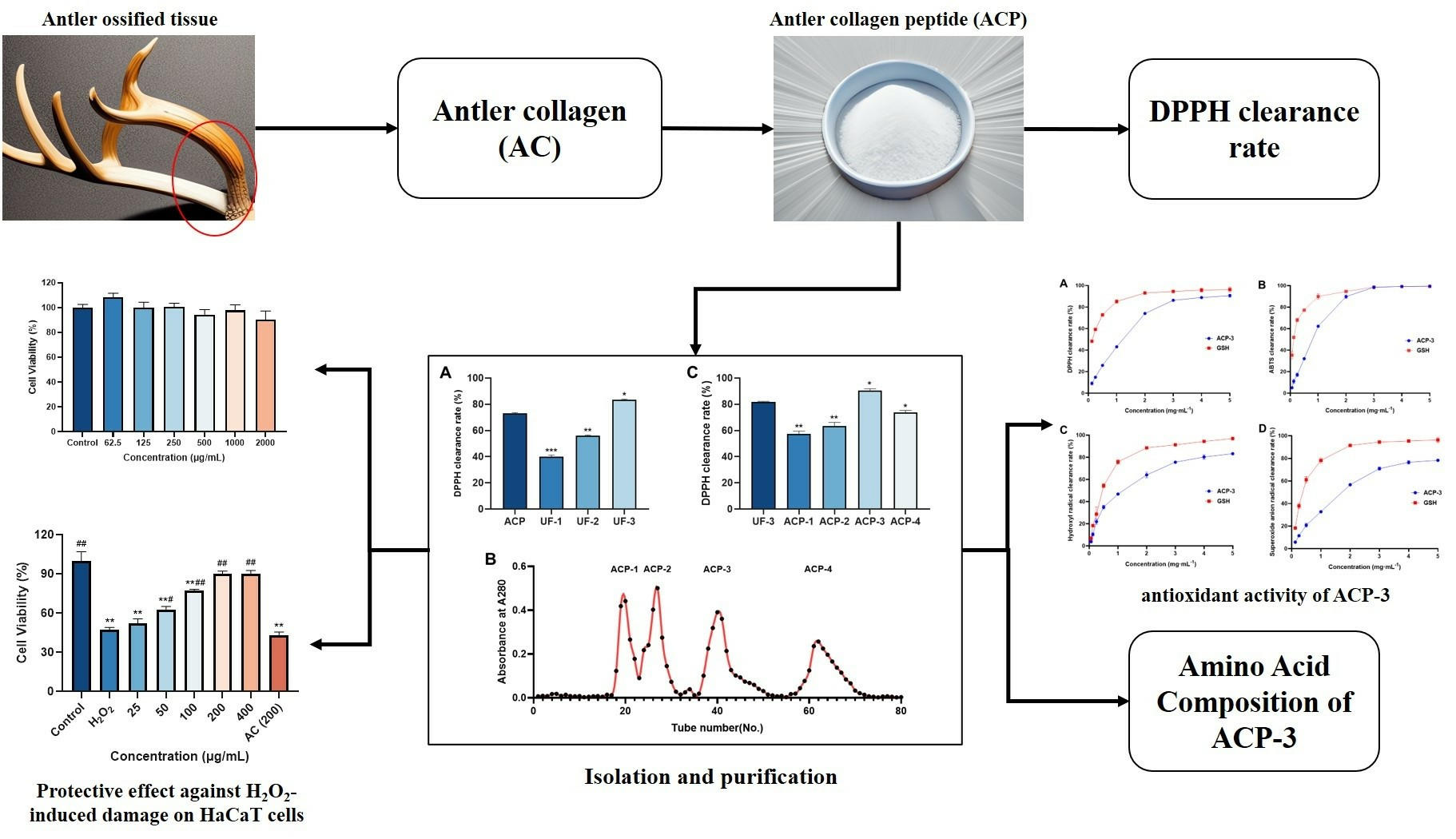
Antioxidant Peptides from the Collagen of Antler Ossified Tissue and Their Protective Effects against H2O2-Induced Oxidative Damage toward HaCaT Cells

Abstract
Share and Cite
Chen, X.; Xia, P.; Zheng, S.; Li, Y.; Fang, J.; Ma, Z.; Zhang, L.; Zhang, X.; Hao, L.; Zhang, H. Antioxidant Peptides from the Collagen of Antler Ossified Tissue and Their Protective Effects against H2O2-Induced Oxidative Damage toward HaCaT Cells. Molecules 2023, 28, 6887. https://doi.org/10.3390/molecules28196887
Chen X, Xia P, Zheng S, Li Y, Fang J, Ma Z, Zhang L, Zhang X, Hao L, Zhang H. Antioxidant Peptides from the Collagen of Antler Ossified Tissue and Their Protective Effects against H2O2-Induced Oxidative Damage toward HaCaT Cells. Molecules. 2023; 28(19):6887. https://doi.org/10.3390/molecules28196887
Chicago/Turabian StyleChen, Xi, Peijun Xia, Shuo Zheng, Yi Li, Jiayuan Fang, Ze Ma, Libo Zhang, Xunming Zhang, Linlin Hao, and Hong Zhang. 2023. "Antioxidant Peptides from the Collagen of Antler Ossified Tissue and Their Protective Effects against H2O2-Induced Oxidative Damage toward HaCaT Cells" Molecules 28, no. 19: 6887. https://doi.org/10.3390/molecules28196887
APA StyleChen, X., Xia, P., Zheng, S., Li, Y., Fang, J., Ma, Z., Zhang, L., Zhang, X., Hao, L., & Zhang, H. (2023). Antioxidant Peptides from the Collagen of Antler Ossified Tissue and Their Protective Effects against H2O2-Induced Oxidative Damage toward HaCaT Cells. Molecules, 28(19), 6887. https://doi.org/10.3390/molecules28196887