Article

Antimicrobial Coatings for Medical Textiles via Reactive Organo-Selenium Compounds

1
Fiber and Biopolymer Research Institute, Department of Plant and Soil Science, Texas Tech University, Lubbock, TX 79409, USA
2
Department of Microbiology and Immunology, Texas Tech University Health Sciences Center, Lubbock, TX 79430, USA
3
Honors College, Texas Tech University, Lubbock, TX 79409, USA
4
Ophthalmology and Visual Sciences, Texas Tech University Health Sciences Center, Lubbock, TX 79430, USA
*
Authors to whom correspondence should be addressed.
Molecules 2023, 28(17), 6381; https://doi.org/10.3390/molecules28176381
Submission received: 31 July 2023 / Revised: 24 August 2023 / Accepted: 29 August 2023 / Published: 31 August 2023

Abstract

:
Bleached and cationized cotton fabrics were chemically modified with reactive organoselenium compounds through the nucleophilic aromatic substitution (SNAr) reaction, which allowed for organo-selenium attachment onto the surface of cotton fabrics via covalent bonds and, in the case of the cationized cotton fabric, additional ionic interactions. The resulting textiles exhibited potent bactericidal activity against S. aureus (99.99% reduction), although only moderate activity was observed against E. coli. Fabrics treated with reactive organo-selenium compounds also exhibited fungicidal activities against C. albicans, and much higher antifungal activity was observed when organo-selenium compounds were applied to the cationized cotton in comparison to the bleached cotton. The treatment was found to be durable against rigorous washing conditions (non-ionic detergent/100 °C). This paper is the first report on a novel approach integrating the reaction of cotton fabrics with an organo-selenium antimicrobial agent. This approach is attractive because it provides a method for imparting antimicrobial properties to cotton fabrics which does not disrupt the traditional production processes of a textile mill.

Graphical Abstract

1. Introduction

One of the markets for technical textiles that is growing the fastest is the market for medical textiles. Medical textiles are used in medical settings to treat wounds or medical conditions, or to provide an appropriate atmosphere for clinical therapy. Medical textiles range from non-woven fabric components of healthcare and hygiene items (diapers and feminine hygiene products) to non-implantable materials (wound dressings, bandages, gauzes, and surgical disposables) and highly specialized textiles used inside the human body. These include extracorporeal devices (such as prostheses) and implantable materials (high-value products utilized for blood filtration, sutures, tissue growth scaffolds, and vascular grafts) [1]. The global market for medical textiles is enormous: its size was estimated to be USD 14.42 billion in 2020 and is projected to reach USD 19.66 billion in 2026, at a compound annual growth rate (CAGR) of 5.3% [2].
Among medical textiles, surgical and hospital textiles used by medical workers are intended to reduce the spread of infection between patients and healthcare personnel and protect them from the transmission of bacteria that can persist on clinical clothing for several days and even after washing [3]. These products benefit from the use of antimicrobial medical textiles that, upon coming into direct contact with endogenous bacteria, have a potential to inhibit the bacterial growth on their surfaces and reduce cross-infections in primary care settings [4]. Because an open wound provides an ideal environment for bacterial and fungal proliferation that might trigger wound inflammation, antimicrobial wound dressings are another type of antimicrobial textile product that are effective in reducing the risks of infection [5,6]. Antimicrobial fabrics are also used as hospital bed sheets, curtains, and patient clothing, etc., to reduce cross-infection [7]. Outside of hospital settings, antimicrobial textiles are being used in regular clothes [8] and sportswear [9].
Antibacterial textiles are traditionally produced by the treatment of textiles with a solution and/or suspension of an antimicrobial agent, which is applied onto fabric via simple dip-coating and/or spraying techniques. The fabric itself is made from non-toxic, non-allergic, non-carcinogenic, and sterilizable fibers, for which the properties important for the final product manufacture—strength, flexibility, elongation, absorbency, and biodegradability—are already known. For a long time, cotton and its derivatives have been used extensively for this purpose. However, while cotton is recognized for its diverse use, durability, and comfort [10], its hydrophilic nature could create an ideal habitat for the development of bacteria. Being made entirely of cellulose [11], daily textiles produce a warm, humid milieu on the skin, encouraging bacterial proliferation. Sweat discharges and skin peeling, etc., contribute to bacterial growth [12], and the bacteria can cause a foul smell, discoloration, fabric degradation, and skin rashes and infection.
Common textile antimicrobial agents include quaternary ammonium compounds (QACs), triclosan, chitosan, polyhexamethylene biguanide (PHMB), N-halamines, and metal nanoparticles (NPs). Despite having numerous advantageous qualities, QACs easily leach from fabric. In terms of their chemical structure, QACs lack reactive functional groups that would allow for covalent attachment to the fibers [13]. Chitosan demonstrates its antibacterial activity only in acidic media because its antibacterial effect is caused by a quaternary ammonium (–NH3+) group, which gets protonated at pH < 6.5 [14]. Chitosan also binds poorly to cellulose fibers. PHMB is acutely toxic for human keratocytes [15]. A significant quantity of adsorbed Cl may accumulate on the surface of fibers after the N-halamine treatment of textiles, which may cause unpleasant odors or even the discoloration of clothes [16]. Metal NPs are toxic and are able to penetrate the skin, lungs, and digestive system, contributing to the production of free radicals, which can induce cell damage [17]. In addition, bacterial resistance is a common drawback for all the aforementioned antibacterial agents.
Recently, a selenium (Se)-based family has been explored for its anticancer [18], antioxidant [19], and antibacterial [20,21,22] activities. In respect to its antibacterial activity, which is of interest to this particular work, bacteria possess thiol-dependent enzyme systems as a part of their Reactive Oxygen Species (ROS) defense. Low-molecular-weight thiols, such as thioredoxin (Trx) and glutathione, etc., act as antioxidants in bacterial cells, helping them to survive oxidative damage. The antibacterial mechanism of organo-selenium compounds is based on their thiol-oxidizing ability and inhibition of the thiol-dependent ROS defense. Namely, the organoselenium anion (R–Se) continuously donates an electron to the molecular oxygen in the bacteria cell, with the formation of a superoxide anion radical (O2●−) and organoselenium radical (R–Se), which, in turn, react with the low-molecular-weight (MW) thiolates (e.g., the anion of glutathione (GS)) [23], with the formation of Se–S bonds, excluding thiols from the ROS defense [24].
It is noteworthy that, while humans also possess a thiol-dependent enzyme system, the reactivity of organoselenium compounds depends on the MW of the thiol, which is significantly larger for mammals [24,25,26]. For instance, the MW of mammalian thioredoxin (Trx) is 55 kDa, whereas the MW of bacterial Trx is 35 kDa [27]. In addition, bacterial Trx is Se-free [27], while mammalian Trx is a Se-containing enzyme able to react with OS compounds to form Se–Se bonds and actually enhance the ROS defense [24].
Several generations of organoselenum compounds (OS) have been synthesized and tested against various microbes for decades [28,29]. The first OS compound was diethyl selenide [30]. Later developed examples include Ebselen and its derivatives (Figure 1a), active against S. aureus, Helicobacter pylori, and Mycobacterium tuberculosis [31,32,33,34]. Diphenyl diselenide (Figure 1b) is known to be effective against Aspergillus sp. [35], Fusarium sp. [36], Cryptococcus sp. [37], Candida glabrata [38], Trichosporon asahii [39], S. aureus, and S. epidermidis [40]. Selenazoles (Figure 1c) and their derivatives have been shown to exhibit antibacterial activity against the fungi Candida albicans and Cryptococcus neoformans [41,42,43], and have been used for the functionalization of known antibiotics to enhance their activity [44]. Selenediazole and its derivatives (Figure 1d) have exhibited antimicrobial action against E. coli, S. aureus, and Mycobacterium tuberculosis [45]. Another class of selenium compounds, selenoesters (Figure 1e) such as methylketone selenoester, have demonstrated promising antibacterial activity against S. aureus, Enterococcus faecalis, Chlamydia trachomatis [46], Salmonella enterica serovar Typhimurium, and Candida sp. [47,48]. These organoselenum compounds have been used as coatings for polyester bandages [49], scleral buckles [50], contact lenses [51], catheter tubing [52], and even cotton socks [53].
To the best of our knowledge, none of these studies have investigated the application of reactive OS compounds, i.e., OS compounds possessing a reactive functional group for covalent attachment to cellulose, the main component of cotton fabric. At the same time, reactive dyes, a common class of textile dyes, are often applied to cotton and react with the hydroxyl groups of cellulose, forming covalent bonds in the presence of alkali via a nucleophilic substitution reaction [54]. These dyes form a permanent attachment to the fiber with a high degree of wash fastness. An example of such a reactive dye is C.I. Reactive Blue 109 (sodium (Z)-5-amino-3-(2-(5-((4,6-dichloro-1,3,5-triazin-2-yl)amino)-2-sulfonatophenyl)hydrazineylidene)-6-((E)-(2,5-disulfonatophenyl)diazenyl)-4-oxo-3,4-dihydronaphthalene-2,7-disulfonate), which contains a 4,6-dichloro-1,3,5-triazin-2-yl fragment. The 4,6-dichloro-1,3,5-triazin-2-yl fragment reacts with cellulose via the nucleophilic aromatic substitution (SNAr) reaction, resulting in the substitution of chlorine atoms with cellulose and the formation of a covalent bond between the cellulose and the dye (Figure 2).
We hypothesized that the incorporation of a 4,6-dichloro-1,3,5-triazin-2-yl fragment into the structure of OS compounds would allow us to reactively attach the OS compounds onto the cotton fabric, and that conditions suitable for dyeing would be applicable for creating a covalent bond between the OS compounds and the cotton cellulose. In addition, chloro- and dichloro-1,3,5-triazin-2-yl-containing molecules are known to exhibit anti-bacterial [55,56] and anti-fungal [57] activities that might be synergistic to selenium activity.
The uptake of reactive dyes can be increased by the means of the cationization of cotton (CC) to introduce cationic sites able to attract negatively charged and/or aromatic dye molecules [54], in addition to covalent attachment. Therefore, we decided to also utilize CC textiles and compare the results with bleached cotton (BC) fabric used as a control.
Attach Chem Inc. (Lubbock TX) synthesized two reactive OS compounds (patent protected, OS-1 and OS-2, Figure 3) for textile application, which differed in their number of Se- atoms and number of potential reactive sites. The objective of this research was to investigate whether the 4-chloro-1,3,5-triazin-2-yl and 4,6-dichloro-1,3,5-triazin-2-yl fragments of the OS compounds were able to react with the cotton cellulose under an alkaline pH, and if the resulting fabric would exhibit antimicrobial properties against Gram-positive (S. aureus), Gram-negative (E. coli) bacteria, and yeast (Candida albicans).

2. Results

2.1. Fabric Cationization

Among the various compounds that have been utilized to produce cationic cotton, a commercially available CR-2000, with the chemical name (3-chloro-2-hydroxypropyl)trimethylammonium chloride (CHPTAC), is a well-established and widely studied cationizing agent [58]. Upon its covalent attachment to cellulose, CHPTAC introduces positive quaternary ammonium groups onto the cotton fabric surface. Our group has previously utilized CHPTAC for the cationization of cotton fabrics [59]; the same protocol for cationization was used in the current study.
The possible two-step mechanism of the reaction of CHPTAC with BC under alkaline conditions is outlined in Figure 4. In the first step, the cellulose of the BC textile is transformed into alkali cellulose and the CHPTAC is converted into its reactive epoxide form. In the second step, the alkali cellulose reacts with the epoxide to produce cationized cotton (CC) fabric. The unreacted epoxide is hydrolyzed with water, with the formation of 2,3-dihydroxy-N,N,N-trimethylpropanaminium chloride that is washed out from the fabric after the reaction.

2.2. Preparation of Organoselenium Textiles

2.2.1. Preparation

After the fabric cationization, the OS compounds were applied to BC and CC fabrics (see Experimental). For the application of the OS compounds to the BC fabric, the fabrics were treated with aqueous solutions of the OS compounds at different concentrations (1%, 3%, and 5%), in the presence of Na2CO3 and Glauber’s salt (Na2SO4). Under alkaline conditions, cellulose-based nucleophile Cell-O entered into a SNAr reaction with the reactive 4-chloro-1,3,5-triazin-2-yl chloride fragments of the OS compounds. Basic Na2CO3 was added to the treatment bath for the adjustment of the pH, and, in order to enhance the fixation of the OS compounds to the BC fabric, Glauber’s salt was used to neutralize the negative zeta potential of the cotton fabric and facilitate the uptake of the OS compounds. Figure 5a illustrates the reaction between the BC fiber and OS-1 compound; the reaction with OS-1 proceeded through the same mechanism and is not shown here.
The protocol was similar for the application of the OS compounds to the CC fabric, with the exception that no Na2SO4 was required due to the presence of positive cationic sites. Figure 5b illustrates the reaction between the CC fiber and OS-2 compound. In addition to the formation of a covalent bond between the OS compounds and the cellulose of the CC fabric as a result of the SNAr reaction, ionic interactions between the quaternary ammonium group of the CC and the aromatic ring of the OS compounds were also established [60], allowing for a higher fixation of the OS compounds on the CC than that on the BC, as is evident in the following sections.

2.2.2. Appearance

Digital photographs of the untreated and OS-treated cotton textile samples are shown in Figure 6. The OS-1 treatment of the cotton textiles produced a reddish hue, whereas the OS-2 treatment of the cotton textiles produced a visible yellow color on the textile surfaces. The color development was concentration dependent and was more prominent when the same compound was applied to the CC than to the BC, for both OS-1 and OS-2.
The evaluation of the color depth (color strength) of the cotton fabrics treated with the OS compounds was conducted through measuring the K/S value (where K is an absorption coefficient and S is a scattering coefficient) using a spectrophotometer. When the OS compounds were applied to fabric, the K/S increased, and, as expected, this increase was proportional to the concentration of the OS in the solution.
As can be seen from Figure 7, the K/S was determined to be 0.2 for the 1% OS-1-treated BC fabrics, 0.3 for the 3% OS-1-treated BC fabric, and 0.4 for the 5% treated BC fabric. There was a substantial increase in the K/S values when OS-2 was applied to BC when compared to those for OS-1. For the OS-2-treated fabrics, the K/S increased from 0.6 for the BC fabric treated with 1% OS-2 to 2.1 for the fabric treated with 3% OS-2 and 4.5 for the fabric treated with 5% OS-2. Overall, for the BC fabric treated with the OS compounds at the highest concentration (5%), a 10-fold increase in the K/S values between OS-1 and OS-2 was observed.
A substantial increase in the K/S values was observed for the CC when compared to the BC, which is attributed to the additional ionic bonding of the CC with either of the OS compounds. Thus, the K/S for the OS-1-treated CC fabrics was found to be in the range from ~0.6 (for the fabric treated with the 1% solution of OS-1) to 2.2 (for the fabric treated with the 5% solution of OS-1). Substantially higher K/S values were obtained for the OS-2-treated fabrics: the K/S was found to be 2.5 for the fabric treated with 1% OS-2, 12.1 for the fabric treated with 3% OS-2, and 48.1 for the fabric treated with 5% OS-2. Overall, the K/S trend was as follows: OS-2@CC >> OS-1@CC > OS2@BC > OS-1@BC, independent of the concentration applied.
To describe the difference between the two colors, a color difference metric (ΔE*) was used in accordance with the International Commission on Illumination (CIE). The ΔE* levels are the difference between the color of the fabric and the reference color standard. Lower ΔE* values indicate similarity to the reference standard, whereas high ΔE* values indicate a significant deviation from the reference.
The ΔE* values of the OS-treated cotton were measured and compared with the reference textile; the results are given in Table 1. The OS-1 treatment of the BC textile produced a ΔE* between 3.40 and 12.38. Similar to the K/S study of the OS-treated textiles, the ΔE* values showed that the OS-2 treatment generated a more prominent color than the OS-1 treatment. Thus, the OS-2 treatment of the BC textile produced a ΔE* between 14.98 and 36.19, depending on the treatment concentration. A substantial increase in the ΔE* values was observed for the CC when compared to the BC. Thus, the OS-1 treatment of the CC textile yielded a ΔE* between 10.81 and 33.03, while the OS-2 treatment of the BC textile produced ΔE* values between 25.06 and 57.69, depending on the treatment concentration.

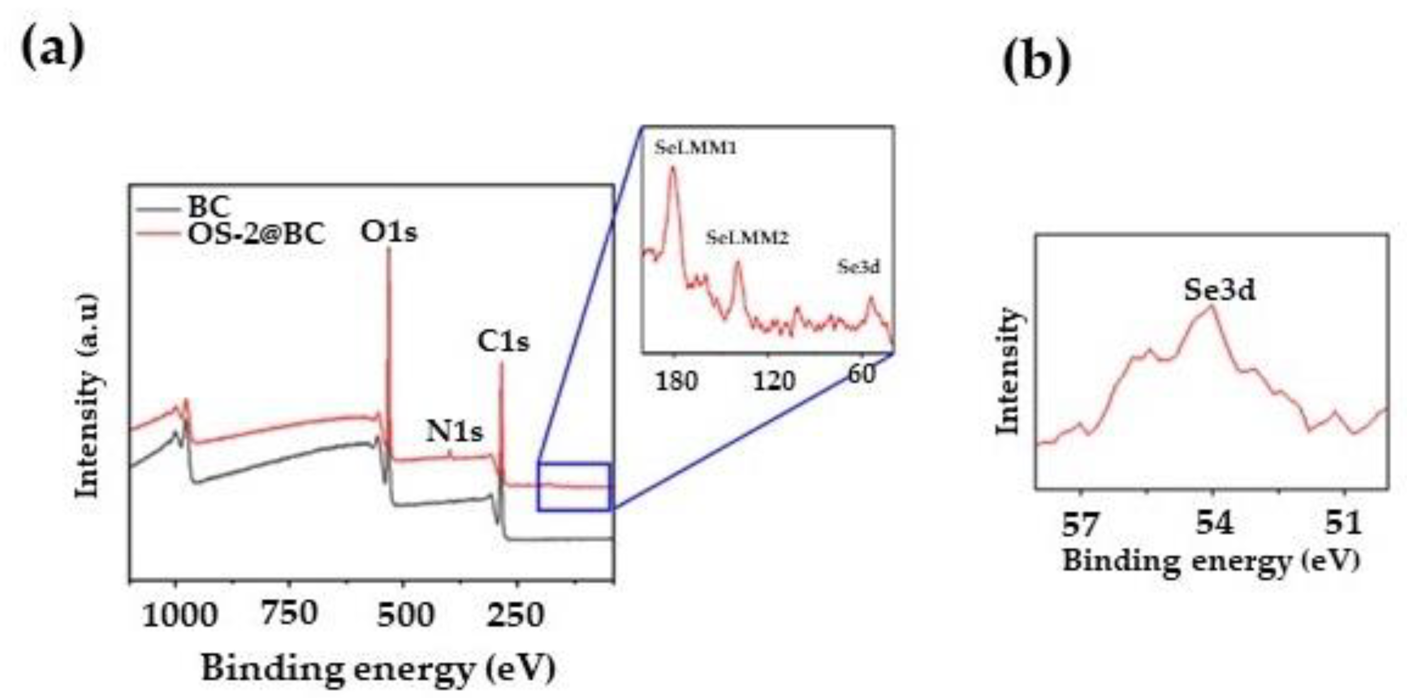
2.3. Textile Characterization

FTIR was found to be not a sensitive enough technique for determining changes on the textile surfaces. Instead, for the surface characterization, X-ray photoelectron spectroscopy (XPS) was conducted on the fabric samples. All the samples were first analyzed through a survey scan (over a binding energy range from 0 to 1000 eV) to determine the elements present within the top 1–10 nm of the surfaces of the samples. Figure 8a shows the survey XPS spectra of the control (BC) and OS-2-treated cotton fabrics. Carbon and oxygen were found as expected for the cellulose, but there was also nitrogen observed in the OS-2-treated BC fabric.
Namely, the survey XPS spectrum of the control fabric showed three peaks for O1s (532 eV), C1s (285 eV), and O2s (24.8 eV) orbitals [61]. Compared to the control fabric, the OS-2-treated fabric exhibited one additional peak N1s (398 eV) orbital [62]. Figure 8b shows the high-resolution Se3d spectrum of the OS-2-treated cotton fabric. A clear peak at 54 eV is obvious in the high-resolution spectrum, which can be attributed to the Se3d orbital [63]. These additional peaks in the OS-2-treated fabric confirm the presence of Se on the surface of the cotton fabric.
It is important to point out that, at this low concentration, these Selenium-based compounds behave more like reactive dyes than crosslinking agents. Typically, if crosslinking occurs between Se-based compounds and cellulose, FTIR spectra would show the presence of corresponding vibrations attributed to crosslinking agents. However, in our case, the FTIR did not show additional vibrations suggesting the occurrence of crosslinking. Further evidence for the absence of crosslinking is the higher reactivity of the Se-based compounds with the cationized cotton fabric as compared to that of the only bleached cotton fabric.

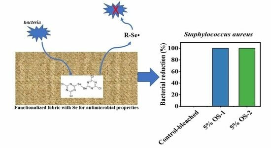
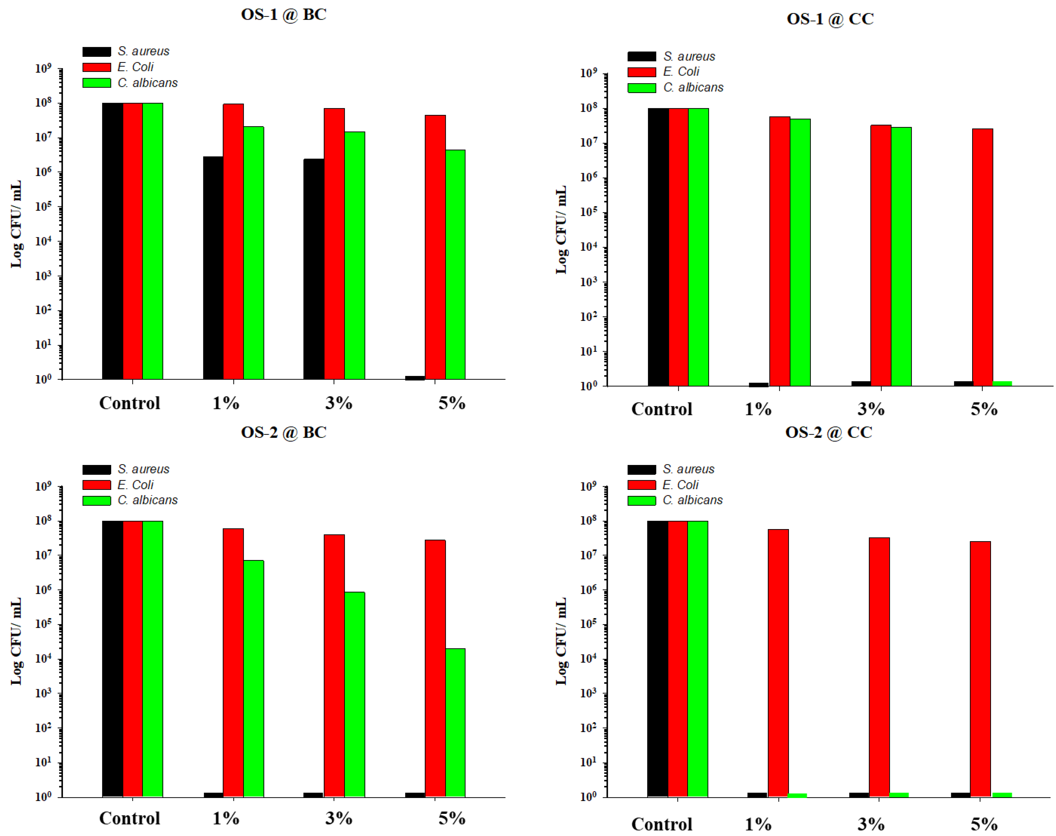
2.4. Antimicrobial Assays

After treatment, the fabric samples were tested for their antimicrobial activity using three strains of microorganisms: S. aureus AH133 GFP (Gram-positive), Escherichia coli MM294 (Gram-negative), and Candida albicans strain 3147 (fungi). S. aureus and Escherichia coli GFP lab strains were chosen as representatives of Gram-positive and Gram-negative bacteria. Candida albicans was chosen to represent a fungal species.
In vitro CFU assays were conducted to determine the antimicrobial activity of the textiles. For conducting the assays, bacteria washed with phosphate-buffered saline (PBS) were suspended in PBS at a concentration of ~108 CFU/mL, and then diluted with fresh PBS to a concentration of 102–103 CFU/mL (see Experimental for details). Fabric samples (1 cm2 area), 10 µL aliquots of inoculum, and 1 mL of fresh PBS buffer were placed into separate wells of a multi-well plate. After 24 h of incubation at 37 °C, the number of colonies formed on the fabrics was counted using dilution and plating techniques. The tests were conducted on the OS-1-treated BC and CC fabrics (OS-1@BC and OS-1@CC, respectively) and OS-2-treated BC and CC textiles (OS-2@BC and OS-2@CC, respectively).
Initial screening was conducted on the fabrics treated with 1, 3, and 5% of OS-1 and OS-2, respectively, to determine the optimal concentration of the OS compounds. Two negative controls—untreated BC and CC fabrics—were also evaluated. Table 2 and Figure 9 summarize the antimicrobial activities of both the BC and CC OS-treated fabric samples, and the bacterial reduction is shown as both % reduction and log reduction.
The bleached fabrics treated with the 1% OS-1 solution allowed for a 97.24% reduction in S. aureus bacterial growth, while the fabrics treated with the 3% OS-1 solution were just slightly more active, yielding a 97.63% reduction in bacteria. With an increase in the OS-1 solution concentration to 5%, the inhibition effects on S. aureus raised to 100%. The bacterial count was significantly higher for the E. coli strain than that of S. aureus, indicating the lower effectiveness of OS-1 BC against E. coli. Thus, the BC fabric treated with 1% OS-1 yielded only a 6.06% E. coli bacterial reduction, the fabric treated with 3% OS-1 provided a 30.30% E. coli reduction, and the fabric treated with 5% OS-1 demonstrated 54.55% activity against E. coli. The activity against the Candida albicans strain was 79.35% at the lowest concentration of OS-1 (1%) and 95.60% at the highest concentration of OS-1 (5%).
The BC fabric treated with OS-2 completely inhibited the growth of S. aureus at its lowest concentration (1% OS-2). Moderate activity, although higher than that in the case of OS-1, was observed against E. coli: 39.39%, 60.61%, and 72.73% for the 1, 3, and 5% OS-2-treated BC fabric, respectively. The antifungal activity was also higher in the case of the OS-2 treatment than that in the case of the OS-1 treatment, even though a complete inhibition of fungi growth was not achieved, and the bacterial reduction values ranged from 92.99% (1% OS-2 treatment) to 99.98 (5% OS-2 treatment).
The CC fabric treated with OS-1 completely inhibited the growth of the S. aureus bacteria at the lowest concentration (1% OS-1). The activity against E. coli was modest: 5% OS-1 killed 74.19% of the bacteria. A fungal reduction of 51.72% was found when the CC fabric was treated with 1% OS-1 and 72.41% when the CC fabric was treated with 3% OS-1, whereas 5% OS-1 yielded a complete elimination of bacterial growth. The CC fabric treated with OS-2 exhibited an excellent 100% bacterial reduction for both the S. aureus bacteria and Candida albicans yeast at its lowest concentration (1% OS-2).
The observed improvement in the antimicrobial activity of OS-2 when compared to OS-1 was likely due to the abundance of Se atoms on the molecule of OS-2, whereas the enhancement in antimicrobial activity in the case of the CC vs. BC cotton was likely attributed to the higher fixation of OS molecules on the CC fabric.
The differences in activities against the different species were likely due to the following. The reason for the high efficacy of OS against the S. aureus bacteria could be due to the abundance of Trx in their cells, which is an attractive target for Se-compounds [64,65]. It also appears that the OS compounds performed significantly better against C. albicans. Like bacteria, fungi also possess low MW Trx as a thiol-dependent ROS defense [66], and the antifungal mechanism of the OS compounds also involves the depletion of low MW thiols and the elevation of ROS. It is reasonable to speculate that there was a similar interaction between fungal Trx and the OS compounds to that in bacteria. An OS compound could also deplete fungal GSH [67].
All types of fabrics demonstrated moderate activity against the E. coli strains. The poor activity of OS against the E. coli strain could be attributed to its limited capacity to penetrate cell walls due to their outer membrane [68]. According to [69], the activity of OS compounds against Escherichia coli bacteria depends on the proportion of their accumulation to the polar amine group of the cell wall. This could be achieved by either introducing polar groups into the OS compounds or applying them in higher doses (>128 (μg/mL)) [69]. In addition, E. coli bacteria possess GSH as their major low MW thiol instead of Trx within their cell [65,70,71,72].

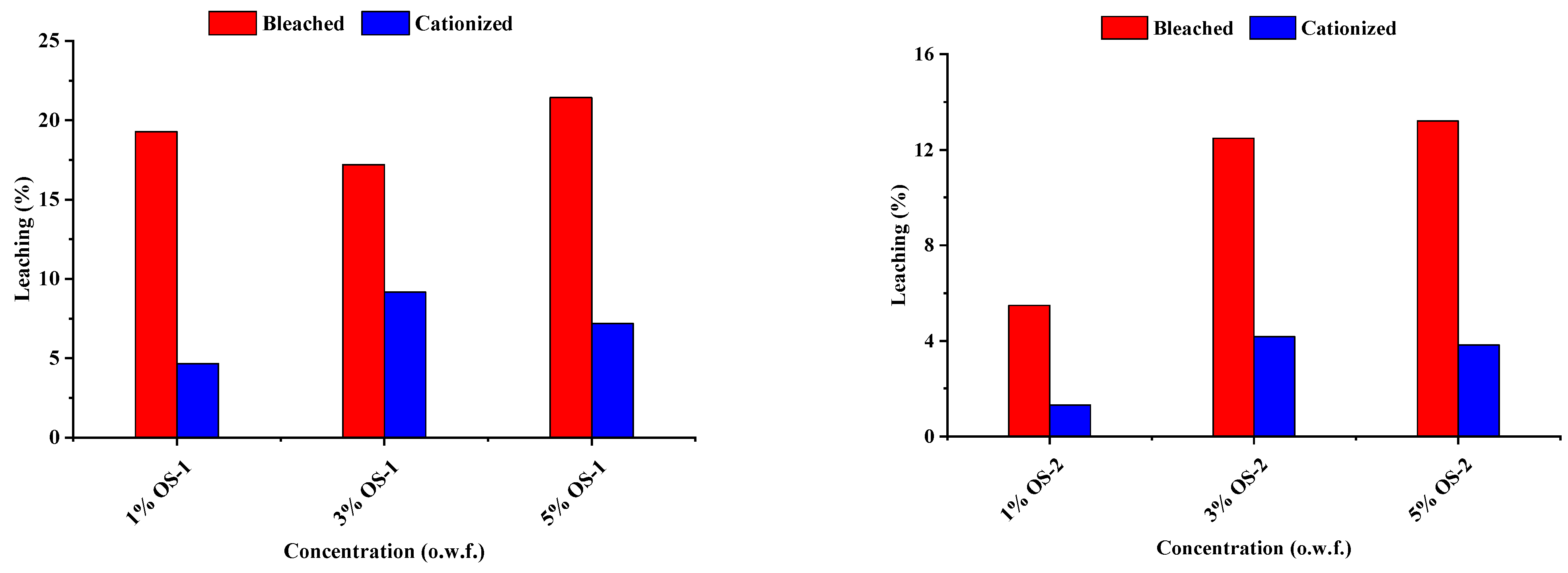
2.5. Leaching Studies

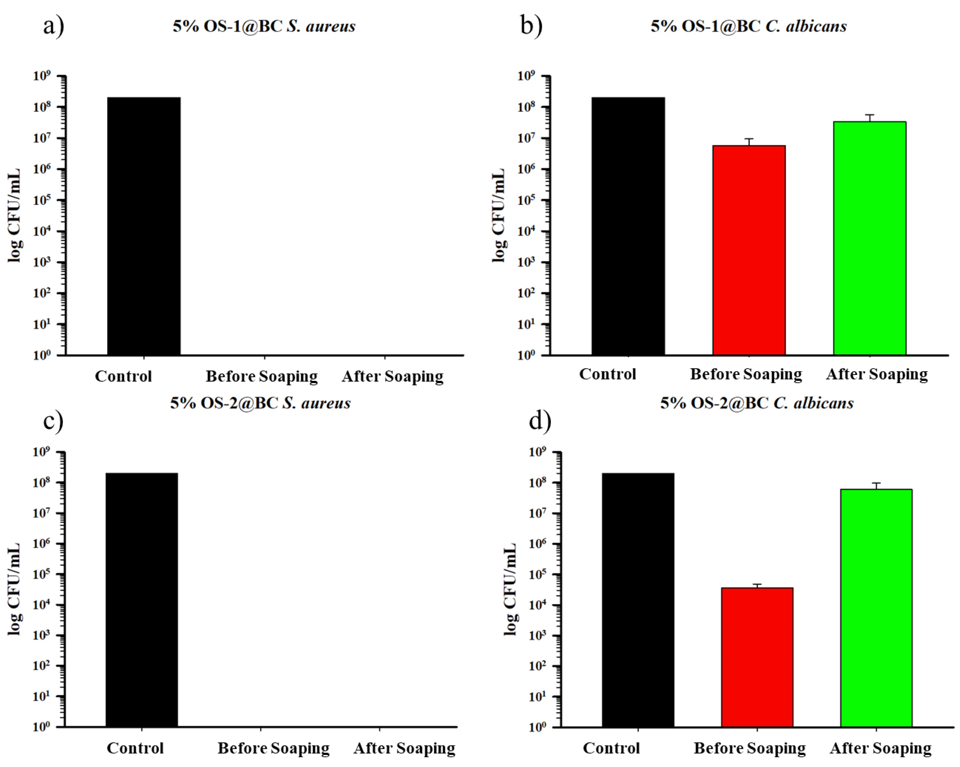
2.5.1. Leaching (%) of OS from Treated Textile Due to Soaping

The degree of leaching (in %) of the OS was determined after washing for 30 min in a non-ionic detergent (Triton X-100) at 100 °C and pH 8 (See Experimental). The degree of leaching (%) of the OS compounds during soaping from the treated textiles is shown in Figure 10.
It is seen that the BC fabric treated with OS-1 released from 17 to 21% of active coating into a water phase, due to the washing with the non-ionic detergent. In contrast, the amount of OS-1 that leached out from the CC fabric was significantly less, from 5 to 9% OS-1.
Among the two compounds, OS-2 was more resistant to leaching than OS-1. Thus, the BC fabric treated with OS-2 liberated less active leaching than the same fabric treated with OS-1, namely from 5 to 13%. Finally, the CC material treated with OS-2 was the most stable in terms of leaching, releasing from only 1 to 4% OS-2 into the aqueous phase. In general, a higher degree of leaching was observed for the bleached fabrics treated with OS compared to the cationized fabrics. This could be due to the better fixation of the OS compounds on the cationized fabric due to the positive amino groups on its surface. This improvement in the leaching resistance of the CC could be attributed to electrostatic interactions between the positively charged OS molecules and negatively charged cationized textile. This leaching behavior can be an indication of the wash fastness of OS-treated textiles. Further work can be conducted to improve the leaching stability or wash fastness behavior of OS compounds.

2.5.2. Durability of Antimicrobial Activity

Textiles are frequently washed in the presence of soap or detergent over their life cycle. Antibacterial materials must be able to withstand repeated washing and laundering in domestic settings. To estimate the durability of the OS treatment of the cotton textiles, the 5% OS (o.w.f.)-treated textiles were washed using rigorous washing conditions (non-ionic detergent Triton X-100 and NaHCO3 at 100 °C for 30 min). The wash durability was measured in terms of in vitro CFU assays.
The results of the in vitro CFU assays for the OS-treated BC textiles are shown in Figure 11, and for the OS-treated CC fabrics in Figure 12. For S. aureus, both the OS-1- and OS-2-treated BC fabrics, washed under rigorous conditions, retained their antimicrobial activity, as shown in Figure 11a,c, completely preventing bacterial growth either before or after washing. An apparent loss in the inhibition of C. albicans biofilms was seen for both the OS-1- and OS-2-treated BC fabrics washed under rigorous conditions (Figure 11b,d for OS-1 and OS-2, respectively), although the difference in the inhibition of C. albicans for the OS-2-treated BC fabric was larger than that for the OS-1 fabric. This indicates a loss of antifungal activity on the BC textile due to soaping. As was shown previously, the OS-treated BC fabric demonstrated little activity against the E. coli strains, which was not expected to change after washing, and the fabric was not re-tested against these bacteria.
The results of the in vitro CFU assays for the OS-treated CC textiles showed that, for S. aureus, both the OS-1- and OS-2-treated CC fabrics, washed under rigorous conditions, behaved similarly to the BC fabric and fully eliminated the S. aureus biofilm growth on both the unwashed and washed textiles. The same was true for C. albicans biofilms for both the OS-1- and OS-2-treated CC fabrics washed under rigorous conditions; a complete inhibition of C. albicans for both the OS-1- and OS-2-treated CC fabric was observed. This durability could be attributed to the strong bonding between the cationic amino groups and aromatic-ring-bearing OS molecules in pre-cationized cotton textiles. As reported earlier, the OS-treated CC fabric was only negligibly active against the E. coli strains, thus the rigorously washed fabric was not tested against this strain.

2.5.3. Release of OS-2 from the Treated Fabric

To determine any potential release of Se from the treated textile into the environment, a leaching study was carried out. The aim of this part of the study was to quantify the release of OS-2 from the OS-2-treated BC (5% OS-2 concentration) into water. The reason behind choosing this particular material for the leaching studies was that OS-2 had a higher activity than OS-1, and its adherence to the BC fabric was weaker than that to the CC; hence, a larger amount of more active compound was expected to leach out.
A calibration curve was first obtained by plotting UV-Vis absorbances at the maximum absorbing wavelength (λmax) of a series of consecutively diluted OS-2 solutions in water at pH 7 as a function of concentration. An OS-treated BC fabric sample was placed into DI water and the OS-2 compound was allowed to leach. The UV-Vis absorbance was recorded every 24 h. The amount of released OS-2 was back-calculated from the calibration equation, considering the dilution factor.
The release kinetics study showed that most of the compound (24 µg/cm2 of OS-2) was released within 24 h, as shown in Figure 12, after which, the release of OS-2 from the treated textile was slow. The highest amount of release of OS-2 (~31 µg/cm2) was obtained after 144 h of leaching. Because OS-2 is a newly synthesized compound and its minimum inhibitory concentration (MIC) and minimum biocidal concentration (MBC) are unknown, we cannot conclude whether the release of OS-2 was above the MIC and MBC of certain microorganisms in the water.

3. Materials and Methods

3.1. Textile Material

Twill-weaved, desized, scoured, and bleached 100% cotton fabric was used for the research (warp 100 yarns per inch, weft 56 yarns per inch, 215 g/m2 pe meter). The fabric was supplied by Fiber and Biopolymer Research Institute, Lubbock, TX, USA.

3.2. Chemicals

Acetic acid (CH3COOH, 99.7%) was obtained from Sigma Aldrich (St. Louis, MO, USA). Acetone (C3H6O, 99.5%), sodium bicarbonate (NaHCO3, 99.7%), sodium hydroxide (NaOH, 98%), and anhydrous sodium carbonate (Na2CO3, 99.5%) were obtained from Fisher Scientific (Hampton, NH, USA). A cationizing agent, 3-chloro-2-hydroxypropyltrimethyl ammonium chloride (CHPTAC), also called CR-2000 (C6H15Cl2NO, 65%), and Triton X-100 (2-[4-(2,4,4-trimethylpentan-2-yl)phenoxy]ethanol) (C16H26O2, 99+%) were obtained from Dow Chemical (Midland, MI, USA). Sodium sulfate anhydrous (Na2SO4, 99.5%) was received from Cooper Natural Resources (Fort Worth, TX, USA). The organoselenium compounds, OS-1 (C6Cl4N6Se2, >99%) and OS-2 (C6H2Cl2N6Se4, >99%), were obtained from Attach Chem (Lubbock, TX USA) and used as received. Deionized (DI) water was obtained from AquaOne (Amarillo, TX, USA) and was used for all purposes.

3.3. Bacterial Strains

S. aureus AH133 GFP (Gram-positive), Escherichia coli MM294 GFP (Gram-negative), and Candida albicans strain 3147 (ATCC 10231TM) were used in this study. Both the S. aureus AH133 GFP (Gram positive) and Escherichia coli MM294 GFP strains were available at the Texas Tech University Health Sciences Center, Lubbock, TX, USA. The Candida albicans strain 3147 (ATCC 10231TM) was purchased from ATCC Manassas, VA, USA. All the microorganisms were cultured in Luria-Bertani (LB) broth or LB agar plates at 37 °C. All the bacterial stock was preserved at −80 °C in an ultra-low-temperature (ULT) refrigerator.

4. Methods

4.1. Fabric Preparation

The cotton fabric was first washed in 2% (v/v) Triton X-100 (at a 1:100 liquor ratio) following the published procedure [73]. Specifically, a hot plate was used to heat and raise the temperature of the DI water (2.5 L) to 50 °C. At this point, 50 mL of Triton X-100 was added to the DI water and mixed well using a magnetic stirrer. Then, 25 g of fabric was immersed into the solution and stirred using a glass rod for 30 min. Then, the fabric was removed and rinsed five times with DI water. The rinsed fabric was dried in a laboratory oven at 80 °C for 60 min. The dried fabric was taken from the oven and then washed in a beaker containing pure acetone for 10 min (at 1:20 liquor ratio) at room temperature. Then, the fabric was removed and rinsed five times with DI water. After that, the rinsed fabric was again dried at 80 °C for 60 min in a laboratory oven.

4.2. Cationization of Cotton Textile

The cotton fabrics were cationized using CR-2000 (65% aqueous solution of CHPTAC), a commercially available cationizing chemical, following a previously published study [59]. CR-2000 (17.5 g) was added to 1 L of DI water at room temperature. The cotton fabric (58.8 g) was immersed into the liquor under constant stirring. Then, NaOH (7.51 g) was added and the mixture was stirred constantly for 20 min. After 20 min, the fabric was taken out and squeezed by hand. The treated fabric was kept in a zipped plastic bag for 24 h to avoid evaporation of the cationizing agent. Then, the fabric was rinsed five times in DI water and neutralized in a 1 g/L acetic acid solution for 5 min at room temperature. Finally, it was again rinsed five times in DI water and dried at 80 °C for 60 min in a laboratory oven to obtain a cationic cotton fabric.

4.3. OS Treatment of Textiles

4.3.1. OS Treatment of Bleached Textile

The bleached cotton fabric was treated with 5% of the OS compound (on the weight of the fabric, o.w.f.) (at 1:40 material to liquor). Triton X-100 solution (200 mL of 0.2 g/L) was prepared at room temperature. The solution was heated to raise its temperature to 60 °C. The OS compound (250 mg) was added to the solution and constantly stirred for 20 min. The bleached cotton fabric (5 g) was immersed in the solution. After 20 min, 10 g of Na2SO4 was added to the bath under constant agitation. After 10 min, 1 g of Na2CO3 was mixed into the solution to adjust the pH to 11. The solution was maintained for a following 60 min under constant stirring. Then, the treated fabric was taken from the bath and rinsed five times in DI water to remove the unfixed OS compound from its surface. Then, acid neutralization was performed to remove the alkalinity. For the acid neutralization, the rinsed fabric was treated in a 0.5 g/L CH3COOH solution at room temperature for 15 min. Then, it was rinsed five times again in DI water. Finally, the material was squeezed by hand and dried in a laboratory oven for 30 min at 100 °C.

4.3.2. OS Treatment of Cationized Textile

To treat the cationized cotton fabric with 5% of an OS compound (o.w.f.), a similar route was followed to that above. The absence of Na2SO4 salt here was the only distinction. Briefly, 0.2 g/L of Triton X-100 solution (200 mL) was prepared in a beaker. The solution was then heated to raise the temperature to 60 °C. At 60 °C, the OS (250 mg) was added to the solution and stirred vigorously using a magnetic stirrer. After 20 min, a piece of cationized cotton fabric (5 g) was immersed in the bath and agitated for 20 min. At this stage, 1 g of Na2CO3 was added to the beaker to raise its pH to 11. The solution was held and stirred for 60 min. After that, the fabric was rinsed five times in DI water. Then, the rinsed fabric was acid neutralized in a 0.5 g/L acetic acid solution for 15 min at room temperature. The neutralized fabric was washed five times using DI water. After squeezing it by hand, the treated fabric sample was dried in an oven at 100 °C for 30 min. Four groups of samples were produced, and the samples were abbreviated as follows for a better understanding (Table 3).

4.4. CFU Antibacterial Assays

The stored bacteria (frozen stocks) were collected from the freezer and washed with phosphate-buffered saline (PBS) (pH 7.4). The washed bacteria were suspended in PBS to an optical density (OD600) of 0.42 to obtain approximately 108 bacterial colonies (CFU) per milliliter. This original inoculum was serially diluted ten-fold (1:10 dilution) up to a dilution faction of 106 (from approximately 102 to 103 CFU/mL) in PBS. The control (untreated) and OS-treated fabric samples were cut into small pieces with a 1 cm2 area each. Three pieces (3 × 1 cm2) of each sample were placed into separate wells of a multi-well plate. Then, 10µL aliquots containing from approximately 102 to 103 CFU/mL (original inoculum) and 1 mL of PBS were added to each well containing the fabric sample. The multi-well plate system was closed using the lid and incubated overnight in an oven at a 37 °C temperature. The next day, the fabric samples were collected from the wells and transferred to sterile microcentrifuge tubes containing 1 mL of PBS to collect the biofilm. The tubes were sonicated in a water bath sonicator for 5–10 min to loosen the biofilm on the surface of the fabric, and then strongly vortexed three times for 1 min to resuspend the bacterial cells. All the bacterial-cell suspensions were serially diluted 10-fold in PBS up to a dilution factor of 106. Then, three drops of the 10 µL aliquots of each dilution were plated or spotted on LB agar plates. They were incubated overnight in an oven at 37 °C. All the assays were performed in triplicate. The following day, the total bacterial colonies were counted for three spots of a certain dilution and averaged to obtain a single CFU. The back-calculation for the bacterial colony was performed using Equation (1):
C F U m L = C F U × D i l u t i o n   f a c t o r × 10 2
where A represents the number of colonies grown in the control sample and B represents the number of colonies grown in the treated sample.

4.5. Color Measurements

An X-Rite Ci7800 benchtop spectrophotometer (X-Rite, Inc., Grand Rapids, MI, USA) equipped with the Color iMatch program was used to measure the reflectance of the untreated control cotton fabrics (bleached and cationic) and OS-treated cotton fabrics. The instrument was initially calibrated using the manufacturer’s recommended two standard tiles (black and white). The color measurements were performed with a 25 mm aperture using one fabric layer. Four measurements were made from each sample (different locations along the front and back side of the control and treated samples) under D65 illumination and 10° standard observer for the wavelength range from 360 to 750 nm. The Color iMatch program provided the average reflectance (%) values for each sample between 360 and 750 nm. The average relative color strength (K/S) values of the control and treated fabric samples were also obtained from the Color iMatch program. This program uses the Kubelka–Munk formula to give relative color strength (K/S) values, as shown in Equation (2):
K S = ( 1 R λ ) 2 2 R λ
where R λ is the reflectance value at the minimum reflecting wavelength (λmin), K is the absorbing coefficient, and S is the scattering coefficient.
The CIE L*, a*, and b* values of the control and OS-treated cotton fabrics were also collected from the Color iMatch program. This program provided the averaged CIE L*, a*, and b* values of the four readings from each sample. Here, L* is defined as lightness. The value of L* can vary between 0 and 100, where 0 means black and 100 means white. The value of a* expresses the redness or greenness of a sample. The positive and negative values of a* indicate the redness and greenness, respectively. The value of b* expresses the degree of the yellowness or blueness of a sample. The positive and negative values of b* indicate the yellowness and blueness, respectively. The color difference (ΔE*) between the reference (untreated) fabric and OS-treated fabric was measured using Equation (3):
Δ E * = ( Δ L * ) 2 + ( Δ a * ) 2 + ( Δ b * ) 2
where ΔL*, Δa*, and Δb* are the differences between the color coordinates of the reference and tested fabric samples.

4.6. Fixation of OS to the Textile

The fixation (%) of the OS compound on the cotton textile was measured from the relative difference in the concentration of the OS solution before and after the treatment. The absorbances of the treatment solution were measured, before and after the treatment, using a Perkin Elmer Lambda 650 UV/Vis Spectrophotometer (PerkinElmer, Inc., Waltham, MA, USA). Using the calibration curve and Beer–Lambert law, the absorbances were converted into concentrations, and the fixation (in %) was calculated using Equation (4):
%   F = C o C t C t × 100
where Co is the concentration of the OS solution before treatment and Ct is the concentration of the OS solution after treatment.

4.7. Release of OS from Treated Textile

A release study of the OS-treated fabric samples in DI water (1 to 50 liquor ratio) per unit area of fabric was conducted using the Ohaus Orbital Shaker (Ohaus Corp., Parsippany, NJ, USA). A 5% OS-2 (o.w.f.)-treated cotton fabric with a 6 cm x 6 cm area was placed in a conical flask containing 50 mL of DI water at room temperature. The flask was then placed in the Ohaus Orbital Shaker (Ohaus Corp., Parsippany, NJ, USA) and continuously shaken at 150 RPM. At every 24 h interval, 3–4 mL of solution was collected from the flask and transferred to a cuvette to collect the absorbance. The absorbance readings at the maximum absorbing wavelength (λmax) were taken using a Perkin Elmer Lambda 650 UV/Vis Spectrophotometer (PerkinElmer, Inc., Waltham, MA, USA). The solution was again placed back into the flask to maintain the initial liquor ratio (1:50). The experiment was run for 168 h. Finally, the release of the OS compound was calculated in µg/cm2 of the treated fabric and plotted in a graph as a function of time.

4.8. Leaching of OS Due to Soaping

The leaching of the OS due to washing with a soaping agent under strong washing conditions was performed. For the leaching study, OS-treated fabric samples were first soaped following the procedure reported in this reference [74]. Briefly, the OS-treated fabric samples were washed (at a 1: 100 liquor ratio) using 2 g/LL of Triton X-100 and 5 g/LL of NaHCO3 at 100 °C for 30 min. After washing, the fabric was rinsed five times in DI water and dried in a laboratory oven at 80 °C for 30 min. The reflectance of the textiles (before and after the soaping process) was taken using an X-Rite Ci7800 benchtop spectrophotometer (X-Rite, Inc., Grand Rapids, MI, USA). The degree of leaching (% L) was calculated from the minimum of the reflectance spectrum (λmin) using Equation (5) [75]:
%   L = R A R B 100 R B × 100   %
where R B represents the reflectance of the treated fabric before soaping and R A represents the reflectance of the treated fabric after soaping. L = 100% denotes a totally decolorized cloth, whereas L = 0 denotes the material as it was originally colored (Figure 13)

4.9. Durability of OS-Treatment

To study the durability of the treatment, 5% OS-treated fabric samples were soaped using 2 g/L of a non-ionic detergent (Triton X-100) and 5 g/L of NaHCO3 at 100 °C for 30 min. They were rinsed several times followed by drying in a laboratory oven at 80 °C for 30 min. The soaped samples were again tested for their antibiofilm activity. The numbers of biofilms formed on the textiles, before and after the soaping, were compared to determine the durability of the OS treatment.

4.10. Fourier Transform Infrared Spectroscopy (FTIR)

The FTIR spectra of the fabric samples were collected using FTIR spectroscopy (Spectrum 400, PerkinElmer, MA, USA) equipped with a ZnSe diamond crystal and pressure arm. Three spectra were collected from each sample at a spectral resolution of 4 cm−1 and 64 co-added scans in the range of 4000–650 cm−1.

4.11. X-ray Photoelectron Spectroscopy (XPS)

Initially, the bleached cotton was treated with 2% OS-2 and sent to the Materials Characterization Center, Edward E. Whitacre Jr. College of Engineering, TTU, for an XPS analysis. The XPS study of the control and OS-2-treated fabric was performed to determine the elemental composition of the fabric surface using a PHI 5000 VersaProbe-II Hybrid X-ray photoelectron spectrometer (Physical Electronics, Inc., Chanhassen, MN, USA). This instrument has an operating pressure of 5 × 10⁻⁷ Pa and minimum probe size of 10 µm. The source electron beam is generated by an LaB6 filament and focused using an electrostatic lens. On the aluminum (Al) anode, a scanning Al Kα X-ray is generated by the scanning electron beam. A quartz crystal monochromator reflects the monochromatic Al Kα scanning X-ray onto the sample as a source beam. When photo electrons are generated by the X-ray and pop out of the surface, an analyzer lens makes an emission angle of 45° to enhance the number of photoelectrons reaching into the analyzer. A hemispherical spherical analyzer filters out the photo electrons of the set energies to finally reach the detector. A dual-beam charge neutralizer was used to eliminate the samples’ static charge.

5. Conclusions

Reactive chlorotriazinyl fragments were incorporated into the structure of two novel organoselenium compounds, namely 1,2-bis(4,6-dichloro-1,3,5-triazin-2-yl)diselane (OS-1) and 6,6′-diselanediylbis(4-chloro-1,3,5-triazine-2-selenol) (OS-2), which allowed for their covalent attachment to cotton cellulose via the nucleophilic aromatic substitution (SNAr) reaction. According to in vitro CFU assays, the OS compounds were strongly active against the Gram-positive S. aureus strain, fully eliminating bacterial growth on these fabrics after the treatment. The inhibition of C. albicans biofilms proceeded to a lower extent on the bleached cotton, but fully eliminated the bacterial growth on the cationized substrate. The OS-treated fabric demonstrated modest activity against the E. coli strains. In all cases with detected antimicrobial activity, the OS-treated cationized fabric exhibited higher activity than the bleached textile, and OS-2 was significantly more active than OS-1.
After washing under rigorous conditions to assess the durability of the treatment, both the OS-1- and OS-2-treated BC fabrics retained the antimicrobial activity against S. aureus, completely preventing bacterial growth. A slight loss in antifungal activity against C. albicans was observed, more so for the OS-2-treated BC fabric than for the OS-1-treated cotton. For the CC fabrics, both the OS-1- and OS-2-treated textiles fully eliminated the S. aureus and C. albicans biofilm growth on the washed textile. The durability of the treatment in the case of the CC could be attributed to the strong bonding between the cationic amino groups and aromatic-ring-bearing OS molecules. In summary, these reactive organo-selenium compounds evaluated as an efficient route for producing antimicrobial textiles did not interrupt the regular production processes of a textile mill.

6. Patents

The work presented here is under consideration by the World Intellectual Property Organization WO 2023/059540 A1.

Author Contributions

Conceptualization, methodology, and resources, N.A., T.W.R. and N.B.; investigation and data curation, P.T., U.J., S.A. and E.H.; original draft preparation, E.H.; review and editing, N.A., T.W.R., N.B. and J.L.S. All authors have read and agreed to the published version of the manuscript.

Funding

This work was supported by the CH Foundation.

Institutional Review Board Statement

Not applicable.

Informed Consent Statement

Not applicable.

Data Availability Statement

Not applicable.

Conflicts of Interest

The authors declare no conflict of interest.

Sample Availability

Not applicable.

References

  1. Azam Ali, M.; Shavandi, A. Medical textiles testing and quality assurance. In Performance Testing of Textiles; Wang, L., Ed.; Woodhead Publishing: Cambridge, UK, 2016; pp. 129–153. [Google Scholar]
  2. Medical Textiles Market Size 2022 Global Growth, Trends, Industry Analysis, Key Players and Forecast to 2026. MarketWatch. Available online: https://www.grandviewresearch.com/industry-analysis/medical-textiles-market#:~:text=The%20global%20medical%20textiles%20market,for%20medical%2Dgrade%20textile%20products. (accessed on 23 August 2023).
  3. Murphy, F.; Tchetchik, A.; Furxhi, I. Reduction of health care-associated infections (HAIs) with antimicrobial inorganic nanoparticles incorporated in medical textiles: An economic assessment. Nanomaterials 2020, 10, 999. [Google Scholar] [CrossRef] [PubMed]
  4. Rouhani, S.A.; Shakeri, M.; Bashari, A. Recent advances in application of chitosan and its derivatives in functional finishing of textiles. In The Impact and Prospects of Green Chemistry for Textile Technology; Shahid ul, I., Butola, B.S., Eds.; Woodhead Publishing: Cambridge, UK, 2019; pp. 107–133. [Google Scholar]
  5. Fahmy, H.M.; Aly, A.A.; Abou-Okeil, A. A non-woven fabric wound dressing containing layer–by–layer deposited hyaluronic acid and chitosan. Int. J. Biol. Macromol. 2018, 114, 929–934. [Google Scholar] [CrossRef] [PubMed]
  6. Sarheed, O.; Ahmed, S.; Souqair, D.; Boateng, J. Antimicrobial dressings for improving wound healing. In Wound Healing—New Insights into Ancient Challenges; Alexandrescu, V.A., Ed.; InTechOpen: London, UK, 2016. [Google Scholar]
  7. Schneider, G.; Bim, F.L.; de Sousa, A.F.L.; Watanabe, E.; Andrade, D.; Fronteira, I. The use of antimicrobial-impregnated fabrics in health services: An integrative review. Rev. Lat. Am. De Enferm. 2021, 29, e3416. [Google Scholar] [CrossRef]
  8. Khan, A.M.; Islam, M.M.; Khan, M.M.R. Chitosan incorporation for antibacterial property improvement of jute-cotton blended denim fabric. J. Text. Inst. 2020, 111, 660–668. [Google Scholar] [CrossRef]
  9. Feng, Y. Nanomaterial antibacterial technology in the design of antibacterial fabrics for sports clothing. Adv. Mater. Sci. Eng. 2021, 2021, 1837729. [Google Scholar]
  10. Voora, V.; Larrea, C.; Bermudez, S. Global Market Report: Cotton; Baliño, S., Ed.; International Institute for Sustainable Development: Geneva, Switzerland, 2020. [Google Scholar]
  11. Tan, L.Y.; Bee, L.T.; Ratnam, C.T.; Woo, K.T.; Tee, T.T.; Rahmat, A.R. A review of antimicrobial fabric containing nanostructures metal-based compound. J. Vinyl Addit. Technol. 2019, 25, E3–E27. [Google Scholar] [CrossRef]
  12. Callewaert, C.; De Maeseneire, E.; Kerckhof, F.M.; de Verliefde Wiele, T.V.; Boon, N. Microbial odor profile of polyester and cotton clothes after a fitness session. Appl. Environ. Microbiol. 2014, 80, 6611–6619. [Google Scholar] [CrossRef]
  13. Simoncic, B.; Tomsic, B. Structures of novel antimicrobial agents for textiles-a review. Text. Res. J. 2010, 80, 1721–1737. [Google Scholar] [CrossRef]
  14. Yilmaz Atay, H. Antibacterial Activity of Chitosan-Based Systems. In Functional Chitosan; Jana, S., Jana, S., Eds.; Springer: Berlin/Heidelberg, Germany, 2019; pp. 457–489. [Google Scholar]
  15. Asiedu-Gyekye, I.J.; Mahmood, A.S.; Awortwe, C.; Nyarko, A.K. Toxicological assessment of polyhexamethylene biguanide for water treatment. Interdiscip. Toxicol. 2015, 8, 193. [Google Scholar] [CrossRef]
  16. Periolatto, M.; Ferrero, F.; Vineis, C.; Varesano, A.; Gozzelino, G. Novel Antimicrobial Agents and Processes for Textile Applications. In Antibacterial Agents; Intechopen: London, UK, 2017; Volume 17. [Google Scholar]
  17. Medici, S.; Peana, M.; Pelucelli, A. An updated overview on metal nanoparticles toxicity. Semin. Cancer Biol. 2021, 76, 17–26. [Google Scholar] [CrossRef]
  18. Ye, S.F.; Yang, Y.; Wu, L.; Ma, W.W.; Zeng, H. Ethaselen: A novel organoselenium anticancer agent targeting thioredoxin reductase 1 reverses cisplatin resistance in drug-resistant K562 cells by inducing apoptosis. Zhejiang Univ. Sci. B 2017, 18, 373–382. [Google Scholar] [CrossRef]
  19. Mugesh, G.; Singh, H.B. Synthetic organoselenium compounds as antioxidants: Glutathione peroxidase activity. Chem. Soc. Rev. 2000, 29, 347–357. [Google Scholar] [CrossRef]
  20. Ratushnaya, E.V.; Kirova, Y.I.; Suchkov, M.A.; Drevko, B.L.; Borodulin, V.B. Synthesis and antibacterial activity of organoselenium compounds. Pharm. Chem. J. 2002, 36, 652–653. [Google Scholar] [CrossRef]
  21. Tran, P.L.; Patel, S.; Hamood, A.N.; Enos, T.; Mosley, T.; Jarvis, C.; Desai, A.; Lin, P.; Reid, W.T. A novel organo-selenium bandage that inhibits biofilm development in a wound by gram-positive and gram-negative wound pathogens. Antibiotics 2014, 3, 435–449. [Google Scholar] [CrossRef]
  22. Zonaro, E.; Lampis, S.; Turner, R.J.; Qazi, S.J.S.; Vallini, G. Biogenic selenium and tellurium nanoparticles synthesized by environmental microbial isolates efficaciously inhibit bacterial planktonic cultures and biofilms. Front. Microbiol. 2015, 6, 584. [Google Scholar] [CrossRef] [PubMed]
  23. Tran, P.; Kopel, J.; Fralick, J.A.; Reid, T.W. The use of an organo-selenium peptide to develop new antimicrobials that target a specific bacteria. Antibiotics 2021, 10, 611. [Google Scholar] [CrossRef]
  24. Dong, C.; Zhou, J.; Wang, P.; Li, T.; Zhao, Y.; Ren, X.; Lu, J.; Wang, J.; Holmgreen, A.; Zou, L. Topical therapeutic efficacy of ebselen against multidrug-resistant staphylococcus aureus LT-1 targeting thioredoxin reductase. Front. Microbiol. 2020, 10, 3016. [Google Scholar] [CrossRef]
  25. Lubos, E.; Loscalzo, J.; Handy, D.E. Glutathione peroxidase-1 in health and disease: From molecular mechanisms to therapeutic opportunities. Antioxid. Redox Signal. 2011, 15, 1957–1997. [Google Scholar] [CrossRef]
  26. Wang, Y.; Han, H.; Cui, B.; Hou, Y.; Wang, Y.; Wang, Q. A glutathione peroxidase from Antarctic psychrotrophic bacterium Pseudoalteromonas sp. ANT506: Cloning and heterologous expression of the gene and characterization of recombinant enzyme. Bioengineered 2017, 8, 742–749. [Google Scholar] [CrossRef] [PubMed]
  27. Lu, J.; Holmgren, A. The thioredoxin antioxidant system. Free Radic. Biol. Med. 2014, 66, 75–87. [Google Scholar] [CrossRef]
  28. Sheikhi-Mohammareh, S.; Shiri, A.; Maleki, E.H.; Matin, M.; Beyzaei, H.; Baranipour, P.; Oroojalian, F.; Memariania, T. Synthesis of various derivatives of [1,3]selenazolo[4,5-d]pyrimidine and exploitation of these heterocyclic systems as antibacterial, antifungal, and anticancer agents. ChemistrySelect 2020, 5, 10060–10066. [Google Scholar] [CrossRef]
  29. Piętka-Ottlik, M.; Wojtowicz-Mlochwska, H.; Kolodziejczyk, K.; Piasecki, E.; Mlochwski, J. New organoselenium compounds active against pathogenic bacteria, fungi and viruses. Chem. Pharm. Bull. 2008, 56, 1423–1427. [Google Scholar] [CrossRef]
  30. Mukherjee, A.J.; Zade, S.S.; Singh, H.B.; Sunoj, R.B. Organoselenium chemistry: Role of intramolecular interactions. Chem. Rev. 2010, 110, 4357–4416. [Google Scholar] [CrossRef] [PubMed]
  31. Bouhafs, R.K.; Jarstrand, C. Effects of antioxidants on surfactant peroxidation by stimulated human polymorphonuclear leukocytes. Free Radic. Res. 2002, 36, 727–734. [Google Scholar] [CrossRef] [PubMed]
  32. Nozawa, R.; Yokota, T.; Fujimoto, T. Susceptibility of methicillin-resistant Staphylococcus aureus to the selenium-containing compound 2-phenyl-1,2-benzoisoselenazol-3(2H)-one (PZ51). Antimicrob. Agents Chemother. 1989, 33, 1388–1390. [Google Scholar] [CrossRef] [PubMed]
  33. Soteropoulos, P.; Vaz, T.; Santangelo, R.; Paderu, P.; Huang, D.Y.; Tamas, M.J.; Perlin, D.S. Molecular characterization of the plasma membrane H+-ATPase, an antifungal target in Cryptococcus neoformans. Antimicrob. Agents Chemother. 2000, 44, 2349–2355. [Google Scholar] [CrossRef]
  34. Wójtowicz, H.; Kloc, K.; Maliszewska, I.; Mlochowski, J.; Pietka, M.; Piasecki, E. Azaanalogues of ebselen as antimicrobial and antiviral agents: Synthesis and properties. Il Farm. 2004, 59, 863–868. [Google Scholar] [CrossRef]
  35. Melo, A.M.; Poester, V.R.; Trapaga, M.; Nogueira, C.W.; Zeni, G.; Martinez, M.; Sass, G.; Stevens, D.A. Diphenyl diselenide and its interaction with antifungals against Aspergillus spp. Med. Mycol. 2021, 59, 528–536. [Google Scholar] [CrossRef]
  36. Venturini, T.P.; Chassot, F.; Loreto, E.S.; Keller, J.T.; Azevedo, M.I.; Zeni, G.; Santurio, J.M.; Alves, S.H. Antifungal activities of diphenyl diselenide and ebselen alone and in combination with antifungal agents against Fusarium spp. Med. Mycol. 2016, 54, 550–555. [Google Scholar] [CrossRef]
  37. Rossato, L.; Loreto, E.; Venturini, T.P.; Azevedo, M.I.; Al-Hatmi, A.M.S.; Santurio, J.M.; Alves, S.H. In vitro combination between antifungals and diphenyl diselenide against Cryptococcus species. Mycoses 2019, 62, 508–512. [Google Scholar] [CrossRef]
  38. Denardi, L.B.; Benardi, L.B.; Mario, D.A.N.; Loerto, E.S.; Nogueira, C.W.; Santurio, J.M.; Alves, S.H. Antifungal activities of diphenyl diselenide alone and in combination with fluconazole or amphotericin B against Candida glabrata. Mycopathologia 2013, 176, 165–169. [Google Scholar] [CrossRef]
  39. Felli Kubiça, T.; Benardi, L.B.; Loreto, E.S.; Zeni, G.; Weiblen, C.; Oliviera, V.; Santurio, J.M.; Alves, S.H. In vitro activity of diphenyl diselenide and ebselen alone and in combination with antifungal agents against Trichosporon asahii. Mycoses 2019, 62, 428–433. [Google Scholar] [CrossRef] [PubMed]
  40. Sancineto, L.; Piccioni, M.; De Marco, S.; Pagiotti, R.; Nascimento, V.; Braga, L.A.; Santi, C.; Pietrella, D. Diphenyl diselenide derivatives inhibit microbial biofilm formation involved in wound infection. BMC Microbiol. 2016, 16, 220. [Google Scholar] [CrossRef]
  41. Kuchar, J.; Rienhold, K.; Rosgen, V.; Nothling, N.; Lehman, C.W.; Mohr, F. Synthesis, reactivity and antimicrobial activity of a series of 2-arylamino-1,3-selenazoles. Molecules 2021, 26, 7695. [Google Scholar] [CrossRef] [PubMed]
  42. Karnik, A.V.; Kulkarni, A.M.; Malviya, N.J.; Mourya, B.R.; Jadhav, B.L. Synthesis and in vitro anti-bacterial evaluation of tetracyclic-ortho-fused 4H-naphtho[1′,2′–5,6]pyrano[3,4-d](1,2,3)selenadiazole and its derivatives. Eur. J. Med. Chem. 2008, 43, 2615–2617. [Google Scholar] [CrossRef]
  43. Gopalakrishnan, M.; Sureshkumar, P.; Thanusu, J.; Kanagarajan, V. Design, synthesis, characterization, antibacterial and antifungal activities of a novel class of 5,7-diaryl-4,4-dimethyl-4,5,6,7-tetrahydropyridino[3,4-d]-1,2,3-selenadiazoles. J. Enzym. Inhib. Med. Chem. 2008, 23, 347–351. [Google Scholar] [CrossRef]
  44. Al-Samadi, M.; Al-Momani, F. Synthesis, characterization an dantimicrobial activity of new 1,2,3-selenadiazoles. Molecules 2008, 13, 2740–2749. [Google Scholar] [CrossRef]
  45. Chitra, S.; Paul, N.; Muthusubramanian, S.; Manisankar, P.; Yogeeswari, P.; Sriram, D. A facile synthesis of carbocycle-fused mono and bis-1,2,3-selenadiazoles and their antimicrobial and antimycobacterial studies. Eur. J. Med. Chem. 2011, 46, 5465–5472. [Google Scholar] [CrossRef] [PubMed]
  46. Mosolygó, T.; Kincses, A.; Csonka, A.; Tönki, Á.S.; Witek, K.; Sanmartín, C.; Marć, M.A.; Handzlik, J.; Kieć-Kononowicz, K.; Domínguez-Álvarez, E.; et al. Selenocompounds as novel antibacterial agents and bacterial efflux pump inhibitors. Molecules 2019, 24, 1487. [Google Scholar] [CrossRef] [PubMed]
  47. Spengler, G.; Kincses, A.; Csonka, A.; Tonki, A.S.; Witek, K.; Sanmartin, C.; Marc, M.A.; Handzilik, J.; Kiec-Kononowicz, K.; Dominguez-Alvarez, E.; et al. Antiviral, Antimicrobial and Antibiofilm Activity of Selenoesters and Selenoanhydrides. Molecules 2019, 24, 4264. [Google Scholar] [CrossRef]
  48. Nové, M.; Kincses, A.; Szalontai, B.; Racz, B.; Blair, J.M.A.; Gonzalez-Pradena, A.; Benito-Lama, M.; Domingez-Alvarez, E.; Spengler, G. Biofilm eradication by symmetrical selenoesters for food-borne pathogens. Microorganisms 2020, 8, 556. [Google Scholar] [CrossRef] [PubMed]
  49. Tran, P.; Enos, T.; Luth, K.; Hamood, A.; Ray, C.; Mitchell, K.; Reid, T.W. Organo-Selenium-Containing Polyester Bandage Inhibits Bacterial Biofilm Growth on the Bandage and in the Wound. Biomedicines 2020, 8, 62. [Google Scholar] [CrossRef]
  50. Tran, P.; Arnett, A.; Jarvis, C.; Moeley, T.; Tran, K.; Hanes, R.; Webster, D.; Mitchell, K.; Dominguez, L.; Hamood, A.; et al. Organo-selenium coatings inhibit gram-negative and gram-positive bacterial attachment to ophthalmic scleral buckle material. Transl. Vis. Sci. Technol. 2017, 6, 1. [Google Scholar] [CrossRef]
  51. Tran, P.L.; Huynh; Pham, P.; Lacky, B.; Jarvis, C.; Moeley, T.; Hamood, A.N.; Hanes, R.; Reid, T. Organoselenium polymer inhibits biofilm formation in polypropylene contact lens case material. Eye Contact Lens 2017, 43, 110–115. [Google Scholar] [CrossRef]
  52. Jacobo, U.; Vopni, R.; Tran, P.; Patel, S.; Jain, S.; de Riese, C.; Reid, T.W.; de Reise, W. Efficacy of Organo-selenium-incorporated urinary catheter tugging for in-vitro growth inhibition of E. coli, K. pneunoniae, P. aeruginosa, and H. influenzae. Int. Urol. Nephrol. 2023, 3, 503–510. [Google Scholar]
  53. Liu, M.; Zhang, X.; Chu, S.; Ge, Y.; Huang, T.; Liu, Y.; Yu, L. Selenization of cotton products with NaHSe endowing the antibacterial activities. Chin. Chem. Lett. 2022, 33, 205–208. [Google Scholar] [CrossRef]
  54. Arivithamani, N.; Dev, V.R.G. Cationization of cotton for industrial scale salt-free reactive dyeing of garments. Clean Technol. Environ. Policy 2017, 19, 2317–2326. [Google Scholar] [CrossRef]
  55. Gavade, S.N.; Markad, V.L.; Kodam, K.M.; Shingare, M.S.; Mane, D.V. Synthesis and biological evaluation of novel 2,4,6-triazine derivatives as antimicrobial agents. Bioorganic Med. Chem. Lett. 2012, 22, 5075–5077. [Google Scholar] [CrossRef] [PubMed]
  56. Solankee, A.; Kapadia, K.; Ćirić, A.; Soković, M.; Irini Doytchinova, I.; Geronikaki, A. Synthesis of some new S-triazine based chalcones and their derivatives as potent antimicrobial agents. Eur. J. Med. Chem. 2010, 45, 510–518. [Google Scholar] [CrossRef]
  57. Singh, P.K. Synthesis and fungicidal activity of novel 3-(substituted/unsubstituted phenylselenonyl)-1-ribosyl/deoxyribosyl-1H-1,2,4-triazole. J. Agric. Food Chem. 2012, 60, 5813–5858. [Google Scholar] [CrossRef]
  58. Farrell, M.J. Cationic cotton, reservations to reality. In Proceedings of the AATCC International Conference, Charlotte, NC, USA, 21–23 March 2012. [Google Scholar]
  59. Acharya, S.; Abidi, N.; Rajbhandari, R.; Meulewater, F. Chemical cationization of cotton fabric for improved dye uptake. Cellulose 2014, 21, 4693–4706. [Google Scholar] [CrossRef]
  60. Nallathambi, A.; Rengaswami, G.D.V. Salt-free reactive dyeing of cotton hosiery fabrics by exhaust application of cationic agent. Carbohydr. Polym. 2016, 152, 1–11. [Google Scholar] [CrossRef]
  61. Zhou, J.; Cai, D.; Xu, Q.; Zhang, Y.; Fu, F.; Diago, H.; Liu, X. Excellent binding effect of l-methionine for immobilizing silver nanoparticles onto cotton fabrics to improve the antibacterial durability against washing. RSC Adv. 2018, 8, 24458–24463. [Google Scholar] [CrossRef] [PubMed]
  62. He, L.; Gao, C.; Li, S.; Chung, C.T.W.; Xin, J.H. Non-leaching and durable antibacterial textiles finished with reactive zwitterionic sulfobetaine. J. Ind. Eng. Chem. 2017, 46, 373–378. [Google Scholar] [CrossRef]
  63. Ruiz-Fresneda, M.A.; Eswayah, A.; Romero-Gonzalez, M.; Gardiner, P.H.; Solari, P.L.; Merroun, M.L. Chemical and structural characterization of SeIV biotransformations by Stenotrophomonas bentonitica into Se0 nanostructures and volatiles Se species. Environ. Sci. Nano 2020, 7, 2140–2155. [Google Scholar] [CrossRef]
  64. Felix, L.; Mylonakis, E.; Fuchs, B.B. Thioredoxin Reductase is a valid target for antimicrobial therapeutic development against gram-positive bacteria. Front. Microbiol. 2021, 12, 663481. [Google Scholar] [CrossRef]
  65. Vido, K.; Diemer, H.; Doresselaer, A.D.; Leize, E.; Juillard, V.; Gruss, A.; Gaudu, P. Roles of thioredoxin reductase during the aerobic life of Lactococcus lactis. J. Bacteriol. 2005, 187, 601–610. [Google Scholar] [CrossRef]
  66. Binder, J.; Shadkchan, Y.; Oshrov, N.; Krappman, S. The essential thioredoxin reductase of the human pathogenic mold Aspergillus fumigatus Is a promising antifungal target. Front. Microbiol. 2020, 11, 1383. [Google Scholar] [CrossRef]
  67. Thangamani, S.; Eldesouky, H.E.; Mohammad, H.; Pascuzzi, P.E.; Avramova, L.; Hazbun, T.R.; Seleem, M.N. Ebselen exerts antifungal activity by regulating glutathione (GSH) and reactive oxygen species (ROS) production in fungal cells. Biochim. Et Biophys. Acta (BBA)-Gen. Subj. 2017, 1861, 3002–3010. [Google Scholar] [CrossRef]
  68. Thangamani, S.; Younis, W.; Seleem, M.N. Repurposing clinical molecule ebselen to combat drug resistant pathogens. PLoS ONE 2015, 10, e0133877. [Google Scholar] [CrossRef]
  69. Chen, C.; Yang, K. Ebselen bearing polar functionality: Identification of potent antibacterial agents against multidrug-resistant Gram-negative bacteria. Bioorganic Chem. 2019, 93, 103286. [Google Scholar] [CrossRef] [PubMed]
  70. Imber, M.; Pietrzyk-Brzezinska, A.J.; Antelmann, H. Redox regulation by reversible protein S-thiolation in Gram-positive bacteria. Redox Biol. 2019, 20, 130–145. [Google Scholar] [CrossRef] [PubMed]
  71. Mugesh, G.; du Mont, W.W.; Sies, H. Chemistry of biologically important synthetic organoselenium compounds. Chem. Rev. 2001, 101, 2125–2180. [Google Scholar] [CrossRef]
  72. Lu, J.; Vlamis-Gardikas, A.; Kandasamy, K.; Zhao, R.; Gustafsson, T.N.; Engstrand, L.; Hoffner, S.; Engman, L.; Holmgen, A. Inhibition of bacterial thioredoxin reductase: An antibiotic mechanism targeting bacteria lacking glutathione. FASEB J. 2013, 27, 1394–1403. [Google Scholar] [CrossRef]
  73. Zhou, C.E.; Kan, C.; Yuen, C.M.; Lo, K.C.; Ho, C.; Lau, K. Regenerable Antimicrobial Finishing of Cotton with Nitrogen Plasma Treatment. BioResources 2016, 11, 1554–1570. [Google Scholar] [CrossRef]
  74. Srikulkit, K.; Santifuengkul, P. Salt-free dyeing of cotton cellulose with a model cationic reactive dye. Color. Technol. 2000, 116, 398–402. [Google Scholar] [CrossRef]
  75. Aķ⋅it, A.C.; Onar, N. Leaching and fastness behavior of cotton fabrics dyed with different type of dyes using sol-gel process. J. Appl. Polym. Sci. 2008, 109, 97–105. [Google Scholar] [CrossRef]
Figure 1. Organo-selenium compounds with antibacterial activity. (a) Ebselen, (b) Diphenily diselenide, (c) 1,3-selenazole, (d) 1,2,3-selenadiazole, (e) Selenoesters.
Molecules 28 06381 g001
Figure 2. Reaction of C.I. Reactive Blue 109 with cellulose.
Molecules 28 06381 g002
Figure 3. OS compounds, 1,2-bis(4,6-dichloro-1,3,5-triazin-2-yl)diselane (OS-1, (a)) and 6,6′-diselanediylbis(4-chloro-1,3,5-triazine-2-selenol) (OS-2, (b)).
Molecules 28 06381 g003
Figure 4. Mechanism of the reaction of CHPTAC with cotton cellulose under alkaline conditions.
Molecules 28 06381 g004
Figure 5. Representative reaction between the cotton fiber and the OS-1 compound: (a) bleached cotton fiber and (b) cationized cotton fiber. The red color represents the newly formed covalent bond while the blue color represents ionic bond.
Molecules 28 06381 g005
Figure 6. Digital photographs of control and OS-treated textiles.
Molecules 28 06381 g006aMolecules 28 06381 g006b
Figure 7. Color strength (K/S) of OS-1-treated textile (a), and OS-2-treated fabric (b). Values represent the means of triplicate experiments ± SD.
Molecules 28 06381 g007
Figure 8. (a) Survey XPS spectra of control OS-2-treated cotton fabric and (b) high-resolution Se3d spectra of OS-2-treated cotton fabric.
Molecules 28 06381 g008
Figure 9. Graph of CFU/mL of S. aureus, E. coli, and C. albicans biofilms formed on the BC (control, left) and CC (right) textiles. Top: OS-1, Bottom: OS-2.
Molecules 28 06381 g009
Figure 10. Leaching (%) of the OS compounds from treated textiles due to the soaping process.
Molecules 28 06381 g010
Figure 11. Graph of CFU/mL of S. aureus (a,c) and C. albicans (b,d) biofilms formed on BC textile treated with the OS compounds before and after the soaping process. Values represent the means of triplicate experiments ± SD.
Molecules 28 06381 g011
Figure 12. Release kinetics of textile sample 5% OS-2-treated BC.
Molecules 28 06381 g012
Figure 13. Difference in reflectance of an OS-2-treated textile before and after soaping procedure.
Molecules 28 06381 g013
Table 1. CIE L*, a*, b*, and ΔE* values of the control and OS-treated textiles.
Bleached Cotton
Sample Namea*b*L*ΔE*
Control (BC)−0.312.6195.500
Control (CC)−0.262.5695.580
Bleached Cotton/OS-1
1% OS-1@BC−0.055.3094.813.40
3% OS-1@BC1.8513.6492.1214.12
5% OS-1@BC0.7012.4293.0212.38
Cationized Cotton/OS-1
1% OS-1@CC2.2914.1691.6614.98
3% OS-1@CC4.1222.8689.2326.16
5% OS-1@CC12.3128.2382.6936.19
Bleached Cotton/OS-2
1% OS-2@BC−1.6910.6794.4010.81
3% OS-2@BC0.0827.7790.5732.55
5% OS-2@BC1.7928.0489.2633.03
Cationized Cotton/OS-2
1% OS-2@CC−1.0922.5789.1725.06
3% OS-2@CC5.1444.7385.0753.24
5% OS-2@CC6.2548.1482.9757.69
L* represents a lightness difference, a* represents the difference in redness or greyness, and b* denotes blueness-yellowness differences between the sample and standard colors.
Table 2. Antimicrobial activity of control (bleached) and OS-1-treated fabrics.
Sample NameS. aureusEscherichia coliCandida albicansS. aureusEscherichia coliCandida albicans
Percent Reduction (%)Log Reduction a
Control (BC)0000.000.000.00
Control (CC)0000.000.000.00
OS-1
Bleached Cotton, BC
1% OS-197.246.0679.351.560.030.69
3% OS-197.6330.3085.001.630.160.82
5% OS-1100.0054.5595.608.000.341.36
Cationized Cotton, CC
1% OS-1100.0043.5551.728.000.250.32
3% OS-1100.0067.7472.418.000.490.56
5% OS-1100.0074.19100.008.000.598.00
OS-2
Bleached Cotton, BC
1% OS-2100.0039.3992.998.000.221.15
3% OS-2100.0060.6199.158.000.402.07
5% OS-2100.0072.7399.988.000.563.70
Cationized Cotton, CC
1% OS-2100.0043.55100.008.000.258.00
3% OS-2100.0067.74100.008.000.498.00
5% OS-2100.0074.19100.008.000.598.00
a Log Reduction = −(log10(−1 × Percent Reduction/100 + 1)). A 1-log reduction corresponds to inactivating 90 percent of a target microbe, with the microbe count being reduced by a factor of 10. Thus, a 2-log reduction corresponds to a 99 percent reduction, or microbe reduction by a factor of 100, and so on.
Table 3. Sample name and details.
AbbreviationDescription
BCBleached cotton fabric
CCCationized cotton fabric
5%OS-1@BC5% o.w.f. OS-1 applied to a bleached cotton fabric
5%OS-2@BC5% o.w.f. OS-2 applied to a bleached cotton fabric
5%OS-1@CC5% o.w.f. OS-1 applied to a cationized cotton fabric
5%OS-2@CC5% o.w.f. OS-2 applied to a cationized cotton fabric
o.w.f. = on the weight of the fabric.
Disclaimer/Publisher’s Note: The statements, opinions and data contained in all publications are solely those of the individual author(s) and contributor(s) and not of MDPI and/or the editor(s). MDPI and/or the editor(s) disclaim responsibility for any injury to people or property resulting from any ideas, methods, instructions or products referred to in the content.

