Article

Synthesis, Reactivity and Coordination Chemistry of Group 9 PBP Boryl Pincer Complexes: [(PBP)M(PMe3)n] (M = Co, Rh, Ir; n = 1, 2)

by
Philipp M. Rutz
1,
Jörg Grunenberg
2 and
Christian Kleeberg
1,*
1
Institute of Inorganic and Analytical Chemistry, Technische Universität Braunschweig, Hagenring 30, 38106 Braunschweig, Germany
2
Institute of Organic Chemistry, Technische Universität Braunschweig, Hagenring 30, 38106 Braunschweig, Germany
*
Author to whom correspondence should be addressed.
Molecules 2023, 28(17), 6191; https://doi.org/10.3390/molecules28176191
Submission received: 29 June 2023 / Revised: 17 August 2023 / Accepted: 18 August 2023 / Published: 22 August 2023

Abstract

:
The unsymmetrical diborane(4) derivative [(d(CH2P(iPr)2)abB)–Bpin] (1) proved to be a versatile PBP boryl pincer ligand precursor for Co(I) (2a, 4a), Rh(I) (23b) and Ir(I/III) (23c, 56c) complexes, in particular of the types [(d(CH2P(iPr)2)abB)M(PMe3)2] (2ac) and [(d(CH2P(iPr)2)abB)M–PMe3] (2bc). Whilst similar complexes have been obtained before, for the first time, the coordination chemistry of a homologous series of PBP pincer complexes, in particular the interconversion of the five- and four-coordinate complexes 2ac/3ac, was studied in detail. For Co, instead of the mono phosphine complex 2a, the dinitrogen complex [(d(CH2P(iPr)2)abB)Co(N2)(PMe3)] (4a) is formed spontaneously upon PMe3 abstraction from 2a in the presence of N2. All complexes were comprehensively characterised spectroscopically in solution via multinuclear (VT-)NMR spectroscopy and structurally in the solid state through single-crystal X-ray diffraction. The unique properties of the PBP ligand with respect to its coordination chemical properties are addressed.

1. Introduction

Since their first report in 2009 by Nozaki, Yamashita and a co-worker, PBP pincer ligands with a diaminoboryl framework have been explored with respect to their coordination chemistry with various transition metals, in particular cobalt, rhodium and iridium, as well as with respect to potential applications in different catalytic and stoichiometric processes [1,2,3,4,5,6,7]. Whilst the majority of boryl pincer complexes are of this PBP diaminoboryl type, a number of boryl pincer complexes with other ligands frameworks, often quite unique ones, have also been reported [8,9,10,11,12,13,14]. Transition metal PBP diaminoboryl pincer complexes are fundamentally accessible through the oxidative addition of a hydridoborane ligand precursor, possibly followed by further modifications, a route already developed by Nozaki and Yamashita in their seminal work [1,2,3,4,5,6,7]. To overcome the inherent obstacles by this ‘B–H oxidative addition route’, we recently developed an unsymmetrical diborane(4), pinB–B(d(CH2P(iPr)2)ab) (1) (pin = (OCMe2)2, d(CH2P(iPr)2)ab = 1,2-(N(CH2P(iPr)2))2C6H4), as a versatile PBP ligand precursor. This precursor provides direct access to PBP complexes through σ bond metathesis, as exemplified with the copper boryl complex [(d(CH2P(iPr)2)abB)Cu]2, and, alternatively, oxidative addition, as exemplified with the platinum bis-boryl complexes cis-[(d(CH2P(iPr)2)abB)(iPr3P)Pt–Bpin] and trans-[(d(CH2P(iPr)2)abB)Pt–Bpin] (Scheme 1) [15].
In the present work, we endeavoured to explore the use of pinB–B(d(CH2P(iPr)2)ab) (1) as a precursor for a series of group 9 PBP boryl pincer complexes and study their fundamental coordination chemistry. To facilitate the access to a range of PBP boryl pincer complexes, we chose three easily available group 9 metal complexes [(Me3P)4Co–Me], [(Me3P)3Rh–Cl] and [(cod)Ir–Cl]2 as precursors [16,17,18].

2. Results

2.1. Cobalt Complexes

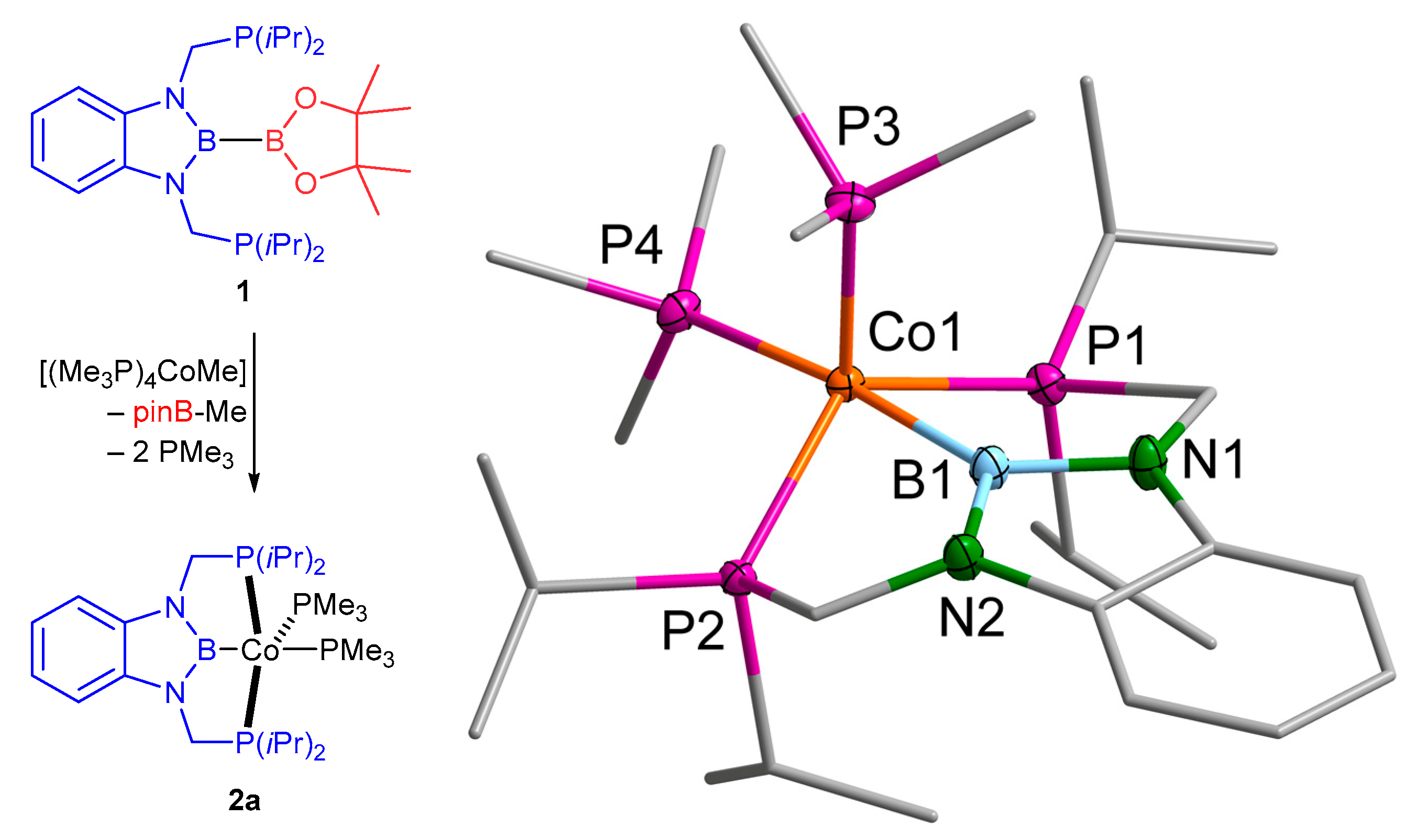
The reaction of 1 with [(Me3P)4Co–Me] results in the mono boryl complex [(d(CH2P(iPr)2)abB)Co–(PMe3)2] (2a) (Scheme 2), presumably via an oxidative addition/reductive elimination pathway [19,20,21]. The reaction delivers 2a after 24 h at 50 °C as dark orange crystals in a 66% isolated yield. A single crystal X-ray diffraction study on 2a revealed a five coordinate 18-valence electron Co(I) complex (Scheme 2). The complex 2a crystallises in an achiral non-centrosymmetric space group of the type Pca21 with four molecules in the unit cell (Z = 4, Z’ = 1) (Supplementary Materials) [22].
The coordination environment at the cobalt atom in 2a is best described as distorted trigonal bipyramidal with the boryl ligand and one PMe3 ligand in the apical positions, and the angle between these positions deviates by 7° from linearity. Moreover, the strong σ donor properties of the boryl ligand result in an elongation of the Co1–P4 distance of the apical PMe3 ligand, compared to distance Co1–P3 of the equatorial PMe3 ligand by 0.03 Å.
The equatorial positions are occupied by the two pincer phosphine donors and a second PMe3 ligand, resulting in a sum of angles in the equatorial plane [P1,P2,P3] of 348.14°, whereby the angle P1–Co1–P2, involving the two pincer phosphorus atoms, is slightly larger than the other angles. For the deviation of the sum of angles, from 360° accounts for the significant displacement of Co1 from the [P1,P2,P3] plane by 0.4372(3) Å towards the P4 atom. This distortion of the trigonal bipyramidal coordination environment at the cobalt atom is due to the restraints imposed by the five-ring chelates in 2a. Whilst the solid-state molecular structure of 2a does not exhibit any crystallographic symmetry, it is virtually Cs symmetric, with a mirror plane through the atoms [B1,P3,P4,Co1] (Figure S37).
An analogous unrestraint mono boryl complex [(PMe3)4Co–Bcat] (cat = 1,2-O2C6H4) exhibits a slightly longer B–Co distance of 1.9545(4) Å and a slightly shorter trans-B P–Co distance of 2.1897(1) Å, together with a less pronounced displacement of the cobalt atom from the equatorial ligand plane [21]. The equatorial Co–P distance in [(PMe3)4Co–Bcat], however, is more equally distributed around 2.17 Å. The closely related square planar PBP complex [(d(CH2P(tBu)2)abB)Co–N2], reported to undergo reversible H2 activation by Peters and a co-worker, exhibits similar Co–B and Co–P distances of 1.946(1) Å and 2.1884(4)/2.1901(3) Å and also significant deviation of the P–Co–P angle from linearity of 156.26(1)° as a result of the five-ring chelation [3].
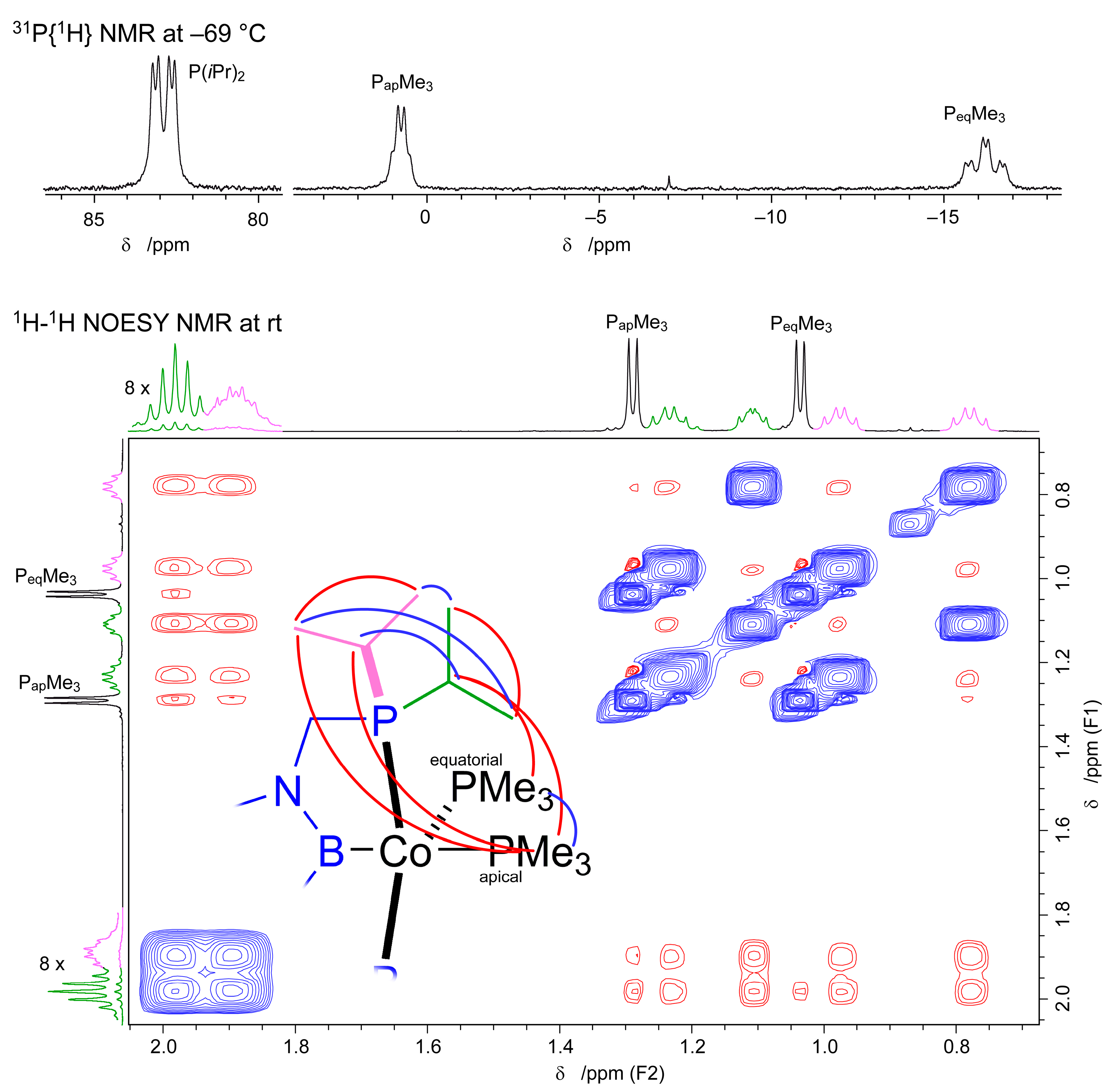
The 31P{1H} NMR spectrum of 2a comprises three distinct signals, two signals of the two distinct PMe3 ligands, one in the apical—trans boryl—position around 0 ppm and the second around –16 ppm for the equatorial PMe3 ligand. The third signal around 83 ppm is assigned to the two equivalent PBP pincer ligand P(iPr)2 groups (Figure 1(top) and Figure S4). Whilst these signals do not exhibit any fine structure at room temperature (Figure S4), at lower temperatures, the signals split in a doublet of doublets at 82.9 ppm (–69 °C) for the P(iPr)2 groups, an apparent broadened quartet at 0.7 ppm (–69 °C) for the apical PMe3 ligand and a triplet of doublets at –16.2 ppm (–69 °C) for the equatorial PMe3 ligand (Figure 1(top) and Figure S4). This is in agreement with the mutual couplings within an A2MN spin system. This agrees with a conformation of the complex in solution similar to the one found in the solid state.
However, the temperature-dependent broadening is indicative of dynamic processes present in solution. A 1H-1H NOESY NMR spectrum at room temperature gives a fitting picture. Distinct NOE contacts between the PMe3 signals and the methine CHMe2 signals allow for the assignment of the PMe3 ligands to the apical and equatorial positions, respectively. Exchange signals are observed between the two PMe3 ligands, but also between pairs of methyl groups of the two distinct isopropyl moieties and the methine protons of these groups (Figure 1 (bottom)). This is fundamentally in agreement with two possible exchange mechanisms: (i) via the dissociation of a PMe3 ligand with a transient four coordinate 16-electron complex [(d(CH2P(iPr)2)abB)Co–PMe3] and the re-association of a PMe3 ligand; (ii) a concerted mechanism exchanging the PMe3 ligand positions via a (distorted) square pyramidal intermediate is feasible. Note also that the NMR data do not suggest any appreciable dissociation of PMe3 from 2a, contrary to the heavier rhodium homolog 2b (vide infra).
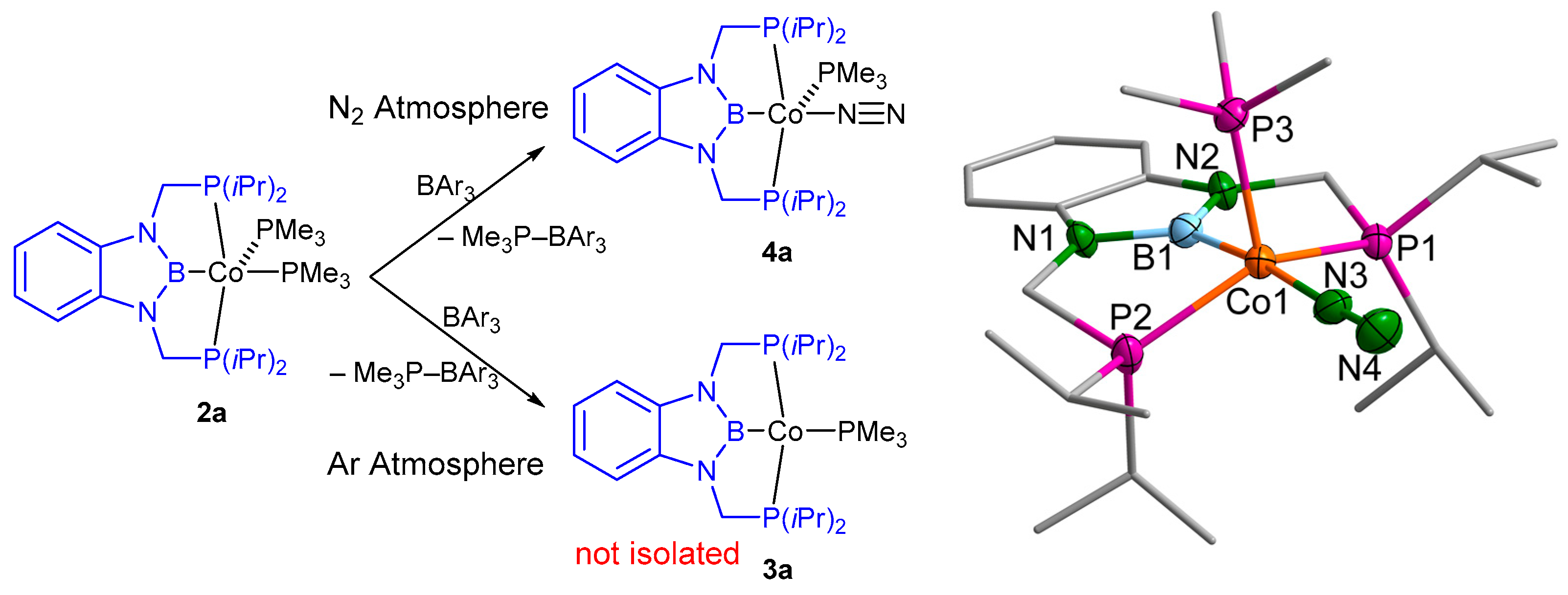
The reaction of 2a with an equimolar amount of BAr3 as a Lewis acid should lead to abstraction of a PMe3 ligand and, after reorganisation, to the complex [(d(CH2P(iPr)2)abB)Co–PMe3] (3a). However, whilst one PMe3 ligand can indeed be abstracted by BPh3, the complex 3a is not isolated. Instead, in a dinitrogen atmosphere, its dinitrogen adduct [(d(CH2P(iPr)2)abB)Co–(N2)(PMe3)] (4a) crystallises in minute amounts after several days at –40° C (Scheme 3).
The N2 complex 4a crystallises in a space group of the type P21/c with two independent molecules in the asymmetric unit (Z = 8, Z’ = 2) (Supplementary Materials) [22]. Both molecules exhibit only a marginal geometric difference, and only one is discussed exemplarily (Figure S40).
As for 2a, the coordination geometry of 4a is best described as distorted trigonal bipyramidal with the boryl ligand and the N2 ligand in the apical positions. The B–Co distance remains virtually unchanged by this substitution of the trans boryl ligand and is also identical to the distance found in the closely related four-coordinate PBP complex [(d(CH2P(tBu)2)abB)Co–N2] reported by Peters et al. of 1.946(1) Å [3]. This indicates again that the B–Co distance is largely determined by the geometrical restraints of the five-ring chelates (vide infra). The close to linear B–Co–N, the angle deviates only by less than 7° from the value found in 2a and in Peter’s N2 complex [3]. The equatorial ligands experience more substantial changes, although their sum of angles around the cobalt atom increases only slightly by 2° to 350.14°, and consistently, the deviation of the cobalt atom from the [P1,P2,P3] plane decreases by 0.05 Å. The angle between the pincer P atoms deviates significantly by 9°; hence, 4a is more distorted from an ideal trigonal bipyramidal geometry towards a square pyramidal arrangement than 2a. However, the reduced steric demand of the ligand in the apical position trans to the boryl ligand in 4a as compared to 2a leads to a relaxation of the B1–Co1–P3 angle by about 7°. The N–N distance in the N2 ligand in 4a compares well with the distance of 1.119(2) Å found in Peter’s N2 complex; the N–Co distance, however, is in 4a slightly—by 0.035 Å—enlarged [3].
As we failed to isolate 4a in any appreciable amounts, we resorted to its spectroscopic in situ characterisation (Figure 2). Performing the reaction of 2a with B(C6F5)3 in toluene and monitoring this reaction via IR spectroscopy gives clear evidence of the immediate formation of an N2 complex, based on the appearance of a strong IR band at 2061 cm−1 if the reaction is conducted under an N2 atmosphere, whereas only a minute signal is observed under an argon atmosphere, presumably due to the presence of adventitious N2 (Scheme 3). This compares well to the N≡N stretching frequency of 2013 cm−1 reported for the related complex [(d(CH2P(tBu)2)abB)Co–N2] (vide supra) by Peters et al. [3].
Following the reaction of 2a with B(C6F5)3 under an N2 atmosphere via 31P and 1111B NMR spectroscopy (Figure 2) gives a consistent picture: upon addition of the Lewis acid, the chemical shifts change from those of 2a (Figure 2). Whilst the 11B{1H} NMR signal shifts only by about 3 ppm, it gives evidence for the presence of the PBP boryl ligand. The changes in the 31P{1H} NMR spectrum are more substantial. The two 31P NMR signals of the PMe3 ligands in 2a change to a broad signal at −13 ppm and a second comparably narrow signal at −7 ppm without an appreciable fine structure. Upon cooling, however, the latter signal broadens, and its intensity reduces, whist the former signal changes into a well-developed triplet (−11.4 ppm, 2JPP = 73 Hz) at −80 °C (Figure 2). The latter triplet corresponds to the doublet at higher chemical shifts (94.6 ppm, 2JPP = 73 Hz). This is readily explained by the abstraction of one PMe3 ligand to give the Lewis acid base adduct Me3P–B(C6F5)3 (δ31P = −6.1 ppm, δ11B = −14.7 ppm in CD2Cl2) and a PBP pincer cobalt complex bearing only one additional PMe3 ligand Me3P–B(C6F5)3 is only sparingly soluble and to a large extend removed prior to the measurement. The remaining dissolved adduct, however, precipitates upon cooling resulting in a reduced 31P NMR signal at lower temperatures. The chemical shift of −11.4 ppm and the P–P coupling constant of around 80 Hz suggest that this PMe3 ligand occupies an equatorial position in a trigonal bipyramidal complex, as it resembles the chemical shift, but in particular, the higher Peq–PPBP coupling constant found in 2a. In other words, the complex that is quantitatively formed is not the four-coordinate complex 3a but a five coordinate complex with a single PMe3 ligand in an equatorial position—the nitrogen complex 4a.
Gas-phase DFT computations on the thermodynamics of the complexes 3a and 4a and their heavier homologues (vide infra) as central atoms indeed show that for cobalt as the central atom, the formation of a five-coordinate N2 complex of the type [(d(CH2P(iPr)2)abB)M–(N2)(PMe3)] is strongly favoured over the four coordinate complex [(d(CH2P(iPr)2)abB)M–(PMe3)] by ΔG298 = −22.4 kJ mol−1E0 = −70.6 kJ mol−1), despite the entropic penalty occurring from the coordination of gaseous N2. However, for the rhodium and iridium analogue, the coordination of an N2 ligand to the latter four-coordinate complex is—in agreement with our observations (vide infra)—disfavoured by ΔG298 = 51.3 kJ mol−1E0 = 8.2 kJmol−1) for rhodium and ΔG298 = 51.4 kJ mol−1E0 = 8.4 kJ mol−1) for iridium (Supplementary Materials) [22]. The computed N≡N stretching frequency in 4a of 2170 cm−1 is by about 100 cm−1 off the experimental values, but within the expected range considering the harmonic nature of the computation and other approximations [22].
Due to an initial computation of the force constant between Co and N2, the bonding in 4a is quite strong (Co–N: 2.33 N cm−1), whilst the trans-B Co–P bond in 2a shows the expected kinetic lability (Co–P: 1.36 N cm−1) of a spectator ligand. More importantly, the electronic coupling in 4a between the N–N bond and the Co–N coordination is pronounced (Co–N/N–N coupling force constant: −0.02 cm N−1) and synergistic (negative sign), pointing to an effective back donation [23]. And indeed, the experimental N2 IR wavenumber of 2061 cm−1 is in line with a modest activation relative to free N2 (~2330 cm−1). Finally, the Co–B bond trans to the N2 ligand seems to be very strong (Co–B: 2.28 N cm−1), reducing the flexibility to access different coordination geometries [24].

2.2. Rhodium Complexes

The reaction of 1 with [(Me3P)3Rh–Cl] in the presence of KOtBu leads to the formation of a rhodium(I) boryl complex (Scheme 4). It may be speculated that the reaction proceeds via an intermediate rhodium alkoxido complex as discussed for the formation of the related complex [(dmabB)Rh(PMe3)3] (dmab = 1,2-(NMe)2C6H4) [20]. However, the reaction of 1 with [(Me3P)3Rh–Cl] in the presence of KOtBu leads to the formation of an equilibrium mixture of the square planar complex [(d(CH2P(iPr)2)abB)Rh–PMe3] (3b) and the five coordinate complex [(d(CH2P(iPr)2)abB)Rh(PMe3)2] (2b) (Scheme 4), whilst in the absence of KOtBu, no reaction is observed (Supplementary Materials) [22] (Figures S12 and S13). After recrystallisation from diethyl ether, the four coordinate complex 3b is obtained as bright orange crystals at a 70% yield, whereas crystallisation from n-pentane in the presence of an excess PMe3 leads to the isolation of the five-coordinate complex 2b as crystalline material at a 43% yield. The spontaneous dissociation of one PMe3 ligand from 2b to give 3b is not contradicting gas-phase DFT computational data (Table S10), suggesting an endothermic (15 kJ mol−1) dissociation from 2b to 3b + PMe3, but overall, an entropy driven exergonic process (−47 kJ mol−1) (Supplementary Materials) [22].
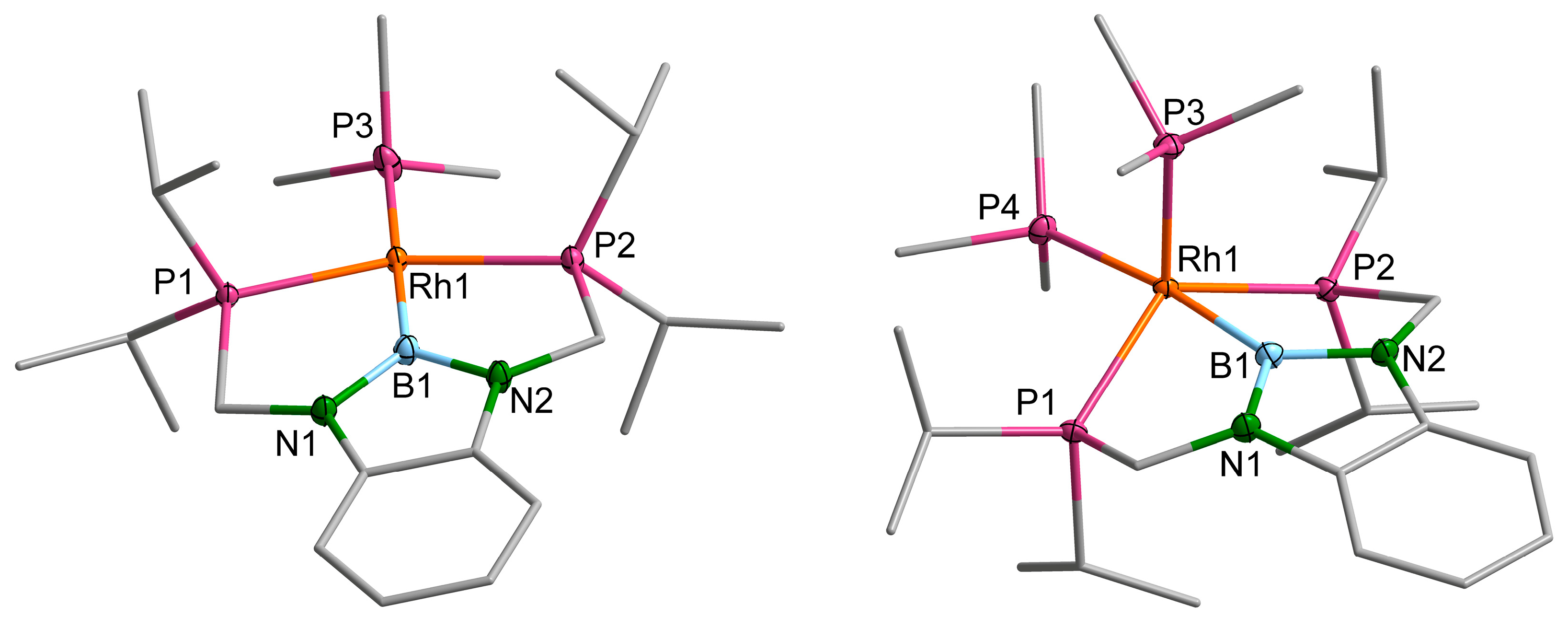
Both complexes 2b and 3b crystallise in monoclinic space groups of the type P21/n and P21/c, respectively, and contain one complex molecule in the asymmetric unit (Z = 4, Z’ = 1) (Supplementary Materials) [22]. The molecular structure of complex 2b is analogous to that of the cobalt homologue 2a (Figure S38). The rhodium ion is distorted trigonal bipyramidaly coordinated by the boryl pincer ligand and one PMe3 ligand in the apical positions (Figure 3, right). The sum of angles in the equatorial plane [P1,P2,P3] comprising the pincer phosphorus atoms and one PMe3 ligand is with 347° only insignificantly smaller than in 2a, whereby the angle P1–Co1–P2, involving the pincer phosphorus atoms, is by about 2° larger than in 2a. The displacement of Rh1 from the [P1,P2,P3] plane is by 0.05 Å larger than in 2a, an effect of the increased radius of the rhodium ion within the restraining pincer coordination environment.
Complex 3b is best described as a distorted square planar complex with a nearly linear B1–Rh1–P3 angle and a significantly (by 27°), from linearity, deviating P1–Rh1–P2 angle. However, this angle is significantly closer to linearity than the respective angle in the five-coordinate complex 2b (Figure 3, left). The change in the Rh···P/B distances between 2a and 3b is comparably small, despite the change in the coordination number. Most pronounced is a decrease in the pincer phosphorus atoms to rhodium distances in comparison to 2b by about 0.06 Å, which may be attributed to the less strained ligand conformation in the more planar 3b.
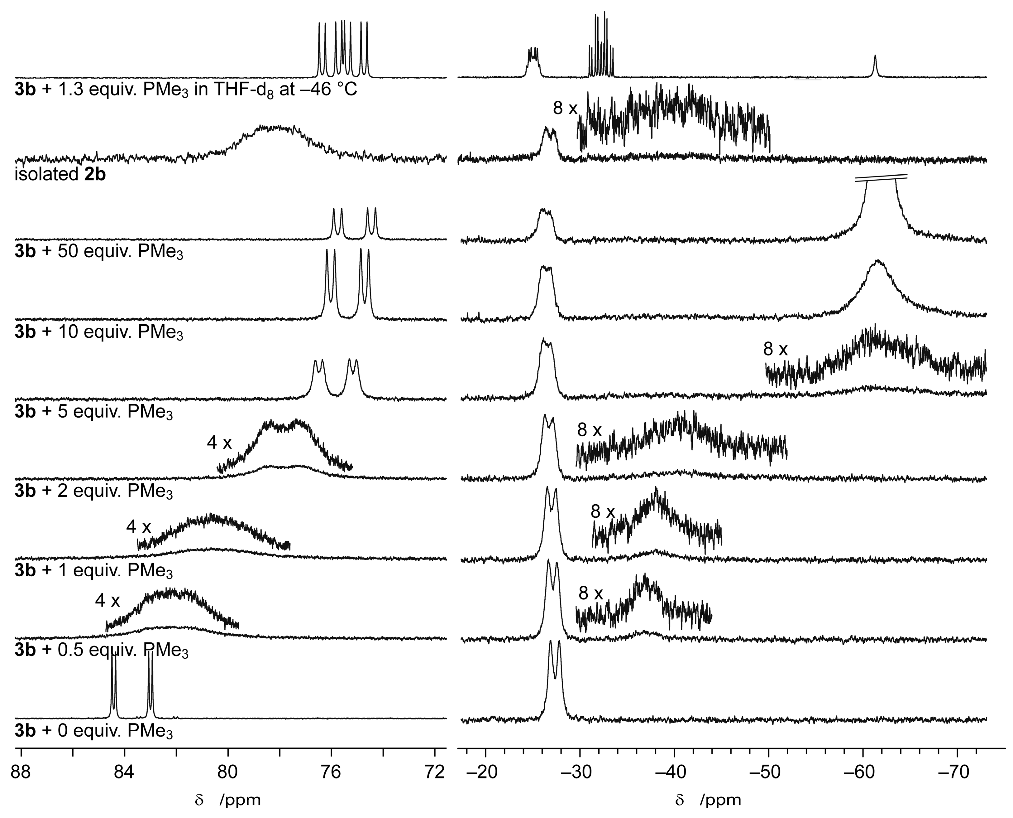
The equilibrium between 2b and 3b, as a fundamental aspect of their coordination chemistry, was further studied via NMR spectroscopy. NMR titration of 3b with increasing amounts of PMe3 shows a highly dynamic behaviour in the 31P{1H} NMR spectra at room temperature (Figure 4 and Figures S17–S19). Only one set of signals of the PBP ligand and the trans-B PMe3 ligand is observed, respectively. Whilst the 31P NMR signal of the PBP ligand changes appreciably from 84 ppm to 75 ppm with increasing amounts of PMe3 added, the signal of the trans-B PMe3 ligand, in 2b, is only marginally influenced (−27.3 to −26.4 ppm). An additional signal is observed shifting from −37 ppm at low amounts of PMe3 to −62 ppm after the addition of an excess of PMe3. This is readily explained by a rapid exchange among 3b, 2b and free PMe3 on the NMR time scale and consequently, the observation of an averaged chemical shift of the exchanging PMe3 moieties throughout this process. In agreement with that, the spectrum observed for isolated 2b is very virtually identical to the spectrum of 3b after the addition of an equimolar amount of PMe3.
At low temperatures, however, the exchange among 3b, 2b and free PMe3 becomes slow on the NMR timescale, and well-resolved signals for 2b and free PMe3 are observed (Figure 4,Figures S14 and S15). The 31P{1H} NMR spectrum of 2b itself at −46 °C comprises three signals (A, M and N) of an A2MNX spin system with the expected 31P-31P and 31P-103Rh couplings (Figure S16, Table S3). Following the reaction of 3b with different amounts of PMe3 via UV-Vis spectroscopy corroborates the rapid equilibrium between 3b and 2b being rather on the side of 3b and free PMe3 (Figures S20 and S21).
In conclusion, it may be stated that the five-coordinate trigonal bipyramidal complex 2b, in contrast to the Co analogue, easily dissociates one PMe3 ligand to give the distorted square planar complex 3b. The virtual indifference in the 31P NMR chemical shift (and line shape) of the apparently not-exchanging trans-B PMe3 ligand around 27 ppm suggests that this exchange does not affect this ligand but involves only the equatorial PMe3 ligand.

2.3. Iridium Complexes

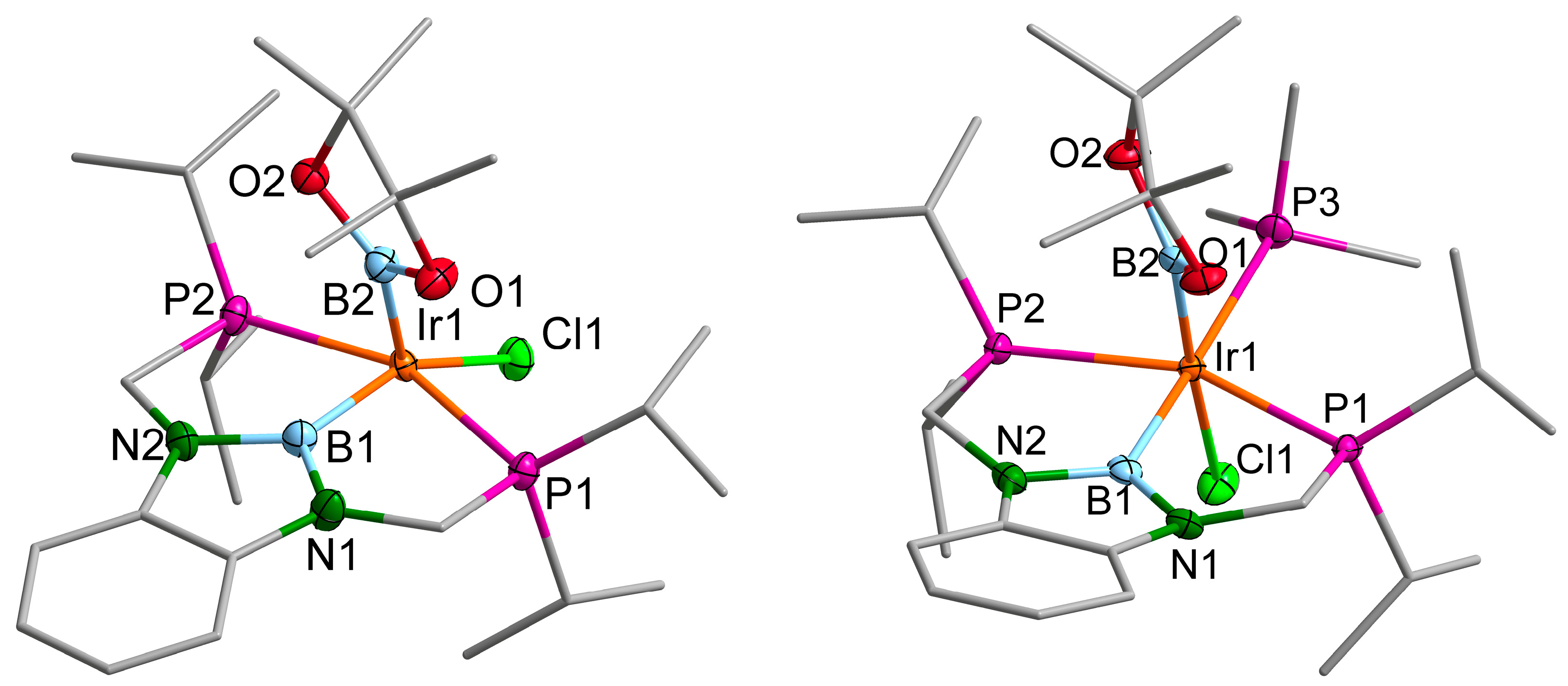
Whilst for the formation of the cobalt and rhodium PBP pincer complexes 2a and 2b/3b, it may be arguable whether activation of the diborane precursor 1 proceeds via a σ bond metathesis or an oxidative addition/reductive elimination pathway, the reaction of 1 with the iridium(I) complex [Ir(cod)Cl]2 (cod = 1,5-cyclooctadien) to give the bis-boryl complex [(d(CH2P(iPr)2)abB)Ir(Bpin)(Cl)] (5c) is obviously an oxidative addition reaction (Scheme 5). This five-coordinate complex reacts with excess PMe3 to give the six-coordinate complex [(d(CH2P(iPr)2)abB)Ir(Bpin)(PMe3)(Cl)] (6c).
Both complexes 5c and 6c crystallise in monoclinic space groups of the type P21/c. The solid-state structure of 5c contains one complex molecule in the asymmetric unit (Z = 4, Z’ = 1), whereas 6c comprises two independent molecules in the asymmetric unit (Z = 8, Z’ = 2). The Bpin moiety in 5c shows some positional disorder that is neglected in the further discussion; for 6c, however, one of the independent molecules shows severe disorder and is not considered for further geometrical analysis (Supplementary Materials) [22].
The trigonal bipyramidal geometry of 5c may be considered typical for a five-coordinate Ir bis-boryl complex with phosphine ligands (Figure 5). All five structurally characterised complexes of this type adopt a trigonal bipyramidal geometry with the two phosphine ligands in the axial positions (P–Ir–P angle 157–172°, for PXP pincer ligands P–Ir–P angle 157–162°) and small B···B distances and B–Ir–B angles in the ranges of 2.22–2.41 Å and 65.8–76.7°, respectively [25,26,27,28,29].
In 6c, the PMe3 ligand adopts a position trans to the PBP pincer boryl ligand, whilst the chlorido ligand occupies a position trans to the Bpin ligand (Figure 5). As a result, 6c may best be described as a strongly distorted octahedral complex with the Bpin and chlorido ligand in the axial positions. Structurally, the extension of the coordination sphere to the distorted octahedral complex 6c is accompanied by some ligand reorganisation. The P1–Ir–P2 angle reduces upon coordination by about 3° to deviate more from linearity, whereas the B–Ir–B angle deviates in 6c by about 6° less from 90° than in 5c (in accordance with the B···B distance increasing from 5c to 6c by 0.25 Å). The d(CH2P(iPr)2)abB ligand backbone in 6c (mean plane [B1,N1,N2,C6H4]) includes an angle of 24.8(8)° with the equatorial plane of the complex (mean plane [P1,P2,P3,B1,Ir1]), 20° more than in the five-coordinate 5c. This is a result of the increased steric encumbrance induced by the extension of the coordination sphere in 6c. The B–Ir distance increases slightly upon PMe3 coordination in 6c because of the presence of trans ligands. This is more significant for B1, which is trans to the stronger trans influencing ligand PMe3 as opposed to the chlorido ligand for B2. The Cl–Ir distance increases accordingly, whereas the P1/P2–Ir1 distances remain virtually unaffected. Again, because of the strong trans influence of the boryl ligand, the P–Ir distance of the PMe3 ligand is longer than those of the pincer phosphine atoms by about 0.06 Å [30].
The solution-state 1H, 31P and 13C NMR spectroscopic data for 5c and 6c fulfil the expectations and can readily be explained by the solid-state structures. Surprising, however, are the 11B NMR chemical shifts. For both complexes, two very distinct, somewhat broadened singlets at chemical shifts of 39.7 ppm (Δw½ = 340 Hz) and 19.9 ppm (Δw½ = 330 Hz) for 5c and of 48.8 ppm (Δw½ = 460 Hz) and 26.6 ppm (Δw½ = 450 Hz) for 6c are observed in THF-d8 at room temperature. This chemical shift range is somewhat different from the 11B NMR data for the reported Ir(III) boryl in a range of 29–35 ppm for five-coordinate and of 30–43 ppm for six-coordinate complexes, respectively [1,2,7,25,26,28,31].
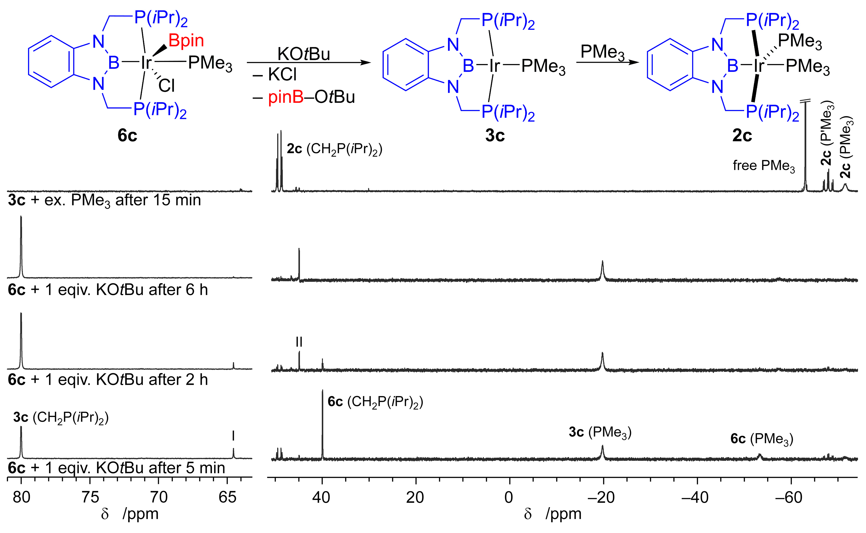
Whilst complex 6c is stable under inert conditions, it reacts readily with an equimolar amount of KOtBu to give the Ir(I) PBP pincer complex 3c (Figure 6, Scheme 5). Monitoring this reaction via in situ NMR spectroscopy (Figure 6 and Figure S34–S36) shows an essentially clean conversion to 3c, as indicated by its characteristic signals around 80 ppm (doublet, JP–P = 5 Hz) for the pincer phosphorus atoms and a broadened singlet for the PMe3 ligand around –20 ppm (Figure S35 Supplementary Materials) [22]. Only minor amounts of a so far unidentified side product with a 31P NMR signal at 45 ppm (II) are observed. However, upon closer evaluation, two transient species are observed during this reaction. At one hand side, the five-coordinate complex 2c (vide infra) is formed in small amounts in the beginning but is later on fully consumed (Figure 6). On the other side, a species with a 31P{1H} NMR singlet signal at 64.5 ppm (I) is observed. In agreement with this, the 11B NMR data suggest the presence of a transient boryl intermediate at 40 ppm, whereas 3c itself exhibits a moderately broad 11B{1H} NMR signal around 56 ppm (Figure S36 Supplementary Materials) [22,32]. It may be assumed that the conversion of 6c to 3c proceeds via the initial coordination of a OtBu ligand followed by (possibly after some reorganisation) the reductive elimination to an Ir(I) PBP complex, 3c or a closely related species. The intermediate presence of 2c may be explained by the intermediate liberation of PMe3 and its transient addition to 3c during this process. An in situ 31P{1H} NMR spectrum of a mixture of 3c and excess PMe3 corroborates the facile formation of 2c (Figure 6, top). Moreover, it must be emphasised that the system 2c/3c/PMe3 exhibits much less dynamic behaviour than the homologous rhodium system 2b/3b/PMe3 (vide supra). Contrary to the latter, even in the presence of excess PMe3 at room temperature, a well-resolved, 31P{1H} NMR spectrum (A2MN spin system) with a narrow linewidth is observed, indicating only comparably slow exchange of a coordinated PMe3 ligand with free PMe3. Contrary to 2b, distinct signals for both PMe3 ligands are observable for 2c at room temperature in the presence of free PMe3 (Figure 6, top). One of these signals (around −70 ppm), however, sharpens upon only moderate cooling to an apparent quartet (Figure S27 Supplementary Materials) [22]. In agreement with that, in situ UV-Vis spectroscopic data of 3c in the presence of different amounts of PMe3 indicate a rapid equilibration, rather on the side of 2c (Figures S30 and S31). The 11B NMR shift of 2c of around 55 ppm is virtually unaffected by the change in the coordination number.
In conclusion, it may be stated that the five-coordinate trigonal bipyramidal complex 2c, similarly to the cobalt analogue 2a and opposed to the rhodium homologue, shows only little dynamic behaviour in solution and does not readily dissociate a PMe3 ligand to give the distorted square planar complex 3c. However, gas-phase DFT computational data suggest similar thermodynamic data for the dissociation of PMe3 from 2cE0 = 16 kJ mol−1, ΔG298 = −48 kJ mol−1) as for the rhodium analogue 2b (Supplementary Materials) [22].
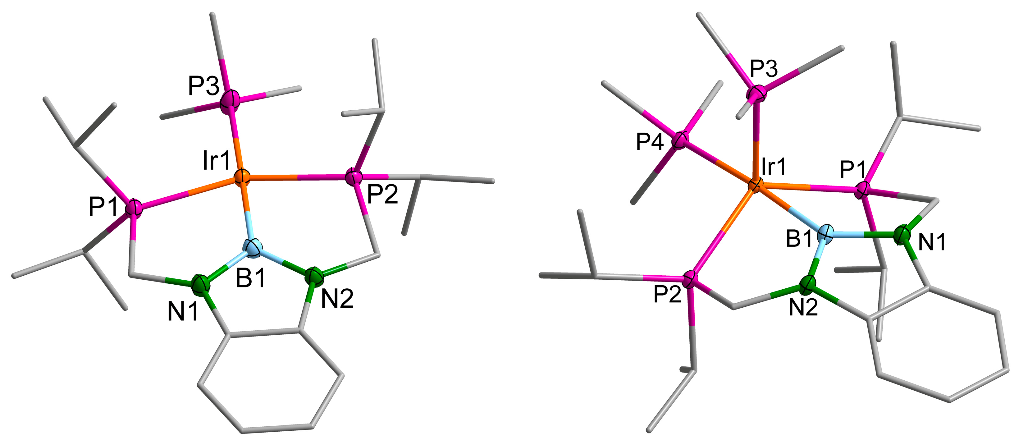
The complexes 2c and 3c crystallise isostructurally with the homologous rhodium complexes in monoclinic space groups of the types P21/n and P21/c, respectively (Z = 4, Z’ = 1) (Supplementary Materials) [22]. As a consequence, the molecular structure of 2c (Figure 7, right) differs only marginally form the structure of the lighter homologue 2b and from the cobalt homologue 2a (Figure S38).
The sum of angles in the equatorial plane [P1,P2,P3] of the distorted trigonal bipyramidal complex 2c is, with 347.76°, only insignificantly different from that in 2a and 2b. The angle P1–Ir1–P2, involving the pincer phosphorus atoms, is larger than that in 2a by about 2° and, hence, virtually identical to that in 2b. The displacement of Ir1 from the [P1,P2,P3] plane is in the middle between the values for two lighter homologues, by 0.03 Å larger than in 2a and by 0.02 Å smaller than in 2b. Generally, the M–P distances, however, increase from 2a to 2b and 2c by about 0.12 Å, most significantly between the cobalt and the rhodium complex.
Overall, the PBP pincer ligand shows, within the series 2a, 2b 2c, a high ability to coordinate different metal ions. The high flexibility of this ligand is also illustrated by a comparison of the five-coordinate complexes 5c and 2c. For both complexes, a trigonal bipyramidal geometry is observed; however, whilst in 2c, the phosphorus atoms of the PBP pincer ligand occupy two equatorial positions and the boryl moiety is bound in an axial position, in 5c, two phosphorus atoms coordinate in the two axial positions and the boron atom in an equatorial position. This is illustrated by P–M–P angles included by the pincer phosphorus atoms decreasing by 30° from 5c to 2c.
The solid-state structure of the distorted square planar complex 3c is again very similar to that of its rhodium homologue 3b (Figure S39) with a nearly linear B1–Ir1–P3 angle and a P1–Ir1–P2 angle of 152.95(2)° deviating significantly from linearity. Noteworthy is the Ir1–B1 distance in 3c that is slightly (0.01 Å) longer, whereas the pincer P–M distances are identical, and the trans-B P–M distance is slightly shorter (0.02 Å) than the respective distance in the rhodium homologue 3b.

3. Discussion

A series of either group 9 PBP diaminoboryl pincer complexes was synthesised using the unsymmetrical diborane(4) 1 as a PBP pincer precursor and fully characterised. In an extension of our earlier work [15], this exemplifies again the versatility of this compound as a PBP pincer ligand precursor. The CoI and RhI complexes [(d(CH2P(iPr)2)abB)Co–(PMe3)2] (2a) and [(d(CH2P(iPr)2)abB)Rh–(PMe3)n] (2b (n = 2), 3b (n = 1)), respectively, were obtained in a one-step reaction from the respective CoI and RhI precursors (Scheme 2 and Scheme 4). Whilst an oxidative addition/reductive elimination pathway is, for both reactions, feasible, in the rhodium case, a σ bond metathesis pathway may be feasible, considering results based on a related non-pincer ligand [20]. The heavier IrI homologue, however, was obtained via the isolated intermediate IrIII complexes [(d(CH2P(iPr)2)abB)Ir(Bpin)(Cl)] (5c) and [(d(CH2P(iPr)2)abB)Ir(Bpin)(PMe3)(Cl)] (6c). Complex 5c is formed upon an oxidative addition reaction of the diborane(4) 1 with [Ir(cod)Cl]2 (cod = 1,5-cyclooctadien) and subsequently reacts via PMe3 addition to 6c. The coordination chemistry of the resulting homologous complexes [(d(CH2P(iPr)2)abB)M(PMe3)2] (2ac) and [(d(CH2P(iPr)2)abB)M–PMe3] (3b,c) was studied structurally in the solid state, as well as spectroscopically in solution. However, for M = Rh and Ir, both complexes are structurally very similar but differ in the dynamic behaviour and the relative accessibility of the four (3b,c) vs. the five (2b,c) coordinated complexes. For Co only the five-coordinate complex 2a is accessible, whereas Lewis acid-promoted PMe3 abstraction under a dinitrogen atmosphere leads to the formation of the surprisingly stable N2 complex [(d(CH2P(iPr)2)abB)Co–(N2)(PMe3)] (4a).
Having, with the unsymmetrical diborane(4) [(d(CH2P(iPr)2)abB)–Bpin] (1), a well accessible and versatile PBP ligand precursor that is capable of oxidative addition (PtII, CoI, RhI (possibly), IrI) and σ bond metathesis (CuI and possibly RhI) reactions [15,20] will stimulate the further development of PBP pincer ligands. In conclusion, PBP diaminoboryl pincer ligands are a ligand class with remarkable ligand properties with respect to their high σ donor strength and weak π acceptor properties—leading to a strong trans effect and influence [30]—that provide stability for the inherently reactive B–M bond due to their pincer framework. Furthermore, PBP pincer ligands are tuneable based on the backbone and P atoms substituents, making them interesting for a broad range of applications from catalysis to the stabilisation of reactive intermediates.

4. Materials and Methods

4.1. General Considerations

pinB–B(d(CH2P(iPr)2)ab) (1), [(Me3P)4CoMe], [(Me3P)3RhCl] and [(cod)IrCl]2 were prepared according to literature procedures [15,16,17,18,33]. All other compounds were commercially available and were used as received; their purity and identity were checked using appropriate spectroscopic methods. Unless otherwise noted, all solvents were dried using an MBraun solvent purification system, deoxygenated using the freeze-pump-thaw method and stored under purified nitrogen. Unless noted otherwise, all manipulations were performed using standard Schlenk techniques under an atmosphere of purified nitrogen or in a nitrogen-filled glove box (MBraun). NMR spectra were recorded on Bruker Avance II 300, Avance III HD 300 and Avance III 400 spectrometers. NMR tubes equipped with screw caps (WILMAD) were used, and the solvents were dried over potassium/benzophenone and degassed. Chemical shifts (δ) are given in ppm, using the (residual) resonance signal of the solvents for calibration (C6D6: 1H NMR: 7.16 ppm, 13C NMR: 128.06 ppm; PhMe-d8: 1H NMR: 2.08 ppm, 13C NMR: 20.43 ppm; THF-d8: 1H NMR: 1.72 ppm, 13C NMR: 25.31 ppm) [34]. 11B and 31P NMR chemical shifts are reported relative to pseudo external BF3·Et2O and 85% H3PO4(aq), respectively. 13C{1H}, 11B{1H} and 31P{1H} NMR spectra were recorded employing composite pulse 1H decoupling. 11B NMR spectra were processed applying a back linear prediction, in order to suppress the broad background signal due to the boron in the NMR tube and instrument. A Lorentz-type window function (LB = 10 Hz) was used, and the spectra were carefully evaluated to ensure that no genuinely broad signals of the sample were suppressed. Simulations were conducted with the TOPSPIN/DAISY program package (Bruker). Melting points were determined in flame-sealed capillaries under nitrogen using a Büchi 535 apparatus and are not corrected. Elemental analyses were performed at the Institut für Anorganische und Analytische Chemie of the Technische Universität Braunschweig using an Elementar vario MICRO cube instrument. A Bruker Vertex 70 spectrometer was used for recording IR spectra. The IR spectra were recorded in PhMe solutions in a cuvette of an approximately 1 mm optical path length equipped with NaCl windows.
X-ray Structure Determination. The single crystals were transferred into inert perfluoroether oil inside a nitrogen-filled glovebox and, outside the glovebox, rapidly mounted on top of a CryoLoop (Hampton Research) and placed on the diffractometer in the cold nitrogen gas stream of a Cryostream 800 cooling system (Oxford Cryosystems) [35]. The data were collected on a Rigaku Oxford Diffraction Synergy-S instrument using either mirror-focused MoKα or CuKα radiation (Rigaku PhotonJet microfocus sources). The reflections were indexed and integrated, and appropriate absorption corrections were applied as implemented in the CrysAlisPro software package [36]. The structures were solved employing the program SHELXT and refined anisotropically for all non-hydrogen atoms via full-matrix least squares based on all F2 values using SHELXL software [37,38,39]. Generally, hydrogen atoms were refined employing a riding model; methyl groups were treated as rigid bodies and were allowed to rotate about the E–CH3 bond. During refinement and analysis of the crystallographic data, the programs OLEX2, PLATON, Mercury and Diamond were used [40,41,42,43]. Unless noted otherwise non-C,H atoms are depicted as ellipsoids at the 50% probability level, whereas the carbon atom framework is depicted as a stick model (grey), and hydrogen atoms are omitted for clarity. Adapted numbering schemes may be used to improve the readability. Further crystallographic details can be found in the Supplementary Materials available.

4.2. Experimental Procedures and Analysis Data

4.2.1. [(d(CH2P(iPr)2)abB)Co(PMe3)2] (2a)

In a Schlenk-flask, d(CH2P(iPr)2)abB–Bpin (1) (100 mg, 0.198 mmol, 1 equiv.) and [(Me3P)4CoMe] (75 mg, 0.198 mmol, 1 equiv.) were dissolved in toluene (50 mL) and stirred for 24 h at 50 °C whilst a reduced pressure was applied for about 50% of the time (the pressure was normalised overnight). The solvent was completely removed in vacuo and the brown residue was dissolved in n-pentane and recrystallised at −40 °C. The resulting dark orange crystals were washed with cold n-pentane (1 mL) and dried in vacuo (77 mg, 0.131 mmol, 66%).
1H NMR (PhMe-d8, 400.4 MHz, rt) δ = 6.96–6.91 (m, 2 H, 3-HCAr), 6.74–6.79 (m, 2 H, 2-HCAr), 3.50 (d, 2JH-H = 11.0 Hz, 2 H, CHH’), 3.47 (d, 2JH-H = 11.0 Hz, 2JH-P = 4 Hz, 2 H, CHH’), 1.98 (app. sept., 3JH-H = 7.6 Hz, 3JH-H = 7.0 Hz, 2 H, CH(CH3)2), 1.89 (m, 3JH-H = 7.0 Hz, 3JH-H = 7.4 Hz, JH-P = 2.5, 4.0 Hz, 2 H, C’H(CH3)2), 1.29 (d, 2JH-P = 5.0 Hz, 9 H, Pap(CH3)3), 1.23 (app. q, 3JH-H = 7.6 Hz, JH-P = 6.8, 6.0 Hz, 6 H, CH(CH3)(C’H3)), 1.11 (m, 3JH-H = 7.0 Hz, JH-P = 5.7, 3.6 Hz, 6 H, CH(CH3)(C’H3)), 1.04 (d, 2JH-P = 4.8 Hz, 9 H, Peq(CH3)3), 0.98 (app. q, 3JH-H = 7.0 Hz, JH-P = 6.9, 6.5 Hz, 6 H, C’H(CH3)(C’H3)), 0.78 (app. q, 3JH-H = 7.4 Hz, JH-P = 6.5, 5.7 Hz, 6 H, C’H(CH3)(C’H3)). 13C{1H} NMR (PhMe-d8, 100.7 MHz, rt) δ = 140.6 (app. t, JC-P = 6 Hz, 1-CAr), 117.2 (s, 3-HCAr), 106.5 (s, 2-HCAr), 44.5 (m, CHH’), 32.0 (app. dt, JC-P = 19, 3 Hz, CH(CH3)2), 29.1 (app. t, JC-P = 5 Hz, C’H(CH3)2), 27.8 (m, Pap(CH3)3), 25.7 (app. dq, JC-P = 15, 4 Hz, Peq(CH3)3), 21.9 (s, CH(CH3)(C’H3)), 20.3 (s, CH(CH3)(C’H3)), 19.4 (s, C’H(CH3)(C’H3)), 18.9 (app. t, JC-P = 3 Hz, C’H(CH3)(C’H3)). 31P{1H} NMR (PhMe-d8, 162.1 MHz, rt) δ = 83.0 (br. s, Δw½ = 217 Hz, CH2P(iPr)2), −1.2 (br. s, Δw½ = 132 Hz, P(CH3)3), −17.4 (br. s, Δw½ = 245 Hz, P(CH3)3). 11B{1H} NMR (PhMe-d8, 128.5 MHz, rt) δ 57.0 (br. s, Δw½ = 360 Hz). 1H NMR (PhMe-d8, 400.4 MHz, −69 °C) δ = 7.20–7.14 (m, 2 H, CAr), 6.95–6.89 (m, 2 H, HCAr), 3.51–3.33 (m, 4 H, CHH’), 1.92 (br. s, 2 H, CH(CH3)2), 1.80 (br. app. sept., JH-H = 7 Hz, 2 H, C’H(CH3)2), 1.22 (d, 2JH-P = 5 Hz, 9 H, P(CH3)3), 1.20 (br. s, 6 H, CH(CH3)(C’H3)), 1.06 (d, 2JH-P = 5 Hz, 9 H, P(CH3)3), 1.03 (br. s, 6 H, CH(CH3)(C’H3)), 0.99–0.90 (m, 6 H, C’H(CH3)(C’H3)), 0.75 (br. s, 6 H, C’H(CH3)(C’H3)). 31P{1H} NMR (PhMe-d8, 162.1 MHz, −69 °C) δ = 82.9 (dd, 2JP-P = 80, 30 Hz, CH2P(iPr)2), 0.7 (app. q, 2JP-P = 30, 28 Hz, Pap(CH3)3), −16.2 (td, q, 2JP-P = 80, 28 Hz, Peq(CH3)3). 1H NMR (C6D6, 300.1 MHz, rt) δ = 7.10–7.03 (m, 2 H, HCAr), 6.92–6.85 (m, 2 H, HCAr), 3.50 (m, 4 H, CHH’), 2.06–1.85 (m, 4 H, CH(CH3)2), 1.29 (d, 2JH-P = 5.0 Hz, 9 H, P(CH3)3), 1.28–1.19 (m, 6 H, CH(CH3)(C’H3)), 1.14–1.07 (m, 6 H, CH(CH3)(C’H3)), 1.07 (d, 2JH-P = 4.8 Hz, 9 H, P(CH3)3), 1.03–0.94 (m, 6 H, C’H(CH3)(C’H3)), 0.87–0.76 (m, 6 H, C’H(CH3)(C’H3)). 11B{1H} NMR (C6D6, 96.3 MHz, rt) δ 57.2 (br. s, Δw½ = 460 Hz). 31P{1H} NMR (C6D6, 121.5 MHz, rt) δ 82.9 (br. s, Δw½ = 200 Hz, CH2P(iPr)2), −1.5 (br. s, Δw½ = 135 Hz, P(CH3)3), −17.5 (br. s, Δw½ = 240 Hz, P’(CH3)3). Anal. Calcd. for C26H54BCoN2P4 (2a): C, 53.08; H, 9.25; N, 4.76. Found: C, 52.84; H, 9.27; N, 5.13. m.p.: 160–163 °C.

4.2.2. [(d(CH2P(iPr)2)abB)Co(N2)(PMe3)] (4a)

Single crystals of 4a: In a nitrogen-filled glovebox, 2a (10 mg, 17 µmol, 1 equiv.) and triphenylborane (4.1 mg, 17 µmol, 1 equiv.) were dissolved in C6D6 (0.7 mL). After 3 d at room temperature, the solution was layered with n-pentane. Colourless crystals of Me3P–BPh3 separated. The supernatant solution was decanted, and the solvent was removed in vacuo. The residue was dissolved in toluene (0.5 mL), and the solution was layered with n-pentane and cooled to −40 °C. Colourless crystals formed overnight, from which the supernatant solution was decanted and cooled again to −40 °C. A few orange single crystals of 4a suitable for x-ray diffraction were obtained from this solution. In situ IR characterisation of 4a was as follows: in a nitrogen-filled glovebox, 2a (10 mg, 17 µmol, 1 equiv.) and tris(pentafluorophenyl)borane (8.7 mg, 17 µmol, 1 equiv.) were dissolved in toluene (0.4 mL) and transferred into an IR cuvette. An IR spectrum of this solution was recorded. The reaction under an Ar atmosphere was conducted analogously in an Ar-filled glovebox. In situ NMR characterisation of 4a was performed as follows: in a nitrogen-filled glovebox, 2a (16.1 mg, 27 µmol, 1 equiv.) and tris(pentafluorophenyl)borane (14 mg, 27 µmol, 1 equiv.) were dissolved in toluene-d8 and filtered through a small pad of celite. NMR spectra of this solution were recorded.
1H NMR (PhMe-d8, 400.4 MHz, rt) δ = 6.88 (br. s, 2 H, HCAr), 6.66 (br. s, 2 H, HCAr), 3.50 (br. s, 2 H, CH2), 3.34 (br. s, 2 H, CH2), 2.11 (overlapping with the residual solvent signal, CH(CH3)2), 1.48–0.65 (P(CH3)3) and CH(CH3)2). 31P{1H} NMR (PhMe-d8, 162.1 MHz, rt) δ = 93.5 (br. s, Δw½ = 211 Hz, CH2P(iPr)2), −13.4 (br. s, Co–P(CH3)3). 11B{1H} NMR (PhMe-d8, 128.5 MHz, rt) δ 54.3 (br. s, Δw½ = 630 Hz). 1H NMR (PhMe-d8, 400.4 MHz, −69 °C) δ = 6.77 (br. s, 2 H, HCAr), 3.35 (br. s, 2 H, CH2), 3.13 (br. d, 2 H, CH2), 1.95 (br. s, 4 H, CH(CH3)2), 1.40 (br. s, 6 H, CH(CH3)2), 1.19 (br. s, 6 H, CH(CH3)2), 1.04 (br. s, 15 H, CH(CH3)2) and P(CH3)3), 0.80 (br. s, 6 H, CH(CH3)2). 31P{1H} NMR (PhMe-d8, 162.1 MHz, −80 °C) δ = 94.6 (d, 2JP-P = 74 Hz, CH2P(iPr)2), −11.4 (t, 2JP-P = 74 Hz, Co–P(CH3)3).

4.2.3. [(d(CH2P(iPr)2)abB)Rh(PMe3)2] (2b)

The reaction was performed as described for 3b on a 55 μmol scale (vide infra). After filtration, an excess of PMe3 (30 µL, 22 mg, 0.3 mmol, 5.5 equiv.) was added, and the resulting yellow solution was cooled to −40 °C. After 48 h, bright yellow crystals suitable for X-ray crystallography had formed. The supernatant solution was decanted, and the crystals were dried in vacuo (15 mg, 24 μmol, 43%). NMR spectra of the isolated material show an equilibrium among 2b, 3b and free PMe3 (Figures S17–S19). NMR spectra of 2b were recorded from a solution of 3b (15 mg, 28 μmol) in THF-d8 (0.7 mL) after the addition of PMe3 (3.7 µL, 2.7 mg, 37 μmol, 1.3 equiv.).
1H NMR (THF-d8, 400.4 MHz, −46° C) δ 6.58–6.47 (m, 4 H, 2,3-HCAr), 3.61–3.42 (m, 4 H, CH2), 2.12 (app. sept., J = 6.9 Hz, 2 H, CH(CH3)2), 1.64 (br. s, 2 H, Δw½ = 25 Hz, CH(CH3)2), 1.43 (d, 2JH-P = 4.8 Hz, 9 H, P(CH3)3), 1.34–1.21 (m, 12 H, CH(CH3)2), 1.11 (d, 2JH-P = 4.8 Hz, 9 H, P’(CH3)3), 1.03 (app. q, J = 5.6 Hz, 6 H, CH(CH3)2), 0.94 (d, 2JH-P = 2 Hz, 2.9 H, free P(CH3)3), 0.7 (app. q, J = 7.1 Hz, 6 H, CH(CH3)2). 11B{1H} NMR (THF-d8, 128.5 MHz, rt) δ 55.4 (s, Δw½ = 365 Hz). 31P{1H} NMR (THF-d8, 162.1 MHz, rt) δ 76.6 (br. d, 1JP-Rh = 153 Hz, Δw½ = 150 Hz, CH2P(iPr)2), −26.4 (br. d, 1JP-Rh = 97 Hz, Δw½ = 105 Hz, P(CH3)3), −37 (br. s, Δw½ = 1000 Hz, P(CH3)3), −54 (br. s, Δw½ = 1600 Hz, free P(CH3)3). 31P{1H} NMR (THF-d8, 162.1 MHz, −46 °C) δ 75.5 (ddd, 1JP-Rh = 157 Hz, 2JP-P = 38, 103 Hz, CH2P(iPr)2), −25.1 (app. dq, 1JP-Rh = 105 Hz, 2JP-P = 43, 38 Hz, Pap(CH3)3), −32.3 (dtd, 1JP-Rh = 157 Hz, 2JP-P = 103, 43 Hz, Peq(CH3)3). Anal. Calcd. for C26H54BN2P4Rh (2b): C, 49.39; H, 8.61; N, 4.30. Found: C, 48.91; H, 8.56; N, 4.47.

4.2.4. [(d(CH2P(iPr)2)abB)Rh(PMe3)] (3b)

In a nitrogen-filled glovebox, d(CH2P(iPr)2)abB–Bpin (1) (41 mg, 82 μmol, 1 equiv.) and [Rh(PMe3)3Cl] (30 mg, 82 μmol, 1 equiv.) were combined and dissolved in toluene (10 mL). A solution of KOtBu (9 mg, 82 μmol, 1 equiv.) in THF (2 mL) was added, and the bright orange solution was stirred for 5 min at room temperature. The solvent was removed in vacuo. The residue was extracted with n-pentane (2 × 3.5 mL) and filtered through a pad of celite. The solvent was removed in vacuo. The orange residue was recrystallised from diethyl ether (3 mL) at −40 °C to give bright orange crystals of [(d(CH2P(iPr)2)abB)Rh(PMe3)] (3b) (30 mg, 56 μmol, 70%).
1H NMR (THF-d8, 400.4 MHz, rt) δ 6.72–6.67 (m, 2 H, HCAr), 6.67–6.62 (m, 2 H, HCAr), 3.64 (app. t, J = 2, 2 Hz, 4 H, NCH2P), 2.18 (app. sept., J = 7, 6, 1.5, 1.5 Hz, 4 H, CH(CH3)2), 1.39 (dd, 2JH-P = 4.3, 3JH-Rh = 0.6 Hz, 9 H, P(CH3)3), 1.19 (app. q, J = 7.0, 7.4, 7.4 Hz, 12 H, CH(CH3)2), 1.09 (app. q, J = 6.0, 6.3, 6.3 Hz, 12 H, CH(CH3)2). 13C{1H} NMR (THF-d8, 101.7 MHz, rt) δ 140.7 (app. td, JC-P = 9 Hz, JC-Rh = 1.5 Hz, 1-CAr), 117.3 (s, HCAr), 104.9 (s, HCAr), 43.9 (m, CH2), 28.6 (app. t, JC-P = 8.5 Hz, CH(CH3)(C’H3)), 23.0 (app. dtd, JC-P = 13, 3 Hz, 2JC-Rh = 1 Hz, P(CH3)3), 20.8 (app. t, JC-P = 5 Hz, CH(CH3)(C’H3)), 20.8 (br. s, CH(CH3)(C’H3)). 11B{1H} NMR (THF-d8, 128.5 MHz, rt) δ 52.4 (s, Δw½ = 400 Hz). 31P{1H} NMR (THF-d8, 162.1 MHz, rt) δ 84.1 (dd, 1JP-Rh = 173 Hz, 2JP-P = 17 Hz, CH2P(iPr)2), −27.1 (br. d, 1JP-Rh = 111 Hz, Δw½ = 90 Hz, P(CH3)3). 31P{1H} NMR (THF-d8, 162.1 MHz, −102 °C) δ 83.8 (dd, 1JP-Rh = 171 Hz, 2JP-P = 17 Hz, CH2P(iPr)2), −25.4 (dt, 1JP-Rh = 113 Hz, 2JP-P = 17 Hz, P(CH3)3). Anal. Calcd. for C23H45BN2P3Rh (3b): C, 49.66; H, 8.15; N, 5.04. Found: C, 49.43; H, 8.11; N, 5.38. m.p.: 199–200 °C.

4.2.5. [(d(CH2P(iPr)2)abB)Ir(PMe3)2] (2c))

In a nitrogen-filled glovebox, 3c (30 mg, 46 μmol, 1 equiv.) was dissolved in THF (5 mL), and PMe3 (23.6 µL, 17.7 mg, 0.23 mmol, 5 equiv.) was added. The solvent was removed under in vacuo conditions. The light yellow residue was recrystallised from diethyl ether (2 mL) at −40 °C to give light yellow crystals of [(d(CH2P(iPr)2)abB)Ir(PMe3)2] (2c) (7 mg, 9.7 µmol, 21%).
1H NMR (PhMe-d8, 400.4 MHz, rt) δ 6.95–6.89 (m, 2 H, 3-HCAr), 6.83–6.77 (m, 2 H, 2-HCAr), 3.53–3.39 (m, 4 H, CH2), 1.96 (app. br. sept., J = 7 Hz, 2 H, CH(CH3)2), 1.63 (br. s, 2 H, Δw½ = 25 Hz, C’H(CH3)2), 1.51 (d, 2JH-P = 5.9 Hz, 9 H, P(CH3)3), 1.24 (d, 2JH-P = 6.3 Hz, 9 H, P’(CH3)3), 1.20–1.08 (m, 12 H, CH(CH3)2), 0.89 (app. q, J = 6.9 Hz, 6 H, C’H(CH3)(C’H3)), 0.64 (app. q, J = 7.1 Hz, 6 H, C’H(CH3)(C’H3)). 11B{1H} NMR (PhMe-d8, 128.5 MHz, rt) δ 55.2 (s, Δw½ = 475 Hz). 31P{1H} NMR (PhMe-d8, 162.1 MHz, −35 °C) δ 50.7 (dd, 2JP-P = 27, 111 Hz, CH2P(iPr)2), −65.7 (td, 2JP-P = 27, 111 Hz, P’(CH3)3), −69.9 (app br. q, 2JP-P = 27, 27 Hz, P(CH3)3). 31P{1H} NMR (PhMe-d8, 162.1 MHz, rt) δ 50.7 (dd, 2JP-P = 27, 112 Hz, CH2P(iPr)2), −65.6 (td, 2JP-P = 27, 112 Hz, P’(CH3)3), −69.9 (br. s, Δw½ = 95 Hz, P(CH3)3). 31P{1H} NMR (THF-d8, 121.5 MHz, rt) δ 50.6 (dd, 2JP-P = 28, 111 Hz, CH2P(iPr)2), −66.4 (td, 2JP-P = 27, 111 Hz, P’(CH3)3), −70.0 (br. s, Δw½ = 100 Hz, P(CH3)3). 13C{1H} NMR (PhMe-d8, 100.7 MHz, rt) δ = 141.2 (app. t, JC-P = 5 Hz, 1-CAr), 117.3 (s, 3-HCAr), 107.5 (s, 2-HCAr), 47.7 (app. td, app. t, JC-P = 19, 10 Hz, CH2), 30.3 (overlapping m, C’H(CH3)2 and P(CH3)3), 28.9 (app. t, JC-P = 11 Hz, CH(CH3)2), 27.9 (br. d, JC-P = 19 Hz, P’(CH3)3), 21.2 (s, CH(CH3)2), 19.8 (s, CH(CH3)2), 19.7 (s, C’H(CH3)(C’H3)), 18.8 (s, C’H(CH3)(C’H3)). Anal. Calcd. for C26H54BN2P4Ir (2c): C, 43.27; H, 7.54; N, 3.88. Found: C, 42.79; H, 7.27; N, 4.02.

4.2.6. [(d(CH2P(iPr)2)abB)Ir(PMe3)] (3c)

In a Schlenk-flask, 5c (50 mg, 68 μmol, 1 equiv.) was dissolved in THF (5 mL). Trimethylphosphine (34.7 µL, 26 mg, 0.342 mmol, 5 equiv.) was added, and the solution was stirred for 5 min at room temperature. The solvent was removed in vacuo. The colourless residue was dissolved in THF (5 mL), and a solution of KOtBu (7.6 mg, 68 μmol, 1 equiv.) in THF (1 mL) was added. The resulting red–green solution was stirred for 2 h at room temperature. The solvent was removed in vacuo, and the residue was extracted with n-pentane (2 × 5 mL). The extract was filtered through a pad of celite and stored at −40 °C. After 24 h, dark red crystals with a greenish hue had separated. The supernatant solution was decanted, and the residue was washed with cold n-pentane (2 × 1 mL) and dried in vacuo (22 mg, 34 μmol, 50%).
1H NMR (PhMe-d8, 400.4 MHz, rt) δ 7.08–7.03 (m, 2 H, 3-HCAr), 6.99–6.93 (m, 2 H, 2-HCAr), 3.63 (app. t, JH-P = 2.1, 2.1 Hz, 4 H, CH2), 2.04 (app. sept. t, 3JH-H = 7.0, 7.0 Hz, JH-P = 2.2, 2.2 Hz, 4 H, CH(CH3)2), 1.42 (d, 2JH-P = 5.7 Hz, 9 H, PMe3), 1.05 (app. q, 3JH-H = 7.0 Hz, JH-P = 7.9, 7.9 Hz, 12 H, CH(CH3)(C’H3)), 0.97 (app. q, 3JH-H = 7.0 Hz, JH-P = 6.1, 6.1 Hz, 12 H, CH(CH3)(C’H3)). 1H NMR (THF-d8, 300.1 MHz, rt) δ 6.73–6.66 (m, 2 H, HCAr), 6.66–6.58 (m, 2 H, HCAr), 3.74 (app. t, JH-P = 2.0, 2.0 Hz, 4 H, CH2), 2.32 (app. sept. t, 3JH-H = 7.0, 7.0 Hz, JH-P = 2.2, 2.2 Hz, 4 H, CH(CH3)2), 1.57 (d, 2JH-P = 5.7 Hz, 9 H, PMe3), 1.17 (app. q, 3JH-H = 7.0 Hz, JH-P = 7.9, 7.9 Hz, 12 H, CH(CH3)(C’H3)), 1.11 (app. q, 3JH-H = 7.0 Hz, JH-P = 6.1, 6.1 Hz, 12 H, CH(CH3)(C’H3)). 13C{1H} NMR (PhMe-d8, 100.7 MHz, rt) δ 140.1 (app. t, JC-P = 8 Hz, CAr), 117.7 (s, 3-HCAr), 108.6 (s, 2-HCAr), 44.4 (app. td, JC-P = 21, 12 Hz, NCH2P), 28.6 (app. t, JC-P = 12 Hz, CH(CH3)2), 24.8 (app. dt, JC-P = 24, 2 Hz, PMe3), 20.2 (app. dt, JC-P = 4 Hz, CH(CH3)(C’H3)), 19.3 (br. s, CH(CH3)(CH3)). 11B{1H} NMR (PhMe-d8, 128.5 MHz, rt) δ 57.5 (s, Δw½ = 430 Hz). 11B{1H} NMR (THF-d8, 96.3 MHz, rt) δ 55.3 (s, Δw½ = 380 Hz). 31P{1H} NMR (PhMe-d8, 162.1 MHz, rt) δ 81.5 (d, JP–P = 5 Hz, CH2P(iPr)2), −18.6 (br s, P(CH3)3). 31P{1H} NMR (THF-d8, 121.5 MHz, rt) δ 80.0 (d, JP–P = 5 Hz, CH2P(iPr)2), −20.1 (br s, P(CH3)3). Anal. Calcd. for C23H45BIrN2P3 (3c): C, 42.79; H, 7.03; N, 4.34. Found: C, 43.22; H, 7.25; N, 4.59. m.p.: 204–206 °C.

4.2.7. [(d(CH2P(iPr)2)abB)IrCl(Bpin)] (5c)

In a Schlenk-flask, 1 (100 mg, 0.198 mmol, 1 equiv.) and [Ir(cod)Cl]2 (66.5 mg, 99 μmol, 1 equiv. Ir) were combined in n-pentane (50 mL). The yellow suspension was stirred at room temperature overnight before all volatiles were removed in vacuo. The bright yellow residue was recrystallised from n-pentane (20 mL) at −40 °C to give 5c as bright yellow crystals (107 mg, 0.146 mmol, 74%).
1H NMR (THF-d8, 400.4 MHz, rt) δ 6.77–6.71 (m, 2 H, 2-HCAr), 6.69–6.64 (m, 2 H, 3-HCAr), 3.87 (app. dt, 2JH-H = 11.6 Hz, JH-P = 2.0, 2.0 Hz, 2 H, CHH’), 3.77 (app. dt, 2JH-H = 11.3 Hz, JH-P = 3.0, 3.0 Hz, 2 H, CHH’), 3.02 (m, 3JH-H = 7.2, 7.1 Hz, JH-P = 3.4, 2.2 Hz, 2 H, CH(CH3)2), 2.99 (m, 3JH-H = 7.6, 7.3 Hz, JH-P = 4.8, 5.8 Hz, 2 H, C’H(CH3)2), 1.47 (m, 3JH-H = 7.6 Hz, JH-P = 7.9, 9.0 Hz, 6 H, C’H(CH3)(C’H3)), 1.45 (app. q, 3JH-H = 7.2 Hz, JH-P = 7.4, 7.4 Hz, 6 H, CH(CH3)(C’H3)), 1.39 (app. q, 3JH-H = 7.3 Hz, JH-P = 6.5, 6.5 Hz, 6 H, C’H(CH3)(C’H3)), 1.14 (app. q, 3JH-H = 7.1 Hz, JH-P = 7.0, 7.0 Hz, 6 H, CH(CH3)(C’H3)), 0.81 (s, 12 H, OC(CH3)2). 13C{1H} NMR (THF-d8, 100.7 MHz, rt) δ 140.8 (app. t, JC-P = 7 Hz, CAr), 118.3 (s, 3-HCAr), 108.2 (s, 2-HCAr), 83.2 (s, OC(CH3)2), 45.7 (app. t, JC-P = 22 Hz, NCH2P), 29.8 (app. t, JC-P = 12 Hz, CH(CH3)2), 28.9 (app. t, JC-P = 12 Hz, C’H(CH3)2), 24.8 (s, OC(CH3)2), 20.4 (app. t, JC-P = 3 Hz, C’H(CH3)(C’H3)), 19.7 (s, CH(CH3)(C’H3)), 18.7(s, CH(CH3)(C’H3)), 18.2 (s, C’H(CH3)(C’H3)). 11B{1H} NMR (THF-d8, 128.5 MHz, rt) δ 39.7 (s, Δw½ = 340 Hz), 19.9 (s, Δw½ = 330 Hz). 31P{1H} NMR (THF-d8, 162.1 MHz, rt) δ 66.4 (s). Anal. Calcd. for C26H48B2N2O2P2IrCl (5c): C, 42.67; H, 6.61; N, 3.83. Found: C, 42.48; H, 6.41; N, 4.06. m.p.: 232–234 °C.

4.2.8. [(d(CH2P(iPr)2)abB)IrCl(Bpin)(PMe3)] (6c)

In a nitrogen-filled glovebox, 5c (15 mg, 20 μmol, 1 equiv.) was dissolved in n-pentane (5 mL), and trimethylphosphine (10.4 µL, 7.8 mg, 0.102 mmol, 5 equiv.) was added. The solvent was removed after 5 min at room temperature to give 6c as a colourless solid. Single crystalline 6c was obtained from the above mixture upon crystallisation at −40 °C (6 mg, 7 μmol, 37%).
1H NMR (THF-d8, 400.4 MHz, rt) δ 6.74–6.69 (m, 2 H, 2-HCAr), 6.67–6.62 (m, 2 H, 3-HCAr), 3.88 (app. dt, 2JH-H = 11.0 Hz, JH-P = 2.2, 2.2 Hz, 2 H, CHH’), 3.64 (app. dt, 2JH-H = 11.0 Hz, JH-P = 2.2, 2.2Hz, 2 H, CHH’), 3.06 (m, 3JH-H = 7.1, 7.1 Hz, JH-P = 3.5, 3.5 Hz, 2 H, CH(CH3)2), 2.61 (m, 3JH-H = 7.2, 7.2 Hz, JH-P = 3.7, 3.7 Hz, 2 H, C’H(CH3)2), 1.68 (d, 2JH-P = 7.2 Hz, 9 H, PMe3), 1.42 (app. q, 3JH-H = 7.1 Hz, JH-P = 7.0, 7.0 Hz, 6 H, CH(CH3)(C’H3)), 1.41 (app. q, 3JH-H = 7.1 Hz, JH-P = 7.0, 7.0 Hz, 6 H, CH(CH3)(C’H3)), 1.38 (app. q, 3JH-H = 7.2 Hz, JH-P = 7.0, 7.0 Hz, 6 H, C’H(CH3)(C’H3)), 1.31 (app. q, 3JH-H = 7.2 Hz, JH-P = 7.1, 7.1 Hz, 6 H, C’H(CH3)(C’H3)), 0.66 (s, 12 H, OC(CH3)2). 13C{1H} NMR (THF-d8, 100.7 MHz, rt) δ 142.4 (app. td, JC-P = 8, 2 Hz, CAr), 117.9 (s, 3-HCAr), 108.5 (s, 2-HCAr), 81.7 (s, OC(CH3)2), 46.3 (app. td, JC-P = 20, 7 Hz, NCH2P), 31.1 (app. t, JC-P = 14 Hz, CH(CH3)2), 27.6 (app. td, JC-P = 11, 2 Hz, C’H(CH3)2), 25.7 (s, OC(CH3)2), 20.5 (d, 2JC-P = 24 Hz, PMe3), 21.1 (s, C’H(CH3)(C’H3)), 20.0 (s, C’H(CH3)(C’H3)), 19.6 (s, CH(CH3)(C’H3)), 19.0 (s, CH(CH3)(CH3)). 11B{1H} NMR (THF-d8, 128.5 MHz, rt) δ 48.8 (s, Δw½ = 460 Hz), 26.6 (s, Δw½ = 450 Hz). 31P{1H} NMR (THF-d8, 162.1 MHz, rt) δ 41.4 (d, JP–P = 12 Hz, CH2P(iPr)2), −51.9 (br. s, Δw½ = 70 Hz, PMe3). Anal. Calcd. for C29H57B2N2O2P3IrCl (6c): C, 43.11; H, 7.11; N, 3.47. Found: C, 42.83; H, 7.03; N, 3.58. m.p.: 182–184 °C (decomp).

Supplementary Materials

The following supporting information can be downloaded at: https://www.mdpi.com/article/10.3390/molecules28176191/s1, additional spectroscopic and experimental details, crystallographic and computational details [44,45,46,47,48,49]. Crystallographic data (including structure factors) for the structures reported in this paper have been deposited with the Cambridge Crystallographic Data Centre. CCDC 2269774–2269781 contains the supplementary crystallographic data for this paper. These data can be obtained free of charge from The Cambridge Crystallographic Data Centre via www.ccdc.cam.ac.uk/structures (accessed on 28 June 2023).

Author Contributions

Conceptualization, C.K.; Funding acquisition, C.K.; Investigation, P.M.R.; Methodology, J.G. and C.K.; Writing—original draft, P.M.R., J.G. and C.K.; Writing—review & editing, P.M.R., J.G. and C.K. All authors have read and agreed to the published version of the manuscript.

Funding

This research was funded by the Deutsche Forschungsgemeinschaft (DFG) (KL 2243/5-1).

Institutional Review Board Statement

Not applicable.

Informed Consent Statement

Not applicable.

Data Availability Statement

The data presented in this study are available in the article or Supplementary Materials.

Acknowledgments

The authors thank AllyChem Co., Ltd. for the generous gift of diboron reagents.

Conflicts of Interest

The authors declare no conflict of interest.

Sample Availability

Not available.

References

  1. Segawa, Y.; Yamashita, M.; Nozaki, K. Syntheses of PBP Pincer Iridium Complexes: A Supporting Boryl Ligand. J. Am. Chem. Soc. 2009, 131, 9201–9203. [Google Scholar] [CrossRef] [PubMed]
  2. Segawa, Y.; Yamashita, M.; Nozaki, K. Diphenylphosphino- or Dicyclohexylphosphino-Tethered Boryl Pincer Ligands: Syntheses of PBP Iridium(III) Complexes and Their Conversion to Iridium-Ethylene Complexes. Organometallics 2009, 28, 6234–6242. [Google Scholar] [CrossRef]
  3. Lin, T.-P.; Peters, J.C. Boryl-Mediated Reversible H2 Activation at Cobalt: Catalytic Hydrogenation, Dehydrogenation, and Transfer Hydrogenation. J. Am. Chem. Soc. 2013, 135, 15310–15313. [Google Scholar] [CrossRef]
  4. van der Vlugt, J.I. Boryl-Based Pincer Systems: New Avenues in Boron Chemistry. Angew. Chem. Int. Ed. 2010, 49, 252–255. [Google Scholar] [CrossRef]
  5. Hasegawa, M.; Segawa, Y.; Yamashita, M.; Nozaki, K. Isolation of a PBP-Pincer Rhodium Complex Stabilized by an Intermolecular C–H σ Coordination as the Fourth Ligand. Angew. Chem. Int. Ed. 2012, 51, 6956–6960. [Google Scholar] [CrossRef] [PubMed]
  6. Lin, T.-P.; Peters, J.C. Boryl−Metal Bonds Facilitate Cobalt/Nickel-Catalyzed Olefin Hydrogenation. J. Am. Chem. Soc. 2014, 136, 13672–13683. [Google Scholar] [CrossRef]
  7. Tanoue, K.; Yamashita, M. Synthesis of Pincer Iridium Complexes Bearing a Boron Atom and iPr-Substituted Phosphorus Atoms: Application to Catalytic Transfer Dehydrogenation of Alkanes. Organometallics 2015, 34, 4011–4017. [Google Scholar] [CrossRef]
  8. Vondung, L.; Frank, N.; Fritz, M.; Alig, L.; Langer, R. Phosphine-Stabilized Borylenes and Boryl Anions as Ligands? Redox Reactivity in Boron-Based Pincer Complexes. Angew. Chem. Int. Ed. 2016, 55, 14450–14454. [Google Scholar] [CrossRef]
  9. Lai, Q.; Bhuvanesh, N.; Ozerov, O.V. Unexpected B/Al Transelementation within a Rh Pincer Complex. J. Am. Chem. Soc. 2020, 142, 20920–20923. [Google Scholar] [CrossRef]
  10. Shih, W.-C.; Gu, W.; MacInnis, M.C.; Timpa, S.D.; Bhuvanesh, N.; Zhou, J.; Ozerov, O.V. Facile Insertion of Rh and Ir into a Boron−Phenyl Bond, Leading to Boryl/Bis(phosphine) PBP Pincer Complexes. J. Am. Chem. Soc. 2016, 138, 2086–2089. [Google Scholar] [CrossRef]
  11. Shih, W.-C.; Ozerov, O.V. Synthesis and Characterization of PBP Pincer Iridium Complexes and Their Application in Alkane Transfer Dehydrogenation. Organometallics 2017, 36, 228–233. [Google Scholar] [CrossRef]
  12. Spokoyny, A.M.; Reuter, M.G.; Stern, C.L.; Ratner, M.A.; Seideman, T.; Mirkin, C.A. Carborane-Based Pincers: Synthesis and Structure of SeBSe and SBS Pd(II) Complexes. J. Am. Chem. Soc. 2009, 131, 9482–9483. [Google Scholar] [CrossRef] [PubMed]
  13. El-Zaria, M.E.; Arii, H.; Nakamura, H. m-Carborane-Based Chiral NBN Pincer-Metal Complexes: Synthesis, Structure, and Application in Asymmetric Catalysis. Inorg. Chem. 2011, 50, 4149–4161. [Google Scholar] [CrossRef]
  14. Eleazer, B.J.; Smith, M.D.; Popov, A.A.; Peryshkov, D.V. (BB)-Carboryne Complex of Ruthenium: Synthesis by Double B−H Activation at a Single Metal Center. J. Am. Chem. Soc. 2016, 138, 10531–10538. [Google Scholar] [CrossRef]
  15. Rutz, P.M.; Grunenberg, J.; Kleeberg, C. Unsymmetrical Diborane(4) as a Precursor to PBP Boryl Pincer Complexes: Synthesis and Cu(I) and Pt(II) PBP Complexes with Unusual Structural Features. Organometallics 2022, 41, 3044–3054. [Google Scholar] [CrossRef]
  16. Klein, H.-F.; Karsch, H.H. Methylkobaltverbindungen mit nicht chelatisierenden Liganden, I. Methyltetrakis(trimethylphosphin)kobalt und seine Derivate. Chem. Berichte 1975, 108, 944–955. [Google Scholar] [CrossRef]
  17. Jones, R.A.; Real, F.M.; Wilkinson, G.; Galas, A.M.R.; Hursthouse, M.B.; Malik, K.M.A. Synthesis of trimethylphosphine complexes of rhodium and ruthenium. X-Ray crystal structures of tetrakis(trimethylphosphine)rhodium(I) chloride and chlorotris(trimethylphosphine)rhodium(I). J. Chem. Soc. Dalton Trans. 1980, 511–518. [Google Scholar] [CrossRef]
  18. Choudhury, J.; Podder, S.; Roy, S. Cooperative Friedel−Crafts Catalysis in Heterobimetallic Regime:  Alkylation of Aromatics by π-Activated Alcohols. J. Am. Chem. Soc. 2005, 127, 6162–6163. [Google Scholar] [CrossRef]
  19. Adams, C.J.; Baber, R.A.; Batsanov, A.S.; Bramham, G.; Charmant, J.P.H.; Haddow, M.F.; Howard, J.A.K.; Lam, W.H.; Lin, Z.; Marder, T.B.; et al. Synthesis and reactivity of cobalt boryl complexes. Dalton Trans. 2006, volume, 1370–1373. [Google Scholar] [CrossRef]
  20. Borner, C.; Brandhorst, K.; Kleeberg, C. Selective B–B Bond Activation in an Unsymmetrical Diborane(4) by [(Me3P)4Rh–X] (X = Me, OtBu): A Switch of Mechanism? Dalton Trans. 2015, 44, 8600–8604. [Google Scholar] [CrossRef]
  21. Drescher, W.; Schmitt-Monreal, D.; Jacob, C.R.; Kleeberg, C. [(Me3P)3Co(Bcat)3]: Equilibrium Oxidative Addition of a B–B Bond and Interconversion between the fac-Tris-Boryl and the mer-Tris-Boryl Complex. Organometallics 2020, 39, 538–543. [Google Scholar] [CrossRef]
  22. Assefa, M.K.; Devera, J.L.; Brathwaite, A.D.; Mosley, J.D.; Duncan, M.A. Vibrational scaling factors for transition metal carbonyls. Chem. Phys. Lett. 2015, 640, 175–179. [Google Scholar] [CrossRef]
  23. Grunenberg, J. Ill-defined concepts in chemistry: Rigid force constants vs. compliance constants as bond strength descriptors for the triple bond in diboryne. Chem. Sci. 2015, 6, 4086–4088. [Google Scholar] [CrossRef] [PubMed]
  24. Del Castillo, T.J.; Thompson, N.B.; Suess, D.L.M.; Ung, G.; Peters, J.C. Evaluating Molecular Cobalt Complexes for the Conversion of N2 to NH3. Inorg. Chem. 2015, 54, 9256–9262. [Google Scholar] [CrossRef]
  25. Schubert, H.; Leis, W.; Mayer, H.A.; Wesemann, L. A bidentate boryl ligand: Syntheses of platinum and iridium complexes. Chem. Commun. 2014, 50, 2738–2740. [Google Scholar] [CrossRef]
  26. Clegg, W.; Lawlor, F.J.; Marder, T.B.; Nguyen, P.; Norman, N.C.; Orpen, A.G.; Quayle, M.J.; Rice, C.R.; Robins, E.G.; Scott, A.J.; et al. Boron–boron bond oxidative addition to rhodium(I) and iridium(I) centres. J. Chem. Soc. Dalton Trans. 1998, 301–309. [Google Scholar] [CrossRef]
  27. Press, L.P.; Kosanovich, A.J.; McCulloch, B.J.; Ozerov, O.V. High-Turnover Aromatic C–H Borylation Catalyzed by POCOP-Type Pincer Complexes of Iridium. J. Am. Chem. Soc. 2016, 138, 9487–9497. [Google Scholar] [CrossRef]
  28. Lee, C.I.; DeMott, J.C.; Pell, C.J.; Christopher, A.; Zhou, J.; Bhuvanesh, N.; Ozerov, O.V. Ligand survey results in identification of PNP pincer complexes of iridium as long-lived and chemoselective catalysts for dehydrogenative borylation of terminal alkynes. Chem. Sci. 2015, 6, 6572–6582. [Google Scholar] [CrossRef]
  29. Foley, B.J.; Bhuvanesh, N.; Zhou, J.; Ozerov, O.V. Combined Experimental and Computational Studies of the Mechanism of Dehydrogenative Borylation of Terminal Alkynes Catalyzed by PNP Complexes of Iridium. ACS Catal. 2020, 10, 9824–9836. [Google Scholar] [CrossRef]
  30. Zhu, J.; Lin, Z.; Marder, T.B. Trans Influence of Boryl Ligands and Comparison with C, Si, and Sn Ligands. Inorg. Chem. 2005, 44, 9384–9390. [Google Scholar] [CrossRef]
  31. Chotana, G.A.; Vanchura, B.A., II; Tse, M.K.; Staples, R.J.; Maleczka, R.E., Jr.; Smith, M.R., III. Getting the sterics just right: A five-coordinate iridium trisboryl complex that reacts with C–H bonds at room temperature. Chem. Commun. 2009, 5731–5733. [Google Scholar] [CrossRef] [PubMed]
  32. Putz, H.; Brandenburg, K. Cole Research Group. 11B NMR Chemical Shifts; SDSU Department of Chemistry & Biochemistr: San Diego, CA, USA, 2015. [Google Scholar]
  33. Price, R.T.; Andersen, R.A.; Muetterties, E.L. Arene C-H bond activation: Reaction of (Me3P)3Rh(Me) with toluene to give (Me3P)3Rh(Ar) where Ar is o-, m- and p-tolyl. J. Organomet. Chem. 1989, 376, 407–417. [Google Scholar] [CrossRef]
  34. Fulmer, G.R.; Miller, A.J.M.; Sherden, N.H.; Gottlieb, H.E.; Nudelman, A.; Stoltz, B.M.; Bercaw, J.E.; Goldberg, K.I. NMR Chemical Shifts of Trace Impurities: Common Laboratory Solvents, Organics, and Gases in Deuterated Solvents Relevant to the Organometallic Chemist. Organometallics 2010, 29, 2176–2179. [Google Scholar] [CrossRef]
  35. Stalke, D. Cryo crystal structure determination and application to intermediates. Chem. Soc. Rev. 1998, 27, 171–178. [Google Scholar] [CrossRef]
  36. Agilent Technologies. CrysalisPro, Version 1.171.40.84–1.171.41.122; Agilent Technologies: Santa Clara, CA, USA, 2020–2021.
  37. Sheldrick, G.M. SHELXT—Integrated space-group and crystal-structure determination. Acta Cryst. 2015, A71, 3–8. [Google Scholar] [CrossRef]
  38. Sheldrick, G.M. Crystal structure refinement with SHELXL. Acta Cryst. 2015, C71, 3–8. [Google Scholar] [CrossRef]
  39. Sheldrick, G.M. A short history of SHELX. Acta Cryst. 2008, A64, 112–122. [Google Scholar] [CrossRef]
  40. Dolomanov, O.V.; Bourhis, L.J.; Gildea, R.J.; Howard, J.A.K.; Puschmann, H. OLEX2: A complete structure solution, refinement and analysis program. J. Appl. Cryst. 2009, 42, 339–341. [Google Scholar] [CrossRef]
  41. Spek, A.L. Structure validation in chemical crystallography. Acta Cryst. 2009, D65, 148–155. [Google Scholar] [CrossRef]
  42. Macrae, C.F.; Sovago, I.; Cottrell, S.J.; Galek, P.T.A.; McCabe, P.; Pidcock, E.; Platings, M.; Shields, G.P.; Stevens, J.S.; Towler, M.; et al. Mercury 4.0: From visualization to analysis, design and prediction. J. Appl. Cryst. 2020, 53, 226–235. [Google Scholar] [CrossRef]
  43. Putz, H.; Brandenburg, K. Diamond—Crystal and Molecular Structure Visualization, Crystal Impact; GbR: Bonn, Germany, 2018. [Google Scholar]
  44. Frisch, M.J.; Trucks, G.W.; Schlegel, H.B.; Scuseria, G.E.; Robb, M.A.; Cheeseman, J.R.; Scalmani, G.; Barone, V.; Mennucci, B.; Petersson, G.A.; et al. Gaussian 09, Revision D.01; Gaussian, Inc.: Wallingford, CT, USA, 2013. [Google Scholar]
  45. Tao, J.; Perdew, J.P.; Staroverov, V.N.; Scuseria, G.E. Climbing the Density Functional Ladder: Nonempirical Meta-Generalized Gradient Approximation Designed for Molecules and Solids. Phys. Rev. Lett. 2003, 91, 146401-1–146401-4. [Google Scholar] [CrossRef] [PubMed]
  46. Staroverov, V.N.; Scuseria, G.E.; Tao, J.; Perdew, J.P. Comparative assessment of a new nonempirical density functional: Molecules and hydrogen-bonded complexes. J. Chem. Phys. 2003, 119, 12129–12137. [Google Scholar] [CrossRef]
  47. Weigend, F.; Ahlrichs, R. Balanced basis sets of split valence, triple zeta valence and quadruple zeta valence quality for H to Rn: Design and assessment of accuracy. Phys. Chem. Chem. Phys. 2005, 7, 3297–3305. [Google Scholar] [CrossRef]
  48. Brandhorst, K.; Grunenberg, J. Efficient computation of compliance matrices in redundant internal coordinates from Cartesian Hessians for nonstationary points. J. Chem. Phys. 2010, 132, 184101-1–184101-7. [Google Scholar] [CrossRef]
  49. Brandhorst, K.; Grunenberg, J. How strong is it? The interpretation of force and compliance constants as bond strength descriptors. Chem. Soc. Rev. 2008, 37, 1558–1567. [Google Scholar] [CrossRef] [PubMed]
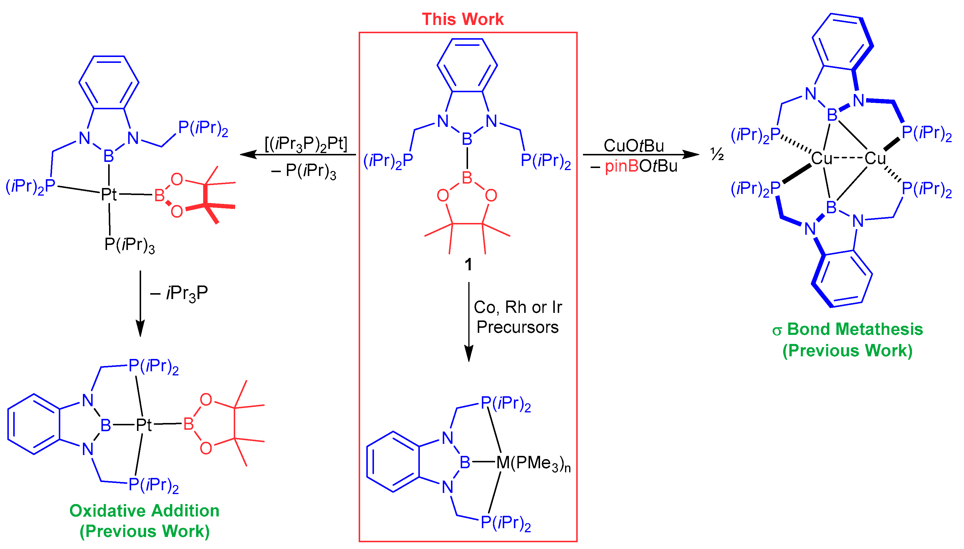
Scheme 1. Formation of PBP pincer boryl complexes from a diborane(4) precursor [15].
Molecules 28 06191 sch001
Scheme 2. Formation of PBP cobalt boryl complexes 2a (left) and its molecular structure (right). Selected distances [Å] and angles [°]: Co1–B1 1.936(2), Co1–P1 2.2063(4), Co1–P2 2.1859(3), Co1–P3 2.1643(4), Co1–P4 2.1968(4), P1–Co1–P2 125.55(2), P2–Co1–P3 112.03(1), P1–Co1–P3 110.56(2), B1–Co1–P4 173.85(5), B1–Co1–P3 83.24(5), Co1–[P1,P2,P3] 0.4372(3).
Molecules 28 06191 sch002
Figure 1. 31P{1H} NMR spectrum of 2a at –69 °C (top), and a section of the 1H-1H NOESY NMR spectrum of 2a (bottom), selected exchange (blue) and NOE (red) correlations are depicted (PhMe-d8, 400.4/162.1 MHz, rt).
Molecules 28 06191 g001
Scheme 3. Reaction of 2a with BAr3 (Ar = Ph, C6F5) and molecular structures of [(d(CH2P(iPr)2)abB)Rh–(N2)(PMe3)] (4a). Selected distances [Å] and angles [°]: Co1–B1 1.942(6), Co1–P1 2.1949(15), Co1–P2 2.2175(16), Co1–P3 2.1798(16), Co1–N3 1.816(5), N3–N4 1.118(7), P1–Co1–P2 134.51(7), P2–Co1–P3 110.78(6), P1–Co1–P3 104.85(6), B1–Co1–P3 89.5(2), B1–Co1–N3 172.2(2), Co1–[P1,P2,P3] 0.3915(9).
Molecules 28 06191 sch003
Figure 2. IR (in PhMe), 31P{1H} NMR and 11B{1H} spectra of 2a + B(C6F5)3 at rt and –80 °C and of isolated 2a (PhMe-d8, 162.1/96.3 MHz, rt).
Molecules 28 06191 g002
Scheme 4. Formation of PBP rhodium boryl complexes 3b and 2b.
Molecules 28 06191 sch004
Figure 3. Molecular structures of the complexes [(d(CH2P(iPr)2)abB)Rh–PMe3] (3b) (left) and [(d(CH2P(iPr)2)abB)Rh(PMe3)2] (2b) (right). Selected distances [Å] and angles [°], 3b: Rh1–B1 2.0221(5), Rh1–P1 2.2658(1), Rh1–P2 2.2794(1), Rh1–P3 2.3555(1), P1–Rh1–P2 152.622(5), B1–Rh1–P3 177.02(2), Rh1–[P1,P2,P3,B1] 0.0264(3); 2b: Rh1–B1 2.0256(7), Rh1–P1 2.3262(2), Rh1–P2 2.3364(2), Rh1–P3 2.3167(2), Rh1–P4 2.3705(5), P1–Rh1–P2 127.894(6), P2–Rh1–P3 108.475(7), P1–Rh1–P3 110.722(7), B1–Rh1–P4 172.54(2), Rh1–[P1,P2,P3] 0.4833(3).
Molecules 28 06191 g003
Figure 4. In situ 31P{1H} NMR spectra of the reaction of 3b with different amounts of PMe3 (121.6 MHz, C6D6, rt), isolated 2b and 3b with 1.3 equiv. PMe3 at −46 °C (162.1 MHz, THF-d8).
Molecules 28 06191 g004
Scheme 5. Consecutive formation of the PBP iridium boryl compels 5c, 6c, 3c and 2c.
Molecules 28 06191 sch005
Figure 5. Molecular structures of the complexes [(d(CH2P(iPr)2)abB)Ir(Bpin)(Cl)] (5c) (left, disorder omitted for clarity) and [(d(CH2P(iPr)2)abB)Ir(Bpin)(Cl)(PMe3)] (6c) (right, one of two independent molecules shown) (Supplementary Materials) [22]. Selected distances [Å] and angles [°], 5c: Ir1–B1 1.986(2), Ir1–B2 2.012(2), Ir1–P1 2.3354(4), Ir1–P2 2.3306(4), Ir1–Cl1 2.4144(4), P1–Ir1–P2 156.45(2), B1–Ir1–B2 72.16(8), B1–Ir1–Cl1 153.23(6), B2–Ir1–Cl1 134.49(6), ∠[P1,P2,B1,Ir1][B1,N1,N2,C6H4] 4.3(2), B1–B2 2.354(3); 6c: Ir1–B1 2.052(4), Ir1–B2 2.050(4), Ir1–P1 2.3391(8), Ir1–P2 2.3627(9), Ir1–P3 2.4155(9), Ir1–Cl1 2.5667(9), P1–Ir1–P2 153.28(3), B1–Ir1–B2 78.9(1), B1–Ir1–Cl1 107.6(1), B1–Ir1–P3 172.6(1), B2–Rh1–Cl1 173.4(1), B2–Rh1–P3 94.0(1), ∠[P1,P2,P3,B1,Ir1][B1,N1,N2,C6H4] 24.78(8), B1–B2 2.605(2).
Molecules 28 06191 g005
Figure 6. In situ 31P{1H} NMR spectra of the reaction of 6c with KOtBu (121.6 MHz, THF-d8, rt).
Molecules 28 06191 g006
Figure 7. Molecular structures of the complexes [(d(CH2P(iPr)2)abB)Ir–PMe3] (3c) and (left) [(d(CH2P(iPr)2)abB)Ir(PMe3)2] (2c) (right). Selected distances [Å] and angles [°], 3c: Ir1–B1 2.034(2), Ir1–P1 2.2764(4), Ir1–P2 2.2662(2), Ir1–P3 2.3355(5), P1–Ir1–P2 152.95(2), B1–Ir1–P3 176.73(6), Ir1–[P1,P2,P3,B1] 0.0386(3); 2c: Ir1–B1 2.055(3), Ir1–P1 2.3113(6), Ir1–P2 2.3165(6), Ir1–P3 2.2911(6), Ir1–P4 2.3521(6), P1–Ir1–P2 127.71(2), P2–Ir1–P3 108.84(8), P1–Ir1–P3 111.21(2), B1–Ir1–P4 172.02(8), Ir1–[P1,P2,P3] 0.4666(3).
Molecules 28 06191 g007
Disclaimer/Publisher’s Note: The statements, opinions and data contained in all publications are solely those of the individual author(s) and contributor(s) and not of MDPI and/or the editor(s). MDPI and/or the editor(s) disclaim responsibility for any injury to people or property resulting from any ideas, methods, instructions or products referred to in the content.