Effects of Quince Gel and Hesperidin Mixture on Experimental Endometriosis
Abstract
1. Introduction
2. Results
2.1. Biochemical Findings
2.2. Morphometric Findings
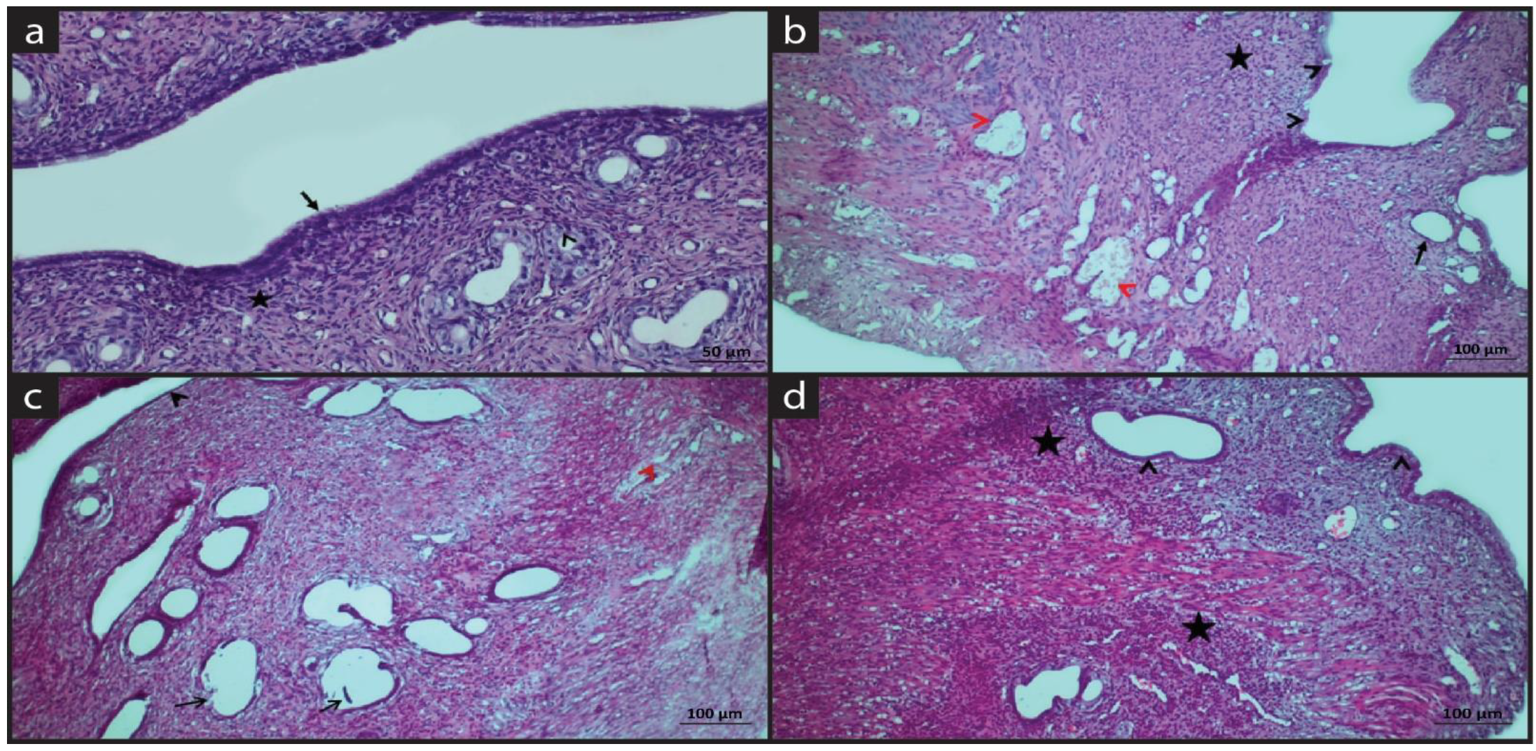
2.3. Histopathological Findings
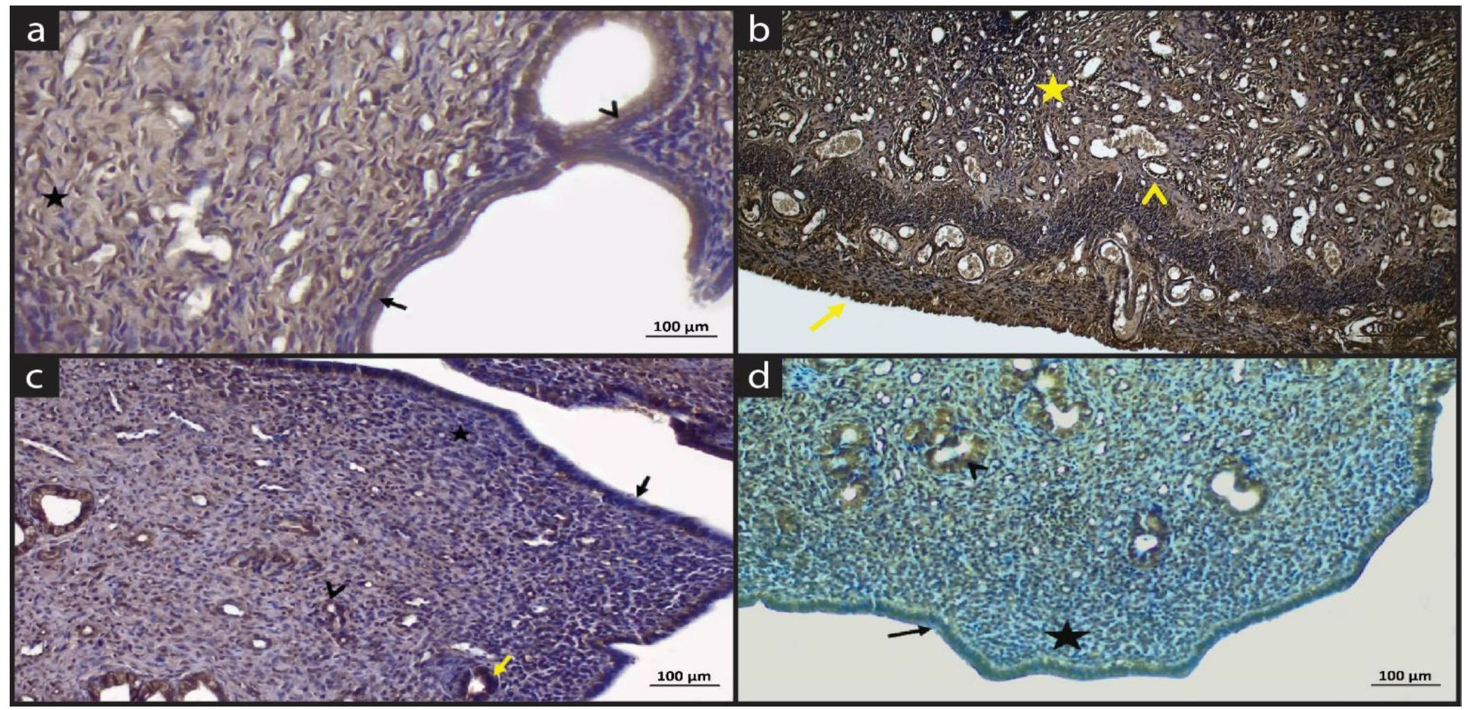
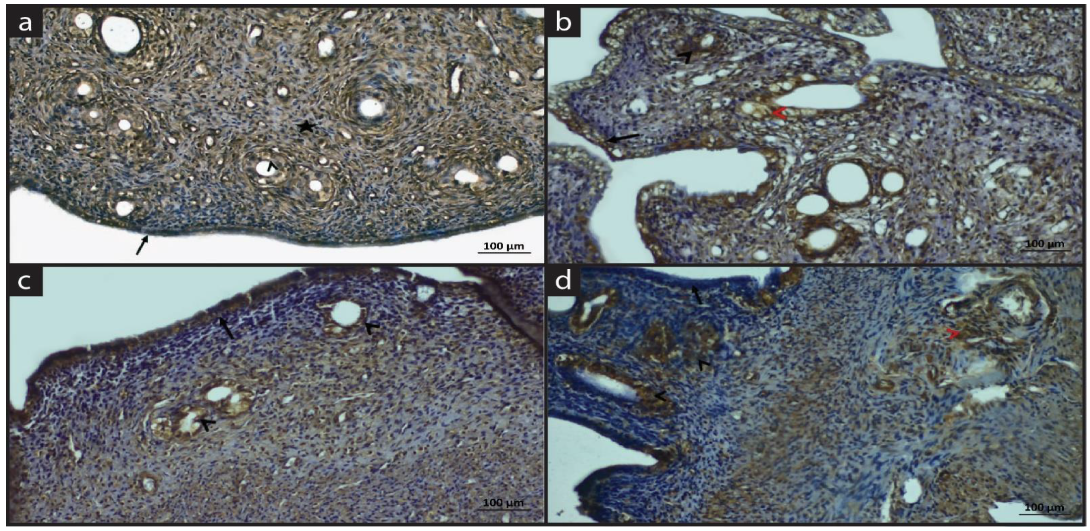
2.4. Immune Staining Findings
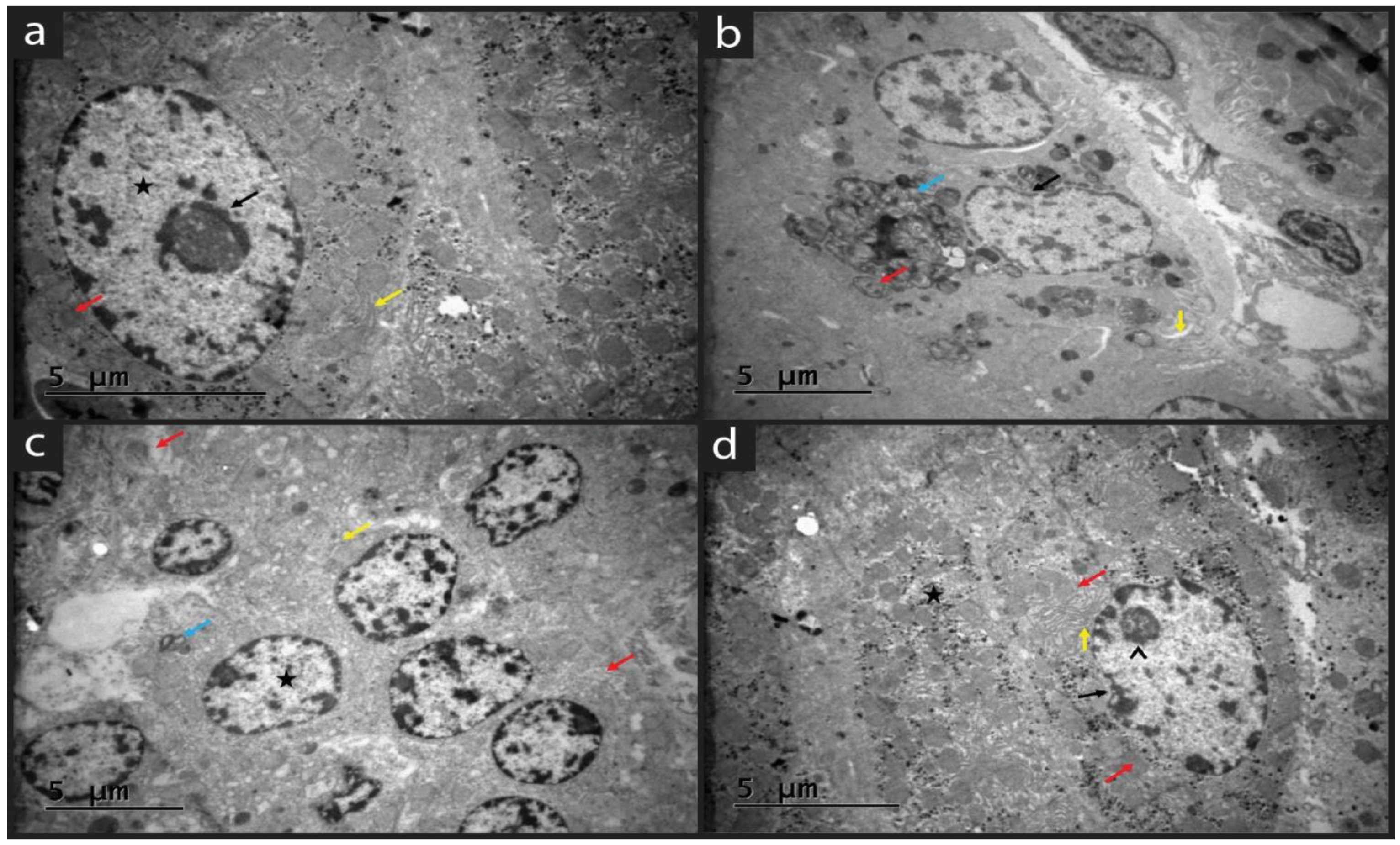
2.5. Ultrastructural Findings
3. Discussion
4. Materials and Methods
4.1. Plant Sampling and Gel Preparation
4.2. Experimental Design
- Sham group (n = 8): Animals underwent operation without any further intervention and no endometrial implantation. Then rats were housed in cages for 7 days and sacrificed at the end of the 7th day.
- Endometriosis (EM) group (n = 8): 3 µg/kg EB was administered subcutaneously to the rats for 7 days. Animals were sacrificed at the end of the 7th day.
- Endometriosis+Quince gel (EM+QG) group (n = 8): 3 µg/kg EB was administered subcutaneously to the rats for 7 days. Concurrently with EB administration, 2cc of QG was applied to the vagina with a thin catheter daily for 7 days and additionally for 14 days (totally 21 days). Animals were sacrificed at the end of the 21st day.
- Endometriosis+Quince gel+hesperidin (EM+QG+HES) group (n = 8): After the estrus stages of female rats were determined, 3 µg/kg EB was administered subcutaneously to the rats for 7 days. Concurrently with EB administration, 50 mg/kg/day Hesperidin with 2cc of QG was applied to the vagina with a thin daily for 7 days and additionally for 14 days (total 21 days). Animals were sacrificed at the end of the 21st day.
4.3. Biochemical Analysis
4.4. Histological Follow-Up
4.5. Histological Staining
4.6. Immunohistochemical Staining
4.7. Ultrastructural Tissue Preparation
4.8. ImageJ Analysis
4.9. Statistical Analysis
5. Conclusions
Author Contributions
Funding
Institutional Review Board Statement
Informed Consent Statement
Data Availability Statement
Conflicts of Interest
Sample Availability
References
- Chauhan, S.; More, A.; Chauhan, V.; Kathane, A. Endometriosis: A Review of Clinical Diagnosis, Treatment, and Pathogenesis. Cureus 2022, 14, e28864. [Google Scholar] [CrossRef] [PubMed]
- Parasar, P.; Ozcan, P.; Terry, K.L. Endometriosis: Epidemiology, Diagnosis and Clinical Management. Curr. Obs. Gynecol. Rep. 2017, 6, 34–41. [Google Scholar] [CrossRef]
- Sourial, S.; Tempest, N.; Hapangama, D.K. Theories on the pathogenesis of endometriosis. Int. J. Reprod. Med. 2014, 2014, 179515. [Google Scholar] [CrossRef]
- Farland, L.V.; Shah, D.K.; Kvaskoff, M.; Zondervan, K.T.; Missmer, S.A. Epidemiological and Clinical Risk Factors for Endometriosis. In Biomarkers for Endometriosis: State of the Art; D’Hooghe, T., Ed.; Springer International Publishing: Cham, Switzerland, 2017; pp. 95–121. [Google Scholar]
- Vinatier, D.; Orazi, G.; Cosson, M.; Dufour, P. Theories of endometriosis. Eur. J. Obs. Gynecol. Reprod. Biol. 2001, 96, 21–34. [Google Scholar] [CrossRef]
- Vannuccini, S.; Clemenza, S.; Rossi, M.; Petraglia, F. Hormonal treatments for endometriosis: The endocrine background. Rev. Endocr. Metab. Disord. 2022, 23, 333–355. [Google Scholar] [CrossRef]
- Chantalat, E.; Valera, M.C.; Vaysse, C.; Noirrit, E.; Rusidze, M.; Weyl, A.; Vergriete, K.; Buscail, E.; Lluel, P.; Fontaine, C.; et al. Estrogen Receptors and Endometriosis. Int. J. Mol. Sci. 2020, 21, 2815. [Google Scholar] [CrossRef] [PubMed]
- Wilmsen, P.K.; Spada, D.S.; Salvador, M. Antioxidant Activity of the Flavonoid Hesperidin in Chemical and Biological Systems. J. Agric. Food Chem. 2005, 53, 4757–4761. [Google Scholar] [CrossRef]
- Pyrzynska, K. Hesperidin: A Review on Extraction Methods, Stability and Biological Activities. Nutrients 2022, 14, 2387. [Google Scholar] [CrossRef]
- Parhiz, H.; Roohbakhsh, A.; Soltani, F.; Rezaee, R.; Iranshahi, M. Antioxidant and anti-inflammatory properties of the citrus flavonoids hesperidin and hesperetin: An updated review of their molecular mechanisms and experimental models. Phytother. Res. 2015, 29, 323–331. [Google Scholar] [CrossRef]
- Kim, J.; Wie, M.B.; Ahn, M.; Tanaka, A.; Matsuda, H.; Shin, T. Benefits of hesperidin in central nervous system disorders: A review. Anat. Cell Biol. 2019, 52, 369–377. [Google Scholar] [CrossRef]
- Beskisiz, S.l.; Asir, F. Hesperidin May Protect Gastric Tissue Against Immobilization Stress. Anal. Quant. Cytopathol. Histopathol. 2021, 43, 399–405. [Google Scholar]
- Silva, B.M.; Andrade, P.B.; Valentão, P.; Ferreres, F.; Seabra, R.M.; Ferreira, M.A. Quince (Cydonia oblonga Miller) fruit (pulp, peel, and seed) and Jam: Antioxidant activity. J. Agric. Food Chem. 2004, 52, 4705–4712. [Google Scholar] [CrossRef] [PubMed]
- Magalhães, A.S.; Silva, B.M.; Pereira, J.A.; Andrade, P.B.; Valentão, P.; Carvalho, M. Protective effect of quince (Cydonia oblonga Miller) fruit against oxidative hemolysis of human erythrocytes. Food Chem. Toxicol. 2009, 47, 1372–1377. [Google Scholar] [CrossRef] [PubMed]
- Najman, K.; Adrian, S.; Sadowska, A.; Świąder, K.; Hallmann, E.; Buczak, K.; Waszkiewicz-Robak, B.; Szterk, A. Changes in Physicochemical and Bioactive Properties of Quince (Cydonia oblonga Mill.) and Its Products. Molecules 2023, 28, 3066. [Google Scholar]
- Hanan, E.; Sharma, V.; Ahmad, F. Nutritional Composition, Phytochemistry and Medicinal Use of Quince (Cydonia oblonga Miller) with Emphasis on its Processed and Fortified Food Products. J. Food Process. Technol. 2020, 11, 1–13. [Google Scholar]
- Silva, B.M.; Andrade, P.B.; Ferreres, F.; Domingues, A.L.; Seabra, R.M.; Ferreira, M.A. Phenolic profile of quince fruit (Cydonia oblonga Miller) (pulp and peel). J. Agric. Food Chem. 2002, 50, 4615–4618. [Google Scholar] [CrossRef]
- Silva, B.M.; Casal, S.; Andrade, P.B.; Seabra, R.M.; Oliveira, M.B.; Ferreira, M.A. Free amino acid composition of quince (Cydonia oblonga Miller) fruit (pulp and peel) and jam. J. Agric. Food Chem. 2004, 52, 1201–1206. [Google Scholar] [CrossRef]
- Hegedűs, A.; Papp, N.; Stefanovits-Bányai, É. A review of nutritional value and putative health-effects of quince (Cydonia oblonga Mill.) fruit. J. Hortic. Sci. 2013, 19, 29–32. [Google Scholar] [CrossRef]
- Ghopur, H.; Usmanova, S.K.; Ayupbek, A.; Aisa, H.A. A new chromone from seeds of Cydonia oblonga. Chem. Nat. Compd. 2012, 48, 562–564. [Google Scholar] [CrossRef]
- Tamri, P.; Hemmati, A.; Boroujerdnia, M.G. Wound healing properties of quince seed mucilage: In vivo evaluation in rabbit full-thickness wound model. Int. J. Surg. 2014, 12, 843–847. [Google Scholar] [CrossRef]
- Alizadeh, H.; Rahnema, M.; Semnani, S.N.; Hajizadeh, N. Detection of compounds and antibacterial effect of quince (Cydonia oblonga Miller) extracts in vitro and in vivo. J. Biol. Act. Prod. Nat. 2013, 3, 303–309. [Google Scholar]
- Izadyari Aghmiuni, A.; Heidari Keshel, S.; Sefat, F.; Akbarzadeh Khiyavi, A. Quince seed mucilage-based scaffold as a smart biological substrate to mimic mechanobiological behavior of skin and promote fibroblasts proliferation and h-ASCs differentiation into keratinocytes. Int. J. Biol. Macromol. 2020, 142, 668–679. [Google Scholar] [CrossRef]
- Wang, L.; Liu, H.M.; Qin, G.Y. Structure characterization and antioxidant activity of polysaccharides from Chinese quince seed meal. Food Chem. 2017, 234, 314–322. [Google Scholar] [CrossRef] [PubMed]
- Mirzaii, M.; Yaeghoobi, M.; Afzali, M.; Amirkhalili, N.; Mahmoodi, M.; Sajirani, E.B. Antifungal activities of quince seed mucilage hydrogel decorated with essential oils of Nigella sativa, Citrus sinensis and Cinnamon verum. Iran. J. Microbiol. 2021, 13, 352–359. [Google Scholar] [CrossRef] [PubMed]
- Smolarz, B.; Szyłło, K.; Romanowicz, H. Endometriosis: Epidemiology, Classification, Pathogenesis, Treatment and Genetics (Review of Literature). Int. J. Mol. Sci. 2021, 22, 10554. [Google Scholar] [CrossRef]
- Jovanovic, S.V.; Steenken, S.; Tosic, M.; Marjanovic, B.; Simic, M.G. Flavonoids as Antioxidants. J. Am. Chem. Soc. 1994, 116, 4846–4851. [Google Scholar] [CrossRef]
- Li, S.; Qin, Q.; Luo, D.; Pan, W.; Wei, Y.; Xu, Y.; Zhu, J.; Shang, L. Hesperidin ameliorates liver ischemia/reperfusion injury via activation of the Akt pathway. Mol. Med. Rep. 2020, 22, 4519–4530. [Google Scholar] [CrossRef]
- Celik, E.; Oguzturk, H.; Sahin, N.; Turtay, M.G.; Oguz, F.; Ciftci, O. Protective effects of hesperidin in experimental testicular ischemia/reperfusion injury in rats. Arch. Med. Sci. 2016, 12, 928–934. [Google Scholar] [CrossRef]
- Kennedy, S.; Bergqvist, A.; Chapron, C.; D’Hooghe, T.; Dunselman, G.; Greb, R.; Hummelshoj, L.; Prentice, A.; Saridogan, E. ESHRE guideline for the diagnosis and treatment of endometriosis. Hum. Reprod. 2005, 20, 2698–2704. [Google Scholar] [CrossRef]
- Mashele, T.; Reddy, Y.; Pather, S. Endometriosis: Three-year histopathological perspective from the largest hospital in Africa. Ann. Diagn. Pathol. 2020, 45, 151458. [Google Scholar] [CrossRef]
- Porter, A.G.; Janicke, R.U. Emerging roles of caspase-3 in apoptosis. Cell Death Differ. 1999, 6, 99–104. [Google Scholar] [CrossRef]
- Hengartner, M.O. The biochemistry of apoptosis. Nature 2000, 407, 770–776. [Google Scholar] [CrossRef] [PubMed]
- Li, M.; Ona, V.; Chen, M.; Kaul, M.; Tenneti, L.; Zhang, X.; Stieg, P.; Lipton, S.; Friedlander, R. Functional role and therapeutic implications of neuronal caspase-1 and-3 in a mouse model of traumatic spinal cord injury. Neuroscience 2000, 99, 333–342. [Google Scholar] [CrossRef]
- Kim, G.T.; Chun, Y.S.; Park, J.W.; Kim, M.S. Role of apoptosis-inducing factor in myocardial cell death by ischemia-reperfusion. Biochem. Biophys. Res. Commun. 2003, 309, 619–624. [Google Scholar] [CrossRef] [PubMed]
- Kaya, C.; Alay, I.; Guraslan, H.; Gedikbasi, A.; Ekin, M.; Ertaş Kaya, S.; Oral, E.; Yasar, L. The Role of Serum Caspase 3 Levels in Prediction of Endometriosis Severity. Gynecol. Obs. Investig. 2018, 83, 576–585. [Google Scholar] [CrossRef] [PubMed]
- Yang, L.; Inokuchi, S.; Roh, Y.S.; Song, J.; Loomba, R.; Park, E.J.; Seki, E. Transforming growth factor-β signaling in hepatocytes promotes hepatic fibrosis and carcinogenesis in mice with hepatocyte-specific deletion of TAK1. Gastroenterology 2013, 144, 1042–1054.e1044. [Google Scholar] [CrossRef]
- Inokuchi, S.; Aoyama, T.; Miura, K.; Osterreicher, C.H.; Kodama, Y.; Miyai, K.; Akira, S.; Brenner, D.A.; Seki, E. Disruption of TAK1 in hepatocytes causes hepatic injury, inflammation, fibrosis, and carcinogenesis. Proc. Natl. Acad. Sci. USA 2010, 107, 844–849. [Google Scholar] [CrossRef]
- Galo, S.; Zúbor, P.; Szunyogh, N.; Kajo, K.; Macháleková, K.; Biringer, K.; Visnovský, J. TNF-alpha serum levels in women with endometriosis: Prospective clinical study. Ceska Gynekol. 2005, 70, 286–290. [Google Scholar]
- Lu, D.; Song, H.; Shi, G. Anti-TNF-α treatment for pelvic pain associated with endometriosis. Cochrane Database Syst. Rev. 2013, 28, CD008088. [Google Scholar] [CrossRef]
- Pearson, G.; Robinson, F.; Beers Gibson, T.; Xu, B.E.; Karandikar, M.; Berman, K.; Cobb, M.H. Mitogen-activated protein (MAP) kinase pathways: Regulation and physiological functions. Endocr. Rev. 2001, 22, 153–183. [Google Scholar] [CrossRef]
- Soares-Silva, M.; Diniz, F.F.; Gomes, G.N.; Bahia, D. The Mitogen-Activated Protein Kinase (MAPK) Pathway: Role in Immune Evasion by Trypanosomatids. Front. Microbiol. 2016, 7, 183. [Google Scholar] [CrossRef]
- Jagodzik, P.; Tajdel-Zielinska, M.; Ciesla, A.; Marczak, M.; Ludwikow, A. Mitogen-Activated Protein Kinase Cascades in Plant Hormone Signaling. Front. Plant. Sci. 2018, 9, 1387. [Google Scholar] [CrossRef] [PubMed]
- Klemmt, P.A.B.; Starzinski-Powitz, A. Molecular and Cellular Pathogenesis of Endometriosis. Curr. Womens Health Rev. 2018, 14, 106–116. [Google Scholar] [CrossRef]
- Koninckx, P.R.; Kennedy, S.H.; Barlow, D.H. Pathogenesis of endometriosis: The role of peritoneal fluid. Gynecol. Obs. Investig. 1999, 47, 23–33. [Google Scholar] [CrossRef] [PubMed]
- Braun, D.P.; Ding, J.; Dmowski, W.P. Peritoneal fluid-mediated enhancement of eutopic and ectopic endometrial cell proliferation is dependent on tumor necrosis factor-alpha in women with endometriosis. Fertil. Steril. 2002, 78, 727–732. [Google Scholar] [CrossRef]
- Iwabe, T.; Harada, T.; Tsudo, T.; Nagano, Y.; Yoshida, S.; Tanikawa, M.; Terakawa, N. Tumor necrosis factor-α promotes proliferation of endometriotic stromal cells by inducing interleukin-8 gene and protein expression. J. Clin. Endocrinol. Metab. 2000, 85, 824–829. [Google Scholar]
- Vernon, M.W.; Wilson, E.A. Studies on the surgical induction of endometriosis in the rat. Fertil. Steril. 1985, 44, 684–694. [Google Scholar] [CrossRef]
- Durgun, C.; Aşır, F. Daidzein alleviated the pathologies in intestinal tissue against ischemia-reperfusion. Eur. Rev. Med. Pharm. Sci. 2023, 27, 1487–1493. [Google Scholar] [CrossRef]
- Durgun, C.; Aşir, F. Effect of ellagic acid on damage caused by hepatic ischemia reperfusion in rats. Eur. Rev. Med. Pharm. Sci. 2022, 26, 8209–8215. [Google Scholar] [CrossRef]
- Crowe, A.R.; Yue, W. Semi-quantitative Determination of Protein Expression using Immunohistochemistry Staining and Analysis: An Integrated Protocol. Bio-Protocol 2019, 9, e3465. [Google Scholar] [CrossRef]


| Groups | TAS | TOS | p |
|---|---|---|---|
| Sham | 1.45 (0.76–1.65) | 15.73 (10.34–55.18) | <0.005 |
| EM | 0.95 (0.58–1.11) a | 35.80 (21.76–88.85) a | |
| EM+QG | 1.09 (0.66–1.21) b | 29.92 (19.48–57.91) b | |
| EM+QG+HES | 1.21 (0.92–1.47) c | 21.42 (16.13–33.47) c |
| Parameters | Sham Median (min-max) | EM Median (min-max) | EM+QG Median (min-max) | EM+QG+HES Median (min-max) |
|---|---|---|---|---|
| Uterine epithelial thickness (μm) | 23.45 (16.45–29.45) | 8.05 (6.88–8.92) * | 14.03 (10.91–18.98) ** | 16.1135 (11.58–19.99) ** |
| Diameter of uterine glands (μm2) | 973.45 (956.45–1023.35) | 1697.51 (1678.71–1714.07) * | 1386.48 (1027.78–1909.82) ** | 1281.85 (958.68–1891.53) ** |
| Blood vessel lumen length (μm) | 77.68 (66.34–84.57) | 97.95 (86.30–106.11) * | 92.63 (85.06–104.66) ** | 86.62 (71.11–99.35) ** |
| Cell degeneration | 1 (1–2) | 3 (3–4) * | 3 (2–4) ** | 3 (1–4) ** |
| Inflammation | 1 (1–2) | 4 (3–4) * | 3 (2–4) ** | 3 (1–4) ** |
| Groups | Sham | EM | EM+QG | EM+QG+HES |
| Caspase-3 | %15.21 | %47.44 | %40.86 | %38.28 |
| Groups | Sham | EM | EM+QG | EM+QG+HES |
| TNF-α | %18.35 | %43.92 | %39.02 | %35.18 |
| Groups | Sham | EM | EM+QG | EM+QG+HES |
| MAPK | %22.45 | %37.67 | %27.83 | %30.56 |
Disclaimer/Publisher’s Note: The statements, opinions and data contained in all publications are solely those of the individual author(s) and contributor(s) and not of MDPI and/or the editor(s). MDPI and/or the editor(s) disclaim responsibility for any injury to people or property resulting from any ideas, methods, instructions or products referred to in the content. |
© 2023 by the authors. Licensee MDPI, Basel, Switzerland. This article is an open access article distributed under the terms and conditions of the Creative Commons Attribution (CC BY) license (https://creativecommons.org/licenses/by/4.0/).
Share and Cite
Ermiş, I.S.; Deveci, E.; Aşır, F. Effects of Quince Gel and Hesperidin Mixture on Experimental Endometriosis. Molecules 2023, 28, 5945. https://doi.org/10.3390/molecules28165945
Ermiş IS, Deveci E, Aşır F. Effects of Quince Gel and Hesperidin Mixture on Experimental Endometriosis. Molecules. 2023; 28(16):5945. https://doi.org/10.3390/molecules28165945
Chicago/Turabian StyleErmiş, Işılay Sezen, Engin Deveci, and Fırat Aşır. 2023. "Effects of Quince Gel and Hesperidin Mixture on Experimental Endometriosis" Molecules 28, no. 16: 5945. https://doi.org/10.3390/molecules28165945
APA StyleErmiş, I. S., Deveci, E., & Aşır, F. (2023). Effects of Quince Gel and Hesperidin Mixture on Experimental Endometriosis. Molecules, 28(16), 5945. https://doi.org/10.3390/molecules28165945


